Embed Size (px)
Citation preview
AACPT CSM Laboratory Values February 2017
Do not copy or distribute without permission of the Academy of Acute Care Physical Therapy 1
Click to edit Master title style
I Found the Lab Value- Now What? Demystifying Lab Values for Patient Management
AUTHORS/INSTITUTIONS: J. Tompkins, Mayo Clinic, Phoenix, AZJ. Dyson, Orlando Health, Orlando, FL
T. Norris, Barnes-Jewish Hospital, Saint Louis, MOK. Levenhagen, Department of Physical Therapy and Athletic
Training, Saint Louis University, Saint Louis, MO
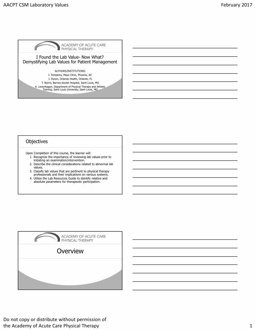
Click to edit Master title styleObjectives
Upon Completion of this course, the learner will:1. Recognize the importance of reviewing lab values prior to
initiating an examination/intervention. 2. Describe the clinical considerations related to abnormal lab
values. 3. Classify lab values that are pertinent to physical therapy
professionals and their implications on various systems. 4. Utilize the Lab Resources Guide to identify relative and
absolute parameters for therapeutic participation.
Click to edit Master title style
Overview
AACPT CSM Laboratory Values February 2017
Do not copy or distribute without permission of the Academy of Acute Care Physical Therapy 2
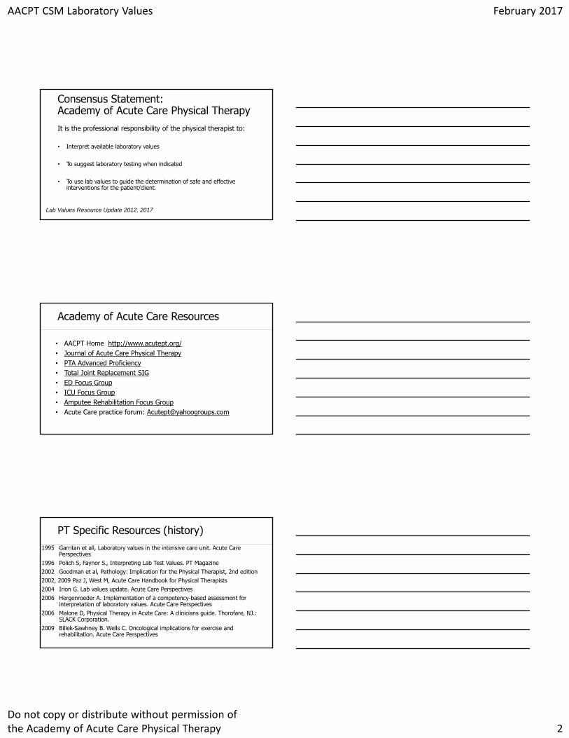
Click to edit Master title styleConsensus Statement: Academy of Acute Care Physical Therapy It is the professional responsibility of the physical therapist to:
• Interpret available laboratory values
• To suggest laboratory testing when indicated
• To use lab values to guide the determination of safe and effective interventions for the patient/client.
Lab Values Resource Update 2012, 2017
Click to edit Master title styleAcademy of Acute Care Resources
• AACPT Home http://www.acutept.org/• Journal of Acute Care Physical Therapy• PTA Advanced Proficiency• Total Joint Replacement SIG• ED Focus Group• ICU Focus Group• Amputee Rehabilitation Focus Group• Acute Care practice forum: [email protected]
Click to edit Master title stylePT Specific Resources (history)1995 Garritan et all, Laboratory values in the intensive care unit. Acute Care
Perspectives1996 Polich S, Faynor S., Interpreting Lab Test Values. PT Magazine2002 Goodman et al, Pathology: Implication for the Physical Therapist, 2nd edition2002, 2009 Paz J, West M, Acute Care Handbook for Physical Therapists 2004 Irion G. Lab values update. Acute Care Perspectives2006 Hergenroeder A. Implementation of a competency-based assessment for
interpretation of laboratory values. Acute Care Perspectives2006 Malone D, Physical Therapy in Acute Care: A clinicians guide. Thorofare, NJ.:
SLACK Corporation. 2009 Billek-Sawhney B. Wells C. Oncological implications for exercise and
rehabilitation. Acute Care Perspectives
AACPT CSM Laboratory Values February 2017
Do not copy or distribute without permission of the Academy of Acute Care Physical Therapy 3
Click to edit Master title stylePT Specific Resources (history)
2008, 2009, 2012, 2017 (pending) - APTA Academy of Acute Care Physical Therapy (formerly Acute Care Section)
2012 Frownfelter D, Dean E , Cardiovascular and Pulmonary Physical Therapy Evidence and Practice (5th ed.). St Louis: Elsevier-Mosby.
2013 Pawlik A, Kress J Issues affecting the delivery of physical therapy services for individuals with critical illness. Physical therapy
2015 Goodman C, Fuller K, Pathology Implications for the physical therapist. St Louis: Elsevier Saunders.
2015 Peterson M. The Impact of Low Hemoglobin on the Percentage of Adverse Events During Physical Therapy in the Acute Care Setting: A Retrospective Study. JACPT.
2016 Hillegass E, Puthoff M, Frese EM, et al. Role of Physical Therapists in the Management of Individuals at Risk for or Diagnosed With Venous Thromboembolism: Evidence-Based Clinical Practice Guideline. Phys Ther..
Click to edit Master title style
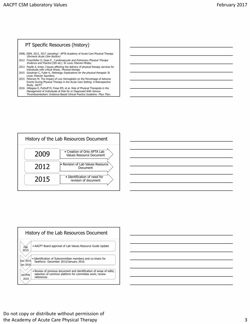
• Creation of Only APTA Lab Values Resource Document2009
• Revision of Lab Values Resource Document2012
• Identification of need for revision of document2015
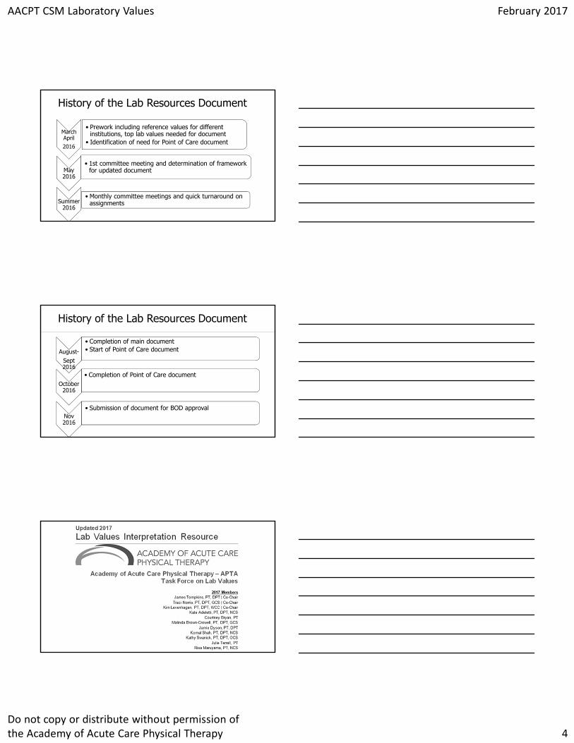
History of the Lab Resources Document
Click to edit Master title style
Fall 2015
• AACPT Board approval of Lab Values Resource Guide Update
Dec 2015Jan 2016
• Identification of Subcommittee members and co-chairs for Taskforce December 2015/January 2016
Jan/Feb2016
• Review of previous document and identification of areas of edits; selection of common platform for committee work; review references
History of the Lab Resources Document
AACPT CSM Laboratory Values February 2017
Do not copy or distribute without permission of the Academy of Acute Care Physical Therapy 4
Click to edit Master title style
March April2016
• Prework including reference values for different institutions, top lab values needed for document
• Identification of need for Point of Care document
May 2016
• 1st committee meeting and determination of framework for updated document
Summer 2016
• Monthly committee meetings and quick turnaround on assignments
History of the Lab Resources Document
Click to edit Master title style
August-Sept 2016
• Completion of main document• Start of Point of Care document
October 2016
• Completion of Point of Care document
Nov 2016
• Submission of document for BOD approval
History of the Lab Resources Document
Overview
AACPT CSM Laboratory Values February 2017
Do not copy or distribute without permission of the Academy of Acute Care Physical Therapy 5
Click to edit Master title styleLaboratory Value Interpretation Resource
Recognize basic laboratory tests and normative values
Recognize absolute and generalized parameters to exercise and therapeutic interventions
Should complement not replace a thorough evaluation and clinical judgment
Follow institutional guides
Should not be used as an excuse not to treat!!!
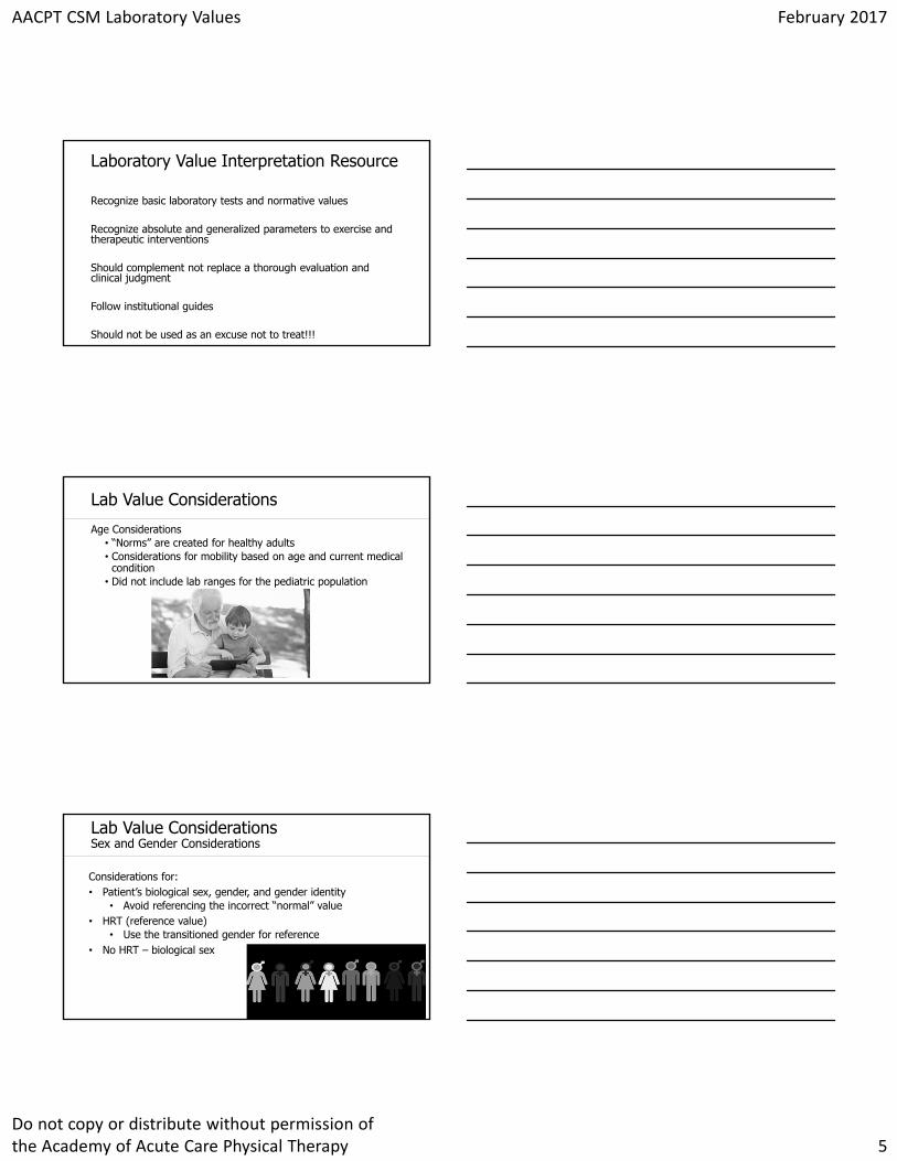
Click to edit Master title styleLab Value Considerations
Age Considerations• “Norms” are created for healthy adults• Considerations for mobility based on age and current medical
condition• Did not include lab ranges for the pediatric population
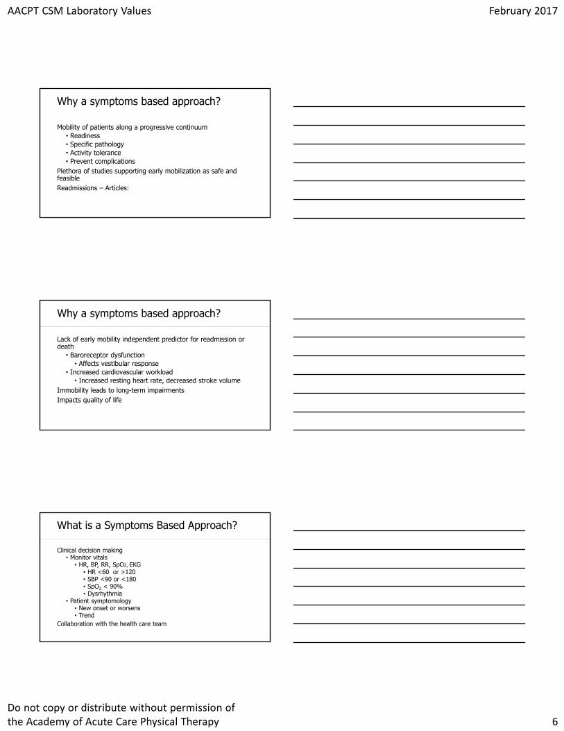
Click to edit Master title styleLab Value ConsiderationsSex and Gender Considerations
Considerations for:• Patient’s biological sex, gender, and gender identity
• Avoid referencing the incorrect “normal” value• HRT (reference value)
• Use the transitioned gender for reference• No HRT – biological sex
AACPT CSM Laboratory Values February 2017
Do not copy or distribute without permission of the Academy of Acute Care Physical Therapy 6
Click to edit Master title styleWhy a symptoms based approach?
Mobility of patients along a progressive continuum• Readiness• Specific pathology• Activity tolerance• Prevent complications
Plethora of studies supporting early mobilization as safe and feasibleReadmissions – Articles:
Click to edit Master title styleWhy a symptoms based approach?
Lack of early mobility independent predictor for readmission or death
• Baroreceptor dysfunction• Affects vestibular response
• Increased cardiovascular workload• Increased resting heart rate, decreased stroke volume
Immobility leads to long-term impairmentsImpacts quality of life
Click to edit Master title styleWhat is a Symptoms Based Approach?
Clinical decision making• Monitor vitals
• HR, BP, RR, SpO2, EKG• HR <60 or >120• SBP <90 or <180• SpO2 < 90%• Dysrhythmia
• Patient symptomology• New onset or worsens• Trend
Collaboration with the health care team
AACPT CSM Laboratory Values February 2017
Do not copy or distribute without permission of the Academy of Acute Care Physical Therapy 7
Click to edit Master title style
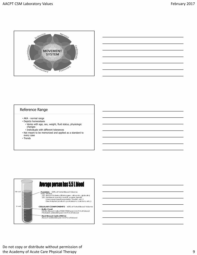
Movement System
Click to edit Master title styleThe Human Movement System
Our Vision-Transforming society by optimizing movement to improve the human
experienceIDENTITY
FOUNDATIONThe Core Of Physical Therapist
Practice, Education, and ResearchAmerican Physical Therapy Association (2015)
Click to edit Master title styleHuman Movement SystemAmerican Physical Therapy Association (2015)
The human movement system comprises the
anatomic structures and physiologic functions that interact to move the body or its component parts.
AACPT CSM Laboratory Values February 2017
Do not copy or distribute without permission of the Academy of Acute Care Physical Therapy 8
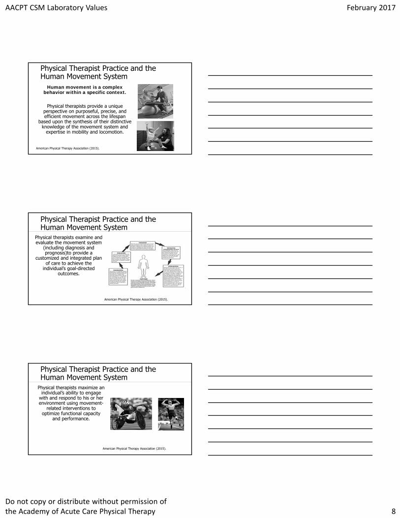
Click to edit Master title stylePhysical Therapist Practice and the Human Movement System
Human movement is a complex behavior within a specific context.
Physical therapists provide a unique perspective on purposeful, precise, and efficient movement across the lifespan
based upon the synthesis of their distinctive knowledge of the movement system and
expertise in mobility and locomotion.
American Physical Therapy Association (2015).
Click to edit Master title stylePhysical Therapist Practice and the Human Movement System
Physical therapists examine and evaluate the movement system
(including diagnosis and prognosis)to provide a
customized and integrated plan of care to achieve the
individual’s goal-directed outcomes.
American Physical Therapy Association (2015).
Click to edit Master title stylePhysical Therapist Practice and the Human Movement System
Physical therapists maximize an individual’s ability to engage
with and respond to his or her environment using movement-
related interventions to optimize functional capacity
and performance.
American Physical Therapy Association (2015).
AACPT CSM Laboratory Values February 2017
Do not copy or distribute without permission of the Academy of Acute Care Physical Therapy 9
Click to edit Master title style
Click to edit Master title styleReference Range
• AKA - normal range• Depicts homeostasis
• Varies with age, sex, weight, fluid status, physiologic changes
• Individuals with different tolerances• Not meant to be memorized and applied as a standard to
every case• Trends
Click to edit Master title style
AACPT CSM Laboratory Values February 2017
Do not copy or distribute without permission of the Academy of Acute Care Physical Therapy 10
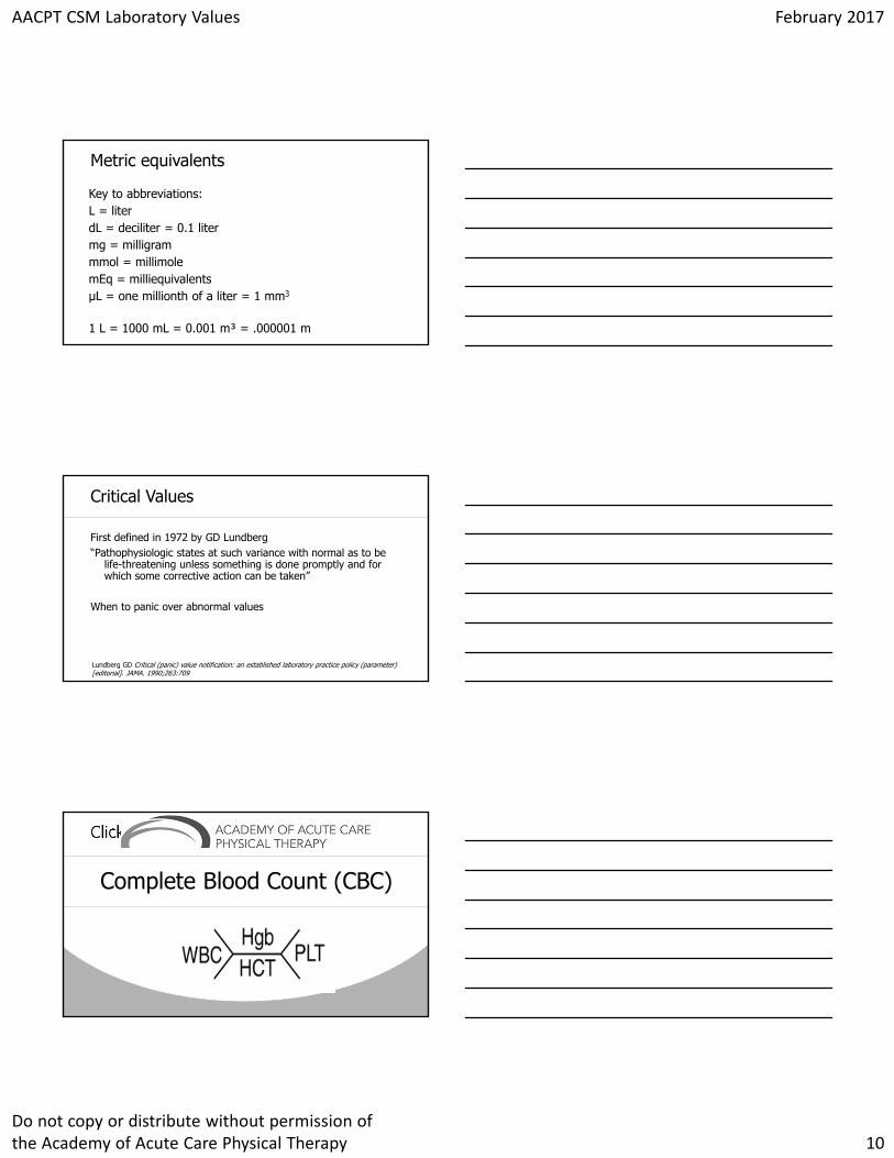
Click to edit Master title styleMetric equivalents
Key to abbreviations:L = literdL = deciliter = 0.1 litermg = milligrammmol = millimolemEq = milliequivalentsμL = one millionth of a liter = 1 mm3
1 L = 1000 mL = 0.001 m³ = .000001 m
Click to edit Master title styleCritical Values
First defined in 1972 by GD Lundberg “Pathophysiologic states at such variance with normal as to be
life-threatening unless something is done promptly and for which some corrective action can be taken”
When to panic over abnormal values
Lundberg GD Critical (panic) value notification: an established laboratory practice policy (parameter) [editorial]. JAMA. 1990;263:709
Click to edit Master title style
Complete Blood Count (CBC)
AACPT CSM Laboratory Values February 2017
Do not copy or distribute without permission of the Academy of Acute Care Physical Therapy 11
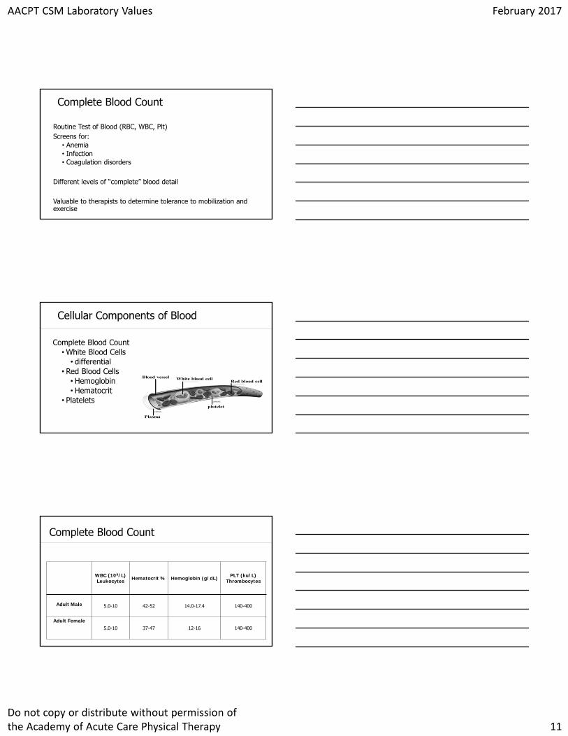
Click to edit Master title styleComplete Blood Count
Routine Test of Blood (RBC, WBC, Plt)Screens for:
• Anemia• Infection• Coagulation disorders
Different levels of “complete” blood detail
Valuable to therapists to determine tolerance to mobilization and exercise
Click to edit Master title styleCellular Components of Blood
Complete Blood Count• White Blood Cells
• differential• Red Blood Cells
• Hemoglobin• Hematocrit
• Platelets
Click to edit Master title styleComplete Blood Count
WBC (109/L)Leukocytes Hematocrit % Hemoglobin (g/dL) PLT (ku/L)
Thrombocytes
Adult Male 5.0-10 42-52 14.0-17.4 140-400
Adult Female5.0-10 37-47 12-16 140-400
AACPT CSM Laboratory Values February 2017
Do not copy or distribute without permission of the Academy of Acute Care Physical Therapy 12
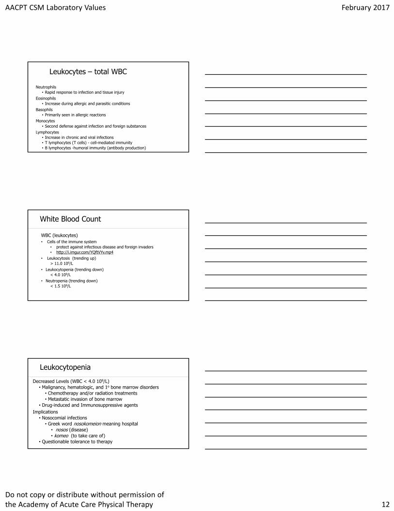
Click to edit Master title styleLeukocytes – total WBC
Neutrophils• Rapid response to infection and tissue injury
Eosinophils• Increase during allergic and parasitic conditions
Basophils • Primarily seen in allergic reactions
Monocytes• Second defense against infection and foreign substances
Lymphocytes• Increase in chronic and viral infections• T lymphocytes (T cells) - cell-mediated immunity• B lymphocytes -humoral immunity (antibody production)
Click to edit Master title styleWhite Blood Count
WBC (leukocytes)• Cells of the immune system
• protect against infectious disease and foreign invaders• http://i.imgur.com/YQftVYv.mp4
• Leukocytosis (trending up)> 11.0 109/L
• Leukocytopenia (trending down)< 4.0 109/L
• Neutropenia (trending down)< 1.5 109/L
Click to edit Master title styleLeukocytopenia
Decreased Levels (WBC < 4.0 109/L)• Malignancy, hematologic, and 1o bone marrow disorders
• Chemotherapy and/or radiation treatments• Metastatic invasion of bone marrow
• Drug-induced and Immunosuppressive agentsImplications
• Nosocomial infections• Greek word nosokomeion meaning hospital
• nosos (disease)• komeo (to take care of)
• Questionable tolerance to therapy
AACPT CSM Laboratory Values February 2017
Do not copy or distribute without permission of the Academy of Acute Care Physical Therapy 13
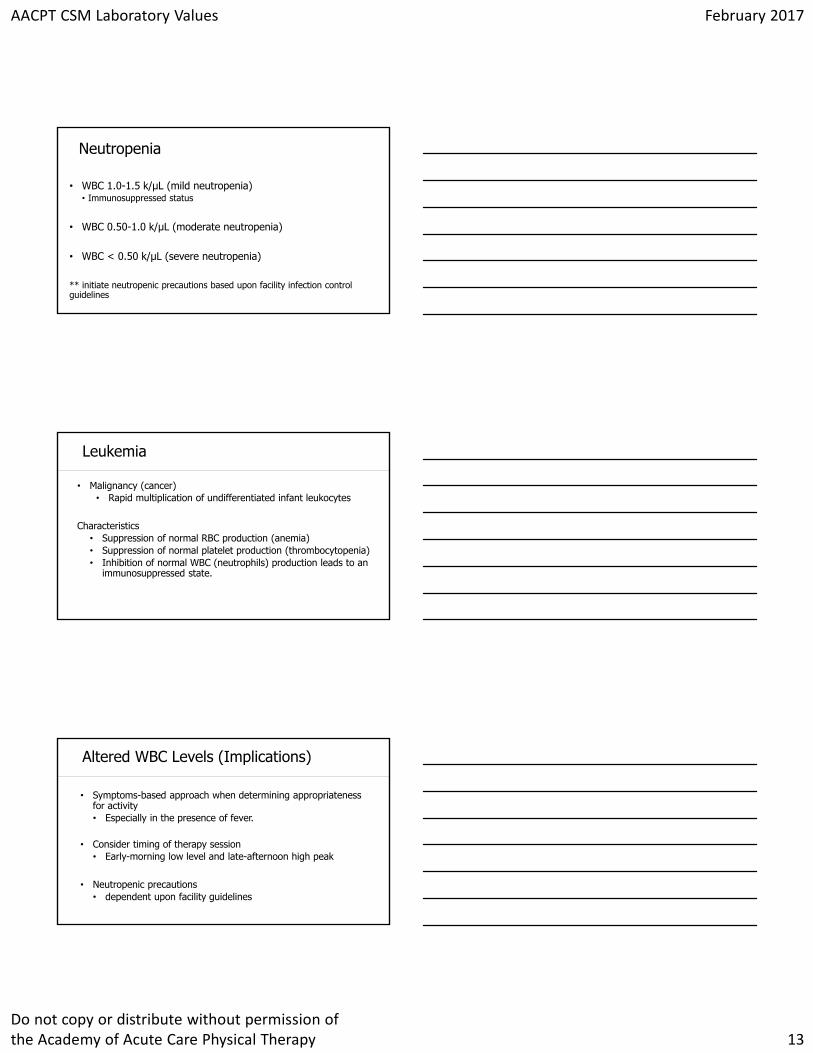
Click to edit Master title styleNeutropenia
• WBC 1.0-1.5 k/μL (mild neutropenia)• Immunosuppressed status
• WBC 0.50-1.0 k/μL (moderate neutropenia)
• WBC < 0.50 k/μL (severe neutropenia)
** initiate neutropenic precautions based upon facility infection control guidelines
Click to edit Master title styleLeukemia
• Malignancy (cancer) • Rapid multiplication of undifferentiated infant leukocytes
Characteristics• Suppression of normal RBC production (anemia)• Suppression of normal platelet production (thrombocytopenia)• Inhibition of normal WBC (neutrophils) production leads to an
immunosuppressed state.
Click to edit Master title styleAltered WBC Levels (Implications)
• Symptoms-based approach when determining appropriateness for activity• Especially in the presence of fever.
• Consider timing of therapy session• Early-morning low level and late-afternoon high peak
• Neutropenic precautions• dependent upon facility guidelines
AACPT CSM Laboratory Values February 2017
Do not copy or distribute without permission of the Academy of Acute Care Physical Therapy 14
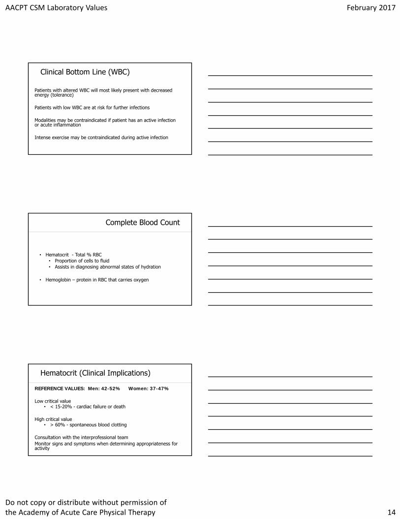
Click to edit Master title styleClinical Bottom Line (WBC)
Patients with altered WBC will most likely present with decreased energy (tolerance)
Patients with low WBC are at risk for further infections
Modalities may be contraindicated if patient has an active infection or acute inflammation
Intense exercise may be contraindicated during active infection
Click to edit Master title styleComplete Blood Count
• Hematocrit - Total % RBC • Proportion of cells to fluid• Assists in diagnosing abnormal states of hydration
• Hemoglobin – protein in RBC that carries oxygen
Click to edit Master title styleHematocrit (Clinical Implications)
REFERENCE VALUES: Men: 42-52% Women: 37-47%
Low critical value • < 15-20% - cardiac failure or death
High critical value• > 60% - spontaneous blood clotting
Consultation with the interprofessional teamMonitor signs and symptoms when determining appropriateness for activity
AACPT CSM Laboratory Values February 2017
Do not copy or distribute without permission of the Academy of Acute Care Physical Therapy 15
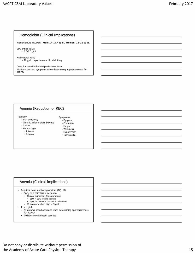
Click to edit Master title styleHemoglobin (Clinical Implications)
REFERENCE VALUES: Men: 14-17.4 g/dL Women: 12-16 g/dL
Low critical value < 5.0-7.0 g/dL
High critical value> 20 g/dL - spontaneous blood clotting
Consultation with the interprofessional teamMonitor signs and symptoms when determining appropriateness for activity
Click to edit Master title styleAnemia (Reduction of RBC)
Etiology• Iron deficiency• Chronic Inflammatory Disease• Cancer• Hemorrhage
• Internal • External
Symptoms• Dyspnea• Confusion• Fatigue• Weakness• Hypotension• Tachycardia
Click to edit Master title styleAnemia (Clinical Implications)
• Requires close monitoring of vitals (BP, HR)• SpO2 to predict tissue perfusion
• Clinical significant (desaturation)• SpO2 < 88% during exercise• SpO2 decrease 4% or more from baseline
• ?? accuracy when Hgb < 9 g/dL• If < 8 g/dL
• Symptoms-based approach when determining appropriateness for activity
• Collaborate with heath care tea
AACPT CSM Laboratory Values February 2017
Do not copy or distribute without permission of the Academy of Acute Care Physical Therapy 16
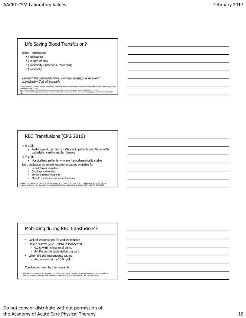
Click to edit Master title styleLife Saving Blood Transfusion?
Blood Transfusions• ↑ utilization • ↑ length of stay• ↑ morbidity (infections, thrombus)• ↑ mortality
Current Recommendations: Primary strategy is to avoid transfusion if at all possible
Scott BH, Seifert FC, Grimson R. Blood transfusion is associated with increased resource utilisation, morbidity and mortality in cardiac surgery. Ann Card Anaesth 2008;11:15-9Shander AS, Goodnough LT; Blood Transfusion as a Quality Indicator in Cardiac Surgery JAMA. 2010;304(14):1610-1611.Hajjar L; TRACS: Differing blood transfusion strategies yield similar complication, death rates in cardiac surgery patients JAMA. 2010;304;1559-1567
Click to edit Master title styleRBC Transfusions (CPG 2016)
< 8 g/dL• Post-surgical, cardiac or orthopedic patients and those with
underlying cardiovascular disease< 7 g/dL
• Hospitalized patients who are hemodynamically stableNo transfusion threshold recommendation available for:
• Hematological disorders• Oncological disorders• Severe thrombocytopenia• Chronic transfusion-dependent anemia
Carson, J. L., Guyatt, G., Heddle, N. M., Grossman, B. J., Cohn, C. S., Fung, M. K., ... & Peterson, N. (2016). Clinical practice guidelines from the AABB: red blood cell transfusion thresholds and storage. JAMA, 316(19), 2025-2035.
Click to edit Master title styleMobilizing during RBC transfusions?
• Lack of evidence re: PT and transfusion• Ohio e-survey (262 PT/PTA respondents)
• 9.2% with institutional policy • 54.8% comfortable delivering care
• When did the respondents say no• Avg ~ minimum of 6.9 g/dL
Conclusion: need further research
Rosenfeldt, A. B., Pilkey, L. M., & Butler, R. S. (2016). A Survey of Physical Therapists Attitudes and Practice Patterns Regarding Intervention During a Red Blood Cell Transfusion. The Journal of Acute Care Physical Therapy.
AACPT CSM Laboratory Values February 2017
Do not copy or distribute without permission of the Academy of Acute Care Physical Therapy 17
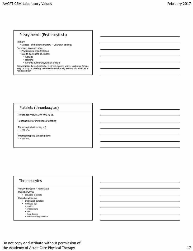
Click to edit Master title stylePolycythemia (Erythrocytosis)
Primary• Disease of the bone marrow – Unknown etiology
Secondary (compensatory)• Physiological manifestation• Due to decreased O2 supply
• Altitude• Nicotine• Chronic pulmonary/cardiac deficits
Presentation: Fever, headache, dizziness, blurred vision, weakness, fatigue, easy bruising or bleeding, decreased mental acuity, sensory disturbances in hands and feet
Click to edit Master title stylePlatelets (thrombocytes)
Reference Value 140-400 k/uL
Responsible for initiation of clotting
Thrombocytosis (trending up)• < 450 k/uL
Thrombocytopenia (trending down)• < 150 k/uL
Click to edit Master title styleThrombocytes
Primary Function – HemostasisThrombocytosis
• Elevated plateletsThrombocytopenia
• Decreased platelets• Reduced by:
• aspirin• medications• diet• liver disease• chemotherapy/radiation
AACPT CSM Laboratory Values February 2017
Do not copy or distribute without permission of the Academy of Acute Care Physical Therapy 18
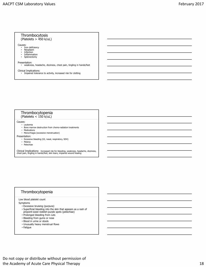
Click to edit Master title styleThrombocytosis(Platelets > 450 k/uL)
Causes: • Iron deficiency• Neoplasm• Infection• Inflammation• Splenectomy
Presentation:• weakness, headache, dizziness, chest pain, tingling in hands/feet
Clinical Implications: • Impaired tolerance to activity, increased risk for clotting
Click to edit Master title styleThrombocytopenia(Platelets < 150 k/uL)
Causes:• Leukemia• Bone marrow destruction from chemo-radiation treatments• Medications• Menorrhagia (excessive menstruation)
Presentation: • Excessive bleeding (GI, nasal, respiratory, SDH)• Melena• Petechiae
Clinical Implications: Increased risk for bleeding, weakness, headache, dizziness, chest pain, tingling in hands/feet, skin tears, impaired wound healing
Click to edit Master title styleThrombocytopenia
Low blood platelet countSymptoms
• Excessive bruising (purpura)• Superficial bleeding into the skin that appears as a rash of
pinpoint-sized reddish-purple spots (petechiae) • Prolonged bleeding from cuts• Bleeding from gums or nose• Blood in urine or stools• Unusually heavy menstrual flows• Fatigue
AACPT CSM Laboratory Values February 2017
Do not copy or distribute without permission of the Academy of Acute Care Physical Therapy 19
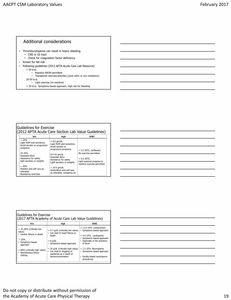
Click to edit Master title styleAdditional considerations
• Thrombocytopenia can result in heavy bleeding • CNS or GI tract• Check for coagulation factor deficiency
• Screen for fall risk• Following guidelines (2013 APTA Acute Care Lab Resource)
> 50 k/uL• Resistive AROM permitted• Therapeutic exercise/activities (cycle with/ or w/o resistance)
20-50 k/uL• Light exercise (no resistive)
< 20 k/uL Symptoms based approach, high risk for bleeding
Click to edit Master title styleGuidelines for Exercise (2012 APTA Acute Care Section Lab Value Guidelines)
Hct Hgb WBC
< 25% Light ROM and isometricsAvoid aerobic or progressive programs
25-35% Essential ADL’sAssistance for safetylight aerobics or weights
> 35% Mobilize and self care as tolerated Resistance exercises
< 8.0 gm/dLLight ROM and isometrics Avoid aerobic or progressive programs
8.0-10 gm/dLEssential ADLsAssistance for safetyLight aerobics or weights
> 10.0 gm/dLAmbulation and self care as tolerated, resistance ex
< 5.0 109/L (w/fever)No exercise permitted
> 5.0 109/L Light exercise progress to resistive exercise permitted
Click to edit Master title styleGuidelines for Exercise (2017 APTA Academy of Acute Care Lab Value Guidelines)
Hct Hgb WBC
< 15-20% (critically low value)• Cardiac failure or death
< 25%• Symptoms based
approach
> 60% (critically high value)• Spontaneous blood
clotting
< 5-7 g/dL (critically low value)• Can lead to heart failure or
death
< 8 g/dL• Symptoms-based approach
> 20 g/dL (critically high value)• Can lead to clogging of
capillaries as a result of hemoconcentration
> 11.0 109/L Leukocytosis• Symptoms based approach
< 4.0 109/L Leukopenia• Symptoms based approach• Especially in the presence
of fever
< 1.5 109/L Neutropenia• Symptoms based approach
• Facility based neutropenicprocedures
AACPT CSM Laboratory Values February 2017
Do not copy or distribute without permission of the Academy of Acute Care Physical Therapy 20
Click to edit Master title style
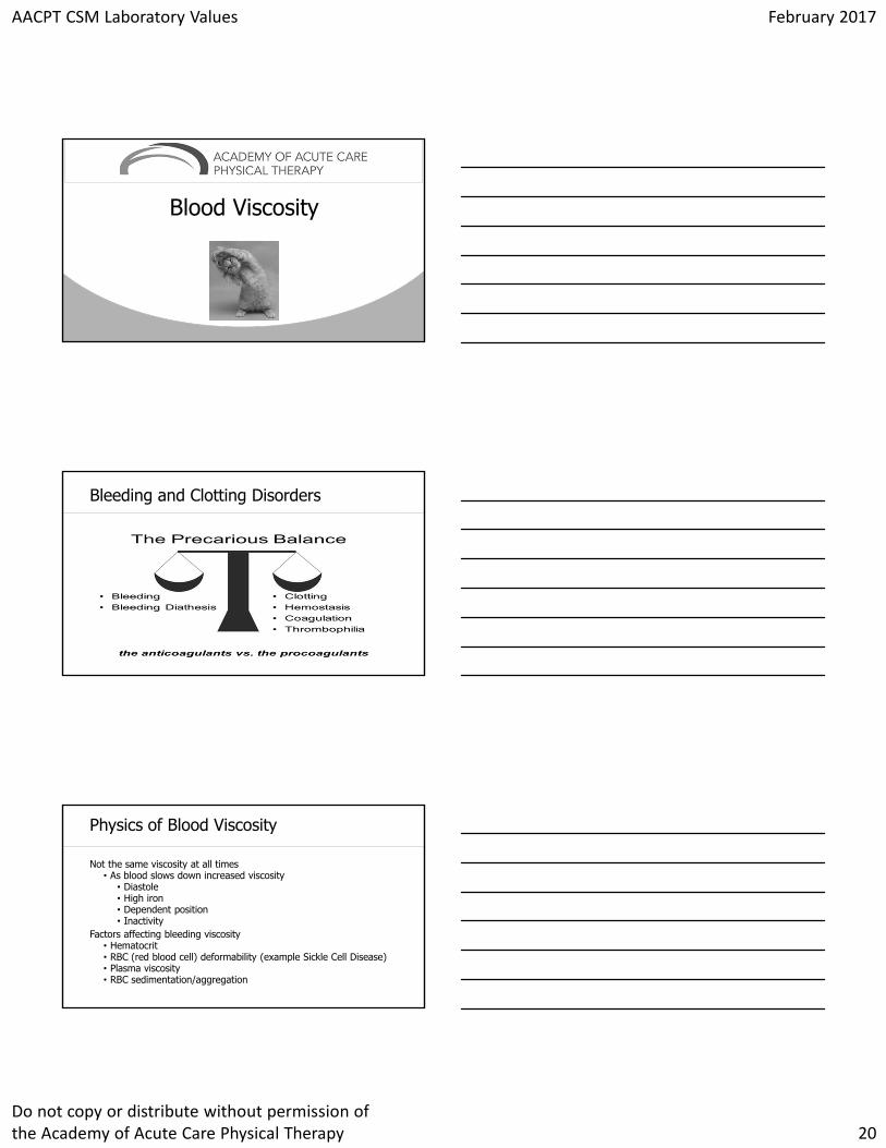
Blood Viscosity
Click to edit Master title styleBleeding and Clotting Disorders
Click to edit Master title stylePhysics of Blood Viscosity
Not the same viscosity at all times• As blood slows down increased viscosity
• Diastole• High iron• Dependent position• Inactivity
Factors affecting bleeding viscosity• Hematocrit• RBC (red blood cell) deformability (example Sickle Cell Disease)• Plasma viscosity• RBC sedimentation/aggregation
AACPT CSM Laboratory Values February 2017
Do not copy or distribute without permission of the Academy of Acute Care Physical Therapy 21
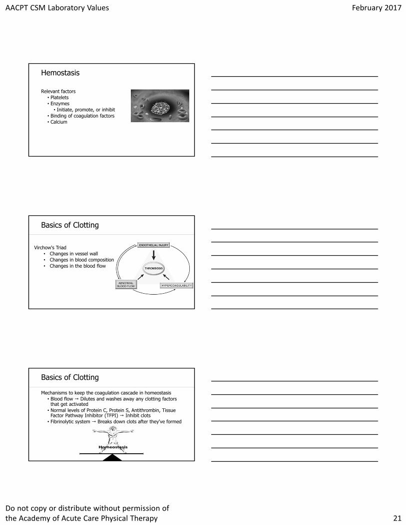
Click to edit Master title styleHemostasis
Relevant factors• Platelets• Enzymes
• Initiate, promote, or inhibit • Binding of coagulation factors• Calcium
Click to edit Master title styleBasics of Clotting
Virchow's Triad • Changes in vessel wall• Changes in blood composition• Changes in the blood flow
Click to edit Master title styleBasics of Clotting
Mechanisms to keep the coagulation cascade in homeostasis• Blood flow Dilutes and washes away any clotting factors
that get activated • Normal levels of Protein C, Protein S, Antithrombin, Tissue
Factor Pathway Inhibitor (TFPI) Inhibit clots• Fibrinolytic system Breaks down clots after they've formed
AACPT CSM Laboratory Values February 2017
Do not copy or distribute without permission of the Academy of Acute Care Physical Therapy 22
Click to edit Master title style
Click to edit Master title styleHemostasis
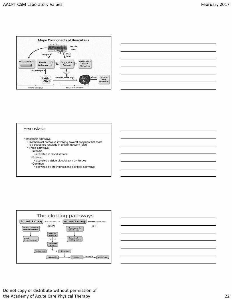
Hemostasis pathways• Biochemical pathways involving several enzymes that react
is a sequence resulting in a fibrin network (clot)• Three pathways
• Intrinsic• activated in blood stream
• Extrinsic• activated outside bloodstream by tissues
• Common• activated by the intrinsic and extrinsic pathways
Click to edit Master title style
aPTTINR/PT
AACPT CSM Laboratory Values February 2017
Do not copy or distribute without permission of the Academy of Acute Care Physical Therapy 23
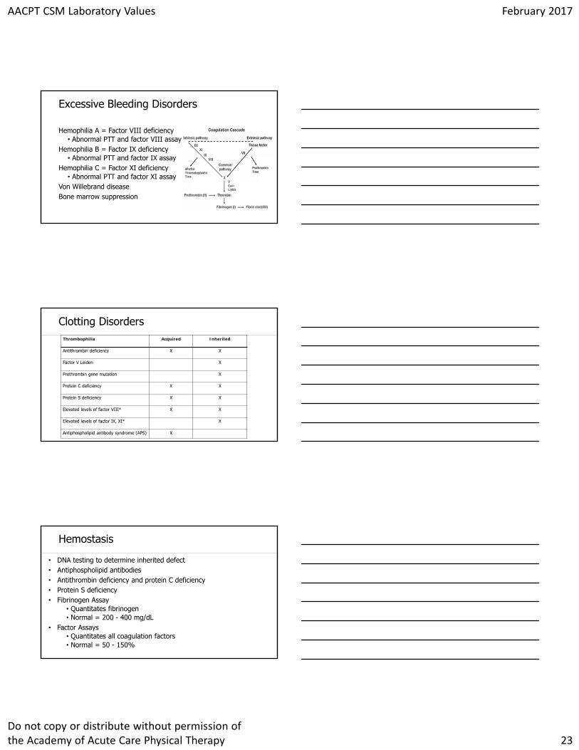
Click to edit Master title styleExcessive Bleeding Disorders
Hemophilia A = Factor VIII deficiency• Abnormal PTT and factor VIII assay
Hemophilia B = Factor IX deficiency• Abnormal PTT and factor IX assay
Hemophilia C = Factor XI deficiency• Abnormal PTT and factor XI assay
Von Willebrand diseaseBone marrow suppression
Click to edit Master title styleClotting DisordersThrombophilia Acquired Inherited
Antithrombin deficiency X X
Factor V Leiden X
Prothrombin gene mutation X
Protein C deficiency X X
Protein S deficiency X X
Elevated levels of factor VIII* X X
Elevated levels of factor IX, XI* X
Antiphospholipid antibody syndrome (APS) X
Click to edit Master title styleHemostasis
• DNA testing to determine inherited defect• Antiphospholipid antibodies• Antithrombin deficiency and protein C deficiency• Protein S deficiency• Fibrinogen Assay
• Quantitates fibrinogen• Normal = 200 - 400 mg/dL
• Factor Assays• Quantitates all coagulation factors• Normal = 50 - 150%
AACPT CSM Laboratory Values February 2017
Do not copy or distribute without permission of the Academy of Acute Care Physical Therapy 24
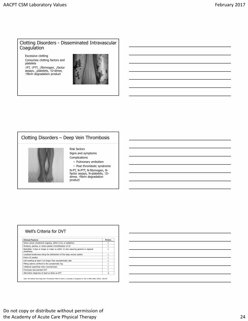
Click to edit Master title style
Excessive clottingConsumes clotting factors and platelets↑PT, ↑PTT, ↓fibrinogen, ↓factor assays, ↓platelets, ↑D-dimer, ↑fibrin degradation product
Clotting Disorders - Disseminated Intravascular Coagulation
Click to edit Master title style
Risk factorsSigns and symptomsComplications
• Pulmonary embolism• Post thrombotic syndrome
N-PT, N-PTT, N-fibrinogen, N-factor assays, N-platelets, ↑D-dimer, ↑fibrin degradation product
Clotting Disorders – Deep Vein Thrombosis
Click to edit Master title styleWell’s Criteria for DVT
Clinical Feature Points
Active cancer (treatment ongoing, within 6 mo, or palliative) 1
Paralysis, paresis, or recent plaster immobilization of LE 1
Bedridden 3 days or longer or major sx within 12 wks requiring general or regional anesthesia
1
Localized tenderness along the distribution of the deep venous system 1
Entire LE swollen 1
Calf swelling at least 3 cm larger than asymptomatic side 1
Pitting edema confined to the symptomatic leg 1
Collateral superficial veins (nonvaricose) 1
Previously documented DVT 1
Alternative diagnosis at least as likely as DVT -2
Does This Patient Have Deep Vein Thrombosis? Wells P, Owen C, Doucette S, Fergusson D, Tran H JAMA 2006; 295(2): 199-207
AACPT CSM Laboratory Values February 2017
Do not copy or distribute without permission of the Academy of Acute Care Physical Therapy 25
Click to edit Master title style
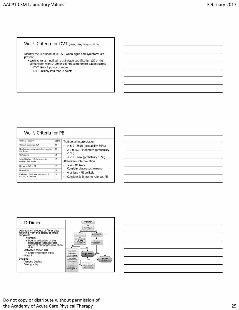
Identify the likelihood of LE DVT when signs and symptoms are present
• Wells criteria modified to a 2-stage stratification (2014) in conjunction with D-Dimer did not compromise patient safety
• DVT likely 2 points or more• DVT unlikely less than 2 points
Well’s Criteria for DVT (Wells, 2014; Hillegass, 2016)
Click to edit Master title styleClinical Feature Score
Clinically suspected DVT 3.0
No alternative diagnosis better explains the illness
3.0
Tachycardia 1.5
Immobilization (>3 d)/ surgery in previous four weeks
1.5
History of DVT or PE 1.5
Hemoptysis 1.0
Malignancy (with treatment within 6 months) or palliative
1.0
Traditional interpretation• > 6.0 - High (probability 59%) • 2.0 to 6.0 - Moderate (probability
29%)• < 2.0 - Low (probability 15%)Alternative interpretation• > 4 - PE likely
Consider diagnostic imaging• 4 or less - PE unlikely • Consider D-Dimer to rule out PE
Well’s Criteria for PE
Click to edit Master title style
Degradation product of fibrin clots resulting from the action of three enzymes:
• Thrombin• Due to activation of the
coagulation cascade that converts fibrinogen into fibrin clots
• Activated factor XIII• Cross-links fibrin clots
• PlasminImaging
• Venous Duplex• Venography
D-Dimer
AACPT CSM Laboratory Values February 2017
Do not copy or distribute without permission of the Academy of Acute Care Physical Therapy 26
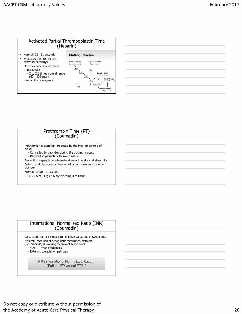
Click to edit Master title style
• Normal: 22 - 31 seconds• Evaluates the intrinsic and
common pathways• Monitors patient on heparin
• Therapeutic • 2 to 2.5 times normal range
(60 - 109 secs) • Variability in reagents
Activated Partial Thromboplastin Time(Heparin)
Click to edit Master title styleProthrombin Time (PT)
(Coumadin)
Prothrombin is a protein produced by the liver for clotting of blood
• Converted to thrombin during the clotting process• Reduced in patients with liver disease
Production depends on adequate vitamin K intake and absorptionDetects and diagnoses a bleeding disorder or excessive clotting disorderNormal Range: 11-13 secsPT > 25 secs - High risk for bleeding into tissue
Click to edit Master title styleInternational Normalized Ratio (INR)(Coumadin)
Calculated from a PT result to minimize variations between labs Monitors how well anticoagulant medication warfarin (Coumadin®) is working to prevent blood clots
• ↑INR ~ ↑risk of bleeding• Extrinsic coagulation pathway
AACPT CSM Laboratory Values February 2017
Do not copy or distribute without permission of the Academy of Acute Care Physical Therapy 27
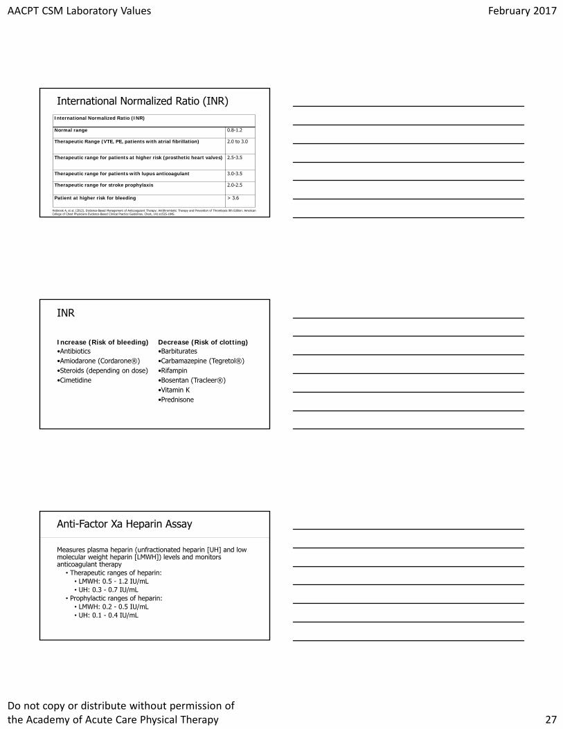
Click to edit Master title styleInternational Normalized Ratio (INR)
Normal range 0.8-1.2
Therapeutic Range (VTE, PE, patients with atrial fibrillation) 2.0 to 3.0
Therapeutic range for patients at higher risk (prosthetic heart valves) 2.5-3.5
Therapeutic range for patients with lupus anticoagulant 3.0-3.5
Therapeutic range for stroke prophylaxis 2.0-2.5
Patient at higher risk for bleeding > 3.6
International Normalized Ratio (INR)
Holbrook A, et al. (2012). Evidence-Based Management of Anticoagulant Therapy: Antithrombotic Therapy and Prevention of Thrombosis 9th Edition: American College of Chest Physicians Evidence-Based Clinical Practice Guidelines. Chest, 141:e152S-184S.
Click to edit Master title style
Increase (Risk of bleeding)•Antibiotics•Amiodarone (Cordarone®)•Steroids (depending on dose)•Cimetidine
Decrease (Risk of clotting)•Barbiturates•Carbamazepine (Tegretol®)•Rifampin•Bosentan (Tracleer®)•Vitamin K•Prednisone
INR
Click to edit Master title styleAnti-Factor Xa Heparin Assay
Measures plasma heparin (unfractionated heparin [UH] and low molecular weight heparin [LMWH]) levels and monitors anticoagulant therapy
• Therapeutic ranges of heparin:• LMWH: 0.5 - 1.2 IU/mL• UH: 0.3 - 0.7 IU/mL
• Prophylactic ranges of heparin:• LMWH: 0.2 - 0.5 IU/mL• UH: 0.1 - 0.4 IU/mL
AACPT CSM Laboratory Values February 2017
Do not copy or distribute without permission of the Academy of Acute Care Physical Therapy 28
Click to edit Master title style
What is the PTs Role?
Click to edit Master title styleExcessive Bleeding Clinical Considerations
Resistance exerciseActivities with risk of fallingEducation
MedicationsFall risk management
Sharp debridement
Click to edit Master title styleManagement of Individuals with Venous Thromboembolism (Hillegass, 2016)
Advocate for a culture of mobility and physical activityScreen for risk of VTE
• Patient interview and physical examinationProvide preventive measures for LE DVT
• Education for signs and symptoms of LE DVT• Activity• Hydration• Mechanical compression
AACPT CSM Laboratory Values February 2017
Do not copy or distribute without permission of the Academy of Acute Care Physical Therapy 29
Click to edit Master title styleManagement of Individuals with Venous Thromboembolism (Hillegass, 2016)
Recommend mechanical compression for patients with LE DVT or when signs and symptoms of post thrombotic syndromeMobilize patients after IVC filter placement once hemodynamically stableVerify the patient is taking an anticoagulant
• Mobilize patients who are at a therapeutic level of anticoagulation
Click to edit Master title style
Oral Medications• Aspirin• Clopidogrel (Plavix)• Fondaparinux (Arixtra)• Warfarin (Coumadin)• Dabigatran (Pradaxa)• Rivaroxaban (Xarelto)• Apixaban (Eliquis) • Savaysa (Edoxaban)
Intravenous • Heparin
• Bridging• Short acting
Subcutaneous• Heparin• Low molecular weight
heparin (LMWH) (Lovenox)
Anticoagulation Medications
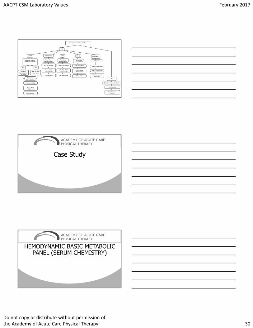
Click to edit Master title styleAlgorithm for Mobilizing Patients with Known Lower-Extremity Deep Vein Thrombosis (Reprinted from Phys Ther. 2016;96:143‐166,with permission of the
American Physical Therapy Association. ©2016 American Physical Therapy Association.)
AACPT CSM Laboratory Values February 2017
Do not copy or distribute without permission of the Academy of Acute Care Physical Therapy 30
Click to edit Master title style
Click to edit Master title style
Case Study
Click to edit Master title style
HEMODYNAMIC BASIC METABOLIC PANEL (SERUM CHEMISTRY)
AACPT CSM Laboratory Values February 2017
Do not copy or distribute without permission of the Academy of Acute Care Physical Therapy 31
Click to edit Master title styleLabs
Blood Chemistry Testing
• Fluid Balance• Body Water
• Electrolyte Balance
Click to edit Master title styleFluid Balance - Hypervolemia
CausesExcess IV fluidsHypertonic FluidInadequate Output
• CHF• Cirrhosis• Renal failure/insuff• Low protein• Steroid use
PresentationPitting edemaSOBAnasarcaJugular distensionHTNTachycardiaCrackles
Click to edit Master title styleFluid Balance - Hypovolemia
CausesLimited oral intake
• CVA• AMS
Excess loss• Vomiting/diarrhea• DM• Burns• Excessive Sweating
PresentationDry Mucus MembranesPoor skin turgorHypotension (orthostatic)TachycardiaTachypneaAMS
AACPT CSM Laboratory Values February 2017
Do not copy or distribute without permission of the Academy of Acute Care Physical Therapy 32
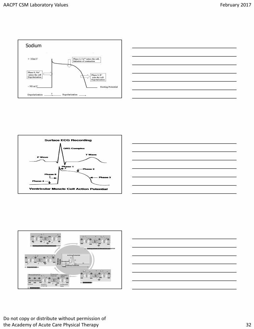
Click to edit Master title styleSodium
Click to edit Master title style
Click to edit Master title style
AACPT CSM Laboratory Values February 2017
Do not copy or distribute without permission of the Academy of Acute Care Physical Therapy 33
Click to edit Master title styleSodiumHypernatremia Na+> 150 mEq/L
CausesHypovolemiaIncreased Na intakeSevere vomitingCHFRenal f/insCushing’s syndromeDiabetes
PresentsIrritabilityAgitationSeizureComaHypotensionTachycardiaWeak pulseDecreased urine output
Click to edit Master title styleSodiumHyponatremia Na+ < 135 mEq/L
CausesDiuretic useGI lossBurns/woundsHypotonic IV useCirrhosis
PresentsHeadacheLethargyDecreased reflexesNausea vomitingDiarrheaSeizure ComaOrthostatic hypotensionPitting edema
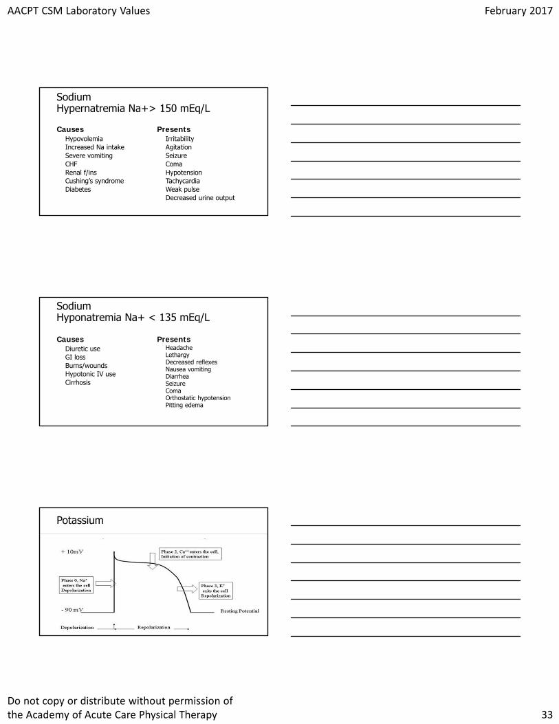
Click to edit Master title stylePotassium
AACPT CSM Laboratory Values February 2017
Do not copy or distribute without permission of the Academy of Acute Care Physical Therapy 34
Click to edit Master title style
Click to edit Master title stylePotassiumHyperkalemia K+ > 5.3 mEq/L
CausesRenal failureMetabolic acidosisDKAAddison’s diseaseExcesses K supplementsBlood transfusions
PresentsMuscle weakness/paralysisParesthesiaBradycardiaHeart BlockV-fibCardiac arrest
Click to edit Master title stylePotassiumHypokalemia K+ < 3.0 mEq/L
CausesDiarrhea/vomitingGI losses/NG suctionDiureticsCushing SyndromeMalnutritionRestrictive dietsETOH abuse
PresentsExtremity weaknessHyporeflexiaParesthesiaLeg CrampsECG changesST depressionInverted TsCardiac arrestHypotensionConstipation
AACPT CSM Laboratory Values February 2017
Do not copy or distribute without permission of the Academy of Acute Care Physical Therapy 35
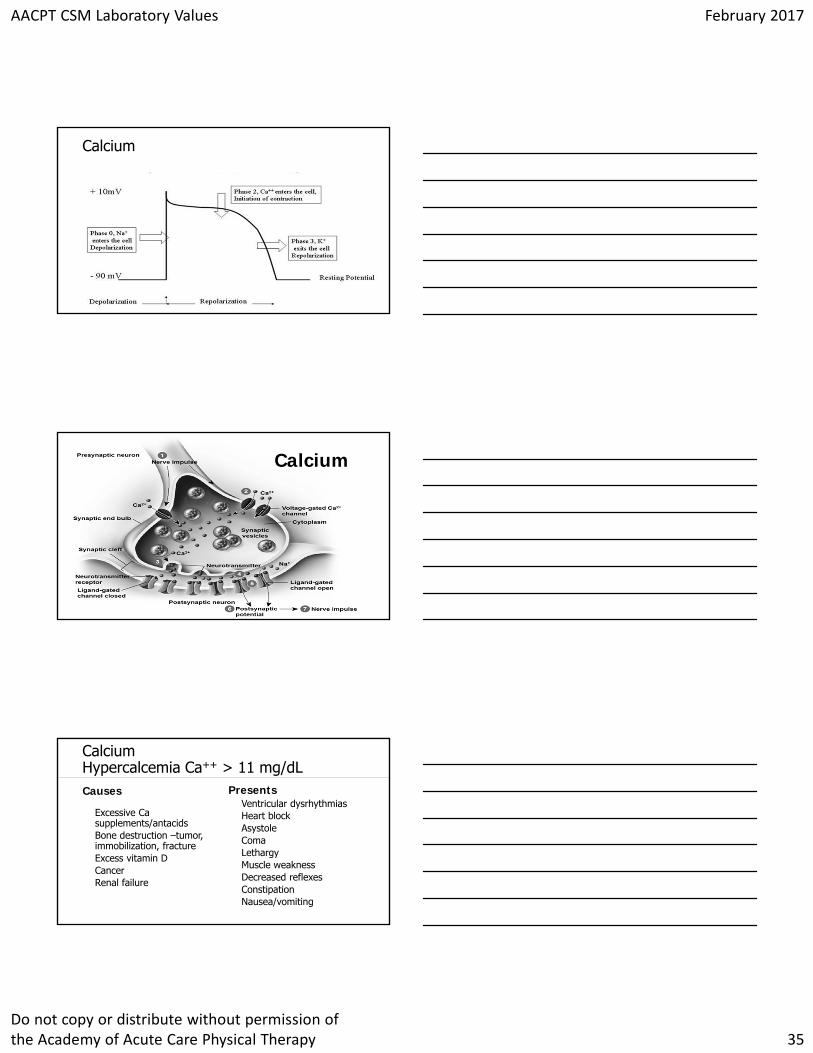
Click to edit Master title styleCalcium
Click to edit Master title styleCalcium
Click to edit Master title styleCalciumHypercalcemia Ca++ > 11 mg/dLCauses
Excessive Ca supplements/antacidsBone destruction –tumor, immobilization, fractureExcess vitamin DCancerRenal failure
PresentsVentricular dysrhythmiasHeart block AsystoleComaLethargyMuscle weaknessDecreased reflexesConstipationNausea/vomiting
AACPT CSM Laboratory Values February 2017
Do not copy or distribute without permission of the Academy of Acute Care Physical Therapy 36
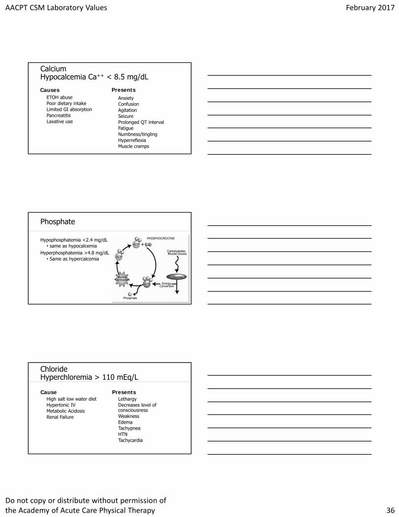
Click to edit Master title styleCalciumHypocalcemia Ca++ < 8.5 mg/dL
CausesETOH abusePoor dietary intakeLimited GI absorptionPancreatitisLaxative use
PresentsAnxietyConfusionAgitationSeizureProlonged QT intervalFatigueNumbness/tinglingHyperreflexiaMuscle cramps
Click to edit Master title stylePhosphate
Hypophosphatemia <2.4 mg/dL• same as hypocalcemia
Hyperphosphatemia >4.8 mg/dL• Same as hypercalcemia
Click to edit Master title styleChlorideHyperchloremia > 110 mEq/L
CauseHigh salt low water dietHypertonic IVMetabolic AcidosisRenal Failure
PresentsLethargyDecreases level of consciousnessWeaknessEdemaTachypneaHTNTachycardia
AACPT CSM Laboratory Values February 2017
Do not copy or distribute without permission of the Academy of Acute Care Physical Therapy 37
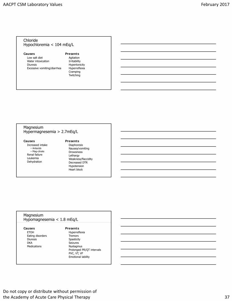
Click to edit Master title styleChlorideHypochloremia < 104 mEq/L
CausesLow salt dietWater intoxicationDiuresisExcessive vomiting/diarrhea
PresentsAgitationIrritabilityHypertonicityHyperreflexiaCrampingTwitching
Click to edit Master title styleMagnesiumHypermagnesemia > 2.7mEq/L
CausesIncreased intake
• Antacids• Mag-citrate
Renal failureLeukemiaDehydration
PresentsDiaphoresisNausea/vomitingDrowsinessLethargyWeakness/flaccidityDecreased DTRHypotensionHeart block
Click to edit Master title styleMagnesiumHypomagnesemia < 1.8 mEq/L
CausesETOHEating disordersDiuresisDKAMedications
PresentsHyperreflexiaTremorsSpasticitySeizuresNystagmusProlonged PR/QT intervalsPVC, VT, VFEmotional lability
AACPT CSM Laboratory Values February 2017
Do not copy or distribute without permission of the Academy of Acute Care Physical Therapy 38
Click to edit Master title styleBUNBlood Urea Nitrogen
Urea forms in the liver from breakdown of proteins and aminos. Normal ranges 10-20 mg/dLUsed to measure renal excretory capacity, estimate protein catabolism and tissue necrosis
• High- High protein diet, renal failure, hypovolemia, CHF, GI Bleed, fever, increased protein catabolism
• Low- liver disease
Click to edit Master title styleCreatinine
Constant excretion each day dependent on body muscle massIncreased levels consistent with renal diseaseNormal Range- 0.9-1.3 mg/dLOther causes of increased levels
• Muscular Dystrophy• Myasthenia Gravis• Rhabdomyolysis• Dehydration
Click to edit Master title style
Cardiac Specific Testing
AACPT CSM Laboratory Values February 2017
Do not copy or distribute without permission of the Academy of Acute Care Physical Therapy 39
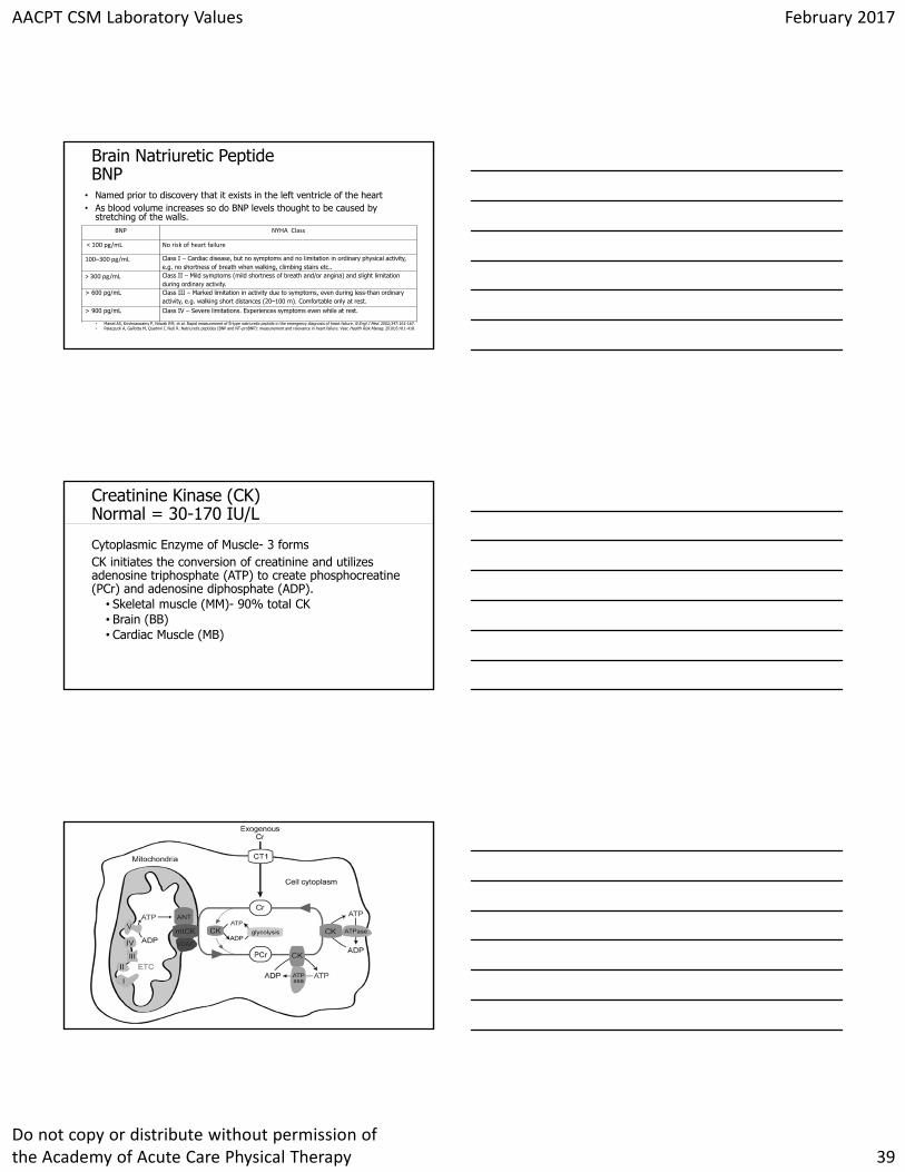
Click to edit Master title styleBrain Natriuretic PeptideBNP
• Named prior to discovery that it exists in the left ventricle of the heart• As blood volume increases so do BNP levels thought to be caused by
stretching of the walls.BNP NYHA Class
< 100 pg/mL No risk of heart failure
100–300 pg/mL Class I – Cardiac disease, but no symptoms and no limitation in ordinary physical activity, e.g. no shortness of breath when walking, climbing stairs etc..
> 300 pg/mL Class II – Mild symptoms (mild shortness of breath and/or angina) and slight limitation during ordinary activity.
> 600 pg/mL Class III – Marked limitation in activity due to symptoms, even during less-than ordinary activity, e.g. walking short distances (20–100 m). Comfortable only at rest.
> 900 pg/mL Class IV – Severe limitations. Experiences symptoms even while at rest.
• Maisel AS, Krishnaswamy P, Nowak RM, et al. Rapid measurement of B-type natriuretic peptide in the emergency diagnosis of heart failure. N Engl J Med. 2002;347:161-167.• Palazzuoli A, Gallotta M, Quatrini I, Nuti R. Natriuretic peptides (BNP and NT-proBNP): measurement and relevance in heart failure. Vasc Health Risk Manag. 2010;6:411-418.
Click to edit Master title styleCreatinine Kinase (CK)Normal = 30-170 IU/L
Cytoplasmic Enzyme of Muscle- 3 formsCK initiates the conversion of creatinine and utilizes adenosine triphosphate (ATP) to create phosphocreatine (PCr) and adenosine diphosphate (ADP).
• Skeletal muscle (MM)- 90% total CK• Brain (BB)• Cardiac Muscle (MB)
Click to edit Master title style
AACPT CSM Laboratory Values February 2017
Do not copy or distribute without permission of the Academy of Acute Care Physical Therapy 40
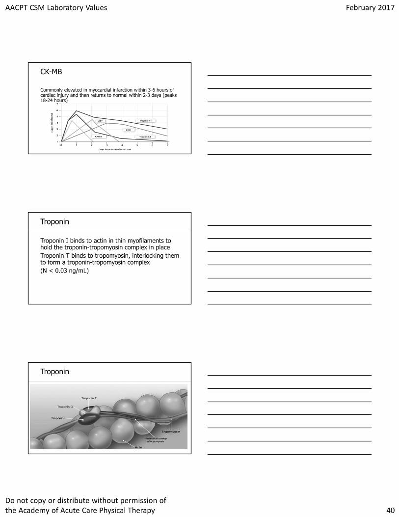
Click to edit Master title styleCK-MB
Commonly elevated in myocardial infarction within 3-6 hours of cardiac injury and then returns to normal within 2-3 days (peaks 18-24 hours)
Click to edit Master title styleTroponin
Troponin I binds to actin in thin myofilaments to hold the troponin-tropomyosin complex in placeTroponin T binds to tropomyosin, interlocking them to form a troponin-tropomyosin complex(N < 0.03 ng/mL)
Click to edit Master title styleTroponin
AACPT CSM Laboratory Values February 2017
Do not copy or distribute without permission of the Academy of Acute Care Physical Therapy 41
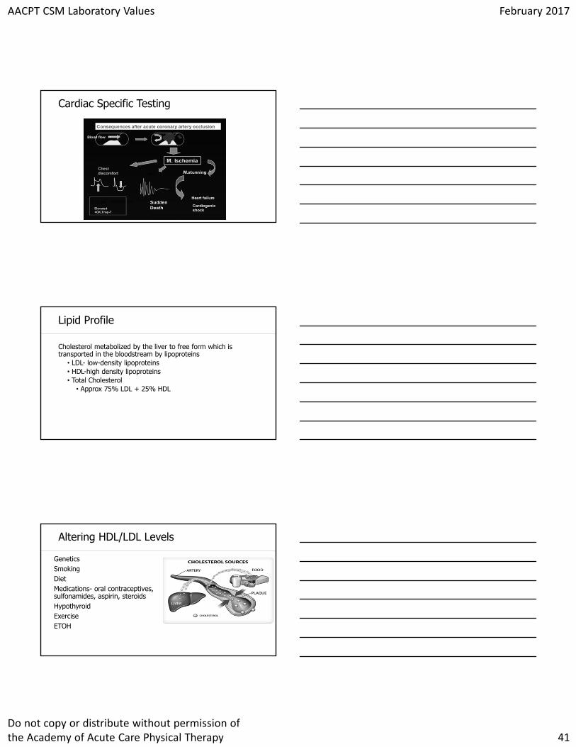
Click to edit Master title styleCardiac Specific Testing
Click to edit Master title styleLipid Profile
Cholesterol metabolized by the liver to free form which is transported in the bloodstream by lipoproteins
• LDL- low-density lipoproteins• HDL-high density lipoproteins• Total Cholesterol
• Approx 75% LDL + 25% HDL
Click to edit Master title styleAltering HDL/LDL Levels
GeneticsSmokingDietMedications- oral contraceptives, sulfonamides, aspirin, steroidsHypothyroidExerciseETOH
AACPT CSM Laboratory Values February 2017
Do not copy or distribute without permission of the Academy of Acute Care Physical Therapy 42
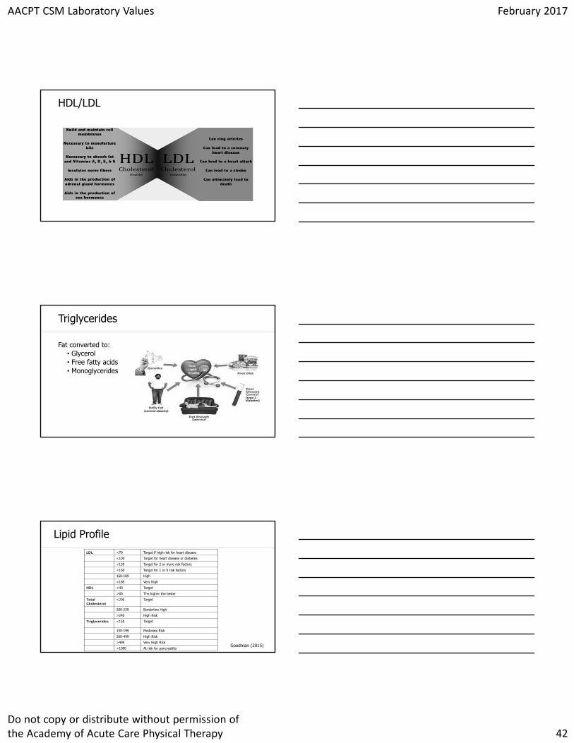
Click to edit Master title styleHDL/LDL
Click to edit Master title styleTriglycerides
Fat converted to:• Glycerol• Free fatty acids• Monoglycerides
Click to edit Master title styleLipid ProfileLDL <70 Target if high risk for heart disease
<100 Target for heart disease or diabetes
<130 Target for 2 or more risk factors
<160 Target for 1 or 0 risk factors
160-189 High
>189 Very High
HDL <40 Target
>60 The higher the better
Total Cholesterol
<200 Target
200-239 Borderline High
>240 High Risk
Triglycerides <150 Target
150-199 Moderate Risk
200-499 High Risk
>499 Very High Risk
>1000 At risk for pancreatitisGoodman (2015)
AACPT CSM Laboratory Values February 2017
Do not copy or distribute without permission of the Academy of Acute Care Physical Therapy 43
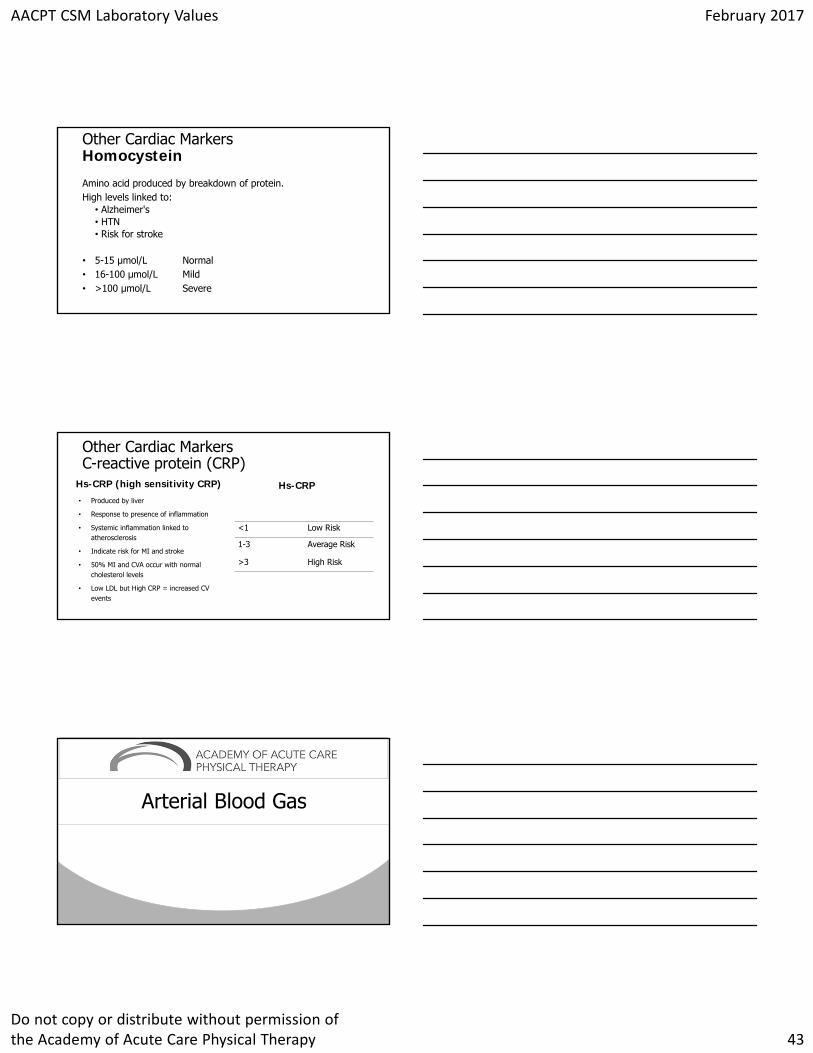
Click to edit Master title styleOther Cardiac MarkersHomocystein
Amino acid produced by breakdown of protein.High levels linked to:
• Alzheimer's• HTN• Risk for stroke
• 5-15 µmol/L Normal• 16-100 µmol/L Mild• >100 µmol/L Severe
Click to edit Master title styleOther Cardiac MarkersC-reactive protein (CRP)
Hs-CRP (high sensitivity CRP)
• Produced by liver
• Response to presence of inflammation
• Systemic inflammation linked to atherosclerosis
• Indicate risk for MI and stroke
• 50% MI and CVA occur with normal cholesterol levels
• Low LDL but High CRP = increased CV events
Hs-CRP
<1 Low Risk
1-3 Average Risk
>3 High Risk
Click to edit Master title style
Arterial Blood Gas
AACPT CSM Laboratory Values February 2017
Do not copy or distribute without permission of the Academy of Acute Care Physical Therapy 44
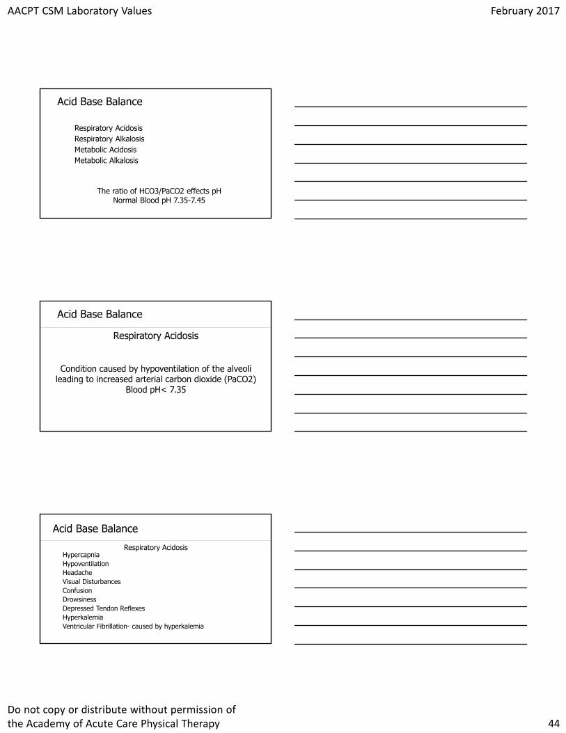
Click to edit Master title styleAcid Base Balance
Respiratory AcidosisRespiratory AlkalosisMetabolic AcidosisMetabolic Alkalosis
The ratio of HCO3/PaCO2 effects pH Normal Blood pH 7.35-7.45
Click to edit Master title styleAcid Base Balance
Respiratory Acidosis
Condition caused by hypoventilation of the alveoli leading to increased arterial carbon dioxide (PaCO2)
Blood pH< 7.35
Click to edit Master title styleAcid Base Balance
HypercapniaHypoventilationHeadacheVisual DisturbancesConfusionDrowsinessDepressed Tendon ReflexesHyperkalemiaVentricular Fibrillation- caused by hyperkalemia
Respiratory Acidosis
AACPT CSM Laboratory Values February 2017
Do not copy or distribute without permission of the Academy of Acute Care Physical Therapy 45
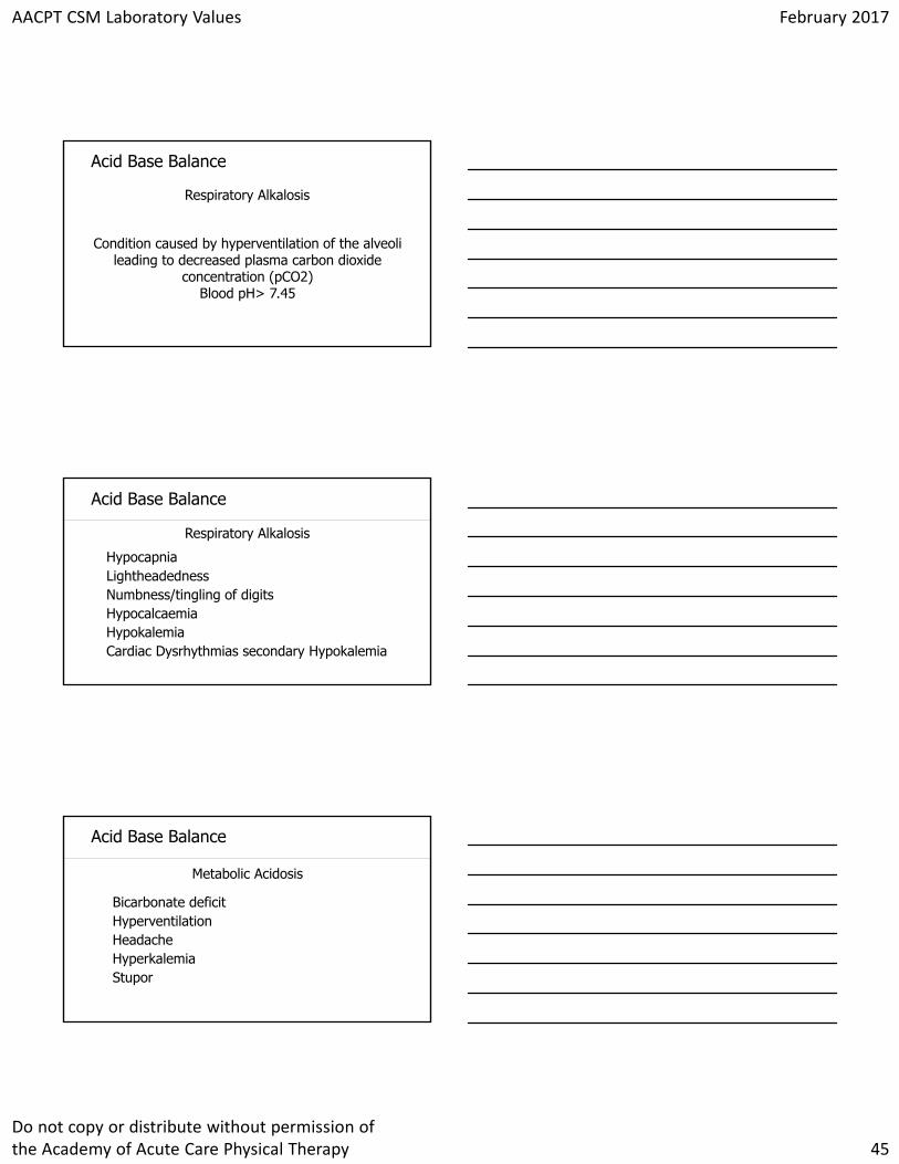
Click to edit Master title styleAcid Base Balance
Respiratory Alkalosis
Condition caused by hyperventilation of the alveoli leading to decreased plasma carbon dioxide
concentration (pCO2)Blood pH> 7.45
Click to edit Master title styleAcid Base Balance
HypocapniaLightheadednessNumbness/tingling of digitsHypocalcaemiaHypokalemiaCardiac Dysrhythmias secondary Hypokalemia
Respiratory Alkalosis
Click to edit Master title styleAcid Base Balance
Bicarbonate deficitHyperventilationHeadacheHyperkalemiaStupor
Metabolic Acidosis
AACPT CSM Laboratory Values February 2017
Do not copy or distribute without permission of the Academy of Acute Care Physical Therapy 46
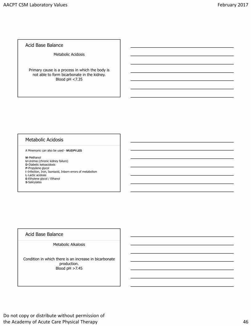
Click to edit Master title styleAcid Base Balance
Metabolic Acidosis
Primary cause is a process in which the body is not able to form bicarbonate in the kidney.
Blood pH <7.35
Click to edit Master title styleMetabolic Acidosis
A Mnemonic can also be used - MUDPILES
M-MethanolU-Uremia (chronic kidney failure)D-Diabetic ketoacidosisP-Propylene glycol I-Infection, Iron, Isoniazid, Inborn errors of metabolismL-Lactic acidosisE-Ethylene glycol / Ethanol S-Salicylates
Click to edit Master title styleAcid Base Balance
Metabolic Alkalosis
Condition in which there is an increase in bicarbonate production.
Blood pH >7.45
AACPT CSM Laboratory Values February 2017
Do not copy or distribute without permission of the Academy of Acute Care Physical Therapy 47
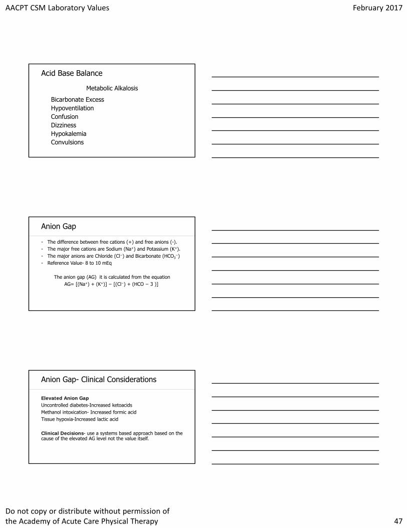
Click to edit Master title styleAcid Base Balance
Bicarbonate ExcessHypoventilationConfusionDizzinessHypokalemiaConvulsions
Metabolic Alkalosis
Click to edit Master title styleAnion Gap
- The difference between free cations (+) and free anions (-). - The major free cations are Sodium (Na+) and Potassium (K+).- The major anions are Chloride (Cl−) and Bicarbonate (HCO3
−)- Reference Value- 8 to 10 mEq
The anion gap (AG) it is calculated from the equationAG= [(Na+) + (K+)] − [(Cl−) + (HCO − 3 )]
Click to edit Master title styleAnion Gap- Clinical Considerations
Elevated Anion GapUncontrolled diabetes-Increased ketoacidsMethanol intoxication- Increased formic acidTissue hypoxia-Increased lactic acid
Clinical Decisions- use a systems based approach based on the cause of the elevated AG level not the value itself.
AACPT CSM Laboratory Values February 2017
Do not copy or distribute without permission of the Academy of Acute Care Physical Therapy 48
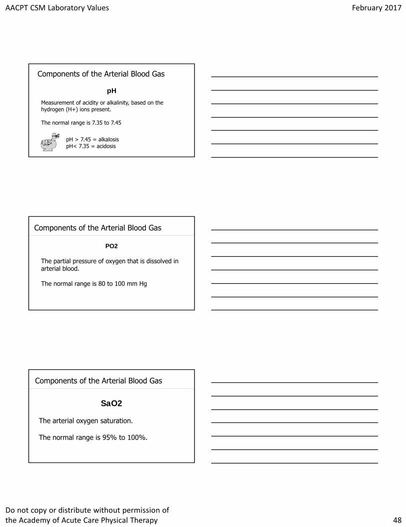
Click to edit Master title styleComponents of the Arterial Blood Gas
pH
Measurement of acidity or alkalinity, based on the hydrogen (H+) ions present.
The normal range is 7.35 to 7.45
pH > 7.45 = alkalosis pH< 7.35 = acidosis
Click to edit Master title styleComponents of the Arterial Blood Gas
PO2
The partial pressure of oxygen that is dissolved in arterial blood.
The normal range is 80 to 100 mm Hg
Click to edit Master title styleComponents of the Arterial Blood Gas
SaO2
The arterial oxygen saturation.
The normal range is 95% to 100%.
AACPT CSM Laboratory Values February 2017
Do not copy or distribute without permission of the Academy of Acute Care Physical Therapy 49
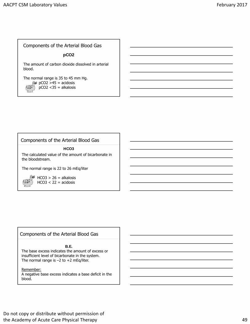
Click to edit Master title styleComponents of the Arterial Blood Gas
pCO2
The amount of carbon dioxide dissolved in arterial blood.
The normal range is 35 to 45 mm Hg. pCO2 >45 = acidosis pCO2 <35 = alkalosis
Click to edit Master title styleComponents of the Arterial Blood Gas
HCO3The calculated value of the amount of bicarbonate in the bloodstream.
The normal range is 22 to 26 mEq/liter
HCO3 > 26 = alkalosis HCO3 < 22 = acidosis
Click to edit Master title styleComponents of the Arterial Blood Gas
B.E. The base excess indicates the amount of excess or insufficient level of bicarbonate in the system. The normal range is –2 to +2 mEq/liter.
Remember:A negative base excess indicates a base deficit in the blood.
AACPT CSM Laboratory Values February 2017
Do not copy or distribute without permission of the Academy of Acute Care Physical Therapy 50
Click to edit Master title style
Click to edit Master title style
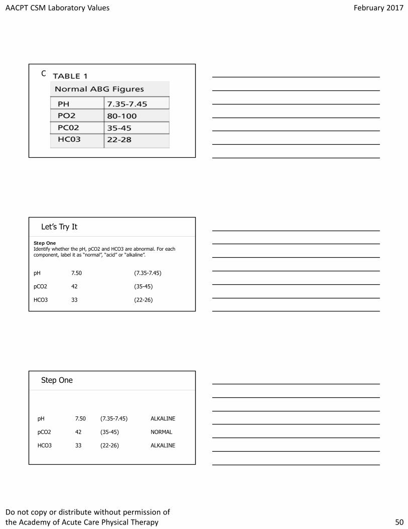
Step One Identify whether the pH, pCO2 and HCO3 are abnormal. For each component, label it as “normal”, “acid” or “alkaline”.
Let’s Try It
pH 7.50 (7.35-7.45)
pCO2 42 (35-45)
HCO3 33 (22-26)
Click to edit Master title style
pH 7.50 (7.35-7.45) ALKALINE
pCO2 42 (35-45) NORMAL
HCO3 33 (22-26) ALKALINE
Step One
AACPT CSM Laboratory Values February 2017
Do not copy or distribute without permission of the Academy of Acute Care Physical Therapy 51
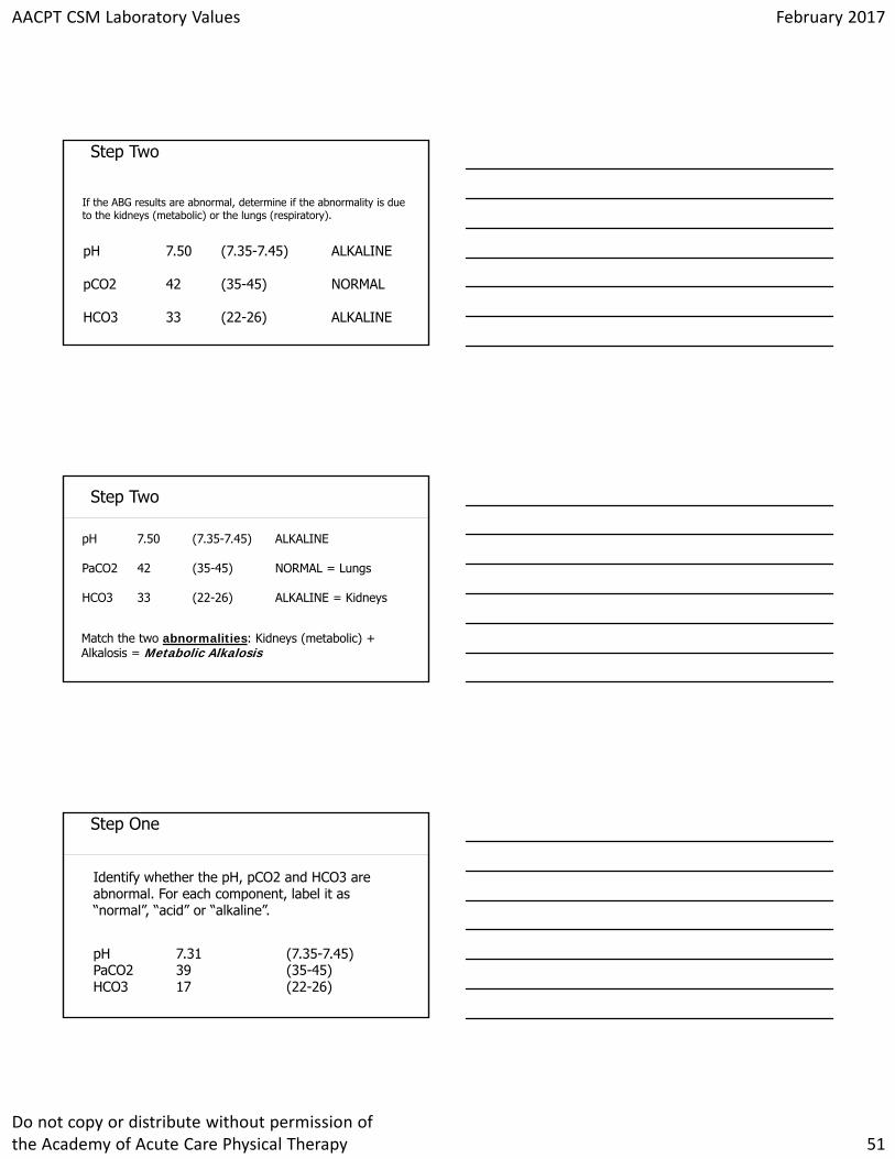
Click to edit Master title styleStep Two
If the ABG results are abnormal, determine if the abnormality is due to the kidneys (metabolic) or the lungs (respiratory).
pH 7.50 (7.35-7.45) ALKALINE
pCO2 42 (35-45) NORMAL
HCO3 33 (22-26) ALKALINE
Click to edit Master title styleStep Two
pH 7.50 (7.35-7.45) ALKALINE
PaCO2 42 (35-45) NORMAL = Lungs
HCO3 33 (22-26) ALKALINE = Kidneys
Match the two abnormalities: Kidneys (metabolic) + Alkalosis = Metabolic Alkalosis
Click to edit Master title styleStep One
Identify whether the pH, pCO2 and HCO3 are abnormal. For each component, label it as “normal”, “acid” or “alkaline”.
pH 7.31 (7.35-7.45) PaCO2 39 (35-45) HCO3 17 (22-26)
AACPT CSM Laboratory Values February 2017
Do not copy or distribute without permission of the Academy of Acute Care Physical Therapy 52
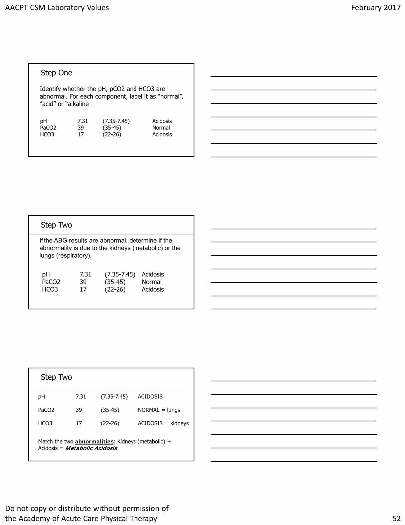
Click to edit Master title styleStep One
Identify whether the pH, pCO2 and HCO3 are abnormal. For each component, label it as “normal”, “acid” or “alkaline”.
pH 7.31 (7.35-7.45) AcidosisPaCO2 39 (35-45) NormalHCO3 17 (22-26) Acidosis
Click to edit Master title styleStep Two
If the ABG results are abnormal, determine if the abnormality is due to the kidneys (metabolic) or the lungs (respiratory).
pH 7.31 (7.35-7.45) AcidosisPaCO2 39 (35-45) NormalHCO3 17 (22-26) Acidosis
Click to edit Master title styleStep Two
pH 7.31 (7.35-7.45) ACIDOSIS
PaCO2 39 (35-45) NORMAL = lungs
HCO3 17 (22-26) ACIDOSIS = kidneys
Match the two abnormalities: Kidneys (metabolic) + Acidosis = Metabolic Acidosis
AACPT CSM Laboratory Values February 2017
Do not copy or distribute without permission of the Academy of Acute Care Physical Therapy 53
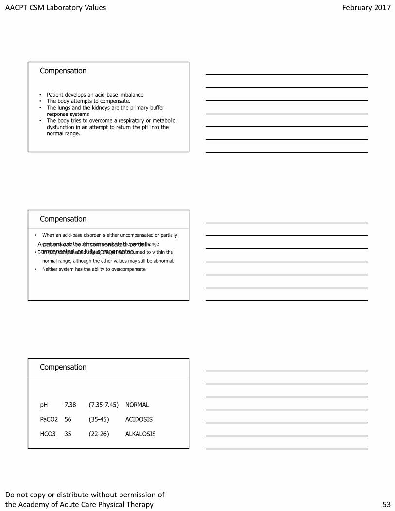
Click to edit Master title styleCompensation
• Patient develops an acid-base imbalance • The body attempts to compensate. • The lungs and the kidneys are the primary buffer
response systems • The body tries to overcome a respiratory or metabolic
dysfunction in an attempt to return the pH into the normal range.
Click to edit Master title styleCompensation
A patient can be uncompensated, partially compensated, or fully compensated.
• When an acid-base disorder is either uncompensated or partially
compensated, the pH remains outside the normal range
• In fully compensated states, the pH has returned to within the
normal range, although the other values may still be abnormal.
• Neither system has the ability to overcompensate
Click to edit Master title styleCompensation
pH 7.38 (7.35-7.45) NORMAL
PaCO2 56 (35-45) ACIDOSIS
HCO3 35 (22-26) ALKALOSIS
AACPT CSM Laboratory Values February 2017
Do not copy or distribute without permission of the Academy of Acute Care Physical Therapy 54
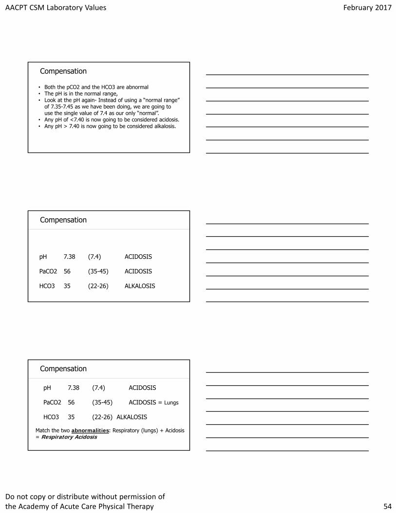
Click to edit Master title styleCompensation
• Both the pCO2 and the HCO3 are abnormal• The pH is in the normal range, • Look at the pH again- Instead of using a “normal range”
of 7.35-7.45 as we have been doing, we are going to use the single value of 7.4 as our only “normal”.
• Any pH of <7.40 is now going to be considered acidosis. • Any pH > 7.40 is now going to be considered alkalosis.
Click to edit Master title styleCompensation
pH 7.38 (7.4) ACIDOSIS
PaCO2 56 (35-45) ACIDOSIS
HCO3 35 (22-26) ALKALOSIS
Click to edit Master title styleCompensation
pH 7.38 (7.4) ACIDOSIS
PaCO2 56 (35-45) ACIDOSIS = Lungs
HCO3 35 (22-26) ALKALOSIS
Match the two abnormalities: Respiratory (lungs) + Acidosis = Respiratory Acidosis
AACPT CSM Laboratory Values February 2017
Do not copy or distribute without permission of the Academy of Acute Care Physical Therapy 55
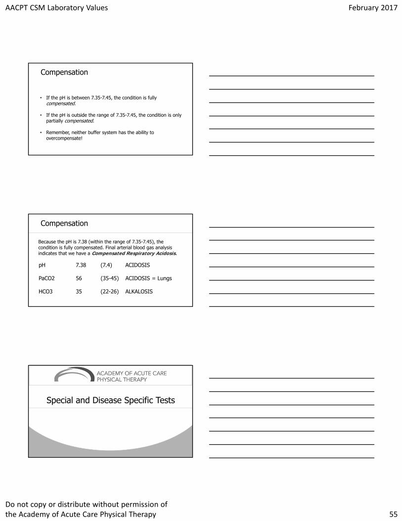
Click to edit Master title styleCompensation
• If the pH is between 7.35-7.45, the condition is fullycompensated.
• If the pH is outside the range of 7.35-7.45, the condition is only partially compensated.
• Remember, neither buffer system has the ability to overcompensate!
Click to edit Master title styleCompensation
Because the pH is 7.38 (within the range of 7.35-7.45), the condition is fully compensated. Final arterial blood gas analysis indicates that we have a Compensated Respiratory Acidosis.
pH 7.38 (7.4) ACIDOSIS
PaCO2 56 (35-45) ACIDOSIS = Lungs
HCO3 35 (22-26) ALKALOSIS
Click to edit Master title style
Special and Disease Specific Tests
AACPT CSM Laboratory Values February 2017
Do not copy or distribute without permission of the Academy of Acute Care Physical Therapy 56
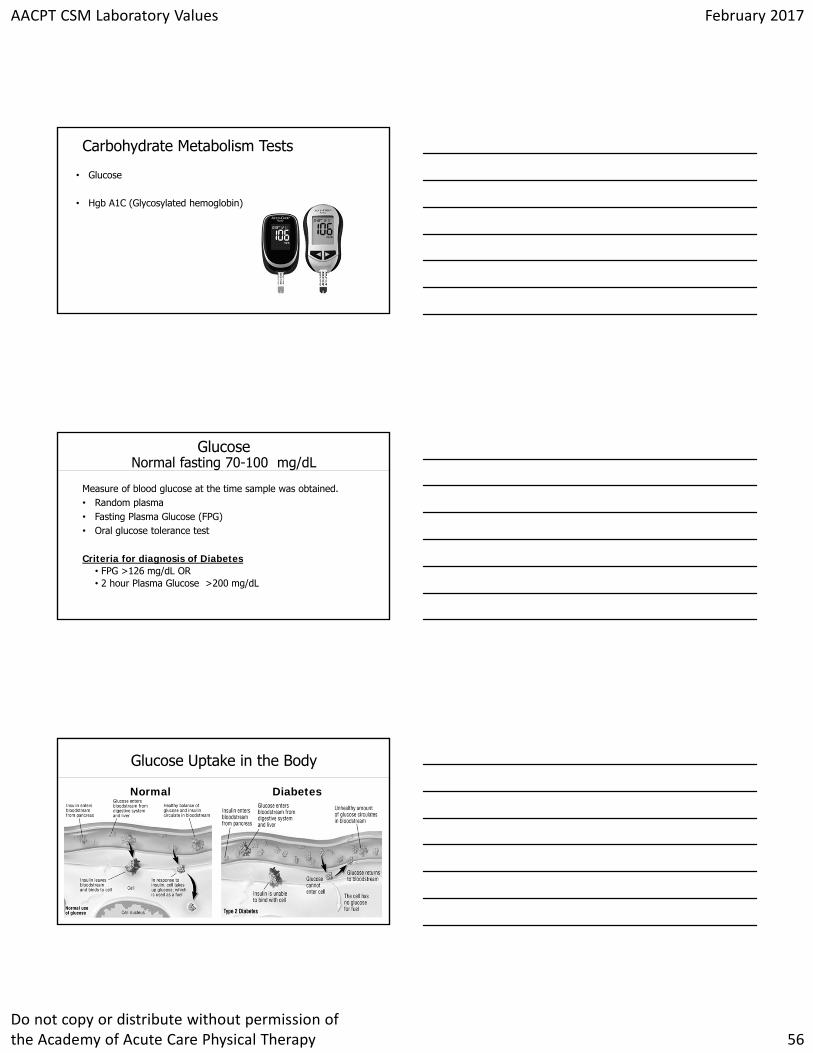
Click to edit Master title styleCarbohydrate Metabolism Tests
• Glucose
• Hgb A1C (Glycosylated hemoglobin)
Click to edit Master title styleGlucoseNormal fasting 70-100 mg/dL
Measure of blood glucose at the time sample was obtained.• Random plasma• Fasting Plasma Glucose (FPG)• Oral glucose tolerance test
Criteria for diagnosis of Diabetes• FPG >126 mg/dL OR • 2 hour Plasma Glucose >200 mg/dL
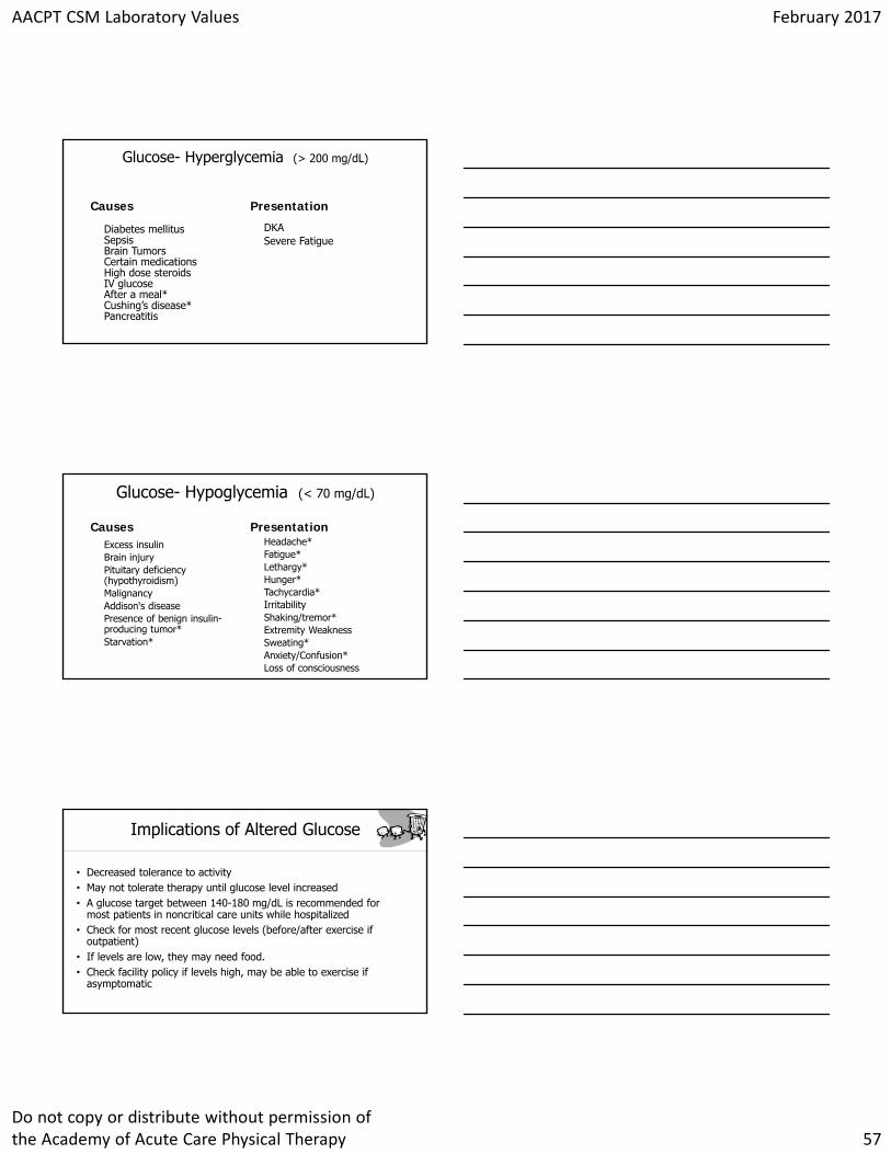
Click to edit Master title styleGlucose Uptake in the Body
Normal Diabetes
AACPT CSM Laboratory Values February 2017
Do not copy or distribute without permission of the Academy of Acute Care Physical Therapy 57
Click to edit Master title styleGlucose- Hyperglycemia (> 200 mg/dL)
Causes
Diabetes mellitusSepsisBrain TumorsCertain medicationsHigh dose steroidsIV glucoseAfter a meal*Cushing’s disease*Pancreatitis
Presentation
DKASevere Fatigue
Click to edit Master title styleGlucose- Hypoglycemia (< 70 mg/dL)
CausesExcess insulinBrain injuryPituitary deficiency (hypothyroidism)MalignancyAddison's diseasePresence of benign insulin-producing tumor*Starvation*
PresentationHeadache* Fatigue*Lethargy*Hunger*Tachycardia*IrritabilityShaking/tremor*Extremity WeaknessSweating*Anxiety/Confusion*Loss of consciousness
Click to edit Master title styleImplications of Altered Glucose
• Decreased tolerance to activity• May not tolerate therapy until glucose level increased• A glucose target between 140-180 mg/dL is recommended for
most patients in noncritical care units while hospitalized• Check for most recent glucose levels (before/after exercise if
outpatient)• If levels are low, they may need food.• Check facility policy if levels high, may be able to exercise if
asymptomatic
AACPT CSM Laboratory Values February 2017
Do not copy or distribute without permission of the Academy of Acute Care Physical Therapy 58
Click to edit Master title styleClinical Signs and Symptoms of Untreated or Uncontrolled Diabetes
•Polyuria•Polydipsia•Polyphagia
–Weight loss•Hyperglycemia (fasting > 126 mg/dL)•Glycosuria•Ketonuria•Fatigue and weakness•Blurred vision•Poor wound healing and recurrent infections
Click to edit Master title styleStrategies for Management of Blood Glucose During/After Exercise
Reduce pre-exercise bolus insulinReduce pre-exercise basal insulinTake extra carbohydrate with exercisePre-exercise or post exercise sprintInsulin pump therapyReduce basal insulin post exercise
Click to edit Master title styleHgb A1C (Glycosylated hemoglobin)
Hgb A1C Test used to look at long term blood glucose levels
• Glucose will stay attached to hemoglobin for 120 days so information is regarding blood glucose levels for past 2-3 months
• ↑ levels indicate poorly controlled DM
AACPT CSM Laboratory Values February 2017
Do not copy or distribute without permission of the Academy of Acute Care Physical Therapy 59
Click to edit Master title styleHgb A1C (Normal < 5.7% )
Normal: < 5.7%• Pre-diabetes mellitus: 5.7 - 6.4%• With diabetes mellitus: > 6.5% (poor glucose control)
Click to edit Master title styleHgb A1C
Causes
Diabetes mellitus
Presentation
Eye diseaseHeart diseaseKidney diseaseNerve damageStrokeGum diseaseNon-traumatic amputations
Click to edit Master title styleImplications of Altered Hgb A1C
• Monitor vitals as a standard of care • Educate importance of exercise for blood sugar control.• Consider wound-care management if levels altered
AACPT CSM Laboratory Values February 2017
Do not copy or distribute without permission of the Academy of Acute Care Physical Therapy 60
Click to edit Master title stylePatient Case
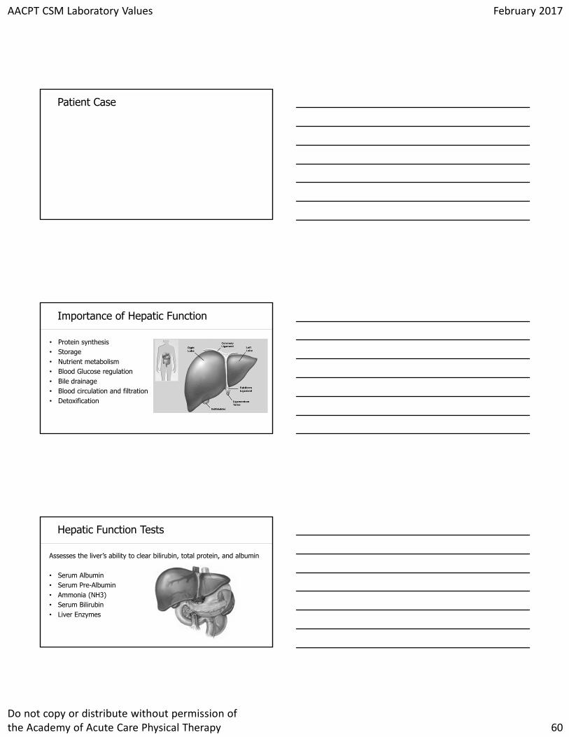
Click to edit Master title styleImportance of Hepatic Function
• Protein synthesis• Storage• Nutrient metabolism• Blood Glucose regulation• Bile drainage• Blood circulation and filtration• Detoxification
Click to edit Master title styleHepatic Function Tests
Assesses the liver’s ability to clear bilirubin, total protein, and albumin
• Serum Albumin• Serum Pre-Albumin• Ammonia (NH3)• Serum Bilirubin• Liver Enzymes
AACPT CSM Laboratory Values February 2017
Do not copy or distribute without permission of the Academy of Acute Care Physical Therapy 61
Click to edit Master title styleSerum Albumin and Serum Prealbumin
Serum Albumin: 3.5-5.2 g/dL• Half-life of 21 days• Required for proper distribution of body fluids between intravascular
compartments & body tissues. • Transports thyroid, other hormones and drugs & buffers pH
Serum Prealbumin: 19-39 mg/dL• Half-life of 2 days• Detects current nutritional status within a patient's body
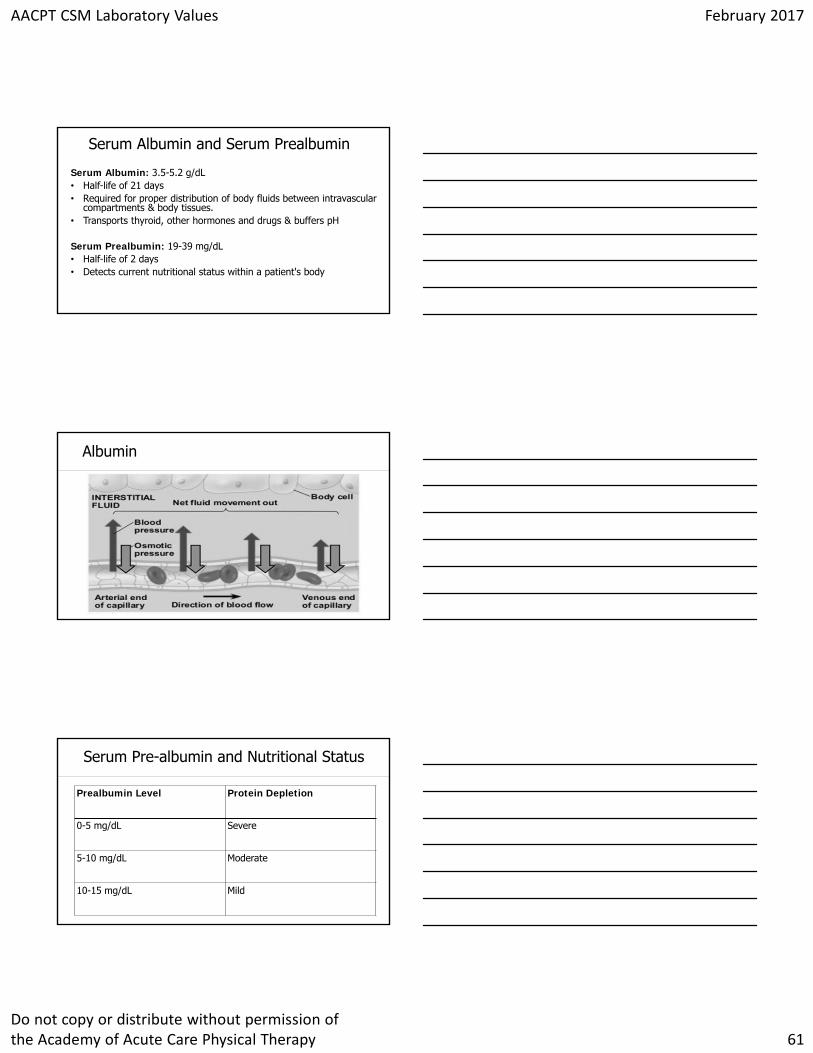
Click to edit Master title styleAlbumin
Click to edit Master title styleSerum Pre-albumin and Nutritional Status
Prealbumin Level Protein Depletion
0-5 mg/dL Severe
5-10 mg/dL Moderate
10-15 mg/dL Mild
AACPT CSM Laboratory Values February 2017
Do not copy or distribute without permission of the Academy of Acute Care Physical Therapy 62
Click to edit Master title styleSerum Albumin and Pre-albumin Trending Upward
Causes
Nutritional compromiseSevere infectionsCongenital disordersSevere dehydrationHepatitisChronic inflammationTuberculosisOverdose of cortisone medicationsCHFRenal DiseaseCancer
Presentation
Clinical features are dependent on the cause
• i.e. renal, cardiac, TB, etc.• Systemic peripheral edema• Delayed wound healing
Click to edit Master title styleSerum Albumin and Pre-albumin Trending Downward
CausesInfectionInflammationLiver diseaseKidney diseaseCrohn's diseaseBurnsMalnutrition/malabsorptionThyroid disease
PresentationPeripheral edemaNon-healing woundHypotension
Click to edit Master title styleHypoalbuminemia
Albumin levels• < 3.0 g/dL nutritionally compromised• < 2.8 g/dL peripheral edema, poor wound healing
Serum Prealbumin: 19-39 mg/dL• < 10 g/dL significant nutritional risk
Clinical Implications• Assess integumentary (incisions) daily• Collaborate with the interdisciplinary team regarding nutrition
AACPT CSM Laboratory Values February 2017
Do not copy or distribute without permission of the Academy of Acute Care Physical Therapy 63
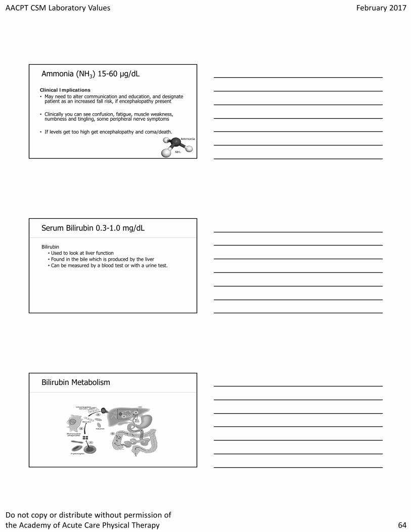
Click to edit Master title styleAmmonia (NH3) 15-60 µg/dL
Ammonia:
Used to evaluate liver function and metabolism. • Results from breakdown of protein in the body.
The liver converts ammonia from blood to urea. • If the liver is damaged, then increased ammonia levels
are noted.
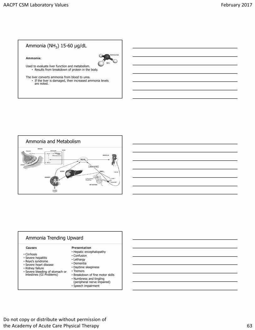
Click to edit Master title styleAmmonia and Metabolism
Click to edit Master title styleAmmonia Trending Upward
Causes
• Cirrhosis • Severe hepatitis• Reye’s syndrome• Severe heart disease• Kidney failure• Severe bleeding of stomach or
intestines (GI Problems)
Presentation• Hepatic encephalopathy• Confusion• Lethargy• Dementia• Daytime sleepiness• Tremors• Breakdown of fine motor skills• Numbness and tingling
(peripheral nerve impaired)• Speech impairment
AACPT CSM Laboratory Values February 2017
Do not copy or distribute without permission of the Academy of Acute Care Physical Therapy 64
Click to edit Master title styleAmmonia (NH3) 15-60 µg/dL
Clinical Implications• May need to alter communication and education, and designate
patient as an increased fall risk, if encephalopathy present
• Clinically you can see confusion, fatigue, muscle weakness, numbness and tingling, some peripheral nerve symptoms
• If levels get too high get encephalopathy and coma/death.
Click to edit Master title styleSerum Bilirubin 0.3-1.0 mg/dL
Bilirubin• Used to look at liver function• Found in the bile which is produced by the liver• Can be measured by a blood test or with a urine test.
Click to edit Master title styleBilirubin Metabolism
AACPT CSM Laboratory Values February 2017
Do not copy or distribute without permission of the Academy of Acute Care Physical Therapy 65
Click to edit Master title style
Causes• Cirrhosis• Hepatitis• Liver metastasis• Hemolytic anemia• Jaundice• Transfusion reaction• Bile duct occlusion• Gallstones• Chemotherapy
Presentation• Patients with severe disease
might have fatigue, anorexia, nausea, fever, and, occasionally, vomiting.
• Might have loose, fatty stools.
• Patients with high levels of bilirubin can lead to jaundice.
Serum Bilirubin Trending Upward
Click to edit Master title styleSerum Bilirubin
Clinical Implications:
• Adapt education if decreased cognition.
• Patients with advanced disease are at risk for osteoporosis and bleeding due to deficiencies of fat soluble vitamins.
• Symptoms-based approach when determining appropriateness for activity
Click to edit Master title styleModel for End-Stage Liver Disease (MELD) and MELD-Na
MELD Score• Serum bilirubin • Serum creatinine• INRMELD-Na• Serum bilirubin • Serum creatinine • INR • Sodium

AACPT CSM Laboratory Values February 2017
Do not copy or distribute without permission of the Academy of Acute Care Physical Therapy 66
Click to edit Master title styleModel for End-Stage Liver Disease (MELD) and MELD-Na
MELD Score and 3 Month Mortality• 40 or more — 71.3% mortality• 30–39 — 52.6% mortality• 20–29 — 19.6% mortality• 10–19 — 6.0% mortality• < 9 — 1.9% mortality
Click to edit Master title styleLiver Enzymes
Click to edit Master title styleLiver Enzymes
Alanine aminotransferase (ALT)-found in cells of liver and kidney• Released with liver damage• Useful in detecting damage related to hepatitis and/or drugsAspartate aminotransferase (AST)-found in liver/heart/muscle cells• Useful in detecting damage due to hepatitis, cirrhosis, drugs toxic to
liver (hepatoxic), alcoholismAlkaline phosphatase (ALP)- found in cells of bile ducts and bones• Useful in detecting blockage of bile ducts, hepatitis, liver cancer,
cirrohosis or hepatoxic drugs
AACPT CSM Laboratory Values February 2017
Do not copy or distribute without permission of the Academy of Acute Care Physical Therapy 67
Click to edit Master title styleClinical Bottom Line
Red Flags for liver dysfunction include:
• Altered cognition or mental status• Ascities• Peripheral edema• Musculoskeletal pain• Right Upper abdominal pain• Weakness• FatigueWe must alter our communication, document changes/inform medical team, be aware of safety risks, and involve caregivers.
Click to edit Master title stylePatient Case
Click to edit Master title styleFK Trough (6-15 ng/mL)
• Also known as the Tacrolimus/Prograf Test• Used to measure the amount of drug in the blood to determine
whether concentration has reached therapeutic levels or is below toxic amounts.
• Tacrolimus is a highly effective immunosuppressant for lowering the risk of organ transplantation.
• The drug is essentially fully metabolized in the liver and intestinal wall, with multiple factors affecting the pharmokinetic and metabolic profile (age, sex, other organ impairment, diet, and concomitant medications).
AACPT CSM Laboratory Values February 2017
Do not copy or distribute without permission of the Academy of Acute Care Physical Therapy 68
Click to edit Master title style
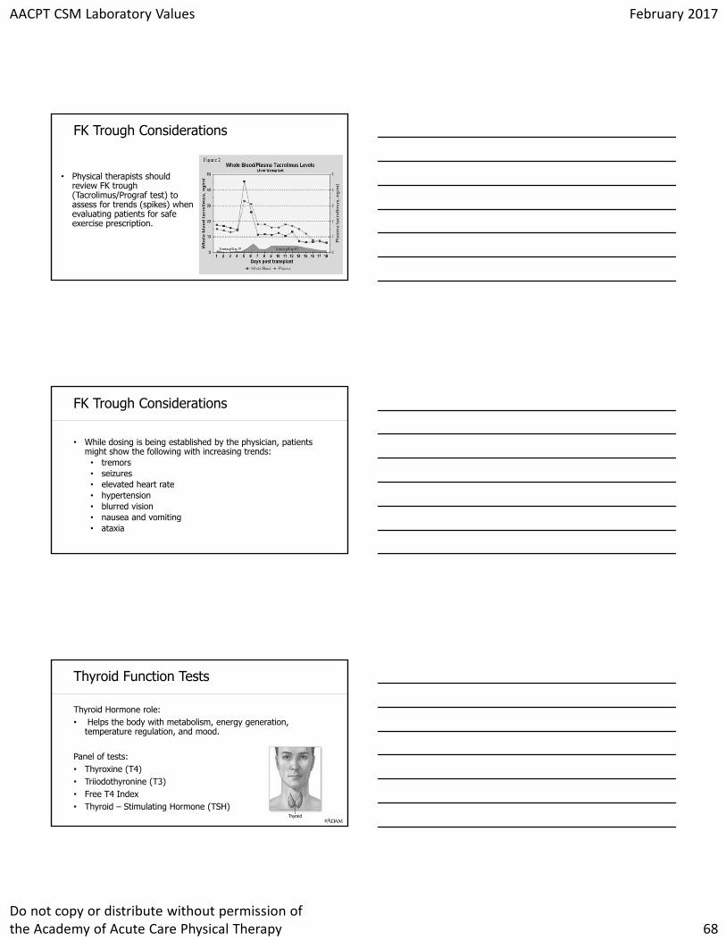
• Physical therapists should review FK trough (Tacrolimus/Prograf test) to assess for trends (spikes) when evaluating patients for safe exercise prescription.
FK Trough Considerations
Click to edit Master title styleFK Trough Considerations
• While dosing is being established by the physician, patients might show the following with increasing trends:
• tremors• seizures• elevated heart rate• hypertension • blurred vision• nausea and vomiting• ataxia
Click to edit Master title styleThyroid Function Tests
Thyroid Hormone role:• Helps the body with metabolism, energy generation,
temperature regulation, and mood.
Panel of tests:• Thyroxine (T4)• Triiodothyronine (T3)• Free T4 Index• Thyroid – Stimulating Hormone (TSH)
AACPT CSM Laboratory Values February 2017
Do not copy or distribute without permission of the Academy of Acute Care Physical Therapy 69
Click to edit Master title styleThyroid Function TestsReference Ranges
• Thyroxine (T4)• 4.5-11.5 µg/dL
• Triiodothyronine (T3)• 80-200 ng/dL
• Free T4 Index• 4.6-11.2 ng/dL
• Thyroid – Stimulating Hormone (TSH)• 0.3-3.0mlU/L
• Increased TSH and decreased T4 = thyroid disease• Decreased TSH= pituitary disease
Click to edit Master title styleThyroid Function Tests
Increased TSH and decreased T4 = thyroid diseaseDecreased TSH= pituitary disease
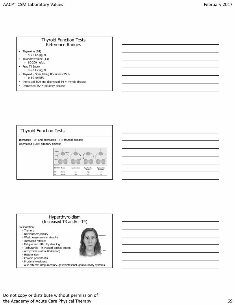
Click to edit Master title styleHyperthyroidism (Increased T3 and/or T4)
Presentation:• Tremors• Nervousness/lability• Weakness/muscular atrophy• Increased reflexes • Fatigue and difficulty sleeping• Tachycardia – increased cardiac output• Arrhythmias (atrial fibrillation)• Hypotension• Chronic periarthritis• Proximal weakness• Also affects: integumentary, gastrointestinal, genitourinary systems
AACPT CSM Laboratory Values February 2017
Do not copy or distribute without permission of the Academy of Acute Care Physical Therapy 70
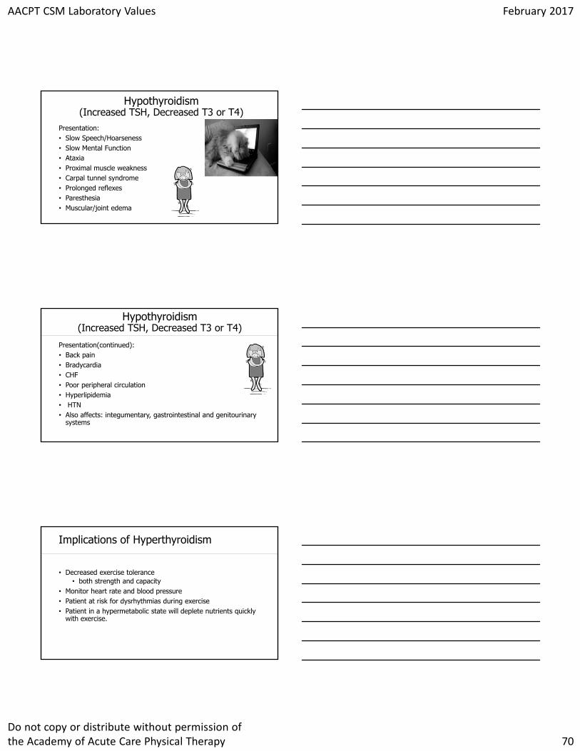
Click to edit Master title styleHypothyroidism (Increased TSH, Decreased T3 or T4)
Presentation:• Slow Speech/Hoarseness• Slow Mental Function• Ataxia• Proximal muscle weakness• Carpal tunnel syndrome• Prolonged reflexes• Paresthesia• Muscular/joint edema
Click to edit Master title styleHypothyroidism (Increased TSH, Decreased T3 or T4)
Presentation(continued):• Back pain• Bradycardia• CHF • Poor peripheral circulation• Hyperlipidemia• HTN • Also affects: integumentary, gastrointestinal and genitourinary
systems
Click to edit Master title styleImplications of Hyperthyroidism
• Decreased exercise tolerance• both strength and capacity
• Monitor heart rate and blood pressure• Patient at risk for dysrhythmias during exercise• Patient in a hypermetabolic state will deplete nutrients quickly
with exercise.
AACPT CSM Laboratory Values February 2017
Do not copy or distribute without permission of the Academy of Acute Care Physical Therapy 71
Click to edit Master title styleImplications of Hypothyroidism
• Hypothyroidism – frequently accompanied by myalgia and CK elevation
• More prone to skin tears• Activity intolerance
• should improve with treatment of hypothyroidism• Rhabdomyolysis, although rare, can appear in the presence of
heavy exercise, alcohol, or medications• Monitor heart rate
• bradycardia
Click to edit Master title styleFluid Analysis and Pathology
• Useful to determine cause of fluid buildup• Used to remove excess fluid• May monitor pressures of fluid in spaces• These results help determine the pathology leading to the
presence of this fluid. • Unlike other laboratory tests, patients may have precautions and
restrictions immediately after this test (before results are available) impacting delivery of therapy services.
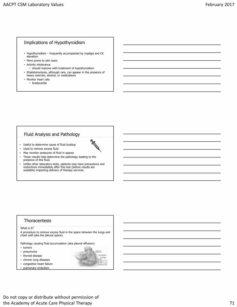
Click to edit Master title styleThoracentesisWhat is it?A procedure to remove excess fluid in the space between the lungs and chest wall (aka the pleural space).
Pathology causing fluid accumulation (aka pleural effusion):• tumors• pneumonia• thyroid disease• chronic lung diseases• congestive heart failure• pulmonary embolism
AACPT CSM Laboratory Values February 2017
Do not copy or distribute without permission of the Academy of Acute Care Physical Therapy 72
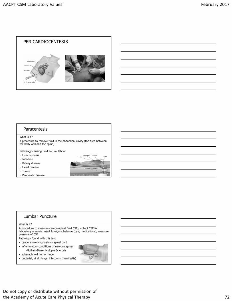
Click to edit Master title stylePERICARDIOCENTESIS
Click to edit Master title styleParacentesis
What is it?A procedure to remove fluid in the abdominal cavity (the area between the belly wall and the spine).
Pathology causing fluid accumulation:• Liver cirrhosis• Infection• Kidney disease• Heart disease• Tumor• Pancreatic disease
Click to edit Master title styleLumbar Puncture
What is it?A procedure to measure cerebrospinal fluid CSF); collect CSF for laboratory analysis, inject foreign substance (dye, medications), measure pressure of CSF Pathology found with this test:• cancers involving brain or spinal cord• inflammatory conditions of nervous system
-Guillain-Barre, Multiple Sclerosis• subarachnoid hemorrhage• bacterial, viral, fungal infections (meningitis)
AACPT CSM Laboratory Values February 2017
Do not copy or distribute without permission of the Academy of Acute Care Physical Therapy 73
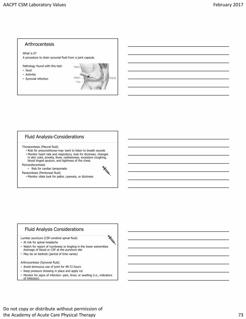
Click to edit Master title styleArthrocentesis
What is it?A procedure to drain synovial fluid from a joint capsule.
Pathology found with this test:• Gout• Arthritis• Synovial infection
Click to edit Master title styleFluid Analysis-Considerations
Thoracentesis (Pleural fluid)• Risk for pneumothorax-may want to listen to breath sounds• Monitor heart rate and respiratory, look for dizziness, changes
in skin color, anxiety, fever, restlessness, excessive coughing, blood tinged sputum, and tightness of the chest.
Pericardiocentesis• Risk for cardiac tamponade
Paracentesis (Peritoneal fluid)• Monitor vitals look for pallor, cyanosis, or dizziness
Click to edit Master title styleFluid Analysis Considerations
Lumbar puncture (CSF-cerebral spinal fluid)• At risk for spinal headache• Watch for report of numbness or tingling in the lower extremities
drainage of blood or CSF at the puncture site• May be on bedrest (period of time varies)
Arthrocentesis (Synovial fluid)• Avoid strenuous use of joint for 48-72 hours• Keep pressure dressing in place and apply ice• Monitor for signs of infection- pain, fever, or swelling (i.e., indicators
of infection)

AACPT CSM Laboratory Values February 2017
Do not copy or distribute without permission of the Academy of Acute Care Physical Therapy 74
Click to edit Master title styleToxicology
What is it?- Urine or blood sample test that determines type and amount
of legal/illegal drugs taken by a patient.
Pathology found/ruled out with this test:• Alcoholism and withdrawal• Fetal alcohol syndrome• Seizure• Delirium and dementia• Analgesic nephropathy• Sexual assault
Click to edit Master title style
Case Studies
Click to edit Master title styleReferences
1.American Physical Therapy Association (2015). Physical therapist practice and the human movement system. Alexandria, VA.
2.Costello, E., Elrod, C., & Tepper, S. (2011). Clinical Decision Making in the Acute Care Environment: A Survey of Practicing Clinicians. The Journal of Acute Care Physical Therapy, 2(2), 46-54.
3.Frownfelter, D., & Dean, E. (2012). Cardiovascular and Pulmonary Physical Therapy Evidence and Practice (5th ed.). St Louis: Elsevier-Mosby. \
4.Goodman, C. C., & Fuller, K. S. (2015). Pathology Implications for the physical therapist. St Louis: Elsevier Saunders.
5.Hall, J. B., Schmidt, G. A., & Kress, J. P. (2015). Principles of Critical Care. New York: Mcgraw-Hill Education.
6.Laposata, M. (2014). Laboratory Medicine: The Diagnosis of Disease in the Clinical Laboratory (2 ed.). New York: McGraw-Hill Education.
7.Lindner, G., & Funk, G. C. (2013). Hypernatremia in critically ill patients. Journal of critical care, 28(2), 216-e11.
8.Malone, D. J. (2006). Physical Therapy in Acute Care: A clinicians guide. Thorofare, NJ.: SLACK Corporation.
9.Pawlik, A. J., & Kress, J. P. (2013). Issues affecting the delivery of physical therapy services for individuals with critical illness. Physical therapy, 93(2), 256-265.
10.Paz, J. C., & West, M. P. (2002). Acute Care Handbook for Physical Therapists (2nd ed.). Boston: Butterworth-Heinemann.
11.Sahrmann, S. A. (2014). The Human Movement System: Our Professional Identity. Physical Therapy, 94(7), 1034-1042. Accessed August 25, 2016.
12.Verbalis, J. G., Goldsmith, S. R., Greenberg, A., Korzelius, C., Schrier, R. W., Sterns, R. H., & Thompson, C. J. (2013). Diagnosis, evaluation, and treatment of hyponatremia: expert panel recommendations. The American journal of medicine, 126(10), S1-S42.