Embed Size (px)
Citation preview

Lab #9 Biology 10 BCC Gallagher Page 1
Lab #9 Biology 10 BCC
Topic: Skeletal and Muscular Systems
Part 1: Directional terms, Planes, Body Cavities
Lab Materials:
male & female surface landmarks models, various models, textbook
Lab Activities: Use models and charts to learn directional terms, planes and body cavities.
Part I: Anatomical Directions:
1. Label diagram and define each of the following anatomical directional terms:
Superior/inferior
anterior/posterior
medial/lateral
dorsal/ventral
proximal/distal
superficial/deep
prone/supine
unilateral/bilateral

Lab #9 Biology 10 BCC Gallagher Page 2
2. Label the diagram and define each of the following planes:
Frontal Plane
Transverse Plane Midsagittal Plane
Part II: Body Cavities and Organs
1. Identify the major body cavities in the diagrams below

Lab #9 Biology 10 BCC Gallagher Page 3
2. Name the organs found in each body cavity
Posterior (dorsal) Cavity
1. Cranial Cavity
2. Spinal(vertebral) Cavity
Anterior (ventral) Cavity
1. Thoracic Cavity
a. Pericardial Cavity
b. Pleural Cavity
2. Abdominal Cavity
3. Pelvic Cavity

Lab #9 Biology 10 BCC Gallagher Page 4
Part III: Organ Systems Overview
Lab Materials:
Tables and Illustrations, Textbook, Internet,
Torso Models
Lab Activities:
Use models and charts to learn the major systems and some of the major organs of each
organ system listed below.
Terminology:
1. Identify the basic structure and function of each of the 11 body systems listed below.
2. List the cells, tissues, organs found in each system.
Integumentary System
Skeletal System
Muscular System
Nervous System
Endocrine System
Circulatory System
Lymphatic System
Immune System
Respiratory System

Lab #9 Biology 10 BCC Gallagher Page 5
Digestive System
Urinary System
Reproductive System
Part IV: The Skeletal System
Lab Materials:
Textbook, models and bones: articulated skeleton
Reminder: Do not use pencils and pens to point to bones and bone markings; use the
blunt or pointed probe in your dissection kit
Lab Activities:
1. Use models, bones, illustrations study the general terminology for types of bones and
be able to recognize examples of each.
2. Distinguish between bones of the axial and appendicular skeleton.
3. Identify the major bones of the axial skeleton
4. Identify the major bones of the appendicular
Types of bones:
1. Using general terminology for types of bones, identify some bones that represent each type
listed below:
Long
Short
Flat
Irregular

Lab #9 Biology 10 BCC Gallagher Page 6
2. Label the types of bones in the picture below.
I. Axial and Appendicular Skeleton: Major Bones: Directions
1. Distinguish between axial and appendicular skeleton
2. Identify all the major bones of the axial skeleton listed on skeletal models.
3. Identify all the major bones of the appendicular skeleton listed on skeletal models.
Axial Skeleton
A. Skull (cranium)
1. Maxilla Bone
2. Nasal Bone
3. Mandible
4. Hard Palate
a. Zygomatic bone
b. Hyoid bone
B. Vertebral Column
1. Cervical Vertebrae
a. Thoracic Vertebrae (facets on body and transverse processes)
2. Lumbar Vertebrae (largest)
3. Sacral Vertebrae (5 fused vertebrae)
4. Coccyx (3 to 5 vestigial vertebrae, body only)
5. Bony Thorax
a. Ribs (12 pairs) (cartilage, floating ribs)
b. Sternum
Appendicular Skeleton
1. Upper Limb
a. Pectoral Girdle
• Scapula
• Clavicle
2. Upper Arm
• Humerus
3. Forearm
• Radius
• Ulna
Lab #9 Biology 10 BCC Gallagher Page 7
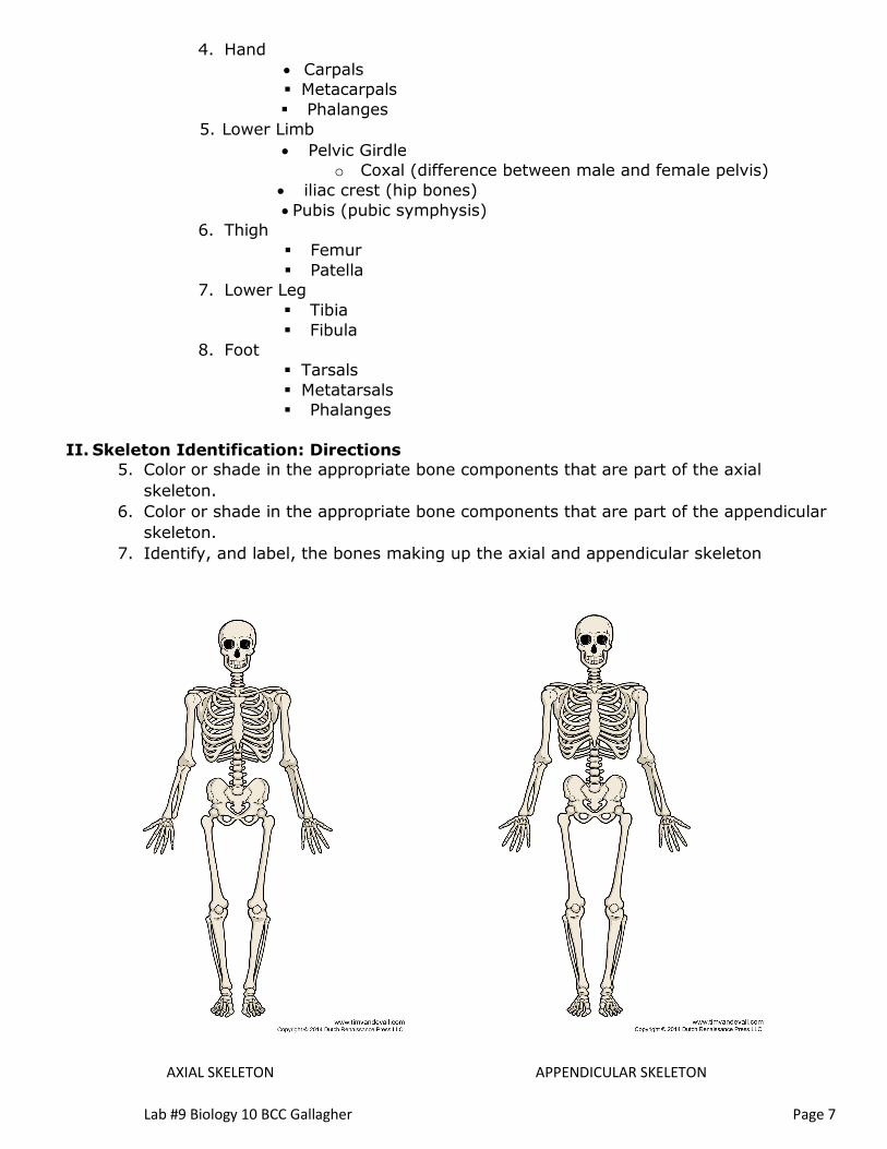
4. Hand
• Carpals
▪ Metacarpals
▪ Phalanges
5. Lower Limb
• Pelvic Girdle
o Coxal (difference between male and female pelvis)
• iliac crest (hip bones)
• Pubis (pubic symphysis)
6. Thigh
▪ Femur
▪ Patella
7. Lower Leg
▪ Tibia
▪ Fibula
8. Foot
▪ Tarsals
▪ Metatarsals
▪ Phalanges
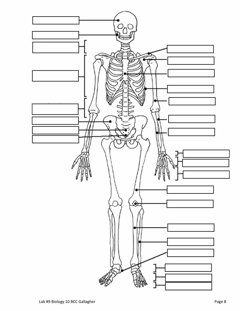
II. Skeleton Identification: Directions
5. Color or shade in the appropriate bone components that are part of the axial
skeleton.
6. Color or shade in the appropriate bone components that are part of the appendicular
skeleton.
7. Identify, and label, the bones making up the axial and appendicular skeleton
AXIAL SKELETON APPENDICULAR SKELETON
Lab #9 Biology 10 BCC Gallagher Page 8

Lab #9 Biology 10 BCC Gallagher Page 9
Part V: Bone Structure
1. Identify and describe the structures of function of the four layers of bone
2. Identify and label these parts of the bone below (not the rest).
Periosteum
Compact Bone
Spongy Bone
Bone Marrow

Lab #9 Biology 10 BCC Gallagher Page 10
Part VI: The Muscular System
Lab Materials:
Models: Muscle cell model
mini and half size human models sagittal heads
muscular arms & legs male and female pelvis
other models showing specific voluntary muscles
Lab Directions:
Using the models and illustrations, identify each of the major muscles listed.
1. Identify the major muscles listed below on the models and diagram below.
2. Know their location and function (see pg 13).
Human Muscular System Diagram
Deltoid
Trapezius Muscle
Latissimus Dorsi Muscle
Biceps
Triceps
Gluteus maximus
Hamstrings
Sartorius
Calf Muscles
• Gastrocnemius
• Soleus Muscle
Pectoralis major
Abdominal Muscles
• External Oblique
• Rectus abdominis
Quadriceps
Pectineus
Serratus Anterior
Fibularis Longus
Trapezius
Deltoid

Lab #9 Biology 10 BCC Gallagher Page 11
3. Draw a line and label the following on the diagram above:
a. Achilles Tendon, frontalis
4. What is the difference between flexors (abductors) and extensors (adductors)?
_________________________________________________________________

Lab #9 Biology 10 BCC Gallagher Page 12
5. Identify and label the following muscles of the head and neck on the model AND the
diagram below:
a. Head Region
• Frontalis
• Orbicularis oculi
• Orbicularis oris
• Masseter
• Frontalis
• Temporalis
• zygomaticus minor and major
• Depressor Anguli Oris
b. Neck Region
• Sternocleidomastoid
• Trapezius
• Ignore and leave blank # 3, 5, and 6.
c. Draw in a line and label the following on your diagram
• Buccinator

Lab #9 Biology 10 BCC Gallagher Page 13
Human Muscles and their Functions
**Identify the Functions of these required muscles.
a. Muscles on the Head and Neck frontalis raises eyebrows orbicularis oris closes mouth; pucker up orbicularis oculi closes eyes; squint extrinsic eye muscles all eye movements masseter closes jaw temporalis closes jaw sternocleidomastoid
flexes and/or rotates head
b. Muscles of the Abdominal Wall
**external oblique supports body wall internal oblique supports body wall transverse abdominis compresses abdomen rectus abdominis
c. Muscles that Move Pectoral
Girdle
flexes vertebral column “6-pack”
**trapezius d. Muscles that Move Upper Arm
elevation and depression of scapula
**pectoralis major flexes humerus main muscle of “pecs” **deltoid abducts upper arm **trapezius extends head; allows several movements of scapula **latissimus dorsi e. Muscles that Move Forearm
adducts & extends humerus “lats”
**biceps brachii flexes forearm “biceps” ** triceps brachii extends forearm “triceps” f. Muscles that Move Hand and Fingers flexors of hand flexes phalanges extensors of hand h. Muscles that Move Thigh
extends phalanges
**gluteus maximus extends thigh most of “glutes” ** sartorius flexes thigh
i. Muscles that Move Lower Leg
biceps femoris extends thigh; flexes lower leg semimembranosus extends thigh; flexes lower leg most of “hamstring”
semitendinosus extends thigh; flexes lower leg rectus femoris extends lower leg vastus lateralis extends lower leg most of “quads” **
vastus medialis extends lower leg j. Muscles that moves Foot gastrocnemius
** achilles tendon plantarflexion of foot ** soleus plantarflexion of foot ** tibialis anterior dorsiflexion of foot
Lab #9 Biology 10 BCC Gallagher Page 14
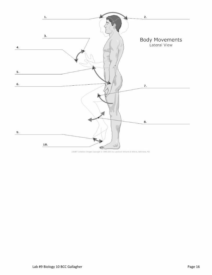
Part VII: Anatomical movements
1. Define and give examples of each Anatomical Movement listed below:
2. Label the diagrams below with anatomical body movements shown.
Flexion/extension
Adduction/abduction
Pronation/supination
Retraction/protraction
Elevation/depression
Rotation/circumduction
External rotation/internal rotation
Inversion/Eversion

Lab #9 Biology 10 BCC Gallagher Page 15
Lab #9 Biology 10 BCC Gallagher Page 16

Lab #9 Biology 10 BCC Gallagher Page 17

Lab #9 Biology 10 BCC Gallagher Page 18
VIII: Joints
1. Identify the main joints in the body and list where each of these joints are found in the
body.
2. Identify and label each of the joints in the pictures below.
Ball and Socket Joint
Hinge Joint
Pivot Joint
Fixed Immovable
Gliding Joint

Lab #9 Biology 10 BCC Gallagher Page 19

Lab #9 Biology 10 BCC Gallagher Page 20
Part VIII: Practice, Practice, Practice
Need study ideas???? ☺
1. Go to the following website and practice skeleton and muscles
www.anatomyarcade.com Whack-a-bone and Poke-a-muscle.
2. Take online Joint quiz and see how you are doing.
3. Watch podcasts on skeleton and muscular system, anatomical movements…
4. Review the powerpoint
5. Review the notes given to you