Embed Size (px)
Citation preview

8/8/2019 L01 Orientation Web (2)
http://slidepdf.com/reader/full/l01-orientation-web-2 1/76
1
Anatomy 403 (MEDADM 403)
The University of Michigan Medical School
Winter 2010
Dr. Ameed Raoof
Lecture One:
An Introduction to The Human Body/
Surface Anatomy
Introduction/Surface anatomy

8/8/2019 L01 Orientation Web (2)
http://slidepdf.com/reader/full/l01-orientation-web-2 2/76
Date Day Sessions( 10 :0 0 - 12 : Faculty Quizzes Location
6-Jan W Lecture(L1) RAOOF 1-23;413-433 NLH- MSII
8-Jan F L2 HILDEBRANDT 92-115 NLH- MSII
11-Jan M L3 STRIBLEY Quiz1 26-88 NLH- MSII
13-Jan W L4 STRIBLEY 116-134 NLH- MSII
15-Jan F L5 RAOOF Skeletal System: development & organization 140-158 NLH- MSII
18-Jan M MLK, JR. DAY NLH- MSII
20-Jan W L6 RAOOF Quiz2 162-210 NLH- MSII
22-Jan F L7 RAOOF Skeletal System - gross anatomy: Appendicular skele 213-246 NLH- MSII25-Jan M L8 RAOOF 250-283 NLH- MSII
27-Jan W LAB VISIT1 NLH- MSII
29-Jan F Practical Quiz NLH- MSII
1-Feb M L9 STRIBLEY Quiz3 287-305 NLH- MSII
3-Feb W L10 STRIBLEY Skeletal Muscles: regional gross anatomy-Axial 309-409 NLH- MSII
5-Feb F L11 STRIBLEY Skeletal Muscles: regional gross anatomy-Appendicular 309-409 NLH- MSII
8-Feb M L12 HILDEBRAND Quiz4 437-452 NLH- MSII
10-Feb W L13 HILDEBRANDT 453-480 NLH- MSII
12-Feb F L14 HILDEBRANDT 481-540 NLH- MSII
15-Feb M L15 HILDEBRAND Quiz5 Cardiovascular system: Lymphatics 541-566 NLH- MSII
17-Feb W L16 RAOOF Nervous System: general organization; development567-580 NLH- MSII
19-Feb F LAB VISIT2 NLH- MSII
22-Feb M Practical Quiz-2 Review Session with Dr. Fisher NLH- MSII
24-Feb W EXAM1 NLH- WLH-MSII
26-Feb F L17 RAOOF 584-608 NLH- MSII
1-Mar M vacation March 1-5 NLH- MSII
8-Mar M L18 RAOOF Quiz6 The Brain and cranial nerves (1) 612-654 NLH- MSII
10-Mar W L19 RAOOF The Brain and cranial nerves (2) 612-654 NLH- MSII
12-Mar F L20 RAOOF Autonomic Nervous System 659-672 NLH- MSII
15-Mar M L21 RAOOF Quiz7 Somatic senses & Motor Control 678-693 NLH- MSII
17-Mar W L22 RAOOF Special senses 697-726 NLH- MSII
19-Mar F L23 STRIBLEY Endocrine System and hormones-1 730-748 NLH- MSII
22-Mar M L24 STRIBLEY Quiz8 Endocrine System and ho rmones-2 730-748 NLH- MSII24-Mar W LAB VISIT3 NLH- MSII
26-Mar F Practical Quiz-3 NLH- MSII
29-Mar M L25 STRIBLEY Quiz9 Digestive System-1 781-814 NLH- MSII
31-Mar W L26 STRIBLEY Digestive System-2 781-814 NLH- MSII
2-Apr F L27 HILDEBRANDT Respiratory System 752-780 NLH- MSII
5-Apr M L28 HILDEBRAND Quiz10 Urinary System 821-845 NLH- MSII
7-Apr W L29 HILDEBRANDT Reproductive System-1-Male 846-859 NLH- MSII
9-Apr F L30 HILDEBRANDT Reproductive System-2-Female 860-892 NLH- MSII
12-Apr M LAB VISIT4 NLH- MSII
14-Apr W Practical Quiz NLH- MSII
16-Apr F Review Session with Dr. Fisher NLH- MSII
22-Apr Th. FINAL EXAM: 10:30 AM-12:30 PM Alternate f inal: 1:30-3:30 NLH- SLH-MSII
*NLH, WLH: North & west Lecture Halls-Medical Sciences II Building next to Taubman Medical Library
Principles: Cells, tissues and organs
The Integumen tary System
Skeletal System - gross anatomy: axial skeleton
Cardiovascular system: Blood vessels & regional circulation
Spinal cord and spinal nerves
Cardiovascular System: The Blood
Cardiovascular System: The Heart
MEDADM 403- Human Anatomy: Structure and Function. Winter 2010 Schedule
Skeletal System - Joints: Classification & structure
Skeletal Muscle: development, organization, & histology
Topic: Page #: Tortora PHA 11
Orientation; principles in biology and anatomy-Surface anato
Development

8/8/2019 L01 Orientation Web (2)
http://slidepdf.com/reader/full/l01-orientation-web-2 3/76

8/8/2019 L01 Orientation Web (2)
http://slidepdf.com/reader/full/l01-orientation-web-2 4/76
4
The course quizzes and exams will make up a total of 300
points (100%):
* Quizzes: 100 points
* Lab quizzes (4): 40
* Exam 1(midterm): 80
* Exam 2 (final): 80

8/8/2019 L01 Orientation Web (2)
http://slidepdf.com/reader/full/l01-orientation-web-2 5/76
5
An Introduction to the Human Body
Anatomy
hysiology
Subdi isions of A &

8/8/2019 L01 Orientation Web (2)
http://slidepdf.com/reader/full/l01-orientation-web-2 6/76
6
Anatomy
Student:
Medical;
Dental;
Under-
graduate

8/8/2019 L01 Orientation Web (2)
http://slidepdf.com/reader/full/l01-orientation-web-2 7/76
7
Introduction
You ar beginning a study of the human body
You will lear n how it is or gani ed
You will lear n how it functions You will lear n anatomical vocabular y so that
you can communicate in common ter ms with
other s who study anatomy

8/8/2019 L01 Orientation Web (2)
http://slidepdf.com/reader/full/l01-orientation-web-2 8/76
8
Definitions
Anatomy - the study of str uctur e and the
r elationshi among str uctur es of the body
Dissection - the car eful cutting apar t of body
str uctur es to examine them
Physiology - the study of the function of
body par ts

8/8/2019 L01 Orientation Web (2)
http://slidepdf.com/reader/full/l01-orientation-web-2 9/76
9
Clinical Connection - Noninvasive
Diagnostic Techniques Inspection - obser ving the body for any
obvious changes f r om nor mal
Palpation - touching the body sur faces with
the hands Auscultation - listening to body sounds,
often with the aid of a stethoscope
Percussion - tapping on the sur face of thebody with f inger tips and listening to ther esulting echo

8/8/2019 L01 Orientation Web (2)
http://slidepdf.com/reader/full/l01-orientation-web-2 10/76
10
Levels of Body Organization - 6 levels
Chemical level - includes atoms and
molecules (two or mor e atoms joined
together)
Cellular level - the basic str uctur al and
functional units of an or ganism
Tissue level - gr oups of cells and the
material surr ounding them that per for m aspecif ic function

8/8/2019 L01 Orientation Web (2)
http://slidepdf.com/reader/full/l01-orientation-web-2 11/76
11
Levels of Body Organization
Or gan level - str uctur es that ar e composed
of two or mor e differ ent types of tissues and
have specif ic functions
System level - consists of r elated or gans
with a common function
Or ganismal level - any living individual, all
the par ts of the body functioning together

8/8/2019 L01 Orientation Web (2)
http://slidepdf.com/reader/full/l01-orientation-web-2 12/76
12
1CHEMICAL LEVEL
Atoms (C, H, O, N, P)
Molecule (DNA)
1 1CHEMICAL LEVEL
Atoms (C, H, O, N, P)
2CELLULAR LEVEL
Molecule (DNA)
Smooth muscle cell
12
3
1CHEMICAL LEVEL
Atoms (C, H, O, N, P)
2CELLULAR LEVEL
Molecule (DNA)
Smooth muscle cell
Smooth muscle tissue
TISSUE LEVEL
12
3 3
4
1CHEMICAL LEVEL
Atoms (C, H, O, N, P)
2CELLULAR LEVEL
Molecule (DNA)
Smooth muscle cell
Smooth muscle tissue
Stomach
Epithelialtissue
ORGAN LEVEL
TISSUE LEVEL
Smooth muscle
tissue layers
12
3
4
Ser ous
membr ane
3
4
5
1CHEMICAL LEVEL
Atoms (C, H, O, N, P)
2CELLULAR LEVEL
Molecule (DNA)
Smooth muscle cell
Smooth muscle tissue
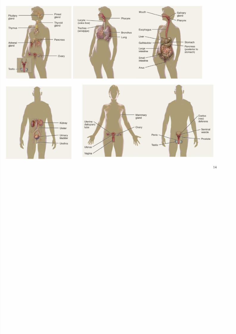
SYSTEM LEVEL
Esophagus
Liver Stomach
Small intestine
Lar ge intestine
Digestive system
Stomach
Epithelialtissue
ORGAN LEVEL
TISSUE LEVEL
Smooth muscle
tissue layers
12
3
4
5
Pancreas
Gallbladder
Ser ous
membr ane
3
4
5
1CHEMICAL LEVEL
Atoms (C, H, O, N, P)
2CELLULAR LEVEL
Molecule (DNA)
Smooth muscle cell
Smooth muscle tissue
ORGANISMAL LEVEL
SYSTEM LEVEL
Esophagus
Liver StomachPancreas
Gallbladder
Small intestine
Lar ge intestine
Digestive system
Stomach
Epithelialtissue
Ser ous
membr aneORGAN LEVEL
TISSUE LEVEL
Smooth muscle
tissue layers
12
3
4
5
6

8/8/2019 L01 Orientation Web (2)
http://slidepdf.com/reader/full/l01-orientation-web-2 13/76
13
8/8/2019 L01 Orientation Web (2)
http://slidepdf.com/reader/full/l01-orientation-web-2 14/76
14

8/8/2019 L01 Orientation Web (2)
http://slidepdf.com/reader/full/l01-orientation-web-2 15/76
15
Basic Anatomical Terminology
Anatomical position - the subject stands
upright, head level, ar ms at the sides with the
palms facing for war d
Pr one - lying face down
Supine - lying face up
Regional names - head, neck, tr unk, upper
limbs, and lower limbs (Fig. 1.2)

8/8/2019 L01 Orientation Web (2)
http://slidepdf.com/reader/full/l01-orientation-web-2 16/76
16
Terminology - Planes and Sections
Planes - imaginar y f lat sur faces that pass
thr ough the body
Sagittal plane - a ver tical plane that divides
the body or or gan into left and right par ts
Midsagittal (median) plane - divides the
body or or gan into equal left and right par ts
Fr ontal (cor onal) plane - divides the body or or gan into f r ont and back por tions

8/8/2019 L01 Orientation Web (2)
http://slidepdf.com/reader/full/l01-orientation-web-2 17/76
17
Planes and Sections
Tr ansverse plane - divides the body or
or gan into upper and lower por tions, also
called cr oss-sectional or hori ontal plane
Oblique plane - passes thr ough the body or
or gan at an angle
Section - one f lat sur face of a thr ee-
dimensional str uctur e or a cut along a plane

8/8/2019 L01 Orientation Web (2)
http://slidepdf.com/reader/full/l01-orientation-web-2 18/76
18
Planes and Sections

8/8/2019 L01 Orientation Web (2)
http://slidepdf.com/reader/full/l01-orientation-web-2 19/76
19
Directional Terms - examples
Superior - towar ds the head; e.g., the noseis superior to the mouth
Inf erior - away f r om the head; e.g., the
stomach is inferior to the hear t Medial - closer to the midline; e.g., the little
f inger is medial to the thumb
Later al - far ther f r om the midline; e.g., thethumb is later al to the index f inger
tc.

8/8/2019 L01 Orientation Web (2)
http://slidepdf.com/reader/full/l01-orientation-web-2 20/76
20
Major Directional Terms

8/8/2019 L01 Orientation Web (2)
http://slidepdf.com/reader/full/l01-orientation-web-2 21/76
21
Superior or Inferior
Dorsal or Ventral
Medial or LateralProximal or Distal
8/8/2019 L01 Orientation Web (2)
http://slidepdf.com/reader/full/l01-orientation-web-2 22/76
22
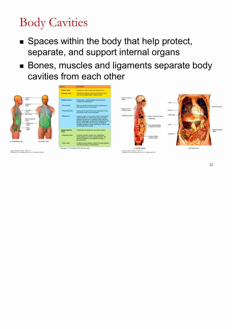
Body Cavities
Spaces within the body that help pr otect,separ ate, and suppor t inter nal or gans
Bones, muscles and ligaments separ ate body
cavities f r om each other

8/8/2019 L01 Orientation Web (2)
http://slidepdf.com/reader/full/l01-orientation-web-2 23/76
23
Body Cavities
Two major cavities, housing the br ain and spinal
cor d, ar e the cr anial cavity and the ver tebr al
(spinal) canal
The two major cavities in the tr unk ar e the thor acic cavity (superior) and the abdominopelvic cavity
(inferior), separ ated by the diaphr agm
Viscer a - the or gans located inside the thor acic and
abdominopelvic cavities The thor acic cavity has thr ee smaller cavities - the
pericar dial cavity and two pleur al cavities

8/8/2019 L01 Orientation Web (2)
http://slidepdf.com/reader/full/l01-orientation-web-2 24/76
24
Body Cavities
Mediastinum - centr al par t of the thor acic
cavity, located between the lungs
Abdominal cavity - contains the stomach,
spleen, liver , gallbladder , small intestine, and
most of the lar ge intestine
Pelvic cavity - contains the urinar y bladder ,
par ts of the lar ge intestine, and inter nal or gans of r epr oduction

8/8/2019 L01 Orientation Web (2)
http://slidepdf.com/reader/full/l01-orientation-web-2 25/76
25
Dorsal Body Cavity
2 subdivisions
cr anial cavity
ver tebr al or spinal canal
eninges

8/8/2019 L01 Orientation Web (2)
http://slidepdf.com/reader/full/l01-orientation-web-2 26/76
26
Ventral Body Cavity
2 subdivisions
thor acic cavity above
diaphr agm
abdominopelvic cavity
below diaphr agm
Diaphr agm
Or gans called viscer a
Or gans cover ed with
ser ous membr ane

8/8/2019 L01 Orientation Web (2)
http://slidepdf.com/reader/full/l01-orientation-web-2 27/76
27
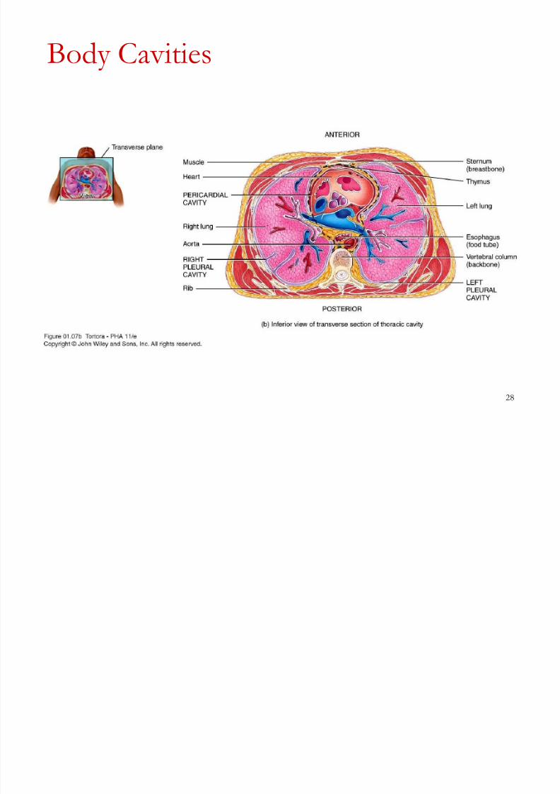
Body Cavities
8/8/2019 L01 Orientation Web (2)
http://slidepdf.com/reader/full/l01-orientation-web-2 28/76
28
Body Cavities

8/8/2019 L01 Orientation Web (2)
http://slidepdf.com/reader/full/l01-orientation-web-2 29/76
29
Body Cavities

8/8/2019 L01 Orientation Web (2)
http://slidepdf.com/reader/full/l01-orientation-web-2 30/76
30
Body Cavities

8/8/2019 L01 Orientation Web (2)
http://slidepdf.com/reader/full/l01-orientation-web-2 31/76
31
Body Cavities
8/8/2019 L01 Orientation Web (2)
http://slidepdf.com/reader/full/l01-orientation-web-2 32/76
32
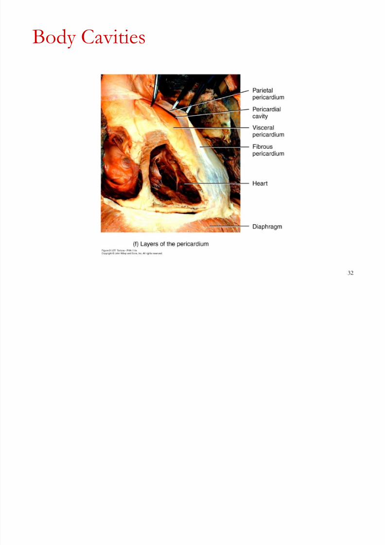
Body Cavities

8/8/2019 L01 Orientation Web (2)
http://slidepdf.com/reader/full/l01-orientation-web-2 33/76
33
Thoracic and Abdominal Cavity
Membranes Ser ous membr ane - a thin, slipper y
membr ane that cover s the viscer a and also
lines the walls of each cavity
P arietal layer - lines the walls of the cavities
V isceral layer - cover s and adher es to the
viscer a within the cavities

8/8/2019 L01 Orientation Web (2)
http://slidepdf.com/reader/full/l01-orientation-web-2 34/76
34
Cavity Membranes
Pleur a - the ser ous membr ane of the pleur al cavities,has a v isceral and a parietal pleura, between thelayer s is the pleur al cavity containing ser ous f luid
Pericar dium - the ser ous membr ane of thepericar dial cavity, between the v isceral pericardiumand parietal pericardium lies the pericar dial cavity
Peritoneum - the ser ous membr ane of theabdominal cavity, has a v isceral peritoneum and parietal peritoneum, with the peritoneal cavitybetween them
8/8/2019 L01 Orientation Web (2)
http://slidepdf.com/reader/full/l01-orientation-web-2 35/76
35
Abdominopel vic Regions and
Quadrants Ther e ar e 9 abdominopelvic r egions
Ther e ar e two hori ontal lines - the top one,
subcostal line, dr awn inferior to the rib cage
Bottom hori ontal line (transtubercular line) dr awn
just inferior to the tops of the hip bones
The two ver tical lines ar e called the left and right
midclav icular lines, dr awn thr ough the midpoints of the clavicles

8/8/2019 L01 Orientation Web (2)
http://slidepdf.com/reader/full/l01-orientation-web-2 36/76
36
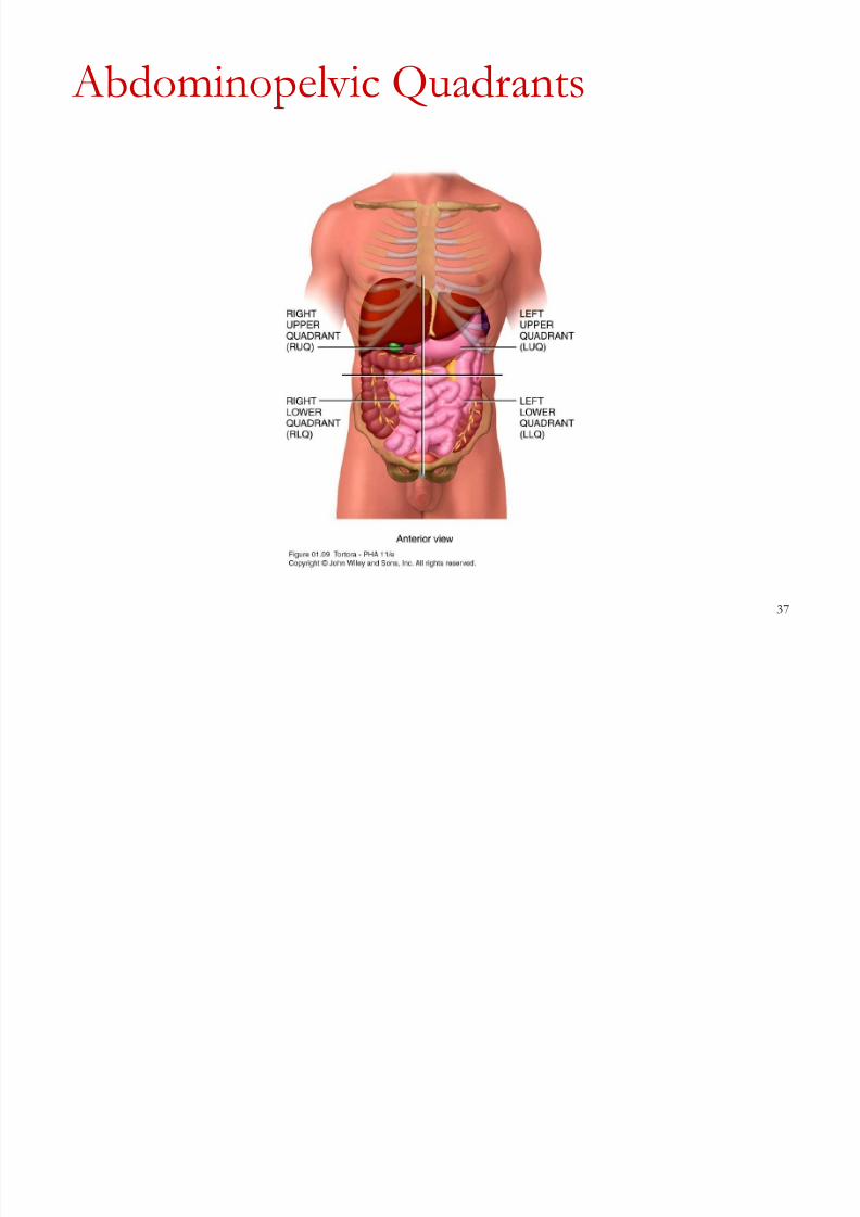
Abdominopel vic Quadrants
The four quadr ants ar e for med by dr awing
two lines, one ver tically and one hori ontally
thr ough the umbilicus
-Right upper quadr ant (RUQ)
-Left upper quadr ant (LUQ)
-Right lower quadr ant (RLQ)
-Left lower quadr ant (LLQ)
8/8/2019 L01 Orientation Web (2)
http://slidepdf.com/reader/full/l01-orientation-web-2 37/76
37
Abdominopel vic Quadrants

8/8/2019 L01 Orientation Web (2)
http://slidepdf.com/reader/full/l01-orientation-web-2 38/76
38
The Human Body and Disease
Disor der - any abnor mality of str uctur e
and/or function
Disease - a mor e specif ic ter m for an illness
wher e signs and symptoms ar e r ecogni ed
Symptoms - ar e subjective, what the patient
r epor ts
Signs - ar e objective, what an examiner obser ves and measur es

8/8/2019 L01 Orientation Web (2)
http://slidepdf.com/reader/full/l01-orientation-web-2 39/76
39
The Human Body and Disease
Epidemiology - the science that deals with
the investigation of diseases and how they
ar e tr ansmitted
Phar macology - the science that deals with
the use of dr ugs in the tr eatment of disease
Diagnosis - the science and skill of
distinguishing one disor der or disease f r omanother

8/8/2019 L01 Orientation Web (2)
http://slidepdf.com/reader/full/l01-orientation-web-2 40/76
40
Plastination

8/8/2019 L01 Orientation Web (2)
http://slidepdf.com/reader/full/l01-orientation-web-2 41/76
41
Which word describes the location of the lungsin relation to the liver?
A. Anterior
B. InferiorC. Superior
D. Lateral
E. Medial
8/8/2019 L01 Orientation Web (2)
http://slidepdf.com/reader/full/l01-orientation-web-2 42/76
42
Introduction- Surface Anatomy
Sur face anatomy is the study of the anatomical landmar ks on theexterior of the body.
Visualization involves looking in a ver y selective and pur poseful
manner at a specif ic par t of the body.
Palpation means using the sense of touch to deter mine the location
of an inter nal par t of the body thr ough the skin.
The var ying depth of differ ent str uctur es f r om the sur face of thebody and due to gender differ ences in the thickness of the der mis
and subcutaneous layer over differ ent par ts of the body, palpations
may r ange f r om light, to moder ate, to deep.
8/8/2019 L01 Orientation Web (2)
http://slidepdf.com/reader/full/l01-orientation-web-2 43/76
43
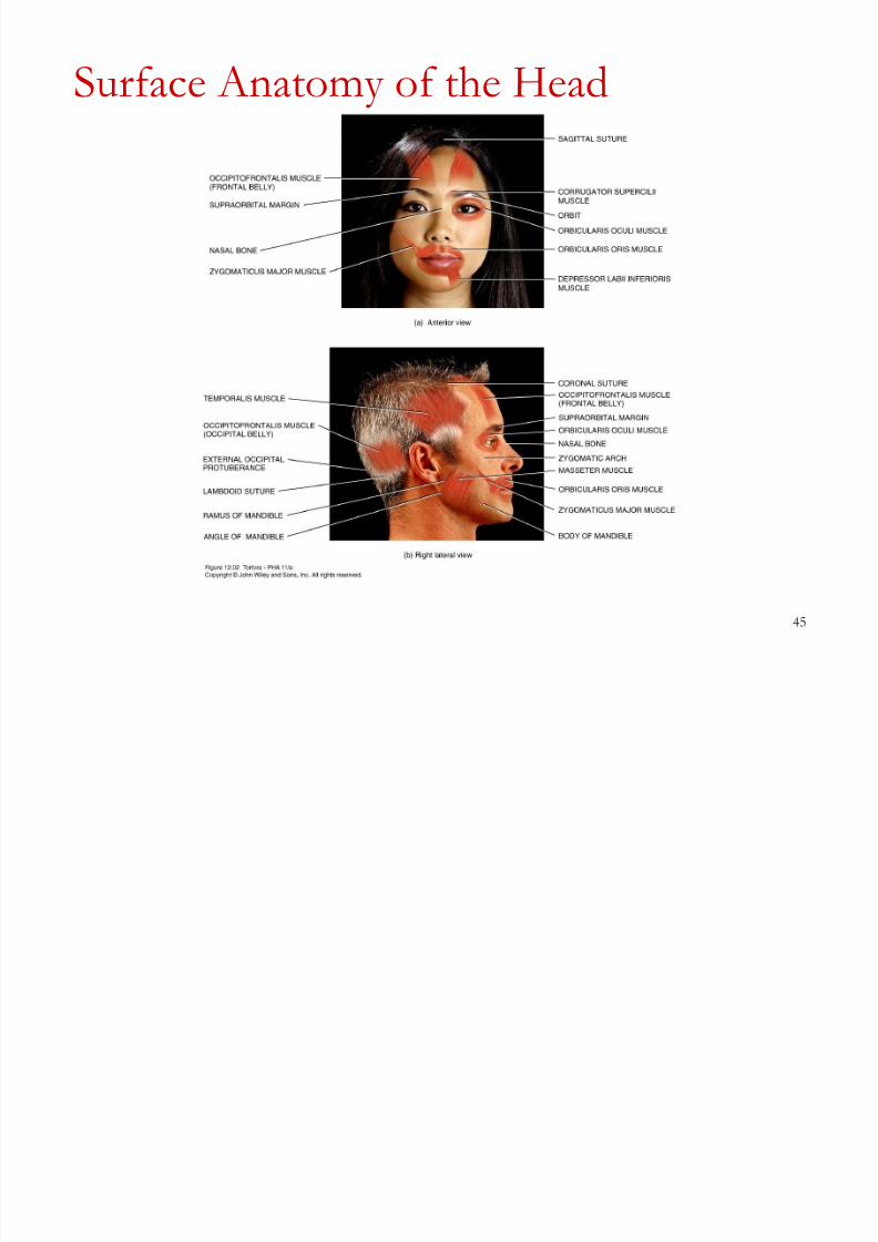
Bony Landmarks of the Head
Sagittal suture, Cor onal and lambdoid sutures. Exter nal occipital pr otuber ance ± pr ominent bony landmar k on
the occipital r egion of the skull
Or bit
The supr aor bital mar gin of the f r ontal bone.
Nasal bones Mandible. The r amus (ver tical por tion), body (hori ontal por tion),
and angle (ar ea wher e the r amus meets the body) of
Zygomatic arch and zygomatic bones.
Mastoid pr ocess - the pr ominent bony landmar k situated posterior
to the ear

8/8/2019 L01 Orientation Web (2)
http://slidepdf.com/reader/full/l01-orientation-web-2 44/76
44
Muscles of Facial Expression
8/8/2019 L01 Orientation Web (2)
http://slidepdf.com/reader/full/l01-orientation-web-2 45/76
45
Surface Anatomy of the Head
8/8/2019 L01 Orientation Web (2)
http://slidepdf.com/reader/full/l01-orientation-web-2 46/76
46
Surface Features of the Eyes
Iris - Cir cular pigmented muscular str uctur e behind the cor nea
Pupil - Opening in the center of the iris thr ough which light tr avels.
Scler a - Fibr ous tissue that cover s the entir e eyeball except the cor nea.
Conjunctiva - Membr ane that cover s the exposed sur face of the eyeball
and lines the eyelids.
Eyelids (palpebr ae) - Folds of skin and muscle.
Palpebr al f issure - Space between the eyelids.
Medial commissure - site of the union of the upper and lower eyelids.
Later al commissure -site of the union of the upper and lower eyelids.
Lacrimal car uncle - Fleshy, yellowish pr ojection of the medial commissur e
contains modif ied sudorifer ous and sebaceous glands.
Eyelashes - Hair s on the mar gin of the eyelids.
Eyebr ows - Sever al r ows of hair s superior to the upper eyelids.

8/8/2019 L01 Orientation Web (2)
http://slidepdf.com/reader/full/l01-orientation-web-2 47/76
47
Surface Anatomy of the Eye

8/8/2019 L01 Orientation Web (2)
http://slidepdf.com/reader/full/l01-orientation-web-2 48/76
48
Surface Features of the Ears
Auricle - The exter nal ear ; also called the pinna. It funnels sound wavesinto the exter nal auditor y canal
Tr agus - Car tilaginous pr ojection anterior to the exter nal auditor y canal.
Just anterior to the tr agus and posterior to the neck of
Antitr agus - Car tilaginous pr ojection opposite the tr agus.
Concha -H
ollow of the auricle. Helix - Superior and posterior f r ee mar gin of the auricle.
Antihelix - Semicir cular ridge superior and posterior to the tr agus.
Triangular fossa - Depr ession in the superior por tion of the antihelix.
Lobule - Inferior por tion of the auricle; it does not contain car tilage
Exter nal auditor y canal (meatus) - Canal about 3 cm (1 in.) long
extending f r om the exter nal ear to the ear dr um (tympanic membr ane). It
contains cer uminous glands that secr ete cer umen (ear wax).

8/8/2019 L01 Orientation Web (2)
http://slidepdf.com/reader/full/l01-orientation-web-2 49/76
49
Surface Features of the Ears

8/8/2019 L01 Orientation Web (2)
http://slidepdf.com/reader/full/l01-orientation-web-2 50/76
50
Surface Features of the Nose and Mouth Root - Superior attachment of the nose at
the for ehead, between the eyes. Apex - Tip of the nose.
Dorsum nasi- Rounded anterior bor der
connecting the r oot and apex
Exter nal naris - xter nal opening into the
nose.
Ala - Convex f lar ed por tion of the inferior
later al sur faces of the nose. Bridge - Superior por tion of the dor sum
nasi.
Philtr um - The ver tical gr oove on the upper
lip that extends along the midline to the
inferior por tion of the nose.
Lips (labia) - Superior and inferior f leshy
bor der s of the or al cavity.

8/8/2019 L01 Orientation Web (2)
http://slidepdf.com/reader/full/l01-orientation-web-2 51/76
51
Surface Anatomy of the Neck
Thyr oid car tilage - The lar gest of the car tilages that compose the
lar ynx (voice box).
Hyoid bone - Located just superior to the thyr oid car tilage.
Cricoid car tilage - A lar yngeal car tilage, located just inferior to thethyr oid car tilage
Thyr oid gland - Two-lobed gland just inferior to the lar ynx
Ster nocleidomastoid muscle- For ms later al aspect of neck.
Ar teries and Veins - Common car otid ar ter y, Inter nal jugular vein,
Subclavian ar ter y, Exter nal car otid ar ter y, Exter nal jugular vein.

8/8/2019 L01 Orientation Web (2)
http://slidepdf.com/reader/full/l01-orientation-web-2 52/76
52
Surface Anatomy of the Neck
Tr apezius muscle - Extends inferiorly and later ally f r om the base
of the skull and occupies a por tion of the later al cer vical r egion.
Ver tebr al spines -The spinous pr ocesses of the cer vical ver tebr ae
may be felt along the midline of the posterior r egion of the neck.
Ver tebr a pr ominens - Pr ominent at the base of the neck, the
spinous pr ocess of the seventh cer vical ver tebr a.

8/8/2019 L01 Orientation Web (2)
http://slidepdf.com/reader/full/l01-orientation-web-2 53/76
53
Surface Anatomy of the Neck

8/8/2019 L01 Orientation Web (2)
http://slidepdf.com/reader/full/l01-orientation-web-2 54/76
54
Surface Features of the Back
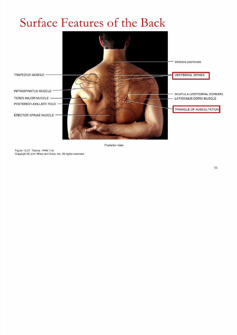
Ver tebr al spines - The spinous pr ocesses of ver tebr ae, ar e quitepr ominent when the ver tebr al column is f lexed.
Scapulae - These easily identif iable sur face landmar ks on the back
lie between ribs 2±7. Various par ts of the scapula, such as the
ver tebr al bor der , axillar y bor der , inferior angle, spine, and acr omion.
Latissimus dorsi muscle - A br oad, f lat, triangular muscle of thelumbar r egion that extends superiorly to the axilla and, when well
developed, gives a V-shape to the tr unk of the body.
Erector spinae (sacr ospinalis) muscle - Located on either side of
the ver tebr al column between the skull and iliac cr ests.
Inf r aspinatus muscle - Located inferior to the spine of the scapula. Rapezius muscle - Extends f r om the cer vical and thor acic
ver tebr ae to the spine and acr omion of the scapula and the later al
end of the clavicle.
8/8/2019 L01 Orientation Web (2)
http://slidepdf.com/reader/full/l01-orientation-web-2 55/76
55
Surface Features of the Back
Teres major muscle - Located inferior to the inf r aspinatus muscle;
together with the latissimus dor si muscle it for ms the inferior bor der
of the posterior axillar y fold.
Posterior axillar y fold - For med by the latissimus dor si and ter esmajor muscles, the posterior axillar y fold.
Triangle of auscultation - A triangular r egion of the back just
medial to the inferior par t of the scapula, wher e the rib cage is not
cover ed by super f icial muscles.It is bounded by the latissimus dor si and tr apezius muscles and ver tebr al bor der of the scapula.
8/8/2019 L01 Orientation Web (2)
http://slidepdf.com/reader/full/l01-orientation-web-2 56/76
56
Surface Features of the Back
8/8/2019 L01 Orientation Web (2)
http://slidepdf.com/reader/full/l01-orientation-web-2 57/76
57
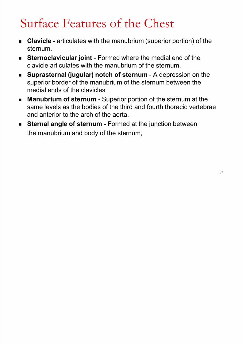
Surface Features of the Chest
Clavicle - ar ticulates with the manubrium (superior por tion) of the
ster num.
Ster noclavicular joint - For med wher e the medial end of the
clavicle ar ticulates with the manubrium of the ster num.
Supr aster nal ( jugular ) notch of ster num - A depr ession on the
superior bor der of the manubrium of the ster num between the
medial ends of the clavicles
Manubrium of ster num - Superior por tion of the ster num at the
same levels as the bodies of the thir d and four th thor acic ver tebr ae
and anterior to the ar ch of the aor ta.
Ster nal angle of ster num - For med at the junction between
the manubrium and body of the ster num,
8/8/2019 L01 Orientation Web (2)
http://slidepdf.com/reader/full/l01-orientation-web-2 58/76
58
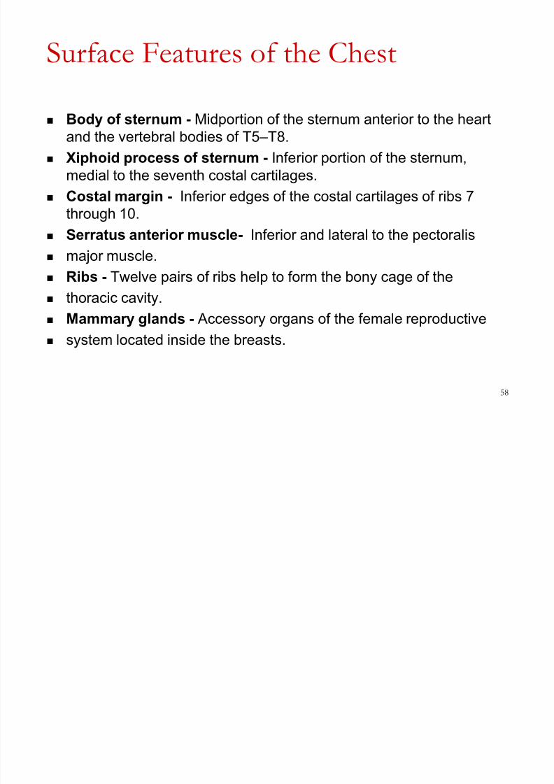
Surface Features of the Chest
Body of ster num - Midpor tion of the ster num anterior to the hear t
and the ver tebr al bodies of T5 ±T8.
Xiphoid pr ocess of ster num - Inferior por tion of the ster num,
medial to the seventh costal car tilages.
Costal mar gin - Inferior edges of the costal car tilages of ribs 7thr ough 10.
Serr atus anterior muscle- Inferior and later al to the pector alis
major muscle.
Ribs - Twelve pair s of ribs help to for m the bony cage of the
thor acic cavity. Mammar y glands - Accessor y or gans of the female r epr oductive
system located inside the br easts.

8/8/2019 L01 Orientation Web (2)
http://slidepdf.com/reader/full/l01-orientation-web-2 59/76
59
Surface Features of the Chest

8/8/2019 L01 Orientation Web (2)
http://slidepdf.com/reader/full/l01-orientation-web-2 60/76
60
Surface Features of the Shoulder
Acr omioclavicular joint
Acr omion
Humer us
Deltoid muscle

8/8/2019 L01 Orientation Web (2)
http://slidepdf.com/reader/full/l01-orientation-web-2 61/76
61
Surface Features of the Armpit
Apex -T
he apex of the axilla is surr ounded by the clavicle, scapula,and f ir st rib.
Base - The base of the axilla is for med by the concave skin and
fascia that extends f r om the ar m to the chest wall
Anterior wall -The anterior wall of the axilla is composed mainly of
the pector alis major muscle
Posterior wall - The posterior wall of the axilla is for med mainly by
the ter es major and latissimus dor si muscles
Medial wall - The medial wall of the axilla is for med by ribs 1±4 and
their corr esponding inter costal muscles, plus the overlying serr atus
anterior muscle.
Later al wall - Finally, the later al wall of the axilla is for med by the
cor acobr achialis and biceps br achii muscles and the superior
por tion of the shaft of the humer us.

8/8/2019 L01 Orientation Web (2)
http://slidepdf.com/reader/full/l01-orientation-web-2 62/76
62
Surface Features of the Arm and
Elbow
Humer us - This ar m bone may be palpated along its entir e length,
especially near the elbow.
Biceps br achii muscle - For ms the bulk of the anterior sur face of
the ar m. On the medial side of the muscle is a gr oove that containsthe br achial ar ter y.
Triceps br achii muscle - For ms the bulk of the posterior sur face of
the ar m.
Medial epicondyle - Medial pr ojection of the humer us near the
elbow. Later al epicondyle - Later al pr ojection of the humer us near the
elbow.

8/8/2019 L01 Orientation Web (2)
http://slidepdf.com/reader/full/l01-orientation-web-2 63/76
63
Surface Features of the Arm and Elbow
Olecr anon - Pr ojection of the pr oximal end of the ulna between andslightly superior to the epicondyles when the for ear m is extended; it
for ms the elbow.
Ulnar nerve - The ³funny bone´ is the r egion wher e the ulnar ner ve
r ests against the medial epicondyle.
Cubital fossa - Triangular space in the anterior r egion of the elbow
Median cubital vein - Cr osses the cubital fossa obliquely and
connects the later al cephalic with the medial basilic veins of the ar m.
Br achial ar ter y - Continuation of the axillar y ar ter y that passes
posterior to the cor acobr achialis muscle and then medial to the
biceps br achii muscle.
Bicipital aponeur osis - An aponeur osis that inser ts the biceps
br achii muscle into the deep fascia in the medial aspect of the
for ear m.

8/8/2019 L01 Orientation Web (2)
http://slidepdf.com/reader/full/l01-orientation-web-2 64/76
64
Surface Features of the Arm and
Elbow

8/8/2019 L01 Orientation Web (2)
http://slidepdf.com/reader/full/l01-orientation-web-2 65/76
65
Surface Features of Forearm and Wrist Ulna - The medial bone of the for ear m
Radius ± The later al bone of the for ear m Br achior adialis muscle - the superior and later al aspect of the
for ear m.
Flexor carpi r adialis muscle. - is on the later al side of the for ear m
Palmaris longus muscle - is medial to the f lexor car pi r adialis
tendon Flexor digitor um super f icialis muscle - is medial to the palmaris
longus tendon.
Flexor carpi ulnaris muscle - is on the medial aspect of the
for ear m.
Radial ar ter y- Located on the later al aspect of the wrist Pisifor m bone. Medial bone of the pr oximal r ow of car pals
Wrist creases - Thr ee mor e or less constant lines on the anterior
aspect of the wrist

8/8/2019 L01 Orientation Web (2)
http://slidepdf.com/reader/full/l01-orientation-web-2 66/76
66
Surface Features of the Forearm and
Wrist

8/8/2019 L01 Orientation Web (2)
http://slidepdf.com/reader/full/l01-orientation-web-2 67/76
67
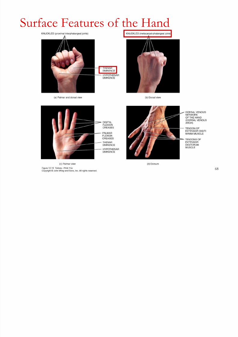
Surface Features of the Hand
Knuckles - Commonly r efer s to the dor sal aspect of the heads of
metacar pals II ±V, but also includes the dor sal aspects of the
metacar pophalangeal and inter phalangeal joints.
Dorsal venous network of the hand (dorsal venous arch).
Super f icial veins on the dorsum of the hand that dr ain blood into
the cephalic vein.
Tendon of extensor digiti minimi muscle - the dor sum of the hand
in line with the phalanx of the little f inger .
Tendons of extensor digitor um muscle - the dor sum of the hand
in line with the phalanges of the ring, middle, and index f inger s.
8/8/2019 L01 Orientation Web (2)
http://slidepdf.com/reader/full/l01-orientation-web-2 68/76
68
Surface Features of the Hand

8/8/2019 L01 Orientation Web (2)
http://slidepdf.com/reader/full/l01-orientation-web-2 69/76
69
Surface Features of the Abdomen and
Pel
vis The abdominal aor ta, which br anches into the right and left
common iliac ar teries anterior to the body of ver tebr a L4
The inf erior vena cava lies to the right of the abdominal aor ta and
is wider ; it arises anterior to the body of ver tebr a L5.
Exter nal oblique muscle - Located inferior to the serr atus anterior
muscle.
Rectus abdominis muscles - Located later al to the midline of the
abdomen.
Linea alba - Flat, tendinous r aphe (inter section of muscle tendons)
Linea semilunaris -The later al edge of the r ectus abdominisMuscle
Iliac crest - Superior mar gin of the ilium of the hip bone

8/8/2019 L01 Orientation Web (2)
http://slidepdf.com/reader/full/l01-orientation-web-2 70/76
70
Surface Features of the Buttock Gluteus maximus muscle - For ms the major por tion of the
pr ominence of the buttocks. Gluteus medius muscle - Superior and later al to the gluteus
maximus muscle.
Gluteal (natal) cleft - Depr ession along the midline that separ ates
the left and right buttocks.
Gluteal fold - Inferior limit of the buttock that r oughly corr esponds
to the inferior mar gin of the gluteus maximus muscle.
Ischial tuber osity - Just superior to the medial side of the gluteal
fold, the tuber osity bear s the weight of the body when seated.
Greater tr ochanter - A pr ojection of the pr oximal end of the femur on the later al side of the thigh.

8/8/2019 L01 Orientation Web (2)
http://slidepdf.com/reader/full/l01-orientation-web-2 71/76
71
Surface Anatomy of the Buttock

8/8/2019 L01 Orientation Web (2)
http://slidepdf.com/reader/full/l01-orientation-web-2 72/76
72
Surface Features of the Thigh and
Knee Sar torius muscle - Super f icial anterior muscle that can be tr aced
f r om the later al aspect of the thigh to the medial aspect of the knee.
Quadriceps f emoris muscle - Thr ee of the four components of the
muscle can be seen: r ectus femoris, vastus medialis and vastus
later alis and the vastus inter medius, is deep to the r ectus femoris Adductor longus muscle. Located at the superior aspect of the
medial thigh. It is the most anterior of the thr ee adductor muscles
(adductor magnus, adductor br evis, and adductor longus.
Femor al triangle - A space at the pr oximal end of the thigh
for med by the inguinal ligament superiorly, the sar torius muscle

8/8/2019 L01 Orientation Web (2)
http://slidepdf.com/reader/full/l01-orientation-web-2 73/76
73
Surface Features of the Thigh and Knee
Hamstring muscles. Super f icial, posterior thigh muscles the biceps
femoris, Tendinosus and semimembr anosus,
Patella - the kneecap, a sesamoid bone.
Patellar ligament - Continuation of the quadriceps femoris tendon
inferior to the patella. Inf r apatellar fat pads cushion it on both sides.
Medial condyle of f emur - Medial pr ojection on the distal end of the
femur .
Medial condyle of tibia - Medial pr ojection on the pr oximal end of
the tibia.
Later al condyle of f emur - Later al pr ojection on the distal end of
the femur .
Later al condyle of tibia - Later al pr ojection on the pr oximal end of
the tibia.
Popliteal fossa - A diamond-shaped ar ea on the posterior aspect
of the knee

8/8/2019 L01 Orientation Web (2)
http://slidepdf.com/reader/full/l01-orientation-web-2 74/76
74
Surface Features of Knee and Thigh

8/8/2019 L01 Orientation Web (2)
http://slidepdf.com/reader/full/l01-orientation-web-2 75/76
75
Surface Features of the Leg, Ank le and Foot
Tibial tuber osity - Bonypr ominence on the superior ,anterior sur face of the tibia intowhich the patellar ligament inser ts.
Tibialis anterior muscle - Liesagainst the later al sur face of thetibia.
Tibia - The medial sur face andanterior bor der (shin) of the tibia
Fibularis (per oneus) longus muscle - A super f icial later al muscle that overlies the f ibula.
Gastr ocnemius muscle - For ms
the bulk of the midpor tion andsuperior por tion of the posterior aspect of the leg.
Soleus muscle - Located deep tothe gastr ocnemius muscle.

8/8/2019 L01 Orientation Web (2)
http://slidepdf.com/reader/full/l01-orientation-web-2 76/76
Surface Features of the Leg, ank le and Foot
Calcaneal (Achilles) tendon - Pr ominent
tendon of the gastr ocnemius and soleusmuscles on the posterior aspect of the ankle
Later al malleolus of f ibula - Pr ojection of the distal end of the f ibula that for ms thelater al pr ominence of the ankle.
Medial malleolus of tibia - Pr ojection of thedistal end of the tibia that for ms the medial pr ominence of the ankle.
Dorsal venous arch - Super f icial veins onthe dor sum of the foot that unite to for m thesmall and gr eat saphenous veins.
Tendons of extensor digitor um longus muscle ± Visible in line with phalanges II ±V(2±5).
Tendon of extensor hallucis longus muscle - Visible in line with phalanx I (gr eattoe).