Embed Size (px)
Citation preview
- 2 -
Molecular characterization of the Dot/Icm-translocated AnkH and AnkJ
eukaryotic-like effectors of Legionella pneumophila
Fabien Habyarimana, Chris T Price, Marina Santic, Souhaila Al-Khodor and Yousef Abu
Kwaik
1Department of Microbiology and Immunology, Room MS-410, College of Medicine,
University of Louisville, KY 40292.
Running title: L. pneumophila AnkH and AnkJ effectors
Key Words: Dot/Icm, Legionnaires’, Anaplasma, Coxiella
* For correspondence: Tel (502) 852-4117, Fax (502) 852-7531
e-mail: [email protected]
Copyright © 2009, American Society for Microbiology and/or the Listed Authors/Institutions. All Rights Reserved.Infect. Immun. doi:10.1128/IAI.00913-09 IAI Accepts, published online ahead of print on 22 December 2009
on June 17, 2018 by guesthttp://iai.asm
.org/D
ownloaded from
- 3 -
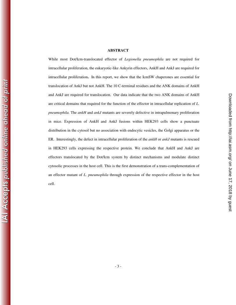
ABSTRACT
While most Dot/Icm-translocated effector of Legionella pneumophila are not required for
intracellular proliferation, the eukaryotic-like Ankyrin effectors, AnkH and AnkJ are required for
intracellular proliferation. In this report, we show that the IcmSW chaperones are essential for
translocation of AnkJ but not AnkH. The 10 C-terminal residues and the ANK domains of AnkH
and AnkJ are required for translocation. Our data indicate that the two ANK domains of AnkH
are critical domains that required for the function of the effector in intracellular replication of L.
pneumophila. The ankH and ankJ mutants are severely defective in intrapulmonary proliferation
in mice. Expression of AnkH and AnkJ fusions within HEK293 cells show a punctuate
distribution in the cytosol but no association with endocytic vesicles, the Golgi apparatus or the
ER. Interestingly, the defect in intracellular proliferation of the ankH or ankJ mutants is rescued
in HEK293 cells expressing the respective protein. We conclude that AnkH and AnkJ are
effectors translocated by the Dot/Icm system by distinct mechanisms and modulate distinct
cytosolic processes in the host cell. This is the first demonstration of a trans-complementation of
an effector mutant of L. pneumophila through expression of the respective effector in the host
cell.
on June 17, 2018 by guesthttp://iai.asm
.org/D
ownloaded from
- 4 -
Introduction
The gram-negative intracellular bacterial pathogen L. pneumophila is found ubiquitously in
aquatic environments where it replicates within a wide range of protozoan hosts (2, 18). Once inhaled by
humans in aerosolized contaminated water, L. pneumophila replicates in human alveolar macrophages
and causes Legionnaires’ disease or a less severe flu-like symptoms designated Pontiac fever (19, 29).
L. pneumophila is equipped with many sophisticated mechanisms that allow it to survive and
replicate within the host cell by creating a specialized ER-like compartment known as the Legionella-
containing vacuole (LCV) (16, 23, 26, 42, 43). Within the LCV, L. pneumophila utilizes its specialized
Dot/Icm type IVB secretion machinery to export a cohort of >200 effectors into the host cell cytosol
that are essential to modulate various cellular processes such as interception of ER-to-Golgi vesicle
traffic, evasion of endocytic traffic, and triggering pro- and anti-apoptotic processes (1, 13, 15, 16, 27,
28, 32, 40, 45). Likewise, L. pneumophila is able to translocate into the host cell cytosol specific
Dot/Icm substrates before its internalization (12, 35). The IcmS and IcmW chaperones facilitate
translocation of few Dot/Icm effectors (7, 10, 12, 36).
In silico analyses of 4 L. pneumophila genomes (Corby, Paris, Lens and Philadelphia-1) have
revealed the presence of many eukaryotic-like genes, which have been suggested to be acquired through
horizontal gene transfer (11, 14). Among the genes encoding eukaryotic-like proteins in L. pneumophila
is a family of at least 11 proteins containing ankyrin eukaryotic-like domains (Ank) (4, 11, 14, 22). The
ankyrin domain (ANK) is a 33-amino acid structural motif, and is the most common protein domain in
the eukaryotic kingdom, where it functions as a scaffold to mediate protein-protein interactions that play
essential roles in various eukaryotic cellular processes, ranging from regulation of transcription,
signaling, cytoskeleton, and cell cycle regulation (3, in press, 5, 8, 34). Therefore, it is predicted that the
on June 17, 2018 by guesthttp://iai.asm
.org/D
ownloaded from

- 5 -
L. pneumophila ankyrin proteins may mimic or interfere with various cellular processes to remodel the
host cell into a proliferative niche.
So far, most of Dot/Icm-exported substrates reported have little or no detectable role in
intracellular proliferation, suggesting a possible functional redundancy among them (16). Strikingly, of
the known Dot/Icm effectors, only loss of SdhA, SidJ or AnkB effectors results in a severe intracellular
growth defect. The sdhA mutant is defective only in macrophages (31) but the sidJ mutant (32) and ankB
mutant (4) are defective in human macrophages and protozoa. Furthermore, two L. pneumophila ankyrin
proteins (AnkH and AnkJ) play a significant role in intracellular replication of L. pneumophila in human
macrophages and in protozoa (22), indicating that they modulate cellular processes that are highly
conserved through evolution from protozoa to mammals.
The AnkH and AnkJ proteins that possess 2 and 3 ANK domains, respectively, have been
reported to be delivered into host cytosol (13, 37). However, their mechanism of translocation and more
importantly the role of the eukaryotic-like ANK domains of AnkH and AnkJ in the intracellular
proliferation of L. pneumophila and in translocation of AnkH and AnkJ proteins remains unknown.
In this study, we show that three of the L. pneumophila ankyrins are delivered into infected cells
by a IcmSW-dependent mechanism. The AnkH and AnkJ are essential in vivo for in intrapulmonary
proliferation in the mouse model. The ANK domains and the last 10 C-terminal residues of AnkH and
AnkJ are required for translocation and for intracellular replication. Importantly, expression of AnkH
and AnkJ in HEK293 cells rescues the intracellular growth defect of the respective effector mutant,
indicating modulation of distinct cytosolic processes by the two effectors to enable intracellular
proliferation.
on June 17, 2018 by guesthttp://iai.asm
.org/D
ownloaded from
- 6 -
Materials and Methods
Bacterial strains, plasmids, primers and media
The parental L. pneumophila serogroup 1 strain AA100/130b (ATCC BAA-74) and its isogenic
dotA, icmS, and icmW mutant strains have been described previously (46). Escherichia coli strain DH5α
was used as surrogate to clone the Cya fusion constructs. L. pneumophila were grown from frozen
stocks on buffered charcoal-yeast extract (BCYE) agar at 37°C or in buffered yeast extract (BYE) broth
at 37°C with shaking (17) for 3 days. The plates and broth used for the cultivation of the L. pneumophila
WT expressing Cya fusion proteins were supplemented with 5 µg /ml of chloramphenicol, whereas the
mutants expressing Cya fusion proteins were supplemented with 5 µg /ml of chloramphenicol and 50 µg
/ml of kanamycin. The plates used for the cultivation of E. coli strains were supplemented with 50 µg
/ml of chloramphenicol on Luria-Bertani (LB) agar plates or broth at 37°C with 5% of CO2 or in LB
broth at 37°C with shaking.
DNA manipulations and Cya reporter constructs
Transfections, restriction enzyme digestions, and DNA manipulation
were performed as
previously described (38). Restriction enzymes and T4 DNA ligase were purchased from Promega
(Madison, WI). L. pneumophila chromosomal DNA was prepared by using the Puregene
DNA isolation
kit (Gentra Systems, Minneapolis, MN ). Plasmid preparations were performed with the Bio-Rad
Quantum miniprep kit. Electroporations were performed with a Bio-Rad Gene Pulser, as recommended
by the manufacturer’s specifications. Purification of DNA fragments from agarose
gels for subcloning
was carried out with a QIAquick gel purification kit (Qiagen, Valencia, CA). Primers (Table 2) used to
amplify the coding sequences of L. pneumophila ank genes utilized to engineer Cya-Ank or GFP-Ank
on June 17, 2018 by guesthttp://iai.asm
.org/D
ownloaded from
- 7 -
constructs by PCR were from Integrated DNA Technologies, Inc. (Coralville, IA). The coding sequences
of L. pneumophila ank genes were fused to the C-terminal of adenylate cyclase (Cya) of B. pertussis
using plasmid pcya-ralF, which is a derivative of pMMB207M45NT (35). All of the L. pneumophila
ank gene PCR products were digested with BamHI and PstI. The pCya-RalF plasmid was digested with
BamHI and PstI to release the ralF gene. Digested products were ligated using T4 DNA ligase.
Resulting Cya-Ank reporter constructs are displayed in Table 2. For in-frame deletion of the ANK
domains and C-terminal of AnkH or AnkJ we used L. pneumophila ank genes cloned into the plasmid
vector pBC-SK+ as previously described (22). To generate domain mutant alleles of ankH and ankJ, an
inverse PCR strategy was employed using pBCSK+ harboring the ankH and ankJ genes as a template.
Briefly, phosphorylated primers (Table 1) were designed to hybridize adjacent to DNA encoding the
ANK domains or C-termini and then the entire plasmid lacking the domain of interest was PCR-
amplified using Phusion DNA polymerase (Finnzymes). The resulting PCR product was then treated
with DpnI restriction to remove residual template DNA from the reaction and then allowed to religate
using T4 DNA ligase. The ligation products were transformed into E. coli DH5α. Domain deletions of
ankH and ankJ were verified by DNA sequencing to ensure the integrity of the ankH and ankJ reading
frames. Recombinant plasmids were then electroporated into the L. pneumophila.
The various pBCSK+ vectors harboring the mutant ankH and ankJ alleles were used as templates
to generate Cya fusions. Primers listed in Table 2 were used in PCR to amplify the mutant ankH and
ankJ alleles and the resulting PCR products were cloned into pCR2.1 via topoisomerization as described
by the manufacturer (Invitrogen Corp, Carlsbad, CA). The mutant ankH and ankJ alleles were then
subcloned into the BamHI-PstI sites of pcya-ralF (Nagai et al., 2005), resulting in replacement of the
ralF gene with ankH and ankJ alleles in frame with cya. Recombinant plasmids were electroporated into
the L. pneumophila.
on June 17, 2018 by guesthttp://iai.asm
.org/D
ownloaded from
- 8 -
To generate the mammalian fusion constructs, ORF of L. pneumophila ankH and ankJ were
cloned into the mammalian expression vectors pAcGFP (Sigma) or p3XFlag-CMV (Sigma) using
primers displayed in table 3 to generate GFP or 3XFlag-AnkH or AnkJ fusion constructs.
Translocation assay
For adenylate cyclase (Cya) activity assays, differentiated U937 cells monolayers grown in 24-
well plates were infected with various strains of L. pneumophila at MOI of 50 for 1h at 37°C. For
cytochalasin D treatment assay, U937 cells were treated with 1µg/ml of cytochalasin D for 30 minutes.
Cells were then infected for 30 minutes, washed 3 times extensively with 1X PBS to remove
extracellular bacteria and subsequently lysed in 200µl of 0.25% dodecyltrimethylammonium bromide in
assay buffer. Cell lysates were processed for the detection of intracellular cAMP using Amersham
cAMP Enzyme Immunoassay kit (GE Healthcare, Piscataway, NJ), as recommended by the
manufacturer.
Macrophage culture and L. pneumophila intracellular replication analysis
Isolation and preparation of the human monocyte-derived macrophages (hMDMs) was carried
out as we previously described (41). The hMDMs and U937 cells were maintained in RPMI-1640 tissue
culture medium (Gibco BRL) supplemented with 10% heat-inactivated fetal bovine serum (FBS) (Gibco
BRL). The cells were cultured under a humidified atmosphere containing 5% CO2 and
95% air at 37°C.
For intracellular proliferation studies, infections of macrophages were performed as we described
previously (22). Briefly, cells were infected with various strains of at a multiplicity of infection (MOI)
of 10 for 1 h followed by treatment with 50µg/ml gentamicin for 1 h to kill extracellular bacteria, and
this was considered the zero time point. The intracellular proliferation was assessed by plating and
on June 17, 2018 by guesthttp://iai.asm
.org/D
ownloaded from
- 9 -
enumerating the colony-forming units (CFU) at 24h and 48h post-infection. Alternatively, the
intracellular replication was examined after 10h post-infection by single cell analysis using laser
scanning microscopy as previously described (22).
Infection of A/J mice with L. pneumophila.
Female pathogen-free, 6- to 8-week-old A/J mice were used for infection by intratracheal inoculation as
described previously (6, 20). The L. pneumophila strain AA100, ankH and ankJ mutants were grown on
BCYE agar plates for 72 h. Three mice per strain were used for intrapulmonary proliferation assay and
3 mice per dose were used for survival assay. The mice were inoculated intratracheally with 50 µl
containing the bacterial dose of 106 for intrapulmonary proliferation assay and 10
7, 10
8,8x10
8 or 10
9 for
survival assay as described previously (6, 20). At 2 h, 24 h, and 48 h, 72h and 7 days post-inoculation,
mice were humanely euthanized, the lungs were removed, and the bacteria were cultured on BCYE agar
for 72 h as described previously (6, 20). For the survival assay, the mice were observed
for 3 days post-
inoculation.
Expression of AnkH and AnkJ in mammalian cells
The human renal epithelial cell line HEK293 cells or HEK293T cells were grown on circular
glass cover slips (Fisher) pretreated with 0.1mg/ml of poly-D-lysine in 24-well culture plates at
concentration of 5x10⁴ cells/ml in DMEM containing 10% FBS overnight. The sub-confluent culture of
HEK293T and HEK293 cells were transiently transfected using calcium phosphate method with the
mammalian expression vectors pAcGFP (Sigma) or BAP3XFlag (Sigma) and constructs of AnkH or
AnkJ fusion GFP or 3XFlag for 18 h. In this study, HEK293T cells were transiently transfected with
GFP or 3XFlag constructs to examine their sub-cellular distribution. Since HEK293T are not suitable for
on June 17, 2018 by guesthttp://iai.asm
.org/D
ownloaded from
- 10 -
generating stable transfected cells, therefore we used HEK293 cells. Transiently transfected HEK293
cells were used to generate stably transfected HEK293 cells. Briefly, 1/10 diluted transiently transfected
HEK293 cells were grown in presence of 1.4mg/ml of geneticin (G418) (Sigma) for 15 days and every
other 3 days the media was replaced by new one containing 1.4mg/ml of geneticin. After 15 days, clones
were picked and screened by confocal laser scanning microscopy and western blot analysis for
expression of the AnkH or AnkJ fusion protein. To study colocalization of AnkH and AnkJ with late
endosomal, nuclear, lysosomal, cis and trans Golgi apparatus and ER markers, we labeled transfected
cells with the primary antibodies anti-Lamp2, anti-Cathepsin D, anti-GM130 (cis), anti-Golgi 58k
(trans) or anti-KDEL. The anti-LAMP-2 (H4B4) monoclonal antibody (developed by J. T.August and J.
E. K.Hildreth) was obtained from the Developmental Studies Hybridoma Bank (University of Iowa, IA).
To label the lysosomes, transfected cells were incubated with mouse monoclonal anti-cathepsin D
antibody (BD Transduction, Franklin Lakes, NJ). Mouse anti-KDEL monoclonal antibody was
purchased from StressGen Biotechnologies (Ann Arbor, Michigan). Rabbit anti-GM130 was obtained
from Santa Cruz Biotechnology, Inc. and monoclonal anti-Golgi 58k was purchased from Sigma. The
polyclonal anti- COX (mitochondria marker) was obtained fro Cell Signaling, monoclonal antibodies
anti-actin and anti-tubulin were obtained from Sigma. Primary antibodies were detected by Alexa Fluor
555-conjugated donkey anti-mouse or anti-rabbit IgG (Molecular Probes). The cells were examined
using an Olympus Fluoview1000 laser scanning confocal microscopy as described previously (41). On
average, 8–15 0.2 mm serial Z sections of each image were captured and stored for further analyses,
using Adobe Design Premium CS3.
Western blot assay
on June 17, 2018 by guesthttp://iai.asm
.org/D
ownloaded from

- 11 -
To detect the Cya-Ank hybrid proteins in L. pneumophila WT, dotA, icmS or icmW strains
harboring empty vector or Cya-Ank constructs, bacteria were grown for 3 days on BCYE in presence of
appropriate antibiotics prior to infection of U937 cells. Total of 5x10⁸
bacteria were isolated by
centrifugation and lysed with B-per bacterial protein extraction reagent (Thermo Scientific, Waltham,
MA) followed by boiling in SDS-PAGE sample buffer. Proteins were transferred onto nitrocellulose
membrane and the Cya-Ank hybrid proteins were detected by using monoclonal anti-M45 antibody at
1:50 dilution (4). After incubation with a secondary antibody conjugated to horseradish peroxidase,
signals were detected by the supersignal west femto maximum sensitivity substrate (Thermo Scientific,
Waltham, MA).
Statistical analysis.
All experiments were performed in triplicate at least three times, and the data shown are
representative of one experiment. To analyze for statistical significant differences between different sets
of data, a two-tailed Student t test was used, and the P value was obtained.
on June 17, 2018 by guesthttp://iai.asm
.org/D
ownloaded from
- 12 -
Results
Potential translocation of L. pneumophila ankyrins upon bacterial attachment to the macrophage
Potential role of the IcmSW chaperon in translocation of the Ank proteins was evaluated using
adenylate cyclase assays. These data showed that the L. pneumophila AnkD, AnkG and AnkJ are
translocated by the Dot/Icm machinery in IcmSW-dependent manner while the translocation of AnkH,
AnkI, AnkK and AnkN is independent of the IcmSW complex (data not shown). Western blots
confirmed equivalent expression of all the proteins in the icmS and IcmW mutants (data not shown).
Some Dot/Icm effectors, such as LepA and LepB are translocated into the host cell upon contact
of L. pneumophila to the host cell prior to bacterial internalization (12, 35). We have recently shown that
AnkB is exported into host cell by extracellular bacteria (39, in presse) Therefore, we examined whether
the other Ank proteins are translocated into macrophages prior to bacterial internalization. Prior to
infection, U937 cells were treated for 30 minutes with 1µg/ml of Cytochalasin D to prevent
phagocytosis. The cells were infected for 30 minutes with the WT strain or the dotA mutant harboring
Cya-Ank fusion constructs or the empty vector. In addition, we infected cells with the WT strain or the
dotA mutant expressing Cya-RalF or Cya-AnkB fusion protein as positive controls. As expected, the WT
strain translocated significantly the RalF into untreated cells compared to the empty vector (student t-
test, p <0.0001) (Fig. 1). The results showed that the AnkB controls was efficiently translocated by
attached extracellular bacteria as indicated by comparable level of cAMP in Cytochalasin D-treated and
untreated cell (student t-test, p >0.5) (Fig. 1). In contrast, the levels of cAMP were significantly different
between Cytochalasin D-treated and untreated cells infected with WT strain harboring any of the tested
Cya-Ank reporters (student t-test, p <0.0001) (Fig. 1).
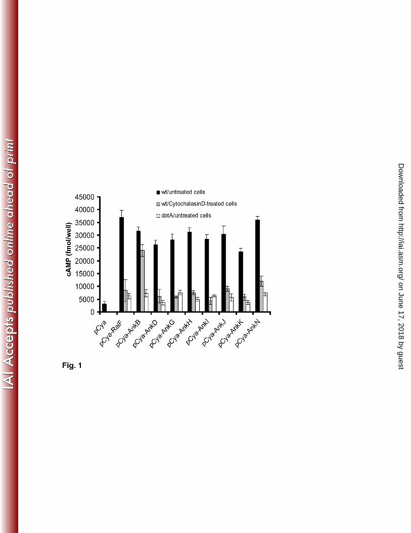
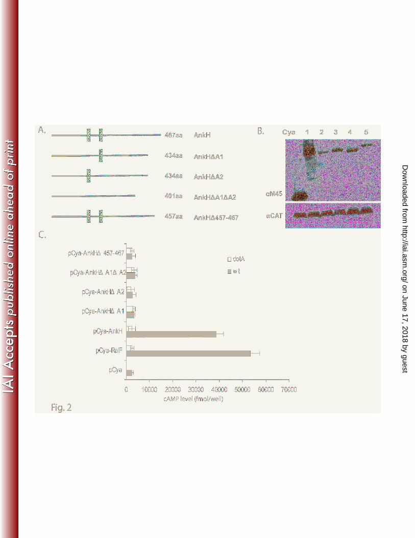
The C-terminus of L. pneumophila AnkH and AnkJ are essential for translocation
on June 17, 2018 by guesthttp://iai.asm
.org/D
ownloaded from
- 13 -
In addition to the existence of a C-terminus secretion motifs, Nagai et al. have shown that some
but not all Dot/Icm substrates harbor conserved hydrophobic residues in the C-terminus (30, 33, 35).
Since AnkH and AnkJ bear hydrophobic residues at the positions -4 or -5 in the C-terminus (Fig. S1),
we tested whether the C-terminal region of AnkH and AnkJ proteins were required for their
translocation. In-frame deletion of the last 10 residues of AnkH and AnkJ were constructed and fused to
Cya (Fig. 2A and Fig. 3A). U937 cells were infected with the WT strain or the dotA mutant expressing
the truncated AnkH and AnkJ reporter fusions. Our results showed a significant low levels of cAMP in
cells infected with the WT strain expressing C-terminal deletion of AnkH and AnkJ with > 10-fold less
compared to cells infected with the WT strain expressing full length AnkH and AnkJ (student t-test,
p<0.0001) (Fig. 2C and Fig. 3C). Thus, the C-terminus of AnkH and AnkJ is required for translocation
into the host cell.
The ANK domains of L. pneumophila are essential for translocation of AnkH and AnkJ
To test whether the ANK domains of AnkH and AnkJ proteins could be required for
translocation of the two proteins into macrophages, single, double or triple in-frame deletions of the
ANK domains of AnkH and AnkJ were fused to Cya (Fig. 2A and Fig. 3A). Immunoblot analysis
revealed equivalent expression of the truncated proteins (Fig. 2B and Fig. 3B). The U937 cells were
infected for 1h with the WT strain or the dotA isogenic mutant expressing each of the truncated AnkH or
AnkJ fusion proteins and intracellular cAMP level was determined in cell lysates. The level of cAMP in
cells infected with L. pneumophila harboring the ANK domains deletion was significantly lower with
>10-fold for truncated AnkH and >7-fold less for truncated AnkJ compared to the full length fusion or
the positive control (student t-test, p <0.0001) (Fig. 2C and 3C). Taken together, these data indicate that
the ANK domains of AnkH and AnkJ are essential for their translocation into the host cell. This is the
on June 17, 2018 by guesthttp://iai.asm
.org/D
ownloaded from
- 14 -
first example of the role of the eukaryotic-like domains in translocation of Dot/Icm effectors into the
host cell. It is also possible that the reduced translocation may be due to a mild reduction in the protein
level of the variant proteins.
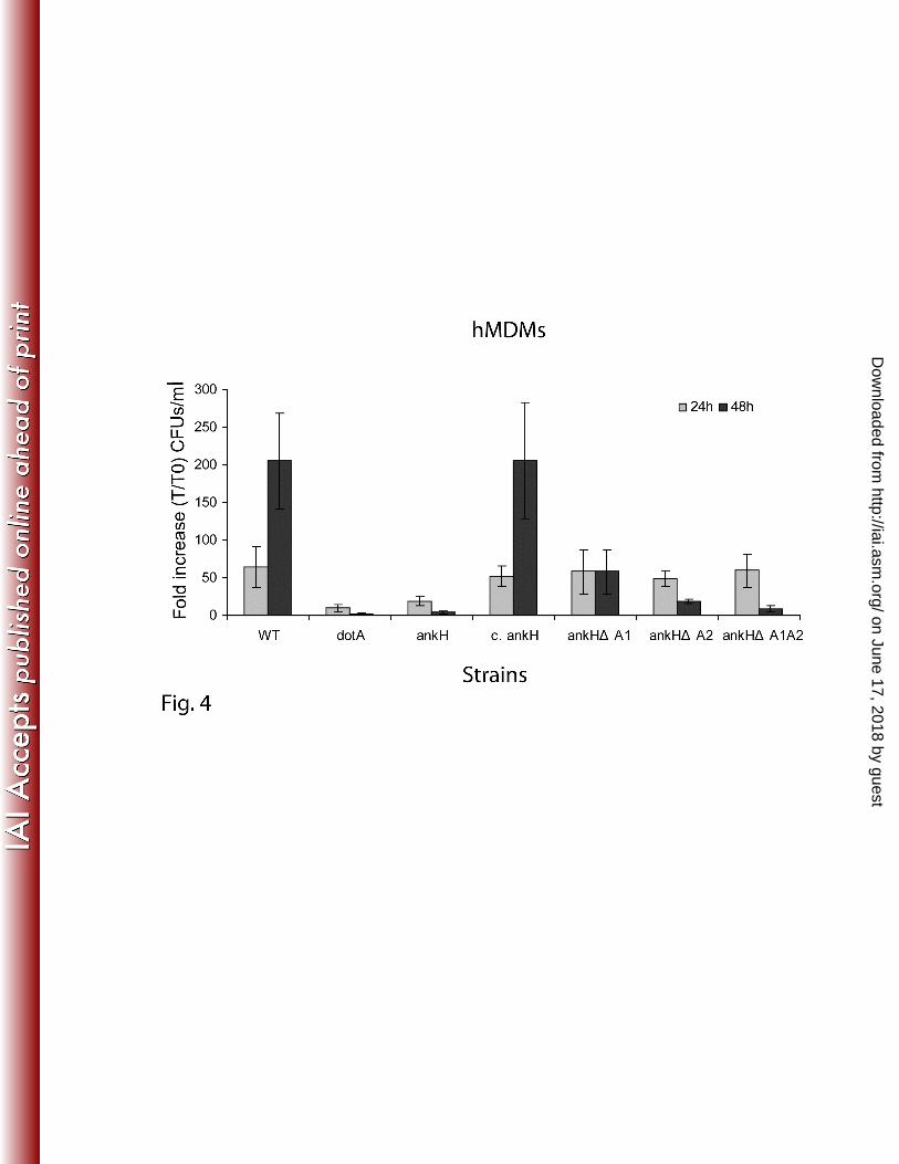
Role of the ANK domains of AnkH and AnkJ in intracellular growth of L. pneumophila
We have previously shown that AnkH and AnkJ are required for intracellular replication of L.
pneumophila (22). The aforementioned role of ANK domains of AnkH and AnkJ in translocation of
AnkH and AnkJ proteins into host cytosol prompted us to examine their role in intracellular replication.
Therefore, we performed single, double or triple in-frame deletion of the ANK domains of AnkH and
AnkJ. The ankH and ankJ mutants were trans-complemented with the full length genes or the
corresponding engineered ANK domain in-frame deletion constructs. The hMDMs were infected with L.
pneumophila WT strain, dotA, ankH or ankJ mutant and ankH and ankJ mutants trans-complemented
with the various constructs. At 24 h post-infection, there was an increase in the number of bacteria in all
strains ranging from 10-fold to 65-fold with an increase in number of bacteria for the WT strain and the
ankH mutant harboring all different constructs compared to dotA and the ankH mutant (student t-test, p
<0.001) (Fig. 4). However, at 48h post-infection there was a significant reduction in the cfus of the ankH
mutant trans-complemented with AnkH∆A1, AnkH∆A2 or AnkH∆A1∆A2 compared to mutant
complemented with the full length gene with > 100-fold increase (student t-test, p <0.0001). As
expected, the dotA and the ankH mutants did not grow in hMDMs (Fig. 4). Because of inconsistent
results in multiple experiments, data from the ankJ mutant trans-complemented with engineered ANK
deletion constructs are not shown. Our results demonstrate that the two ANK domains of AnkH play a
vital role in intracellular proliferation of L. pneumophila. It is also possible that the reduced intracellular
growth may be due to a reduced translocation of the truncated proteins.
on June 17, 2018 by guesthttp://iai.asm
.org/D
ownloaded from
- 15 -
L. pneumophila ankH and ankJ mutants are attenuated in intrapulmonary replication in mice
The AnkH and AnkJ effectors are essential for intracellular proliferation of L. pneumophila
within human monocyte-derived macrophages (hMDMs) and protozoa (22), but whether the two
effectors are required for the infection in vivo in animal models is not known. Moreover, the role of
Dot/Icm-translocated effectors in the pulmonary infection in animal models has never been examined
for any Dot/Icm effector. To determine whether the mutation in ankH or ankJ caused a decrease in
mortality in the A/J mouse model, we infected mice intratracheally(6, 9, 20) with doses of 107-10
9 cfus.
By the first day post-infection with a high dose of 109 cfus there was a mortality rate of 80% in mice
infected by the WT strain compared to the mortality rate of 20% in mice infected by ankB, ankH or ankJ
mutant with the similar dose (Fig. 5A) (student t-test, p <0.001). The AnkB attenuated mutant was used
as a negative control (39, in press). These data show that the two Ank effectors contribute the lethality of
Legionnaires’ disease in the mice model of the disease, consistent with their role in intracellular
proliferation within cultured macrophages.
To investigate whether AnkH and AnkJ are required for intrapulmonary proliferation of L.
pneumophila, we infected A/J mice with 106 of the L. pneumophila WT strain, the ankH, or the ankJ
mutant. Multiplication of L. pneumophila in lungs of infected mice was assessed by CFU enumeration
after 24h, 48h, 72h and 7 days post-infection. At 48h and 72h post-infection, the CFUs of L.
pneumophila recovered from mice infected with the ankH or the ankJ mutant were significantly lower
than the WT strain (student t-test, p <0.001) with at least a 1000-fold and 100-fold less bacteria,
respectively, were recovered from the lungs for both mutants (Fig. 5B). The ankB attenuated mutant (39,
in press), which was used as a control, was severely defective in intra-pulmonary replication. These data
on June 17, 2018 by guesthttp://iai.asm
.org/D
ownloaded from
- 16 -
show that the ankH and ankJ mutants are defective in intrapulmonary proliferation and mice mortality,
consistent with their in vitro intracellular growth defect in cultured macrophages (22). Taken together,
our data show that the two Ank effectors are the first effectors of L. pneumophila shown to be required
for intrapulmonary proliferation and lethality of L. pneumophila in the mice model of Legionnaires’
disease.
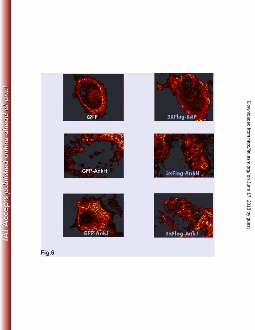
Expression and trafficking of AnkH and AnkJ in mammalian cells
Ectopic expression of bacterial proteins in eukaryotic cells has been an important strategy to
study localization of bacterial effector proteins translocated into host cell and may provide key insights
into the function of the effectors (13, 30, 37). Therefore, GFP-tagged or 3XFlag-tagged AnkH and AnkJ
were constructed and transiently or stably expressed in HEK293T or HEK293 cells to study their sub-
cellular distribution and trafficking in mammalian cells. Constructs of GFP or bacterial alkaline
phosphatase (BAP) fusion was used as a negative control. Transient transfection of HEK293T cells with
plasmids harboring GFP-tagged or 3xFlag-tagged ankH and ankJ using calcium phosphate yielded 70%-
75% transfection efficiency. Transient or stable expression of AnkH and AnkJ were not toxic to the
HEK293 cells (data not shown). Transient or stable expression of GFP-tagged or 3XFlag-tagged AnkH
and AnkJ were distributed in the cytoplasm with punctuate-like distribution but neither of the two
effectors were detected in the nucleus (Fig. 6), suggesting that AnkH and AnkJ were associated with
host cell vesicles. Nevertheless, these punctuate-like structures did not colocalize with L. pneumophila.
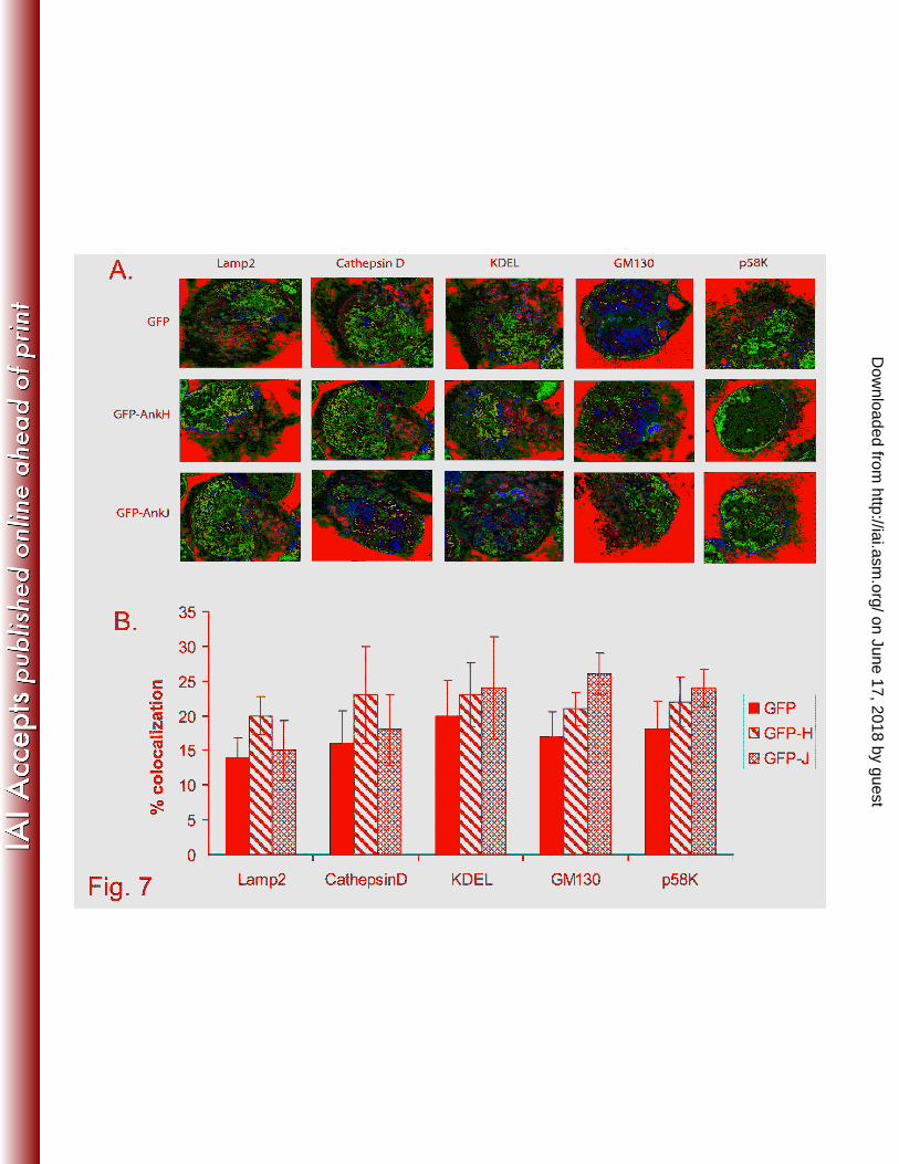
We utilized laser scanning confocal microscopy to determine trafficking and potential co-
localization of the two effectors with endosomal, lysosomal, Golgi, ER, microfilament, microtubules,
mitochondria, and nuclear compartments using specific markers Lamp2, cathepsin D, GM130 or P58-k,
KDEL, actin, tubulin, mitochondrial protein, and nuclear dye (DAPI), respectively. The data showed
on June 17, 2018 by guesthttp://iai.asm
.org/D
ownloaded from
- 17 -
that there were no significant differences in association of the above markers with GFP-AnkH or GFP-
AnkJ fusion proteins compared to GFP negative control (13-20%) (student t-test, p >0.5), indicating that
these proteins are not associated with endosomal, lysosomal, ER, or Golgi vesicles (Fig. 7A and B). We
conclude that despite the punctuate distribution of the two Ank effectors in mammalian cells, they do
not co-localize with any sub-cellular compartment.
L. pneumophila ankH and ankJ mutants are rescued in HEK 293 cells expressing AnkH and
AnkJ-GFP fusion proteins
Since L. pneumophila ankH and ankJ mutants exhibited intracellular growth defect and the
AnkH and AnkJ are translocated into host cells by the Dot/Icm system, we examined whether stable
HEK293 cells expressing L. pneumophila AnkH or AnkJ fusion proteins could rescue the respective
mutant for the defect in intracellular proliferation. Therefore, HEK293 cells with stable expression of
GFP, GFP-AnkH or GFP-AnkJ were infected with L. pneumophila WT, dotA, ankH or ankJ mutant.
After 10h post-infection, infected cells were labeled with anti-L. pneumophila antibody to evaluate the
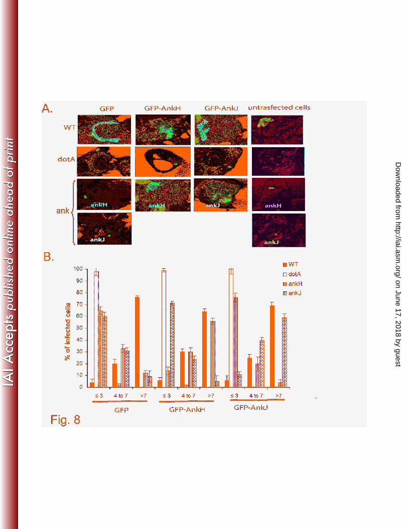
intracellular replication by single cell analysis using confocal microscopy. The data showed that by 10h
post-infection, ~70% of the WT strain-infected cells harbored more than 7 bacteria/cell in HEK293 cells
expressing GFP, GFP-AnkH or GFP-AnkJ. In contrast, the dotA mutant did not replicate with 1-3
bacteria/infected cell. Similar to the WT-infected cells expressing GFP-AnkH or GFP-AnkJ, ~60% of
the ankH mutant-infected cells and ankJ mutant-infected cells harbored more than 7 bacteria (Fig. 8A
and B). In contrast, the ankH and ankJ mutants did not replicate in HEK293 cells expressing GFP alone
with >60% of the ankH and ankJ mutant-infected cells harbored ≤3 bacteria/cell (Fig. 8A and B). The
ankH mutant was not rescued in cells expressing ankJ, or vice versa (data not shown). These data show
that the ankH and ankJ mutants are rescued in mammalian cells expressing L. pneumophila AnkH and
on June 17, 2018 by guesthttp://iai.asm
.org/D
ownloaded from

- 18 -
AnkJ, respectively. Collectively, our data indicate that AnkH and AnkJ modulate distinct processes in
the host cell cytosol, consistent with their distinct structure and mode of export. This is the first example
of a trans-complementation of a Dot/Icm effector mutant of L. pneumophila through expression of the
effector in the host cell.
on June 17, 2018 by guesthttp://iai.asm
.org/D
ownloaded from

- 19 -
Discussion
The ANK domains are the most abundant domain in the eukaryotic kingdom, where they
function as scaffold to mediate protein-protein interactions required for various eukaryotic cellular
processes, ranging from regulation of transcription, signaling, cytoskeleton, and cell cycle regulation (5,
8, 34). Recently, genomic analyses have shown that L. pneumophila genome encodes a large family of
eukaryotic-like ankyrin proteins (4, 11, 14, 22). It is thought that these proteins have been acquired by L.
pneumophila through horizontal gene transfer through co-evolution with its natural protozoan hosts to
perhaps mimic or interfere with host cell processes to establish a replicative niche within host cells. In
addition to AnkB, the AnkH and AnkJ proteins play a significant role in intracellular replication of L.
pneumophila in human macrophages and protozoa (22), indicating that these Dot/Icm-translocated
effectors modulate cellular processes that are highly conserved through evolution from protozoa to
mammals. Importantly, our data show that the AnkH and AnkJ Dot/Icm-translocated effectors are
essential for intrapulmonary proliferation in vivo in A/J mice. These data are consistent with the role of
the two effectors in intracellular growth of L. pneumophila in macrophages (22). To our knowledge, this
is the first demonstration for an essential role of Dot/Icm-translocated effectors in intrapulmonary
proliferation in animal models.
In several other pathogens, such as Agrobacterium, Bordetella, Helicobacter, Anaplasma,
Coxiella and Brucella, the TFSS is essential for delivery of host cell-modulating effectors. In agreement
with two different studies (13, 37), our data show that 7 L. pneumophila Ank proteins are delivered into
the host cytosol. In contrast to de Filipe et al. study (13), using a different strategy our data show that
AnkG/LegA7 is part of the cohort of Dot/Icm-translocated effectors. Consistent with the L. pneumophila
Ank proteins being translocated by the Dot/Icm secretion system, there is a recent report of 13 Dot/Icm-
translocated ankyrin proteins of Coxiella burnetii when expressed in L. pneumophila as a surrogate host
on June 17, 2018 by guesthttp://iai.asm
.org/D
ownloaded from

- 20 -
(37, 44). Moreover, Anaplasma phagocytophilum encodes an ankyrin protein (AnkA), which is
translocated by the TFSS into the host cell cytosol and nucleus (21, 24, 25). Given the rising number of
translocated effector proteins by L. pneumophila into the host cell and its large spectrum of
environmental hosts, it is possible that L. pneumophila selectively deploy a specific set of effectors that
best promote its survival and proliferation within a specific host cell in the environment or in humans
during infection.
The components of the Dot/Icm TFSS engage some of its effector proteins through a recognition
of a translocation signal predicted to be at the C-terminus of Dot/Icm substrates (10, 35). Our data show
that deletion of the last 10 residues of AnkH and AnkJ abrogates their translocation, indicating that their
translocation signal is located at C-terminus (4, 12, 35).
The deletion of the ANK domains in AnkH and AnkJ reduced substantially their translocation,
suggesting that the ANK domains actively participate in their translocation. Two explanations appear to
validate the involvement of the ANK domains in AnkH and AnkJ proteins delivery into host cytosol.
First, the ANK domains may participate in folding or unfolding of the AnkH or AnkJ proteins for
suitable presentation of their C-terminal translocation signal to the Dot/Icm components. Second, the
ANK domains may be involved in interaction between AnkH or AnkJ proteins and the Dot/Icm
components in order to be properly delivered into host cells, where they interfere with host cell
processes to facilitate bacterial proliferation. However, it is possible that the ANK deletion proteins are
translocated into host cell and the enzymatic activity of adenylate cyaclase (Cya) is inhibited due to
misfolding of the truncated protein, thereby Cya become inaccessible to the activating host calmodulin,.
In addition, possible misfolding of the in-frame deletion of the Ank protein may have rendered it
unrecognizable to be delivered by the Dot/Icm system.
on June 17, 2018 by guesthttp://iai.asm
.org/D
ownloaded from
- 21 -
Interestingly, expression of AnkH and AnkJ in mammalian cells show a punctate distribution
throughout the cytosol, but our data indicate no association of AnkH or AnkJ with endosomal,
lysosomal, ER, mitochondria and Golgi vesicles, or actin and tubulin. These data are consistent with our
previous findings that the phagosomes harboring the L. pneumophila ankH and ankJ mutants are
trafficked in similar manner as the ones harboring the WT strain (22), suggesting that these proteins are
not involved in trafficking of LCV or recruitment of the ER to LCV. It is unlikely that the punctuate
distribution of proteins in mammalian cells is due to protein aggregation, since the ectopically expressed
protein is functional in trans-rescue of the mutants for the defect in intracellular proliferation. Sub-
cellular localization as well as the host cell targets of the Ank effectors still to be identified.
The complementation of the ankH mutant by in frame-deletions of the ANK domains of AnkH
does not restore its intracellular growth defect (22). However, deletion of the ANK domains of AnkH
abrogates its translocation, indicating that the role of AnkH in intracellular replication requires its ANK
domains that are also indispensable for its translocation. Whether translocation of the in-frame deletion
of the Ank protein would render it functional in the host cell cytosol is not known.
Remarkably, our data indicate that ankH and ankJ mutants exhibit a severe intrapulmonary
replication defect resulting in less mortality compared to the WT strain. This is consistent with our
previous ex vivo results in human macrophages and alveolar epithelial cells (22) and corroborates with
high rate of survival of animal infected by the ankH or ankJ mutants. This is the first demonstration for
the role of Dot/Icm effectors in the development of Legionnaires’ disease in animal models.
Interestingly, when HEK293 cells expressing AnkH and AnkJ are infected with the ankH and
ankJ mutants, the intracellular growth defect of the respective mutant is rescued. These data completely
support the findings that the AnkH and AnkJ are translocated into the host cell cytosol to modulate
on June 17, 2018 by guesthttp://iai.asm
.org/D
ownloaded from

- 22 -
distinct cytosolic processes needed to sustain the intracellular proliferation of the ankH and ankJ
mutants. This is the first finding that ectopic expression of a Dot/Icm effector in mammalian cells can
rescue the growth of an effector mutant.
In summary, our data show that 3 L. pneumophila Ank proteins are delivered into the host cells
in an IcmSW complex-dependent manner, and none of the 7 translocated Ank proteins tested are
delivered into the host cell by attached extracellular bacteria. Furthermore, our data indicate that the
ANK domains and the C-terminus of the AnkH and AnkJ are indispensable for translocation into the
host cell, which is essential for intracellular proliferation of L. pneumophila. Ectopic expression in
mammalian cells and co-localization studies show that AnkH and AnkJ proteins are distributed in
punctuate-like structures throughout the cytosol and are not associated with nuclear, endosomal,
lysosomal, Golgi or ER compartments. Our data show that the L. pneumophila ankH and ankJ mutants
are rescued for their intracellular growth defect in HEK293 cells expressing the respective effector.
on June 17, 2018 by guesthttp://iai.asm
.org/D
ownloaded from
- 23 -
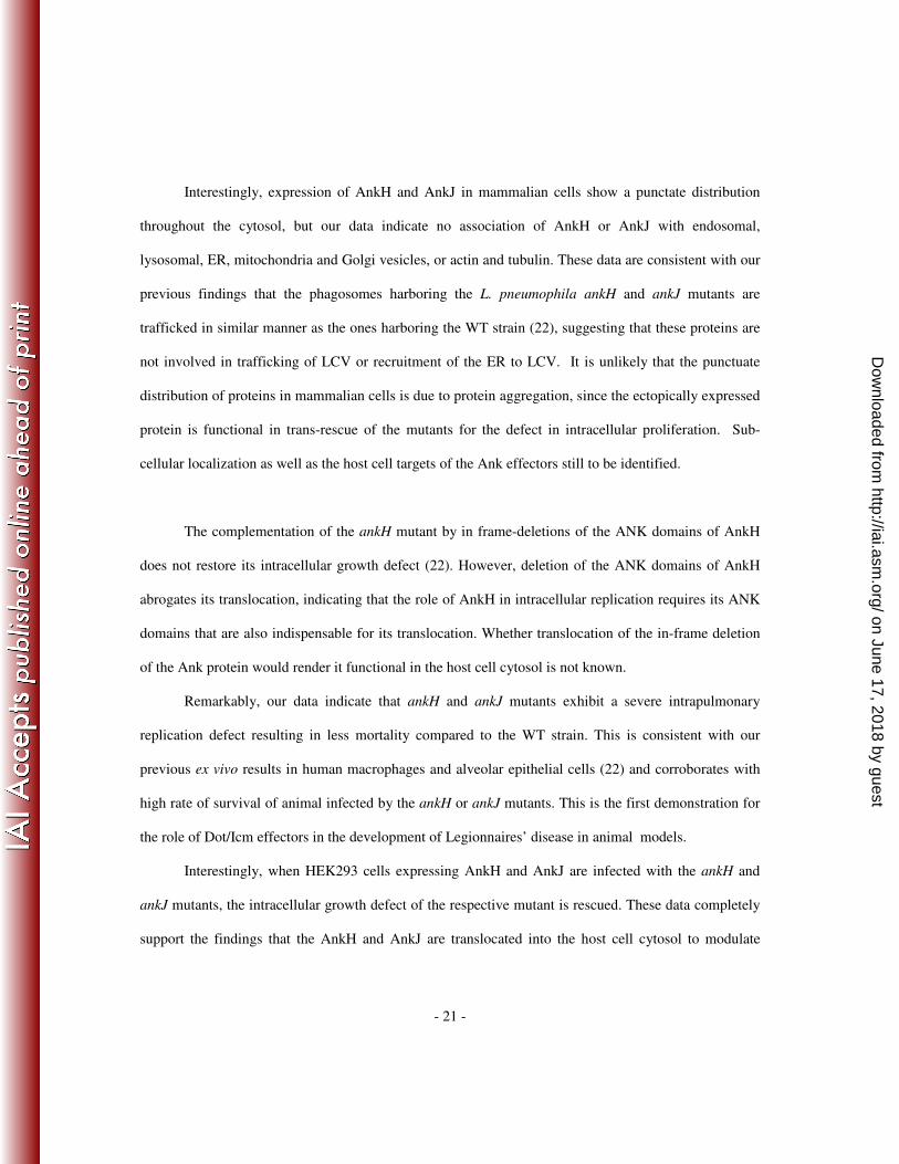
Table 1. List of ANK deletion constructs and primers used to generate ANK deletions in
ankH and ankJ.
F: TGATGCTATTTTCATTTC
R: TACGCCCCCCCCACTTATG
F: GGGGGGGGCGTACTGATT
R: CCTATCAACTATCACAAAG
F: ATAGTTGATAGGATAATCR: CTGGTGAATAGCACCAATAT
F: TTATTCTTCAAAACGACTCTCTGGAAC
R: GGAGAAATACCTCCTTCAAGAA
ankJ∆A1
ankJ∆A2
ankJ∆A3
ankJ∆797-807
F: CCAGACGTCACAGGACGC
R: TTCATCGATATCATCCAAAG
F: TACACTCGTAATGGTCTTTG
R: GACGTCTGGCTTGTTGATAT
F: TTAATTAGGATTAATCCCACAATCATCCAGAATT
R: TAACCCGTAAAGGAAATAATTTATT
ankH∆A1
ankH∆A2
ankH∆1391-1401
Primer Sequences (5'-3‘)Constructs
All primers are 5’-phosphorylated
on June 17, 2018 by guesthttp://iai.asm
.org/D
ownloaded from
- 24 -
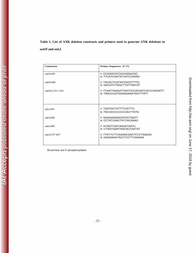
Table 2. List of Cya-Ank reporter constructs and primers used to generate ank fusions.
Constructs Primer Sequences (5'-3') Restrition sites
pCya-ankB F: GGATCCTTATGAAAAAGAATTTTTTTTCTG
R: CTGCAGTTAACAAACAAGGCACTTGCT BamHI
PstI
pCya-ankC F: CCCGGATCCTTATGGATTTTGTAAGTGAAATG
R: CCCCTGCAGTTACTATTTTAGGACAACTCGT BamHI
PstI
pCya-ankD F: CCCGGATCCTTATGTTGACTCCTCCGCCTGACT
R: CCCCTGCAGTTAGTCCTGAGGATTTTCTTTA BamHI
PstI
pCya-ankG F: CCCGGATCCTTCTGAATTCATTATGGATAGC
R: CCCCTGCAGTTATTTCATACCAAAACGAG BamHI
PstI
pCya-ankH F: CCCGGATCCTTATGAGTATTGCAAAC
R: CCCCTGCAGTTATAGGCCTGTCGCAACAGGATBamHI
PstI
pCya-ankI F: CCCGGATCCTTATGATTATTTTATATGATTTT
R: CCCCTGCAGTTAAAAAAACTTGCTTTCAAGTGBamHI
PstI
pCya-ankJ F: CCCGGATCCTTGTGATTAAAATGGGTAGA
R: CCCCTGCAGTTAAAGTGCGTTTTTAGGGGTATBamHI
PstI
pCya-ankK F: CCCGGATCCTTATGCCTAGAGTTTATAATCTTA
R: CCCCTGCAGTTAGATTTTATTCTTTGATAGTGABamHI
PstI
pCya-ankN F: CCCGGATCCTTGGTAAAAATTATGCC
R: CCCCTGCAGTTACTACCATTTTAATTTCAAG BamHI
PstI
pCya-ankQ F: CCCGGATCCTTATGCTTATGGCCG
R: CCCCTGCAGTTATGCTTATGGCCGCAACAA BamHI
PstI
on June 17, 2018 by guesthttp://iai.asm
.org/D
ownloaded from
- 25 -
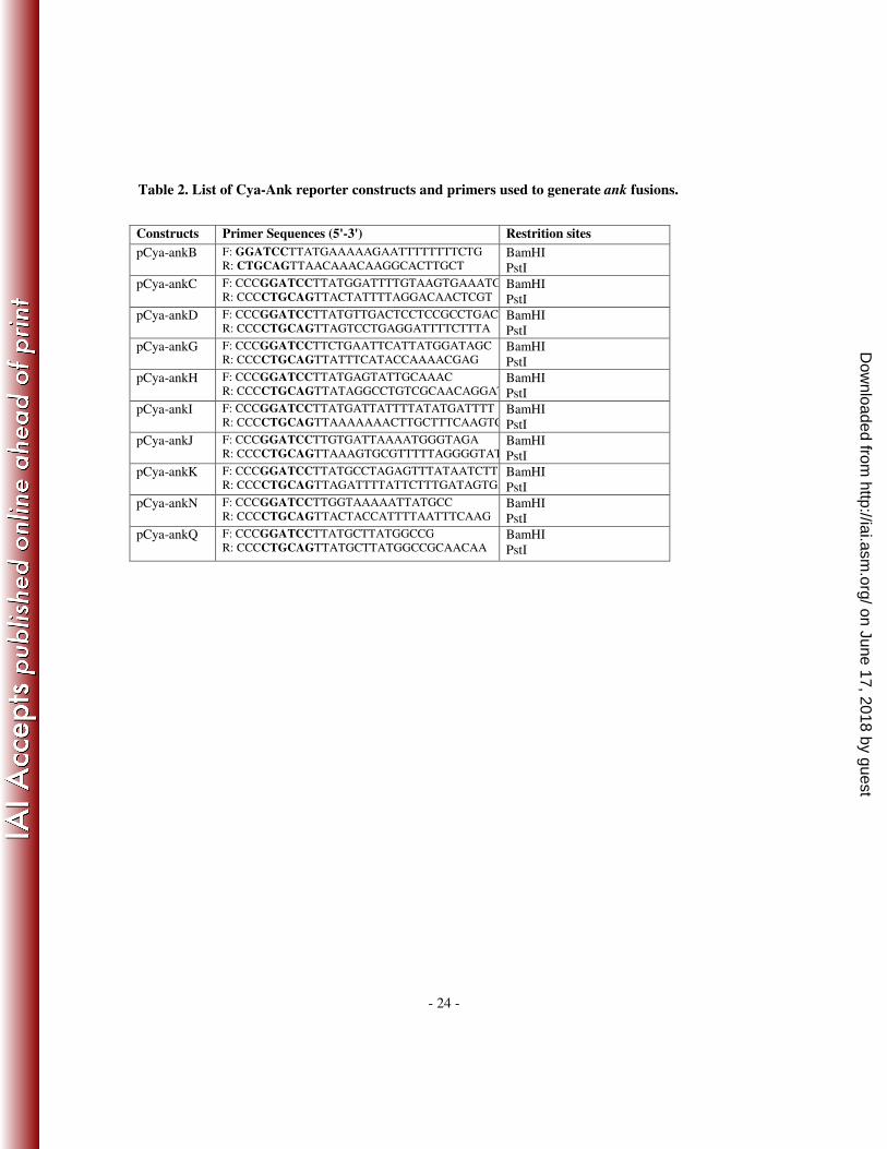
Table 3. List of GFP/3xFlag-Ank fusion constructs and primers used to generate ank
fusions.
Constructs Primer Sequences (5'-3') Restrition sites
p3xflag-ankH F:AGATCTGATGAGTATTGCAAACGATA
R:GGATCCTTATAGGCCTGTCGCAACAGGATT BglII
BamH
p3xflag-ankJ F:AAGCTTGTGATTAAAATGGGTAGA
R:AGATCTTTAAAGTGCGTTTTTAGGGGTATC HindIII
BglII
pAcGFP-ankH F:AGATCTGATGAGTATTGCAAACGATA
R:TCTAGATTATAGGCCTGTCGCAACAGGATT BglII
XbaI
pAcGFP-ankJ F: AGATCTGTGATTAAAATGGGTAGA
R: TCTAGATTAAAGTGCGTTTTTAGGGGTATC BglII
XbaI
on June 17, 2018 by guesthttp://iai.asm
.org/D
ownloaded from
- 26 -
References
1. Abu-Zant, A., S. Jones, R. Asare, J. Suttles, C. Price, J. Graham, and Y. A.
Kwaik. 2006. Anti-apoptotic signalling by the Dot/Icm secretion system of L.
pneumophila. Cell Microbiol.
2. Abu Kwaik, Y., L.-Y. Gao, B. J. Stone, C. Venkataraman, and O. S. Harb.
1998. Invasion of protozoa by Legionella pneumophila and its role in bacterial
ecology and pathogenesis. Appl.Environ.Microbiol. 64:3127-3133.
3. Al-Khodor1, S. 2010. Ankyrin-repeat containing proteins of microbes: a
conserved structure with functional diversity. Trends in Microbiology. In Press.
4. Al-Khodor, S., C. T. Price, F. Habyarimana, A. Kalia, and Y. Abu Kwaik.
2008. A Dot/Icm-translocated ankyrin protein of Legionella pneumophila is
required for intracellular proliferation within human macrophages and protozoa.
Mol Microbiol 70:908-923.
5. Amstutz, P., H. K. Binz, P. Parizek, M. T. Stumpp, A. Kohl, M. G. Grutter,
P. Forrer, and A. Pluckthun. 2005. Intracellular kinase inhibitors selected from
combinatorial libraries of designed ankyrin repeat proteins. J Biol Chem
280:24715-22.
6. Asare, R., M. Santic, I. Gobin, M. Doric, J. Suttles, J. Graham, C. Price, and
Y. A. Kwaik. 2007. Genetic susceptibility to Legionella longbeachae and caspase
activation within mice and human macrophages is distinct from L. pneumophila.
Infect Immun.
7. Bardill, J. P., J. L. Miller, and J. P. Vogel. 2005. IcmS-dependent translocation
of SdeA into macrophages by the Legionella pneumophila type IV secretion
system. Mol Microbiol 56:90-103.
8. Batchelor, A. H., D. E. Piper, F. C. de la Brousse, S. L. McKnight, and C.
Wolberger. 1998. The structure of GABPalpha/beta: an ETS domain- ankyrin
repeat heterodimer bound to DNA. Science 279:1037-41.
9. Brieland, J., P. Freeman, R. Kunkel, C. Chrisp, M. Hurley, J. Fantone, and
C. Engleberg. 1994. Replicative Legionella pneumophila lung infection in
intratracheally inoculated A/J mice. A murine model of human Legionnaires'
disease. Am J Pathol 145:1537-46.
10. Cambronne, E. D., and C. R. Roy. 2007. The Legionella pneumophila IcmSW
complex interacts with multiple Dot/Icm effectors to facilitate type IV
translocation. PLoS Pathog 3:e188.
11. Cazalet, C., C. Rusniok, H. Bruggemann, N. Zidane, A. Magnier, L. Ma, M.
Tichit, S. Jarraud, C. Bouchier, F. Vandenesch, F. Kunst, J. Etienne, P.
Glaser, and C. Buchrieser. 2004. Evidence in the Legionella pneumophila
genome for exploitation of host cell functions and high genome plasticity. Nat
Genet 36:1165-73.
12. Chen, J., M. Reyes, M. Clarke, and H. A. Shuman. 2007. Host cell-dependent
secretion and translocation of the LepA and LepB effectors of Legionella
pneumophila. Cell Microbiol 9:1660-71.
on June 17, 2018 by guesthttp://iai.asm
.org/D
ownloaded from
- 27 -
13. de Felipe, K. S., R. T. Glover, X. Charpentier, O. R. Anderson, M. Reyes, C.
D. Pericone, and H. A. Shuman. 2008. Legionella eukaryotic-like type IV
substrates interfere with organelle trafficking. PLoS Pathog 4:e1000117.
14. De Felipe, K. S., S. Pampou, O. S. Jovanovic, C. D. Pericone, S. F. Ye, S.
Kalachikov, and H. A. Shuman. 2005. Evidence for acquisition of Legionella
type IV secretion substrates via interdomain horizontal gene transfer. J Bacteriol
187:7716-26.
15. Derre, I., and R. R. Isberg. 2004. Legionella pneumophila replication vacuole
formation involves rapid recruitment of proteins of the early secretory system.
Infect Immun 72:3048-53.
16. Ensminger, A. W., and R. R. Isberg. 2009. Legionella pneumophila Dot/Icm
translocated substrates: a sum of parts. Curr Opin Microbiol.
17. Feeley, J. C., R. J. Gibson, G. W. Gorman, N. C. Langford, J. K. Rasheed, D.
C. Mackel, and W. B. Baine. 1979. Charcoal-yeast extract agar: primary
isolation medium for Legionella pneumophila. J.Clin.Microbiol. 10:437-441.
18. Fields, B. S., G. N. Sanden, J. M. Barbaree, W. E. Morrill, R. M. Wadowsky,
E. H. White, and J. C. Feeley. 1989. Intracellular multiplication of Legionella
pneumophila in amoebae isolated from hospital hot water tanks. Curr.Microbiol.
18:131-137.
19. Fraser, D. W., T. R. Tsai, W. Orenstein, W. E. Parkin, H. J. Beecham, R. G.
Sharrar, J. Harris, G. F. Mallison, S. M. Martin, J. E. McDade, C. C.
Shepard, and P. S. Brachman. 1977. Legionnaires' disease: description of an
epidemic of pneumonia. N Engl J Med 297:1189-97.
20. Gao, L. Y., B. J. Stone, J. K. Brieland, and Y. Abu Kwaik. 1998. Different
fates of Legionella pneumophila pmi and mil mutants within macrophages and
alveolar epithelial cells. Microb Pathog 25:291-306.
21. Garcia-Garcia, J. C., K. E. Rennoll-Bankert, S. Pelly, A. M. Milstone, and J.
S. Dumler. 2009. Silencing of host cell CYBB gene expression by the nuclear
effector AnkA of the intracellular pathogen Anaplasma phagocytophilum. Infect
Immun 77:2385-91.
22. Habyarimana, F., S. Al-Khodor, A. Kalia, J. E. Graham, C. T. Price, M. T.
Garcia, and Y. A. Kwaik. 2008. Role for the Ankyrin eukaryotic-like genes of
Legionella pneumophila in parasitism of protozoan hosts and human
macrophages. Environ Microbiol 10:1460-74.
23. Horwitz, M. A. 1983. The Legionnaires' disease bacterium (Legionella
pneumophila) inhibits phagosome-lysosome fusion in human monocytes.
J.Exp.Med. 158:2108-2126.
24. IJdo, J., A. C. Carlson, and E. L. Kennedy. 2007. Anaplasma phagocytophilum
AnkA is tyrosine-phosphorylated at EPIYA motifs and recruits SHP-1 during
early infection. Cell Microbiol 9:1284-96.
25. Ijdo, J. W., A. C. Carlson, and E. L. Kennedy. 2007. Anaplasma
phagocytophilum AnkA is tyrosine-phosphorylated at EPIYA motifs and recruits
SHP-1 during early infection. Cell Microbiol.
26. Isberg, R. R., T. J. O'Connor, and M. Heidtman. 2009. The Legionella
pneumophila replication vacuole: making a cosy niche inside host cells. Nat Rev
Microbiol 7:13-24.
on June 17, 2018 by guesthttp://iai.asm
.org/D
ownloaded from
- 28 -
27. Kagan, J. C., and C. R. Roy. 2002. Legionella phagosomes intercept vesicular
traffic from endoplasmic reticulum exit sites. Nat Cell Biol 4:945-954.
28. Kagan, J. C., M. P. Stein, M. Pypaert, and C. R. Roy. 2004. Legionella subvert
the functions of rab1 and sec22b to create a replicative organelle. J Exp Med
199:1201-11.
29. Kaufmann, A. F., J. E. McDade, C. M. Patton, J. V. Bennett, P. Skaliy, J. C.
Feeley, D. C. Anderson, M. E. Potter, V. F. Newhouse, M. B. Gregg, and P. S.
Brachman. 1981. Pontiac fever: isolation of the etiologic agent (Legionella
pneumophilia) and demonstration of its mode of transmission. Am J Epidemiol
114:337-47.
30. Kubori, T., A. Hyakutake, and H. Nagai. 2008. Legionella translocates an E3
ubiquitin ligase that has multiple U-boxes with distinct functions. Mol Microbiol
67:1307-19.
31. Laguna, R. K., E. A. Creasey, Z. Li, N. Valtz, and R. R. Isberg. 2006. A
Legionella pneumophila-translocated substrate that is required for growth within
macrophages and protection from host cell death. Proc Natl Acad Sci U S A
103:18745-50.
32. Liu, Y., and Z. Q. Luo. 2007. The Legionella pneumophila effector SidJ is
required for efficient recruitment of endoplasmic reticulum proteins to the
bacterial phagosome. Infect Immun 75:592-603.
33. Luo, Z. Q., and R. R. Isberg. 2004. Multiple substrates of the Legionella
pneumophila Dot/Icm system identified by interbacterial protein transfer. Proc
Natl Acad Sci U S A 101:841-6.
34. Mosavi, L. K., T. J. Cammett, D. C. Desrosiers, and Z. Y. Peng. 2004. The
ankyrin repeat as molecular architecture for protein recognition. Protein Sci
13:1435-48.
35. Nagai, H., E. D. Cambronne, J. C. Kagan, J. C. Amor, R. A. Kahn, and C. R.
Roy. 2005. A C-terminal translocation signal required for Dot/Icm-dependent
delivery of the Legionella RalF protein to host cells. Proc Natl Acad Sci U S A
102:826-31.
36. Ninio, S., D. M. Zuckman-Cholon, E. D. Cambronne, and C. R. Roy. 2005.
The Legionella IcmS-IcmW protein complex is important for Dot/Icm-mediated
protein translocation. Mol Microbiol 55:912-26.
37. Pan, X., A. Luhrmann, A. Satoh, M. A. Laskowski-Arce, and C. R. Roy.
2008. Ankyrin repeat proteins comprise a diverse family of bacterial type IV
effectors. Science 320:1651-4.
38. Pomerantsev, A. P., and V. M. Pavlov. 1999. [pCSE4 plasmid for cloning of
promoter-containing DNA fragments in francisella tularensis]. Vestn Ross Akad
Med Nauk:29-32.
39. Price, C. T. 2010. Acquisition of poly-ubiquitinated proteins by Legionella
pneumophila containing phagosome requires a bacterial F-box containing
Ankyrin effector. PLoS Pathogens, In Press.
40. Ragaz, C., H. Pietsch, S. Urwyler, A. Tiaden, S. S. Weber, and H. Hilbi. 2008.
The Legionella pneumophila phosphatidylinositol-4 phosphate-binding type IV
substrate SidC recruits endoplasmic reticulum vesicles to a replication-permissive
vacuole. Cell Microbiol 10:2416-33.
on June 17, 2018 by guesthttp://iai.asm
.org/D
ownloaded from
- 29 -
41. Santic, M., M. Molmeret, and Y. Abu Kwaik. 2005. Maturation of the
Legionella pneumophila-containing phagosome into a phagolysosome within
gamma interferon-activated macrophages. Infect. Immun. 73:3166-3171.
42. Swanson, J. A., and S. C. Baer. 1995. Phagocytosis by zippers and triggers.
Trends in Cell Biology 5:89-93.
43. Tilney, L. G., O. S. Harb, P. S. Connelly, C. G. Robinson, and C. R. Roy.
2001. How the parasitic bacterium Legionella pneumophila modifies its
phagosome and transforms it into rough ER: implications for conversion of
plasma membrane to the ER membrane. J Cell Sci 114:4637-50.
44. Voth, D. E., D. Howe, P. A. Beare, J. P. Vogel, N. Unsworth, J. E. Samuel,
and R. A. Heinzen. 2009. The Coxiella burnetii ankyrin repeat domain-
containing protein family is heterogeneous, with C-terminal truncations that
influence Dot/Icm-mediated secretion. J Bacteriol 191:4232-42.
45. Weber, S. S., C. Ragaz, K. Reus, Y. Nyfeler, and H. Hilbi. 2006. Legionella
pneumophila exploits PI(4)P to anchor secreted effector proteins to the replicative
vacuole. PLoS Pathog 2:e46.
46. Zink, S. D., L. Pedersen, N. P. Cianciotto, and Y. Abu Kwaik. 2002. The
Dot/Icm type IV secretion system of Legionella pneumophila is essential for the
induction of apoptosis in human macrophages. Infect. Immun. 70:1657-1663.
on June 17, 2018 by guesthttp://iai.asm
.org/D
ownloaded from
- 30 -
Figure Legends
Fig. 1 Potential translocation of L. pneumophila Ankyrin proteins by attached extracellular
bacteria. Untreated or cytochaladinD-treated U937 cells were infected with the wild-type strain of L.
pneumophila expressing the indicated Cya hybrid proteins. After 30 minutes of infection, cultured cells
were lysed and cAMP was quantified by ELISA and the amount of cAMP is indicated as fmol/well. The
experiment was performed three times. The data points are the average of cAMP concentration for one
representative experiment performed in triplicate. Error bars represent standard deviations of triplicate
samples.
Fig. 2. The ANK repeats and the C-terminus of L. pneumophila AnkH are required for delivery
into host cell
(A) Organization of the AnkH and different ANK domain or C-terminus deletions. Each protein
size and truncation are displayed.
(B ) Immunoblots of whole-cell bacterial extracts expressing indicated Cya hybrid proteins from
wild-type (WT), probed with monoclonal antibody specific to the M45 epitope and reprobed with anti-
CAT as loading control. The numbers represent the different truncated Cya-AnkH hybrid proteins: Cya-
AnkH (1), Cya-AnkH∆A1(2), Cya-AnkH∆A2 (3), Cya-AnkH∆A1∆A2 (4), Cya-AnkJ∆457-467(5).
(C) U937 cells were infected with the wild-type strain or the dotA mutant of L. pneumophila
expressing the indicated Cya hybrid proteins. After 1 h of infection, cultured cells were lysed and cAMP
was quantified by ELISA and the amount of cAMP is indicated as fmol/well. The experiment was
performed three times and the data are the average of cAMP concentration for one representative
experiment performed in triplicate. Error bars represent standard deviations of triplicate samples.
on June 17, 2018 by guesthttp://iai.asm
.org/D
ownloaded from
- 31 -
Fig. 3. The ANK repeats and the C-terminus of L. pneumophila AnkJ are required for delivery
into host cell
(A) Organization of the AnkJ and different ANK repeats or C-terminus in-frame deletions. Each
protein size and different truncation are displayed.
(B) Immunoblots of whole-cell bacterial extracts expressing the indicated Cya hybrid were
probed with monoclonal antibody specific to the M45 epitope and reprobed with anti-CAT as a loading
control. The numbers represent the different Cya-AnkJ truncated hybrid proteins: Cya-AnkJ (1), Cya-
AnkJ∆A1(2), Cya-AnkJ∆A2 (3), Cya-AnkJ∆A3 (4), Cya-AnkJ∆A1∆A2 (5), Cya-AnkJ∆A1∆A3 (6),
Cya-AnkJ∆A2 ∆A3 (7), Cya-AnkJ∆A1∆A2 ∆A3 (8), Cya-AnkJ∆259-269 (9).
(C) U937 cells were infected with the wild-type strain or the dotA mutant of L. pneumophila
expressing the indicated Cya hybrid proteins. After 1 h of infection, cultured cells were lysed and cAMP
was quantified by ELISA and the amount of cAMP is indicated as fmol/well. The experiment was
performed three times and the data are the average of cAMP concentration for one representative
experiment performed in triplicate. Error bars represent standard deviations of triplicate samples.
Fig. 4. The ANK repeats of AnkH are indispensable for intracellular growth of L. pneumophila
Monolayers of hMDMs were infected with L. pneumophila WT strain, dotA or ankH mutants
complemented with full length AnkH or constructs with in-frame deletion of the ANK domains. The
infection was carried out in triplicate with an MOI of 10 for 1h followed by 1h gentamicin treatment to
kill extracellular bacteria. At 24h and 48h, L. pneumophila-infected cells were lysed and plated onto
BCYE plates for CFU enumeration. The WT strain was used as a positive control and dotA mutant strain
as a negative control. The results are represented as fold increase (T/T0). The experiment was performed
on June 17, 2018 by guesthttp://iai.asm
.org/D
ownloaded from
- 32 -
three times. The data points are the average of one representative experiment performed in triplicate.
Error bars represent standard deviations of triplicate samples.
Fig. 5. The L. pneumophila ankH and ankJ mutants are defective in the A/J mice model
(A.) Groups of 30 mice were infected with 107, 10
8, 8x10
8 or 10
9 cfus of L. pneumophila WT
strain, ankH or ankJ mutant compared to the ankB mutant. After 1, 2, 3, 4 and 5 days post-infection
mortality was determined. After 3 days there was no mortality.
(B.) A/J mice were infected with 106 of L. pneumophila WT strain, ankH or ankJ mutants
compared to the ankB mutant. After 1, 2, 3 and 7 days post-infection, 3 mice were sacrificed and lungs
were collected for CFU enumeration.
Fig. 6. Subcellular localization of AnkH and AnkJ
HEK293T cells were transiently transfected with the empty vector pAcGFP, the pAcGFP-AnkH
or pAcGFP-AnkJ fusion constructs or with pBAP-3XFlag, and pAnkH-3XFlag or pAnkJ-3XFlag fusion
constructs for 18 h. After transfection, localization of AnkH and AnkJ fusion proteins was examined by
confocal laser scanning microscopy. Similar results were obtained in stable transfections (data not
shown).
Fig. 7. Colocalization of AnkH and AnkJ with endosomal, lysosomal, Golgi and ER compartments
Stable transfected HEK293 cells expressing GFP, the GFP-AnkH or GFP-AnkJ fusion proteins
were fixed and stained with anti-Lamp2, anti-cathepsinD, anti-KDEL, anti-GM130 and anti-p58k
antibodies. Their association with endosomal, lysosomal and ER compartments was assessed by
confocal laser scanning microscopy (A) and quantitation of colocalization is shown in (B). The results
on June 17, 2018 by guesthttp://iai.asm
.org/D
ownloaded from
- 33 -
shown are representative of three independent experiments performed in triplicate. The data represent
means ± standard deviation.
Fig. 8. The L. pneumophila ankH or ankJ mutants are rescued within HEK293 cells expressing
AnkH or AnkJ-GFP fusion proteins, respectively. HEK293 cells with stable expression of GFP,
AnkH and AnkJ-GFP fusion proteins were infected with the wild-type strain, the dotA, ankH or ankJ
mutant at MOI of 5. After 10h post-infection, cells were stained for laser scanning confocal microscopy
analysis (A). Percentage of infected cells harboring ≤3, 4-7 and >7 bacteria per cell were determined
based on analyses of 100 infected cells shown in (B). The results shown are representative of three
independent experiments performed in triplicate. The data represent means ± standard deviation.
on June 17, 2018 by guesthttp://iai.asm
.org/D
ownloaded from


![advcloudfiles.advantech.com · TW06/00913.OO, continued Advantech Co., Ltd] Advanixs Corp. ISO Issue 7 Detailed scope 1. Design and Manufacture of Industrial Automation Series Product](https://img.pdfslide.us/doc/110x75/5e730faa403e1156767a2a11/tw0600913oo-continued-advantech-co-ltd-advanixs-corp-iso-issue-7-detailed.jpg)