Embed Size (px)
Citation preview
Old but Gold: The Importance of Medical History in DiagnosingNeutrophilic Dermatoses Characterized by Pathergy. A Case of PyodermaGangrenosumAlessia Paganelli*, Federico Garbarino, Chiara Fiorentini, Laura Bigi and Cristina Magnoni
Clinica Dermatologica, A.O.U. Policlinico di Modena, University of Modena and Reggio Emilia, Italy*Corresponding author: Alessia Paganelli, Clinica Dermatologica, A.O.U. Policlinico di Modena, University of Modena and Reggio Emilia, Italy, Tel: +393496440085; E-mail: [email protected]
Received date: August 10, 2017; Accepted date: August 21, 2017; Published date: August 24, 2017
Copyright: © 2017 Paganelli A, et al. This is an open-access article distributed under the terms of the Creative Commons Attribution License, which permits unrestricteduse, distribution, and reproduction in any medium, provided the original author and source are credited.
Abstract
We describe the case of an 84-years old woman referred to our clinic without an established diagnosis in 2015 forthe presence of multiple cervical ulcers since 2013, when she underwent vascular surgery of the carotid arteries.The ulcers had infiltrated and actively inflamed violaceous borders; they were itchy and showed signs of scratching.The patient had already been prescribed different antibiotic treatments, without any clinical improvement. Multiplebiopsies had also been performed, but histology was not diagnostic, showing a non-specific dermal inflammatoryinfiltrate. Despite optimal wound care treatment, we observed a dramatic worsening of the skin lesions, spreadingfrom the neck to the vertex, especially at sites of minor trauma (for example, starting from scratch lesions). Thepresence of pathergy, which consists in the occurrence of lesions at sites of trauma, suggested the diagnosis ofpyoderma gangrenosum. Systemic glucocorticoid therapy was then prescribed, with quick improvement and nearlycompletes clinical remission. Our case confirms the importance of anamnesis and detailed collection of symptomsassociated with the clinical manifestations in dermatological dermatoses such as pyoderma gangrenosum whereimaging, histology and laboratory findings are often not very helpful for a correct diagnosis.
Keywords: Pyoderma gangrenosum; Pathergy; Ulcers; Medicalhistory; Glucocorticoids
IntroductionPyoderma Gangrenosum (PG) is a neutrophilic dermatosis which
typically presents with painful exudating ulcers, with infiltrated andactively inflamed violaceous borders [1]. One helpful clinical featurefor diagnosing active PG may be the presence of “pathergy” or theoccurrence of lesions at sites of trauma, in up to 30% of the patients[2-4]. PG may be idiopathic, but it is associated with an underlyingdisease in 50% of patients [5].
Clinical CaseWe describe the case of an 84-years old patient who was referred to
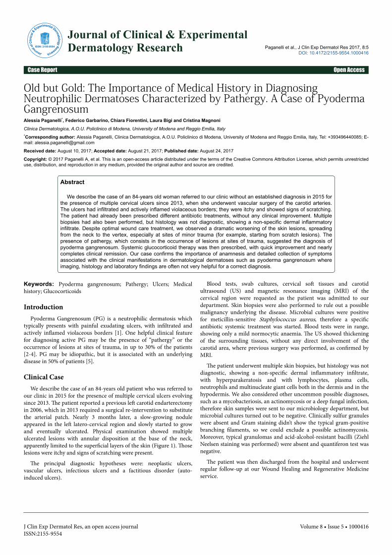
our clinic in 2015 for the presence of multiple cervical ulcers evolvingsince 2013. The patient reported a previous left carotid endarterectomyin 2006, which in 2013 required a surgical re-intervention to substitutethe arterial patch. Nearly 3 months later, a slow-growing noduleappeared in the left latero-cervical region and slowly started to growand eventually ulcerated. Physical examination showed multipleulcerated lesions with annular disposition at the base of the neck,apparently limited to the superficial layers of the skin (Figure 1). Thoselesions were itchy and signs of scratching were present.
The principal diagnostic hypotheses were: neoplastic ulcers,vascular ulcers, infectious ulcers and a factitious disorder (auto-induced ulcers).
Blood tests, swab cultures, cervical soft tissues and carotidultrasound (US) and magnetic resonance imaging (MRI) of thecervical region were requested as the patient was admitted to ourdepartment. Skin biopsies were also performed to rule out a possiblemalignancy underlying the disease. Microbial cultures were positivefor meticillin-sensitive Staphylococcus aureus, therefore a specificantibiotic systemic treatment was started. Blood tests were in range,showing only a mild normocytic anaemia. The US showed thickeningof the surrounding tissues, without any direct involvement of thecarotid area, where previous surgery was performed, as confirmed byMRI.
The patient underwent multiple skin biopsies, but histology was notdiagnostic, showing a non-specific dermal inflammatory infiltrate,with hyperparakeratosis and with lymphocytes, plasma cells,neutrophils and multinucleate giant cells both in the dermis and in thehypodermis. We also considered other uncommon possible diagnoses,such as a mycobacteriosis, an actinomycosis or a deep fungal infection,therefore skin samples were sent to our microbiology department, butmicrobial cultures turned out to be negative. Clinically sulfur granuleswere absent and Gram staining didn’t show the typical gram-positivebranching filaments, so we could exclude a possible actinomycosis.Moreover, typical granulomas and acid-alcohol-resistant bacilli (ZiehlNeelsen staining was performed) were absent and quantiferon test wasnegative.
The patient was then discharged from the hospital and underwentregular follow-up at our Wound Healing and Regenerative Medicineservice.
Paganelli et al., J Clin Exp Dermatol Res 2017, 8:5DOI: 10.4172/2155-9554.1000416
Case Report Open Access
J Clin Exp Dermatol Res, an open access journalISSN:2155-9554
Volume 8 • Issue 5 • 1000416
Journal of Clinical & ExperimentalDermatology ResearchJourna
l of C
linic
al &
Experimental Dermatology Research
ISSN: 2155-9554
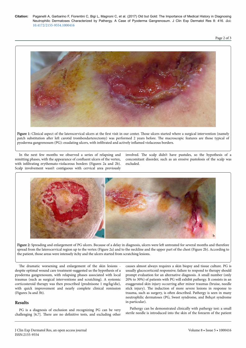
Figure 1: Clinical aspect of the laterocervical ulcers at the first visit in our center. Those ulcers started where a surgical intervention (namelypatch substitution after left carotid trombendarterectomy) was performed 2 years before. The macroscopic features are those typical ofpyoderma gangrenosum (PG): exudating ulcers, with infiltrated and actively inflamed violaceous borders.
In the next few months we observed a series of relapsing andremitting phases, with the appearance of confluent ulcers of the vertex,with infiltrating erythemato-violaceous borders (Figures 2a and 2b).Scalp involvement wasn’t contiguous with cervical area previously
involved. The scalp didn’t have pustules, so the hypothesis of aconcomitant disorder, such as an erosive pustolosis of the scalp wasexcluded.
Figure 2: Spreading and enlargement of PG ulcers. Because of a delay in diagnosis, ulcers were left untreated for several months and thereforespread from the laterocervical region up to the vertex (Figure 2a) and to the neckline and the upper part of the chest (Figure 2b). According tothe patient, those areas were intensely itchy and the ulcers started from scratching lesions.
The dramatic worsening and enlargement of the skin lesions -despite optimal wound care treatment-suggested us the hypothesis of apyoderma gangrenosum, with relapsing phases associated with localtraumas (such as surgical interventions and scratching). A systemiccorticosteroid therapy was then prescribed (prednisone 1 mg/kg/die),with quick improvement and nearly complete clinical remission(Figures 3a and 3b).
ResultsPG is a diagnosis of exclusion and recognizing PG can be very
challenging [6,7]. There are no definitive tests, and excluding other
causes almost always requires a skin biopsy and tissue culture. PG isusually glucocorticoid responsive; failure to respond to therapy shouldprompt evaluation for an alternative diagnosis. A small number (only20% to 30%) of patients with PG will exhibit pathergy. It consists in anexaggerated skin injury occurring after minor traumas (bruise, needlestick injury). The induction of more severe lesions in response totrauma, such as surgery, is often described. Pathergy is seen in manyneutrophilic dermatoses (PG, Sweet syndrome, and Behçet syndromein particular).
Pathergy can be demonstrated clinically with pathergy test: a smallsterile needle is introduced into the skin of the forearm of the patient
Citation: Paganelli A, Garbarino F, Fiorentini C, Bigi L, Magnoni C, et al. (2017) Old but Gold: The Importance of Medical History in DiagnosingNeutrophilic Dermatoses Characterized by Pathergy. A Case of Pyoderma Gangrenosum. J Clin Exp Dermatol Res 8: 416. doi:10.4172/2155-9554.1000416
Page 2 of 3
J Clin Exp Dermatol Res, an open access journalISSN:2155-9554
Volume 8 • Issue 5 • 1000416

and the test is considered positive when a small pustule and/or anerythematous papule are observed at site of injection after 24-48 h.
Figure 3: Clinical remission of head (Figure 3a) and cervical (Figure 3b) lesions. As expected, PG ulcers quickly responded to systemicglucocorticoids. The only new lesions (Fig. 3b, arrows) arose from the patch previously applied on the dressing, thus confirming the presenceof pathergy in this case of PG.
Management is threefold: wound care, evaluation of underlyingcauses, and treatment of PG as a chronic inflammatory disease.Treating PG can be very tricky, and if there is an associated underlyingdisease, therapy should be directed at controlling that process. PG isoften responsive to topical or intralesional glucocorticoids.Cyclosporine and infliximab may also be effective, with infliximabhaving the highest quality evidence, although most clinicians usesystemic glucocorticoids as first-line therapy [8-10].
AcknowledgementsWe conduct our studies in accordance with recognized international
standards, including the principles of the Declaration of Helsinki.
References1. Alavi A, French LE, Davis MD, Brassard A, Kirsner RS, et al. (2017)
Pyoderma Gangrenosum: An Update on Pathophysiology, Diagnosis andTreatment. Am J Clin Dermatol 18: 355-372.
2. Liaqat M, Elsensohn AN, Hansen CD, Maughan JA, Petersen MJ, et al.(2014) Acute postoperative pyoderma gangrenosum case and review ofliterature identifying chest wall predominance and no recurrencefollowing skin grafts. J Am AcadDermatol 71: e145-e146.
3. Tolkachjov SN, Fahy AS, Cerci FB, Wetter DA, Cha SS, et al. (2016)Postoperative Pyoderma Gangrenosum: A Clinical Review of PublishedCases. Mayo Clin Proc 91: 1267-1279.
4. Iosifescu AG, Boiangiu CI, Comănescu CM, Iliescu VA (2012) Pyodermagangrenosum—a postoperative "pseudo-infection". Chirurgia (Bucur)107: 119-121.
5. Marzano AV, Borghi A, Meroni PL, Cugno M (2016) Pyodermagangrenosum and its syndromic forms: evidence for a link withautoinflammation. Br J Dermatol 175: 882-891.
6. Braswell SF, Kostopoulos TC, Ortega-Loayza AG (2015) Pathophysiologyof pyoderma gangrenosum (PG): an updated review. J Am AcadDermatol 73: 691-698.
7. Wong WW, Machado GR, Hill ME (2011) Pyoderma gangrenosum: thegreat pretender and a challenging diagnosis. J Cutan Med Surg 15:322-328.
8. Patel F, Fitzmaurice S, Duong C, He Y, Fergus J, et al. (2015) Effectivestrategies for the management of pyoderma gangrenosum: acomprehensive review. Acta Derm Venereol 95: 525-531.
9. Quist SR, Kraas L (2017) Treatment options for pyoderma gangrenosum.J Dtsch Dermatol Ges 15: 34-40.
10. Soto Vilches F, Vera-Kellet C (2017) Pyoderma gangrenosum: Classic andemerging therapies. Med Clin (Barc) S0025-7753: 30343-30343.
Citation: Paganelli A, Garbarino F, Fiorentini C, Bigi L, Magnoni C, et al. (2017) Old but Gold: The Importance of Medical History in DiagnosingNeutrophilic Dermatoses Characterized by Pathergy. A Case of Pyoderma Gangrenosum. J Clin Exp Dermatol Res 8: 416. doi:10.4172/2155-9554.1000416
Page 3 of 3
J Clin Exp Dermatol Res, an open access journalISSN:2155-9554
Volume 8 • Issue 5 • 1000416
![l c a l Derma Journal of Clinical & Experimental t i n o i ......malignant erythroderma and idiopathic erythroderma [3]. Erythrodermic psoriasis represents 25 % of cases presenting](https://img.pdfslide.us/doc/110x75/5e4761ab9511660af751b382/l-c-a-l-derma-journal-of-clinical-experimental-t-i-n-o-i-malignant.jpg)


![l c a l Derma Journal of Clinical & Experimental t i n i ... · hepatocellular carcinoma, cholangiocarcinoma, ovarian cancer) [3-6]. The typical histological feature of porokeratosis](https://img.pdfslide.us/doc/110x75/5f7fe512dc08fc6a376d97a7/l-c-a-l-derma-journal-of-clinical-experimental-t-i-n-i-hepatocellular.jpg)


