Embed Size (px)
Citation preview
A Review of Lupus Miliaris Disseminatus Faciei-Like Histopathologic Changesin 10 CasesKathryn Echols1*, Fiona Fang2 and James W. Patterson2,3
1Medical University of South Carolina, Department of Dermatology and Dermatologic Surgery, Charleston, South Carolina2University of Virginia, Department of Dermatology, Charlottesville, Virginia3University of Virginia, Department of Pathology, Charlottesville, Virginia*Corresponding author: Kathryn F Echols, Medical University of South Carolina, Department of Dermatology and Dermatologic Surgery, 135 Rutledge Avenue; MSC578 Charleston, SC 29425, South Carolina, Tel: 843-792-6963; E-mail: [email protected]
Received date: May 19, 2014, Accepted date: June 28, 2014, Published date: July 5, 2014
Copyright: © 2014 Echols K. This is an open-access article distributed under the terms of the Creative Commons Attribution License, which permits unrestricted use,distribution, and reproduction in any medium, provided the original author and source are credited.
Abstract
Objective: To determine the clinical and histopathologic features of all lesions diagnosed as lupus miliarisdisseminatus faciei via biopsy over the past 16 years at a single institution. Clinical features reviewed included ageof patient, location and number of lesions, duration, description of primary lesion, size, and suspected clinicaldiagnosis or differential diagnosis. Histopathologic features reviewed included presence of caseation necrosis, depthof granuloma, presence of lymphocytic infiltrate, disruption of hair follicles, and presence of multinucleated giantcells.
Methods: The records of 10 patients (mean age, 50.4 years; range, 6 to 79 years) with characteristic histologicfeatures of lupus miliaris disseminatus faciei were reviewed and the histopathologic findings and clinical featureswere analyzed. Formalin-fixed, paraffin-embedded specimens were examined by hematoxylin-eosin staining.
Results: The most common clinical appearance was a single papule located on the face. Two cases with solitary,extrafacial distributions were reported. All cases demonstrated epithelioid granulomas with a central area ofcaseation necrosis. The majority of granulomas were perifollicular in location and were comprised of histiocytes,lymphocytes, and multinucleated giant cells.
Conclusion: The 10 cases we report demonstrate the importance of recognizing the entity in solitary as well asextrafacial forms. Limiting the histologic diagnosis to fully developed lesions demonstrating epithelioid granulomaswith caseation necrosis serves to clarify the diagnosis in the setting of diverse clinical presentations. Furtherinformation is needed to clarify the diagnosis, etiology, and pathogenesis of this disease, but an unusual hostresponse to folliculitis or follicular injury likely plays a role in most cases.
Keywords: Lupus miliaris disseminatus faciei; Caseation necrosis,Caseous necrosis; Lupus miliaris; Acne agminata; Follicle centeredgranuloma; Epithelioid granuloma
IntroductionLupus miliaris disseminatus faciei (LMDF) is an uncommon but
distinct, chronic, inflammatory dermatosis characterized by abruptdevelopment of generally asymptomatic, single to multiple, 1-3 mmbrown-red, brown, to yellowish dome-shaped papules or nodules withoccasional mild scaling [1-5]. Small pustules may rarely accompanythe papules [3,6]. Distribution tends to be symmetrical, primarilyinvolving the central and lateral face with the lower eyelids being mostfrequently affected [1-3,5]. However, multiple extrafacial sites ofinvolvement and one case without any facial involvement have beenreported [1,6,7]. Diascopy may reveal apple-jelly nodules [1-3]. LMDFmost commonly affects young adults of both sexes although casesamong children and the elderly have been reported [5,8]. Spontaneousresolution of the lesions is reported to occur over 1-4 years, oftenleaving small, pitted scars [1,5,6]. Microscopic findings are essentialfor diagnosis and characteristically reveal superficial granulomatousinflammation surrounding caseation necrosis that is often
perifollicular in distribution, although LMDF is now regarded as aspectrum classified into three histological stages: early, fully developed,and late [1]. Each stage has distinct histological findings. Fullydeveloped lesions are further broken down into 4 groups based on thetype of granulomatous reaction [1-4,9]. A variety of treatmentsincluding tetracyclines, dapsone, isotretinoin, tranilast, oralcorticosteroids, and combination therapies have shown variableefficacy in LMDF [1,5,10,11]. Though efficacy is difficult to determinein this spontaneously resolving dermatosis, early diagnosis andtreatment has demonstrated prevention of scar formation [1].
The etiology and pathogenesis of LMDF are unknown. It isconsidered by some to be part of a spectrum between granulomatousrosacea and sarcoidosis [6]. Others postulate an immune response topilosebaceous units or a foreign body reaction to sebum, keratin, orDemodex folliculorum from ruptured follicles [1,3,4,9]. Studiesrevealing intense lysozyme reaction in LMDF suggest that aninfectious agent may induce cell-mediated immunity, with subsequentformation of epithelioid cell granulomas [12]. The following reportdescribes the clinical and histopathologic findings in 10 cases of LMDFseen in our institution over a 16-year period.
Echols et al., J Clin Exp Dermatol Res 2014, 5:4 DOI: 10.4172/2155-9554.1000223
Research Article Open Access
J Clin Exp Dermatol ResISSN:2155-9554 JCEDR an open access journal
Volume 5 • Issue 4 • 1000223
Journal of Clinical & ExperimentalDermatology ResearchJourna
l of C
linic
al &
Experimental Dermatology Research
ISSN: 2155-9554
Materials and MethodsFrom January 1996 to September 2011, the records of 10 patients
with a histopathologic diagnosis of lupus miliaris disseminatus facieiwere collected from our dermatopathology archive. The records andarchival slides were reviewed to determine the clinical appearance anddistribution of skin lesions, their duration, and dermatologic history.Skin biopsy specimens stained with hematoxylin-eosin were reviewed,and additional sections obtained from the paraffin blocks were stainedwith Ziehl-Neelsen (AFB) and period acid-Schiff (PAS) stains.
ResultsThe case series included 6 men and 4 women from 6 to 79 years of
age (mean, 50.4 years). Seven patients presented with a single lesion,
the other three with multiple lesions. Locations included the cheek,cutaneous lip, medial canthus, eyelid, temple, antihelix, neck, andlower back (Table 1). One patient presented with a solitary lesion ofthe lower back with no facial involvement and another presented witha solitary lesion of the antihelix without facial involvement. Thelesions had reportedly been present for “months” up to one year. Eightof 10 patients had a history of previous or coexisting dermatologicdisease. One patient had a history of similar lesions previouslydiagnosed as rosacea, and two other patients had a history of faciallesions diagnosed as cystic acne.
Case No. Age (y) Sex Site Lesion No. Clinical diagnosis BeforeBiopsy Onset Dermatologic History Size (mm)
1 36 F Cheek 1 EIC Months Cystic acne 8
2 67 M Lower Back 1 Intradermal nevus vs. BCC Months SCC, BCC 4
3 79 F Cutaneous Lip 1 Indurated AK 3 months LPP 3
4 6 M Face MultipleGA vs. sarcoidosis vs.deep molluscum vs.mucinosis
Months None 0.5-2
5 57 M Medial Canthus 1 Cyst vs. BCC 1 y Rosacea 2
6 39 M Antihelix 1 BCC … Plaque psoriasis 3
7 34 M Eyelids, MedialCanthus, Neck Multiple
GA vs. sarcoidosis vs.deep molluscum vs.mucinosis vs. BCC
… GA …
8 59 F Lower Eyelid 1 BCC … … …
9 58 F Eyelids, MedialCanthus Multiple Granulomatous Rosacea 8-10 mo Nodulocystic acne 03-May
10 69 M Temple 1 BCC … BCC …
Table 1: Clinical features of 10 cases of lupus miliaris disseminatus faciei. Abbreviations: EIC, epidermal inclusion cyst; BCC, basal cellcarcinoma; SCC, squamous cell carcinoma; AK, actinic keratosis; LPP, lichen planopilaris; GA, granuloma annulare; F, female; M, male.
Eight of the lesions were described as papules, the other two asnodules. One lesion had a centrally located pustule, and one had slightscaling. The average size of the lesions was 3.6 mm in diameter,ranging from 0.5 to 8 mm (Table 1). One patient was symptomaticwith complaints of mild tenderness over the lesion. The clinicaldiagnosis or differential diagnosis at time of biopsy included basal cellcarcinoma (BCC) in six cases and sarcoidosis, granuloma annulare(GA), and epidermal cyst in two cases each. Granulomatous rosacea,mucinosis, intradermal nevus, actinic keratosis, and deep molluscumwere listed as clinical differential diagnoses in one case each (Table 1).
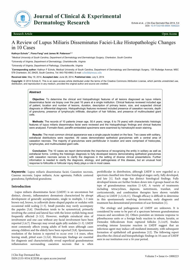
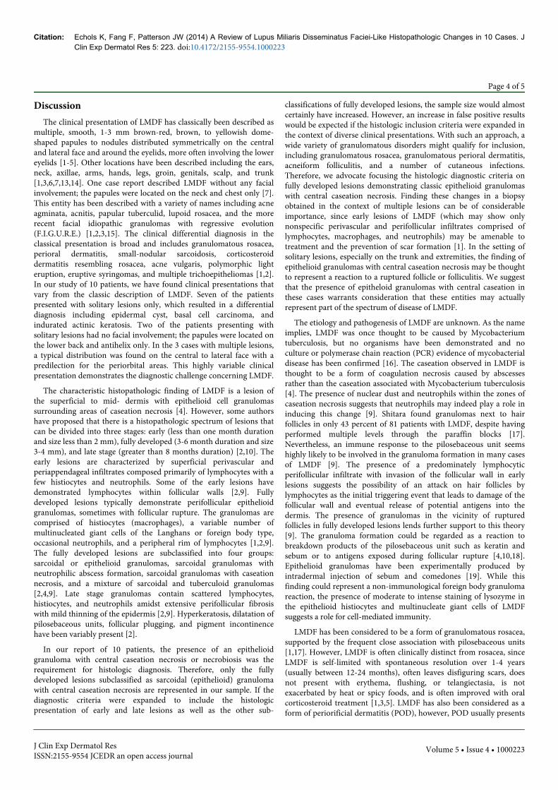
Figure 1: Histologic spectrum of LMDF. A. Extensive caseousnecrosis surrounded by a layer of histiocytes and multinucleatedhistiocytes with a peripheral rim of lymphocytes (40 ͯmagnification). B. Predominance of cellular components withminimal central necrosis (40 ͯ magnification).
Citation: Echols K, Fang F, Patterson JW (2014) A Review of Lupus Miliaris Disseminatus Faciei-Like Histopathologic Changes in 10 Cases. JClin Exp Dermatol Res 5: 223. doi:10.4172/2155-9554.1000223
Page 2 of 5
J Clin Exp Dermatol ResISSN:2155-9554 JCEDR an open access journal
Volume 5 • Issue 4 • 1000223
Histopathologically, there were epithelioid (sarcoidal) granulomaswith central areas of necrobiosis or caseation necrosis in all specimens(Figure 1).
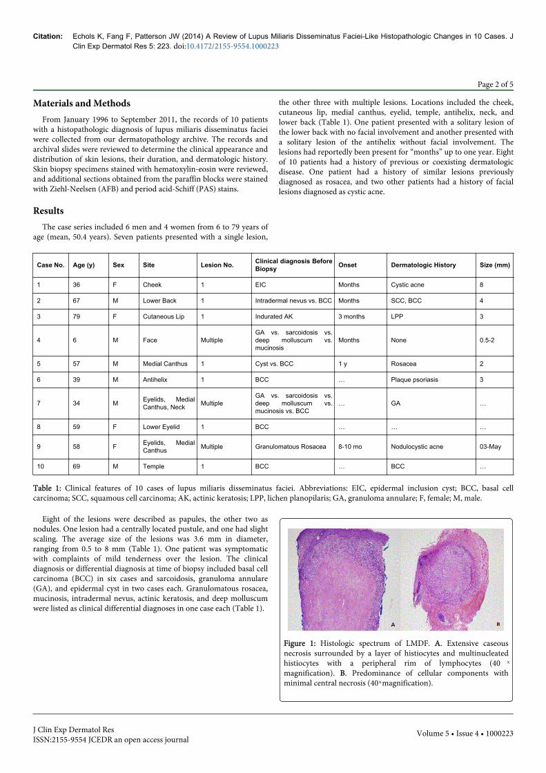
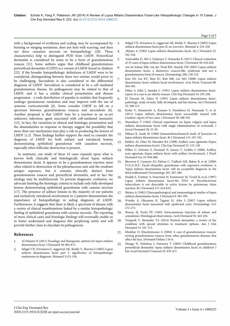
The granulomas were located in the upper dermis in six cases andin the mid dermis in two cases. In seven of the cases, a perifolliculardistribution of the granulomas was noted (Figure 2).
Figure 2: Involvement of folliculo-sebaceous unit. A. Folliculardisruption and extensive caseous necrosis (100 ͯ magnification). B.Focal, minimal perifolliculitis (200 ͯ magnification).
Lymphocytes were seen in eight cases (Figure 1-3); the lymphocytesformed an outer layer surrounding the granuloma in four cases(Figure 3).
Figure 3: Granulomatous component of LMDF. A. Granulomaformation with multinucleated giant cells (100 ͯ magnification). B.Langhans giant cell within granuloma (400 ͯ magnification).
Disruption of hair follicles in approximation to the granulomas wasnoted in two cases (Figure 1B and Figure 2B). One case demonstrateda perifollicular lymphocytic infiltrate with minimal follicular invasion.Multinucleated giant cells were seen in eight cases (Figure 3). AFB andPAS stains were performed on five of the cases and were all negative(Table 2).
Case No. Granuloma Type Caseation Necrosis Granuloma location Granuloma contents Disruption of hairfollicle
MN giantcells
AFB &PAS
1 Epithelioid (sarcoidal) Yes Unable TBD* Histiocytes, sparselymphocytes No Yes Neg
2 Epithelioid Yes Upper dermis Histiocytes, outer layer oflymphocytes Yes No Neg
3 Epithelioid Yes Upper dermis(perifollicular)
Histiocytes, outer layer oflymphocytes No Yes NA
4 Epithelioid Yes Upper dermis(perifollicular) Histiocytes, lymphocytes Yes Yes NA
5 Epithelioid Yes Mid dermis(perifollicular)
Histiocytes, outer layer oflymphocytes No Yes NA
6 Epithelioid Yes Upper dermis(perifollicular) Histiocytes, lymphocytes No Yes Neg
7 Epithelioid Yes Upper dermis(perifollicular)
Histiocytes, outer layerlymphocytes No Yes Neg
8 Epithelioid Yes Upper dermis(perifollicular) …* …* …* NA
9 Epithelioid Yes Mid dermis(perifollicular) Histiocytes, lymphocytes No Yes Neg
10 Epithelioid Yes Unable TBD* Histiocytes …* Yes NA
Table 2: Histologic features of 10 cases of lupus miliaris disseminatus faciei. Fields marked with * indicate the data was not able to be determinedbecause the original slides and paraffin block were no longer available for review and the data was not included in the original dermatopathologyreport. Abbreviations: TBD, to be determined; MN, multinucleated; AFB, acid fast bacteria; PAS, periodic acid-schiff; NA, Not applicable (stainnot done); Neg, negative.
Citation: Echols K, Fang F, Patterson JW (2014) A Review of Lupus Miliaris Disseminatus Faciei-Like Histopathologic Changes in 10 Cases. JClin Exp Dermatol Res 5: 223. doi:10.4172/2155-9554.1000223
Page 3 of 5
J Clin Exp Dermatol ResISSN:2155-9554 JCEDR an open access journal
Volume 5 • Issue 4 • 1000223
DiscussionThe clinical presentation of LMDF has classically been described as
multiple, smooth, 1-3 mm brown-red, brown, to yellowish dome-shaped papules to nodules distributed symmetrically on the centraland lateral face and around the eyelids, more often involving the lowereyelids [1-5]. Other locations have been described including the ears,neck, axillae, arms, hands, legs, groin, genitals, scalp, and trunk[1,3,6,7,13,14]. One case report described LMDF without any facialinvolvement; the papules were located on the neck and chest only [7].This entity has been described with a variety of names including acneagminata, acnitis, papular tuberculid, lupoid rosacea, and the morerecent facial idiopathic granulomas with regressive evolution(F.I.G.U.R.E.) [1,2,3,15]. The clinical differential diagnosis in theclassical presentation is broad and includes granulomatous rosacea,perioral dermatitis, small-nodular sarcoidosis, corticosteroiddermatitis resembling rosacea, acne vulgaris, polymorphic lighteruption, eruptive syringomas, and multiple trichoepitheliomas [1,2].In our study of 10 patients, we have found clinical presentations thatvary from the classic description of LMDF. Seven of the patientspresented with solitary lesions only, which resulted in a differentialdiagnosis including epidermal cyst, basal cell carcinoma, andindurated actinic keratosis. Two of the patients presenting withsolitary lesions had no facial involvement; the papules were located onthe lower back and antihelix only. In the 3 cases with multiple lesions,a typical distribution was found on the central to lateral face with apredilection for the periorbital areas. This highly variable clinicalpresentation demonstrates the diagnostic challenge concerning LMDF.
The characteristic histopathologic finding of LMDF is a lesion ofthe superficial to mid- dermis with epithelioid cell granulomassurrounding areas of caseation necrosis [4]. However, some authorshave proposed that there is a histopathologic spectrum of lesions thatcan be divided into three stages: early (less than one month durationand size less than 2 mm), fully developed (3-6 month duration and size3-4 mm), and late stage (greater than 8 months duration) [2,10]. Theearly lesions are characterized by superficial perivascular andperiappendageal infiltrates composed primarily of lymphocytes with afew histiocytes and neutrophils. Some of the early lesions havedemonstrated lymphocytes within follicular walls [2,9]. Fullydeveloped lesions typically demonstrate perifollicular epithelioidgranulomas, sometimes with follicular rupture. The granulomas arecomprised of histiocytes (macrophages), a variable number ofmultinucleated giant cells of the Langhans or foreign body type,occasional neutrophils, and a peripheral rim of lymphocytes [1,2,9].The fully developed lesions are subclassified into four groups:sarcoidal or epithelioid granulomas, sarcoidal granulomas withneutrophilic abscess formation, sarcoidal granulomas with caseationnecrosis, and a mixture of sarcoidal and tuberculoid granulomas[2,4,9]. Late stage granulomas contain scattered lymphocytes,histiocytes, and neutrophils amidst extensive perifollicular fibrosiswith mild thinning of the epidermis [2,9]. Hyperkeratosis, dilatation ofpilosebaceous units, follicular plugging, and pigment incontinencehave been variably present [2].
In our report of 10 patients, the presence of an epithelioidgranuloma with central caseation necrosis or necrobiosis was therequirement for histologic diagnosis. Therefore, only the fullydeveloped lesions subclassified as sarcoidal (epithelioid) granulomawith central caseation necrosis are represented in our sample. If thediagnostic criteria were expanded to include the histologicpresentation of early and late lesions as well as the other sub-
classifications of fully developed lesions, the sample size would almostcertainly have increased. However, an increase in false positive resultswould be expected if the histologic inclusion criteria were expanded inthe context of diverse clinical presentations. With such an approach, awide variety of granulomatous disorders might qualify for inclusion,including granulomatous rosacea, granulomatous perioral dermatitis,acneiform folliculitis, and a number of cutaneous infections.Therefore, we advocate focusing the histologic diagnostic criteria onfully developed lesions demonstrating classic epithelioid granulomaswith central caseation necrosis. Finding these changes in a biopsyobtained in the context of multiple lesions can be of considerableimportance, since early lesions of LMDF (which may show onlynonspecific perivascular and perifollicular infiltrates comprised oflymphocytes, macrophages, and neutrophils) may be amenable totreatment and the prevention of scar formation [1]. In the setting ofsolitary lesions, especially on the trunk and extremities, the finding ofepithelioid granulomas with central caseation necrosis may be thoughtto represent a reaction to a ruptured follicle or folliculitis. We suggestthat the presence of epitheloid granulomas with central caseation inthese cases warrants consideration that these entities may actuallyrepresent part of the spectrum of disease of LMDF.
The etiology and pathogenesis of LMDF are unknown. As the nameimplies, LMDF was once thought to be caused by Mycobacteriumtuberculosis, but no organisms have been demonstrated and noculture or polymerase chain reaction (PCR) evidence of mycobacterialdisease has been confirmed [16]. The caseation observed in LMDF isthought to be a form of coagulation necrosis caused by abscessesrather than the caseation associated with Mycobacterium tuberculosis[4]. The presence of nuclear dust and neutrophils within the zones ofcaseation necrosis suggests that neutrophils may indeed play a role ininducing this change [9]. Shitara found granulomas next to hairfollicles in only 43 percent of 81 patients with LMDF, despite havingperformed multiple levels through the paraffin blocks [17].Nevertheless, an immune response to the pilosebaceous unit seemshighly likely to be involved in the granuloma formation in many casesof LMDF [9]. The presence of a predominately lymphocyticperifollicular infiltrate with invasion of the follicular wall in earlylesions suggests the possibility of an attack on hair follicles bylymphocytes as the initial triggering event that leads to damage of thefollicular wall and eventual release of potential antigens into thedermis. The presence of granulomas in the vicinity of rupturedfollicles in fully developed lesions lends further support to this theory[9]. The granuloma formation could be regarded as a reaction tobreakdown products of the pilosebaceous unit such as keratin andsebum or to antigens exposed during follicular rupture [4,10,18].Epithelioid granulomas have been experimentally produced byintradermal injection of sebum and comedones [19]. While thisfinding could represent a non-immunological foreign body granulomareaction, the presence of moderate to intense staining of lysozyme inthe epithelioid histiocytes and multinucleate giant cells of LMDFsuggests a role for cell-mediated immunity.
LMDF has been considered to be a form of granulomatous rosacea,supported by the frequent close association with pilosebaceous units[1,17]. However, LMDF is often clinically distinct from rosacea, sinceLMDF is self-limited with spontaneous resolution over 1-4 years(usually between 12-24 months), often leaves disfiguring scars, doesnot present with erythema, flushing, or telangiectasia, is notexacerbated by heat or spicy foods, and is often improved with oralcorticosteroid treatment [1,3,5]. LMDF has also been considered as aform of periorificial dermatitis (POD), however, POD usually presents
Citation: Echols K, Fang F, Patterson JW (2014) A Review of Lupus Miliaris Disseminatus Faciei-Like Histopathologic Changes in 10 Cases. JClin Exp Dermatol Res 5: 223. doi:10.4172/2155-9554.1000223
Page 4 of 5
J Clin Exp Dermatol ResISSN:2155-9554 JCEDR an open access journal
Volume 5 • Issue 4 • 1000223
with a background of erythema and scaling, may be accompanied byburning or stinging sensations, does not heal with scarring, and doesnot show caseation necrosis on histopathology [20]. Thesecharacteristics help to distinguish POD from LMDF. Periorificialdermatitis is considered by some to be a form of granulomatousrosacea [21]. Some authors argue that childhood granulomatousperiorificial dermatitis (CGPD) is a variant of LMDF found in children[22]. If the broader histopathologic definitions of LMDF were to beconsidered, distinguishing between these two entities would prove tobe challenging. Sarcoidosis is also considered in the differentialdiagnosis of LMDF. Sarcoidosis is considered to be a cell mediatedgranulomatous disease. Its pathogenesis may be related to that ofLMDF, and it has a similar clinical presentation and diseaseprogression - a wide distribution of papules to nodules that frequentlyundergo spontaneous resolution and may improve with the use ofsystemic corticosteroids [6]. Some consider LMDF to fall on aspectrum between granulomatous rosacea and sarcoidosis [6].Another proposal is that LMDF may be a reaction to an as-yetunknown infectious agent associated with cell-mediated immunity[12]. In fact, the variations in clinical and histologic presentation andthe dissimilarities in response to therapy suggest the possibility thatmore than one mechanism may play a role in producing the lesions ofLMDF [1,3]. These findings further support the need to consider thediagnosis of LMDF for both solitary and extrafacial lesionsdemonstrating epithelioid granulomas with caseation necrosis,especially when follicular destruction is present.
In summary, our study of these 10 cases expands upon what isknown both clinically and histologically about lupus miliarisdisseminatus faciei. It appears to be a granulomatous reaction mostoften related to destruction of the pilosebaceous unit and the resultantantigen exposure, but it remains clinically distinct fromgranulomatous rosacea and periorificial dermatitis, and in fact theetiology may be multifactorial. To prevent diagnostic confusion, weadvocate limiting the histologic criteria to include only fully developedlesions demonstrating epithelioid granulomas with caseous necrosis[17]. The presence of solitary lesions in the majority of our patientsand exclusively extrafacial involvement in 2 patients demonstrates theimportance of histopathology in aiding diagnosis of LMDF.Furthermore, it suggests that there is likely a spectrum of disease witha variety of clinical manifestations linked by a similar histopathologicfinding of epithelioid granuloma with caseous necrosis. The reportingof more clinical cases and histologic findings will eventually enable usto better understand and diagnose this perplexing entity and willprovide further clues to elucidate its pathogenesis.
References1. Al-Mutairi N (2011) Nosology and therapeutic options for lupus miliaris
disseminatus faciei. J Dermatol 38: 864-873.2. Sehgal VN, Srivastava G, Aggarwal AK, Reddy V, Sharma S (2005) Lupus
miliaris disseminatus faciei part I: significance of histopathologicundertones in diagnosis. Skinmed 3:151-156.
3. Sehgal VN, Srivastava G, Aggarwal AK, Reddy V, Sharma S (2005) Lupusmiliaris disseminatus faciei part II: an overview. Skinmed 4: 234-238.
4. .Shitara A (1984) Lupus miliaris disseminatus faciei. Int J Dermatol 23:542-544.
5. Amiruddin D, Mii S, Fujimura T, Katsuoka K (2011) Clinical evaluationof 35 cases of lupus miliaris disseminatus faciei. J Dermatol 38: 618-620.
6. van de Scheur MR, van der Waal RIF, Starink TM (2003) Lupus miliarisdisseminatus faciei: a distinctive rosacea-like syndrome and not agranulomatous form of rosacea. Dermatology 206: 120-123.
7. Kim DS, Lee KY, Shin JU, Roh MR, Lee MG (2008) Lupus miliarisdisseminatus faciei without facial involvement. Acta Derm Venereol 88:504-505.
8. Dekio S, Jidoi J, Imaoka C (1991) Lupus miliaris disseminatus faciei-report of a case in an elderly woman. Clin Exp Dermatol 16: 295-296.
9. El Darouti M, Zaher H (1993) Lupus miliaris disseminatus faciei-pathologic study of early, fully developed, and late lesions. Int J Dermatol32: 508-511.
10. Koike Y, Hatamochi A, Koyano S, Namikawa H, Hamasaki Y, et al.(2011) Lupus miliaris disseminatus faciei successfully treated withtranilast: report of two cases. J Dermatol 38: 588-592.
11. Matsubara T (1962) Clinical experiment on lupus vulgaris and lupusmiliaris disseminatus faciei with single administration of corticoids. JInvest Dermatol 38: 25-29.
12. Mihara K, Isoda M (1986) Immunohistochemical study of lysozyme inlupus miliaris disseminatus faciei. Br J Dermatol 115: 187-192.
13. Bedlow AJ, Otter M, Marsden RA (1998) Axillary acne agminata (lupusmiliaris disseminatus faciei). Clin Exp Dermatol 23: 125-128.
14. Hillen U, Schroter S, Denisjuk N, Jansen T, Grabbe S (2006) Axillaryacne agminata (lupus miliaris faciei with axillary involvement). J DtschDermatol Ges 10: 858-860.
15. Skowron F, Causeret AS, Pabion C, Viallard AM, Balme B, et al. (2000)F.I.G.U.R.E.: Facial idiopathic granulomas with regressive evolution: is‘lupus miliaris disseminatus faciei’ still an acceptable diagnosis in thethird millennium? Dermatology 201: 287-289.
16. Hodak E, Trattner A, Feuerman H, Feinmesser M, Tsvieli R, et al. (1997)Lupus miliaris disseminatus faciei-the DNA of Mycobacteriumtuberculosis is not detectable in active lesions by polymerase chainreaction. Br J Dermatol 137: 614-619.
17. Shitara A (1982) Clinicopathological and immunological studies of lupusmiliaris disseminatus faciei. J Dermatol 9: 383-395.
18. Watabe A, Okuyama R, Tagami H, Aiba S (2007) Lupus miliarisdisseminatus faciei associated with epidermal cysts. Dermatology 214:272-273.
19. Strauss JS, Pochi PE (1965) Intracutaneous injection of sebum andcomedones: Histological observations. Arch Dermatol 92: 443-456.
20. Tempark T, Shwayder TA (2014) Perioral dermatitis: a review of thecondition with special attention to treatment options. Am J ClinDermatol 15: 101-113.
21. Khokhar O, Khachemoune A (2004) A case of granulomatous rosacea:sorting granulomatous rosacea from other granulomatous diseases thataffect the face. Dermatol Online J 10: 6.
22. Misago N, Nakafusa J, Narisawa Y (2005) Childhood granulomatousperiorificial dermatitis: lupus miliaris disseminatus faciei in children? JEur Acad Dermatol Venereol 19: 470-473.
Citation: Echols K, Fang F, Patterson JW (2014) A Review of Lupus Miliaris Disseminatus Faciei-Like Histopathologic Changes in 10 Cases. JClin Exp Dermatol Res 5: 223. doi:10.4172/2155-9554.1000223
Page 5 of 5
J Clin Exp Dermatol ResISSN:2155-9554 JCEDR an open access journal
Volume 5 • Issue 4 • 1000223


![%UDFNHWV [CT] High School 106content.ciacsports.com/pdfs/wrestling_open_info_2020.pdf[L1]James Lunt Xavier - Gr12 L L L L L L L L L L L L L L L L L L L L L L L L &,$&6WDWH2SHQ3UHOLPLQDU\%UDFNHWV](https://img.pdfslide.us/doc/110x75/6056cf3169537459b5566dee/udfnhwv-ct-high-school-l1james-lunt-xavier-gr12-l-l-l-l-l-l-l-l-l-l-l-l-l.jpg)


![Ch 5: ARIMA model · 1.1 Non-Stationary Data [ToC] Dow Jones Index From Aug. 28 to Dec. 18, 1972 l l l l l ll l l l l l l l l l l l l l l l l l l l l l l l l l l l l l l l l l l l](https://img.pdfslide.us/doc/110x75/5ee0213ead6a402d666b5f8b/ch-5-arima-model-11-non-stationary-data-toc-dow-jones-index-from-aug-28-to.jpg)