Embed Size (px)
Citation preview

Kristina M. Nowitzki, M.D., Ph.D. and Hao S. Lo, M.D. University of Massachusetts Medical School, Worcester, MA
Outline
Would you like to skip the brief introduction? Click the menu button at any point to skip/return to the case menu:
I. Introduction highlighting normal renal enhancement physiology including normal CT nephrogram phases.
II. Cases organized in a quiz format, with etiologies including:
a. obstructive b. vascular c. traumatic d. infectious/inflammatory e. neoplastic
Intr
od
uct
ion
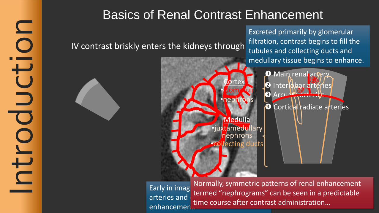
Basics of Renal Contrast Enhancement
IV contrast briskly enters the kidneys through the renal arteries:
Cortex Medulla
Early in imaging, the distribution of arteries and capillaries governs renal enhancement.
Main renal artery
Cortical radiate arteries
Interlobar arteries Arcuate arteries
•glomeruli •nephrons
•juxtamedullary nephrons
Excreted primarily by glomerular filtration, contrast begins to fill the tubules and collecting ducts and medullary tissue begins to enhance.
Normally, symmetric patterns of renal enhancement termed “nephrograms” can be seen in a predictable time course after contrast administration…
•collecting ducts
Intr
od
uct
ion
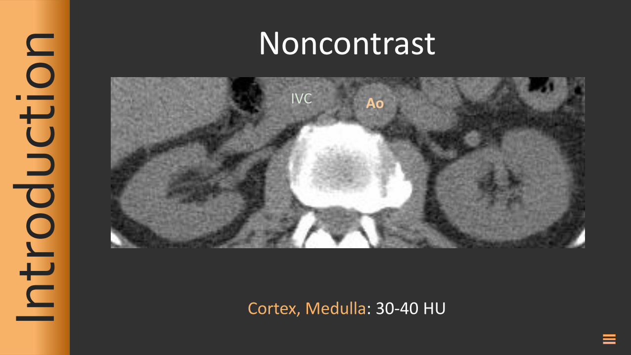
Noncontrast
Ao IVC
Cortex, Medulla: 30-40 HU
Intr
od
uct
ion
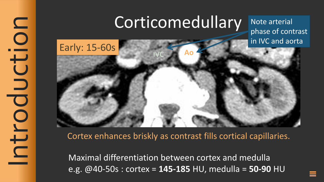
Corticomedullary
Ao IVC Early: 15-60s
Cortex enhances briskly as contrast fills cortical capillaries.
Maximal differentiation between cortex and medulla e.g. @40-50s : cortex = 145-185 HU, medulla = 50-90 HU
Note arterial phase of contrast in IVC and aorta
Intr
od
uct
ion
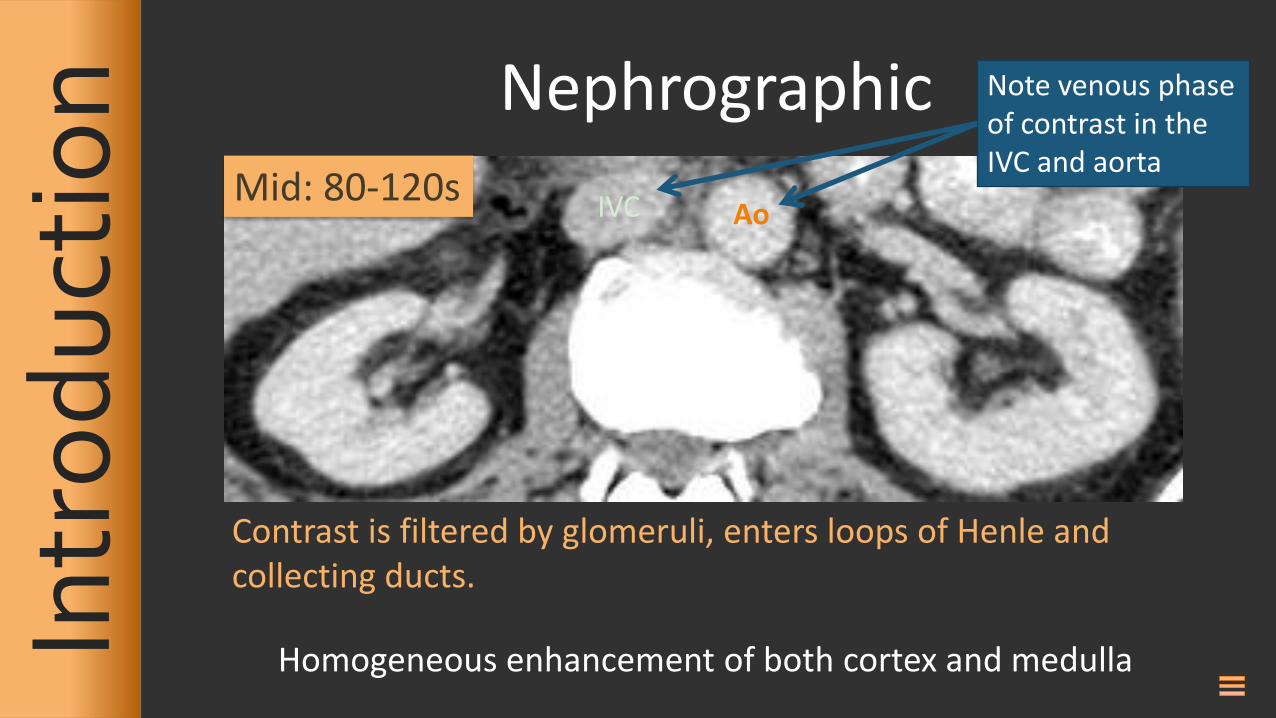
Nephrographic
Contrast is filtered by glomeruli, enters loops of Henle and collecting ducts.
Mid: 80-120s Ao IVC
Homogeneous enhancement of both cortex and medulla
Note venous phase of contrast in the IVC and aorta
Intr
od
uct
ion
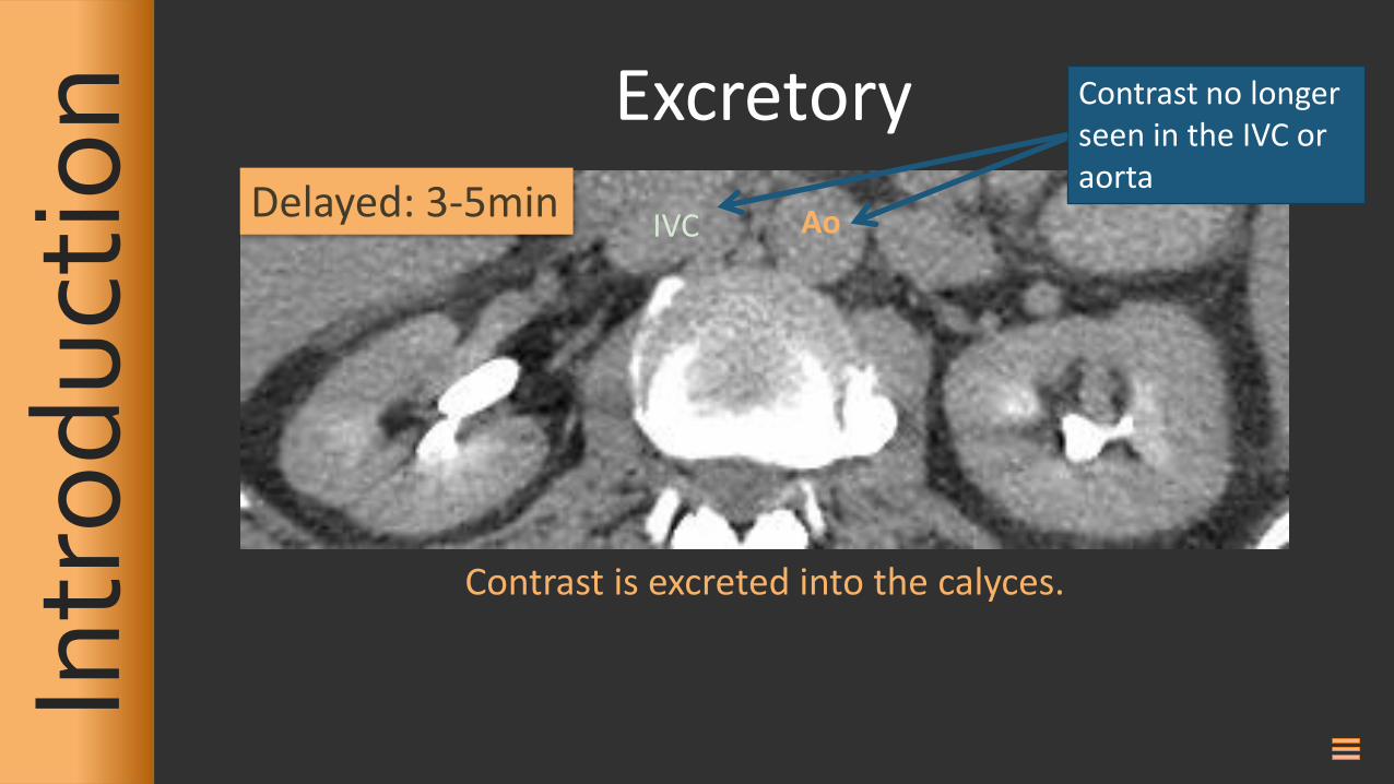
Excretory
Delayed: 3-5min
Contrast is excreted into the calyces.
Ao IVC
Contrast no longer seen in the IVC or aorta
Intr
od
uct
ion
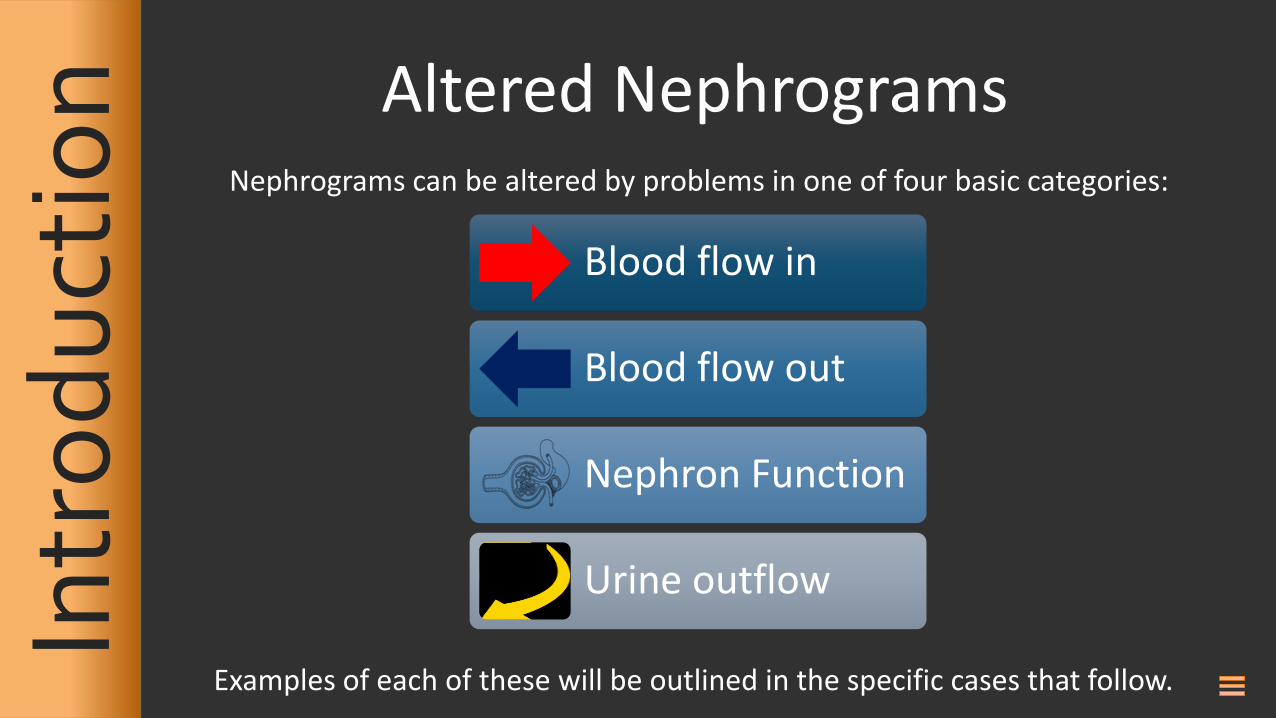
Altered Nephrograms
Nephrograms can be altered by problems in one of four basic categories:
Blood flow in
Blood flow out
Nephron Function
Urine outflow
Examples of each of these will be outlined in the specific cases that follow.

Case 2
Case 3
Case 4
Case 5
Case 6
Case 7
Case 8
Case 1 Case 9
Case 10
Case 11
Case 12
Case 13
Case 14
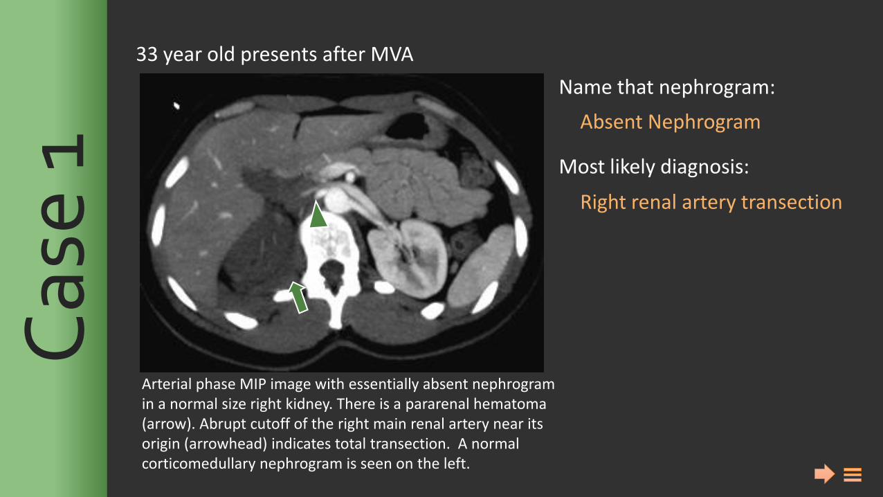
Name that nephrogram: Most likely diagnosis:
Absent Nephrogram
Right renal artery transection
Arterial phase MIP image with essentially absent nephrogram in a normal size right kidney. There is a pararenal hematoma (arrow). Abrupt cutoff of the right main renal artery near its origin (arrowhead) indicates total transection. A normal corticomedullary nephrogram is seen on the left.
33 year old presents after MVA C
ase
1
Name that nephrogram: Most likely diagnosis:
Absent Nephrogram
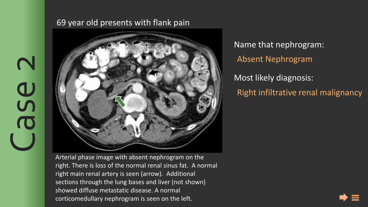
Right infiltrative renal malignancy
Arterial phase image with absent nephrogram on the right. There is loss of the normal renal sinus fat. A normal right main renal artery is seen (arrow). Additional sections through the lung bases and liver (not shown) showed diffuse metastatic disease. A normal corticomedullary nephrogram is seen on the left.
69 year old presents with flank pain C
ase
2
Name that nephrogram: Most likely diagnosis:
Absent Nephrogram
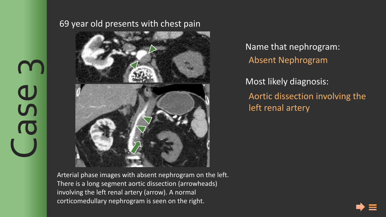
Aortic dissection involving the left renal artery
Arterial phase images with absent nephrogram on the left. There is a long segment aortic dissection (arrowheads) involving the left renal artery (arrow). A normal corticomedullary nephrogram is seen on the right.
69 year old presents with chest pain C
ase
3
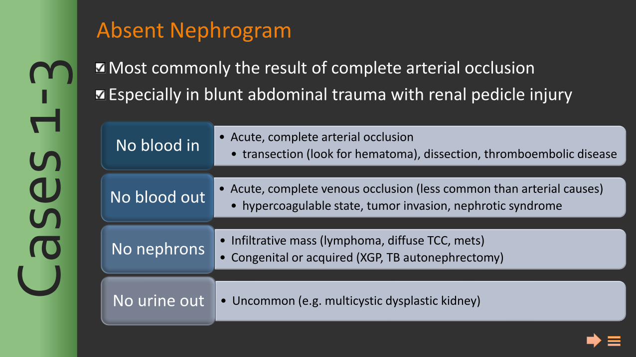
Absent Nephrogram
Most commonly the result of complete arterial occlusion
Especially in blunt abdominal trauma with renal pedicle injury
• Acute, complete arterial occlusion
• transection (look for hematoma), dissection, thromboembolic disease No blood in
• Acute, complete venous occlusion (less common than arterial causes)
• hypercoagulable state, tumor invasion, nephrotic syndrome No blood out
• Infiltrative mass (lymphoma, diffuse TCC, mets)
• Congenital or acquired (XGP, TB autonephrectomy) No nephrons
• Uncommon (e.g. multicystic dysplastic kidney) No urine out Cas
es 1
-3
Name that nephrogram: Most likely diagnosis:
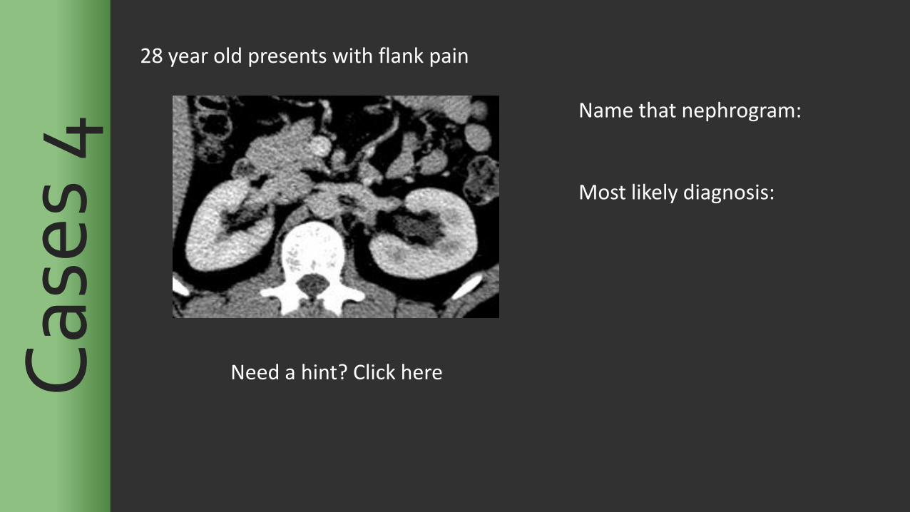
Need a hint? Click here Cas
es 4
28 year old presents with flank pain
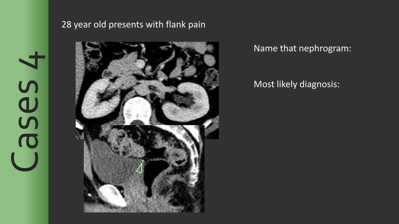
Name that nephrogram: Most likely diagnosis:
Cas
es 4
28 year old presents with flank pain
Name that nephrogram: Most likely diagnosis:
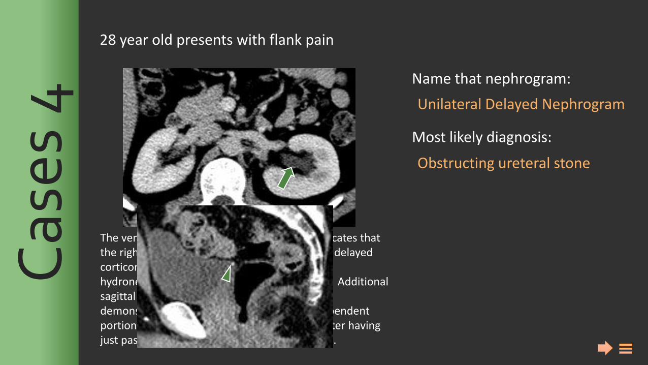
28 year old presents with flank pain
Unilateral Delayed Nephrogram
Obstructing ureteral stone
The venous phase of intravascular contrast indicates that the right nephrographic phase is normal with a delayed corticomedullary nephrogram on the left. Mild hydronephrosis is also seen on the left (arrow). Additional sagittal view of the bladder in the same patient demonstrates a punctate stone in the most dependent portion of the urinary bladder, settling there after having just passed through the left ureter (arrowhead).
Cas
es 4
Name that nephrogram: Most likely diagnosis:
Need a hint? Click here Cas
es 5
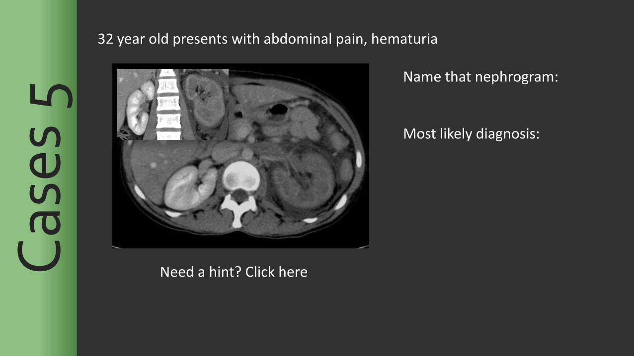
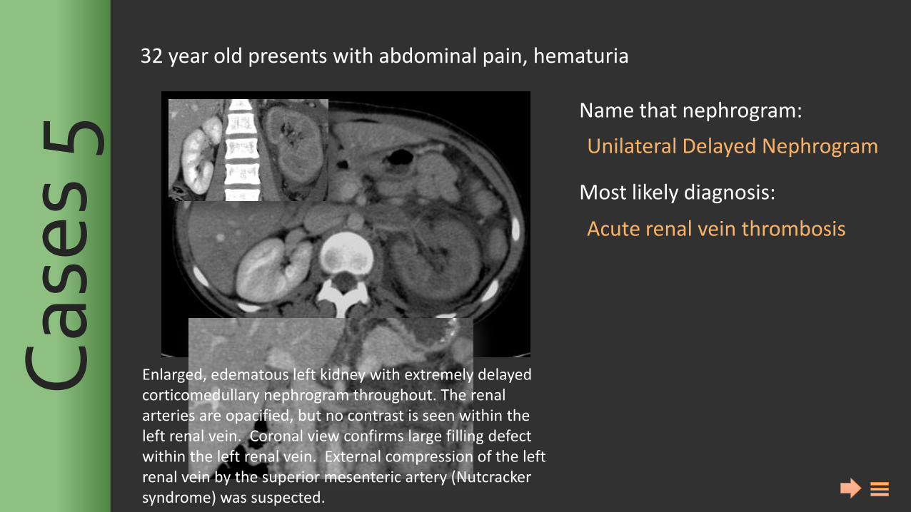
32 year old presents with abdominal pain, hematuria
Name that nephrogram: Most likely diagnosis:
Cas
es 5
32 year old presents with abdominal pain, hematuria
Name that nephrogram: Most likely diagnosis:
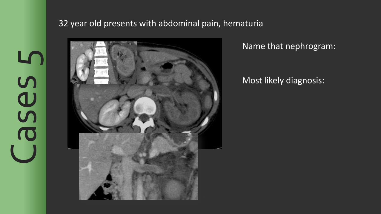
32 year old presents with abdominal pain, hematuria
Unilateral Delayed Nephrogram
Acute renal vein thrombosis
Enlarged, edematous left kidney with extremely delayed corticomedullary nephrogram throughout. The renal arteries are opacified, but no contrast is seen within the left renal vein. Coronal view confirms large filling defect within the left renal vein. External compression of the left renal vein by the superior mesenteric artery (Nutcracker syndrome) was suspected.
Cas
es 5
Name that nephrogram: Most likely diagnosis:
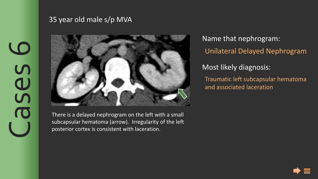
Unilateral Delayed Nephrogram
Traumatic left subcapsular hematoma and associated laceration
There is a delayed nephrogram on the left with a small subcapsular hematoma (arrow). Irregularity of the left posterior cortex is consistent with laceration.
35 year old male s/p MVA C
ases
6
Name that nephrogram: Most likely diagnosis:
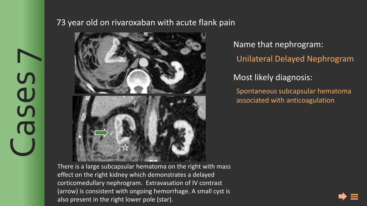
Unilateral Delayed Nephrogram
Spontaneous subcapsular hematoma associated with anticoagulation
There is a large subcapsular hematoma on the right with mass effect on the right kidney which demonstrates a delayed corticomedullary nephrogram. Extravasation of IV contrast (arrow) is consistent with ongoing hemorrhage. A small cyst is also present in the right lower pole (star).
73 year old on rivaroxaban with acute flank pain C
ases
7
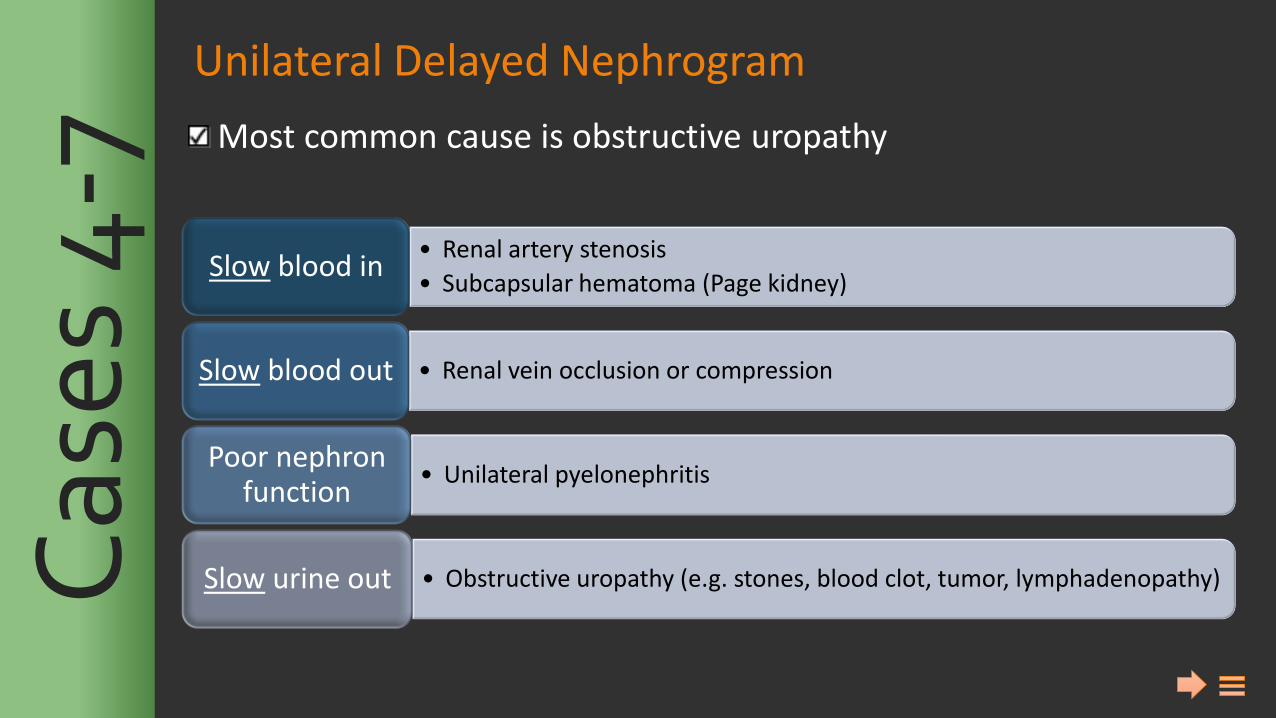
Unilateral Delayed Nephrogram
Most common cause is obstructive uropathy
• Renal artery stenosis
• Subcapsular hematoma (Page kidney) Slow blood in
• Renal vein occlusion or compression Slow blood out
• Unilateral pyelonephritis Poor nephron
function
• Obstructive uropathy (e.g. stones, blood clot, tumor, lymphadenopathy) Slow urine out Cas
es 4
-7
Name that nephrogram: Most likely diagnosis:
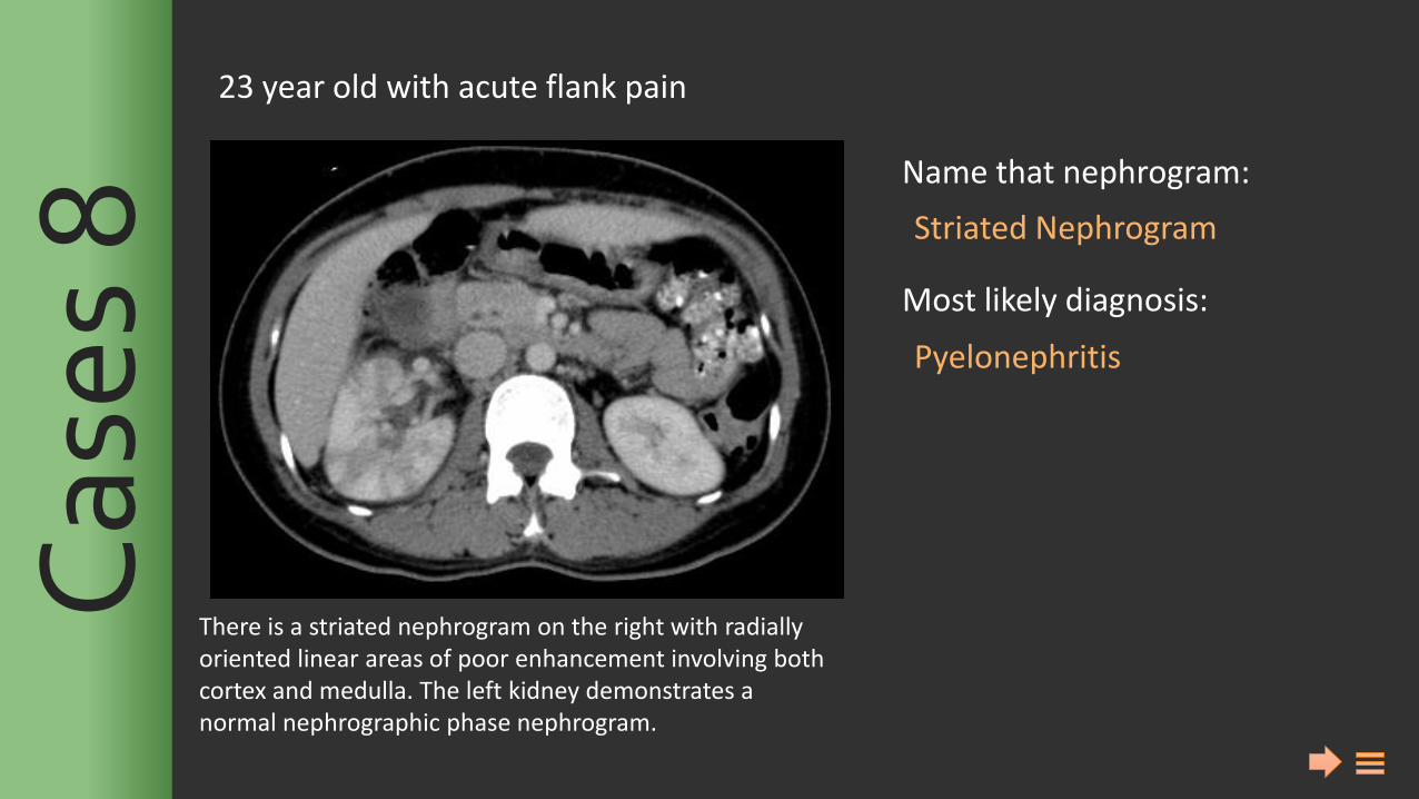
Striated Nephrogram
Pyelonephritis
There is a striated nephrogram on the right with radially oriented linear areas of poor enhancement involving both cortex and medulla. The left kidney demonstrates a normal nephrographic phase nephrogram.
23 year old with acute flank pain C
ases
8
Name that nephrogram: Most likely diagnosis:
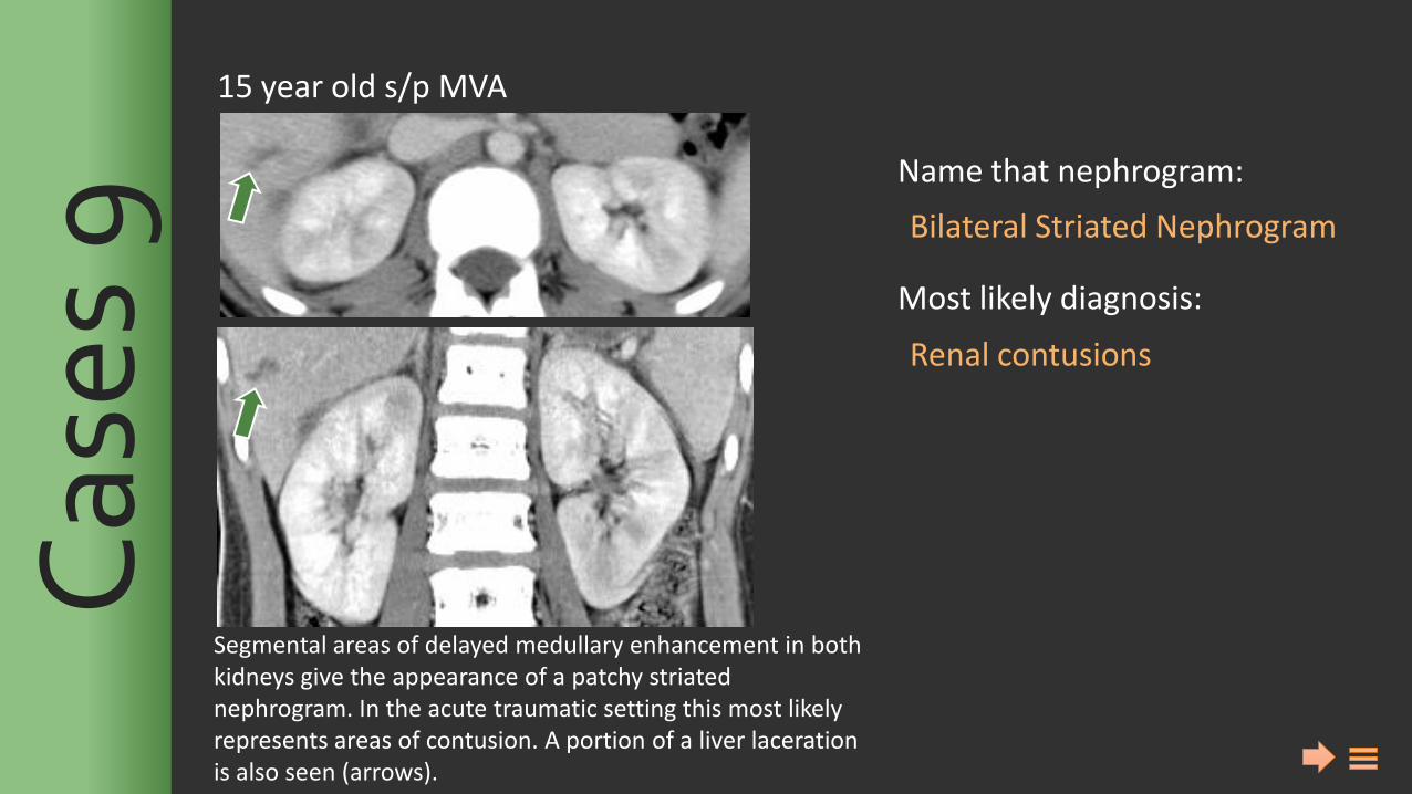
Bilateral Striated Nephrogram
Renal contusions
15 year old s/p MVA C
ases
9
Segmental areas of delayed medullary enhancement in both kidneys give the appearance of a patchy striated nephrogram. In the acute traumatic setting this most likely represents areas of contusion. A portion of a liver laceration is also seen (arrows).
Name that nephrogram: Most likely diagnosis:
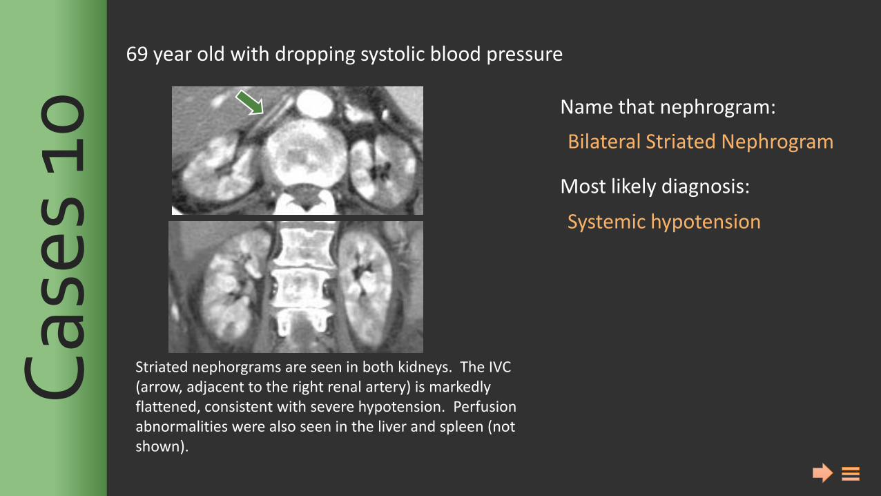
Bilateral Striated Nephrogram
Systemic hypotension
69 year old with dropping systolic blood pressure C
ases
10
Striated nephorgrams are seen in both kidneys. The IVC (arrow, adjacent to the right renal artery) is markedly flattened, consistent with severe hypotension. Perfusion abnormalities were also seen in the liver and spleen (not shown).
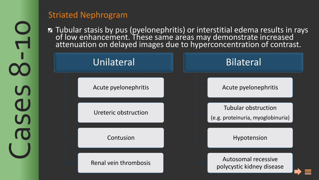
Striated Nephrogram
Tubular stasis by pus (pyelonephritis) or interstitial edema results in rays of low enhancement. These same areas may demonstrate increased attenuation on delayed images due to hyperconcentration of contrast.
Unilateral
Acute pyelonephritis
Ureteric obstruction
Contusion
Renal vein thrombosis
Bilateral
Acute pyelonephritis
Tubular obstruction
(e.g. proteinuria, myoglobinuria)
Hypotension
Autosomal recessive polycystic kidney disease
Cas
es 8
-10
Name that nephrogram: Most likely diagnosis:
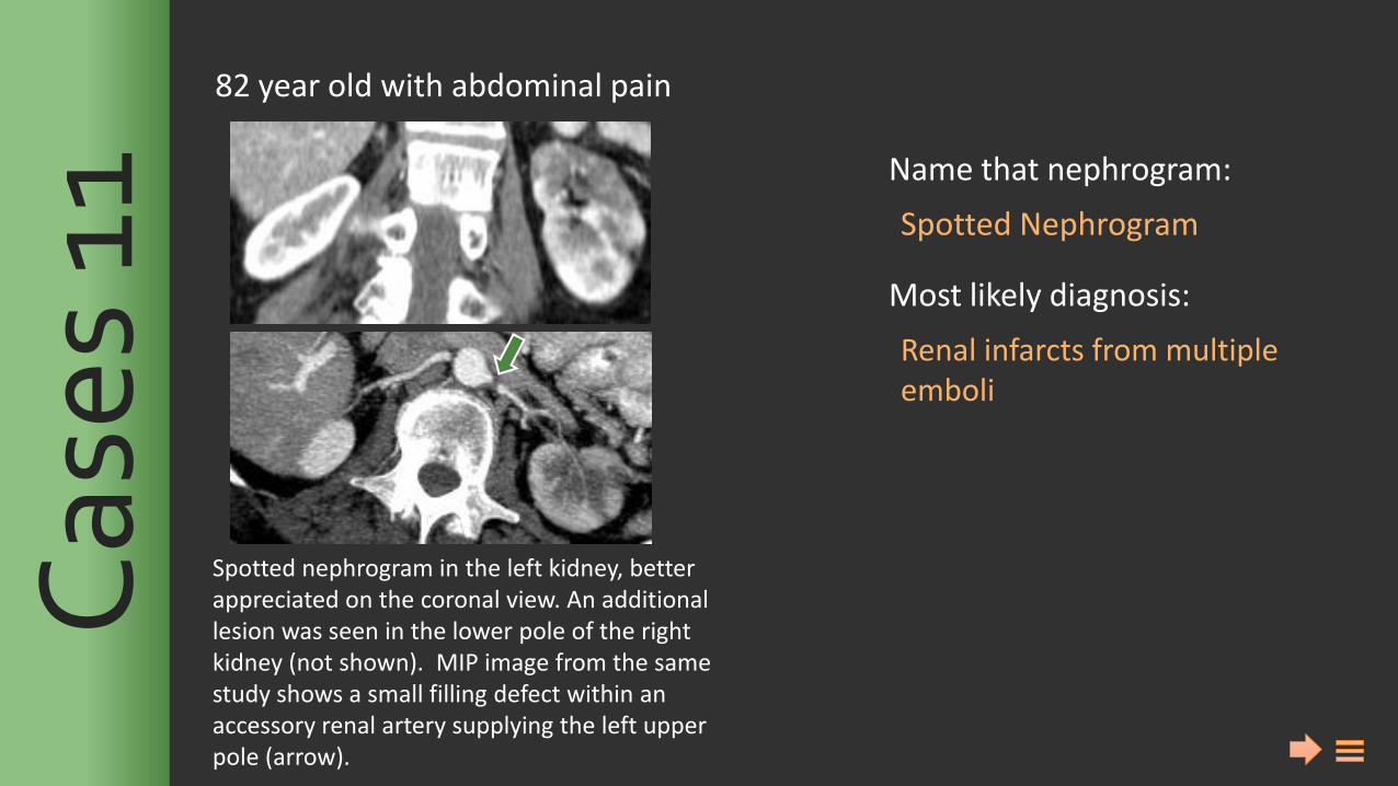
Spotted Nephrogram
Renal infarcts from multiple emboli
82 year old with abdominal pain C
ases
11
Spotted nephrogram in the left kidney, better appreciated on the coronal view. An additional lesion was seen in the lower pole of the right kidney (not shown). MIP image from the same study shows a small filling defect within an accessory renal artery supplying the left upper pole (arrow).
Name that nephrogram: Most likely diagnosis:
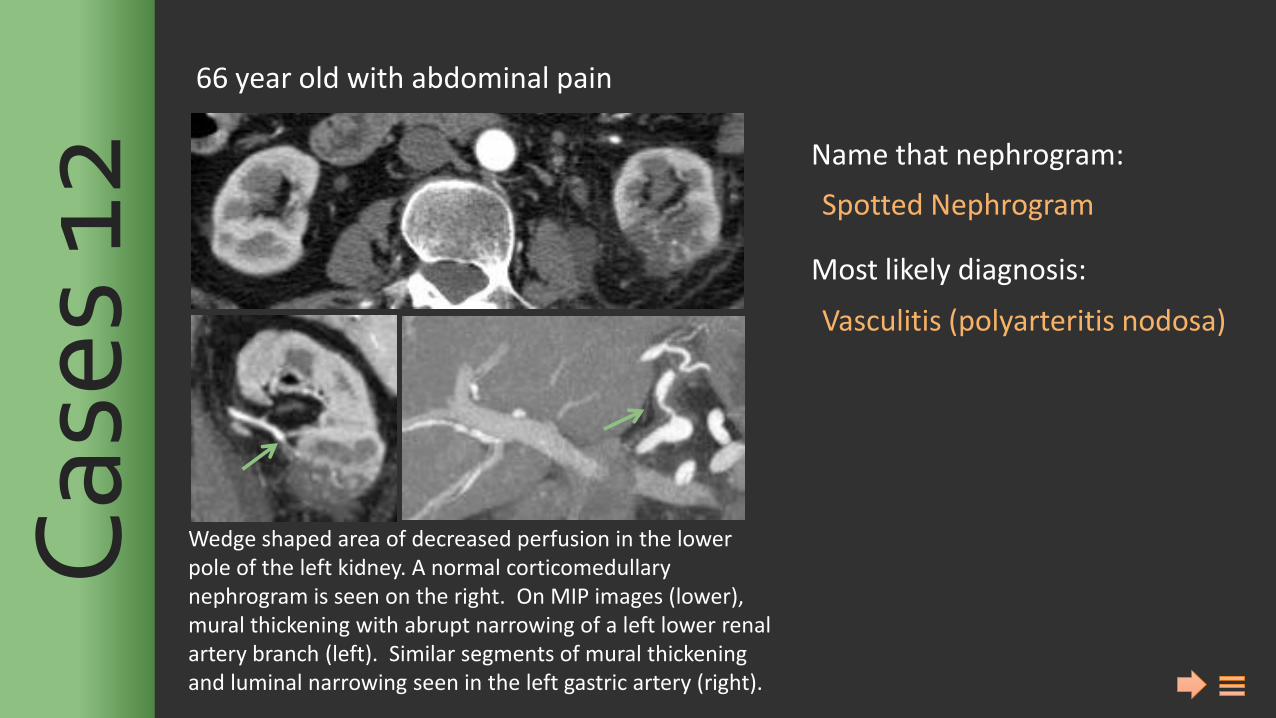
Spotted Nephrogram
Vasculitis (polyarteritis nodosa)
66 year old with abdominal pain C
ases
12
Wedge shaped area of decreased perfusion in the lower pole of the left kidney. A normal corticomedullary nephrogram is seen on the right. On MIP images (lower), mural thickening with abrupt narrowing of a left lower renal artery branch (left). Similar segments of mural thickening and luminal narrowing seen in the left gastric artery (right).
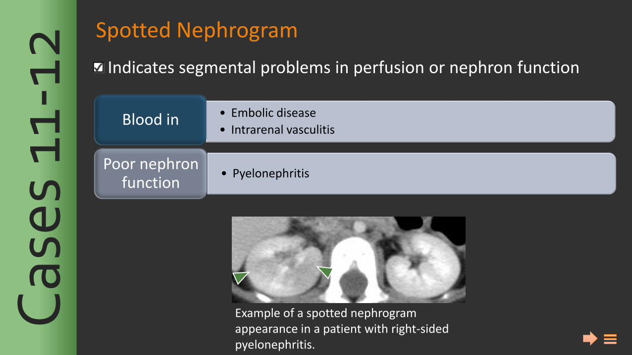
Spotted Nephrogram
Indicates segmental problems in perfusion or nephron function C
ases
11-
12
• Embolic disease
• Intrarenal vasculitis Blood in
• Pyelonephritis Poor nephron
function
Example of a spotted nephrogram appearance in a patient with right-sided pyelonephritis.
Name that nephrogram: Most likely diagnosis:
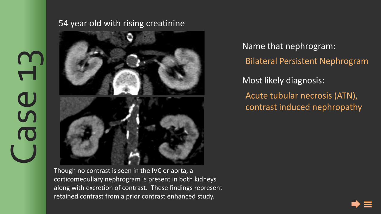
Bilateral Persistent Nephrogram
Acute tubular necrosis (ATN), contrast induced nephropathy
54 year old with rising creatinine C
ase
13
Though no contrast is seen in the IVC or aorta, a corticomedullary nephrogram is present in both kidneys along with excretion of contrast. These findings represent retained contrast from a prior contrast enhanced study.
Name that nephrogram: Most likely diagnosis:
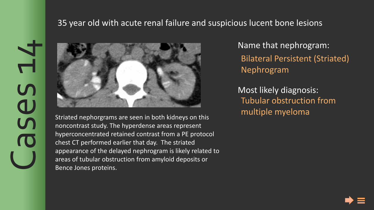
Bilateral Persistent (Striated) Nephrogram
Tubular obstruction from multiple myeloma
35 year old with acute renal failure and suspicious lucent bone lesions C
ases
14
Striated nephorgrams are seen in both kidneys on this noncontrast study. The hyperdense areas represent hyperconcentrated retained contrast from a PE protocol chest CT performed earlier that day. The striated appearance of the delayed nephrogram is likely related to areas of tubular obstruction from amyloid deposits or Bence Jones proteins.
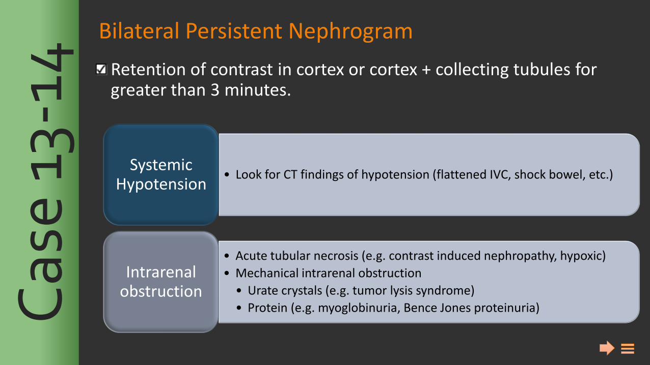
Bilateral Persistent Nephrogram
Retention of contrast in cortex or cortex + collecting tubules for greater than 3 minutes.
• Look for CT findings of hypotension (flattened IVC, shock bowel, etc.) Systemic
Hypotension
• Acute tubular necrosis (e.g. contrast induced nephropathy, hypoxic)
• Mechanical intrarenal obstruction
• Urate crystals (e.g. tumor lysis syndrome)
• Protein (e.g. myoglobinuria, Bence Jones proteinuria)
Intrarenal obstruction C
ase
13-1
4
Su
mm
ary
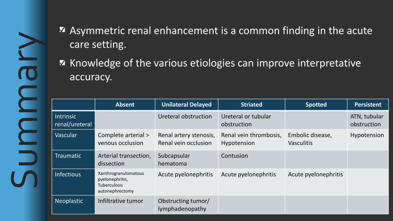
Absent Unilateral Delayed Striated Spotted Persistent
Intrinsic renal/ureteral
Ureteral obstruction Ureteral or tubular obstruction
ATN, tubular obstruction
Vascular Complete arterial > venous occlusion
Renal artery stenosis, Renal vein occlusion
Renal vein thrombosis, Hypotension
Embolic disease, Vasculitis
Hypotension
Traumatic Arterial transection, dissection
Subcapsular hematoma
Contusion
Infectious Xanthrogranulomatous pyelonephritis, Tuberculosis autonephrectomy
Acute pyelonephritis Acute pyelonephritis
Acute pyelonephritis
Neoplastic Infiltrative tumor Obstructing tumor/ lymphadenopathy
Asymmetric renal enhancement is a common finding in the acute care setting.
Knowledge of the various etiologies can improve interpretative accuracy.

Saunders, H. S., R. B. Dyer, R. Y. Shifrin, E. S. Scharling, R. E. Bechtold, and R. J. Zagoria. “The CT Nephrogram: Implications for Evaluation of Urinary Tract Disease.” Radiographics: A Review Publication of the Radiological Society of North America, Inc 15, no. 5 (September 1995): 1069–85; discussion 1086–88. doi:10.1148/radiographics.15.5.7501851. Wolin, Ely A., David S. Hartman, and J. Ryan Olson. “Nephrographic and Pyelographic Analysis of CT Urography: Principles, Patterns, and Pathophysiology.” AJR. American Journal of Roentgenology 200, no. 6 (June 2013): 1210–14. doi:10.2214/AJR.12.9691. Wolin, Ely A., David S. Hartman, and J. Ryan Olson. “Nephrographic and Pyelographic Analysis of CT Urography: Differential Diagnosis.” AJR. American Journal of Roentgenology 200, no. 6 (June 2013): 1197–1203. doi:10.2214/AJR.12.9692. R
efer
ence
s