Embed Size (px)
Citation preview
Version: 5 Last updated: 23 January 2019
Instructions for Use
For the rapid and simple separation of mitochondrial, cytosolic and nuclear fractions.
View kit datasheet: www.abcam.com/ab109719(use www.abcam.cn/ab109719 for China, or www.abcam.co.jp/ab109719 for Japan)
This product is for research use only and is not intended for diagnostic use.
ab109719 Cell Fractionation Kit - Standard

Table of Contents
1. Introduction 2
2. Protocol Summary 5
3. Kit Contents 7
4. Storage and Handling 7
5. Additional Materials Required 7
6. Protocol 8
7. Protocol Notes 12
8. Data Analysis 15
1

1. Introduction
ab109719 (MS861) provides a method and reagents for a rapid
preparation of cytosolic, mitochondrial and nuclear fractions. The kit
is based on sequential detergent-extraction of cytosolic and
mitochondrial proteins without the need for mechanical disruption of
cells, and thus fractionates cells into cytosol-containing,
mitochondria-containing and nuclei-containing fractions. These
fractions are referred throughout the protocol as cytosolic,
mitochondrial and nuclear fractions. With this kit sufficient sample
material can be prepared for subsequent Western blot analysis, or
for analysis by microplate ELISA or dipstick assay.
ab109719 is designed to allow the measurement of any proteins
which are differentially represented in the cytosol, mitochondria and
nuclei, and is particularly applicable to studies of proteins that
translocate between these three cellular compartments. As an
example, the use of the kit is described throughout this protocol in
relation to the following of cytochrome c release from the
mitochondria to the cytosol during apoptosis (see Figures 2, 3 and
4), as this is perhaps the best known mitochondrial protein
translocation event and it is an important component of apoptosis
research. Similarly, the kit was successfully used to measure the
release of Smac/Diablo from the mitochondria to the cytosol and the
translocation of Bax from cytosol to mitochondria during apoptosis as
2

well as cleavage on nuclear poly (ADP-ribose) polymerase (PARP),
see Figure 4.
ab109719 provides a rapid method to obtain cytosolic, mitochondrial
and nuclear fractions, thus avoiding time consuming and inefficient
cell disruption and differential centrifugation. The kit is based on
sequential and selective extraction of cytosolic and mitochondrial
proteins with proprietary detergents that allow sequential release of
cytosolic and mitochondrial proteins to the extracellular buffer. In the
first step, the plasma membrane is selectively permeabilized with
Detergent I. The cytosol-containing fraction is separated from the
remainder of cells containing intact mitochondria and nuclei by a
simple centrifugation step. In the second step, mitochondrial proteins
are then extracted with Detergent II and separated from the nuclei-
containing fraction by a second centrifugation step.
In control cells, mitochondrial intermembrane space proteins
including cytochrome c and Smac/Diablo remain in the mitochondrial
fraction (Figures 1, 2, 3 and 4). However, if cytochrome c and
Smac/Diablo are released from the mitochondrial intermembrane
space into cytosol, as frequently occurs in apoptosis, the cytosolic
cytochrome c and Smac/Diablo are found in the cytosolic fraction
with other cytosolic proteins (Figures 2, 3 and 4).
The three distinct fractions generated can be analyzed by Western
blot or by ELISA microplate. For Western blot analysis, ApoTrack™
Cytochrome c Apoptosis WB Cocktail is recommended
3

(ab110415/MSA12) (typical results shown below), which contains an
antibody against cytochrome c (ab110325/MSA06 Anti-cytochrome c
monoclonal antibody) plus antibodies against key mitochondrial and
cytosolic markers. For the analysis of cytochrome c by microplate
ELISA assay, Abcam’s ab110172 (MSA41) Cytochrome c Protein
Quantity Microplate Assay Kit is recommended. These methods
were verified on HeLa cells, 143B osteosarcoma cells, SHSY5Y
neuroblastoma cells, HepG2 cells and HdFN fibroblast cells treated
with Staurosporine or Jurkat TIB 152 cells incubated with
Staurosporine or anti-Fas antibody to undergo apoptosis. The
proportion of cytochrome c found in the cytosol-containing fractions
by this method correlated with the results of immunocytochemical
analysis using Abcam’s ab110417/MSA07 ApoTrack™
Cytochrome c Apoptosis for Immunocytochemistry (see Figure 5).
4

2. Protocol Summary
.
5
Grow cells in two 100 mm dishes, approximately 2.5 x10⁶ cells. Induce apoptosis in one dish by a desired method Harvest cells by centrifugation at 300 x g for 5 min
Re-suspend cells in 5 ml of 1X Buffer A Determine cell count and total cell number Centrifuge cells at 300 x g for 5 min
Re-suspend cells in buffer A to 6.6 x 10⁶ cells/ml Prepare Buffer B by 1000-fold dilution of Detergent I in Buffer A Dilute the cell suspension with equal volume of Buffer B Incubate the tube with constant mixing for 7 min at RT
Centrifuge the cell suspension at 5,000 x g for 1 min at 4°C Remove and save supernatant, save also pellet Centrifuge the supernatant at 10,000 x g for 1 min at 4°C Save the final supernatant; this is fraction C
Re-suspend and combine both sequential pellets in Buffer A to the original volume of cell suspension prior the addition of Buffer B
Prepare Buffer C by 25-fold dilution of Detergent II in Buffer A Dilute the suspension with equal volume of Buffer C Incubate the tube with constant mixing for 10 min at RT
Centrifuge the cell suspension at 5,000 x g for 1 min at 4°C Remove and save supernatant, save also pellet Centrifuge the supernatant at 10,000 x g for 1 min at 4°C Save the final supernatant; this is fraction M
Re-suspend and combine both sequential pellets in Buffer A to the original volume of suspension after the addition of Buffer C; this is fraction N

WESTERN BLOT ANALYSIS OF CYTOCHROME C RELEASE USING ANTIBODY COCKTAIL ab110415/MSA12:
6
Mix four volumes of sample with one volume of 5X SDS-PAGE Sample Buffer
Vortex thoroughly Incubate 10 minutes at 37°C Load the samples on the gel
Incubate the blocked membrane with provided antibody cocktail diluted 250-fold in PBS containing 5% non-fat milk powder for 2 hrs at RT
Calculate the cytosolic cytochrome c in both untreated and treated cells: Cyt c C (%) = 100 x Cyt c C/ (Cyt c C + Cyt c M + Cyt c N)
Calculate the treatment-specific release of cytochrome c into the cytosol:Cyt c C Released (%) = Cyt c C Treated (%) - Cyt c C
Untreated (%)

3. Kit Contents
Sufficient materials are provided for fractionation of 1 x10⁸ cells or
for preparation of 40 samples, each corresponding to one 100 mm
plate at 2.5 x 10⁶ cells/plate.
2X Buffer A: 175 mL
Detergent I: 25 µL
Detergent II: 1 mL
5X SDS Sample Buffer: 10 mL
4. Storage and Handling
Store all components at -20°C, except Detergent I stored at -80°C.
Ship on dry ice.
5. Additional Materials Required
Tube rotator for 1.5 ml tubes
Cell counting device such as hematocytometer
7
6. Protocol
Note: This protocol contains detailed steps for preparation of subcellular fractions and analysis by Western blot or microplate ELISA. Be completely familiar with the protocol and protocol notes before beginning the assay. Do not deviate from the specified protocol steps or optimal results may not be obtained.
1. Grow cells. Seed two 100 mm tissue culture plates at an
equal density and grow them to semi-confluent density.
6.2. Induce apoptosis. Incubate cells in one dish in the
presence of inducer of apoptosis at desired concentration
and for desired time. In parallel, incubate the uninduced
control cells in another dish.
6.3. Equilibrate 2X Buffer A to room temperature (RT, see
note ii) and add equal volume of water to make 1X Buffer
A.
6.4. Collect cells. For adherent cells, remove and save
medium. Detach cells by treatment with 8 ml of 0.25%
Trypsin-EDTA and add the detached cells into the saved
medium. Rinse the plate with additional 4 ml of 0.25%
Trypsin-EDTA and add the rinse to the pooled cells.
Collect cells by centrifugation for 5 min at 300 x g at RT in
a swinging bucket rotor centrifuge.
8

6.5. 1X Buffer A wash. Re-suspend cell pellets in 5 ml of 1X
Buffer A. Take a small aliquot (~25 μL) of un-induced
control cells for counting. Note the volumes of both cell
suspensions. Collect cells by centrifugation for 5 min at
300 x g at RT.
6.6. Count cells. While centrifugation proceeds, count the
uninduced cells using hematocytometer and determine the
total cell number in the control sample.
6.7. Prepare cell suspension in 1X Buffer A. Discard
supernatants and re-suspend control cell pellet in 1X
Buffer A to 6.6 x 10⁶ cells/ml. Re-suspend the induced cell
pellet in the same volume of 1X Buffer A. See Note iii.
6.8. Prepare Buffer B. To prepare Buffer B, dilute Detergent I
1000-fold in 1X Buffer A. For example, to 5 ml of 1X Buffer
A add 5 μl of Detergent I. Mix well by pipetting. Prepare
only amount needed for immediate use.
6.9. Cytosol Extraction. Transfer a volume of the cell
suspensions into a new set of tubes. Add the equal
volume of Buffer B to the cell suspensions. Mix by
pipetting. Incubate samples for 7 minutes on a rotator at
RT.
9

6.10. Centrifugation. Centrifuge samples at 5,000 x g for 1 min
at 4°C. Carefully remove all supernatants and transfer
them to a new set of tubes. Save pellets on ice. Re-
centrifuge the supernatant fractions at 10,000 x g for
1 min.
6.11. Preparation of cytosolic fractions. Transfer the resulting
supernatants containing cytosolic proteins into a new set
of tubes. These are the cytosolic fractions (C).
6.12. Prepare suspensions of the cytoplasm-depleted “cells”
(containing mitochondria and nuclei) in 1X Buffer A. Re-
suspend and combine the sequential cytoplasm-depleted
“cell” pellets, generated in Steps 10 and 11, in 1X Buffer A.
Use the same volume of 1X Buffer A as was used to
suspend the intact control cells, to 6.6 x 10⁶ cells/m in
Step 9 prior to addition of Buffer B.
6.13. Prepare Buffer C. To prepare Buffer C, dilute Detergent II
25-fold in 1X Buffer A. For example, to 4.8 m of 1X Buffer
A add 0.2 ml of Detergent II. Mix well by pipetting. Prepare
only amount needed for immediate use.
6.14. Mitochondria Extraction. Transfer a volume of the cytosol-
depleted samples generated in Step 12 into a new set of
tubes, for example 9/10 of the re-suspended sample. Add
exactly the same volume of Buffer C to the suspensions.
10
Mix by pipetting. Incubate samples for 10 minutes on a
rotator at RT.
6.15. Centrifugation. Centrifuge samples at 5,000 x g for 1 min
at 4°C. Carefully remove all supernatants and transfer
them to a new set of tubes. Save pellets on ice. Re-
centrifuge the supernatant fractions at 10,000 x g for
1 min.
6.16. Preparation of mitochondrial fraction. Transfer the
resulting supernatants containing mitochondrial proteins
into a new set of tubes. These are the mitochondrial
fractions (M).
6.17. Preparation of nuclear fraction. Re-suspend and combine
the sequential cell pellets, generated in Steps 15 and 16,
in 1X Buffer A to the original volume of suspensions after
the addition of Buffer C in Step 14. These fractions contain
re-suspended cytosol- and mitochondria-depleted
remainder of cells containing nuclei and thus represent the
nuclear fractions (N).
6.18. Analysis by ELISA. For analysis of fractions by Abcam’s
Cytochrome c Protein Quantity Microplate Assay Kit
(ab110172/ MSA41), follow the provided protocol.
11

Note: Analysis by SDS-PAGE/Western blotting. Mix four
volumes of sample with one volume of 5X SDS-PAGE Sample
Buffer. The samples prepared from nuclear fractions (N) may
become very viscous due to the presence of DNA. To avoid
pipetting errors it is important to shear the DNA. This is best
done using a probe sonicator; alternatively the DNA may be
sheered by very vigorous vortexing for about two minutes.
Incubate the samples containing SDS PAGE Sample Buffer for
10 min in 60°C water bath and vortex again briefly. Load
samples of equal volumes of fractions C, M and N side by side
onto gel immediately.
6.19. Western Blotting. Proceed with Western Blot analysis.
7. Protocol Notes
i. Cell collection. Since cell detachment during apoptosis is a
common phenomenon, it is compulsory to collect, in the
case of adherent cells, any cells floating in the medium in
addition to the cells attached to the dish see Step 4.
ii. 2X Buffer A thawing. After 2X Buffer A is thawed or brought
to 1X with water, the formation of white precipitate is normal.
To dissolve the precipitate, incubate the samples 10 min in
hot water bath with occasional inversion.
12

iii. Detergent I permeabilization. The appropriate
permeabilization conditions depend on ratio of Detergent I to
the total cellular mass, see Data Analysis section. Since
cells vary in their size, the recommended cell concentration
(3.3 x 10⁶ cells/ml) during the treatment with Detergent I was
determined to be optimal for 143B osteosarcoma cells, HeLa
cells, HepG2 cells, HdFN cells and SHSY5Y cells. To keep
the ratio of detergent to total cellular mass constant, small
cells, like Jurkat cells, need to be re-suspended to 20 x 106
cells/ml at Step 7, to obtain 10 x106 cells/ml during the
treatment with Detergent I. The same rules apply to
extraction of mitochondrial proteins by Detergent II.
iv. For other cell types, we recommend to initially optimize the
ratio of Detergent I to the total cellular protein. Prepare a
two-fold dilution set of cell suspensions in 1X Buffer A. To
each sample add equal volume of diluted detergent as in
Step 9. Then follow the steps in the protocol. Determine the
sample with the lowest ratio of detergent to cell number in
which GAPDH signal is present in C fraction and absent in
the corresponding M fraction. Use this ratio to analyze
translocation of proteins between mitochondria and cytosol
in a drug-treated sample of this particular cell line.
13

v. The cytosolic, mitochondrial and nuclear fractions prepared,
respectively, in Steps 11, 16 and 17 may be flash-frozen and
stored at -80°C.
vi. Protein assay (optional). Save a small aliquot of cell
suspension from Step 5 for subsequent determination of
total protein in the whole cell suspension. Subsequently,
when fractions are analyzed, load fractions derived from
untreated and treated cells in amounts proportional,
respectively, to the equal amount of protein in the untreated
and treated whole cell suspension.
vii. If desired, mock-Detergent I treated samples can be
prepared by dilution of cell suspension prepared in Step 7
with equal volume of the 1X Buffer A. Similarly, mock-
Detergent II treated samples can be prepared by dilution of
suspension of cytosol-depleted remainder of cells prepared
in Step 12 with equal volume of the 1X Buffer A.
viii. If desired, 1X Buffer A can be supplemented with protease
inhibitors, such as Protease Inhibitor Cocktail to minimize
nonspecific proteolysis during the fractionation.
14
8. Data Analysis
1. Control of fractionation. The complete permeabilization of
the plasma membrane by Detergent I and thus release of
cytosolic proteins from the cells, as well as complete
extraction of mitochondrial proteins by Detergent II and thus
separation of mitochondrial and nuclear compartments are
prerequisite for assaying redistribution of cytochrome c, and
others intermembrane-space localized pro-apoptotic
proteins, from mitochondrial intermembrane space into
cytosol or nucleus. The ApoTrack™ Cytochrome c
Apoptosis WB Antibody Cocktail (ab110415/ MSA12) allows
monitoring, in addition to cytochrome c, of glyceraldehyde-3-
phosphate dehydrogenase (GAPDH) and pyruvate
dehydrogenase E1 α,(PDH E1 α) a mitochondrial matrix
protein of 44 kDa, to verify internally the permeabilization
process. ab109719 is optimized to deliver complete
Detergent I-driven permeabilization of HeLa, 143B, HepG2,
SHSY5Y, HepG2, HdFN and Jurkat cells. When these cells
are used, the great majority of GAPDH, a cytosolic protein of
about 38 kDa, is present in the C fraction, while little or no
signal is present in the M fraction, indicating sufficient
permeabilization by Detergent I to release cytosolic proteins
out of the cells. In the untreated control cells, the great
majority of cytochrome c, an intermembrane space protein of
15

~13 kDa, is present in the M fraction indicating intactness of
mitochondrial outer membrane towards the Detergent I. In
cells induced to undergo apoptosis, while cytochrome c
redistributes from fraction M to fraction C, the great majority
of PDH E1 α remains in the M fraction, indicating the
intactness of the mitochondrial inner membrane. ab109719
is also optimized to deliver complete Detergent II-driven
extraction of mitochondrial proteins, while preserving
majority of nuclear proteins in the detergent-resistant nuclear
fraction. Thus in control HeLa or HepG2 cells the great
majority of cytochrome c and PDH E1 α is present in the M
fraction while little or no signal of these proteins is present in
the N fraction. At the same time, the great majority of
nuclear markers PARP and transcriptional factor SP1 are
found in the nuclear fraction while little or no signal of these
proteins is present in the C and M fractions.
2. General mitochondrial marker. The kit allows comparison
and normalization of the amounts of mitochondria among
different cell types or treatments of cells by assaying for the
mitochondrial inner membrane protein, Complex V α (~55
kDa).
3. Determination of the distribution of a protein between
cytosolic, mitochondrial and nuclear fractions. The
distribution of a protein between C, M and N fractions is
calculated as percentage of the protein present in a
16

fraction out of the sum of the protein present in C, M and N
fractions. For example, the determination of cytosolic
cytochrome c is indicated by the formula below. Cytochrome c fraction C (%) = 100 x cytochrome c fraction C/
(cytochrome c fraction C + cytochrome c fraction M + cytochrome c
fraction N)
If a drug or conditions change the distribution of a protein,
the protein distribution before and after the treatment can be
compared and protein translocation specific to the treatment
can be calculated. For example, the release of cytochrome c
caused by a drug treatment is indicated by the formula
below.
Released Cytochrome c fraction C (%) = Cytochrome c fraction C
of treated cell (%) - Cytochrome c fraction C of untreated cells (%)
17

Figure 1. Cytosolic (C), mitochondrial (M) and nuclear (N) fractions of HepG2 cells were prepared as described in the Protocol. Fractions were analyzed by Western blotting using ab110415 ApoTrack™ Cytochrome c Apoptosis WB Antibody Cocktail (MSA12) containing antibodies against mitochondrial matrix (pyruvate dehydrogenase subunit E1α, PDH E1α), mitochondrial inner membrane (F1-ATPase α), mitochondrial intermembrane space (cytochrome c) and cytosolic (glyceraldehyde-3-phosphate dehydrogenase, GAPDH) markers as well as with antibodies against additional mitochondrial matrix (Hsp70) and nuclear (poly (ADP-ribose) polymerase, PARP and SP1) markers, followed by appropriate HRP-conjugated goat secondary antibodies and ECL detection. Representative blots as well as the quantitative analysis, as described in Data Analysis, are shown.
18

Figure 2. HeLa cells were treated for 4 hrs with 1 μM Staurosporine (STS) or were left untreated (CONTROL). The cytosolic fraction (S) and mitochondria-containing reminder of the cells (P) were prepared by Detergent I extraction (“+”, 1X Buffer A containing Detergent I, as described in the Protocol), or by mock-extraction (“-“, 1X Buffer A without Detergent I). The samples derived from 12 μg of whole cells protein were analyzed by Western blotting ab110415 ApoTrack™ Cytochrome c Apoptosis WB Antibody Cocktail (MSA12), an alkaline phosphatase-conjugated goat anti-mouse secondary antibody, and AP Conjugate Substrate Kit. A representative blot as well as the quantitative analysis, as described in Data Analysis, is shown.
19
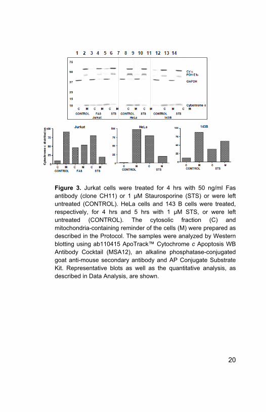
Figure 3. Jurkat cells were treated for 4 hrs with 50 ng/ml Fas antibody (clone CH11) or 1 μM Staurosporine (STS) or were left untreated (CONTROL). HeLa cells and 143 B cells were treated, respectively, for 4 hrs and 5 hrs with 1 μM STS, or were left untreated (CONTROL). The cytosolic fraction (C) and mitochondria-containing reminder of the cells (M) were prepared as described in the Protocol. The samples were analyzed by Western blotting using ab110415 ApoTrack™ Cytochrome c Apoptosis WB Antibody Cocktail (MSA12), an alkaline phosphatase-conjugated goat anti-mouse secondary antibody and AP Conjugate Substrate Kit. Representative blots as well as the quantitative analysis, as described in Data Analysis, are shown.
20

Figure 4. HeLa cells were treated with 0, 125 or 250 nM Staurosporine. Cytosolic (C), mitochondrial (M) and nuclear (N) fractions were prepared as described in the Protocol. Fractions were analyzed by Western blotting using antibodies against Bax cytochrome c, Smac, Hsp70 (ab2799) and PARP (following with appropriate HRP-conjugated goat secondary antibodies and
21

ECL detection. Quantitative analyses of representative blots, as described in Data Anaylsis, are shown.
Figure 5. Jurkat cells were treated for 4 hrs with 1 μM Fas antibody (clone CH11) or 1 μM Staurosporine (STS) or were left untreated (CONTROL) and processed for immunocytochemistry using Abcam’s ApoTrack™ Cytochrome c Apoptosis Kit (ab110417/MSA07) for Immunocytochemistry. Overlays of cytochrome c (in green) and Complex V-α (in red) staining are shown in left panels, cytochrome c only staining in middle panels and overlays of Complex V-α and DAPI (in blue) staining in right panels.
22

UK, EU and ROWEmail: [email protected]: +44 (0)1223 696000www.abcam.com
US, Canada and Latin AmericaEmail: [email protected]: 888-77-ABCAM (22226)www.abcam.com
China and Asia Pacific Email: [email protected]: 400 921 0189 / +86 21 2070 0500www.abcam.cn
JapanEmail: [email protected]: +81-(0)3-6231-0940www.abcam.co.jp
23
Copyright © 2019 Abcam, All Rights Reserved. The Abcam logo is a registered trademark.
All information / detail is correct at time of going to print.


![Untersuchungen zum Wirkmechanismus von 6-Amino-11,12 ... · PARP Poly [ADP-ribose] polymerase PBGD Porphobilinogen deaminase PBS Phosphate buffered saline PCR Polymerase chain reaction](https://img.pdfslide.us/doc/110x75/5d5cbcc088c9939b368b7c27/untersuchungen-zum-wirkmechanismus-von-6-amino-1112-parp-poly-adp-ribose.jpg)