Embed Size (px)
Citation preview
King’s Research Portal
DOI:10.1007/s00784-016-1739-x
Document VersionPublisher's PDF, also known as Version of record
Link to publication record in King's Research Portal
Citation for published version (APA):Niazi, S. A., Al Kharusi, H. S., Patel, S., Bruce, K., Beighton, D., Foschi, F., & Mannocci, F. (2016). Isolation ofPropionibacterium acnes among the microbiota of primary endodontic infections with and without intraoralcommunication. CLINICAL ORAL INVESTIGATIONS. DOI: 10.1007/s00784-016-1739-x
Citing this paperPlease note that where the full-text provided on King's Research Portal is the Author Accepted Manuscript or Post-Print version this maydiffer from the final Published version. If citing, it is advised that you check and use the publisher's definitive version for pagination,volume/issue, and date of publication details. And where the final published version is provided on the Research Portal, if citing you areagain advised to check the publisher's website for any subsequent corrections.
General rightsCopyright and moral rights for the publications made accessible in the Research Portal are retained by the authors and/or other copyrightowners and it is a condition of accessing publications that users recognize and abide by the legal requirements associated with these rights.
•Users may download and print one copy of any publication from the Research Portal for the purpose of private study or research.•You may not further distribute the material or use it for any profit-making activity or commercial gain•You may freely distribute the URL identifying the publication in the Research Portal
Take down policyIf you believe that this document breaches copyright please contact [email protected] providing details, and we will remove access tothe work immediately and investigate your claim.
Download date: 30. May. 2018
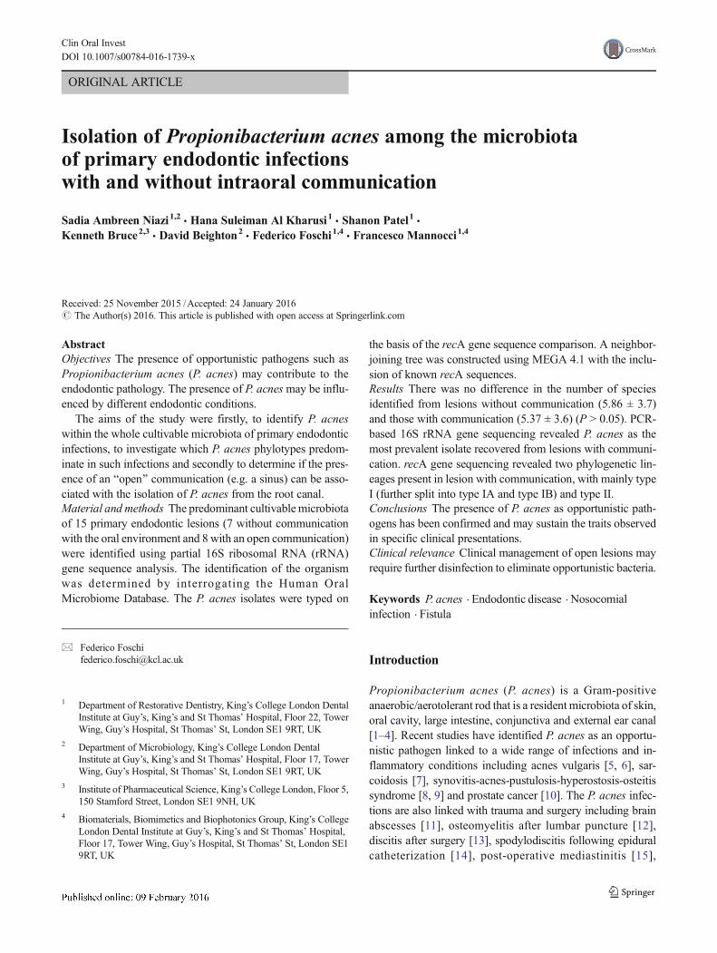
ORIGINAL ARTICLE
Isolation of Propionibacterium acnes among the microbiotaof primary endodontic infectionswith and without intraoral communication
Sadia Ambreen Niazi1,2 & Hana Suleiman Al Kharusi1 & Shanon Patel1 &
Kenneth Bruce2,3 & David Beighton2& Federico Foschi1,4 & Francesco Mannocci1,4
Received: 25 November 2015 /Accepted: 24 January 2016# The Author(s) 2016. This article is published with open access at Springerlink.com
AbstractObjectives The presence of opportunistic pathogens such asPropionibacterium acnes (P. acnes) may contribute to theendodontic pathology. The presence of P. acnesmay be influ-enced by different endodontic conditions.
The aims of the study were firstly, to identify P. acneswithin the whole cultivable microbiota of primary endodonticinfections, to investigate which P. acnes phylotypes predom-inate in such infections and secondly to determine if the pres-ence of an Bopen^ communication (e.g. a sinus) can be asso-ciated with the isolation of P. acnes from the root canal.Material and methods The predominant cultivablemicrobiotaof 15 primary endodontic lesions (7 without communicationwith the oral environment and 8 with an open communication)were identified using partial 16S ribosomal RNA (rRNA)gene sequence analysis. The identification of the organismwas determined by interrogating the Human OralMicrobiome Database. The P. acnes isolates were typed on
the basis of the recA gene sequence comparison. A neighbor-joining tree was constructed using MEGA 4.1 with the inclu-sion of known recA sequences.Results There was no difference in the number of speciesidentified from lesions without communication (5.86 ± 3.7)and those with communication (5.37 ± 3.6) (P > 0.05). PCR-based 16S rRNA gene sequencing revealed P. acnes as themost prevalent isolate recovered from lesions with communi-cation. recA gene sequencing revealed two phylogenetic lin-eages present in lesion with communication, with mainly typeI (further split into type IA and type IB) and type II.Conclusions The presence of P. acnes as opportunistic path-ogens has been confirmed and may sustain the traits observedin specific clinical presentations.Clinical relevance Clinical management of open lesions mayrequire further disinfection to eliminate opportunistic bacteria.
Keywords P. acnes . Endodontic disease . Nosocomialinfection . Fistula
Introduction
Propionibacterium acnes (P. acnes) is a Gram-positiveanaerobic/aerotolerant rod that is a resident microbiota of skin,oral cavity, large intestine, conjunctiva and external ear canal[1–4]. Recent studies have identified P. acnes as an opportu-nistic pathogen linked to a wide range of infections and in-flammatory conditions including acnes vulgaris [5, 6], sar-coidosis [7], synovitis-acnes-pustulosis-hyperostosis-osteitissyndrome [8, 9] and prostate cancer [10]. The P. acnes infec-tions are also linked with trauma and surgery including brainabscesses [11], osteomyelitis after lumbar puncture [12],discitis after surgery [13], spodylodiscitis following epiduralcatheterization [14], post-operative mediastinitis [15],
* Federico [email protected]
1 Department of Restorative Dentistry, King’s College London DentalInstitute at Guy’s, King’s and St Thomas’ Hospital, Floor 22, TowerWing, Guy’s Hospital, St Thomas’ St, London SE1 9RT, UK
2 Department of Microbiology, King’s College London DentalInstitute at Guy’s, King’s and St Thomas’ Hospital, Floor 17, TowerWing, Guy’s Hospital, St Thomas’ St, London SE1 9RT, UK
3 Institute of Pharmaceutical Science, King’s College London, Floor 5,150 Stamford Street, London SE1 9NH, UK
4 Biomaterials, Biomimetics and Biophotonics Group, King’s CollegeLondon Dental Institute at Guy’s, King’s and St Thomas’ Hospital,Floor 17, Tower Wing, Guy’s Hospital, St Thomas’ St, London SE19RT, UK
Clin Oral InvestDOI 10.1007/s00784-016-1739-x
endophthalmitis [16] and endocarditis [17]. Moreover, foreignbody implants are one of the predisposing factors associatedwith P. acnes infections [10, 12, 14].
P. acnes have been classified into four highly distinct evo-lutionary lineages: type IA, IB, II and III, which display dif-ferences in inflammatory properties, production of virulencedeterminants and association with various conditions [18–23].Type I is usually associated with skin colonization [18],whereas type II and III are almost exclusively associated withinfections of implanted prosthesis [19, 24] suggesting theirpossible role in the pathogenesis of the implant infections. Aprevious study concluded that P. acnes as an opportunisticpathogen was associated much more often than previouslyreported, with apical periodontitis in teeth with an existingroot canal treatment (secondary endodontic infection) [25].
Bacteria may penetrate into the root canal space via caries,trauma and also during the root canal treatment procedureitself due to poor cross infection control and/or inadequateisolation of the operatory field [26]. It has been shown thatP. acnes type II and III are associated with refractory endodon-tic infections [25]. It is possible that P. acnes may be intro-duced into the root canal at the time of the root canal obtura-tion or may infect the root canal post-treatment as a result ofbacterial leakage at the tooth-restoration interface [25, 27].
The ecology of primary endodontic infection is unique,harboring microbiota, which depend on the presence or ab-sence of a frank communication with the oral cavity. Variationin oxygen tension, availability of metabolites/substrates andreplenishment of the biofilm via continuous re-colonizationthrough Bopen^ communications with the oral cavity associ-ated with the sinus tract, full depth periodontal pocket and ahistory of trauma (resulting in root fracture, tooth avulsion orsubluxation) may all significantly alter the microbiota presentin primary endodontic infections [26].
The primary aim of the present study was to determine ifP. acnes is present within the whole cultivable microbiota ofprimary endodontic infections. The secondary aims were toinvestigate which specific recA phylotypes of P. acnes pre-dominate in such infections, and finally to determine if thepresence of an Bopen^ communication with the endodonticniche can be associated with a more frequent isolation ofP. acnes from the root canal space.
Methods
Patients selections
Patients recruited into the study received treatment in theEndodontic Department of the Dental Institute at Guys’ andSt Thomas’ Hospital, King’s College London. The projectwas approved by the local ethics committee (South LondonREC: 05/Q0705/051). The patient had verbal and written
information about the purpose of the study, and they gave theirwritten informed consent prior to their inclusion. The selectedteeth were single rooted with necrotic pulp (primary lesions).Samples were assigned according to clinical history to groupA (absence [Table 1]), or group B (presence [Table 2]) of acommunication with the oral environment. In group A, (n = 7)samples were collected from the root canal system from teethwhich had no macroscopic communication between the toothand the oral cavity (Table 1). In group B, (n = 8) samples werecollected from the root canal system from teeth that had evi-dence of communication with the oral environment eitherthrough the presence of a sinus tract, periodontal probingdepths of at least 8 mm and/or recent history of traumaresulting in subluxation, luxation and horizontal root fracturein the presence of deep periodontal pocketing (Table 2). Allpatients were healthy with no active, acute or chronic medicalconditions and had not been prescribed antibiotics during theprevious month. All significant details about the dental histo-ry, clinical and radiographic signs of the involved tooth wererecorded (Tables 1 and 2).
Samples collection
After local anesthesia, the tooth to be treated was isolated withthe rubber dam. The tooth the clamp and the surrounding damwere cleaned with 30 % (v/v) hydrogen peroxide anddecontaminated with 2.5 % sodium hypochlorite followedby sodium thiosulphate. After decontamination, the isolatedtooth and surrounding dam were swabbed to check for con-tamination. The existing restoration and caries were removedwith a sterile bur, after which the access cavity was completedwith a new sterile high-speed carbide bur without water-cooling until the pulp chamber was exposed. The content ofthe root canal space was removed using a combination ofsterile K-type or Hedstrom files (Dentsply Maillefer,Baillagues, Switzerland) as well as rotary instruments(ProTaper, Dentsply Maillefer) without any irrigation. Allthe tissues removed were transferred into 1 ml PRAS medium(Oxyrase, Mansfield, OH, USA). All samples were immedi-ately transported on ice to the laboratory, after which end-odontic treatment was completed.
Microbial analysis of samples
Each sample was dispersed by vortexing with sterile glassbeads, diluted in fastidious anaerobe broth (Lab M, UK) andplated onto non-selective media; duplicate plates of fastidiousanaerobe agar (FAA) supplemented with 5 % horse blood(Lab M, UK). The FAA plates were incubated anaerobicallyfor 7 days. After incubation, colonies were counted, andpredetermined number of colonies (n = 30 per patient) wererandomly selected by using the grid to divide the plate into
Clin Oral Invest
different sections for gram-staining and molecularidentification.
The swabs taken from the prepared teeth prior to the re-moval of the restoration were plated directly onto FAA andincubated anaerobically for 7 days.
Identification of isolates
All randomly selected isolates were subcultured on FAA platesand grown anaerobically for 24 h. Bacterial genomic DNAwasextracted by boiling 100 μl of a suspension of the cultured cellsprepared in sterile distilled H2O for 10 min, followed by coolingon ice for 10 min and centrifuged at 13,000×g for 2 min [18].The supernatant containing the genomic DNA was stored at−20 °C prior to analysis. DNA amplification of a partial frag-ment of the 16S ribosomal (rRNA) gene of the isolates waspe r f o rmed us i ng un ive r s a l p r ime r s : 9F (5 ′ - 3 ′GAGTTTGATCCTGGCTCA) and 907R (5 ′ - 3 ′CGTCAATTCCTTTGAGTT) [28]. The amplification was car-ried out with the following reaction mixture (final volume,25 μl): 0.5 μl of 9F forward primers (concentration 10 pmol/μl; MWG, UK), 0.5 μl of 907R reverse primer (concentration10 pmol/μl; MWG), 23 μl of Reddymix buffer (Thermo
Scientific, UK) and 1 μl DNA extract. The thermal cyclingconditions included initial denaturation at 94 °C for 10 min,denaturation at 94 °C for 30 s, annealing at 49 °C for 30 s andextension at 72 °C for 90 s, repeated for 34 cycles and finalextension at 72 °C for 5 min. The amplified products were runon a 0.5 % agarose gel (Sigma, UK) and visualized under UVtransillumination. PCR products were cleaned for the sequencereaction with Microclean (Sigma, UK) according to the manu-facturer’s instructions.
Amplicon sequencing was performed by using an ABIPrism BigDye Terminator sequencing kit (AppliedBiosystems) with 30 cycles of denaturation at 96 °C for10 s, annealing at 50 °C for 5 s and extension at 60 °C for2 min. Sequencing reaction products were run on an ABI3730xl sequencer Applied Biosystems). All DNA sequenceswere analyzed, trimmed and aligned using BioEdit software(version 7.0.0; http://www.mbio.ncsu.edu/BioEdit/bioedit.html). The partial gene sequences were identified by aBLAST search of the NCBI database (http://0-www.ncbi.nlm.nih.gov.ilsprod.ilb.neu.edu/BLAST/), the Human OralMicrobiome database (http://www.homd.org/) or theRibosome Database Project database http://rdp.cme.msu.edu/). Phylogenetic trees were constructed by the neighbor-
Table 1 Group A samples without communication
Sample Sex Age(Year)
Coronalrestoration
Periodontalpockets (mm)
Clinical findings Tooth Tx provided
1 M 30 – 2 Necrotic discolored PA lesion UL1 RCT bleaching
2 F 24 Amalgam 1 Deep restoration, necrotic PA lesion LR5 RCT PFM crown
3 F 19 Composite 1 Old trauma Necrotic PA lesion UR2 RCT bleaching
4 M 42 – 3 Enamel infraction necrotic discolored UR1 RCT bleaching
5 F 27 IRM 2 Deep cavity failed pulp capping LL3 RCT ceramic crown
6 F 35 Composite 2 Necrotic discolored PA lesion LR2 RCT Composite core
7 M 20 Composite 1 Deep MOD filling necrotic UL4 RCT fiber post-composite core
8 M 11 - 2 Old trauma necrotic discolored PA lesion symptomatic UR1 RCT bleaching
Table 2 Group B samples with communication
Sample Sex Age(Year)
Coronalrestoration
Periodontalpockets (mm)
Clinical findings Tooth Tx provided
9 F 21 − 5+ Horizontal root fracture, deep periodontalpocket communicating with the fracture
UR1 RCT MTA Composite build-up
10 F 23 Composite 3 Necrotic abscess sinus symptomatic UR2 RCT bleaching composite build-up
11 M 15 − 3 Trauma avulsed surgically repositioned and splinted UR1 RCT bleaching composite build-up
12 M 32 Composite 4 Long standing abscess, sinus for 5 yrs.Symptomatic h/o flare-up mid Tx
UR1 RCT bleaching composite core
13 F 29 − 3 Trauma Lateral luxation Repositioned and splinted LR2 RCT composite core
14 M 17 − 3 Trauma sub-luxation Repositioned and splinted UL4 RCT Composite core
15 M 19 − 4 Trauma avulsed surgically repositioned and splinted LL3 RCT bleaching composite build-up
Clin Oral Invest
joining (NJ) method, based on 16S rRNA gene sequence com-parisons, using theMEGA (version 4.1) program (http://www.megasoftware.net/).
Phylotyping of P. acnes isolates
Endodontic P. acnes isolates identified by partial 16S rRNAgene sequencing were typed by partial recA gene sequencing[18]. All selected P. acnes isolates were subcultured on FAAand grown for 24 h.
The P. acnes recA gene was amplified using primer PAR-1(positions −96 to −75; 5′-AGCTCGGTGGGGTTCTCTCATC-3′) and primer PAR-2 (positions +1105 to +1083; 5′-GCTTCCTCATACCACTGGTCATC-3′), which generated a1201-bp amplicon [18]. The reaction mix (final volume, 25 μl)
comprised of 0.5 μl of PAR-1 (concentration 10 pmol/μl;Sigma), 0.5 μl of PAR-2 (concentration 10 pmol/μl; Sigma),23 μl of Reddymix (Thermo Scientific) and 1 μl DNA extract.
The thermal cycling conditions included initial denatur-ation at 95 °C for 3 min, denaturation at 95 °C for 1 min,annealing at 55 °C for 30 s, and extension at 72 °C for 90 s,repeated for 35 cycles and a final extension at 72 °C for10 min. Amplified products were run on a 0.5 % agarose geland visualized under UV transillumination.
Sequencing was performed as described above. TheP. acnes recA sequences were compared with GenBank se-quences AY642055 (type IA), EU687255 (type IB),AY642061 (type II) and DQ672252 (type III). NJ trees wereconstructed using the Jukes-Cantor method with MEGA (ver-sion 4.1) software (www.megasoftware.net/).
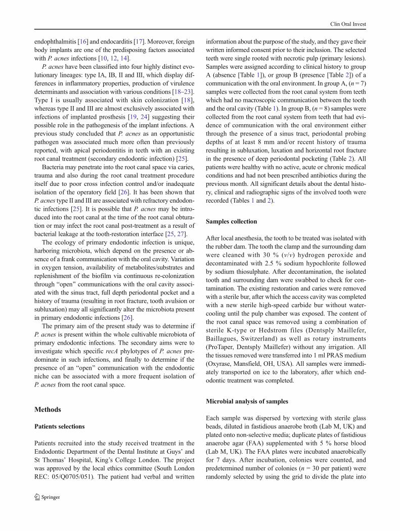
Fig. 1 Prevalence of the 33microbial taxa in 7 primaryendodontic lesions withoutcommunications with the oralenvironment
Clin Oral Invest
Statistical analysis
Data distributions were compared using χ2 tests; means werecompared using the Mann-Whitney U test in SPSSPC(Version 21. IBM, USA). Linear regression analysis was usedto determine the effect of open or closed oral communicationon the presence of P. acnes.
Results
No organisms were recovered from the samples taken fromthe disinfected tooth surfaces prior to making access into theroot canal. The range of organisms cultured from the 15 pri-mary endodontic infections is shown in Tables 1 and 2. Fromthe 15 samples, a mean of 5.6 ± 3.5 taxa were detected. The
number of species identified from lesions without communi-cation (5.86 ± 3.7) was not different (P > 0.05) than the num-ber from lesions with communication (5.37 ± 3.6).
Cultivable taxa from primary endodontic infectionwithout Bopen^ communications with the oral cavity
In the 7 primary endodontic infections without communica-tion with the oral cavity, 33 cultivable bacterial taxa weredetected amongst the 132 isolates recovered (Fig. 1). Themicrobiota of these samples were dominated by the Gram-positive facultative anaerobes, which accounted for 23 of the31 identified taxa. Furthermore, three Gram-positive obligateanaerobes including Atopobium parvulum, Mogibacteriumdiversum and Olsenella uli were also identified in thesesamples. Only a minimal number of Gram-negative bacteria
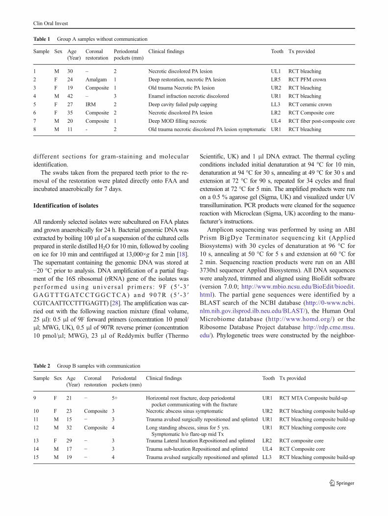
Fig. 2 Phylogenetic tree showingall 33 bacterial taxa belonging to3 phyla from 132 isolatesidentified from the 7 primaryendodontic cases withoutcommunications. The tree wasconstructed by the neighbor-joining method based on 16SrRNA gene sequencecomparisons. The scale barrepresents 0.05 substitutions pernucleotide position. The numbersat the node of the tree indicatebootstrap values for each node outof 500 bootstrap resampling
Clin Oral Invest
were identified in these primary endodontic cases, which com-prised obligate anaerobes including two Prevotella species(Prevotella melaninogenica and P. nigrescens) and three spe-cies of Veillonella (Veillonella parvula, Veillonella dispar andVeillonella atypical) (Fig. 2). No Gram-negative facultativeanaerobes were found in these samples. Actinomycesnaeslundii was the most prevalent bacterial taxa present in57 % of the cases. This was followed by Streptococcusgordonii, Streptococcus mitis bv, Streptococcus sanguinis,V. dispar and V. parvula (28.57 % of cases), and all the restof the taxa were present in 14.29 %.of the cases (Fig. 1).
Cultivable taxa from primary endodontic infectionwith Bopen^ communications with the oral cavity
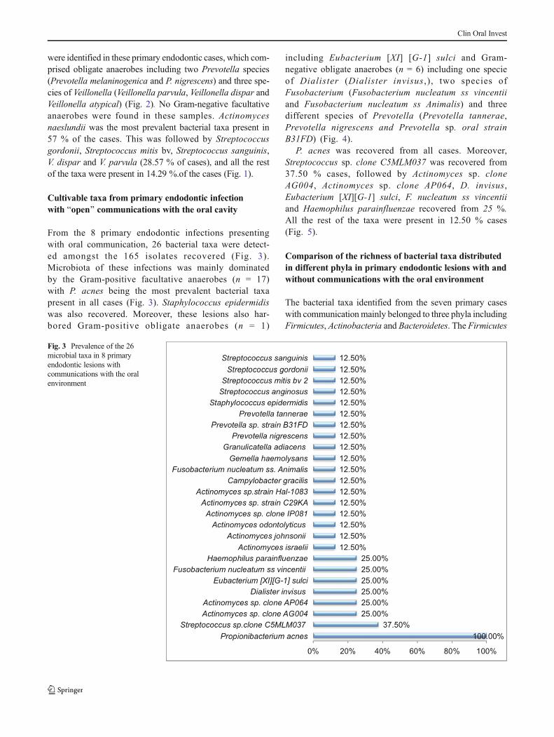
From the 8 primary endodontic infections presentingwith oral communication, 26 bacterial taxa were detect-ed amongst the 165 isolates recovered (Fig. 3).Microbiota of these infections was mainly dominatedby the Gram-positive facultative anaerobes (n = 17)with P. acnes being the most prevalent bacterial taxapresent in all cases (Fig. 3). Staphylococcus epidermidiswas also recovered. Moreover, these lesions also har-bored Gram-positive obligate anaerobes (n = 1)
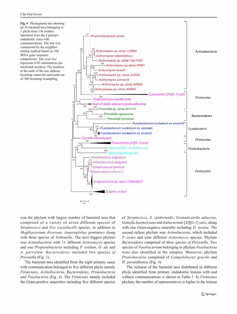
including Eubacterium [XI] [G-1] sulci and Gram-negative obligate anaerobes (n = 6) including one specieof Dialister (Dialister invisus ,), two species ofFusobacterium (Fusobacterium nucleatum ss vincentiiand Fusobacterium nucleatum ss Animalis) and threedifferent species of Prevotella (Prevotella tannerae,Prevotella nigrescens and Prevotella sp. oral strainB31FD) (Fig. 4).
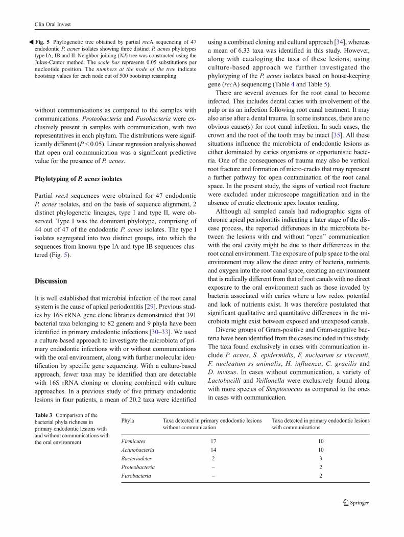
P. acnes was recovered from all cases. Moreover,Streptococcus sp. clone C5MLM037 was recovered from37.50 % cases, followed by Actinomyces sp. cloneAG004, Actinomyces sp. clone AP064, D. invisus,Eubacterium [XI][G-1] sulci, F. nucleatum ss vincentiiand Haemophilus parainfluenzae recovered from 25 %.All the rest of the taxa were present in 12.50 % cases(Fig. 5).
Comparison of the richness of bacterial taxa distributedin different phyla in primary endodontic lesions with andwithout communications with the oral environment
The bacterial taxa identified from the seven primary caseswith communicationmainly belonged to three phyla includingFirmicutes, Actinobacteria and Bacteroidetes. The Firmicutes
Fig. 3 Prevalence of the 26microbial taxa in 8 primaryendodontic lesions withcommunications with the oralenvironment
Clin Oral Invest
was the phylum with largest number of bacterial taxa thatcomprised of a variety of seven different species ofStreptococci and five Lactobacilli species, in addition toMogibacterium diversum, Anaeroglobus geminates alongwith three species of Veillonella. The next biggest phylumwas Actinobacteria with 11 different Actinomyces speciesand one Propionibacteria including P. avidum, O. uli andA. parvulum. Bacteroidetes included two species ofPrevotella (Fig. 2).
The bacterial taxa identified from the eight primary caseswith communication belonged to five different phyla namelyFirmicutes, Actinobacteria, Bacteroidetes, Proteobacteriaand Fusobacteria (Fig. 4). The Firmicutes mainly includedthe Gram-positive anaerobes including five different species
of Streptococci, S. epidermidis, Granulicatella adiacens,Gemella haemolysans and Eubacterium [XI][G-1] sulci, alongwith one Gram-negative anaerobe including D. invisus. Thesecond richest phylum was Actinobacteria, which includedP. acnes and nine different Actinomyces species. PhylumBacteroidetes comprised of three species of Prevotella. Twospecies of Fusobacterium belonging to phylum Fusobacteriawere also identified in the samples. Moreover, phylumProteobacteria comprised of Campylobacter gracilis andH. parainfluenza (Fig. 4).
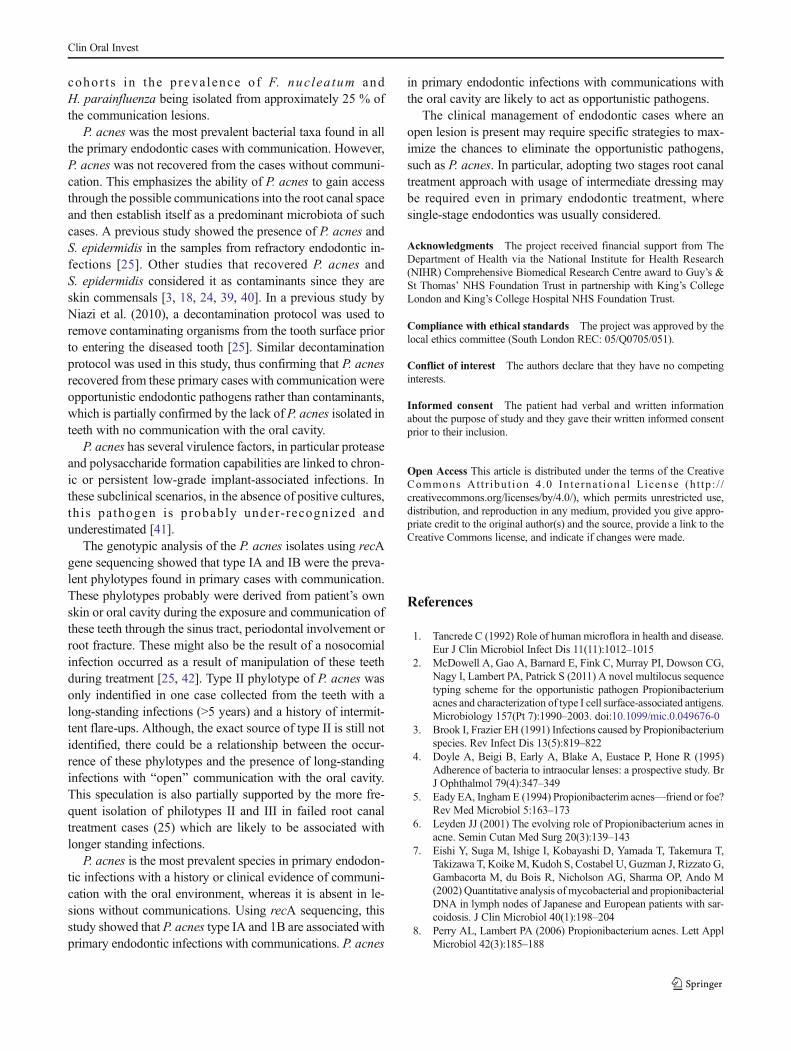
The richness of the bacterial taxa distributed in differentphyla identified from primary endodontic lesions with andwithout communications is shown in Table 3. In Firmicutesphylum, the number of representatives is higher in the lesions
Fig. 4 Phylogenetic tree showingall 26 bacterial taxa belonging to5 phyla from 136 isolatesidentified from the 8 primaryendodontic cases withcommunications. The tree wasconstructed by the neighbor-joining method based on 16SrRNA gene sequencecomparisons. The scale barrepresents 0.05 substitutions pernucleotide position. The numbersat the node of the tree indicatebootstrap values for each node outof 500 bootstrap resampling.
Clin Oral Invest
Clin Oral Invest
without communications as compared to the samples withcommunications. Proteobacteria and Fusobacteria were ex-clusively present in samples with communication, with tworepresentatives in each phylum. The distributions were signif-icantly different (P < 0.05). Linear regression analysis showedthat open oral communication was a significant predictivevalue for the presence of P. acnes.
Phylotyping of P. acnes isolates
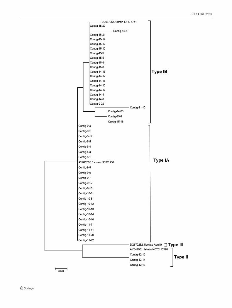
Partial recA sequences were obtained for 47 endodonticP. acnes isolates, and on the basis of sequence alignment, 2distinct phylogenetic lineages, type I and type II, were ob-served. Type I was the dominant phylotype, comprising of44 out of 47 of the endodontic P. acnes isolates. The type Iisolates segregated into two distinct groups, into which thesequences from known type IA and type IB sequences clus-tered (Fig. 5).
Discussion
It is well established that microbial infection of the root canalsystem is the cause of apical periodontitis [29]. Previous stud-ies by 16S rRNA gene clone libraries demonstrated that 391bacterial taxa belonging to 82 genera and 9 phyla have beenidentified in primary endodontic infections [30–33]. We useda culture-based approach to investigate the microbiota of pri-mary endodontic infections with or without communicationswith the oral environment, along with further molecular iden-tification by specific gene sequencing. With a culture-basedapproach, fewer taxa may be identified than are detectablewith 16S rRNA cloning or cloning combined with cultureapproaches. In a previous study of five primary endodonticlesions in four patients, a mean of 20.2 taxa were identified
using a combined cloning and cultural approach [34], whereasa mean of 6.33 taxa was identified in this study. However,along with cataloging the taxa of these lesions, usingculture-based approach we further investigated thephylotyping of the P. acnes isolates based on house-keepinggene (recA) sequencing (Table 4 and Table 5).
There are several avenues for the root canal to becomeinfected. This includes dental caries with involvement of thepulp or as an infection following root canal treatment. It mayalso arise after a dental trauma. In some instances, there are noobvious cause(s) for root canal infection. In such cases, thecrown and the root of the tooth may be intact [35]. All thesesituations influence the microbiota of endodontic lesions aseither dominated by caries organisms or opportunistic bacte-ria. One of the consequences of trauma may also be verticalroot fracture and formation of micro-cracks that may representa further pathway for open contamination of the root canalspace. In the present study, the signs of vertical root fracturewere excluded under microscope magnification and in theabsence of erratic electronic apex locator reading.
Although all sampled canals had radiographic signs ofchronic apical periodontitis indicating a later stage of the dis-ease process, the reported differences in the microbiota be-tween the lesions with and without Bopen^ communicationwith the oral cavity might be due to their differences in theroot canal environment. The exposure of pulp space to the oralenvironment may allow the direct entry of bacteria, nutrientsand oxygen into the root canal space, creating an environmentthat is radically different from that of root canals with no directexposure to the oral environment such as those invaded bybacteria associated with caries where a low redox potentialand lack of nutrients exist. It was therefore postulated thatsignificant qualitative and quantitative differences in the mi-crobiota might exist between exposed and unexposed canals.
Diverse groups of Gram-positive and Gram-negative bac-teria have been identified from the cases included in this study.The taxa found exclusively in cases with communication in-clude P. acnes, S. epidermidis, F. nucleatum ss vincentii,F. nucleatum ss animalis, H. influenza, C. gracilis andD. invisus. In cases without communication, a variety ofLactobacilli and Veillonella were exclusively found alongwith more species of Streptococcus as compared to the onesin cases with communication.
�Fig. 5 Phylogenetic tree obtained by partial recA sequencing of 47endodontic P. acnes isolates showing three distinct P. acnes phylotypestype IA, IB and II. Neighbor-joining (NJ) tree was constructed using theJukes-Cantor method. The scale bar represents 0.05 substitutions pernucleotide position. The numbers at the node of the tree indicatebootstrap values for each node out of 500 bootstrap resampling
Table 3 Comparison of thebacterial phyla richness inprimary endodontic lesions withand without communications withthe oral environment
Phyla Taxa detected in primary endodontic lesionswithout communication
Taxa detected in primary endodontic lesionswith communications
Firmicutes 17 10
Actinobacteria 14 10
Bacteriodetes 2 3
Proteobacteria – 2
Fusobacteria – 2
Clin Oral Invest
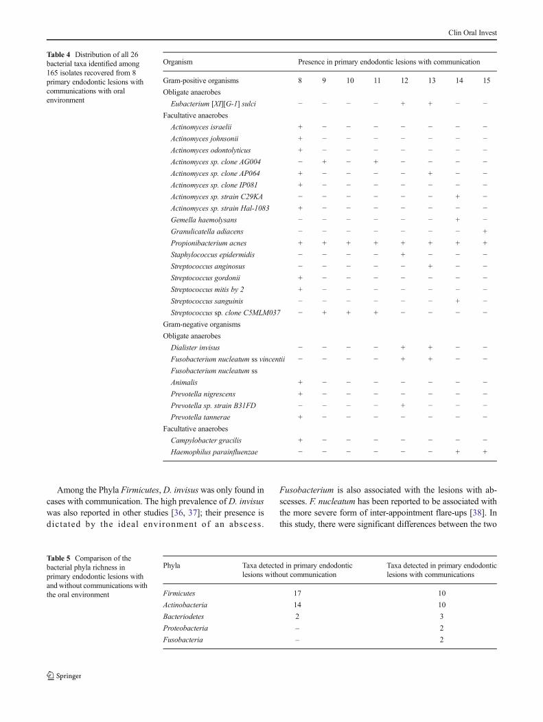
Among the Phyla Firmicutes, D. invisus was only found incases with communication. The high prevalence of D. invisuswas also reported in other studies [36, 37]; their presence isdictated by the ideal environment of an abscess.
Fusobacterium is also associated with the lesions with ab-scesses. F. nucleatum has been reported to be associated withthe more severe form of inter-appointment flare-ups [38]. Inthis study, there were significant differences between the two
Table 4 Distribution of all 26bacterial taxa identified among165 isolates recovered from 8primary endodontic lesions withcommunications with oralenvironment
Organism Presence in primary endodontic lesions with communication
Gram-positive organisms 8 9 10 11 12 13 14 15
Obligate anaerobes
Eubacterium [XI][G-1] sulci − − − − + + − −Facultative anaerobes
Actinomyces israelii + − − − − − − −Actinomyces johnsonii + − − − − − − −Actinomyces odontolyticus + − − − − − − −Actinomyces sp. clone AG004 − + − + − − − −Actinomyces sp. clone AP064 + − − − − + − −Actinomyces sp. clone IP081 + − − − − − − −Actinomyces sp. strain C29KA − − − − − − + −Actinomyces sp. strain Hal-1083 + − − − − − − −Gemella haemolysans − − − − − − + −Granulicatella adiacens − − − − − − − +
Propionibacterium acnes + + + + + + + +
Staphylococcus epidermidis − − − − + − − −Streptococcus anginosus − − − − − + − −Streptococcus gordonii + − − − − − − −Streptococcus mitis by 2 + − − − − − − −Streptococcus sanguinis − − − − − − + −Streptococcus sp. clone C5MLM037 − + + + − − − −
Gram-negative organisms
Obligate anaerobes
Dialister invisus − − − − + + − −Fusobacterium nucleatum ss vincentii − − − − + + − −Fusobacterium nucleatum ss
Animalis + − − − − − − −Prevotella nigrescens + − − − − − − −Prevotella sp. strain B31FD − − − − + − − −Prevotella tannerae + − − − − − − −
Facultative anaerobes
Campylobacter gracilis + − − − − − − −Haemophilus parainfluenzae − − − − − − + +
Table 5 Comparison of thebacterial phyla richness inprimary endodontic lesions withand without communications withthe oral environment
Phyla Taxa detected in primary endodonticlesions without communication
Taxa detected in primary endodonticlesions with communications
Firmicutes 17 10
Actinobacteria 14 10
Bacteriodetes 2 3
Proteobacteria – 2
Fusobacteria – 2
Clin Oral Invest
cohor t s in the preva lence of F. nuc lea tum andH. parainfluenza being isolated from approximately 25 % ofthe communication lesions.
P. acnes was the most prevalent bacterial taxa found in allthe primary endodontic cases with communication. However,P. acnes was not recovered from the cases without communi-cation. This emphasizes the ability of P. acnes to gain accessthrough the possible communications into the root canal spaceand then establish itself as a predominant microbiota of suchcases. A previous study showed the presence of P. acnes andS. epidermidis in the samples from refractory endodontic in-fections [25]. Other studies that recovered P. acnes andS. epidermidis considered it as contaminants since they areskin commensals [3, 18, 24, 39, 40]. In a previous study byNiazi et al. (2010), a decontamination protocol was used toremove contaminating organisms from the tooth surface priorto entering the diseased tooth [25]. Similar decontaminationprotocol was used in this study, thus confirming that P. acnesrecovered from these primary cases with communication wereopportunistic endodontic pathogens rather than contaminants,which is partially confirmed by the lack of P. acnes isolated inteeth with no communication with the oral cavity.
P. acnes has several virulence factors, in particular proteaseand polysaccharide formation capabilities are linked to chron-ic or persistent low-grade implant-associated infections. Inthese subclinical scenarios, in the absence of positive cultures,this pathogen is probably under-recognized andunderestimated [41].
The genotypic analysis of the P. acnes isolates using recAgene sequencing showed that type IA and IB were the preva-lent phylotypes found in primary cases with communication.These phylotypes probably were derived from patient’s ownskin or oral cavity during the exposure and communication ofthese teeth through the sinus tract, periodontal involvement orroot fracture. These might also be the result of a nosocomialinfection occurred as a result of manipulation of these teethduring treatment [25, 42]. Type II phylotype of P. acnes wasonly indentified in one case collected from the teeth with along-standing infections (>5 years) and a history of intermit-tent flare-ups. Although, the exact source of type II is still notidentified, there could be a relationship between the occur-rence of these phylotypes and the presence of long-standinginfections with Bopen^ communication with the oral cavity.This speculation is also partially supported by the more fre-quent isolation of philotypes II and III in failed root canaltreatment cases (25) which are likely to be associated withlonger standing infections.
P. acnes is the most prevalent species in primary endodon-tic infections with a history or clinical evidence of communi-cation with the oral environment, whereas it is absent in le-sions without communications. Using recA sequencing, thisstudy showed that P. acnes type IA and 1B are associated withprimary endodontic infections with communications. P. acnes
in primary endodontic infections with communications withthe oral cavity are likely to act as opportunistic pathogens.
The clinical management of endodontic cases where anopen lesion is present may require specific strategies to max-imize the chances to eliminate the opportunistic pathogens,such as P. acnes. In particular, adopting two stages root canaltreatment approach with usage of intermediate dressing maybe required even in primary endodontic treatment, wheresingle-stage endodontics was usually considered.
Acknowledgments The project received financial support from TheDepartment of Health via the National Institute for Health Research(NIHR) Comprehensive Biomedical Research Centre award to Guy’s &St Thomas’ NHS Foundation Trust in partnership with King’s CollegeLondon and King’s College Hospital NHS Foundation Trust.
Compliance with ethical standards The project was approved by thelocal ethics committee (South London REC: 05/Q0705/051).
Conflict of interest The authors declare that they have no competinginterests.
Informed consent The patient had verbal and written informationabout the purpose of study and they gave their written informed consentprior to their inclusion.
Open Access This article is distributed under the terms of the CreativeCommons At t r ibut ion 4 .0 In te rna t ional License (h t tp : / /creativecommons.org/licenses/by/4.0/), which permits unrestricted use,distribution, and reproduction in any medium, provided you give appro-priate credit to the original author(s) and the source, provide a link to theCreative Commons license, and indicate if changes were made.
References
1. Tancrede C (1992) Role of human microflora in health and disease.Eur J Clin Microbiol Infect Dis 11(11):1012–1015
2. McDowell A, Gao A, Barnard E, Fink C, Murray PI, Dowson CG,Nagy I, Lambert PA, Patrick S (2011) A novel multilocus sequencetyping scheme for the opportunistic pathogen Propionibacteriumacnes and characterization of type I cell surface-associated antigens.Microbiology 157(Pt 7):1990–2003. doi:10.1099/mic.0.049676-0
3. Brook I, Frazier EH (1991) Infections caused by Propionibacteriumspecies. Rev Infect Dis 13(5):819–822
4. Doyle A, Beigi B, Early A, Blake A, Eustace P, Hone R (1995)Adherence of bacteria to intraocular lenses: a prospective study. BrJ Ophthalmol 79(4):347–349
5. Eady EA, Ingham E (1994) Propionibacterim acnes—friend or foe?Rev Med Microbiol 5:163–173
6. Leyden JJ (2001) The evolving role of Propionibacterium acnes inacne. Semin Cutan Med Surg 20(3):139–143
7. Eishi Y, Suga M, Ishige I, Kobayashi D, Yamada T, Takemura T,Takizawa T, Koike M, Kudoh S, Costabel U, Guzman J, Rizzato G,Gambacorta M, du Bois R, Nicholson AG, Sharma OP, Ando M(2002) Quantitative analysis of mycobacterial and propionibacterialDNA in lymph nodes of Japanese and European patients with sar-coidosis. J Clin Microbiol 40(1):198–204
8. Perry AL, Lambert PA (2006) Propionibacterium acnes. Lett ApplMicrobiol 42(3):185–188
Clin Oral Invest
9. Schaeverbeke T, Lequen L, de Barbeyrac B, Labbe L, Bebear CM,Morrier Y, Bannwarth B, Bebear C, Dehais J (1998)Propionibacterium acnes isolated from synovial tissue and fluid ina patient with oligoarthritis associated with acne and pustulosis.Arthritis Rheum 41(10):1889–1893
10. Cohen RJ, B.A. S, J.E. MN, Shannon T, K.L. G (2005)Propionibacterium acnes associated with inflammation in radicalprostatectomy specimens: a possible link to cancer evolution? JUrol 173:1969–1974
11. Mathisen GE, D. MR, L. GW, M. CD (1984) Brain abscess andcerebritis. Rev Infect Dis 6(Suppl.1):101–106
12. Abolnik IZ, Eaton JV, Sexton DJ (1995) Propionibacterium acnesvertebral osteomyelitis following lumbar puncture: case report andreview. Clin Infect Dis 21(3):694–695
13. Harris AE, Hennicke C, Byers K, Welch WC (2005) Postoperativediscitis due to Propionibacterium acnes: a case report and review ofthe literature. Surg Neurol 63(6):538–541 discussion 541
14. Halkic N, Blanc C, Corthesy ME, Corpataux JM (2001) Lumbarspondylodiscitis after epidural anaesthesia at a distant site.Anaesthesia 56(6):602–603
15. Friberg O, Svedjeholm R, Kallman J, Soderquist B (2007)Incidence, microbiological findings, and clinical presentation ofsternal wound infections after cardiac surgery with and withoutlocal gentamicin prophylaxis. Eur J Clin Microbiol Infect Dis26(2):91–97
16. Benz MS, Scott IU, Flynn HW Jr, Unonius N, Miller D (2004)Endophthalmitis isolates and antibiotic sensitivities: a 6-year reviewof culture-proven cases. Am J Ophthalmol 137(1):38–42
17. Gunthard H, Hany A, Turina M, Wust J (1994) Propionibacteriumacnes as a cause of aggressive aortic valve endocarditis and impor-tance of tissue grinding: case report and review. J Clin Microbiol32(12):3043–3045
18. McDowell A, Valanne S, Ramage G, Tunney MM, Glenn JV,McLorinan GC, Bhatia A, Maisonneuve JF, Lodes M, PersingDH, Patrick S (2005) Propionibacterium acnes types I and II repre-sent phylogenetically distinct groups. J Clin Microbiol 43(1):326–334
19. McDowell A, Perry AL, Lambert PA, Patrick S (2008) A newphylogenetic group of Propionibacterium acnes. J Med Microbiol57(Pt 2):218–224
20. Valanne S, McDowell A, Ramage G, Tunney MM, Einarsson GG,O’Hagan S, Wisdom GB, Fairley D, Bhatia A, Maisonneuve JF,Lodes M, Persing DH, Patrick S (2005) CAMP factor homologuesin Propionibacterium acnes: a new protein family differentiallyexpressed by types I and II. Microbiology 151(Pt 5):1369–1379
21. Lodes MJ, Secrist H, Benson DR, Jen S, Shanebeck KD, GuderianJ, Maisonneuve JF, Bhatia A, Persing D, Patrick S, Skeiky YA(2006) Variable expression of immunoreactive surface proteins ofPropionibacterium acnes. Microbiology 152(12):3667–3681
22. Nagy I, Pivarcsi A, Kis K, Koreck A, Bodai L, McDowell A,Seltmann H, Patrick S, Zouboulis CC, Kemeny L (2006)Propionibacterium acnes and lipopolysaccharide induce the expres-sion of antimicrobial peptides and proinflammatory cytokines/chemokines in human sebocytes. Microbes and Infection/InstitutPasteur 8(8):2195–2205. doi:10.1016/j.micinf.2006.04.001
23. McDowell A, Barnard E, Nagy I, Gao A, Tomida S, Li H, Eady A,Cove J, Nord CE, Patrick S (2012) An expanded multilocus se-quence typing scheme for Propionibacterium acnes: investigationof ‘pathogenic’, ‘commensal’ and antibiotic resistant strains. PLoSOne 7(7):e41480. doi:10.1371/journal.pone.0041480
24. Sampedro MF, Piper KE, McDowell A, Patrick S, Mandrekar JN,Rouse MS, Steckelberg JM, Patel R (2009) Species ofPropionibacterium and Propionibacterium acnes phylotypes
associated with orthopedic implants. Diagn Microbiol Infect Dis64(2):138–145 doi:S0732-8893(09)00029–7
25. Niazi SA, Clarke D, Do T, Gilbert SC, Mannocci F, Beighton D(2010) Propionibacterium acnes and Staphylococcus epidermidisisolated from refractory endodontic lesions are opportunistic path-ogens. J Clin Microbiol 48(11):3859–3869. doi:10.1128/jcm.01326-10
26. Narayanan LL, Vaishnavi C (2010) Endodontic microbiology. JConserv Dent 13(4):233–239. doi:10.4103/0972-0707.73386
27. Patel S, Wilson R, Dawood A, Foschi F, Mannocci F (2012) Thedetection of periapical pathosis using digital periapical radiographyand cone beam computed tomography—part 2: a 1-year post-treat-ment follow-up. Int Endod J 45(8):711–723. doi:10.1111/j.1365-2591.2012.02076.x
28. Lane DJ (ed) (1991) 16S/23S rRNA sequencing. In nucleic acidtechniques in bacterial systematics. Wiley, Chichester
29. Kakehashi S, Stanley HR, Fitzgerald RJ (1965) The effects of sur-gical exposures of dental pulps in germ-free and conventional lab-oratory rats. Oral Surg Oral Med Oral Pathol 20:340–349
30. Siqueira JF Jr, Rocas IN (2009) Diversity of endodontic microbiotarevisited. J Dent Res 88(11):969–981
31. Lee YJ, KimMK, Hwang HK, Kook JK (2005) Isolation and iden-tification of bacteria from the root canal of the teeth diagnosed asthe acute pulpitis and acute periapical abscess. J Korean AcadConserv Dent 30(5):13
32. Kim SY, Choi HY, Park SH, Choi B (2003) Distribution of oralpathogens in infection of endodontic origin. J Korean AcadConserv Dent 28(4):303–313
33. Kum KY, Foud AF (2003) PCR-based identification ofEubacteirum species in endodontic infection. J Korean AcadConserv Dent 28(3):241–248
34. Munson MA, Pitt-Ford T, Chong B, Weightman A, Wade WG(2002) Molecular and cultural analysis of the microflora associatedwith endodontic infections. J Dent Res 81(11):761–766
35. van Steenbergen TJ, vanWinkelhoff AJ, Mayrand D, Grenier D, deGraaff J (1984) Bacteroides endodontalis sp. nov., an asaccharolyticblack-pigmented Bacteroides species from infected dental root ca-nals. Int J Sys Bact 34:118–120
36. Fouad AF, Barry J, Caimano M, Clawson M, Zhu Q, Carver R,Hazlett K, Radolf JD (2002) PCR-based identification of bacteriaassociated with endodontic infections. J Clin Microbiol 40(9):3223–3231
37. Siqueira JF Jr, Rocas IN (2002) Dialister pneumosintes can be asuspected endodontic pathogen. Oral Surg Oral Med Oral PatholOral Radiol Endod 94(4):494–498
38. LE C d PV (2002) Fusobacterium nucleatum in endodontic flare-ups. Oral Surg OralMed Oral Pathol Oral Radiol Endod 93(2):179–183
39. Tunney MM, Patrick S, Gorman SP, Nixon JR, Anderson N, DavisRI, Hanna D, Ramage G (1998) Improved detection of infection inhip replacements. A currently underestimated problem. J BoneJoint Surg Br Volume 80(4):568–572
40. Lazar JM, SchulmanDS (1992) Propionibacterium acnes prostheticvalve endocarditis: a case of severe aortic insufficiency. ClinCardiol 15(4):299–300
41. Achermann Y, Goldstein EJ, Coenye T, Shirtliff ME (2014)Propionibacterium acnes: from commensal to opportunisticbiofilm-associated implant pathogen. Clin Microbiol Rev 27(3):419–440. doi:10.1128/CMR.00092-13
42. Patel S, Wilson R, Dawood A, Mannocci F (2012) The detection ofperiapical pathosis using periapical radiography and cone beamcomputed tomography—part 1: pre-operative status. Int Endod J45(8):702–710. doi:10.1111/j.1365-2591.2011.01989.x
Clin Oral Invest