Embed Size (px)
Citation preview

University of Wisconsin MilwaukeeUWM Digital Commons
Theses and Dissertations
May 2013
Kinematic and Kinetic Comparisons of Arm andHand Reaching Movements with Mild andModerate Gravity-Supported, Computer-Enhanced Armeo®spring: A Case StudyQussai M. ObiedatUniversity of Wisconsin-Milwaukee
Follow this and additional works at: https://dc.uwm.edu/etdPart of the Occupational Therapy Commons
This Thesis is brought to you for free and open access by UWM Digital Commons. It has been accepted for inclusion in Theses and Dissertations by anauthorized administrator of UWM Digital Commons. For more information, please contact [email protected].
Recommended CitationObiedat, Qussai M., "Kinematic and Kinetic Comparisons of Arm and Hand Reaching Movements with Mild and Moderate Gravity-Supported, Computer-Enhanced Armeo®spring: A Case Study" (2013). Theses and Dissertations. 322.https://dc.uwm.edu/etd/322
KINEMATIC AND KINETIC COMPARISONS OF ARM AND HAND REACHING
MOVEMENTS WITH MILD AND MODERATE GRAVITY-SUPPORTED,
COMPUTER-ENHANCED ARMEO®
SPRING: A CASE STUDY
by
Qussai M. Obiedat
A Thesis Submitted in
Partial Fulfillment of the
Requirements for the Degree of
Master of Science
in Occupational Therapy
at
The University of Wisconsin-Milwaukee
May 2013
ii
ABSTRACT
KINEMATIC AND KINETIC COMPARISONS OF ARM AND HAND REACHING
MOVEMENTS WITH MILD AND MODERATE GRAVITY-SUPPORTED,
COMPUTER-ENHANCED ARMEO®
SPRING: A CASE STUDY
by
Qussai M. Obiedat
The University of Wisconsin-Milwaukee, 2013
Under the Supervision of Professor Ying-Chih Wang
Background: Stroke has been recognized as a leading cause of serious long-term
disability in the United States (U.S.) with 795,000 people experience a new or recurrent
stroke each year (Roger et al., 2011). The most apparent defect after stroke is motor
impairments (Masiero, Armani, & Rosati, 2011). Statistically, half of stroke survivors
suffer from upper extremity hemiparesis and approximately one quarter become
dependent in activities of daily living (Sanchez et al., 2006). There is strong evidence that
intensity and task specificity are the main drivers in an effective treatment program after
stroke. In addition, this training should be repetitive, functional, meaningful, and
challenging for a patient (Van Peppen et al., 2004). The use of robotic systems to
complement standard poststroke multidisciplinary programs is a recent approach that
looks very promising. Robotic devices can provide high-intensity, repetitive, task-
specific, interactive treatment of the impaired limb and can monitor patients' motor
progress objectively and reliably, measuring changes in quantitative movement
kinematics and forces (Masiero, Armani, & Rosati, 2011).
iii
Objective: The purpose of this study was to examine the role of Armeo®Spring
(Hocoma, Inc.), a gravity-supported, computer-enhanced robotic devise, on reaching
movements while using two different gravity-support levels (mild and moderate weight
support) on individuals with stroke.
Methods: One stroke subject and one gender-matched healthy control
participated in this study after gaining their informed consent. Both subjects performed a
computer-based game (picking apples successfully and placing them in a shopping cart)
under two gravity weight-support conditions (mild and moderate) provided by the
Armeo®Spring device. The game tasks were described as a reaching cycle which
consisted of five phases (initiation, reaching, grasping, transporting, and releasing). Joint
angles for the glenohumeral and elbow joints throughout the reaching cycle were found.
Three kinematic parameters (completion time, moving velocity, acceleration) and one
kinetic parameter (vertical force acting on the forearm) was calculated for various
instances and phases of the reaching motion. In addition, the muscle activation patterns
for anterior deltoid, middle deltoid, biceps, triceps, extensor digitorum, flexor digitorum,
and brachioradialis were found and the mean magnitude of the electromyography (EMG)
signal during each phase of the reaching cycle was found as a percentage of the subject’s
maximum voluntary contraction (MVC).
Results: Within the healthy control subject, results demonstrated no
significant differences in mean completion time, moving velocity, or acceleration
between mild to moderate gravity-support levels during all phases of the cycle. The
stroke subject results revealed a significant decrease in the cycle mean completion time
(p= 0.042) between the two gravity-support levels, specifically in mean completion time
iv
of the grasping phase. A significant increase was found in the initiation phase moving
velocity (p=0.039) and a significant decrease was found in the grasping phase (p=0.048)
between two gravity-support levels in the stroke subject. Between subjects, significant
increase in the cycle mean completion time was found under both mild and moderate
conditions (p<.001 for both conditions). Additionally, significant decreases in the moving
velocities were found in all phases of the cycle between the healthy control and the stroke
subject under both conditions. With increasing weight support, the healthy control subject
showed an increase in abduction and flexion degrees at the glenohumeral joint level, and
an increase in flexion degrees of the elbow joint. On the other hand, the stroke subject
showed a decrease in abduction degrees and an increase in flexion degrees at the
glenohumeral joint level, and a decrease in flexion degrees of the elbow joint after
increasing the weight-support level. Results demonstrated an increase in the mean of
vertical forces when changing gravity-support levels from mild to moderate during all
phases of the cycle in both stroke and healthy subjects. Last, the average EMG magnitude
during the reaching cycle phases was reduced for muscles acting against gravity (anterior
deltoid, middle deltoid, biceps, and brachioradialis) in both the healthy control and the
stroke subject.
Conclusion: The significant differences in movement performance between mild
and moderate physical weight support suggested a preliminary result that the gravity-
supported mechanism provides a mean to facilitate functional upper limb motor
performance in individuals with stroke. Future studies should examine such effects with
larger sample sizes.
v
TABLE OF CONTENTS
Introduction ................................................................................................................... 1
Significance of the Study .................................................................................. 3
Background and Literature Review .............................................................................. 4
Types of Stroke ................................................................................................. 4
Ischemic Stroke ..................................................................................... 4
Hemorrhagic Stroke .............................................................................. 6
Transient Ischemic Attack .................................................................... 7
Symptoms & Complications ............................................................................. 7
Motor Recovery ................................................................................................ 9
Robot-assisted Therapy ..................................................................................... 12
Gravity Compensation ...................................................................................... 14
Reaching Studies ...............................................................................................15
Specific Aims and Hypothesis ...................................................................................... 16
Methods......................................................................................................................... 17
Participants ........................................................................................................ 17
Inclusion and exclusion criteria ........................................................................ 17
Device: ArmeoSpring ....................................................................................... 18
Armeo®Spring Weight-Support System .......................................................... 20
Game: Fruit Shopping ....................................................................................... 21
Data Collection ................................................................................................. 22
Procedure .......................................................................................................... 23
MVC Procedure ................................................................................................ 24
Kinematic Model .............................................................................................. 24
Data Analysis .................................................................................................... 27
vi
Results ........................................................................................................................... 28
Kinematic parameters ....................................................................................... 29
Joint Angles ......................................................................................................32
Forearm Vertical Forces ................................................................................... 32
Electromyography (EMG) ................................................................................ 33
Discussion .....................................................................................................................38
References ..................................................................................................................... 42
Appendix A: Previous Reaching Studies in the Literature ........................................... 52
Appendix B: Text Descriptions .................................................................................... 59
vii
LIST OF FIGURES
Figure 1. The Armeo®Spring study setup .....................................................................19
Figure 2. Armeo®Spring weight support system ..........................................................20
Figure 3. The print screen of the fruit shopping game ..................................................22
Figure 4. The mean completion time between the two gravity-support levels .............30
Figure 5. Joint angle changes during the reaching cycle for the healthy subject
(upper panel) and the stroke subject (lower panel) .......................................................33

viii
LIST OF TABLES
Table 1. Brunnstrom and Twitechell motor recovery stages ........................................11
Table 2: Armeo®Spring support levels ........................................................................21
Table 3. Markers used in the motion caption procedure ...............................................24
Table 4. MVC testing positions ....................................................................................26
Table 5. Anatomical coordinate systems ......................................................................26
Table 6. Armeo®Spring mild and moderate weight-support levels for stroke
and healthy subjects ......................................................................................................29
Table 7. Kinematic parameters of the healthy subject with mild & moderate
weight support ...............................................................................................................30
Table 8. Kinematic parameters of the stroke subject with mild & moderate
weight support ...............................................................................................................31
Table 9. Vertical support forces for healthy control and stroke subject with
mild & moderate weight support ..................................................................................35
Table 10.Healthy subject EMG average magnitude (% of MVC) ................................35
Table 11. Stroke subject EMG average magnitude (% of MVC) .................................36
Table 12. Healthy subject EMG minimum magnitude (% of MVC) ............................36
Table 13. Stroke subject EMG minimum magnitude (% of MVC) ..............................37
Table 14. Healthy subject EMG maximum magnitude (% of MVC) ...........................37

ix
Table 15. Stroke subject EMG maximum magnitude (% of MVC) .............................38
Table 16. P-values for between-subjects average EMG magnitude .............................38
1
Introduction
Recently, stroke has been recognized as one of the leading causes of serious long-
term disability in the United States (U.S.). Approximately 795,000 people experience a
new or recurrent stroke each year (Roger et al., 2011). Although the medical treatment
improvements of the complications caused by stroke decreased the mortality rate of the
disease, 90% of the survivors still suffer from significant neurological deficits (Volpe,
Krebs, & Hogan, 2001). The most common defects after stroke are upper extremity
functional impairments and disability in activities of daily living (Masiero, Armani, &
Rosati, 2011). Statistically, half of stroke survivors suffer from upper extremity
impairments and approximately one quarter become dependent in activities of daily
living (Sanchez et al., 2006).
Loureiro, Harwin, Nagai, and Johnson (2011) categorized current, available
upper extremity stroke rehabilitation methodologies and technologies as: conventional
physical and occupational therapy, constraint-induced movement therapy, and robotic-
aided and sensor-based therapy systems. Although an increased effort has been made to
enhance the recovery process following stroke, patients generally do not reach their full
recovery potential when discharged from hospital following initial rehabilitation. This
can be attributed to the economic pressure and the lack of available human resources
(Loureiro, Harwin, Nagai, & Johnson, 2011). These facts lead to focus more on robot-
assisted therapy as an equivalent in quality to traditional methods. The use of robot
assisted therapy will deliver therapy at reduced cost and provide a solution to overcome
the labor-intensive, one-to-one stroke rehabilitation.

2
The development, preliminary clinical use, and effectiveness of the
Armeo®Spring, a gravity-supported, computer-enhanced robotic devise, for individuals
with upper limb motor dysfunction have been supported (Gijbels et al., 2011; Housman,
Scott, & Reinkensmeyer, 2009; Sanchez et al., 2006). A study conducted by Sanchez et
al., (2006) demonstrated that individuals with chronic stroke whose arm function is
compromised in a normal gravity environment could perform reaching and drawing
movements while using T-WREX (the prototype version of the Armeo®Spring). The
patients improved their motor function (mean change in Fugl-Meyer score was 5 points)
over a period of eight weeks. Results from Housman, Scott, & Reinkensmeyer, (2009)
showed that, using the T-WREX can improve arm movement ability after chronic severe
hemiparesis with brief one-on-one assistance from a therapist (approximately 4 minutes
per session). Additionally, the 3-dimensional weight support, instant visual movement
feedback, and simple virtual reality software provided by T-WREX were associated with
modest sustained gains at 6-month follow-up (mean change in Fugl-Meyer score was 3.6
points) when compared with the conventional approach (mean change in Fugl-Meyer
score was 1.5 points). The study conducted by Gijbels et al., (2011) was in multiple
sclerosis (MS) and thus results were not described here.
The fundamental kinematic and kinetic comparisons of arm and hand reaching
movements with gravity-supported, computer-enhanced Armeo®spring have not been
studied. Specifically, how the change of the weight level of support in the Armeo®Spring
device may affect the reaching performance of patients with severe stroke. This project
aimed to examine the role of the Armeo®Spring on reaching movements while using two
different gravity-support levels.

3
Significance of the Study
Stroke rehabilitation is an important public health issue that needs to be addressed
by all health care professionals. It gains this importance because of the increase of the
prevalence and incidence of those with stroke disability due to population aging and
improved survival after the initial injury (Volpe et al., 2009). Krebs, Volpe, Aisen, &
Hogan (2000) described three ways to maximize the productivity in the delivery of
rehabilitation without sacrificing the quality of care patients receive. These three methods
include: develop evidence-based therapy, re-allocate personnel and tasks, and increase
the productivity of each caregiver that can be achieved by providing therapists with
appropriate tools.
The increase of the prevalence and incidence of stroke along with the economical
pressure and lack of human resources stimulates the interest in the use of robot-assisted
techniques to enhance the efficiency and effectiveness of post-stroke rehabilitation
(Burgar et al., 2011). On the other hand, it is important to investigate the efficiency of
each device and to make sure that it provides realistic clinical expectations as it is
supposed to achieve.
Post-stroke rehabilitation has tremendous implication for most of health care
professions and stands as an intrinsic part of occupational therapy practice. As
“Occupational therapy (OT) aims at facilitating task performance by improving relevant
performing skills or developing and teaching compensatory strategies to overcome lost
performance skills” (Steultjens et al., 2003). Providing therapists with the proper tools to
4
promote the quality of care provided will play a key role in enhancing occupational
therapy interventions and enable therapists to increase their productivity levels.
Background and Literature Review
The World Health Organization (WHO) defines stroke as “a clinical syndrome
with rapidly developing clinical signs of focal or global disturbance of cerebral function,
lasting more than 24 hours or leading to death with no apparent cause other than of
vascular origin.” (Broeks, Lankhorst, Rumping, & Prevo, 1999). It occurs when a blood
clot blocks an artery, which is a blood vessel that carries blood from the heart to the
body, or when a blood vessel bursts, causing an interruption in the blood flow to an area
of the brain. When either of these scenarios happens, brain cells begin to die leading to
brain damage (National Stroke Association, 2011). In addition, stroke, or cerebrovascular
accident (CVA), can be defined as “a sudden ischemic or hemorrhagic disturbance in the
blood supply to brain tissue that results in partial loss of brain function.” (Prange,
Jannink, Groothuis-Oudshoorn, Hermens, & Ijzerman, 2006).
Types of Stroke
Stroke has been categorized by the National Stroke Association (2011) according
to its underlying cause into two major types: ischemic and hemorrhagic stroke.
Ischemic Stroke. Ischemic stroke accounts for about 87 percent of all cases
(American Heart Association, 2011). Naturally, blood clotting is a beneficial
physiological process which aims to slow and eventually stop the bleeding from a wound.

5
However, these clots maybe a source of danger in the case of stroke because they can
block arteries and cut off blood flow and oxygen supply to certain areas of the brain, A
process which is known as Ischemia (National Stroke Association, 2011).
Ischemic stroke has two subtypes according to the clot formation origin: (a)
embolic stroke, (b) thrombotic stroke.
a. Embolic Stroke: the blood clot that causes embolic stroke is formed somewhere in
the body, usually the heart, and travels through the bloodstream to the brain. The
clot travels in the brain blood vessels until it reaches a small enough vessel to
block its passage causing a stroke. The medical word used to describe this type of
blood clot is embolus (National Stroke Association, 2011).
b. Thrombotic Stroke: the blood clot causing this type of strokes is formed on a
blood vessel causing the blockage to one or more of the arteries supplying blood
to the brain. The process leading to this blockage is known as thrombosis
referring to the medical description for a clot that forms on a blood-vessel deposit
which is thrombus. This blood clot can happen as a result of unhealthy blood
vessels clogged with a buildup of fatty deposits and cholesterol. The body reacts
regarding these buildups as a multiple, tiny and repeated injuries to the blood
vessel wall, as if a bleeding from a wound is present, it responds by forming clots.
Two types of thrombosis can cause stroke: large vessel thrombosis and small
vessel disease/lacunar infarction (National Stroke Association, 2011).
i. Large Vessel Thrombosis: large vessel thrombosis is the most common and
best understood type of thrombotic stroke. Most of this type of strokes is

6
caused by a combination of long-term atherosclerosis followed by rapid
blood clot formation. Patients who have suffered this type of brain attack are
more likely to have coronary artery disease, and heart attack is a frequent
cause of death (National Stroke Association, 2011).
ii. Small Vessel Disease/Lacunar Infarction: occurs when blood flow is
blocked to a very small arterial vessel. Little is known about the causes of
small vessel disease, but it is closely linked to high blood pressure or as
known as hypertension (National Stroke Association, 2011).
Hemorrhagic Stroke. Hemorrhagic stroke accounts for about 13 percent of
stroke cases (American Heart Association, 2011). This type of strokes is caused by the
breakage or burst of a blood vessel in the brain. The medical word that describes this type
of breakage is hemorrhage which can be caused by a number of disorders that affect the
blood vessels, including long-standing high blood pressure and cerebral aneurysms. An
aneurysm is defined as a weak or thin spot on a blood vessel wall, which is usually
present at birth or develop over a number of years, and usually don't cause detectable
problems until they break (National Stroke Association, 2011).
Hemorrhagic stroke is categorized into two subtypes: (a) subarachnoid
hemorrhage and (b) intracerebral hemorrhage
a. Subarachnoid hemorrhage (SAH): when an aneurism bursts in a large artery on or
near the thin, delicate membrane surrounding the brain, the blood spills into the
area around the brain which is filled with a protective fluid, causing the brain to
be surrounded by blood-contaminated fluid (National Stroke Association, 2011).

7
b. Intracerbral hemorrhage: occurs when bleeding from vessels within the brain is
present. The primary cause of this type of hemorrhage is hypertension (National
Stroke Association, 2011).
Transient Ischemic Attack (TIA). Transient ischemic attack (TIA) is often
labeled as a “mini-stroke.” It is more accurately characterized as a “warning stroke”. Like
stroke, TIA is caused by a clot but the only difference between a stroke and TIA is that
with TIA the blockage of the blood vessel is transient (temporary). TIA symptoms occur
rapidly and last for a relatively short time (less than five minutes; the average is about a
minute). Unlike a stroke, when a TIA is over, there’s no permanent injury to the brain
(National Stroke Association, 2011).
Symptoms & Complications
According to the World Health Organization (WHO) (WHO, 2011) the most
common symptom of a stroke is sudden weakness or numbness of the face, arm or leg,
mostly on one side of the body. Other symptoms include: confusion, difficulty speaking
or understanding speech; difficulty seeing with one or both eyes; difficulty walking,
dizziness, loss of balance or coordination; severe headache with no apparent cause; as
well as fainting or unconsciousness.
The severity and effects of a stroke depend on where the stroke occurs in the brain
(location) and how much the brain is damaged (lesion size) (Volpe, Krebs, & Hogan,
2001; WHO, 2011), resulting in deficits of the cognitive, sensory, affective, and motor
functions (Krebs, Volpe, Aisen, & Hogan, 2000).
8
The most disabling motor deficit following stroke is the loss of arm function.
About 85% of stroke survivors have a sensorimotor deficit in the arm which is
characterized by muscle weakness, abnormal muscle tone, abnormal movement
synergies, lack of mobility between structures at the shoulder girdle, and incoordination
during voluntary movement (Cirstea, Ptito, & Levin, 2003). Deficits in the coordinated
use of the limb are most evident in the limb contralateral to the damaged side of the brain
(Levin, 1996). Attempts to make goal-directed movements with the affected limb in
stroke survivors are typically characterized by decreased range of motion (ROM),
movement speed, smoothness, coordination, and abnormal pattern of muscle activation
(Johnson, Feng, Johnson, & Winters, 2007; Levin, 1996).
The development of upper extremity spastic paresis is a common complication
following stroke. It is comprised of positive and negative symptoms that occur to varying
degrees in each patient. Positive symptoms include spasticity, hypertonia, increased
muscle stiffness, and excessive co-contraction between agonist and antagonist muscles
(Leonard, Gardipee, Koontz, Anderson, & Wilkins, 2006). Spasticity is defined as a
velocity dependent hyperexcitability of muscles to stretch and is characterized by
exaggerated tendon reflexes, increased resistance to passive movement and hypertonia
resulting from loss of upper motor neuron inhibitory control (Watkins et al., 2002).
Negative symptoms include muscle paresis and discoordination (Leonard, Gardipee,
Koontz, Anderson, & Wilkins, 2006). After stroke, spasticity contributes to motor
impairments and activity limitations and may become a severe problem for some patients
(Sommerfeld, Eek, Svensson, Holmqvist, & von Arbin, 2004). In the upper limb,
spasticity may present in two types of synergies. A flexor synergy which consists of
9
forearm supination and elbow flexion combined with shoulder flexion, abduction and
external rotation, or extensor synergy which is characterized by forearm pronation and
elbow extension associated with shoulder extension, adduction and internal rotation
(Levin, 1996).
Motor Recovery
Generally, the largest proportion of the recovery process takes place during the
weeks and months that immediately follow stroke occurrence (Volpe, Krebs, & Hogan,
2001). Even though, the rehabilitation process should not be stopped after the acute
rehabilitation hospital event. In fact, home training or home training enhanced with
devices managed by therapists has the potential to contribute to recovery goals (Volpe,
Krebs, & Hogan, 2001).
Motor learning have been defined loosely by motor control scientists by
considering it a fuzzy term that encompasses motor adaptation, skill acquisition, and
decision making (Huang & Krakauer, 2009). The neuro-rehabilitation science is built up
on two basic assumptions, the first one is that motor learning principles apply to motor
recovery, and the second assumption is that patients can learn (Huang & Krakauer, 2009).
The recent motor control models suggest that the central nervous system learns a
new novel task through practice by constructing a pattern of control variables or making
an internal model for that task, and once the new process is earned, it is stored in memory
and available for recall (Cirstea, Ptito, & Levin, 2003).
10
The majority of motor deficit recovery occurs within 6-months post-stroke
(Macclellan et al., 2005). According to the available scientific literature, post-stroke
rehabilitation intervention is suggested to be significantly more effective when it is
delivered in the early phase of recovery. Evidence supports that the better functional
outcome is determined by rehabilitation that is initiated promptly and based on intensive
multisensory stimulation which is associated with increased adaptive plasticity of the
brain in the early post-stroke stages (Masiero, Armani, & Rosati, 2011).
The restoration of motor function in the arm and leg after stroke has been
described as an ordered, predictable, stepwise progression by Twitechell (Twitchell,
1951). The initial stage of this progression is flaccid paralysis, after that the development
of a basic stereotypical synergy of voluntary movements, and then to normal patterns of
voluntary movements. Based on these observations Brunnstrom (Brunnstrom, 1966)
divided the progression into 6 sequential stages of motor recovery (Table 1) (Crow &
Harmeling-van der Wel, 2008). The early stages are characterized by the appearance of
spasticity and the development of stereotypic movement patterns while isolated joint
movements are jeopardized. In later stages, spasticity declines and the patient is able to
make movements out of synergy. Still later, isolated joint movement and control returns
(Levin, 1996).
To understand stroke recovery on a more mechanistic level, two main
assumptions are encompassing the recovery process. The first one is that parallel brain
regions in the unaffected hemisphere conduct the functions of the contra-lateral
hemisphere necrotic tissue by sending its commands via uncrossed pathways. The second
assumption is that the adjacent areas of undamaged brain tissue recognize and conduct

11
the functions of the necrotic tissue in the same hemisphere (Krebs, Hogan, Aisen, &
Volpe, 1998). The cortical maps reorganization process has been demonstrated in the
motor, sensory, auditory, and visual maps. Furthermore, the environment has an influence
on the degree of reorganization of the remaining undamaged cortex (as recent animal
studies on primates have shown) which suggest that exercising the patients’ affected
limbs might have a positive effect on neurological restoration of the limb function
(Krebs, Hogan, Aisen, & Volpe, 1998).
Table 1. Brunnstrom & Twitechell motor recovery stages (Crow & Harmeling-van der Wel, 2008)
Twitchell Brunnstrom
Flaccid paralysis with areflexia Stage 1
Flaccid paralysis
Reflex activity returns/spasticity develops Stage 2
Development of minimal movement in synergies
Voluntary movement in stereotyped flexor and
extensor synergies/spasticity is at maximal level
Stage 3
Voluntary movement synergy dependent
Voluntary movement with breaking up of
synergies/spasticity is reducing
Stage 4
Some movements out of synergy
Stage 5
Movements almost independent of synergy
Normal voluntary movement with normal speed and
dexterity/slight hyperactivity of the tendon reflexes
Stage 6
Normal movement with normal speed
Stroke survivors have the tendency to use their unaffected arm in real world tasks
(Johnson, Feng, Johnson, & Winters, 2007). Part of the standard treatment for the
sensorimotor impairment focus on teaching patients to use the unaffected limb to adapt,
compensate, and improve motor abilities with respect to feeding, grooming, and toileting
(Volpe et al., 2009). The other emphasis of acute rehabilitation is to teach compensatory
rather than restorative methods (Burgar et al., 2011). On the other hand, different studies
reported that several approaches, including repetitive passive exercises, forced use of the
paretic limb or constraint-induced movement therapy, functional electrical stimulation,
increased amounts of therapy including external manipulation, and biofeedback provided

12
positive outcomes on the motor recovery of the affected limb (Krebs, Volpe, Aisen, &
Hogan, 2000; Masiero, Armani, & Rosati, 2011).
The literature supports that in order for the therapy to be effective it should
contain elements of repetition, intense practice, motivation, and task application. Patient
involvement and empowerment along with the use of functional and purposeful tasks in
an enriched environment play a key role in increasing patient’s motivation and recovery
(Wisneski & Johnson, 2007).
The rehabilitation process of the impaired upper limb focuses on reducing
impairment and improving independent function on various activities of daily living
(ADLs) salient to patients’ real life environment. If the patients are able to transfer motor
and functional gains achieved during supervised therapy to their daily life this process is
considered effective and successful (Johnson, Feng, Johnson, & Winters, 2007).
Robot-assisted Therapy
The use of rehabilitation robots to complement standard post-stroke rehabilitation
is a new promising tradition that has been developed intensively in the past few decades
(Masiero, Armani, & Rosati, 2011). Examples of upper extremity rehabilitation robots
that are currently available in the market or in research labs are Armeo®Spring (Hocoma,
Inc), Armeo®Power (Hocoma, Inc), ARMin (Nef, Mihelj, & Riener, 2007), MIT-
MANUS (Krebs, Hogan, Aisen, & Volpe, 1998), and T-WREX (Housman, Scott, &
Reinkensmeyer, 2009). They have been developed to aid in rehabilitation, alter the
physical burden on a therapist to overcome the limited availability of one-to-one stroke
rehabilitation, and potentially improve a clinic’s productivity (Wagner et al., 2011;
13
Wang, Wang, Zhang, Wang, & Wang, 2011). Robotic devices can provide repetitive,
task-specific, and high-intensity interactive treatment of the impaired limb. They can also
measure patients’ motor progress objectively and measure changes in movement
kinematics and forces (Masiero, Armani, & Rosati, 2011).
A common misperception about robot-assisted therapy is that it would ultimately
replace human-administered therapy (Krebs, Volpe, Aisen, & Hogan, 2000). In fact, it is
most appropriate to consider the robot as an advanced tool that is used under the therapist
supervision to implement relatively simple and labor-intensive therapies (Masiero,
Armani, & Rosati, 2011). As the systematic reviews of robot-assisted therapy suggest,
these devices met the criteria for improving proximal upper extremity strength and have
the potential to promote motor recovery to a greater extent than traditional therapy
(Housman, Scott, & Reinkensmeyer, 2009). Individuals who suffer from acute or chronic
stroke and receive more therapy with a robotic device can recover more movement
ability, and those with chronic stroke who receive matched amounts of robotic and
conventional therapy produced comparable therapeutic benefits (Sanchez et al., 2006).
Rehabilitation robots for the upper limb can be classified into passive, active, and
interactive systems (Loureiro, Harwin, Nagai, & Johnson, 2011; Riener, Nef, &
Colombo, 2005). In passive systems, no actuation is implemented to move patient limbs.
Instead, the system constrains the patient’s arm to a determined range of motion. They
often consist of mechanical linkages that move easily when pushed and their technical
components typically include stiff frames, bearings and pulleys, and ropes with counter-
weights (Loureiro, Harwin, Nagai, & Johnson, 2011; Riener, Nef, & Colombo, 2005).
Active systems are equipped with electromechanical, pneumatic, hydraulic and other
14
drives to move the patient’s arm actively through a predefined path. Either the devices
are open-loop controlled, or simple position-control strategies are implemented to take a
patient’s arm from a predefined position to a new position using a certain velocity profile
(Loureiro, Harwin, Nagai, & Johnson, 2011; Riener, Nef, & Colombo, 2005). Interactive
systems react to the patient’s input and characterized not only by actuators but also by
sophisticated impedance and other control strategies. They are usually back-drivable and
possess low, intrinsic, end point impedance (Loureiro, Harwin, Nagai, & Johnson, 2011;
Riener, Nef, & Colombo, 2005).
Gravity Compensation
Little information regarding the effects of gravity compensation on upper limb
recovery after stroke was found in the literature. It was reported that stroke patients
showed an improved arm function after nine weeks of training using gravity
compensation provided by sling suspension, which suggest that the application of gravity
compensation may be considered a valuable tool to stimulate functional improvement in
stroke rehabilitation (Prange et al., 2009). Another research has shown that gravity
compensation in upper limbs decreases the required shoulder abduction torques during
two dimensional reaching movements at shoulder height, causing a decrease in coupled
elbow flexion leading to an increase in the range of elbow extension (Krabben et al.,
2012). Furthermore, the maximal reaching distance during a 3-dimensional movement,
starting with the hand at waist height and reaching to a target at shoulder height, is
slightly larger when gravity compensation is applied to the arm of stroke patients
(Prange et al., 2009).
15
Reaching Studies
Many studies have examined the reaching movements in stroke (Archambault,
Pigeon, Feldman, & Levin, 1999; Cirstea, Ptito, & Levin, 2003; Jannink et al., 2007;
Kamper, McKenna-Cole, Kahn, & Reinkensmeyer, 2002; Krabben et al., 2012; Leonard,
Gardipee, Koontz, Anderson, & Wilkins, 2006; Levin, 1996; Prange et al., 2009). The
analytical variables that have been used to quantify the reaching movements varied
among different studies, which included (but not limit to) speed accuracy and efficiency
of reaching, peak wrist velocity, endpoint error, reach path ratio, peak speed ratio,
number of speed peaks, interjoint coordination, linearity of hand motion, movement
direction variability, muscle co-contraction, muscle activation, and trunk compensation.
Different analysis methods have been used to determine the movement onsets and offsets.
For example, Cirstea and Levin (2000) used the times at which the tangential velocity
exceeded or fell below 10% of the peak velocity, while Butler et al. (2010) defined the
beginning (i.e., initiation) of each cycle as the first instant when the velocity of the wrist
marker exceeded 5% of peak reaching velocity. Kinematic data were low-pass filtered at
5 Hz and 6 Hz (Kamper, McKenna-Cole, Kahn, & Reinkensmeyer, 2002; Wagner,
Dromerick, Sahrmann, & Lang, 2007). In general, studies have shown that in stroke
subjects multi-joint pointing movements are characterized by decreased movement speed
and increased movement variability, by increased movement segmentation and by spatial
and temporal incoordination between adjacent arm joints with respect to healthy subjects
(Archambault, Pigeon, Feldman, & Levin, 1999; Cirstea & Levin, 2000). Stroke subjects
also showed the use of compensatory movement patterns (Cirstea & Levin, 2000).
Previous reaching studies available in the literature are illustrated in Appendix.

16
Specific Aims and Hypothesis
Aim 1: To compare reaching biomechanics between two different gravity-support levels
(mild and moderate weight support) in a healthy control using the gravity-supporting
exoskeleton apparatus (Armeo®Spring)
Hypothesis 1: We hypothesize that different gravity-support levels do not affect
reaching movements in healthy controls.
Aim 2: To compare reaching biomechanics between two different gravity-support levels
(mild and moderate weight support) in a stroke subject using the gravity-supporting
exoskeleton apparatus (Armeo®Spring)
Hypothesis 2: We hypothesize that the stroke subject will improve the reaching
performance under the higher weight support condition. Specifically, we
hypothesize that the gravity-supporting facilitates the stroke subject’s upper limb
movement and thus the stroke subject is able to complete the task more efficiently
and with less physical efforts. The moving time would reduce and mean reaching
speed would increase.
Aim 3: To compare the biomechanics of reaching movements between a healthy control
and a stroke subject using the gravity-supporting exoskeleton apparatus (Armeo®Spring)
Hypothesis 3: We hypothesize that, comparing to the healthy control, the stroke
subject would have (a) a longer moving time, (b) slower moving speed, and (c)
different muscle activation patterns in the muscles acting against gravity in the
upper limb during reaching.
17
Methods
Participants
This study was conducted at the Gait and Biodynamics Laboratory at the
University Services and Research (USR) building on the University of Wisconsin
Milwaukee campus. The recruitment process was done through flyers distributed around
campus and in the surrounding community and through word-of-mouth. Subjects
completed a questionnaire over the phone to determine their eligibility. The study took
approximately 2 hours over a one-day course for each participant. Prior to testing, the
participants signed an informed consent form to participate in the study per the protocol
approved by the University of Wisconsin Milwaukee Institutional Review Board for
human subject research.
For the proposed study, one stroke subject and one healthy control were recruited
in this study after gaining their informed consent.
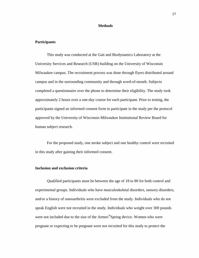
Inclusion and exclusion criteria
Qualified participants must be between the age of 18 to 80 for both control and
experimental groups. Individuals who have musculoskeletal disorders, sensory disorders,
and/or a history of osteoarthritis were excluded from the study. Individuals who do not
speak English were not recruited in the study. Individuals who weight over 300 pounds
were not included due to the size of the Armeo®Spring device. Women who were
pregnant or expecting to be pregnant were not recruited for this study to protect the
18
unborn child and the mother from the risks during testing. Stroke survivors were
excluded from the study if they had more than 3 score in the modified Ashworth Scale,
onset of stroke is less than 6 months, and/or unstable health conditions in the judgment of
the Principal Investigator (PI) and Co-PI would prevent them from participating in this
study.
Device: Armeo®Spring
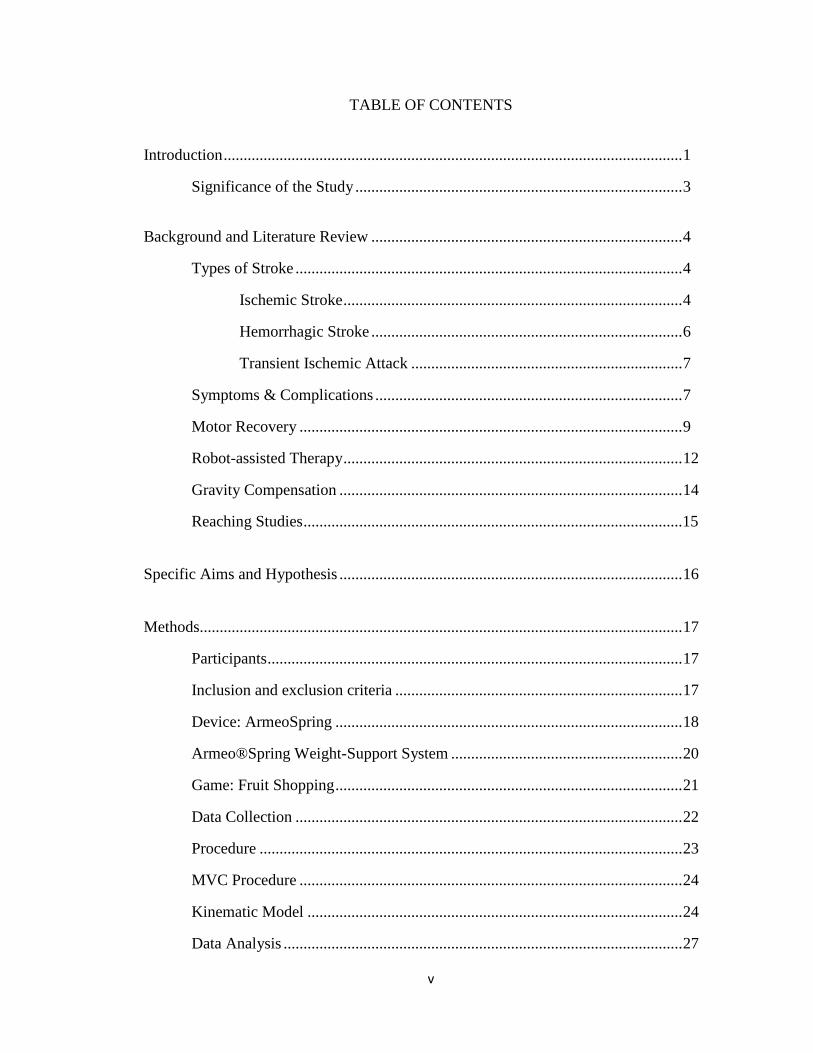
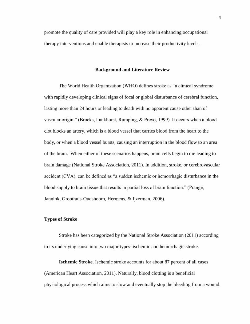
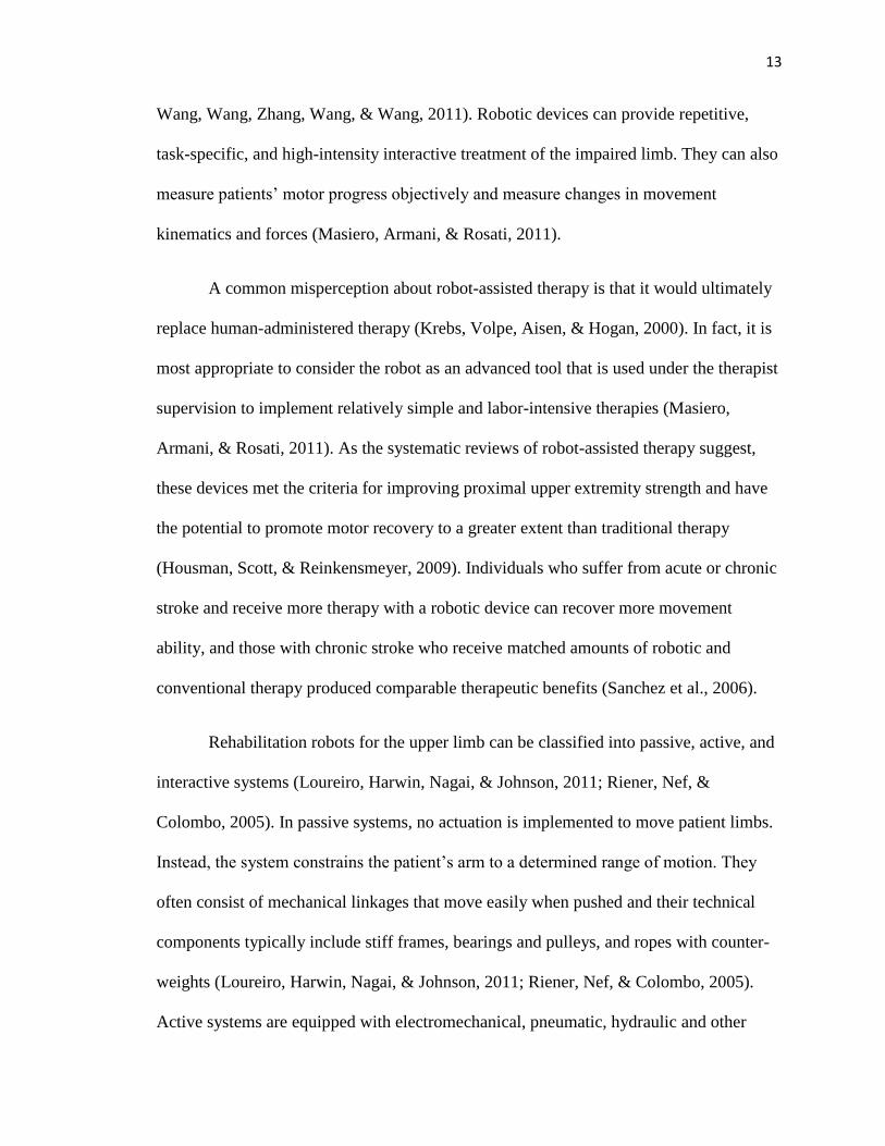
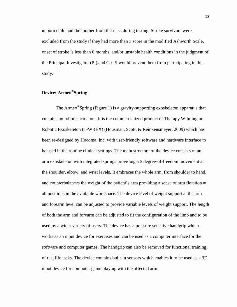
The Armeo®Spring (Figure 1) is a gravity-supporting exoskeleton apparatus that
contains no robotic actuators. It is the commercialized product of Therapy Wilmington
Robotic Exoskeleton (T-WREX) (Housman, Scott, & Reinkensmeyer, 2009) which has
been re-designed by Hocoma, Inc. with user-friendly software and hardware interface to
be used in the routine clinical settings. The main structure of the device consists of an
arm exoskeleton with integrated springs providing a 5 degree-of-freedom movement at
the shoulder, elbow, and wrist levels. It embraces the whole arm, from shoulder to hand,
and counterbalances the weight of the patient’s arm providing a sense of arm flotation at
all positions in the available workspace. The device level of weight support at the arm
and forearm level can be adjusted to provide variable levels of weight support. The length
of both the arm and forearm can be adjusted to fit the configuration of the limb and to be
used by a wider variety of users. The device has a pressure sensitive handgrip which
works as an input device for exercises and can be used as a computer interface for the
software and computer games. The handgrip can also be removed for functional training
of real life tasks. The device contains built-in sensors which enables it to be used as a 3D
input device for computer game playing with the affected arm.
19
The device comes with computer software (Armeocontrol) which contains an
extensive library of game-like movement exercises. The games are designed to mimic
functional arm movements, to provide training in a simple virtual reality environment,
and to achieve the goal of enabling repetitive task-specific practice.
In all functional exercises, the exercises are mapped into a cubic workspace,
which can be adjusted to the movement abilities of each individual. Before starting the
exercise session, the workspace has to be defined (i.e., the maximum distance a person
can bring his/her hand up, down, left, and right, and how far and close to the body while
using the Armeo®Spring) to adjust to the movement abilities of each individual.
Figure 1. The Armeo®Spring study setup
Computer
software
interface
Markers for
motion caption
system

20
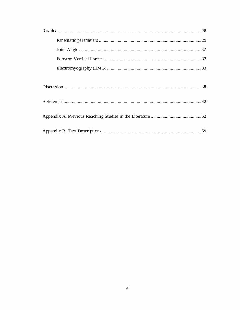
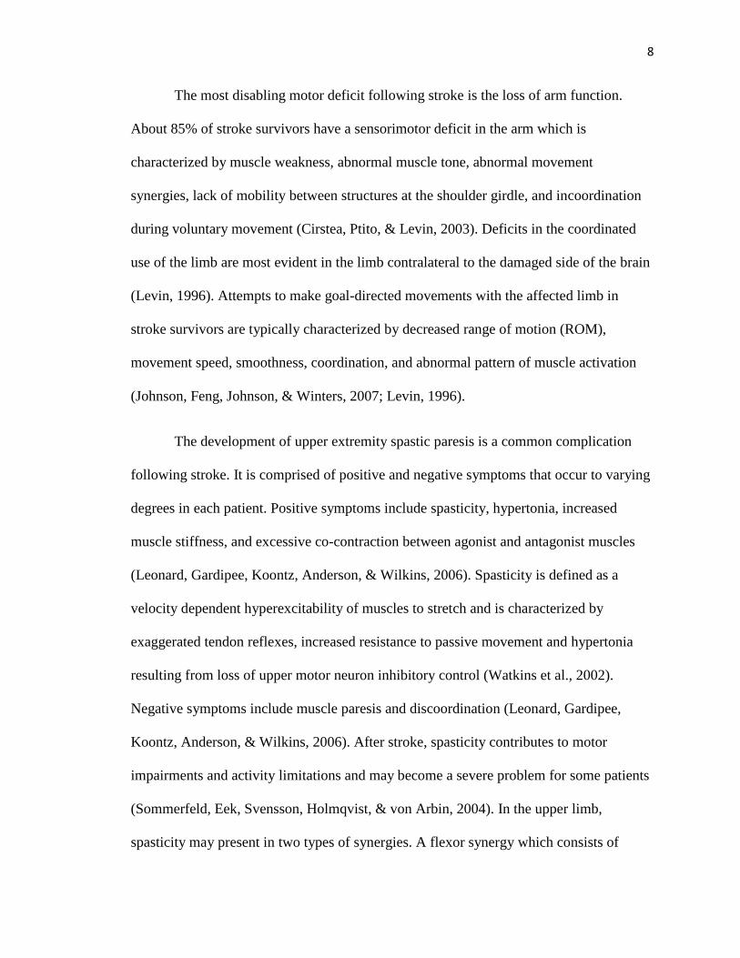
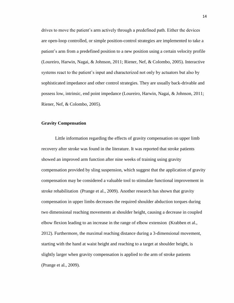
Armeo®Spring Weight-Support System
The level of weight support is device related (no standardized measuring units
have been used to describe level of support) for both arm and forearm (Figure 2). Using
the device scale of arm (A-K) and forearm support (1-5), the mild weight-support level
was defined as (C – D) support levels and (1 – 2) support levels at the arm and forearm
respectively. The moderate weight-support level was defined as (E – G) support levels
and (3 – 4) support levels at the arm and forearm respectively. In order to clarify the
weight-support system of the device, the differences between variable weight-support
levels in both arm and forearm were measured manually using a tension gauge. Results
are displayed in table 2.
Figure 2. Armeo®Spring weight support system
There is a load cell embedded just underneath
the middle of the forearm brace to record the
tension force (i.e., vertical supporting force).

21
Table 2: Armeo®Spring support levels
* The moments at the shoulder level were computed for shoulder flexion movement only. The moments at
the elbow joint level were computed for elbow flexion movements.
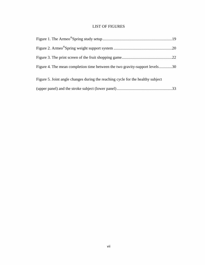
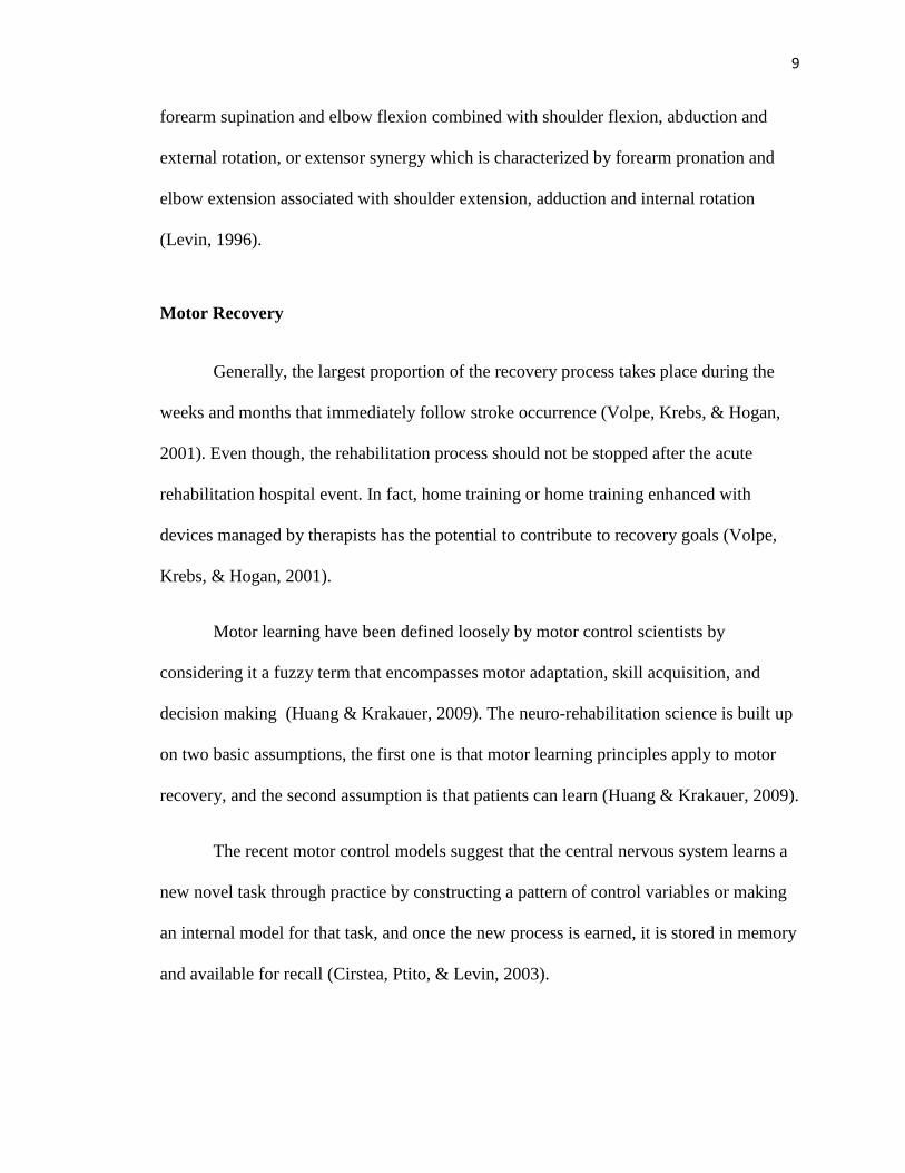
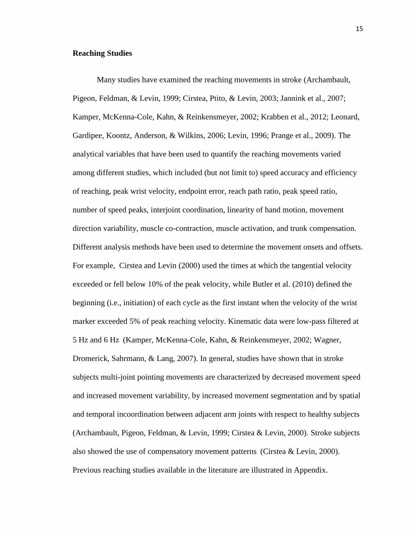
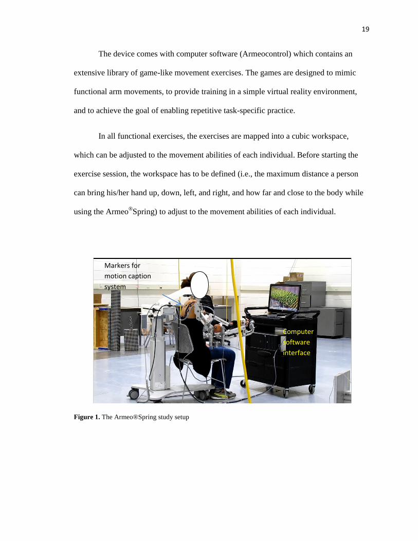
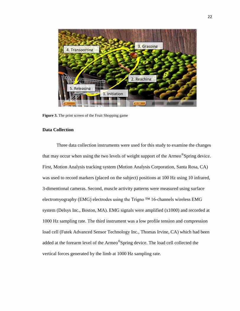
Game: Fruit Shopping
The Fruit Shopping (Figure 3) is one of the games included with Armeocontrol
games library. It is about picking apples and placing them in a shopping cart. The apples
are green in color and will show up one at a time across a computer screen while the
shopping cart is placed at the lower left corner of the screen (for a right-hand user). To
complete the game, the user should move a hand-like pointer using the Armeo®Spring
from the initial start position to reach an apple that turns from green to red in color.
When the pointer is over the red apple, the user should squeeze/grasp the pressure
sensitive handgrip of the Armeo®Spring device to hold the apple and transport the apple
to the shopping cart. When the color of the cart changes the user should take the pressure
off the device handgrip to release the apple. The phases and tasks of the Fruit Shopping
cycle are displayed in Figure 3.
Body Part ArmeoSpring Support
Measured Support
(N.m)
Arm
A 0
B 0.79
C 1.65
D 2.43
E 3.24
F 4.01
G 4.92
H 5.91
I 6.91
J 7.85
K 8.73
Forearm
1 0
2 0.81
3 1.63
4 2.41
5 3.28
22
Figure 3. The print screen of the Fruit Shopping game
Data Collection
Three data collection instruments were used for this study to examine the changes
that may occur when using the two levels of weight support of the Armeo®
Spring device.
First, Motion Analysis tracking system (Motion Analysis Corporation, Santa Rosa, CA)
was used to record markers (placed on the subject) positions at 100 Hz using 10 infrared,
3-dimentional cameras. Second, muscle activity patterns were measured using surface
electromyography (EMG) electrodes using the Trigno ™ 16-channels wireless EMG
system (Delsys Inc., Boston, MA). EMG signals were amplified (x1000) and recorded at
1000 Hz sampling rate. The third instrument was a low profile tension and compression
load cell (Futek Advanced Sensor Technology Inc., Thomas Irvine, CA) which had been
added at the forearm level of the Armeo®Spring device. The load cell collected the
vertical forces generated by the limb at 1000 Hz sampling rate.
4. Transporting
5. Releasing
2. Reaching
1. Initiation
3. Grasping
23
Procedure
Before data collection, subjects were informed to wear tight fitting clothing on the
scheduled data collection date. Upon their arrival, and after signing the informed consent
form, clinical assessments including the Fugl Meyer-Upper Arm Scale and the modified
Ashworth Scale were administered by the PI to assess the stroke severity of the stroke
subject. Afterwards, a total number of 26 reflective markers were placed on the subjects’
chests, backs, shoulders, upper arms, and forearms using a double-sided adhesion tape
directly to the skin. Marker names and positions are illustrated in Table 3. After that, a
total of 7 bipolar surface EMG electrodes were placed to record the activities in the
anterior deltoid, middle deltoid, biceps, triceps, extensor digitorum, flexor digitorum, and
brachioradialis muscles. Before applying the electrodes, the skin beneath the electrode
placing positions was cleaned with alcohol prep pad. Excessive hair, if present, was
shaved using a razor. After applying the electrodes, an initial signal check was performed
to ensure that the EMG electrodes were functioning. Then, the Maximal Voluntary
Contraction (MVC) of each muscle was recorded.
After applying all the markers and EMG electrodes, subjects wore the
Armeo®Spring device while sitting on a stationary chair with no arm support. Then, the
subjects were instructed to practice the Fruit Shopping game by using Armeo®
Spring as
an input device for 3-5 minutes. After that, three trials were recorded for each subject
while using the Armeo®Spring with mild weight support and three trials with moderate
weight support. Within each trial of the Fruit Shopping game, the computer continued to
provide the subject an apple for reaching until (a) the end of time (total duration is 3
minutes), or (b) the subject had picked up all the apple (n =17) within the time limit.
24
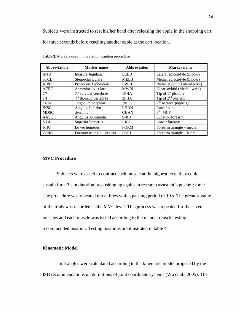
Subjects were instructed to rest his/her hand after releasing the apple in the shopping cart
for three seconds before reaching another apple at the cart location.
Table 3. Markers used in the motion caption procedure
Abbreviation Marker name Abbreviation Marker name
INJU Incisura Jugularis LELB Lateral epicondyle (Elbow)
STCL Sternoclaviculare MELB Medial epicondyle (Elbow)
XIPH Processus Xiphoideus LWRI Radial styloid (Lateral wrist)
ACRO Acromioclaviculare MWRI Ulnar styloid (Medial wrist)
C7 7th
cervical vertebrae 1PHA Tip of 1st phalanx
T4 4th
thoracic vertebrae 2PHA Tip of 2nd
phalanx
TRSC Trigonum Scapulae 2MCP 2nd
Metacarpophalgel
INSC Angulus Inferior LHAN Lower hand
MDSC dynamic CHAN 5th
MCP
AASC Angulus Acromialis S-RU Superior forearm
S-HU Superior humerus I-RU Lower forearm
I-HU Lower humerus FORM Forearm triangle – medial
FORC Forearm triangle – central FORL Forearm triangle – lateral
MVC Procedure
Subjects were asked to contract each muscle at the highest level they could
sustain for ∼3 s in duration by pushing up against a research assistant’s pushing force.
The procedure was repeated three times with a pausing period of 10 s. The greatest value
of the trials was recorded as the MVC level. This process was repeated for the seven
muscles and each muscle was tested according to the manual muscle testing
recommended position. Testing positions are illustrated in table 4.
Kinematic Model
Joint angles were calculated according to the kinematic model proposed by the
ISB recommendations on definitions of joint coordinate systems (Wu et al., 2005). The
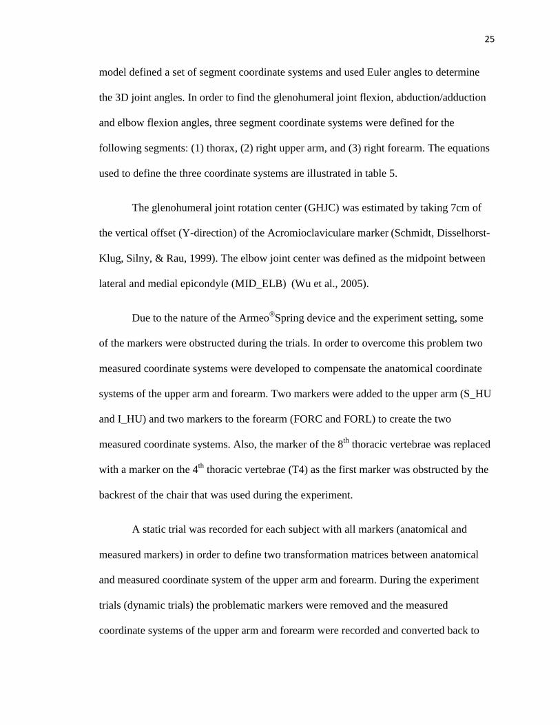
25
model defined a set of segment coordinate systems and used Euler angles to determine
the 3D joint angles. In order to find the glenohumeral joint flexion, abduction/adduction
and elbow flexion angles, three segment coordinate systems were defined for the
following segments: (1) thorax, (2) right upper arm, and (3) right forearm. The equations
used to define the three coordinate systems are illustrated in table 5.
The glenohumeral joint rotation center (GHJC) was estimated by taking 7cm of
the vertical offset (Y-direction) of the Acromioclaviculare marker (Schmidt, Disselhorst-
Klug, Silny, & Rau, 1999). The elbow joint center was defined as the midpoint between
lateral and medial epicondyle (MID_ELB) (Wu et al., 2005).
Due to the nature of the Armeo®Spring device and the experiment setting, some
of the markers were obstructed during the trials. In order to overcome this problem two
measured coordinate systems were developed to compensate the anatomical coordinate
systems of the upper arm and forearm. Two markers were added to the upper arm (S_HU
and I_HU) and two markers to the forearm (FORC and FORL) to create the two
measured coordinate systems. Also, the marker of the 8th
thoracic vertebrae was replaced
with a marker on the 4th
thoracic vertebrae (T4) as the first marker was obstructed by the
backrest of the chair that was used during the experiment.
A static trial was recorded for each subject with all markers (anatomical and
measured markers) in order to define two transformation matrices between anatomical
and measured coordinate system of the upper arm and forearm. During the experiment
trials (dynamic trials) the problematic markers were removed and the measured
coordinate systems of the upper arm and forearm were recorded and converted back to

26
the anatomical coordinate systems using the two transformation matrices defined in the
static trial.
The angles between coordinate systems were calculated using Euler rotation
following ZX’Y’’ sequence. The Z-axis is the flexion/extension axis of the glenohumeral
and elbow joints. The X-axis is the abduction/adduction axis of the glenohumeral and
elbow joints, and the Y-axis internal/external axis of the upper arm and forearm.
Table 4. MVC testing positions
Muscle Position
Anterior deltoid While seated and elbow in slight flexion position, the subject flex
their arm to 90° against the resistance force provided above the
elbow joint
Middle deltoid While seated and elbow in slight flexion position, the subject
abduct their arm to 90° against the resistance force provided above
the elbow joint
Biceps While seated and with slight shoulder flexion and forearm is
supinated, the subject flex elbow to 90 against the resistance force
provided above the wrist joint
Triceps While seated and with slight shoulder flexion and forearm is
supinated, the subject extend elbow from 90 of flexion against the
resistance force provided above the wrist joint
Extensor digitorum While forearm resting on a table and pronated, the subject extend
their wrist against the resistance force provided at subject’s hand
Flexor digitorum While forearm resting on a table and supinated, the subject flex
their wrist against the resistance force provided at subject’s hand
Brachioradialis While seated and with slight shoulder flexion and forearm is
pronated, the subject flex elbow to 90 against the resistance force
provided above the wrist joint
Table 5. Anatomical coordinate systems
Segment Coordinate System
Thorax Origin: GHJC
Yt: ((INJU+C7)/2) – ((XIPH+T4)/2), pointing upward
Zt: cross product of Yt and (C7-INJU), pointing to the right
Xt: cross product of Yt and Zt, pointing forward
Upper arm Origin: GHJC
Yh: GHJC – MID_ELB, pointing to GHJC
Zh: cross product of (MWRI - MID_ELB) and Yh, pointing to the right
27
Xh: cross product of Yh and Zh, pointing forward
Forearm Origin: MWRI
Yf: MID_ELB – MWRI, pointing proximally
Xf: cross product of Yf and (LWRI – MWRI), pointing forward
Zf: cross product of Xf and Yf, pointing to the right
Data Analysis
The data collected using motion capture system, the load cell, and Trigno ™
wireless EMG system were processed and labeled using Cortex 2.4.0 motion analysis
software. The motion analysis data were low-pass filtered at 12 Hz using a Butterworth
filter (Butler et al., 2010). Joint angles for three primary motions of the arm:
glenohumeral joint flexion-extension, abduction-adduction and elbow flexion–extension
were calculated using Matlab (MathWorks, Natick, MA). Each EMG sensor is equipped
with band-pass filter with cut-off frequencies 20- 450 Hz. The EMG signal was full-wave
rectified and smoothed using Root Mean Square (RMS) function using 0.3 seconds time
window (Stoeckmann, Sullivan, & Scheidt, 2009). The muscle activations were measured
as percentages of the MVC value.
The Fruit Shopping cycle consisted of five phases: (1) initiation, (2) reaching, (3)
grasping, (4) transporting, and (5) releasing & resting. The cycle phases were defined
based on the 2MCP marker (base of the index finger on the dorsal side of the hand)
coordinates and velocity. The resting periods between the cycles were used to initially
segregate the cycles. The beginning (i.e., initiation) of each cycle was identified as the
first instant when the velocity of the 2MCP marker exceeded 5% of peak reaching
velocity and continued to increase until it reached 30% of peak reaching velocity while
the 2MCP marker coordinates increased in two axes at least (Butler et al., 2010). The
reaching phase started when the peak reaching velocity exceeded 30% and continued

28
until 2MCP marker reach back to 5% of its peak reaching velocity. The grasping phase
started when the 2MCP marker reached 5% of its peak velocity after the reaching phase
and ended when the 2MCP marker reach back to the last 5% of its peak velocity before
reaching to 30% again. The transporting phase started when the velocity of 2MCP marker
exceeded 5% of its peak velocity following grasping phase and ended when the 2MCP
marker reached back to 5% of its peak velocity. Then, the end of the cycle was signified
by a decrease in 2MCP marker velocity to less than 5% of the peak velocity upon
returning the arm to the initial position.
For each phase, three kinematic parameters (completion time, moving velocity,
acceleration) and one kinetic parameter (arm vertical supporting force) were calculated.
Velocity and acceleration parameters were computed based on the 2MCP marker using
the 3-point central difference method. In addition, the average magnitude of the EMG
envelope was calculated for each phase. For visual inspection purpose, we plotted the
joint angles during one reaching cycle and compared the changes under mild and
moderate weight support conditions.
Independent t-test was used to compare between-group differences (stroke
subjects vs. healthy controls). Sample t-test was used to compare within group
differences (i.e., data from the same stroke subject or data from the same healthy control).
Results
Two subjects were recruited for this case study. A healthy control subject (female,
35 years, 110 lb, 1.52m, right side dominant) and a stroke subject (female, 54 years, 110
29
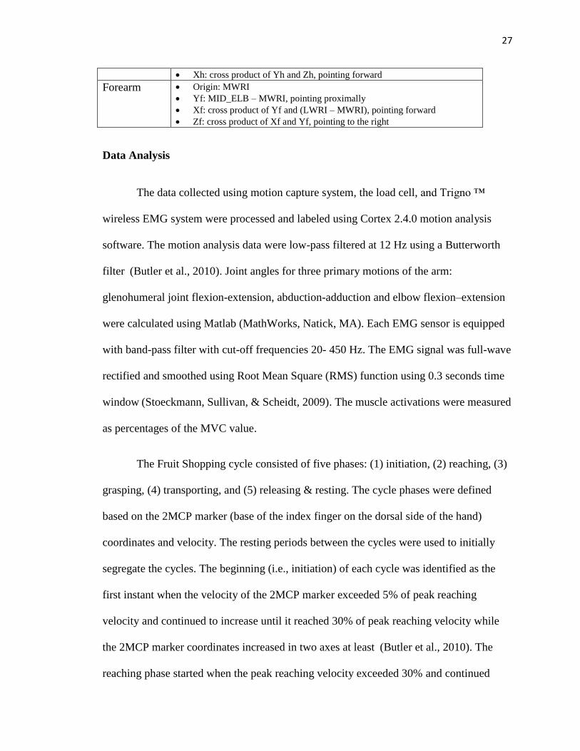
lb, 1.47m, right side dominant). The stroke subject had a stroke for 18 months in her right
side with a Fugl-meyer score of 27/66. Descriptions of subjects’ mild and moderate
weight-support levels provided by the Armeo®Spring device are illustrated in table 6.
Table 6. Armeo®Spring mild and moderate weight-support levels for stroke and healthy subjects.
Subject Body Part Level of Support Support Difference
(N.m) Mild Moderate
Healthy Arm D G 1.58
Forearm 2 4 1.63
Stroke Arm C E 2.50
Forearm 1 3 1.61
* Different baseline support (i.e., mild weight support) was adjusted accordingly depending on the weight
of the subject’s arm, such that with the mild weight support provided by the Armeo®Spring the subject’s
hand was floating just above the knee height in a sitting position. With the moderate weight support, which
was increased with 2 to 3 units weight support (e.g., from C to E was a 2-level increase), the subject’s arm
was floating near the theoretic but not exceeding the shoulder height.
Kinematic parameters
The first two research hypotheses pertained to the within group differences in
reaching performance. As hypothesized within the healthy control subject, results
demonstrated no significant differences in mean completion time, moving velocity, or
acceleration between mild to moderate gravity-support levels during all phases of the
cycle (Table 7). As predicted within the stroke subject (Table 8), results revealed a
significant decrease in the cycle mean completion time (p= 0.042). Specifically, a
significant decrease was found in mean completion time of the grasping phase (p=0.043)
between the two gravity-support levels (Figure 4). When comparing the moving velocity
within the stroke subject, a significant increase was found in the initiation phase moving
velocity (p=0.039) and a significant decrease was found in the grasping phase (p=0.048)

30
between two gravity-support levels. No significant differences were found in all phases
of the cycle when comparing the movement acceleration between the two gravity-support
levels.
Figure 4. Mean completion time between the two gravity-support levels
Table 7. Kinematic parameters of the healthy subject with mild & moderate weight support
Support
Level Parameter
Phase
Initiation Reaching Grasping Transporting Cycle
Mild
Completion
Time
Mean
(s) 0.21 1.05 0.89 1.30 3.46
SD 0.06 0.28 0.39 0.60 0.74
Velocity
Mean
(mm/s) 85.97 220.24 30.38 198.64 153.76
SD 5.13 29.02 7.89 20.72 26.73
Acceleration
Mean
(mm/s2)
577.10 -127.63 4.88 -2.18 -1.51
SD 64.36 18.40 3.66 1.96 2.75
Moderate
Completion
Time
Mean
(s) 0.23 1.11 0.83 1.21 3.38
SD 0.06 0.29 0.35 0.15 0.39
Velocity
Mean
(mm/s) 87.30 222.03 29.96 195.88 145.95
SD 8.30 18.14 5.85 22.19 26.14
Acceleration
Mean
(mm/s2)
556.46 -120.61 3.70 -2.69 -1.50
SD 58.83 23.89 3.89 2.71 2.82
0
2
4
6
8
10
12
Initiation Reaching Grasping Transporting Duration
Me
an C
om
leti
on
Tim
e (
s)
Phase
Mild Support (Healthy)
Moderate Support (Healthy)
Mild Support (Stroke)
Moderate Support (Stroke)
*
*
31
Table 8. Kinematic parameters of the stroke subject with mild & moderate weight support
Support
Level Parameter
Phase
Initiation Reaching Grasping Transporting Cycle
Mild
Completion
Time
Mean
(s) 0.25 1.50 5.41 1.81 8.96
SD 0.16 0.95 5.60 0.49 6.06
Velocity
Mean
(mm/s) 50.47 124.80 54.50 90.49 77.10
SD 5.84 28.97 14.71 26.84 17.36
Acceleration
Mean
(mm/s2)
441.88 -68.59 -0.15 0.52 0.38
SD 263.92 41.05 3.72 3.19 0.84
Moderate
Completion
Time
Mean
(s) 0.22 1.34 2.98 1.75 6.28
SD 0.10 0.59 1.86 0.67 2.35
Velocity
Mean
(mm/s) 54.46 130.33 46.09 96.28 79.94
SD 8.33 34.13 16.71 28.62 18.28
Acceleration
Mean
(mm/s2)
442.27 -73.03 -1.78 1.90 0.20
SD 191.04 37.50 7.03 7.69 1.04
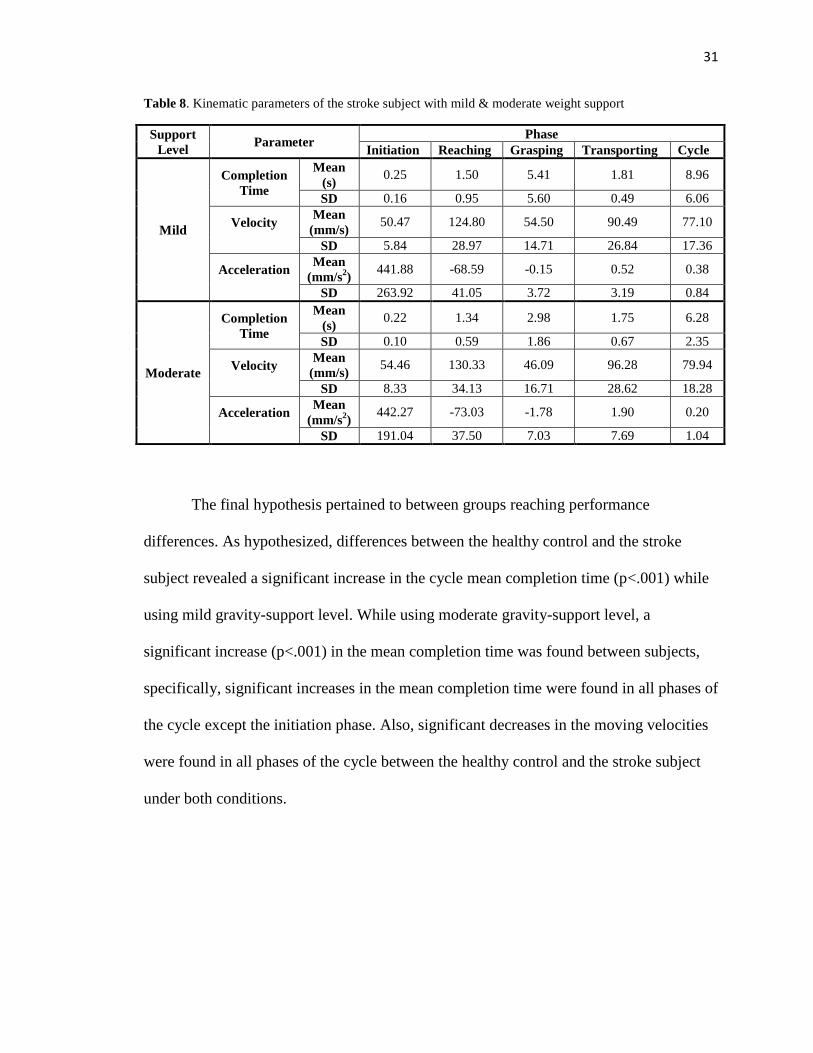
The final hypothesis pertained to between groups reaching performance
differences. As hypothesized, differences between the healthy control and the stroke
subject revealed a significant increase in the cycle mean completion time (p<.001) while
using mild gravity-support level. While using moderate gravity-support level, a
significant increase (p<.001) in the mean completion time was found between subjects,
specifically, significant increases in the mean completion time were found in all phases of
the cycle except the initiation phase. Also, significant decreases in the moving velocities
were found in all phases of the cycle between the healthy control and the stroke subject
under both conditions.
32
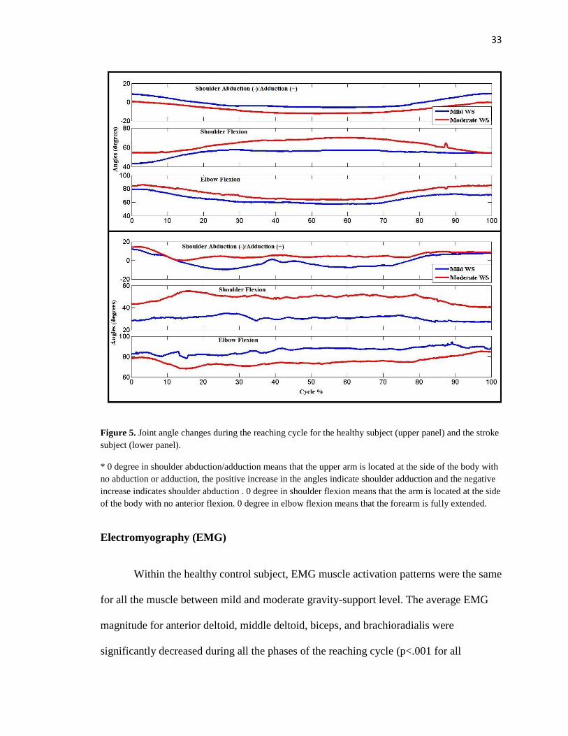
Joint Angles
After increasing the weight-support provided by the Armeo®Spring device, the
healthy control subject showed an increase in abduction and flexion degrees at the
glenohumeral joint level, and an increase in flexion degrees of the elbow joint. On the
other hand, the stroke subject showed a decrease in abduction degrees and an increase in
flexion degrees at the glenohumeral joint level, and a decrease in flexion degrees of the
elbow joint after increasing the weight-support level. Figure 5 displays the average joint
angles during the reaching cycle for the healthy subject (upper panel) and the stroke
subject (lower panel).
Forearm Vertical Forces
Results demonstrated an increase in the mean of vertical forces when changing
gravity-support levels from mild to moderate during all phases of the cycle in both stroke
and healthy subjects. Differences between the healthy control and the stroke subject
revealed an increase in the cycle mean of vertical forces (1.78 lbs) while using mild
gravity-support level. While using moderate gravity-support level, an increase in the
cycle mean of vertical forces (2.67 lbs) was found between subjects. The average vertical
forces for the two subjects during each phase of the reaching cycle are illustrated in table
9 for both weight-support levels.
33
Figure 5. Joint angle changes during the reaching cycle for the healthy subject (upper panel) and the stroke
subject (lower panel).
* 0 degree in shoulder abduction/adduction means that the upper arm is located at the side of the body with
no abduction or adduction, the positive increase in the angles indicate shoulder adduction and the negative
increase indicates shoulder abduction . 0 degree in shoulder flexion means that the arm is located at the side
of the body with no anterior flexion. 0 degree in elbow flexion means that the forearm is fully extended.
Electromyography (EMG)
Within the healthy control subject, EMG muscle activation patterns were the same
for all the muscle between mild and moderate gravity-support level. The average EMG
magnitude for anterior deltoid, middle deltoid, biceps, and brachioradialis were
significantly decreased during all the phases of the reaching cycle (p<.001 for all
34
muscles) when changing the weight-support level from mild to moderate support.
Furthermore, no significant difference was found in the average EMG magnitude for the
triceps, extensor digitorum, and flexor digitorum muscles during all phases of the
reaching cycle between the two weight-support levels. Table 10 displays the average
EMG magnitude between two support levels for the healthy control subject.
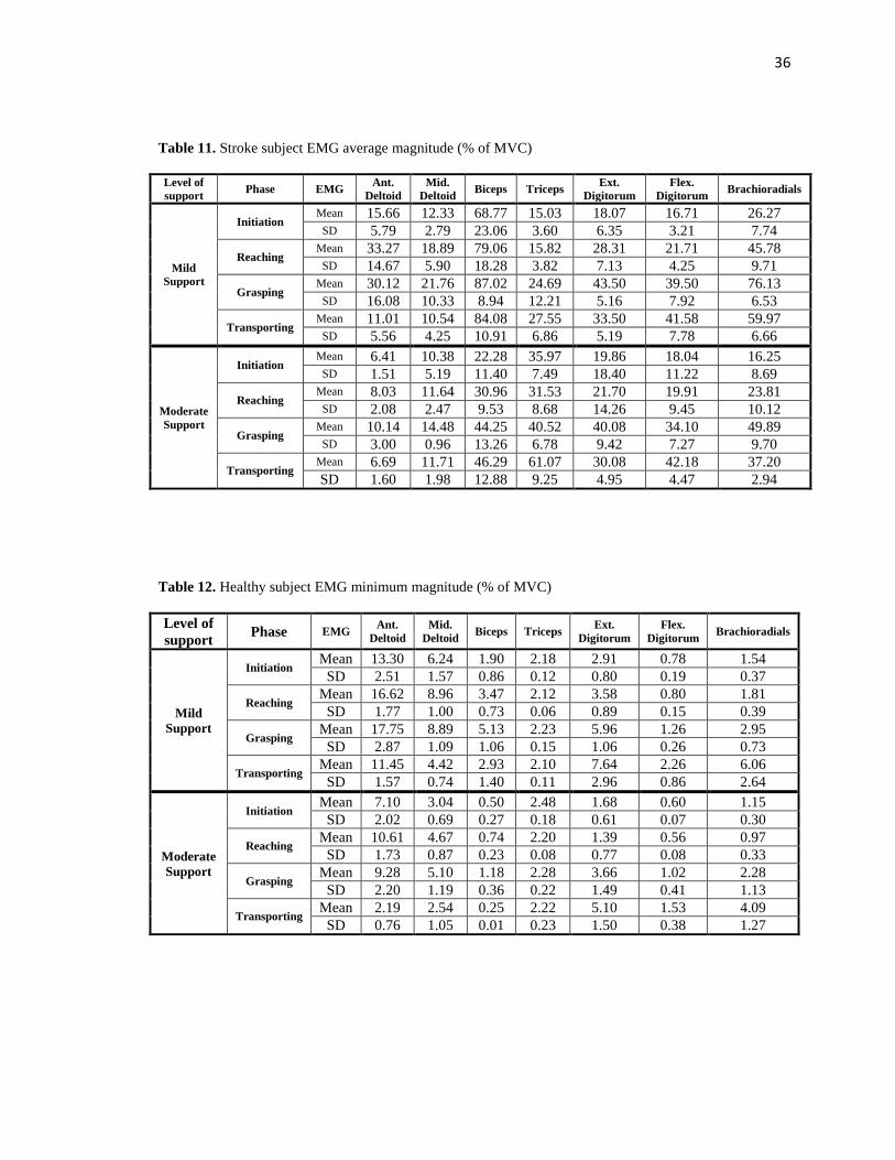
Within the stroke subject, the average EMG magnitude for the anterior deltoid,
biceps, and brachioradialis muscles were significantly decreased in all phases of the
reaching cycle when changing weight-support level from mild to moderate support. On
the other hand, the average EMG magnitude of the triceps muscle was significantly
increased in all phases of the cycle (p<0.001 during initiation, p=0.001 during reaching,
p=0.005 during grasping, and p<0.001 during transporting). No significant difference was
found in the middle deltoid muscle average EMG magnitude during the phases of the
cycle except a significant decrease in the reaching phase (p=0.006) between two weight-
support levels. Furthermore, no significant difference was found in the average EMG
magnitude for the extensor digitorum and flexor digitorum muscles during all phases of
the reaching cycle between the two weight-support levels. Table 11 displays the average
EMG magnitude between two support levels for the stroke subject.
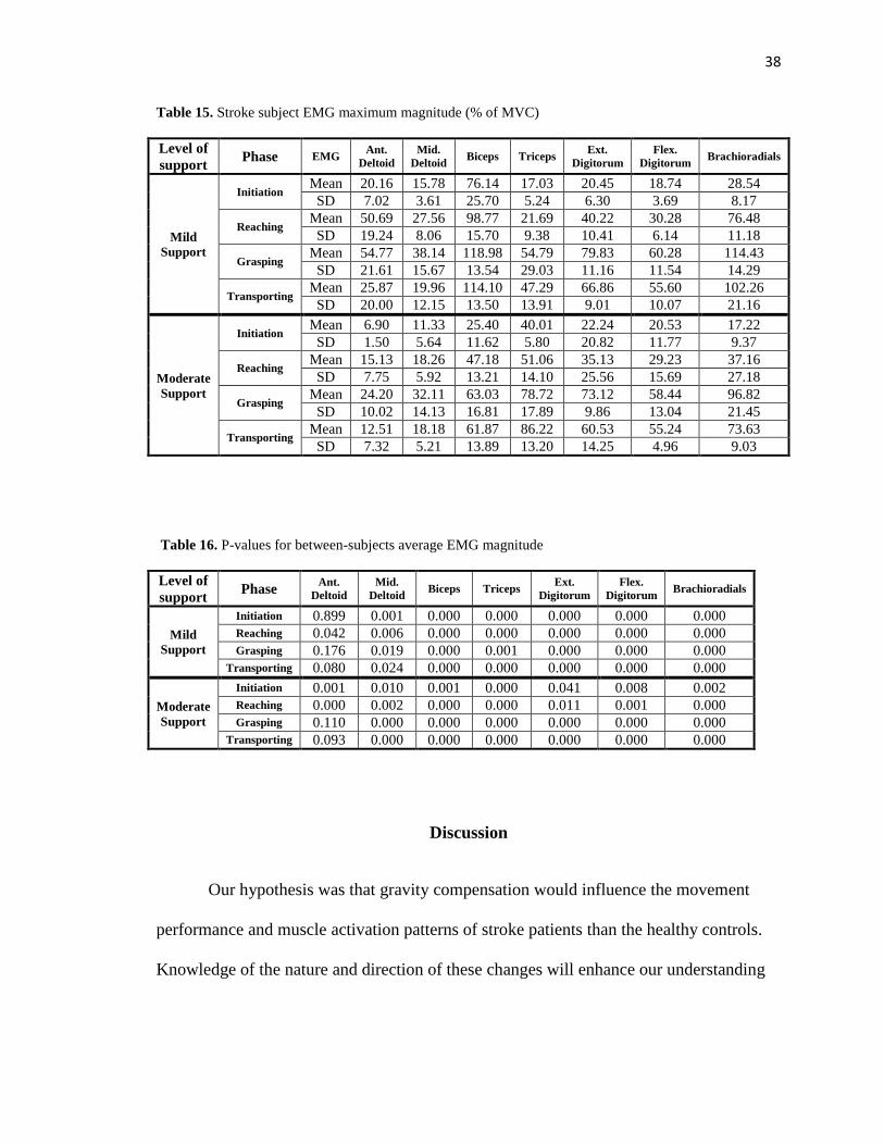
When comparing two subjects under the two weight-support conditions, the
results revealed significant decrease in the average EMG magnitude for all muscles
during all phases of the reaching cycle except for the anterior deltoid muscle. Under the
mild weight-support condition, no significant difference was found in the average EMG
magnitude during the initiation, grasping, and transporting phases. Under the moderate

35
weight-support, no significant difference was found during the grasping and transporting
phases. P-values for between-subjects average EMG magnitude are illustrated in table 16.
Table 9. Vertical support forces for healthy control and stroke subject with mild & moderate weight
support
Subject Support
Level Vertical Force
Phase
Initiation Reaching Grasping Transporting
Healthy
Mild Mean (lb) 4.73 2.18 1.43 5.10
SD 0.32 0.58 0.43 0.57
Moderate Mean (lb) 7.39 5.03 4.72 7.38
SD 0.42 0.51 0.64 0.43
Stroke
Mild Mean (lb) 7.20 5.99 6.14 6.89
SD 0.43 0.15 0.68 0.85
Moderate Mean (lb) 8.44 7.78 7.59 8.25
SD 0.63 0.46 0.36 0.33
Table 10. Healthy subject EMG average magnitude (% of MVC)
Level of support
Phase EMG Ant.
Deltoid Mid.
Deltoid Biceps Triceps
Ext. Digitorum
Flex. Digitorum
Brachioradials
Mild Support
Initiation Mean 15.40 7.82 2.75 2.27 3.86 0.92 1.97
SD 1.96 1.20 0.72 0.14 0.65 0.18 0.39
Reaching Mean 21.42 11.73 5.31 2.35 4.68 1.02 2.34
SD 1.61 1.23 1.20 0.15 0.77 0.18 0.45
Grasping Mean 22.08 11.67 6.59 2.52 13.22 3.49 9.62
SD 3.73 1.83 1.31 0.21 2.60 0.78 2.71
Transporting Mean 14.76 6.58 4.52 2.52 11.56 3.52 9.07
SD 1.61 0.94 1.26 0.32 2.46 0.74 1.90
Moderate Support
Initiation Mean 9.17 3.97 0.65 2.36 3.64 3.31 1.28
SD 1.90 0.69 0.29 0.19 0.66 0.15 0.34
Reaching Mean 13.79 7.48 1.27 2.37 4.55 1.04 1.40
SD 1.25 1.11 0.26 0.16 0.56 1.37 0.38
Grasping Mean 12.21 6.47 1.51 2.63 12.15 3.26 7.28
SD 2.07 1.24 0.39 0.29 2.28 0.58 1.55
Transporting Mean 5.44 4.57 0.49 2.64 10.70 3.45 6.19
SD 1.61 0.92 0.11 0.13 1.46 1.06 0.82
36
Table 11. Stroke subject EMG average magnitude (% of MVC)
Level of
support Phase EMG
Ant.
Deltoid
Mid.
Deltoid Biceps Triceps
Ext.
Digitorum
Flex.
Digitorum Brachioradials
Mild
Support
Initiation Mean 15.66 12.33 68.77 15.03 18.07 16.71 26.27
SD 5.79 2.79 23.06 3.60 6.35 3.21 7.74
Reaching Mean 33.27 18.89 79.06 15.82 28.31 21.71 45.78
SD 14.67 5.90 18.28 3.82 7.13 4.25 9.71
Grasping Mean 30.12 21.76 87.02 24.69 43.50 39.50 76.13
SD 16.08 10.33 8.94 12.21 5.16 7.92 6.53
Transporting Mean 11.01 10.54 84.08 27.55 33.50 41.58 59.97
SD 5.56 4.25 10.91 6.86 5.19 7.78 6.66
Moderate
Support
Initiation Mean 6.41 10.38 22.28 35.97 19.86 18.04 16.25
SD 1.51 5.19 11.40 7.49 18.40 11.22 8.69
Reaching Mean 8.03 11.64 30.96 31.53 21.70 19.91 23.81
SD 2.08 2.47 9.53 8.68 14.26 9.45 10.12
Grasping Mean 10.14 14.48 44.25 40.52 40.08 34.10 49.89
SD 3.00 0.96 13.26 6.78 9.42 7.27 9.70
Transporting Mean 6.69 11.71 46.29 61.07 30.08 42.18 37.20
SD 1.60 1.98 12.88 9.25 4.95 4.47 2.94
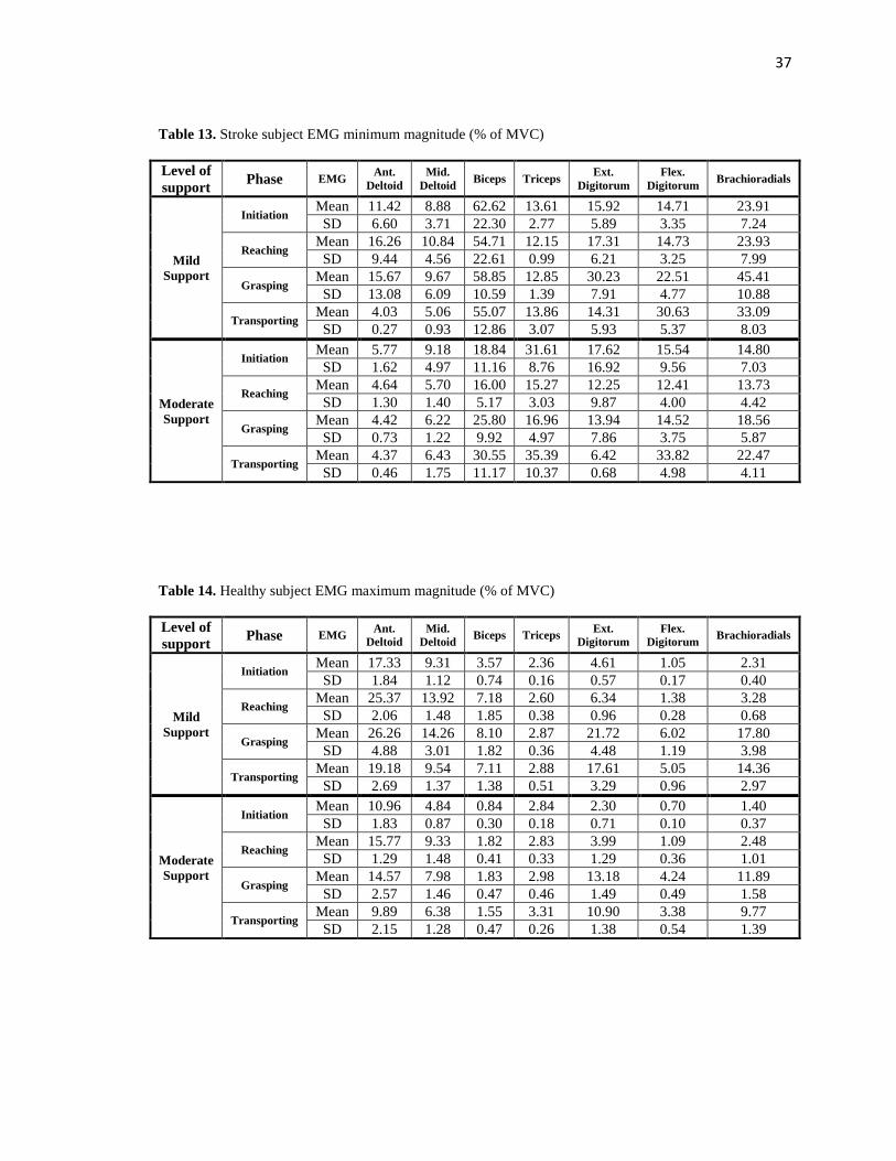
Table 12. Healthy subject EMG minimum magnitude (% of MVC)
Level of
support Phase EMG
Ant.
Deltoid
Mid.
Deltoid Biceps Triceps
Ext.
Digitorum
Flex.
Digitorum Brachioradials
Mild
Support
Initiation Mean 13.30 6.24 1.90 2.18 2.91 0.78 1.54
SD 2.51 1.57 0.86 0.12 0.80 0.19 0.37
Reaching Mean 16.62 8.96 3.47 2.12 3.58 0.80 1.81
SD 1.77 1.00 0.73 0.06 0.89 0.15 0.39
Grasping Mean 17.75 8.89 5.13 2.23 5.96 1.26 2.95
SD 2.87 1.09 1.06 0.15 1.06 0.26 0.73
Transporting Mean 11.45 4.42 2.93 2.10 7.64 2.26 6.06
SD 1.57 0.74 1.40 0.11 2.96 0.86 2.64
Moderate
Support
Initiation Mean 7.10 3.04 0.50 2.48 1.68 0.60 1.15
SD 2.02 0.69 0.27 0.18 0.61 0.07 0.30
Reaching Mean 10.61 4.67 0.74 2.20 1.39 0.56 0.97
SD 1.73 0.87 0.23 0.08 0.77 0.08 0.33
Grasping Mean 9.28 5.10 1.18 2.28 3.66 1.02 2.28
SD 2.20 1.19 0.36 0.22 1.49 0.41 1.13
Transporting Mean 2.19 2.54 0.25 2.22 5.10 1.53 4.09
SD 0.76 1.05 0.01 0.23 1.50 0.38 1.27
37
Table 13. Stroke subject EMG minimum magnitude (% of MVC)
Level of
support Phase EMG
Ant.
Deltoid
Mid.
Deltoid Biceps Triceps
Ext.
Digitorum
Flex.
Digitorum Brachioradials
Mild
Support
Initiation Mean 11.42 8.88 62.62 13.61 15.92 14.71 23.91
SD 6.60 3.71 22.30 2.77 5.89 3.35 7.24
Reaching Mean 16.26 10.84 54.71 12.15 17.31 14.73 23.93
SD 9.44 4.56 22.61 0.99 6.21 3.25 7.99
Grasping Mean 15.67 9.67 58.85 12.85 30.23 22.51 45.41
SD 13.08 6.09 10.59 1.39 7.91 4.77 10.88
Transporting Mean 4.03 5.06 55.07 13.86 14.31 30.63 33.09
SD 0.27 0.93 12.86 3.07 5.93 5.37 8.03
Moderate
Support
Initiation Mean 5.77 9.18 18.84 31.61 17.62 15.54 14.80
SD 1.62 4.97 11.16 8.76 16.92 9.56 7.03
Reaching Mean 4.64 5.70 16.00 15.27 12.25 12.41 13.73
SD 1.30 1.40 5.17 3.03 9.87 4.00 4.42
Grasping Mean 4.42 6.22 25.80 16.96 13.94 14.52 18.56
SD 0.73 1.22 9.92 4.97 7.86 3.75 5.87
Transporting Mean 4.37 6.43 30.55 35.39 6.42 33.82 22.47
SD 0.46 1.75 11.17 10.37 0.68 4.98 4.11
Table 14. Healthy subject EMG maximum magnitude (% of MVC)
Level of
support Phase EMG
Ant.
Deltoid
Mid.
Deltoid Biceps Triceps
Ext.
Digitorum
Flex.
Digitorum Brachioradials
Mild
Support
Initiation Mean 17.33 9.31 3.57 2.36 4.61 1.05 2.31
SD 1.84 1.12 0.74 0.16 0.57 0.17 0.40
Reaching Mean 25.37 13.92 7.18 2.60 6.34 1.38 3.28
SD 2.06 1.48 1.85 0.38 0.96 0.28 0.68
Grasping Mean 26.26 14.26 8.10 2.87 21.72 6.02 17.80
SD 4.88 3.01 1.82 0.36 4.48 1.19 3.98
Transporting Mean 19.18 9.54 7.11 2.88 17.61 5.05 14.36
SD 2.69 1.37 1.38 0.51 3.29 0.96 2.97
Moderate
Support
Initiation Mean 10.96 4.84 0.84 2.84 2.30 0.70 1.40
SD 1.83 0.87 0.30 0.18 0.71 0.10 0.37
Reaching Mean 15.77 9.33 1.82 2.83 3.99 1.09 2.48
SD 1.29 1.48 0.41 0.33 1.29 0.36 1.01
Grasping Mean 14.57 7.98 1.83 2.98 13.18 4.24 11.89
SD 2.57 1.46 0.47 0.46 1.49 0.49 1.58
Transporting Mean 9.89 6.38 1.55 3.31 10.90 3.38 9.77
SD 2.15 1.28 0.47 0.26 1.38 0.54 1.39
38
Table 15. Stroke subject EMG maximum magnitude (% of MVC)
Level of
support Phase EMG
Ant.
Deltoid
Mid.
Deltoid Biceps Triceps
Ext.
Digitorum
Flex.
Digitorum Brachioradials
Mild
Support
Initiation Mean 20.16 15.78 76.14 17.03 20.45 18.74 28.54
SD 7.02 3.61 25.70 5.24 6.30 3.69 8.17
Reaching Mean 50.69 27.56 98.77 21.69 40.22 30.28 76.48
SD 19.24 8.06 15.70 9.38 10.41 6.14 11.18
Grasping Mean 54.77 38.14 118.98 54.79 79.83 60.28 114.43
SD 21.61 15.67 13.54 29.03 11.16 11.54 14.29
Transporting Mean 25.87 19.96 114.10 47.29 66.86 55.60 102.26
SD 20.00 12.15 13.50 13.91 9.01 10.07 21.16
Moderate
Support
Initiation Mean 6.90 11.33 25.40 40.01 22.24 20.53 17.22
SD 1.50 5.64 11.62 5.80 20.82 11.77 9.37
Reaching Mean 15.13 18.26 47.18 51.06 35.13 29.23 37.16
SD 7.75 5.92 13.21 14.10 25.56 15.69 27.18
Grasping Mean 24.20 32.11 63.03 78.72 73.12 58.44 96.82
SD 10.02 14.13 16.81 17.89 9.86 13.04 21.45
Transporting Mean 12.51 18.18 61.87 86.22 60.53 55.24 73.63
SD 7.32 5.21 13.89 13.20 14.25 4.96 9.03
Table 16. P-values for between-subjects average EMG magnitude
Level of
support Phase
Ant.
Deltoid
Mid.
Deltoid Biceps Triceps
Ext.
Digitorum
Flex.
Digitorum Brachioradials
Mild
Support
Initiation 0.899 0.001 0.000 0.000 0.000 0.000 0.000
Reaching 0.042 0.006 0.000 0.000 0.000 0.000 0.000
Grasping 0.176 0.019 0.000 0.001 0.000 0.000 0.000
Transporting 0.080 0.024 0.000 0.000 0.000 0.000 0.000
Moderate
Support
Initiation 0.001 0.010 0.001 0.000 0.041 0.008 0.002
Reaching 0.000 0.002 0.000 0.000 0.011 0.001 0.000
Grasping 0.110 0.000 0.000 0.000 0.000 0.000 0.000
Transporting 0.093 0.000 0.000 0.000 0.000 0.000 0.000
Discussion
Our hypothesis was that gravity compensation would influence the movement
performance and muscle activation patterns of stroke patients than the healthy controls.
Knowledge of the nature and direction of these changes will enhance our understanding
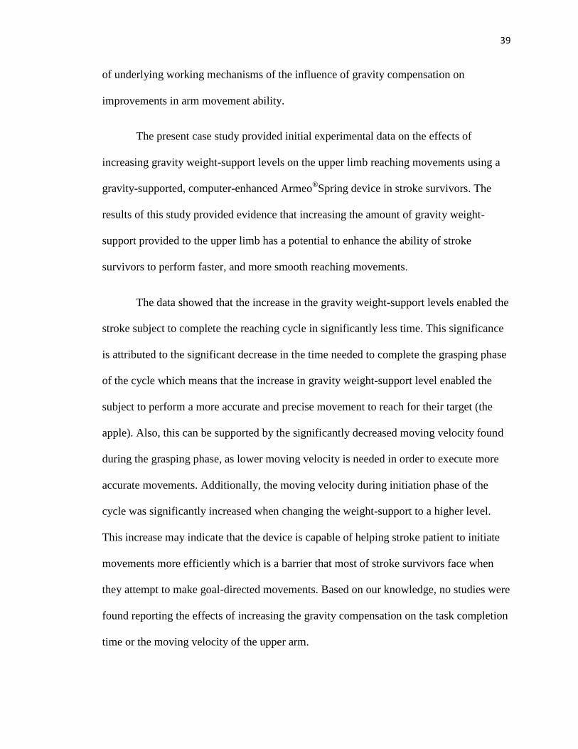
39
of underlying working mechanisms of the influence of gravity compensation on
improvements in arm movement ability.
The present case study provided initial experimental data on the effects of
increasing gravity weight-support levels on the upper limb reaching movements using a
gravity-supported, computer-enhanced Armeo®Spring device in stroke survivors. The
results of this study provided evidence that increasing the amount of gravity weight-
support provided to the upper limb has a potential to enhance the ability of stroke
survivors to perform faster, and more smooth reaching movements.
The data showed that the increase in the gravity weight-support levels enabled the
stroke subject to complete the reaching cycle in significantly less time. This significance
is attributed to the significant decrease in the time needed to complete the grasping phase
of the cycle which means that the increase in gravity weight-support level enabled the
subject to perform a more accurate and precise movement to reach for their target (the
apple). Also, this can be supported by the significantly decreased moving velocity found
during the grasping phase, as lower moving velocity is needed in order to execute more
accurate movements. Additionally, the moving velocity during initiation phase of the
cycle was significantly increased when changing the weight-support to a higher level.
This increase may indicate that the device is capable of helping stroke patient to initiate
movements more efficiently which is a barrier that most of stroke survivors face when
they attempt to make goal-directed movements. Based on our knowledge, no studies were
found reporting the effects of increasing the gravity compensation on the task completion
time or the moving velocity of the upper arm.
40
When comparing muscle activity levels during the cycle between two different
support levels, we found that the level of muscle activity was lower during movements
with higher weight-support in the anterior deltoid, middle deltoid, biceps, and
brachioradialis muscles in both the stroke subject and the control subject. In other words,
in both stroke and control subjects, the increase in weight-support level reduced the level
of muscle activity needed to hold the arm in a certain orientation during the cycle. These
results are consistent with findings reported by Jannink et al. (2007) and Prange et al.
(2009). This finding supports that the Armeo®Spring device enable the stroke patients to
generate movements with less efforts. Perry, Powell, and Rosen (2009) reported that the
majority of human arm joint torques are devoted to supporting the human arm position in
space while compensating gravitational loads whereas a minor portion is dedicated to arm
motion itself.
During the reaching cycle, the results showed a decrease in the glenohumeral joint
abduction movements accompanied with an increase in the elbow joint extension
movements in the stroke subject while using a higher weight-support level. These results
suggest that the increase in weight-support level changed the motion control mechanism
to depend more on the movement of the distal joint (elbow) with less contribution from
the proximal joint (glenohumeral) to complete the reaching cycle. This suggestion can
explain the increased EMG magnitude found in triceps muscle after increasing the level
of weight-support. This finding can be supported by a recent research that showed that
the arm support decreases the required shoulder abduction torques during two
dimensional reaching movements at shoulder height, subsequently causing a decrease in
coupled elbow flexion, leading to an increase in the range of elbow extension (Iwamuro,

41
Cruz, Connelly, Fischer, & Kamper, 2008; Krabben et al., 2012). The increase in the
triceps EMG magnitude can be attributed also to force needed to push against the extra
weight support provided by the Armeo®Spring device under the moderate weight-support
condition.
The stroke subject showed larger shoulder adduction movements comparing to the
healthy control, however, these results were difficult to conclude as the moving range
(i.e., working space) was calculated and defined for each subject to allow each individual
to be able to complete the Fruit Shopping task.
There were several limitations of this study. First, we encountered challenges in
study setup as the markers were sometimes obstructed by the Armeo®Spring devices
while subjects were performing the reaching task. We have spent considerable amount of
time to perform data cleaning (fill in gaps, correct switching maker data) to ensure the
quality and validity of the data. Second, we did not randomize the two conditions (mild
and moderate weight-support). Subjects were instructed to complete the Fruit Shopping
under mild weight support followed by moderate weight support. As a result, one could
argue that observed changes under these two conditions might be due to practice or
fatigue effects.
In summary, the significant differences in movement performance between mild
and moderate physical weight support suggest the gravity-supported mechanism provides
a mean to facilitate functional upper limb motor performance in individuals with stroke.
42
References
Aisen, M. L., Krebs, H. I., Hogan, N., McDowell, F., & Volpe, B. T. (1997). The effect
of robot-assisted therapy and rehabilitative training on motor recovery following
stroke. Archives of Neurology, 54(4), 443-446.
American Heart Association. (2011). Retrieved 10/17, 2011, from
http://www.strokeassociation.org/STROKEORG/AboutStroke/TypesofStroke/Types
-of-Stroke_UCM_308531_SubHomePage.jsp
Archambault, P., Pigeon, P., Feldman, A., & Levin, M. (1999). Recruitment and
sequencing of different degrees of freedom during pointing movements involving the
trunk in healthy and hemiparetic subjects. Experimental Brain Research.
Experimentelle Hirnforschung. Expérimentation Cérébrale, 126(1), 55-67.
Bosecker, C., Dipietro, L., Volpe, B., & Krebs, H. I. (2010). Kinematic robot-based
evaluation scales and clinical counterparts to measure upper limb motor performance
in patients with chronic stroke. Neurorehabilitation and Neural Repair, 24(1), 62-69.
doi: 10.1177/1545968309343214
Broeks, J. G., Lankhorst, G. J., Rumping, K., & Prevo, A. J. (1999). The long-term
outcome of arm function after stroke: Results of a follow-up study. Disability and
Rehabilitation, 21(8), 357-364.
Brunnstrom, S. (1966). Motor testing procedures in hemiplegia: Based on sequential
recovery stages. Physical Therapy, 46(4), 357-375.
43
Burgar, C. G., Lum, P. S., Scremin, A. M., Garber, S. L., Van der Loos, H. F., Kenney,
D., & Shor, P. (2011). Robot-assisted upper-limb therapy in acute rehabilitation
setting following stroke: Department of veterans affairs multisite clinical trial.
Journal of Rehabilitation Research and Development, 48(4), 445-458.
Butler, E. E., Ladd, A. L., Louie, S. A., Lamont, L. E., Wong, W., & Rose, J. (2010).
Three-dimensional kinematics of the upper limb during a reach and grasp cycle for
children. Gait & Posture, 32(1), 72-77. doi: 10.1016/j.gaitpost.2010.03.011
Cirstea, M. C., & Levin, M. F. (2000). Compensatory strategies for reaching in stroke.
Brain : A Journal of Neurology, 123 ( 5), 940-953.
Cirstea, M. C., Ptito, A., & Levin, M. F. (2003). Arm reaching improvements with short-
term practice depend on the severity of the motor deficit in stroke. Experimental
Brain Research.Experimentelle Hirnforschung.Experimentation Cerebrale, 152(4),
476-488. doi: 10.1007/s00221-003-1568-4
Crow, J. L., & Harmeling-van der Wel, B. C. (2008). Hierarchical properties of the motor
function sections of the fugl-meyer assessment scale for people after stroke: A
retrospective study. Physical Therapy, 88(12), 1554-1567. doi:
10.2522/ptj.20070186
Dirnagl, U., Iadecola, C., & Moskowitz, M. A. (1999). Pathobiology of ischaemic stroke:
An integrated view. Trends in Neurosciences, 22(9), 391-397.

44
Engelhardt, K. G. (1986). Current status and future prospects for robotic technology in
health care delivery. Central Nervous System Trauma : Journal of the American
Paralysis Association, 3(1), 111-126.
Gijbels, D., Lamers, I., Kerkhofs, L., Alders, G., Knippenberg, E., & Feys, P. (2011). The
armeo spring as training tool to improve upper limb functionality in multiple
sclerosis: A pilot study. Journal of Neuroengineering and Rehabilitation, 8, 5. doi:
10.1186/1743-0003-8-5
Hatano, S. (1976). Experience from a multicentre stroke register: A preliminary report.
Bulletin of the World Health Organization, 54(5), 541-553.
Hingtgen, B., McGuire, J. R., Wang, M., & Harris, G. F. (2006). An upper extremity
kinematic model for evaluation of hemiparetic stroke. Journal of Biomechanics,
39(4), 681-688. doi: 10.1016/j.jbiomech.2005.01.008
Housman, S. J., Scott, K. M., & Reinkensmeyer, D. J. (2009). A randomized controlled
trial of gravity-supported, computer-enhanced arm exercise for individuals with
severe hemiparesis. Neurorehabilitation and Neural Repair, 23(5), 505-514. doi:
10.1177/1545968308331148
Hsieh, Y. W., Wu, C. Y., Liao, W. W., Lin, K. C., Wu, K. Y., & Lee, C. Y. (2011).
Effects of treatment intensity in upper limb robot-assisted therapy for chronic stroke:
A pilot randomized controlled trial. Neurorehabilitation and Neural Repair, 25(6),
503-511. doi: 10.1177/1545968310394871
45
Huang, V. S., & Krakauer, J. W. (2009). Robotic neurorehabilitation: A computational
motor learning perspective. Journal of Neuroengineering and Rehabilitation, 6, 5.
doi: 10.1186/1743-0003-6-5
Iwamuro, B. T., Cruz, E. G., Connelly, L. L., Fischer, H. C., & Kamper, D. G. (2008).
Effect of a gravity-compensating orthosis on reaching after stroke: Evaluation of the
therapy assistant WREX. Archives of Physical Medicine and Rehabilitation, 89(11),
2121-2128. doi: 10.1016/j.apmr.2008.04.022; 10.1016/j.apmr.2008.04.022
Jannink, M. J. A., Prange, G. B., Stienen, A. H. A., van der Kooij, H., Kruitbosch, J. M.,
Ijzerman, M. J., & Hermens, H. J. (2007). Reduction of muscle activity during
repeated reach and retrieval with gravity compensation in stroke patients.
Rehabilitation Robotics, 2007. ICORR 2007. IEEE 10th International Conference
on, 472-476.
Johnson, M. J., Feng, X., Johnson, L. M., & Winters, J. M. (2007). Potential of a suite of
robot/computer-assisted motivating systems for personalized, home-based, stroke
rehabilitation. Journal of Neuroengineering and Rehabilitation, 4, 6. doi:
10.1186/1743-0003-4-6
Kamper, D. G., McKenna-Cole, A. N., Kahn, L. E., & Reinkensmeyer, D. J. (2002).
Alterations in reaching after stroke and their relation to movement direction and
impairment severity. Archives of Physical Medicine and Rehabilitation, 83(5), 702-
707.

46
Krabben, T., Prange, G. B., Molier, B. I., Stienen, A. H., Jannink, M. J., Buurke, J. H., &
Rietman, J. S. (2012). Influence of gravity compensation training on synergistic
movement patterns of the upper extremity after stroke, a pilot study. Journal of
Neuroengineering and Rehabilitation, 9, 44-0003-9-44. doi: 10.1186/1743-0003-9-
44; 10.1186/1743-0003-9-44
Krebs, H. I., Hogan, N., Aisen, M. L., & Volpe, B. T. (1998). Robot-aided
neurorehabilitation. IEEE Transactions on Rehabilitation Engineering : A
Publication of the IEEE Engineering in Medicine and Biology Society, 6(1), 75-87.
Krebs, H. I., Volpe, B. T., Aisen, M. L., & Hogan, N. (2000). Increasing productivity and
quality of care: Robot-aided neuro-rehabilitation. Journal of Rehabilitation Research
and Development, 37(6), 639-652.
Leonard, C. T., Gardipee, K. A., Koontz, J. R., Anderson, J. H., & Wilkins, S. A. (2006).
Correlation between impairment and motor performance during reaching tasks in
subjects with spastic hemiparesis. Journal of Rehabilitation Medicine : Official
Journal of the UEMS European Board of Physical and Rehabilitation Medicine,
38(4), 243-249. doi: 10.1080/16501970600609808
Levin, M. F. (1996). Interjoint coordination during pointing movements is disrupted in
spastic hemiparesis. Brain : A Journal of Neurology, 119 ( Pt 1)(Pt 1), 281-293.
Lo, A. C., Guarino, P. D., Richards, L. G., Haselkorn, J. K., Wittenberg, G. F., Federman,
D. G., . . . Peduzzi, P. (2010). Robot-assisted therapy for long-term upper-limb
47
impairment after stroke. The New England Journal of Medicine, 362(19), 1772-
1783. doi: 10.1056/NEJMoa0911341
Loureiro, R. C., Harwin, W. S., Nagai, K., & Johnson, M. (2011). Advances in upper
limb stroke rehabilitation: A technology push. Medical & Biological Engineering &
Computing, 49(10), 1103-1118. doi: 10.1007/s11517-011-0797-0
Lum, P. S., Burgar, C. G., Shor, P. C., Majmundar, M., & Van der Loos, M. (2002).
Robot-assisted movement training compared with conventional therapy techniques
for the rehabilitation of upper-limb motor function after stroke. Archives of Physical
Medicine and Rehabilitation, 83(7), 952-959.
Macclellan, L. R., Bradham, D. D., Whitall, J., Volpe, B., Wilson, P. D., Ohlhoff, J., . . .
Bever, C. T.,Jr. (2005). Robotic upper-limb neurorehabilitation in chronic stroke
patients. Journal of Rehabilitation Research and Development, 42(6), 717-722.
Masiero, S., Armani, M., & Rosati, G. (2011). Upper-limb robot-assisted therapy in
rehabilitation of acute stroke patients: Focused review and results of new
randomized controlled trial. Journal of Rehabilitation Research and Development,
48(4), 355-366.
Meyers, L. S., Gamst, G., & Guarino, A. J. (2006). Applied multivariate research: Design
and interpretation. (1st ed.). Thousand Oaks, CA: Sage publications, Inc.
National Stroke Association. (2011). Retrieved 10/16, 2011, from
http://www.stroke.org/site/PageServer?pagename=stroke

48
Nef, T., Mihelj, M., & Riener, R. (2007). ARMin: A robot for patient-cooperative arm
therapy. Med Bio Eng Comput, 45, 887-900. doi: 10.1007/s11517-007-0226-6
Perry, J., Powell, J., & Rosen, J. (2009). Isotropy of an upper limb exoskeleton and the
kinematics and dynamics of the human arm. Applied Bionics and Biomechanics,
6(2), 175-191. doi: 10.1080/11762320902920575
Prange, G. B., Jannink, M. J., Groothuis-Oudshoorn, C. G., Hermens, H. J., & Ijzerman,
M. J. (2006). Systematic review of the effect of robot-aided therapy on recovery of
the hemiparetic arm after stroke. Journal of Rehabilitation Research and
Development, 43(2), 171-184.
Prange, G. B., Jannink, M. J., Stienen, A. H., van der Kooij, H., Ijzerman, M. J., &
Hermens, H. J. (2009). Influence of gravity compensation on muscle activation
patterns during different temporal phases of arm movements of stroke patients.
Neurorehabilitation and Neural Repair, 23(5), 478-485. doi:
10.1177/1545968308328720
Riener, R., Nef, T., & Colombo, G. (2005). Robot-aided neurorehabilitation of the upper
extremities. Medical & Biological Engineering & Computing, 43(1), 2-10.
Roger, V. L., Go, A. S., Lloyd-Jones, D. M., Benjamin, E. J., Berry, J. D., Borden, W. B.,
. . . on behalf of the American Heart Association Statistics Committee and Stroke
Statistics Subcommittee. (2011). Heart disease and stroke statistics--2012 update: A
report from the american heart association. Circulation, doi:
10.1161/CIR.0b013e31823ac046
49
Sainburg, R. L., & Kalakanis, D. (2000). Differences in control of limb dynamics during
dominant and nondominant arm reaching. Journal of Neurophysiology, 83(5), 2661-
2675.
Sanchez, R. J., Liu, J., Rao, S., Shah, P., Smith, R., Rahman, T., . . . Reinkensmeyer, D. J.
(2006). Automating arm movement training following severe stroke: Functional
exercises with quantitative feedback in a gravity-reduced environment. IEEE
Transactions on Neural Systems and Rehabilitation Engineering : A Publication of
the IEEE Engineering in Medicine and Biology Society, 14(3), 378-389. doi:
10.1109/TNSRE.2006.881553
Schmidt, R., Disselhorst-Klug, C., Silny, J., & Rau, G. (1999). A marker-based
measurement procedure for unconstrained wrist and elbow motions. Journal of
Biomechanics, 32(6), 615-621.
Sommerfeld, D. K., Eek, E. U., Svensson, A. K., Holmqvist, L. W., & von Arbin, M. H.
(2004). Spasticity after stroke: Its occurrence and association with motor
impairments and activity limitations. Stroke; a Journal of Cerebral Circulation,
35(1), 134-139. doi: 10.1161/01.STR.0000105386.05173.5E
Steultjens, E., Dekker, J., Bouter, L., van de Nes, J., Cup, E., & van den Ende, C. (2003).
Occupational therapy for stroke patients : A systematic review. American Heart
Association, doi: 10.1161/01.STR.0000057576.77308.30
50
Stoeckmann, T. M., Sullivan, K. J., & Scheidt, R. A. (2009). Elastic, viscous, and mass
load effects on poststroke muscle recruitment and co-contraction during reaching: A
pilot study. Physical Therapy, 89(7), 665-678. doi: 10.2522/ptj.20080128
Twitchell, T. E. (1951). The restoration of motor function following hemiplegia in man.
Brain : A Journal of Neurology, 74(4), 443-480.
Van Peppen, R. P., Kwakkel, G., Wood-Dauphinee, S., Hendriks, H. J., Van der Wees, P.
J., & Dekker, J. (2004). The impact of physical therapy on functional outcomes after
stroke: What's the evidence? Clinical Rehabilitation, 18(8), 833-862.
Volpe, B. T., Huerta, P. T., Zipse, J. L., Rykman, A., Edwards, D., Dipietro, L., . . .
Krebs, H. I. (2009). Robotic devices as therapeutic and diagnostic tools for stroke
recovery. Archives of Neurology, 66(9), 1086-1090. doi:
10.1001/archneurol.2009.182
Volpe, B. T., Krebs, H. I., & Hogan, N. (2001). Is robot-aided sensorimotor training in
stroke rehabilitation a realistic option? Current Opinion in Neurology, 14(6), 745-
752.
Wagner, J. M., Dromerick, A. W., Sahrmann, S. A., & Lang, C. E. (2007). Upper
extremity muscle activation during recovery of reaching in subjects with post-stroke
hemiparesis. Clinical Neurophysiology : Official Journal of the International
Federation of Clinical Neurophysiology, 118(1), 164-176. doi:
10.1016/j.clinph.2006.09.022
51
Wagner, T. H., Lo, A. C., Peduzzi, P., Bravata, D. M., Huang, G. D., Krebs, H. I., . . .
Guarino, P. D. (2011). An economic analysis of robot-assisted therapy for long-term
upper-limb impairment after stroke. Stroke; a Journal of Cerebral Circulation,
42(9), 2630-2632. doi: 10.1161/STROKEAHA.110.606442
Wang, C. J., Wang, C. X., Zhang, L., Wang, Y. L., & Wang, Y. J. (2011). Advances in
stroke care and research in 2010. Clinical and Experimental Pharmacology &
Physiology, 38(9), 562-569. doi: 10.1111/j.1440-1681.2011.05566.x;
10.1111/j.1440-1681.2011.05566.x
Watkins, C. L., Leathley, M. J., Gregson, J. M., Moore, A. P., Smith, T. L., & Sharma, A.
K. (2002). Prevalence of spasticity post stroke. Clinical Rehabilitation, 16(5), 515-
522.
WHO. (2011). Retrieved 10/15, 2011, from
http://www.who.int/topics/cerebrovascular_accident/en/
Wisneski, K. J., & Johnson, M. J. (2007). Quantifying kinematics of purposeful
movements to real, imagined, or absent functional objects: Implications for
modelling trajectories for robot-assisted ADL tasks. Journal of Neuroengineering
and Rehabilitation, 4, 7. doi: 10.1186/1743-0003-4-7
Wu, G., van der Helm, F. C., Veeger, H. E., Makhsous, M., Van Roy, P., Anglin, C., . . .
International Society of Biomechanics. (2005). ISB recommendation on definitions
of joint coordinate systems of various joints for the reporting of human joint motion-
-part II: Shoulder, elbow, wrist and hand. Journal of Biomechanics, 38(5), 981-992.

52
52
Appendix A: Previous Reaching Studies in the Literature
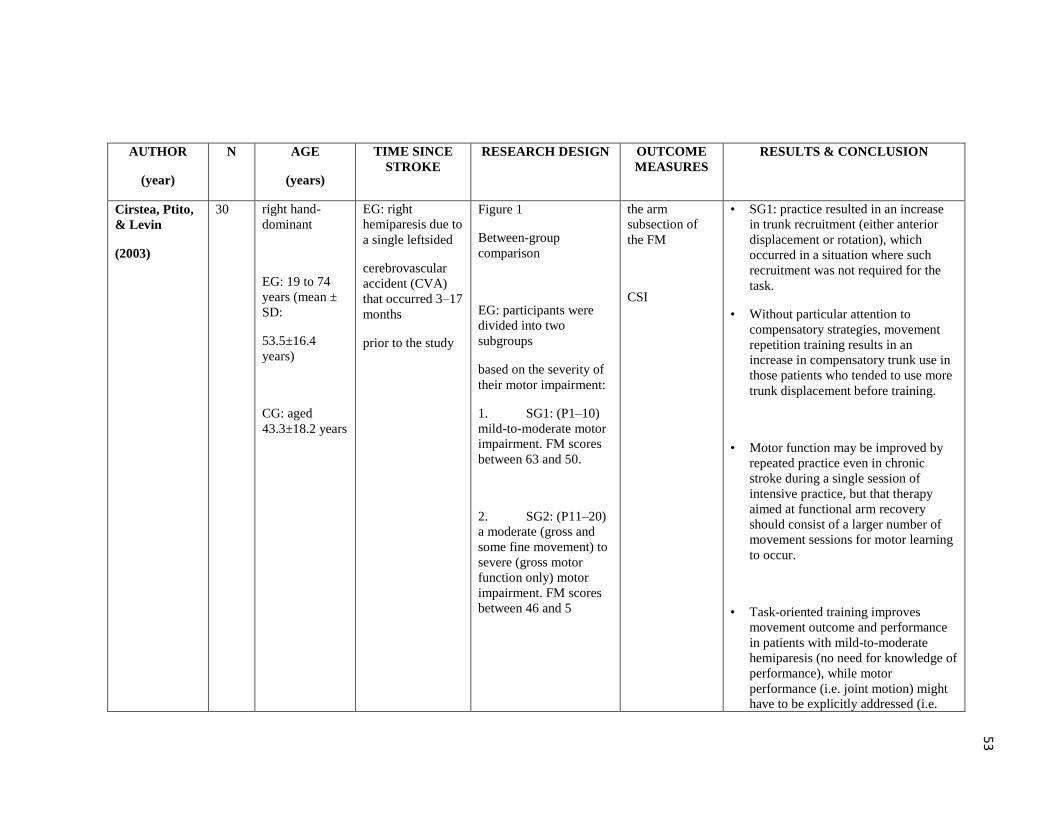
53
AUTHOR
(year)
N AGE
(years)
TIME SINCE
STROKE
RESEARCH DESIGN OUTCOME
MEASURES
RESULTS & CONCLUSION
Cirstea, Ptito,
& Levin
(2003)
30 right hand-
dominant
EG: 19 to 74
years (mean ±
SD:
53.5±16.4
years)
CG: aged
43.3±18.2 years
EG: right
hemiparesis due to
a single leftsided
cerebrovascular
accident (CVA)
that occurred 3–17
months
prior to the study
Figure 1
Between-group
comparison
EG: participants were
divided into two
subgroups
based on the severity of
their motor impairment:
1. SG1: (P1–10)
mild-to-moderate motor
impairment. FM scores
between 63 and 50.
2. SG2: (P11–20)
a moderate (gross and
some fine movement) to
severe (gross motor
function only) motor
impairment. FM scores
between 46 and 5
the arm
subsection of
the FM
CSI
• SG1: practice resulted in an increase
in trunk recruitment (either anterior
displacement or rotation), which
occurred in a situation where such
recruitment was not required for the
task.
• Without particular attention to
compensatory strategies, movement
repetition training results in an
increase in compensatory trunk use in
those patients who tended to use more
trunk displacement before training.
• Motor function may be improved by
repeated practice even in chronic
stroke during a single session of
intensive practice, but that therapy
aimed at functional arm recovery
should consist of a larger number of
movement sessions for motor learning
to occur.
• Task-oriented training improves
movement outcome and performance
in patients with mild-to-moderate
hemiparesis (no need for knowledge of
performance), while motor
performance (i.e. joint motion) might
have to be explicitly addressed (i.e.

54
knowledge of performance provided)
for patients with more severe
impairments.
(Hingtgen,
McGuire,
Wang, &
Harris, 2006)
8 51.37±14.8
years
subjects had
experienced a
stroke and were
scheduled to
receive BOTOXs
(Botulinum Toxin
Type A)
Figure 2
subjects seated in a chair
at a therapy table, and
verbally instructed to
place their hand against
their sternum. Next, the
patients were instructed
to reach as far as they
can at their own pace to
the target directly in
front of them. After
reaching the target, the
subjects end the reaching
cycle with their hand
against their sternum.
kinematic
variables of
movement time,
range of motion,
peak angular
velocity, and
percentage of
reach where
peak velocity
occurs
• An UE kinematic model for motion
analysis is proposed
• The current model provides
calculations of the joint angular
velocity profile of reaching cycles.
• The static and dynamic evaluation
tests confirm the system’s accuracy
and precision in describing 3D upper
extremity motion.
• The current model was useful in
detecting significant differences
between affected and unaffected
metrics (range of motion, peak angular
velocity)
(Kamper,
McKenna-Cole,
Kahn, &
Reinkensmeyer,
2002)
20 EG: 16 age
from 30 to 85
years
CG: 4
Chronic stroke
patients from 9
months to 6 years
and in arm
impairment level
from severe to
mild.
Figure 3
Participants reached
toward a screen of 75
targets spanning an
approximate range from
± 90° side to side and
from waist to head.
Chedoke-
McMaster
Stroke
Arm
Assessment,
distance,
velocity,
smoothness,
straightness,
and direction of
• Reaches performed with the impaired
arms showed significant degradation
in all performance measures. Although
only modestly dependent on the target
location, these features correlated
strongly with impairment level, as
well as with each other. Reaching
distance showed the strongest
correlations with the other parameters.
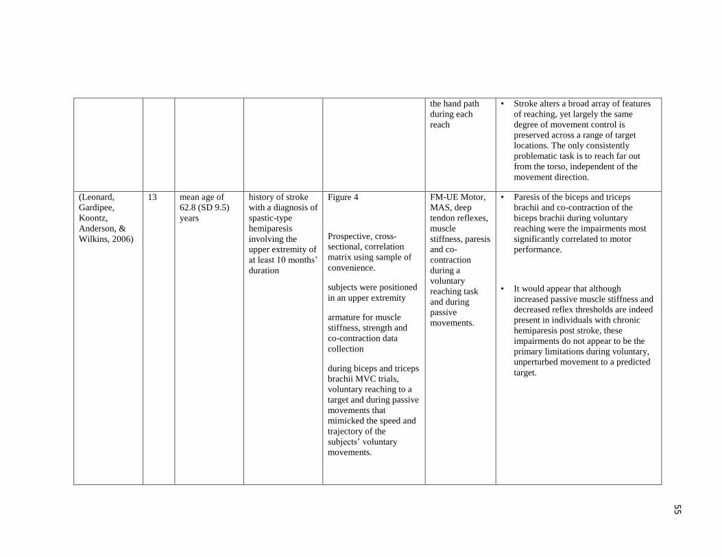
55
the hand path
during each
reach
• Stroke alters a broad array of features
of reaching, yet largely the same
degree of movement control is
preserved across a range of target
locations. The only consistently
problematic task is to reach far out
from the torso, independent of the
movement direction.
(Leonard,
Gardipee,
Koontz,
Anderson, &
Wilkins, 2006)
13 mean age of
62.8 (SD 9.5)
years
history of stroke
with a diagnosis of
spastic-type
hemiparesis
involving the
upper extremity of
at least 10 months’
duration
Figure 4
Prospective, cross-
sectional, correlation
matrix using sample of
convenience.
subjects were positioned
in an upper extremity
armature for muscle
stiffness, strength and
co-contraction data
collection
during biceps and triceps
brachii MVC trials,
voluntary reaching to a
target and during passive
movements that
mimicked the speed and
trajectory of the
subjects’ voluntary
movements.
FM-UE Motor,
MAS, deep
tendon reflexes,
muscle
stiffness, paresis
and co-
contraction
during a
voluntary
reaching task
and during
passive
movements.
• Paresis of the biceps and triceps
brachii and co-contraction of the
biceps brachii during voluntary
reaching were the impairments most
significantly correlated to motor
performance.
• It would appear that although
increased passive muscle stiffness and
decreased reflex thresholds are indeed
present in individuals with chronic
hemiparesis post stroke, these
impairments do not appear to be the
primary limitations during voluntary,
unperturbed movement to a predicted
target.
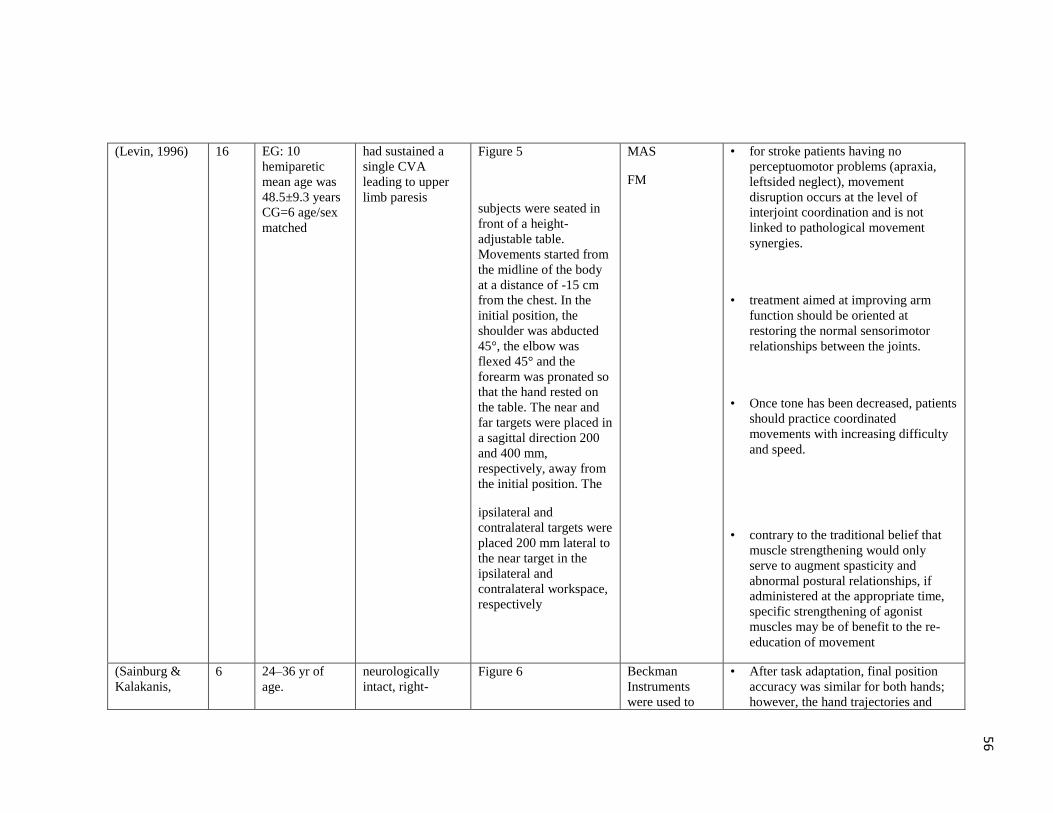
56
(Levin, 1996) 16 EG: 10
hemiparetic
mean age was
48.5±9.3 years
CG=6 age/sex
matched
had sustained a
single CVA
leading to upper
limb paresis
Figure 5
subjects were seated in
front of a height-
adjustable table.
Movements started from
the midline of the body
at a distance of -15 cm
from the chest. In the
initial position, the
shoulder was abducted
45°, the elbow was
flexed 45° and the
forearm was pronated so
that the hand rested on
the table. The near and
far targets were placed in
a sagittal direction 200
and 400 mm,
respectively, away from
the initial position. The
ipsilateral and
contralateral targets were
placed 200 mm lateral to
the near target in the
ipsilateral and
contralateral workspace,
respectively
MAS
FM
• for stroke patients having no
perceptuomotor problems (apraxia,
leftsided neglect), movement
disruption occurs at the level of
interjoint coordination and is not
linked to pathological movement
synergies.
• treatment aimed at improving arm
function should be oriented at
restoring the normal sensorimotor
relationships between the joints.
• Once tone has been decreased, patients
should practice coordinated
movements with increasing difficulty
and speed.
• contrary to the traditional belief that
muscle strengthening would only
serve to augment spasticity and
abnormal postural relationships, if
administered at the appropriate time,
specific strengthening of agonist
muscles may be of benefit to the re-
education of movement
(Sainburg &
Kalakanis,
6 24–36 yr of
age.
neurologically
intact, right-
Figure 6 Beckman
Instruments
were used to
• After task adaptation, final position
accuracy was similar for both hands;
however, the hand trajectories and

57
2000) handed adults
subjects sat facing a
computer screen with
each arm supported, over
a horizontal table top at
shoulder height, by a
frictionless air-jet
system. All joints distal
to the elbow were
immobilized using an air
splint. The scapulae and
trunk were immobilized
using a custom-fit
butterfly-shaped chest
restraint.
monitor the
elbow and
shoulder joint
angles.
joint coordination patterns during the
movement were systematically
different. The trajectories of both
hands were not straight but exhibited
oppositely directed curvatures.
• Results show interlimb differences in
the relative timing, magnitude, and
direction of muscle torques at the
shoulder and elbow that are more
likely to result from differences in
neural activation.
Abbreviations: CG - Control Group; CSI: the Composite Spasticity Index; EG - Experimental Group; FM: Fugl-Meyer; MVC:
Maximal Voluntary Contraction; MAS: modified Ashworth scale; UL - Upper limb, UE - Upper extremity
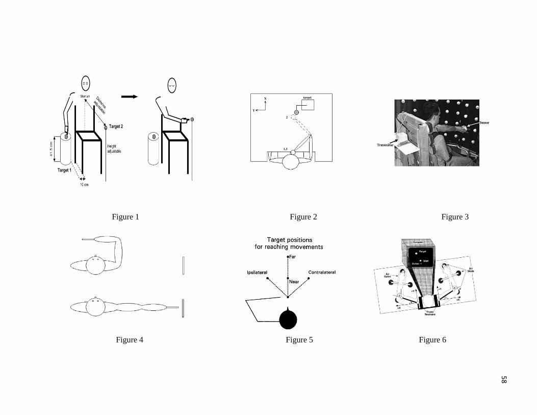
58
Figure 1 Figure 2 Figure 3
Figure 4 Figure 5 Figure 6

59
Appendix B: Text Descriptions
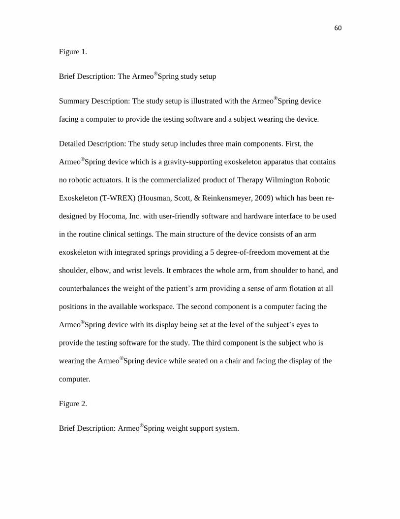
60
Figure 1.
Brief Description: The Armeo®Spring study setup
Summary Description: The study setup is illustrated with the Armeo®Spring device
facing a computer to provide the testing software and a subject wearing the device.
Detailed Description: The study setup includes three main components. First, the
Armeo®Spring device which is a gravity-supporting exoskeleton apparatus that contains
no robotic actuators. It is the commercialized product of Therapy Wilmington Robotic
Exoskeleton (T-WREX) (Housman, Scott, & Reinkensmeyer, 2009) which has been re-
designed by Hocoma, Inc. with user-friendly software and hardware interface to be used
in the routine clinical settings. The main structure of the device consists of an arm
exoskeleton with integrated springs providing a 5 degree-of-freedom movement at the
shoulder, elbow, and wrist levels. It embraces the whole arm, from shoulder to hand, and
counterbalances the weight of the patient’s arm providing a sense of arm flotation at all
positions in the available workspace. The second component is a computer facing the
Armeo®Spring device with its display being set at the level of the subject’s eyes to
provide the testing software for the study. The third component is the subject who is
wearing the Armeo®Spring device while seated on a chair and facing the display of the
computer.
Figure 2.
Brief Description: Armeo®Spring weight support system.
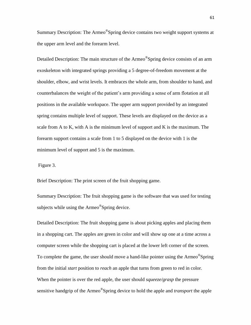
61
Summary Description: The Armeo®Spring device contains two weight support systems at
the upper arm level and the forearm level.
Detailed Description: The main structure of the Armeo®Spring device consists of an arm
exoskeleton with integrated springs providing a 5 degree-of-freedom movement at the
shoulder, elbow, and wrist levels. It embraces the whole arm, from shoulder to hand, and
counterbalances the weight of the patient’s arm providing a sense of arm flotation at all
positions in the available workspace. The upper arm support provided by an integrated
spring contains multiple level of support. These levels are displayed on the device as a
scale from A to K, with A is the minimum level of support and K is the maximum. The
forearm support contains a scale from 1 to 5 displayed on the device with 1 is the
minimum level of support and 5 is the maximum.
Figure 3.
Brief Description: The print screen of the fruit shopping game.
Summary Description: The fruit shopping game is the software that was used for testing
subjects while using the Armeo®Spring device.
Detailed Description: The fruit shopping game is about picking apples and placing them
in a shopping cart. The apples are green in color and will show up one at a time across a
computer screen while the shopping cart is placed at the lower left corner of the screen.
To complete the game, the user should move a hand-like pointer using the Armeo®
Spring
from the initial start position to reach an apple that turns from green to red in color.
When the pointer is over the red apple, the user should squeeze/grasp the pressure
sensitive handgrip of the Armeo®Spring device to hold the apple and transport the apple

62
to the shopping cart. When the color of the cart changes the user should take the pressure
off the device handgrip to release the apple. This process is illustrated in five phases
shown in the figure; the initiation, reaching, grasping, transporting and releasing phase.
Figure 4.
Brief Description: The mean completion time between the two gravity-support levels.
Summary Description: The mean time needed to complete each phase of the reaching
cycle and the total mean duration needed to complete the reaching cycle for a healthy
subject and a stroke subject under the mild and moderate level of support provided by the
Armeo®Spring device is displayed in column graph.
Detailed Description: The mean time needed to complete each phase of the reaching
cycle and the total mean duration needed to complete the reaching cycle for a healthy
subject and a stroke subject under the mild and moderate level of support provided by the
Armeo®Spring device is displayed in column graph. The x-axis represents the four phases
of the reaching cycle (Initiation, Reaching, Grasping, and Transporting) and the total
duration. In each phase and the total duration, four columns are displayed side by side.
The first column is blue in color and represents the healthy subject while using the mild
weight-support. The second column is red in color and represents the healthy subject
while using the moderate level of support. The third one is green in color and represents
the stroke subject while using the mild weight-support and the last column is purple in
color and represents the stroke subject while using the moderate level of weight-support.
The y-axis represents the time in seconds.
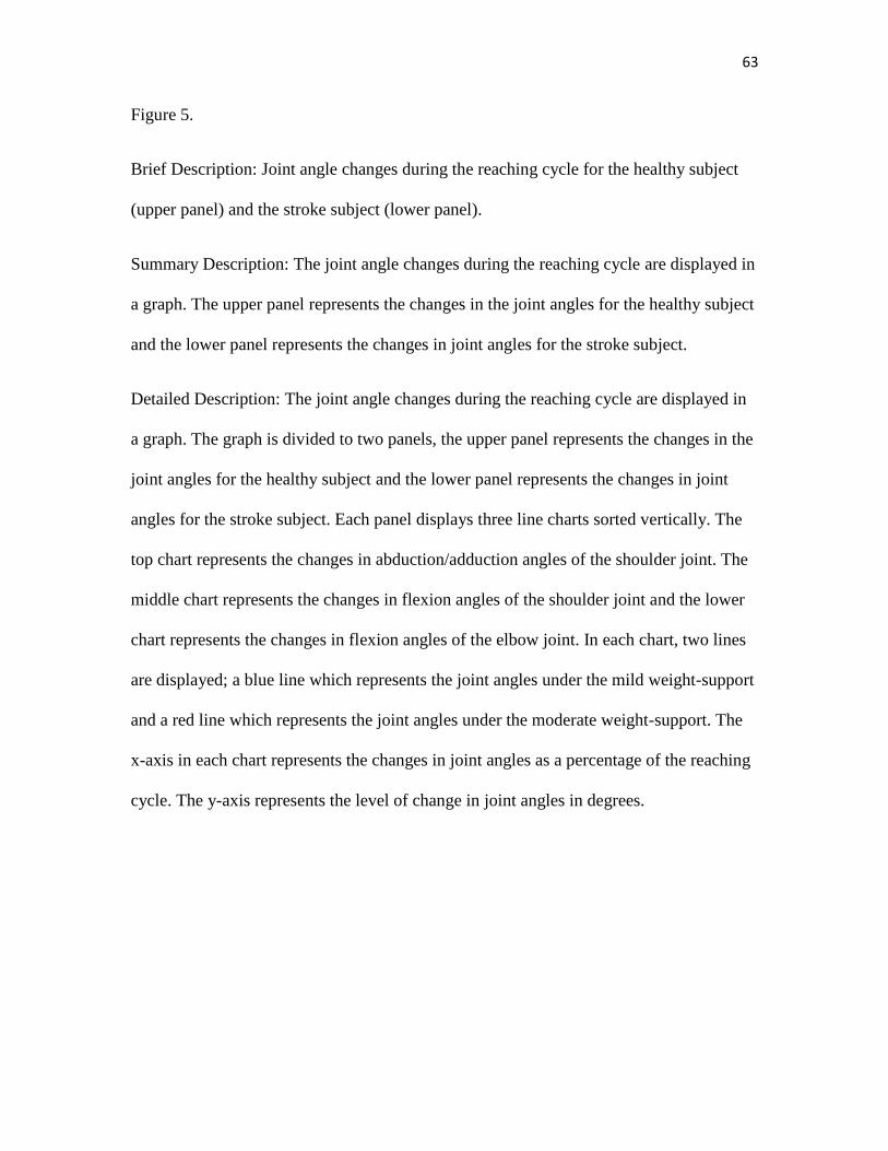
63
Figure 5.
Brief Description: Joint angle changes during the reaching cycle for the healthy subject
(upper panel) and the stroke subject (lower panel).
Summary Description: The joint angle changes during the reaching cycle are displayed in
a graph. The upper panel represents the changes in the joint angles for the healthy subject
and the lower panel represents the changes in joint angles for the stroke subject.
Detailed Description: The joint angle changes during the reaching cycle are displayed in
a graph. The graph is divided to two panels, the upper panel represents the changes in the
joint angles for the healthy subject and the lower panel represents the changes in joint
angles for the stroke subject. Each panel displays three line charts sorted vertically. The
top chart represents the changes in abduction/adduction angles of the shoulder joint. The
middle chart represents the changes in flexion angles of the shoulder joint and the lower
chart represents the changes in flexion angles of the elbow joint. In each chart, two lines
are displayed; a blue line which represents the joint angles under the mild weight-support
and a red line which represents the joint angles under the moderate weight-support. The
x-axis in each chart represents the changes in joint angles as a percentage of the reaching
cycle. The y-axis represents the level of change in joint angles in degrees.


![Dyrobes Rotordynamics Software ://dyrobes.com/wp-content/uploads/2016/12/...nonlinearities associated with structural kinematic and kinetic effects [4], 3) easily accommodates various](https://img.pdfslide.us/doc/110x75/5fdda5998da647596b12bff2/dyrobes-rotordynamics-software-nonlinearities-associated-with-structural.jpg)