Embed Size (px)
Citation preview

Cardiac tumors
e-Poster: C-184
Congress: ECR 2009
Type: Educational Exhibit
Topic: Cardiac / MRI
Authors: , M.A. Portilha , B. Goncalves , H. Rodrigues , P. Donato , F. Caseiro-Alves ; I. Santiago1 2 2 2 2 2 1
Aveiro/PT, Coimbra/PT2
Keywords: Cardiac tumors, Pericardial tumors
Any information contained in this pdf file is automatically generated from digital material submittedto EPOS by third parties in the form of scientific presentations. References to any names, marks,products, or services of third parties or hypertext links to third-party sites or information are providedsolely as a convenience to you and do not in any way constitute or imply ECR’s endorsement,sponsorship or recommendation of the third party, information, product, or service. ECR is notresponsible for the content of these pages and does not make any representations regarding thecontent or accuracy of material in this file.As per copyright regulations, any unauthorised use of the material or parts thereof as well ascommercial reproduction or multiple distribution by any traditional or electronically basedreproduction/publication method is strictly prohibited.You agree to defend, indemnify, and hold ECR harmless from and against any and all claims, damages,costs, and expenses, including attorneys’ fees, arising from or related to your use of these pages.Please note: Links to movies, ppt slideshows and any other multimedia files are not available in the pdfversion of presentations.www.myESR.org

1. Learning objectives
To illustrate the CT and MR imaging findings of various types of cardiac tumors diagnosed at ourInstitution. To outline the advantages and disadvantages of CT and MR for cardiac tumor diagnosis. Todiscuss the pertinent literature on distinguishing clinical and imaging features of each type of cardiactumor presented.
2. Background
Cardiac masses are rare. The estimated cumulative prevalence of primary cardiac tumors at autopsy isonly 0.002%–0.3%. The prevalence of metastasis to the heart is 1,2%, approximately 40 times higherthan that of primary cardiac tumors, being found at autopsy in 10-12 % of patients with knownmalignancies.
About 75% of primary cardiac tumors are benign, the most common in adults being Myxoma.Malignant cardiac tumors constitute approximately 25% of all primary cardiac tumors, the mostcommon in adults being Angiosarcomas (33%).
Cardiac tumors are frequently asymptomatic. Their clinical presentation may also be similar to that ofmuch more common disorders such as heart failure, stroke and coronary artery disease, includingsymptoms like dispnea, peripheral edema and thoracic pain, thus frequently delaying the definitivediagnosis.
Innocuous, low cost and widely available transthoracic and transesophageal echocardiographyprovide high-resolution, real-time images. Transthoracic echocardiography, being non-invasive, isfrequently the first imaging modality used when a cardiac tumor is suspected. Both are limited,though, by operator experience, by a restricted field of view and low soft tissue contrast.
CT and MR allow evaluation of the hole mediastinum and possible extracardiac extent of the tumor.These methods are also more flexible in the selection of image planes and allow accurate comparisonbetween examinations. They are very important for prognosis determination and surgery planning. CToffers the possibility of hole thoracic evaluation, including the lungs, and detection of calcifications.MR has better soft tissue contrast resolution and allows better tissue characterization. Disadvantagesof these two imaging modalities include the need for electrocardiographic gating, which, in thepresence of arrhythmia, may lead to acquisition artifacts.
In this educational exhibit, we review the clinical, morphological and CT and MR imaging findings ofseveral different types of benign and malignant cardiac tumors and tumor-like lesions, including atrialmyxomas, lipoma, lipomatous hipertrophy of the interatrial septum, fibroma, rhabdomyoma,bronchogenic cyst, pericardial hamartoma, metastasis, undifferentiated sarcoma and lymphoma,based on selected images of cases diagnosed at our Institution between 1992 and 2008.
3. Imaging findings OR Procedure details
BENIGN ATRIAL TUMORS
MYXOMA
Myxoma is the most common primary cardiac tumor, comprising approximately 50% of all primary

cardiac masses. It is slightly more common in male patients, with a mean age onset of 50 years. 75% ofmyxomas occur in the left atrium, typically at the level of the fossa ovalis, in the interatrial septum;18% on the right atrium and only 7% on the ventricles. Although they can be asymptomatic, there is aclassic triad of obstructive symptoms related to the chamber where the tumor is based; constitutionalsymptoms, such as fever, malaise, weight loss and anemia; and embolic events, involving mainly thebrain, kidneys and lower limbs with left-sided myxomas, and the pulmonary circulation withright-sided myxomas. These tumors usually present as an endocardial-based pedunculated polypoidmass, with a smooth or villous surface, moving freely inside the atrium, sometimes protuding to theventricle during diastole. An important differential diagnosis is the presence of an intracavitarythrombus. However, thrombus do not have a pedicle or enhance after IV contrast ( ).Figure 3
The imaging characteristics consist of a spherical or ovoid mass with a lobulated contour, hipo orCTisodense compared with the myocardium, usually heterogeneous, sometimes with coarse orpunctuate calcifications, and heterogeneous enhancement. There are some possible associatedfindings such as chamber dilation, signs of cardiac failure or of embolic events ( ).Figure 1
Figure 1

Figure 1
93 yo male patient with left atrial (LA) myxoma. CT Pulmonary Angiogram performed in theEmergency Department for suspected, not confirmed, pulmonary thromboembolism. a) A welldefined, soft-tissue density, slightly enhancing mass (arrow), located in the LA wasfound. b) Sagitalreformatted view, better depicting the lesion´s pedicle.
The imaging findings include the presence of a spherical or ovoid mass with lobulated contours,MRwith heterogeneous signal intensity, predominately isointense with respect to the myocardium onT1-weighted images and increased signal intensity on T2-weighted images, with heterogeneousenhancement after gadolinium. There is a better correlation between the point of attachment of theMyxoma described on MR than with CT imaging, when compared with the surgical description (
, ).Figure 2 [Video 1] video 1

Figure 2
Figure 2
Same patient as in Video 1. Cardiac MR. a) SSFP image shows the myxoma (*) has moved to the leftventricle during diastole b) IRGE image shows no areas of delayed enhancement.
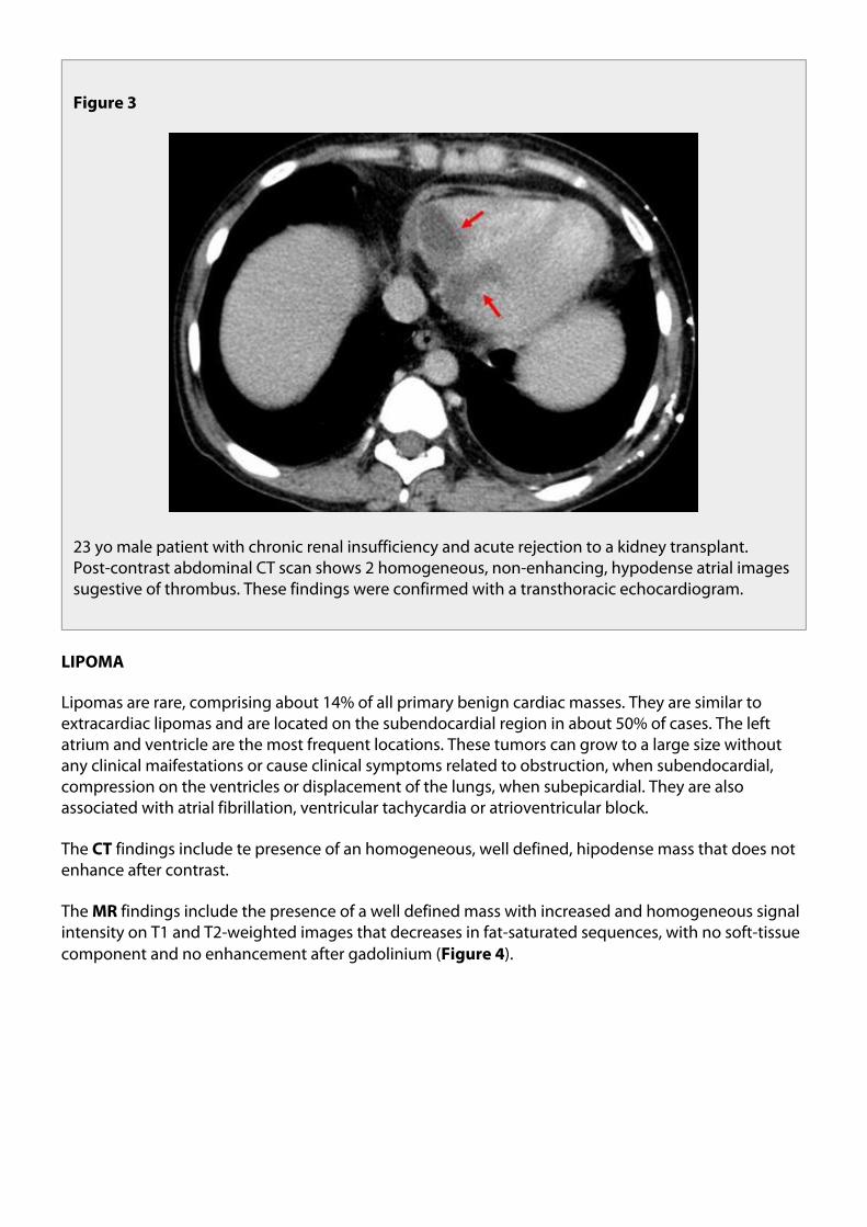
Figure 3
23 yo male patient with chronic renal insufficiency and acute rejection to a kidney transplant.Post-contrast abdominal CT scan shows 2 homogeneous, non-enhancing, hypodense atrial imagessugestive of thrombus. These findings were confirmed with a transthoracic echocardiogram.
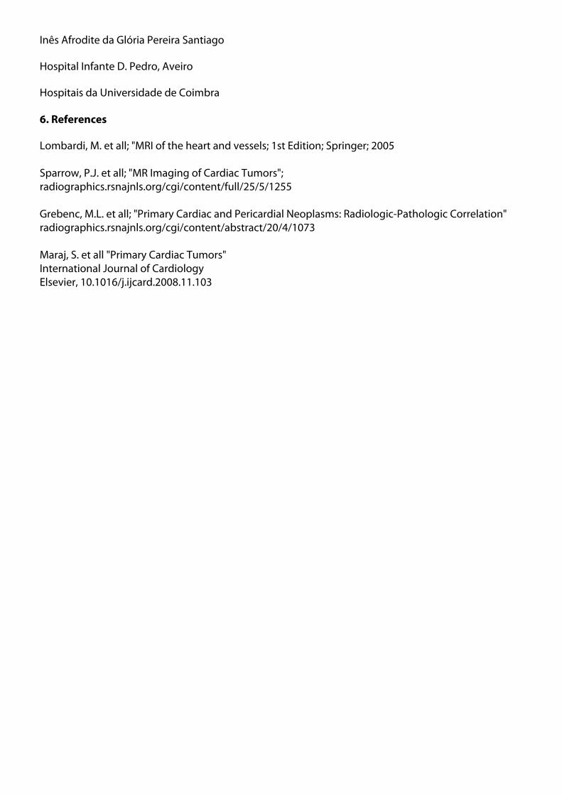
LIPOMA
Lipomas are rare, comprising about 14% of all primary benign cardiac masses. They are similar toextracardiac lipomas and are located on the subendocardial region in about 50% of cases. The leftatrium and ventricle are the most frequent locations. These tumors can grow to a large size withoutany clinical maifestations or cause clinical symptoms related to obstruction, when subendocardial,compression on the ventricles or displacement of the lungs, when subepicardial. They are alsoassociated with atrial fibrillation, ventricular tachycardia or atrioventricular block.
The findings include te presence of an homogeneous, well defined, hipodense mass that does notCTenhance after contrast.
The findings include the presence of a well defined mass with increased and homogeneous signalMRintensity on T1 and T2-weighted images that decreases in fat-saturated sequences, with no soft-tissuecomponent and no enhancement after gadolinium ( ).Figure 4
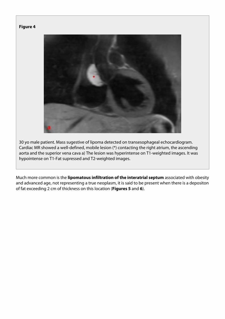
Figure 4
30 yo male patient. Mass sugestive of lipoma detected on transesophageal echocardiogram.Cardiac MR showed a well-defined, mobile lesion (*) contacting the right atrium, the ascendingaorta and the superior vena cava a) The lesion was hyperintense on T1-weighted images. It washypointense on T1-Fat supressed and T2-weighted images.
Much more common is the associated with obesitylipomatous infiltration of the interatrial septumand advanced age, not representing a true neoplasm, it is said to be present when there is a depositonof fat exceeding 2 cm of thickness on this location ( and ).Figures 5 6

Figure 5
Figure 5
61 yo female patient with morbid obesity and lipomatous hypertrophy of the interatrial septum(IAS). CT scan performed for staging of folicular lymphoma. a) Post-contrast CT showsnon-enhancing, well defined, homogeneous fat density mass confined to the IAS (betweenarrows). b) Axillary and upper mediastinal lymphadenopathy (*) are also shown.
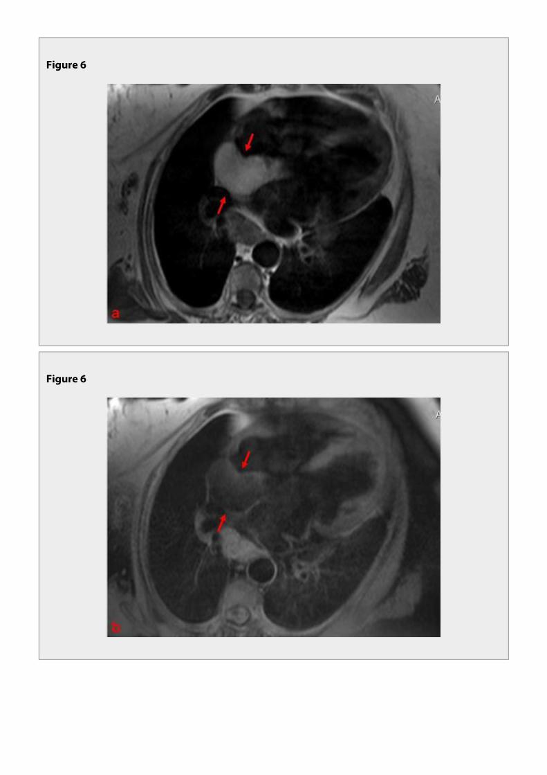
Figure 6
Figure 6
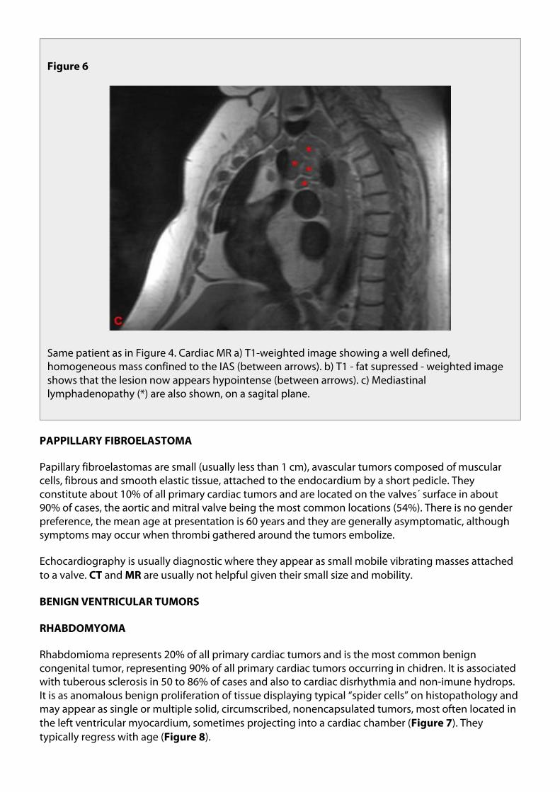
Figure 6
Same patient as in Figure 4. Cardiac MR a) T1-weighted image showing a well defined,homogeneous mass confined to the IAS (between arrows). b) T1 - fat supressed - weighted imageshows that the lesion now appears hypointense (between arrows). c) Mediastinallymphadenopathy (*) are also shown, on a sagital plane.
PAPPILLARY FIBROELASTOMA
Papillary fibroelastomas are small (usually less than 1 cm), avascular tumors composed of muscularcells, fibrous and smooth elastic tissue, attached to the endocardium by a short pedicle. Theyconstitute about 10% of all primary cardiac tumors and are located on the valves´ surface in about90% of cases, the aortic and mitral valve being the most common locations (54%). There is no genderpreference, the mean age at presentation is 60 years and they are generally asymptomatic, althoughsymptoms may occur when thrombi gathered around the tumors embolize.
Echocardiography is usually diagnostic where they appear as small mobile vibrating masses attachedto a valve. and are usually not helpful given their small size and mobility.CT MR
BENIGN VENTRICULAR TUMORS
RHABDOMYOMA
Rhabdomioma represents 20% of all primary cardiac tumors and is the most common benigncongenital tumor, representing 90% of all primary cardiac tumors occurring in chidren. It is associatedwith tuberous sclerosis in 50 to 86% of cases and also to cardiac disrhythmia and non-imune hydrops.It is as anomalous benign proliferation of tissue displaying typical “spider cells” on histopathology andmay appear as single or multiple solid, circumscribed, nonencapsulated tumors, most often located inthe left ventricular myocardium, sometimes projecting into a cardiac chamber ( ). TheyFigure 7typically regress with age ( ).Figure 8
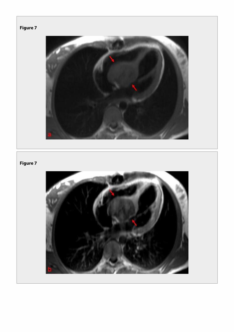
Figure 7
Figure 7

Figure 7
21 yo female patient with disrythmia. Cardiac MR shows a well defined, rounded mass located inthe interventricular septum (between arrows), compatible with a rhabdomyoma. The mass isisointense with the miocardium on T1-weighted images (a), enhancing less than the surroundingmyocardium and heterogeneously after gadolinium (b) and hypointense on T2-weighted images(c).
Figure 8

Figure 8
Figure 8

Figure 8

Figure 8
23 yo female patient with tuberous sclerosis and an involuted left ventricle rhabdomyoma withadipose substitution (arrow). Cardiac MR. a) T1-weighted image showing a hyperintense linearlesion on the lateral wall of the left ventricle. b) T1-weighted image after gadolinium shows thatthe lesion does not enhance significantly. c) On T2-weighted images, the lesion appearshypointense. d) and e) IRGE images, long and short axis, respectively, showing delayedenhancement of the lesion.
FIBROMA
Fibroma is a rare tumor that primarily affects children, being the 2 most common benign primarynd
cardiac tumors in children after rhabdomioma. It may occur as part of Gorlin or basal cell nevussyndrome. Although one third are incidentally found, these tumors are sometimes associated witharrhythmias, heart failure and sudden death. Fibromas are composed of fibroblasts and large amountsof collagen and are tipically located in the ventricles, most often the ventricular septum and leftventricular free wall.
At , they appear as a soft-tissue density, homogeneous mass, frequently with dystrophcCTcalcifications, either well circumscribed or infiltrative, with litlle or no enhancement.
At these tumors are usually homogeneously hypointense on T2-weighted images and isointenseMRwhen compared to the myocardium n T1-weighted images, showing little or no enhancement aftergadolinium ( and ).Figures 9 10

Figure 9
Figure 9

Figure 9
Figure 9
41 yo male patient with a history of acute myocardial infarction. Cardiac MR showing a fibroma inthe ventricular septum a) T1-weighted image shows an oval mass in the interventricular septumthat is isodense to the myocarium (arrow) b) T1-weighted image after gadolinium shows minimalenhancement (arrow) c) IRGE shows delayed enhancement of the lesion (arrow) d) Subendocardialdelayed enhancement at the inferior wall of the left ventricle related to previous infarct (arrow).

Figure 10
Figure 10

Figure 10
Figure 10

Figure 10
72 yo male with a history of acute myocardial infarction and congestive heart failure. Transthoracicechocardiogram detected an encapsulated ecogenic mass protuding to the interatrial septum.Cardiac MR shows an infero-septal, left ventricle based fibroma (between arrows). a), b) and c) OnT1-weighted images (axial, coronal and sagital plane, respectively) the lesion was slightlyhypointense. d) Post-gadolinium T1-weighted image shows that the lesion enhances less than themyocardium. e) the lesion was hypointense on T2-weighted images.
BENIGN PERICARDIAL TUMORS
PERICARDIAL CYSTS
Pericardial cysts are usually located in the right cardiophrenic space and may be congenital oriatrogenic. They are usually asymptomatic but may rarely cause symptoms like dyspnea, arrhythmia orretrosternal pain.
At , they present as a homogeneous, well-defined, fluid attenuation lesion with a smooth wall,CTalthough sometimes the attenuation may be greater.
At , they have a characteristic low signal intensity on T1-weighted images and high signal intensityMRon T2-weighted images ( ).Figure 11
These lesions do not enhance after IV contrast.

Figure 11
Figure 11
Incidentally found pericardial cyst in a 78 yo woman. a) Axial T2 Haste weighted image and b)Coronal T2 Haste weighted image showing an homogeneous, well-defined, hyperintense image inthe left cardiophrenic space (an unusual location).
Other benign pericardial lesions include and ( ).teratoma hamartoma Figure 12

Figure 12
Figure 12
6 yo child with pericardia hamartoma. a) Thoracic CT shows nodular hipodense image with corseperipheral calcifications. b) coronal reformat image showing the longitudinal extension of thelesion.

MALIGNANT TUMORS
METASTASIS
Metastases are 20-40 times more prevalent than primary cardiac tumors, the most common primarysources being bronchogenic carcinoma, lymphoma, leucemia, breast carcinoma, esophaguscarcinoma melanoma, sarcoma, hepatoma, adrenal adenocarcinoma and renal cell carcinoma. Theymay involve the heart by contiguous extension, lymphatic or hematogenous spread. The commonestsite of involvement is the pericardium with or without invasion of the underlying myocardium (Figure
). In approximately one-third of patients with involvement, death will be directly13 cardiacattributable to the metastases as a result of pericardial tamponade, congestive failure, orcardiaccoronary artery invasion Presenting symptoms may include shortness of breath, chest wall pain, andperipheral edema. As with other malignancies, arrhythmias may also be a feature. There arecardiacno specific appearances of metastases or direct extension to the heart.
Figure 13

Figure 13

Figure 13
74 yo male patient with a history of progressive dispnea and peripheral edema for the past 3months. CT scan showed right central hilar mass with associated right lower lobe colapse andbilateral pleural effusion. The mass invaded the mediastinum. Thoracic MR requested to betterdepict mediastinal invasion. a), b) and c) are T2 Haste weighted images, a and b in an axial planes (Bat a higher level than A) and c) in a sagital plane. We can see an intermediate signal mass involvingand invading the right atrium and the emergence of the aorta and pulmonary arteries (*). Uppermediastinal lymphadenopathies (º) and bilateral pleural effusion (+) are also shown.
ANGIOSARCOMA
Angiosarcoma is the most common primary cardiac malignancy in adults, representing 37% of allprimary cardiac malignancies, is two times more common in men than in women. It is composed ofill-defined anastomotic vascular spaces lined by atypical endothelial cells. It´s located in the rightatrium in 80% of cases, growing either as a well-defined mass protruding into the right atrium or as aninfiltrating mass extending into the pericardium, the tricuspid valve or the superior or inferior venacava. They are frequently advanced and metastatic at the time of presentation, the lung being themost common site for metastatic disease. Angiosarcomas cause symptoms by obstructing rightcardiac filling and causing pericardial tamponade.
At , they present as a low attenuation right atrial mass, either ill-defined or nodular, withCTheterogeneous enhancement after contrast. Pericardial thickening and effusion may be seen.
At , angiosarcoma presents as a large heterogeneous mass in the right atrium, sometimesMRinvolving the pericardium causing pericardial thickening or nodularity and hemorrhagic pericardialeffusion. It is heterogeneous on T1-weighted images, with areas of intermediate, low and high signalintensity due to the presence of tumor tissue, necrosis and methemoglobin, respectively. They areheterogeneous, predominantly hyperintense on T2-weighted images. The enhancement aftergadolinium is marked at the surface – sunray appearance – and heterogeneous.
UNDIFFERENTIATED SARCOMA
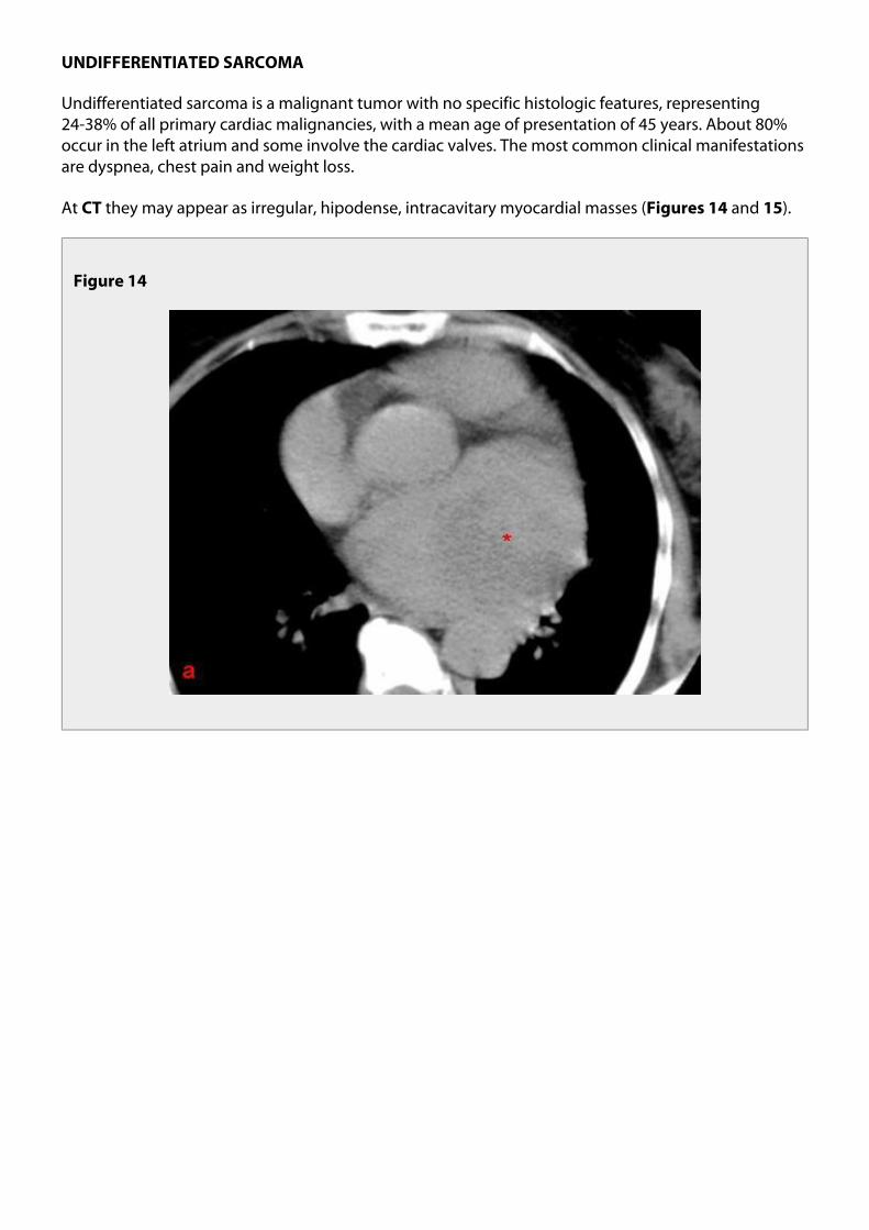
Undifferentiated sarcoma is a malignant tumor with no specific histologic features, representing24-38% of all primary cardiac malignancies, with a mean age of presentation of 45 years. About 80%occur in the left atrium and some involve the cardiac valves. The most common clinical manifestationsare dyspnea, chest pain and weight loss.
At they may appear as irregular, hipodense, intracavitary myocardial masses ( and ).CT Figures 14 15
Figure 14
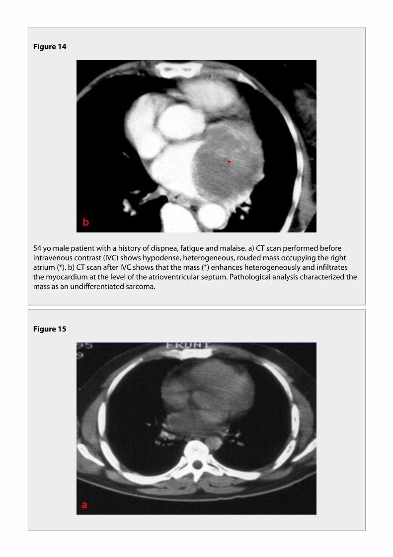
Figure 14
54 yo male patient with a history of dispnea, fatigue and malaise. a) CT scan performed beforeintravenous contrast (IVC) shows hypodense, heterogeneous, rouded mass occupying the rightatrium (*). b) CT scan after IVC shows that the mass (*) enhances heterogeneously and infiltratesthe myocardium at the level of the atrioventricular septum. Pathological analysis characterized themass as an undifferentiated sarcoma.
Figure 15

Figure 15
63 yo male with disrythmia. a) Thoracic CT scan performed before IV contrast shows a hypodensemass on the left atrium (arrow). b) CT scan after IV contrast shows that the mass is lobulated andenhances heterogeneously (arrow). Pathological analysis revealed an undifferentiated sarcoma.
At , they may show as polypoid, isointense, infiltrative masses on T1-weighted images ( ).MR Figure 16The tumor may also manifest as a hemorrhagic mass replacing the pericardium, similar toangiosarcoma.

Figure 16
55 yo male patient with biopsy proven undifferentiated sarcoma of the left ventricle. a)T1-weighted image showing an heterogeneous mass with some hyperintense areas infiltrating thewall of the left ventricle.
Other, rarer primary sarcomas of the heart are (11% - 24%), malignant fibrous histiocytoma (8% - 9%), (4% - 7%) and (3% - 9%).leiomyosarcoma rhabdomyosarcoma osteosarcoma
LYMPHOMA
Primary cardiac lymphomas are exceedingly rare, are typically of the non-Hodgkin B-cell type, and areconfined to the heart or pericardium. They usually occur in immunocompromised patients but are notrestricted solely to this group. Presentation is with rapidly worsening heart failure, obstructivesymptoms, or arrhythmias. They most commonly involve the right side of the heart, in particular theright atrium, with frequent involvement of more than one chamber and invasion of the pericardiumwith pericardial effusion. Microscopically, they consist of firm homogeneous nodules, not prone tohemorrhage or necrosis.
The imaging findings are nonspecific ( and ).CT Figures 17 18

Figure 17

Figure 17
63 yo immunocompetent male patient with a history of fatigue and peripheral edema with amonth duration. Transthoracic echocardiogram showed pericardial effusion and a lobulated masslocated at the tricuspid valve, insinuating to the interatrial septum. a) Thoracic CT before theadministration of IVC shows pericardial (*) and bilateral pleural effusion (+). b) Cardiac CT after IVCshows a hypodense, hypoattenuating, labulated mass located at the level of the tricuspid valve,extending to the interatrial septum. The patient died a few weeks later. Pathological analysisrevealed a primary B-Cell lymphoma of the heart.

Figure18
Figure 18

Figure 19
83 yo woman. CT performed for staging of lymphoma (supraclavicular lymphadenopatiespreviously biopsed). a) Post-IV contrast CT axial image showing supraclavicular lymphadenopaties(*). b) Lobulated mass involving the right atrial and ventricular myocardium. c) Coronal reformatimage showing the longitudinal extension of the mass.
At imaging, they are isointense on T1-weighted images and heterogeneously hyperintense on T2-MRweighted images; they demonstrate heterogeneous enhancement after administration of gadoliniumcontrast material, with areas of low enhancement in the center of the lesion compared to theperiphery.
MALIGNANT PERICARDIAL TUMORS
Most malignant pericardial tumors are . Primary malignancies are pericardial metastasis, that represent less than 1% of all mesotheliomas but 50% of all primary pericardialmesotheliomas
tumors.
4. Conclusion
CT and MR imaging findings help differentiate benign from malignant cardiac tumors, sometimeseven further narrowing the differential diagnosis; therefore, influencing their management.
5. Personal Information
Inês Afrodite da Glória Pereira Santiago
Hospital Infante D. Pedro, Aveiro
Hospitais da Universidade de Coimbra
6. References
Lombardi, M. et all; "MRI of the heart and vessels; 1st Edition; Springer; 2005
Sparrow, P.J. et all; "MR Imaging of Cardiac Tumors";radiographics.rsnajnls.org/cgi/content/full/25/5/1255
Grebenc, M.L. et all; "Primary Cardiac and Pericardial Neoplasms: Radiologic-Pathologic Correlation"radiographics.rsnajnls.org/cgi/content/abstract/20/4/1073
Maraj, S. et all "Primary Cardiac Tumors"International Journal of CardiologyElsevier, 10.1016/j.ijcard.2008.11.103

7. Mediafiles
Figure 1
93 yo male patient with left atrial (LA) myxoma. CT Pulmonary Angiogram performed in theEmergency Department for suspected, not confirmed, pulmonary thromboembolism. a) A welldefined, soft-tissue density, slightly enhancing mass (arrow), located in the LA wasfound. b) Sagitalreformatted view, better depicting the lesion´s pedicle.

Figure 1
Figure 2

Figure 2
Same patient as in Video 1. Cardiac MR. a) SSFP image shows the myxoma (*) has moved to the leftventricle during diastole b) IRGE image shows no areas of delayed enhancement.

Figure 3
23 yo male patient with chronic renal insufficiency and acute rejection to a kidney transplant.Post-contrast abdominal CT scan shows 2 homogeneous, non-enhancing, hypodense atrial imagessugestive of thrombus. These findings were confirmed with a transthoracic echocardiogram.
Figure 4
30 yo male patient. Mass sugestive of lipoma detected on transesophageal echocardiogram.Cardiac MR showed a well-defined, mobile lesion (*) contacting the right atrium, the ascendingaorta and the superior vena cava a) The lesion was hyperintense on T1-weighted images. It washypointense on T1-Fat supressed and T2-weighted images.
Figure 5

Figure 5
61 yo female patient with morbid obesity and lipomatous hypertrophy of the interatrial septum(IAS). CT scan performed for staging of folicular lymphoma. a) Post-contrast CT showsnon-enhancing, well defined, homogeneous fat density mass confined to the IAS (betweenarrows). b) Axillary and upper mediastinal lymphadenopathy (*) are also shown.

Figure 6
Same patient as in Figure 4. Cardiac MR a) T1-weighted image showing a well defined,homogeneous mass confined to the IAS (between arrows). b) T1 - fat supressed - weighted imageshows that the lesion now appears hypointense (between arrows). c) Mediastinallymphadenopathy (*) are also shown, on a sagital plane.
Figure 6

Figure 6
Figure 7

Figure 7
21 yo female patient with disrythmia. Cardiac MR shows a well defined, rounded mass located inthe interventricular septum (between arrows), compatible with a rhabdomyoma. The mass isisointense with the miocardium on T1-weighted images (a), enhancing less than the surroundingmyocardium and heterogeneously after gadolinium (b) and hypointense on T2-weighted images(c).
Figure 7

Figure 8
Figure 8
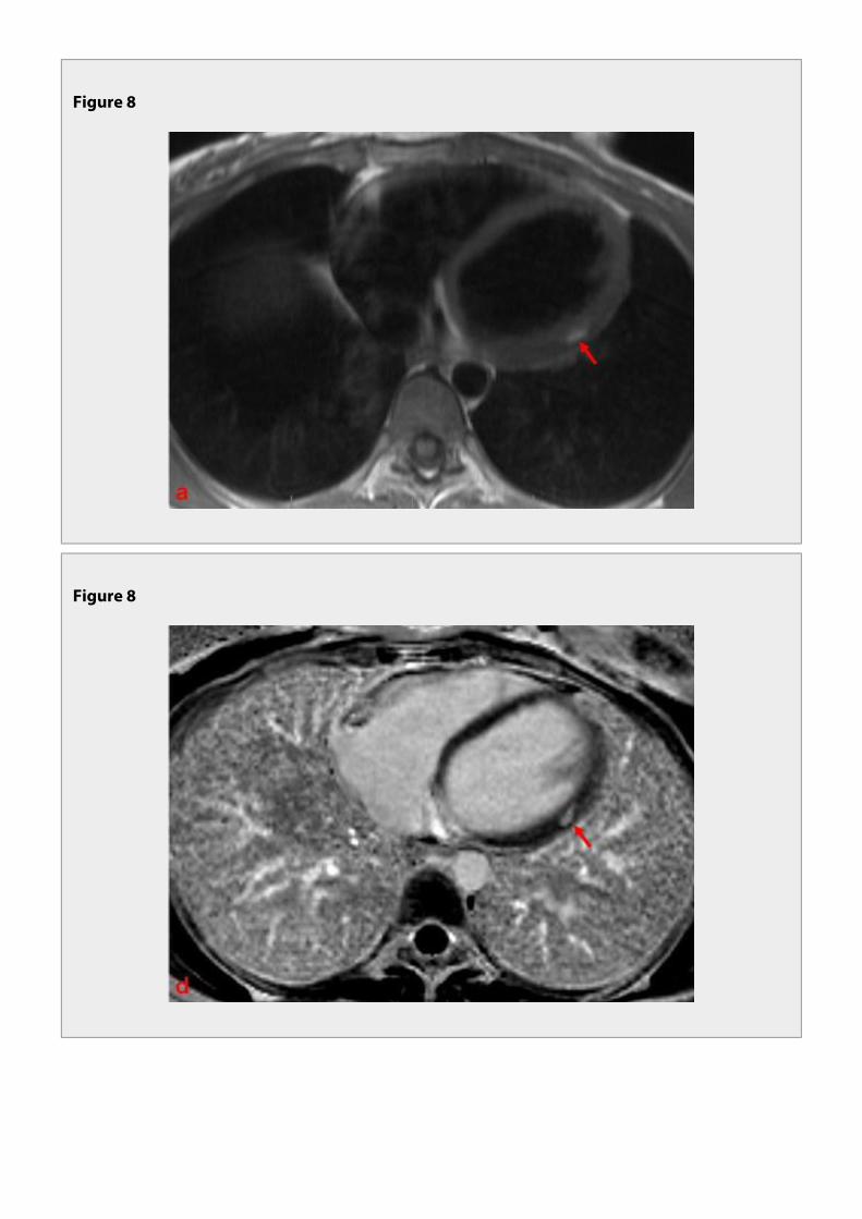
Figure 8
Figure 8

Figure 8
23 yo female patient with tuberous sclerosis and an involuted left ventricle rhabdomyoma withadipose substitution (arrow). Cardiac MR. a) T1-weighted image showing a hyperintense linearlesion on the lateral wall of the left ventricle. b) T1-weighted image after gadolinium shows thatthe lesion does not enhance significantly. c) On T2-weighted images, the lesion appearshypointense. d) and e) IRGE images, long and short axis, respectively, showing delayedenhancement of the lesion.

Figure 9
41 yo male patient with a history of acute myocardial infarction. Cardiac MR showing a fibroma inthe ventricular septum a) T1-weighted image shows an oval mass in the interventricular septumthat is isodense to the myocarium (arrow) b) T1-weighted image after gadolinium shows minimalenhancement (arrow) c) IRGE shows delayed enhancement of the lesion (arrow) d) Subendocardialdelayed enhancement at the inferior wall of the left ventricle related to previous infarct (arrow).
Figure 9
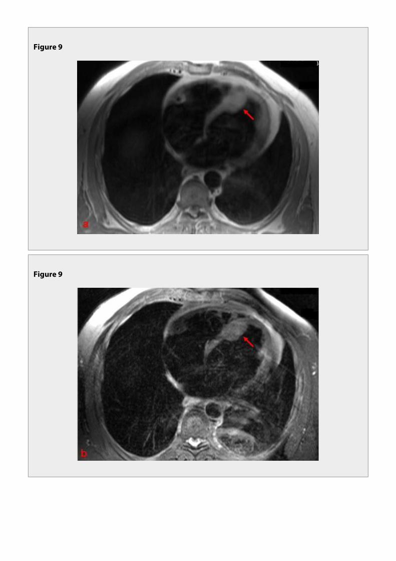
Figure 9
Figure 9

Figure 10
Figure 10
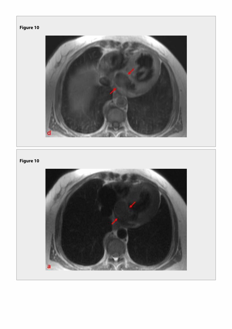
Figure 10
Figure 10

Figure 10
72 yo male with a history of acute myocardial infarction and congestive heart failure. Transthoracicechocardiogram detected an encapsulated ecogenic mass protuding to the interatrial septum.Cardiac MR shows an infero-septal, left ventricle based fibroma (between arrows). a), b) and c) OnT1-weighted images (axial, coronal and sagital plane, respectively) the lesion was slightlyhypointense. d) Post-gadolinium T1-weighted image shows that the lesion enhances less than themyocardium. e) the lesion was hypointense on T2-weighted images.

Figure 11
Incidentally found pericardial cyst in a 78 yo woman. a) Axial T2 Haste weighted image and b)Coronal T2 Haste weighted image showing an homogeneous, well-defined, hyperintense image inthe left cardiophrenic space (an unusual location).
Figure 11

Figure 12
6 yo child with pericardia hamartoma. a) Thoracic CT shows nodular hipodense image with corseperipheral calcifications. b) coronal reformat image showing the longitudinal extension of thelesion.
Figure 12

Figure 13
74 yo male patient with a history of progressive dispnea and peripheral edema for the past 3months. CT scan showed right central hilar mass with associated right lower lobe colapse andbilateral pleural effusion. The mass invaded the mediastinum. Thoracic MR requested to betterdepict mediastinal invasion. a), b) and c) are T2 Haste weighted images, a and b in an axial planes (Bat a higher level than A) and c) in a sagital plane. We can see an intermediate signal mass involvingand invading the right atrium and the emergence of the aorta and pulmonary arteries (*). Uppermediastinal lymphadenopathies (º) and bilateral pleural effusion (+) are also shown.
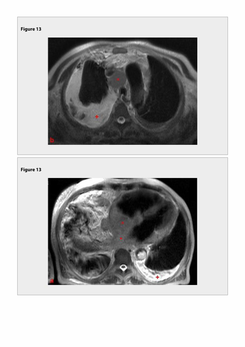
Figure 13
Figure 13

Figure 14
Figure 14
54 yo male patient with a history of dispnea, fatigue and malaise. a) CT scan performed beforeintravenous contrast (IVC) shows hypodense, heterogeneous, rouded mass occupying the rightatrium (*). b) CT scan after IVC shows that the mass (*) enhances heterogeneously and infiltratesthe myocardium at the level of the atrioventricular septum. Pathological analysis characterized themass as an undifferentiated sarcoma.

Figure 15
Figure 15
63 yo male with disrythmia. a) Thoracic CT scan performed before IV contrast shows a hypodensemass on the left atrium (arrow). b) CT scan after IV contrast shows that the mass is lobulated andenhances heterogeneously (arrow). Pathological analysis revealed an undifferentiated sarcoma.
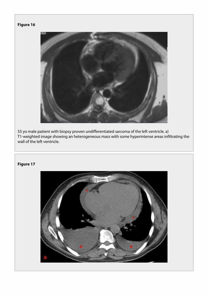
Figure 16
55 yo male patient with biopsy proven undifferentiated sarcoma of the left ventricle. a)T1-weighted image showing an heterogeneous mass with some hyperintense areas infiltrating thewall of the left ventricle.
Figure 17

Figure 17
63 yo immunocompetent male patient with a history of fatigue and peripheral edema with amonth duration. Transthoracic echocardiogram showed pericardial effusion and a lobulated masslocated at the tricuspid valve, insinuating to the interatrial septum. a) Thoracic CT before theadministration of IVC shows pericardial (*) and bilateral pleural effusion (+). b) Cardiac CT after IVCshows a hypodense, hypoattenuating, labulated mass located at the level of the tricuspid valve,extending to the interatrial septum. The patient died a few weeks later. Pathological analysisrevealed a primary B-Cell lymphoma of the heart.

Figure 18
Figure18

Figure 19
83 yo woman. CT performed for staging of lymphoma (supraclavicular lymphadenopatiespreviously biopsed). a) Post-IV contrast CT axial image showing supraclavicular lymphadenopaties(*). b) Lobulated mass involving the right atrial and ventricular myocardium. c) Coronal reformatimage showing the longitudinal extension of the mass.
Video 1
68 yo male patient with fatigue and malaise. Transthoracic echocardiogram detected a mobile,pedunculated mass in the left atrium sugestive of a myxoma. Cardiac MR SSFP video, long axisplane, depicting the myxoma´s movement to the left ventricle during diastole.