Embed Size (px)
Citation preview
Spine www.spinejournal.com 865
RANDOMIZED TRIAL
SPINE Volume 40 , Number 12 , pp 865 - 875 ©2015, Wolters Kluwer Health, Inc. All rights reserved.
DOI: 10.1097/BRS.0000000000000906
Study Design. The KAST (Kiva Safety and Effectiveness Trial) study was a pivotal, multicenter, randomized control trial for evaluation of safety and effectiveness in the treatment of patients with painful, osteoporotic vertebral compression fractures (VCFs). Objective. The objective was to demonstrate noninferiority of the Kiva system to balloon kyphoplasty (BK) with respect to the composite primary endpoint. Summary of Background Data. Annual incidence of osteoporotic VCFs is prevalent. Optimal treatment of VCFs should address pain, function, and deformity. Kiva is a novel implant for vertebral augmentation in the treatment of VCFs. Methods. A total of 300 subjects with 1 or 2 painful osteoporotic VCFs were randomized to blindly receive Kiva (n = 153) or BK (n = 147). Subjects were followed through 12 months. The primary endpoint was a composite at 12 months defi ned as a reduction in fracture pain by at least 15 mm on the visual analogue scale, maintenance or improvement in function on the Oswestry Disability Index, and absence of device-related serious adverse events. Secondary endpoints included cement usage, extravasation, and adjacent level fracture.
From the * Department of Radiology, Vascular/Interventional Radiology, Medical College of Wisconsin, Milwaukee, WI ; † Department of Orthopedic and Trauma Surgery, Universitätsklinikum, Bonn, Germany ; ‡ Sutter Health Vascular and Vein Institute, Roseville, CA ; § Clinical Radiology of Oklahoma, Edmond, OK ; ¶ Vascular and Interventional Radiology, Chicago, Hinsdale, IL; and � Department of Orthopaedic Surgery, University of California, San Diego, San Diego, CA.
Acknowledgment date: August 13, 2014. First revision date: November 18, 2014. Second revision date: January 9, 2015. Acceptance date: March 4, 2015.
The device(s)/drug(s) is/are FDA-approved or approved by corresponding national agency for this indication.
Benvenue Medical, Inc., funds were received in support of this work.
Relevant fi nancial activities outside the submitted work: board membership, consultancy, payment for lectures, royalties, stocks.
Address correspondence and reprint requests to Sean M. Tutton, MD, FSIR, Department of Radiology, Vascular/Interventional Radiology, Medical College of Wisconsin, Froedtert Memorial Lutheran Hospital, Room 2803, 9200 West Wisconsin Ave, Milwaukee, WI 53226; E-mail: [email protected]
The reported annual incidence of osteoporotic vertebral compression fractures (VCFs) in the United States has been estimated to be more than 700,000. 1 , 2 Patients may
experience chronic pain, decreased physical function, signifi -cant spinal deformity, social isolation, decrease in quality of life, and complications that can result in death, which is often related to the kyphotic deformity and decreased activity. 3 , 4 The optimal treatment of VCFs should address these issues.
For patients with refractory pain, impaired physical func-tion, and progressive deformity, minimally invasive vertebral augmentation techniques such as vertebroplasty and balloon kyphoplasty (BK) are excellent options. With vertebroplasty, polymethylmethacrylate (PMMA) is injected using imaging guidance into the vertebral body to stabilize the fracture. Ver-tebral BK involves the creation of a void within the vertebral body, most commonly created with an infl atable bone tamp (balloon), to reduce the amount of compression from the fracture. This void is then fi lled with PMMA for immediate
Results. A mean improvement of 70.8 and 71.8 points in the visual analogue scale score and 38.1 and 42.2 points in the Oswestry Disability Index was noted in Kiva and BK, respectively. No device-related serious adverse events occurred. Despite signifi cant differences in risk factors favoring the control group at baseline, the primary endpoint demonstrated noninferiority of Kiva to BK. Analysis of secondary endpoints revealed superiority with respect to cement use and site-reported extravasation and a positive trend in adjacent level fracture warranting further study. Conclusion. The KAST study successfully established that the Kiva system is noninferior to BK based on a composite primary endpoint assessment incorporating pain-, function-, and device-related serious adverse events for the treatment of VCFs due to osteoporosis. Kiva was shown to be noninferior to BK and revealed a positive trend in several secondary endpoints. Key words: vertebral augmentation , vertebral compression fracture (VCF) , Kiva system , balloon kyphoplasty (BK) . Level of Evidence: 1 Spine 2015;40:865–875
KAST Study: The Kiva System As a Vertebral Augmentation Treatment—A Safety and Effectiveness Trial
A Randomized, Noninferiority Trial Comparing the Kiva System With Balloon Kyphoplasty in Treatment of Osteoporotic Vertebral Compression Fractures
Sean M. Tutton , MD, FSIR, * Robert Pfl ugmacher , MD, † Mark Davidian , MD, ‡ Douglas P. Beall , MD, § Francis R. Facchini , MD, FSIR, ¶ and Steven R. Garfi n , MD �
Copyright © 2015 Wolters Kluwer Health, Inc. Unauthorized reproduction of this article is prohibited.
SPINE141054_LR 865SPINE141054_LR 865 21/05/15 10:31 AM21/05/15 10:31 AM

RANDOMIZED TRIAL KAST Study: The Kiva System • Tutton et al
866 www.spinejournal.com June 2015
stabilization. Both techniques reduce pain, improve func-tion, and are generally safe. 1–5 Recent studies demonstrate the advantage of using BK over vertebroplasty as the control arm, citing improved mortality rates and cost savings in the BK group. 2 , 5
This article reports on a novel vertebral augmentation technique, the Kiva VCF treatment system (Benvenue Medical Inc., Santa Clara, CA). The Kiva system uses a small implant that is delivered through a single, small-diameter incision offering structural support to the vertebral body and a res-ervoir to direct and contain the bone cement used to repair VCFs. The Kiva Safety and Effectiveness Trial (KAST) was designed to demonstrate noninferiority of Kiva to BK for the safety and effectiveness in the treatment of osteoporotic VCFs.
MATERIALS AND METHODS The trial was approved by the Food and Drug Administra-tion and each investigational site’s institutional review board. Written informed consent for enrollment into the trial was obtained from each subject.
Study Design The KAST study was a prospective, multicenter, random-ized, controlled trial designed to demonstrate noninferiority of Kiva to BK with respect to the primary endpoint. Study inclusion and exclusion criteria are provided in Table 1 . Randomization in the KAST trial followed a 1:1 (treatment/control) allocation ratio and was stratifi ed by the site and number of intended treatment levels, using a blocked random-ization scheme with blocks of randomly varying sizes. Assign-ments were allocated via a secure Web-based system admin-istered by an independent data coordinating center. Given the nature of the 2 treatment arms under study, investigators could not be blinded and were disclosed the randomization assignment prior to surgery. Patients were blinded until after the procedure was completed. Investigators were experienced with vertebral augmentation procedures, specifi cally BK. Pro-cedures were conducted in a hospital setting, with the major-ity of treated patients being outpatient for recovery. The study design and schedule of assessments are outlined in Table 2 . An independent imaging core laboratory (CL; BioClinica, New-town, PA) was used to provide an unbiased assessment of all radiographical measurements. In addition, an independent physician adjudicator (IPA) reviewed all site-reported serious adverse events (SAE), potentially device- or procedure-related adverse events, adjacent level compression fractures, extrava-sation, and any technical events that occurred in the study, along with the associated imaging laboratory assessments.
Vertebral Augmentation Techniques The Kiva system consists of a spiraled coil implant made of PEEK-OPTIMA, loaded with 15% barium sulfate to render the implant visible by fl uoroscopy ( Figure 1 ). A fl uoroscopi-cally guided transpedicular approach is used, similar to the BK technique, starting with an 11-gauge bone access needle and a guide pin inserter. The Kiva cannula is inserted over the guide pin. Through the cannula, a nitinol Kiva coil is deployed. The
shaped memory of the nitinol coil reconfi gures itself into a stack of uniform diameter loops within the vertebral body, where up to 4 loops can develop. The Kiva implant is then deployed over the coil ( Figure 2A ). The Kiva coil is retracted, leaving the Kiva implant in place in the cancellous bone ( Figure 2B ). PMMA cement is then injected through a deliv-ery system and fl ows through small slots directed centrally in the implant. PMMA is contained by the PEEK implant ( Figure 3A ). The implant ( Figure 3B ) is then separated from the cannula and the delivery cannula is removed. In cases of hard/sclerotic bone, the Kiva-Pilot (Benvenue Medical, Inc.) was used to create channels in the bone to facilitate deploy-ment of the Kiva coil and the Kiva implant.
The control treatment arm used BK with the Kyphon infl at-able bone tamps, bone fi ller devices, and cement (Medtronic, Inc., Minneapolis, MN). The procedure was conducted according to the Instructions for Use via a bilateral approach using 2 balloons. In the event that hard bone was encountered during access or infl ation, a BK curette was used to create space in the hard bone.
Outcome Measures The primary study endpoint was a composite endpoint at 12 months including reduction in pain by 15 mm or more from baseline on the 100-mm visual analogue scale (VAS), 6 maintenance (did not worsen by ≥ 10 points) or improvement in function from baseline on the 100-point Oswestry Disabil-ity Index (ODI), 6 and absence of device-related SAEs. Second-ary endpoints specifi ed for hypothesis testing included volume of bone cement usage, extravasation, VAS score change from baseline, ODI score change from baseline, and subsequent adjacent level fracture rates.
Statistical Analysis The study was designed using Bayesian statistical methods to adaptively determine sample size, with a minimum of 200 and a maximum of 425 subjects scheduled to be enrolled, curtailing enrollment at an interim analysis if the predictive probability of eventually passing the primary objective was high. This analysis probabilistically predicted unobserved 12-month outcomes, based on interim observed outcomes (at 30 d, 6 mo). Once enrollment stopped and follow-up con-cluded, the primary objective (difference in proportions) was tested for noninferiority, using a margin δ = 0.125; if non-inferiority was established, superiority was tested. Success required a posterior probability exceeding 96.6% to maintain type I error of 5% or more.
All parameters (proportions, means, and variances) were assigned noninformative prior distributions. Posterior prob-abilities of noninferiority/superiority and equal-tailed 95% Bayesian credible intervals were calculated for quantities of interest. Bayesian credible intervals indicate the plausible ranges of unknown quantities; for between-group differences, Bayesian credible intervals that exclude zero indicate a statis-tical difference between groups.
Effi cacy analyses were conducted primarily on the as-treated (AT) population, constituting randomized subjects
Copyright © 2015 Wolters Kluwer Health, Inc. Unauthorized reproduction of this article is prohibited.
SPINE141054_LR 866SPINE141054_LR 866 21/05/15 10:31 AM21/05/15 10:31 AM
RANDOMIZED TRIAL KAST Study: The Kiva System • Tutton et al
Spine www.spinejournal.com 867Copyright © 2015 Wolters Kluwer Health, Inc. Unauthorized reproduction of this article is prohibited.
TABLE 1. KAST Study Inclusion and Exclusion Criteria Inclusion criteria
1. The patient is at least 50 yr of age.
2. The patient has a score on the back pain VAS of ≥ 70 mm after 2–6 wk of conservative care OR a VAS score of ≥ 50 mm after 6 wk of conservative care.
3. The patient has an ODI score of ≥ 30%.
4. The patient has radiographical evidence of 1 or 2 (1–2) A 1.1, A 1.2, A 1.3. fractures as classifi ed by the AO Spine Fracture classifi ca-tion, due to primary or secondary osteoporosis in the thoracic and/or lumbar spine. Note that patients are eligible if they have had treatment of or healed VCF(s) at any nonindex level.
5. The patient has central pain over the spinous process(es) upon palpation at the index level(s).
6. The index fracture(s) is (are) acute or persistent (not healed), as demonstrated by T2-weighted STIR MRI (or bone scan if patient is contraindicated for MRI).
7. The index fracture(s) has(have) failed conservative care of at least 2 wk but no longer than 6 mo.
8. The index fracture(s) shows (show) radiographical evidence of at least 5% vertebral collapse.
9. The pedicle identifi ed for access to the index fracture has a diameter that is ≥ 6 mm.
10. The patient is mentally capable and willing to sign a study-specifi c informed consent as documentation of the informed consent process prior to any study procedures.
11. The patient is willing and able to comply with all study requirements including follow-up visits and radiographical assessments.
Exclusion criteria
Patients were not allowed to participate in the study if they met any of the following exclusion criteria:
1. The index fracture(s) has (have) been caused by high-energy trauma.
2. The index fracture(s) has (have) known tumor involvement.
3. The index fracture(s) is (are) diagnosed as an osteonecrotic fracture(s) by the treating physician.
4. The index fracture(s) is a (are) translational force fracture(s) (A2.1, 2.2, 2.3).
5. The index fracture(s) is a (are) burst fracture(s) (A 3.3, B, or C type) or pedicle fracture(s) with posterior cortical wall disruption.
6. The index fracture(s) has (have) posterior vertebral wall displacement occupying >20% of the cross-sectional area of the spinal canal.
7. The index fracture(s) has (have) severe deformity with reduction of > 75% in any height and accompanying area, using adjacent level as comparison.
8. The index level(s) has (have) undergone previous surgical treatment of a vertebral body compression fracture or other surgical proce-dure at the index level(s).
9. Angulation of index fracture(s) makes treatment with the Kiva system impossible (at discretion of surgeon).
10. The pedicle identifi ed for access to the index fracture has a diameter of < 6 mm.
11. The patient has Paget disease.
12. The patient has a BMI of > 35 kg/m 2 .
13. The patient has uncontrolled diabetes as characterized by glycated hemoglobin A 1c of > 7% and/or blood glucose of > 180 mg/dL.
14. The patient has severe cardiopulmonary defi ciencies.
15. The patient has myelopathy.
16. The patient is receiving long-term steroid therapy (steroid dose ≥ 30 mg/d for > 3 mo).
17. A medical contraindication to spinal surgery and/or general anesthesia, such as coagulopathy (with a threshold for normal being INR ≤ 1.5, PTT within laboratory reference range, and platelet count of > 100,000).
18. The patient has spinal canal compromise causing clinical manifestations of cord, neural foramen, or nerve root compression at the level(s) to be treated.
19. The patient has neurological symptoms or defi cits or radiculopathy related to the VCF.
( Continued )
SPINE141054_LR 867SPINE141054_LR 867 21/05/15 10:31 AM21/05/15 10:31 AM

RANDOMIZED TRIAL KAST Study: The Kiva System • Tutton et al
868 www.spinejournal.com June 2015
undergoing the intended procedure with technically success-ful procedures at all levels. Technical failure was defi ned as lack of Kiva implant placement or lack of bilateral bone tamp infl ation. Additional analyses were conducted on the per pro-tocol population, constituting subjects with 12-month data and no major protocol deviations.
RESULTS The KAST study was determined to have adequate sample size at the second planned interim analysis when 300 subjects had been randomized. Enrollment was then closed. Twenty-one sites in North America and Europe enrolled and random-ized a total of 300 subjects (Kiva: n = 153; and BK: n = 147). Ninety-fi ve percent (285/300) of subjects met the criteria for the AT analysis population (Kiva: n = 144; and BK: n = 141). Figure 4 presents subject accountability. Two hundred fi fty-three subjects (Kiva: n = 127; and BK: n = 126) completed the trial through the 12-month follow-up.
The Kiva and BK treatment groups were comparable with respect to most baseline characteristics including age, sex, duration, and type of prior conservative care, baseline VAS and ODI scores, and dual-energy x-ray absorptiometry spine T-scores. Kiva subjects had more risk factors for VCFs,
including a statistically higher percentage of former smokers (Kiva: 41.7%; and BK: 29.8%) and prior thoracolumbar junc-tion fractures (Kiva: 29.2%; and BK: 19.1%). Additional risk factors that did not reach statistical signifi cance included a history of osteoporotic VCFs (Kiva: 48.6%; and BK: 38.3%) and prior spinal surgical procedures (Kiva: 24.3%; and BK: 19.1%). Table 3 shows the baseline characteristics of the AT subjects.
Approximately a quarter of subjects in both groups had 2 treated index levels, with the majority at the thoracolumbar junction (T11–L1). There were 177 fractures (n = 144 sub-jects) in the Kiva group and 178 fractures (n = 141 subjects) in the BK group.
Technical success rates were similar in both groups (Kiva: 98.6%; and BK: 97.9%). Subjects treated with Kiva had an average of 2.6 loops deployed, and almost a quarter with more than 3 loops deployed. Total volume of cement deliv-ered was 50% less in Kiva subjects (2.4 cm 3 ) than in BK sub-jects (5.4 cm 3 ). Because of hard bone, the Kiva-Pilot was used in 24 of 184 (13.0%) fractures and a curette was used in 11 of 184 (6.0%) BK fractures.
One technical device observation was associated with an adverse event; a fractured pedicle was associated with
Copyright © 2015 Wolters Kluwer Health, Inc. Unauthorized reproduction of this article is prohibited.
TABLE 1. ( Continued ) 20. The patient has pain based on clinical diagnosis of herniated nucleus pulposus or severe spinal stenosis (progressive weakness or
paralysis).
21. The patient has indications of instability related to the index fracture ( ≥ 30 ° of kyphosis, translation > 4mm, interspinous process widening, greater than grade 1 spondylolisthesis and/or > 25 ° of scoliosis if the index level is included in the curve).
22. The patient has planned spine surgery for any disorder during or up to 30 d after the study treatment.
23. The patient has had spine surgery for any disorder in the 30 d prior to enrollment.
24. The patient has a documented active systemic or local infection, such as osteomyelitis or discitis, with a WBC > 15.0 × 10 3 / μ L and a temperature of > 101.5 ° F.
25. The patient has a known allergy to the investigational device materials and/or acrylics/polymethylmethacrylate or a hypersensitivity to monomers.
26. The patient has a diagnosis of hemorrhagic diathesis.
27. The patient has uncontrolled psychiatric illness or severe dementia.
28. The patient has a baseline back pain VAS score of < 50 mm if patient has at least 6 wk of conservative care or < 70 mm if patient has 2–6 wk of conservative care.
29. The patient has a baseline ODI score of < 30%.
30. The patient is currently receiving anticancer therapy or anti-HIV therapy.
31. Patient has autoimmune or infl ammatory rheumatic disease.
32. Patient’s life expectancy is less than the study duration or undergoing palliative care.
33. The patient is known to be a current alcohol or drug abuser.
34. The patient is known to be involved in medical litigation including Workmen’s Compensation.
35. The patient is a prisoner.
36. The patient is participating in another investigational study that has a potential for effect to the study treatment or the study endpoints.
37. The patient is pregnant or considering getting pregnant during study participation.
VAS indicates visual analogue scale; ODI, Oswestry Disability Index; VCF, vertebral compression fracture; STIR MRI, short tau inversion recovery magnetic reso-nance imaging; INR, international normalized ratio; PTT, partial thromboplastin time; WBC, white blood cell; BMI, body mass index.
SPINE141054_LR 868SPINE141054_LR 868 21/05/15 10:31 AM21/05/15 10:31 AM

RANDOMIZED TRIAL KAST Study: The Kiva System • Tutton et al
Spine www.spinejournal.com 869
the use of the Kiva-Pilot in the setting of sclerotic bone. This resulted in back pain at the time of patient discharge, which was managed with analgesics. There were no Kiva device–related adverse events. Three Kiva subjects expe-rienced procedure-related events (Herpes zoster, postpro-cedural pain, pruritus), and 4 BK subjects experienced a procedure-related event (airway complication of anesthe-sia, back pain, ischemic stroke, rash). The rate of SAEs through 12 months was similar between the 2 groups
(Kiva: 28.8%; and BK: 34.7%). No subjects in either group experienced a device-related adverse event that required reintervention.
For the primary composite endpoint, 94.5% of Kiva sub-jects and 97.6% of BK subjects were successful at 12-month follow-up. The posterior probability for noninferiority on the primary endpoints was 99.9%, which exceeded the 96.6% required for demonstration that Kiva is noninferior to BK ( Table 4 ).
Copyright © 2015 Wolters Kluwer Health, Inc. Unauthorized reproduction of this article is prohibited.
TABLE 2. KAST Study Design and Schedule of Assessments Treatment group: Kiva system
Control group: Balloon kyphoplasty
Randomization: 1:1 (treatment/control)
Number of sites: 21 enrolling sites (15 US, 6 OUS)
Number of subjects: Adaptive design with minimum of 200 and a maximum of 425 subjects (300 enrolled and followed from August 2010 to May 2013)
Primary endpoint: Composite endpoint incorporating back pain on VAS, ODI function, and device-related serious adverse events
Schedule of Assessments Baseline Procedure 7 d 30 d 6 mo 12 mo Unscheduled
Subject history* X †
Physical/Neuro examination* X † X X X
X (if visit is related to back pain or the study procedure)
DXA scan*X (within 3 mo prior to
the procedure. Or up to 30 d postprocedure)
ODI ‡ X † X X XX (if visit is related to
back pain or the study procedure)
SF-36 ‡ X † X X X
Back Pain on VAS ‡ X † X X X XX (if visit is related to
back pain or the study procedure)
Patient satisfaction X X X X
Rehabilitation methods data collection* X X X X
Procedure data* X
Standing anteroposterior and lateral radiographs of thoracolumbar spine*
X (within 1 d preprocedure) X (prior to d/c) X X X
X (if visit is related to back pain or the study procedure)
Adverse events*X (from start of the
procedure and until d/c)
X X X X X
*Assessments conducted by the investigator and/or study coordinator during in-patient visit. † Assessments were to be completed within 14 days before the procedure. If the procedure was delayed, and the original assessments were going to be more than 14 days before the procedure, those assessments were to be repeated before the procedure. ‡ Outcomes measured by patient self-assessment during in-patient visit. VAS indicates visual analogue scale; ODI, Oswestry Disability Index; DXA, dual-energy x-ray absorptiometry; d/c, discharge.
SPINE141054_LR 869SPINE141054_LR 869 21/05/15 10:31 AM21/05/15 10:31 AM

RANDOMIZED TRIAL KAST Study: The Kiva System • Tutton et al
870 www.spinejournal.com June 2015
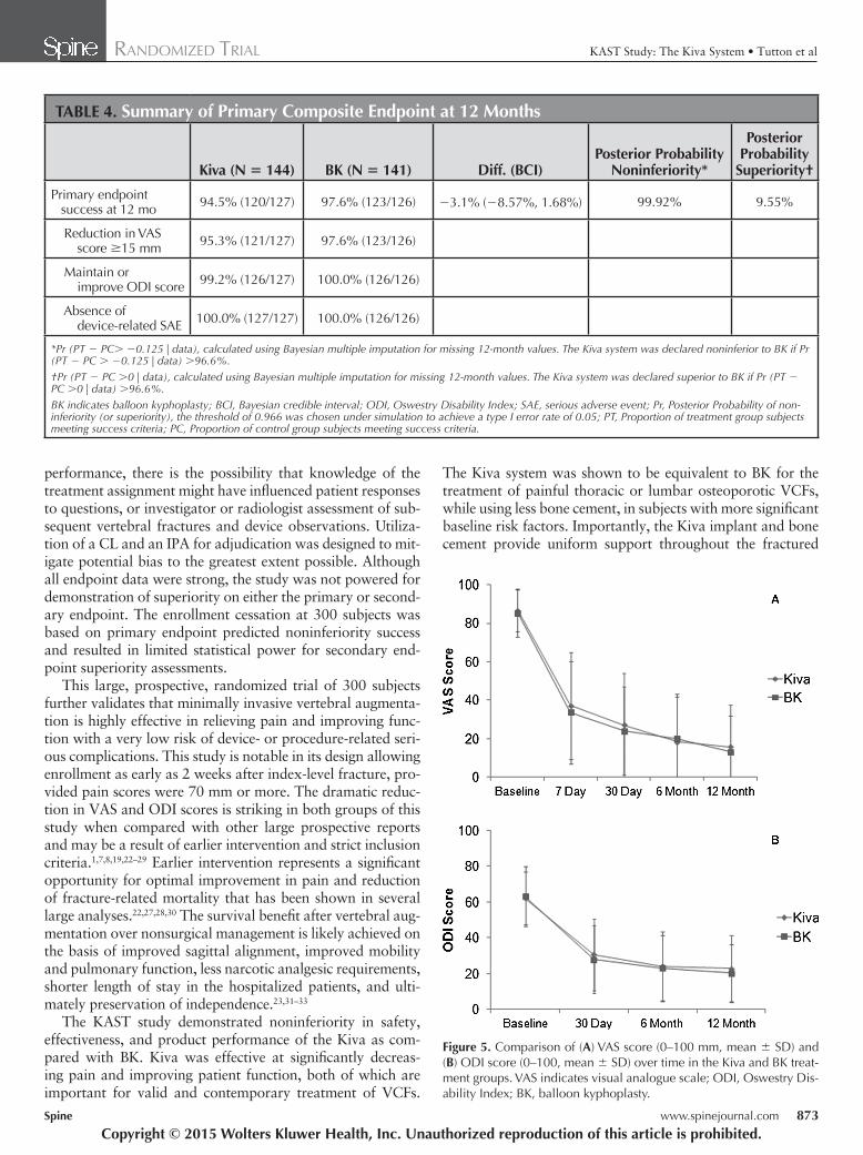
In the 285 AT subjects, VAS scores improved signifi cantly over baseline in both groups after 12 months (Kiva: 70.8 ± 26.3; and BK: 71.8 ± 23.5). ODI scores also improved sig-nifi cantly at 12 months over baseline in both groups (Kiva: 38.1 ± 19.8; and BK: 42.2 ± 21.7). A comparison of VAS
( Figure 5A ) and ODI ( Figure 5B ) scores over time across groups is presented.
In the AT subjects at 12 months, 20.9% (28/134) of the Kiva group and 22.3% (29/130) of the BK group had expe-rienced a new adjacent level compression fracture (symptom-atic and asymptomatic combined rate), meeting the endpoint of noninferiority in the Kiva group. An analysis of the per protocol population showed that 13.8% (16/116) of the Kiva group and 20.2% (23/114) of the BK group had experienced a new adjacent level fracture.
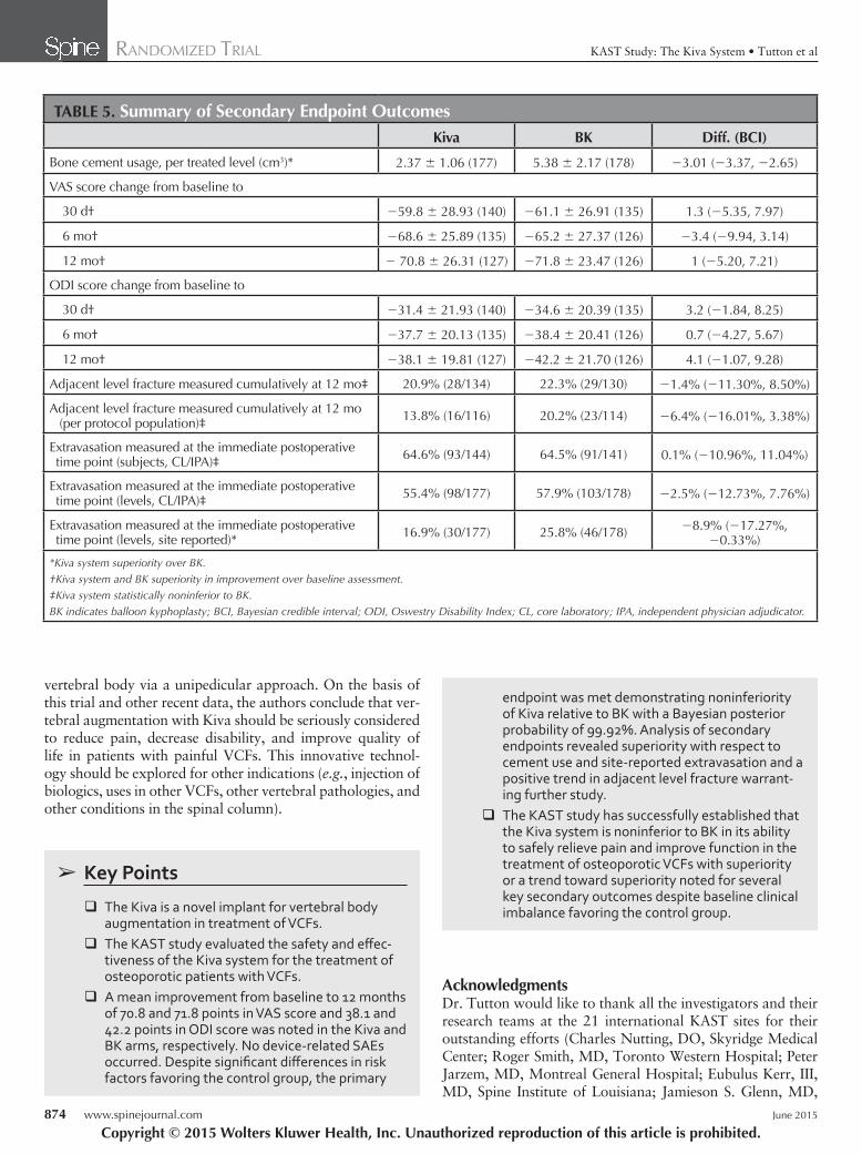
Extravasation of bone cement observed at the procedure was assessed independently by the CL and IPA. Statisti-cal noninferiority was met for the extravasation rate by CL and IPA. The extravasation rate as reported by the site was signifi cantly lower in the Kiva group when compared with BK (Kiva: 16.9%; and BK: 25.8%). Secondary endpoints are summarized in Table 5 .
DISCUSSION The KAST study was a prospective, multicenter, randomized controlled trial designed to evaluate the safety and effective-ness of the Kiva system, a novel implant-based vertebral aug-mentation device. Given its randomized controlled design, large cohort, and 12-month follow-up, it provides level 1 evidence in the study of 2 treatment arms for osteoporotic compression fractures. Focal tenderness and marrow edema on magnetic resonance imaging were required inclusion cri-teria. These refi ned inclusion criteria identifi ed patients with acute or subacute osteoporotic VCF-related pain, unlike ear-lier reported randomized controlled trials without correlative examination or strict imaging criteria. 1 , 7 The KAST study incorporated the use of an independent CL for radiographical evaluations and used an IPA for adjudication of safety data in an effort to remove bias in its assessment of effi cacy and safety.
The KAST study was able to prove that Kiva is noninferior to BK in its ability to safely relieve pain and improve func-tion in the treatment of osteoporotic VCFs. Pain and func-tion were signifi cantly improved from baseline at 30 days, 6 months, and 12 months in both groups, demonstrating the clinical success of these techniques in treating painful VCFs.
No cement-related clinical complications were reported in the 300 subjects enrolled. The observed rate of extravasation
Copyright © 2015 Wolters Kluwer Health, Inc. Unauthorized reproduction of this article is prohibited.
Figure 1. Image of the Kiva system used for vertebral augmentation.
Figure 2. Image under fl uoroscopy of ( A ) the Kiva implant being de-ployed during treatment of VCF and ( B ) post-VCF treatment with the Kiva system. The Kiva implant provides a predictable reservoir for bone cement and the vertebra is restored. VCF indicates vertebral compres-sion fracture.
Figure 3. Image of ( A ) polymethylmethacrylate being injected and con-tained within the Kiva implant. ( B ) The Kiva implant.
SPINE141054_LR 870SPINE141054_LR 870 21/05/15 10:31 AM21/05/15 10:31 AM

RANDOMIZED TRIAL KAST Study: The Kiva System • Tutton et al
Spine www.spinejournal.com 871
in both treatment arms in the KAST study was at the upper limit of that reported in the literature. 8–15 Independent review of plain radiographs in KAST was rigid, including anything outside the confi nes of the vertebral body as evidence of extravasation, without consideration for clinical relevance. This approach resulted in an increased observed frequency of extravasation over the site-reported rate. Site-reported extrav-asation showed a 34% reduction in Kiva over BK. A lower level of extravasation associated with less cement usage has been similarly reported in the literature. 16
Adjacent level fracture rates for Kiva were noninferior to BK. This is notable, given that the Kiva group used sig-nifi cantly less cement and had both a higher percentage of prior fractures and more subjects with a history of smoking. Subsequent fracture risk is increased in patients with a his-tory of smoking and fractures, and the greater the number of vertebral fractures, the greater the risk of additional frac-tures. 17–20 The Kiva implant has been previously reported to be associated with signifi cantly fewer additional VCFs, so
it is possible that the patient characteristics seen in this trial may have neutralized any potential advantage of the implant giving rise to fewer additional VCFs. The fi nding of noninfe-riority in these patients with potentially more complications should be considered in treatment selection. In the analyses of the per protocol population (representing > 80% of the total AT analysis cohort), the Kiva group was observed to have a 30% reduction over BK in adjacent level new fracture rate. This analysis is consistent with a recent study comparing Kiva with BK. 21 In the study of 26 matched pairs, the Kiva and BK groups had an incidence of adjacent and subsequent fractures of 11.5% and 53.8%, respectively. 21 In light of the baseline clinical imbalance favoring the control group, and prior stud-ies demonstrating the benefi t of Kiva with respect to adjacent fracture rates, this trend in the KAST trial warrants further investigation because it is a signifi cant cost driver in the treat-ment of VCFs. 21
The KAST study had several limitations. Because treat-ment was only blinded until just prior to the procedure
Copyright © 2015 Wolters Kluwer Health, Inc. Unauthorized reproduction of this article is prohibited.
Figure 4. Subject accountability of the Kiva and bal-loon kyphoplasty treatment groups at each assessment interval.
SPINE141054_LR 871SPINE141054_LR 871 21/05/15 10:31 AM21/05/15 10:31 AM

RANDOMIZED TRIAL KAST Study: The Kiva System • Tutton et al
872 www.spinejournal.com June 2015
Copyright © 2015 Wolters Kluwer Health, Inc. Unauthorized reproduction of this article is prohibited.
TABLE 3. Baseline Characteristics in Subjects in the Kiva and BK Treatment Groups
Kiva* BK* Diff. (BCI) †
Age
Mean (yr) ± SD (N) 76.03 ± 8.82 (144) 75.09 ± 9.62 (141) 0.94 ( − 1.22, 3.10)
Median (Min, Max) 76.97 (51.62, 96.69) 75.03 (50.40, 91.99)
Female sex [% (n/N)] 72.9% (105/144) 75.2% (106/141) − 2.3% ( − 12.33%, 7.90%)
Former smoker [% (n/N)] 41.7% (60/144) 29.8% (42/141) 11.9% (0.75%, 22.61%) ‡
History of osteoporotic VCF [% (n/N)] 48.6% (70/144) 38.3% (54/141) 10.3% ( − 1.19%, 21.49%)
Thoracic (levels T1–T10) 20.1% (29/144) 20.6% (29/141) − 0.5% ( − 9.78%, 8.90%)
Thoracolumbar junction (levels T11, T12, L1) 29.2% (42/144) 19.1% (27/141) 10.1% (0.04%, 19.66%) ‡
Lumbar (levels L2–5) 22.2% (32/144) 19.1% (27/141) 3.1% ( − 6.32%, 12.38%)
Prior spinal surgical procedures [% (n/N)] 24.3% (35/144) 19.1% (27/141) 5.2% ( − 4.44%, 14.60%)
Single-level procedures 13.2% (19/144) 10.6% (15/141) 2.6% ( − 5.07%, 10.14%)
Multilevel procedures 11.8% (17/144) 10.6% (15/141) 1.2% ( − 6.30%, 8.58%)
Total duration of conservative treatment [% (n/N)]
≤ 6 wk 50.7% (73/144) 54.6% (77/141) − 3.86% ( − 15.29%, 7.63%)
> 6 wk– ≤ 3 mo 32.6% (47/144) 32.6% (46/141) 0.0% ( − 10.79%, 10.82%)
> 3 mo 16.7% (24/144) 12.8% (18/141) 3.83% ( − 4.43%, 12.08%)
DXA spine T-score
Mean ± SD (N) − 1.96 ± 1.58 (133) − 1.89 ± 1.49 (121) − 0.07 ( − 0.45, 0.31)
Median (Min, Max) − 2.10 ( − 5.40, 4.00) − 2.00 ( − 5.60, 2.10)
Baseline VAS score
Mean ± SD (N) 86.67 ± 11.13 (144) 85.29 ± 12.12 (141) 1.38 ( − 1.35, 4.11)
Median (Min, Max) 89.00 (59.00, 100.00) 87.00 (52.00, 100.00)
Baseline ODI score
Mean ± SD (N) 62.22 ± 14.59 (144) 63.17 ± 16.48 (141) − 0.95 ( − 4.60, 2.70)
Median (Min, Max) 62.36 (30.00, 97.78) 64.44 (30.00, 97.78)
Number of treated fractures [% (n/N)]
1 77.1% (111/144) 73.8% (104/141) 3.3% ( − 6.67%, 13.19%)
2 22.9% (33/144) 26.2% (37/141) − 3.3% ( − 13.19%, 6.67%)
Treated fracture Location [% (n/N)]
Thoracic (levels T1–T10) 19.2% (34/177) 12.4% (22/178) 6.8% ( − 0.76%, 14.38%)
Thoracolumbar junction (levels T11, T12, L1) 52.0% (92/177) 52.2% (93/178) − 0.2% ( − 10.58%, 10.04%)
Lumbar (levels L2–L5) 28.8% (51/177) 35.4% (63/178) − 6.6% ( − 16.08%, 3.13%)
*As-treated subjects. † Difference take for comparability between the 2 groups (Kiva − BK). 95% BCI for the difference in means (or proportions). ‡ BCIs that exclude zero indicate a statistical difference between groups. BK, balloon kyphoplasty; BCI, Bayesian credible interval; VCF, vertebral compression fracture; DXA, dual-energy x-ray absorptiometry; ODI, Oswestry Disability Index.
SPINE141054_LR 872SPINE141054_LR 872 21/05/15 10:31 AM21/05/15 10:31 AM
RANDOMIZED TRIAL KAST Study: The Kiva System • Tutton et al
Spine www.spinejournal.com 873
performance, there is the possibility that knowledge of the treatment assignment might have infl uenced patient responses to questions, or investigator or radiologist assessment of sub-sequent vertebral fractures and device observations. Utiliza-tion of a CL and an IPA for adjudication was designed to mit-igate potential bias to the greatest extent possible. Although all endpoint data were strong, the study was not powered for demonstration of superiority on either the primary or second-ary endpoint. The enrollment cessation at 300 subjects was based on primary endpoint predicted noninferiority success and resulted in limited statistical power for secondary end-point superiority assessments.
This large, prospective, randomized trial of 300 subjects further validates that minimally invasive vertebral augmenta-tion is highly effective in relieving pain and improving func-tion with a very low risk of device- or procedure-related seri-ous complications. This study is notable in its design allowing enrollment as early as 2 weeks after index-level fracture, pro-vided pain scores were 70 mm or more. The dramatic reduc-tion in VAS and ODI scores is striking in both groups of this study when compared with other large prospective reports and may be a result of earlier intervention and strict inclusion criteria. 1 , 7 , 8 , 19 , 22–29 Earlier intervention represents a signifi cant opportunity for optimal improvement in pain and reduction of fracture-related mortality that has been shown in several large analyses. 22 , 27 , 28 , 30 The survival benefi t after vertebral aug-mentation over nonsurgical management is likely achieved on the basis of improved sagittal alignment, improved mobility and pulmonary function, less narcotic analgesic requirements, shorter length of stay in the hospitalized patients, and ulti-mately preservation of independence. 23 , 31–33
The KAST study demonstrated noninferiority in safety, effectiveness, and product performance of the Kiva as com-pared with BK. Kiva was effective at signifi cantly decreas-ing pain and improving patient function, both of which are important for valid and contemporary treatment of VCFs.
The Kiva system was shown to be equivalent to BK for the treatment of painful thoracic or lumbar osteoporotic VCFs, while using less bone cement, in subjects with more signifi cant baseline risk factors. Importantly, the Kiva implant and bone cement provide uniform support throughout the fractured
Copyright © 2015 Wolters Kluwer Health, Inc. Unauthorized reproduction of this article is prohibited.
TABLE 4. Summary of Primary Composite Endpoint at 12 Months
Kiva (N = 144) BK (N = 141) Diff. (BCI)Posterior Probability
Noninferiority*
Posterior Probability
Superiority †
Primary endpoint success at 12 mo 94.5% (120/127) 97.6% (123/126) − 3.1% ( − 8.57%, 1.68%) 99.92% 9.55%
Reduction in VAS score ≥ 15 mm 95.3% (121/127) 97.6% (123/126)
Maintain or improve ODI score 99.2% (126/127) 100.0% (126/126)
Absence of device-related SAE 100.0% (127/127) 100.0% (126/126)
*Pr (PT − PC > − 0.125 | data), calculated using Bayesian multiple imputation for missing 12-month values. The Kiva system was declared noninferior to BK if Pr (PT − PC > − 0.125 | data) > 96.6%. †Pr (PT − PC > 0 | data), calculated using Bayesian multiple imputation for missing 12-month values. The Kiva system was declared superior to BK if Pr (PT − PC > 0 | data) > 96.6%. BK indicates balloon kyphoplasty; BCI, Bayesian credible interval; ODI, Oswestry Disability Index; SAE, serious adverse event; Pr, Posterior Probability of non-inferiority (or superiority), the threshold of 0.966 was chosen under simulation to achieve a type I error rate of 0.05; PT, Proportion of treatment group subjects meeting success criteria; PC, Proportion of control group subjects meeting success criteria.
Figure 5. Comparison of ( A ) VAS score (0–100 mm, mean ± SD) and ( B ) ODI score (0–100, mean ± SD) over time in the Kiva and BK treat-ment groups. VAS indicates visual analogue scale; ODI, Oswestry Dis-ability Index; BK, balloon kyphoplasty.
SPINE141054_LR 873SPINE141054_LR 873 21/05/15 10:31 AM21/05/15 10:31 AM
RANDOMIZED TRIAL KAST Study: The Kiva System • Tutton et al
874 www.spinejournal.com June 2015
vertebral body via a unipedicular approach. On the basis of this trial and other recent data, the authors conclude that ver-tebral augmentation with Kiva should be seriously considered to reduce pain, decrease disability, and improve quality of life in patients with painful VCFs. This innovative technol-ogy should be explored for other indications ( e.g. , injection of biologics, uses in other VCFs, other vertebral pathologies, and other conditions in the spinal column).
➢ Key Points
The Kiva is a novel implant for vertebral body augmentation in treatment of VCFs.
The KAST study evaluated the safety and eff ec-tiveness of the Kiva system for the treatment of osteoporotic patients with VCFs.
A mean improvement from baseline to 12 months of 70.8 and 71.8 points in VAS score and 38.1 and 42.2 points in ODI score was noted in the Kiva and BK arms, respectively. No device-related SAEs occurred. Despite signifi cant diff erences in risk factors favoring the control group, the primary
endpoint was met demonstrating noninferiority of Kiva relative to BK with a Bayesian posterior probability of 99.92%. Analysis of secondary endpoints revealed superiority with respect to cement use and site-reported extravasation and a positive trend in adjacent level fracture warrant-ing further study.
The KAST study has successfully established that the Kiva system is noninferior to BK in its ability to safely relieve pain and improve function in the treatment of osteoporotic VCFs with superiority or a trend toward superiority noted for several key secondary outcomes despite baseline clinical imbalance favoring the control group.
Acknowledgments Dr. Tutton would like to thank all the investigators and their research teams at the 21 international KAST sites for their outstanding efforts (Charles Nutting, DO, Skyridge Medical Center; Roger Smith, MD, Toronto Western Hospital; Peter Jarzem, MD, Montreal General Hospital; Eubulus Kerr, III, MD, Spine Institute of Louisiana; Jamieson S. Glenn, MD,
Copyright © 2015 Wolters Kluwer Health, Inc. Unauthorized reproduction of this article is prohibited.
TABLE 5. Summary of Secondary Endpoint Outcomes Kiva BK Diff. (BCI)
Bone cement usage, per treated level (cm 3 )* 2.37 ± 1.06 (177) 5.38 ± 2.17 (178) − 3.01 ( − 3.37, − 2.65)
VAS score change from baseline to
30 d † − 59.8 ± 28.93 (140) − 61.1 ± 26.91 (135) 1.3 ( − 5.35, 7.97)
6 mo † − 68.6 ± 25.89 (135) − 65.2 ± 27.37 (126) − 3.4 ( − 9.94, 3.14)
12 mo † − 70.8 ± 26.31 (127) − 71.8 ± 23.47 (126) 1 ( − 5.20, 7.21)
ODI score change from baseline to
30 d † − 31.4 ± 21.93 (140) − 34.6 ± 20.39 (135) 3.2 ( − 1.84, 8.25)
6 mo † − 37.7 ± 20.13 (135) − 38.4 ± 20.41 (126) 0.7 ( − 4.27, 5.67)
12 mo † − 38.1 ± 19.81 (127) − 42.2 ± 21.70 (126) 4.1 ( − 1.07, 9.28)
Adjacent level fracture measured cumulatively at 12 mo ‡ 20.9% (28/134) 22.3% (29/130) − 1.4% ( − 11.30%, 8.50%)
Adjacent level fracture measured cumulatively at 12 mo (per protocol population) ‡ 13.8% (16/116) 20.2% (23/114) − 6.4% ( − 16.01%, 3.38%)
Extravasation measured at the immediate postoperative time point (subjects, CL/IPA) ‡ 64.6% (93/144) 64.5% (91/141) 0.1% ( − 10.96%, 11.04%)
Extravasation measured at the immediate postoperative time point (levels, CL/IPA) ‡ 55.4% (98/177) 57.9% (103/178) − 2.5% ( − 12.73%, 7.76%)
Extravasation measured at the immediate postoperative time point (levels, site reported)* 16.9% (30/177) 25.8% (46/178) − 8.9% ( − 17.27%,
− 0.33%)
*Kiva system superiority over BK. † Kiva system and BK superiority in improvement over baseline assessment. ‡ Kiva system statistically noninferior to BK. BK indicates balloon kyphoplasty; BCI, Bayesian credible interval; ODI, Oswestry Disability Index; CL, core laboratory; IPA, independent physician adjudicator.
SPINE141054_LR 874SPINE141054_LR 874 21/05/15 10:31 AM21/05/15 10:31 AM

RANDOMIZED TRIAL KAST Study: The Kiva System • Tutton et al
Spine www.spinejournal.com 875
References 1. Buchbinder R , Osborne RH , Ebeling PR , et al. A randomized trial
of vertebroplasty for painful osteoporotic vertebral fractures . N Engl J Med 2009 ; 361 : 557 – 68 .
2. Chen AT , Cohen DB , Skolasky RL . Impact of nonoperative treat-ment, vertebroplasty, and kyphoplasty on survival and morbidity after vertebral compression fracture in the Medicare population . J Bone Joint Surg Am 2013 ; 95 : 1729 – 36 .
3. Brunton S , Carmichael B , Gold D , et al. Vertebral compression frac-tures in primary care: recommendations from a consensus panel . J Fam Pract 2005 ; 54 : 781 – 8 .
4. Gold DT . Osteoporosis and quality of life psychosocial out-comes and interventions for individual patients . Clin Geriatr Med 2003 ; 19 : 271 – 80 , vi.
5. Edidin AA , Ong KL , Lau E , et al. Cost-effectiveness analysis of treatments for vertebral compression fractures . Appl Health Econ Health Policy 2012 ; 10 : 273 – 84 .
6. Ostelo RW , Deyo RA , Stratford P , et al. Interpreting change scores for pain and functional status in low back pain: towards inter-national consensus regarding minimal important change . Spine 2008 ; 33 : 90 – 4 .
7. Kallmes DF , Comstock BA , Heagerty PJ , et al. A randomized trial of vertebroplasty for osteoporotic spinal fractures . N Engl J Med 2009 ; 361 : 569 – 79 .
8. Bae H , Shen M , Maurer P , et al. Clinical experience using Cortoss for treating vertebral compression fractures with vertebroplasty and kyphoplasty: twenty-four month follow-up . Spine 2010 ; 35 : E1030 – 6 .
9. Becker S , Meissner J , Tuschel A , et al. Cement leakage into the posterior spinal canal during balloon kyphoplasty: a case report . J Orthop Surg 2007 ; 15 : 222 – 5 .
10. Hou C . Balloon kyphoplasty and vertebroplasty for verte-bral compression fracture: a systemic review . Osteoporosis Int 2011 ; 22 : S738 – 9 .
11. Korovessis P , Vardakastanis K , Repantis T , et al. Balloon kypho-plasty versus KIVA vertebral augmentation—comparison of 2 techniques for osteoporotic vertebral body fractures: a prospective randomized study . Spine 2013 ; 38 : 292 – 9 .
12. Lee MJ , Dumonski M , Cahill P , et al. Percutaneous treatment of vertebral compression fractures: a meta-analysis of complications . Spine 2009 ; 34 : 1228 – 32 .
13. Taylor RS , Fritzell P , Taylor RJ . Balloon kyphoplasty in the man-agement of vertebral compression fractures: an updated systematic review and meta-analysis . Eur Spine J 2007 ; 16 : 1085 – 100 .
14. Taylor RS , Taylor RJ , Fritzell P . Balloon kyphoplasty and vertebro-plasty for vertebral compression fractures: a comparative system-atic review of effi cacy and safety . Spine 2006 ; 31 : 2747 – 55 .
15. Zhang J , Li X , Lu F , et al. Balloon kyphoplasty combined with bone cement for the treatment of osteoporotic vertebral fracture in 58 cases . J Clin Rehabil Tissue Eng Res 2009 ; 13 : 9373 – 6 .
16. Potet J , Weber-Donat G , Curis E , et al. Incidence of pulmonary cement embolism after real-time CT fl uoroscopy-guided vertebro-plasty . J Vasc Int Radiol 2013 ; 24 : 1853 – 60 .
17. Gehlbach S , Saag KG , Adachi JD , et al. Previous fractures at mul-tiple sites increase the risk for subsequent fractures: the global longitudinal study of osteoporosis in women . J Bone Miner Res 2012 ; 27 : 645 – 53 .
18. Melton LJ III , Atkinson EJ , Cooper C , et al. Vertebral fractures predict subsequent fractures . Osteoporosis Int 1999 ; 10 : 214 – 21 .
19. Siris ES , Genant HK , Laster AJ , et al. Enhanced prediction of frac-ture risk combining vertebral fracture status and BMD . Osteoporo-sis Int 2007 ; 18 : 761 – 70 .
20. Kanis JA , Johnell O , De Laet C , et al. A meta-analysis of previous fracture and subsequent fracture risk . Bone 2004 ; 35 : 375 – 82 .
21. Otten LA , Bornemnn R , Jansen TR , et al. Comparison of balloon kyphoplasty with the new Kiva(R) VCF system for the treatment of vertebral compression fractures . Pain Phys 2013 ; 16 : E505 – 12 .
22. Papanastassiou ID , Filis A , Gerochristou MA , et al. Controversial issues in kyphoplasty and vertebroplasty in osteoporotic vertebral fractures . BioMed Res Int 2014 ; 2014 : 934206 .
23. Zampini JM , White AP , McGuire KJ . Comparison of 5766 ver-tebral compression fractures treated with or without kyphoplasty . Clin Orthop Relat Res 2010 ; 468 : 1773 – 80 .
24. Wardlaw D , Cummings SR , Van Meirhaeghe J , et al. Effi cacy and safety of balloon kyphoplasty compared with non-surgical care for vertebral compression fracture (FREE): a randomised controlled trial . Lancet 2009 ; 373 : 1016 – 24 .
25. Santiago FR , Abela AP , Alvarez LG , et al. Pain and functional out-come after vertebroplasty and kyphoplasty. A comparative study . Eur J Radiol 2010 ; 75 : e108 – 13 .
26. Li X , Yang H , Tang T , et al. Comparison of kyphoplasty and verte-broplasty for treatment of painful osteoporotic vertebral compres-sion fractures: twelve-month follow-up in a prospective nonran-domized comparative study . J Spinal Disord Tech 2012 ; 25 : 142 – 9 .
27. Gerling MC , Eubanks JD , Patel R , et al. Cement augmentation of refractory osteoporotic vertebral compression fractures: survivor-ship analysis . Spine 2011 ; 36 : E1266 – 9 .
28. Edidin AA , Ong KL , Lau E , et al. Mortality risk for operated and nonoperated vertebral fracture patients in the Medicare population . J Bone Miner Res 2011 ; 26 : 1617 – 26 .
29. Chen C , Wei H , Zhang W , et al. Comparative study of kyphoplasty for chronic painful osteoporotic vertebral compression fractures via unipedicular versus bipedicular approach . J Spinal Disord Techn 2011 ; 24 : E62 – 5 .
30. Lau E , Ong K , Kurtz S , et al. Mortality following the diagnosis of a vertebral compression fracture in the Medicare population . J Bone Joint Surg Am 2008 ; 90 : 1479 – 86 .
31. Chandra RV , Yoo AJ , Hirsch JA . Vertebral augmentation: update on safety, effi cacy, cost effectiveness and increased survival . Pain Physician 2013 ; 16 : 309 – 20 .
32. Lange A , Kasperk C , Alvares L , et al. Survival and cost compari-son of kyphoplasty and percutaneous vertebroplasty using German claims data . Spine 2014 ; 39 : 318 – 26 .
33. Stevenson M , Gomersall T , Lloyd Jones M , et al. Percutaneous vertebroplasty and percutaneous balloon kyphoplasty for the treat-ment of osteoporotic vertebral fractures: a systematic review and cost-effectiveness analysis . Health Technol Assess 2014 ; 18 : 1 – 290 .
CORE Orthopaedic Medical Center; Johannes Hierholzer, MD, Klinikum Ernst von Bergmann; Hervé Deramond, MD, CHU Amiens; James Zucherman, MD, St. Marys Spine Cen-ter; Bruce Frankel, MD, Medical University of South Caro-lina; David Kallmes, MD, Mayo Clinic–Rochester; Thach Dam Nguyen, MD, Penn State Hershey; Marc Alonzo, MD, Evanston Hospital; Frederic Schils, MD, CHC Saint Joseph; Jeffrey A. Stone, MD, Mayo Clinic–Jacksonville; Fabio Kom-los, MD, El Camino Hospital; James Rappaport, MD, Sierra Regional Spine Institute), as well as the study statistician (Andrew Mugglin, PhD, Paradigm Biostatistics, LLC), the study core laboratory (BioClinica, Inc.), and the study CRO (Boston Biomedical Associates) for their contributions.
Copyright © 2015 Wolters Kluwer Health, Inc. Unauthorized reproduction of this article is prohibited.
SPINE141054_LR 875SPINE141054_LR 875 21/05/15 10:31 AM21/05/15 10:31 AM