Embed Size (px)
Citation preview
Proc. Natl. Acad. Sci. USAVol. 87, pp. 187-190, January 1990Neurobiology
Nerve growth factor receptor immunoreactivity is transientlyassociated with the subplate neurons of the mammaliancerebral cortex
(trophic factors/neural development/cell death/brain/transient neurons)
KAREN L. ALLENDOERFER*, DAVID L. SHELTONt, ERIC M. SHOOTER, AND CARLA J. SHATZDepartment of Neurobiology, Stanford University School of Medicine, Stanford, CA 94305
Communicated by Gunther S. Stent, October 13, 1989
ABSTRACT Nerve growth factor and its receptor (NGFR)are known to be present in diverse embryonic and neonatalcentral nervous system tissues, including the cerebral cortex.However, the identity of the cortical cells expressing NGFRimmunoreactivity has not been established. We have usedimmunolabeling coupled with [3HJthymidine autoradiographyto identify such cells in ferret and cat brain. Polyclonalantibodies raised against a synthetic peptide corresponding toa conserved amino acid sequence of the NGFR were used forthis purpose. Western (immunologic) blot analyses show thatthese antibodies specifically recognize NGFR and precursorproteins. In both species, NGFR immunoreactivity is primarilyassociated with the early generated and transient subplateneuron population of the developing neocortex, as indicated bythe following evidence: the immunoreactive cells (i) are locateddirectly beneath the developing cortical plate, (a) frequentlyhave the inverted pyramid shape characteristic of subplateneurons, and (iii) can be labeled by an injection of[3H]thymidine on embryonic day (E) 28, a time when onlysubplate neurons are being generated. Intense NGFR immu-nostaining is seen on the cell bodies of these neurons as early asE30, several days after their last round of cell division, and thisimmunostaining remains strong for -3 weeks. The NGFRimmunoreactivity begins to decline around E52 and has dis-appeared from the region altogether by E60, at which timesubplate neurons begin to die. The cellular localization andtiming of expression suggest that the NGFR may play a role inthe maintenance of subplate neurons and in the maturation ofthe cerebral cortex.
During the development of the mammalian brain, the forma-tion of the adult six-layered cerebral cortex is preceded by aperiod in which a special class of transient neurons is gener-ated (1-3). These neurons are among the first postmitotic cellsof the telencephalon and, in higher mammals, undergo theirfinal round of cell division well before the neurons ofthe adultcortical plate [cat at embryonic day (E) 24-30 (1, 4); ferret atE20-24 (3)]. These earliest generated cells then migrate awayfrom the germinal zone and initially take up positions imme-diately beneath the pial surface. However, as the neurons ofthe future cortical plate migrate out and assume their finallocations, the early generated neuronal population is split intotwo parts, the subplate, which is located directly beneath thedeveloping cortical plate, and the marginal zone, which islocated directly above it (4).
Subplate neurons mature rapidly and express immunoreac-tivity not only to neurotransmitters but also to microtubule-associated protein 2 (MAP2) long before the cortical neurons
do (5, 6). Furthermore, they receive functional synapticinputs (refs. 5 and 7; E. Friauf, S. K. McConnell, and C.J.S.,unpublished data) and pioneer elaborate projections to cor-tical and subcortical targets (5, 8). Subplate neurons are alsothought to interact with the many ingrowing axonal systemsfrom the thalamus and cortex that are known to wait in thesubplate zone (2, 9, 10) before finally invading the corticalplate, at -1 week before birth (11). After the waiting periodends, subplate neurons begin a programmed cell death, whicheliminates over 90% of the population, until finally only1%o remain by 3 months of age (12, 13). Here we examine
the possibility that nerve growth factor (NGF) plays a role inthe maintenance of subplate neurons during early corticaldevelopment and in their subsequent death. We studied thedistribution of NGF receptor (NGFR) immunoreactivity ofthe subplate neurons from the time they become postmitoticuntil after they begin to die, in view of recent reports that thereceptor is likely to be transiently expressed in the centralnervous system and, in particular, in the telencephalon,during embryonic and neonatal life (14-17).
MATERIALS AND METHODSEight cat brains between E35 and E60 and four ferret brains,ages postnatal days (P)2 and 10 were studied. Cat fetal tissuewas obtained by sterile surgery on timed pregnant cats, asdescribed (1, 4). Timed pregnant sable ferrets were obtainedfrom Marshall Laboratories (North Rose, NY). As discussedbelow, the pace and pattern of development of cat and ferretcortex are strikingly similar.
Preparation of Antibody. The peptide YNSRPVNQTPP-PEGEKLHSD (in one-letter code), corresponding to resi-dues 258-276 of the rat low-affinity NGFR (18) plus aterminal tyrosine, was synthesized commercially usingFMOC (9-fluorenylmethoxycarbonyl) chemistry. This se-quence is part of the intracellular domain of the protein andis completely conserved at the amino acid level between therat and human low-affinity NGFR (18, 19). The peptide wascrosslinked to porcine thyroglobulin by using 1-ethyl-3-(3-dimethylaminopropyl)-carbodiimide. One milligram ofthyroglobulin conjugate (corresponding to 100 ,.g of peptide)was mixed with 100 pug of free peptide, emulsified with 3 volof Freund's complete adjuvant, and injected into 10 intra-muscular sites of four female New Zealand White rabbits.The animals were reinjected subcutaneously with the sameamount of antigen in Freund's incomplete adjuvant at
Abbreviations: NGF, nerve growth factor; NGFR, NGF receptor; E,embryonic day; P, postnatal day; MAP2, microtubule-associatedprotein 2.*To whom reprint requests should be addressed.tPresent address: Genentech, Inc., 460 Point San Bruno, South SanFrancisco, CA 94080.
187
The publication costs of this article were defrayed in part by page chargepayment. This article must therefore be hereby marked "advertisement"in accordance with 18 U.S.C. §1734 solely to indicate this fact.
Dow
nloa
ded
by g
uest
on
Janu
ary
30, 2
022
188 Neurobiology: Allendoerfer et al.
monthly intervals. Rabbits were bled 2 weeks after eachreinjection, and antipeptide antibodies were affinity-purifiedwith a column of peptide crosslinked via glutaraldehyde tobovine serum albumin bound to Affi-Gel (Bio-Rad) and elutedwith glycine hydrochloride, pH 2.5. Before use, antibodiesthus obtained were adsorbed with a similar column contain-ing a NGFR-unrelated peptide bound to bovine serum albu-min. All ofthe experiments in this study were carried out withthe antibodies obtained from a single bleeding of one rabbit.Western (Immunologic) Blots. Telencephalon tissue (in-
cluding cortical plate and subplate; excluding hippocampus)was dissected immediately after euthanasia of animals of theappropriate age, homogenized in 10.8% sucrose, and centri-fuged at 3000 x g for 5 min. The pellet was dissolved in 0.1M phosphate-buffered saline with 0.5% Nonidet P-40 (Sig-ma), incubated on ice for 10 min, and microcentrifuged. Theoptical density at 280 gm was read to estimate the proteincontent of the Nonidet P-40 soluble fraction, and -200 ,ug ofprotein was applied per lane to an SDS/polyacrylamide gel(7.5%) for electrophoresis at 30 V. The protein was trans-ferred to nitrocellulose, and the blot was incubated withanti-NGFR antibody, iodinated by using lodogen (Pierce) toa specific 125I activity of 6.5 x 10⁸ cpm/Ag.
Immunohistochemistry. Brains were prepared for immuno-histochemistry by transcardial perfusion of the cat fetuses orferret neonates with 4% (wt/vol) paraformaldehyde in 0.1 Msodium phosphate buffer followed by 6-12 hr postfixation inthe same solution. Tissue was equilibrated in 20% (wt/vol)sucrose, and coronal cryostat sections (20 Am) were cut andmounted on gelatin/albumin-coated slides. Sections wereincubated in 1:1000 anti-MAP2 serum (5, 6) (provided by R.Vallee, Worcester Foundation for Experimental Biology),anti-NGFR antibody at 1 ,g/ml, or anti-NGFR antibody at 1,ug/ml blocked by 100-fold excess of the peptide againstwhich the antibody was raised. Antibody binding was visu-alized by using peroxidase immunohistochemistry (ABC,Vector Laboratories). [3H]Thymidine was injected into fe-tuses in utero at E28 and visualized autoradiographically atE47 by coating mounted brain sections with Kodak NTB-2emulsion and exposing them in the dark at 40C for 4 to 6 weeksas described (1). The immunohistochemical results are sum-marized in Table 1.
<. C1) _' C S
RESULTSTo investigate the presence and distribution of NGFR in thecerebral cortex during development, we immunostained sec-tions from fetal cat brain at ages between E30 and E60 andneonatal ferret brain at postnatal day (P) 2 and P10 (see Table1). Many previous studies have demonstrated that these twospecies have a remarkably similar pace of cortical develyp-ment during fetal life, even though the ferret is born at 41gestational days (3, 20), whereas the cat is born at 65 days (1)(e.g., a ferret at P2 is developmentally equivalent to a cat atE43). After the ferret is born, its development speeds up inrelation to that of the cat, which remains in utero, so that byP10 a ferret cortex resembles that of an E60 cat.
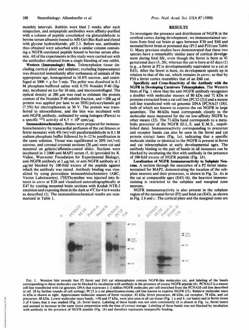
Specificity and Cross-Reactivity of the Antibody with theNGFR in Developing Carnivore Telencephalon. The Westernblots of Fig. 1 show that the anti-NGFR antibody recognizesa doublet with molecular masses of 80 kDa and 71 kDa inproteins extracted from a rat cell line [PC12 (21)] and a mousecell line transfected with rat genomic DNA [PCNA15 (18)],both of which are known to express the rat NGFR in largequantities. The 80-kDa band corresponds, closely to themolecular mass measured for the rat low-affinity. NGFR byother means (22). The 71-kDa band corresponds to a meta-bolic precursor of the NGFR (D.L.S. and E.M.S., unpub-lished data). Immunoreactivity corresponding to precursorand receptor bands can also be seen in the ferret and catprotein extract lanes (Fig. la), indicating that a specificmolecule similar or identical to the NGFR is present in ferretand cat telencephalon at early developmental ages. Theantibody binding to the pair of bands in all instances can beblocked by incubating the blot with antibody in the presenceof 100-fold excess of NGFR peptide (Fig. lb).
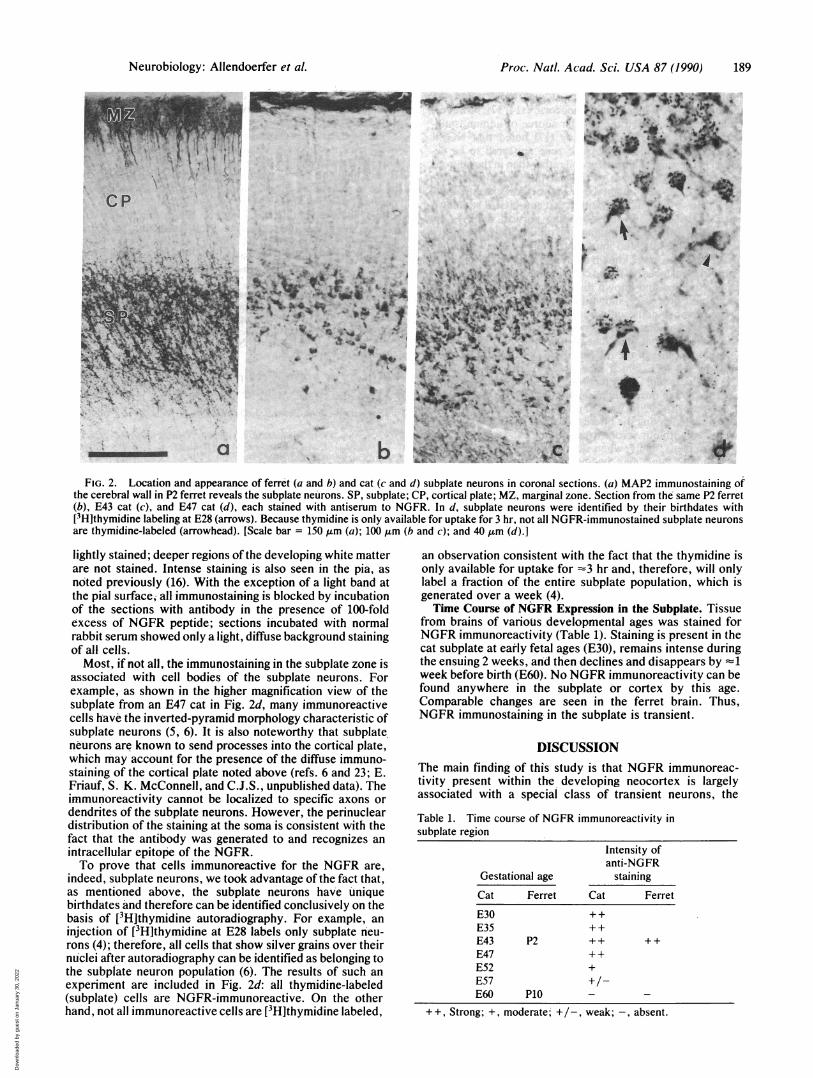
Localization of NGFR Immunoreactivity to Subplate Neu-rons. A section through the neocortex of a P2 ferret immu-nostained for MAP2, demonstrating the location of the sub-plate neurons and their processes, is shown in Fig. 2a. As inthe cat at comparable ages (E43) (6), the heaviest immuno-staining is restricted to the subplate and marginal zoneneurons.NGFR immunoreactivity is also present in the subplate
region of the neonatal ferret (P2) and fetal cat (E43), as shownin Fig. 2 b and c. The cortical plate and the marginal zone are
Yto \ \l
rp
412*
.
4w. _~~~~~~~~- "
<- 1
FIG. 1. Western blot reveals that P2 ferret and E43 cat telencephalon contain NGFR-like molecules (a), and labeling of the bandscorresponding to these molecules can be blocked by incubation with antibody in the presence of excess NGFR peptide (b). PCNA15 is a mousecell line transfected with rat genomic DNA that expresses 1-2 million NGFR molecules per cell (enriched from the PCNA10 cell line describedin ref. 18 by further rounds of cell sorting); PC12 is a rat pheochromocytoma cell line known to express NGFR (21). Relative molecular massin kDa is shown at right. Approximate molecular masses of ferret receptor, 82 kDa; ferret precursor, 68 kDa; cat receptor, 78 kDa; and catprecursor, 68 kDa. Lower molecular mass bands, -50 and 37 kDa, were also seen in all cat tissue (Fig. 1 a and b, cat lanes) and in ferret tissue2 of 4 times that it was studied (Fig. lb, ferret lanes). Labeling of these bands was not seen consistently (it is absent in Fig. la, ferret lanes)and seemed .to increase in the same tissue preparation with the time of extract storage. Labeling of these bands was not blocked by incubationwith antibody in the presence of NGFR peptide (Fig. lb) and therefore represents nonspecific binding.
Proc. Natl. Acad. Sci. USA 87 (1990)
.:..M.,. .:zI 1. .- .:
-Awbm.%-Wr
Dow
nloa
ded
by g
uest
on
Janu
ary
30, 2
022
Proc. Natl. Acad. Sci. USA 87 (1990) 189
* S
~b
-,~ - ~
4.
,Ka'~
* .. . f %~~q.PI .9 .. ...... ..
s S'N e-s, rs3 .> J%- .0
.4iRR *~~~41_I
_a e
t..FIG. 2. Location and appearance of ferret (a and b) and cat (c and d) subplate neurons in coronal sections. (a) MAP2 immunostaining of
the cerebral wall in P2 ferret reveals the subplate neurons. SP, subplate; CP, cortical plate; MZ, marginal zone. Section from the same P2 ferret(b), E43 cat (c), and E47 cat (d), each stained with antiserum to NGFR. In d, subplate neurons were identified by their birthdates with[3H]thymidine labeling at E28 (arrows). Because thymidine is only available for uptake for 3 hr, not all NGFR-immunostained subplate neuronsare thymidine-labeled (arrowhead). [Scale bar = 150 ,um (a); 100 ,um (b and c); and 40,um (d).]
lightly stained; deeper regions of the developing white matterare not stained. Intense staining is also seen in the pia, asnoted previously (16). With the exception of a light band atthe pial surface, all immunostaining is blocked by incubationof the sections with antibody in the presence of 100-foldexcess of NGFR peptide; sections incubated with normalrabbit serum showed only a light, diffuse background stainingof all cells.Most, if not all, the immunostaining in the subplate zone is
associated with cell bodies of the subplate neurons. Forexample, as shown in the higher magnification view of thesubplate from an E47 cat in Fig. 2d, many immunoreactivecells have the inverted-pyramid morphology characteristic ofsubplate neurons (5, 6). It is also noteworthy that subplateneurons are known to send processes into the cortical plate,which may account for the presence of the diffuse immuno-staining of the cortical plate noted above (refs. 6 and 23; E.Friauf, S. K. McConnell, and C.J.S., unpublished data). Theimmunoreactivity cannot be localized to specific axons ordendrites of the subplate neurons. However, the perinucleardistribution of the staining at the soma is consistent with thefact that the antibody was generated to and recognizes anintracellular epitope of the NGFR.To prove that cells immunoreactive for the NGFR are,
indeed, subplate neurons, we took advantage of the fact that,as mentioned above, the subplate neurons have uniquebirthdates and therefore can be identified conclusively on thebasis of [3H]thymidine autoradiography. For example, aninjection of [3Hlthymidine at E28 labels only subplate neu-rons (4); therefore, all cells that show silver grains over theirnuclei after autoradiography can be identified as belonging tothe subplate neuron population (6). The results of such anexperiment are included in Fig. 2d: all thymidine-labeled(subplate) cells are NGFR-immunoreactive. On the otherhand, not all immunoreactive cells are [3H]thymidine labeled,
an observation consistent with the fact that the thymidine isonly available for uptake for -3 hr and, therefore, will onlylabel a fraction of the entire subplate population, which isgenerated over a week (4).Time Course of NGFR Expression in the Subplate. Tissue
from brains of various developmental ages was stained forNGFR immunoreactivity (Table 1). Staining is present in thecat subplate at early fetal ages (E30), remains intense duringthe ensuing 2 weeks, and then declines and disappears by -1week before birth (E60). No NGFR immunoreactivity can befound anywhere in the subplate or cortex by this age.Comparable changes are seen in the ferret brain. Thus,NGFR immunostaining in the subplate is transient.
DISCUSSIONThe main finding of this study is that NGFR immunoreac-tivity present within the developing neocortex is largelyassociated with a special class of transient neurons, the
Table 1. Time course of NGFR immunoreactivity insubplate region
Intensity ofanti-NGFR
Gestational age stainingCat Ferret Cat Ferret
E30 ++E35 ++E43 P2 ++ ++E47 + +E52 +E57 +/-E60 P10 - -
++, Strong; +, moderate; +/-, weak; -, absent.
*CP
a
Neurobiology: Allendoerfer et al.
Ivr--.. I v 1%
V 0
v. 'I ., pIs ..r I ( i
% ..; ,I.1 1 II 4 % (I
t I "
k
Dow
nloa
ded
by g
uest
on
Janu
ary
30, 2
022
190 Neurobiology: Allendoerfer et al.
subplate neurons. Two previous studies have noted thepresence ofNGFR in the rodent neocortex (14, 17); however,neither study identified the cellular source of immunostain-ing. In their study, Koh and Loy (17) noted that NGFRimmunoreactivity in the subplate zone appeared to be de-posited in the surrounding neuropil rather than in the neuronsthemselves, leading them to suggest that radial glial cells,present at this age, might be a source for extraneuronalNGFR. However, our observation that anti-NGFR staining isrestricted to subplate and overlying cortical plate is notconsistent with the morphology of radial glial cells, whichhave cell bodies located immediately above the ventricularsurface and processes known to stretch between the pial andventricular surfaces (24). The coincidence between pattern ofimmunostaining and known distribution of subplate neuronsomata and processes shown here and the fact that NGFRimmunoreactive cells could be [3H]thymidine-labeled duringthe period of subplate neurogenesis indicate that it is subplateneurons, not radial glia, that express the NGFR. Further-more, the authors of the rodent studies used the monoclonalantibody 192 IgG, which recognizes a cell-surface epitope(25), whereas our polyclonal serum was generated to asequence in the intracellular domain of NGFR. The stainingassociated with 192 IgG would probably outline neuronal cellbodies while leaving the interior unstained, whereas stainingassociated with our serum would fill the cell interior, exceptfor the nucleus. Thus, when stained with 192 IgG, NGFR inthe subplate zone may be more difficult to localize withcertainty.
Subplate neurons are immunoreactive for, the NGFR asearly as E30, just as the last of these neurons becomepostmitotic (1, 4) and the first of them begin to project axonsto subcortical targets (8). These neurons remain immunore-active during the month-long period in which they mature andingrowing axons from thalamus and other regions of cortexaccumulate around them. Then NGFR immunoreactivity islost by E60, a time when the waiting axonal systems have justleft the subplate to invade the cortical plate (2) and at a timewhen subplate neurons, though still present in appreciablenumbers, have begun to disappear by cell death (6, 12, 13).The complete loss of NGFR immunoreactivity at this agecannot be due to the disappearance of the neurons them-selves because many MAP2-immunoreactive cells and pro-cesses can still be seen in the subplate zone at E60, althoughtheir density is beginning to decrease (6, 12, 13). Further-more, between E52 and E57, a progressive decrease in theoverall intensity ofNGFR immunostaining on each neuron isseen, not a decrease in the total number of NGFR-immunoreactive neurons, suggesting a down-regulation ofreceptor expression on the cells rather than elimination ofNGFR-immunoreactive neurons by cell death at this time.These correlations raise several possibilities concerning
the functional significance of NGFR expression on subplateneurons. (i) The presence of its receptor at early ages couldindicate that NGF guides the growth of subplate axons in amanner similar to that suggested for the guidance of axons ofsympathetic neurons in vivo (26) or chicken sensory neuronsin vitro (refs. 27 and 28; for an alternate view, see ref. 29). (ii)NGF has also been shown to regulate the expression ofneuropeptide genes in sensory neurons (30) and could do thesame for neuropeptides in the subplate. (iii) Alternatively,loss of NGFR from subplate neurons may play a role inbringing the axonal waiting period to a close. (iv) A finalpossibility is that, as in the development of the peripheralnervous system (31-33) and perhaps also that of the basalforebrain (34, 35), NGF might act to sustain subplate neu-
rons. If so, the loss ofNGFR on subplate neurons may be thefirst step in a cascade of events that eventually leads toprogrammed cell death.The question then arises as to the cellular source of NGF.
In the rodent neocortex, the level ofNGF mRNA is very low(36, 37) at ages when NGFR immunoreactivity in the subplateis very high (14, 17), suggesting that cortex itself is not likelyto be a source of NGF during this early developmentalperiod. Probable candidates include other targets and affer-ents of the subplate neurons, such as the thalamus, tectum,or even subplate neurons themselves. Whatever the source ofNGF, the presence of NGFR on this transient population ofneurons suggests that NGF participates in the developmentalprocesses that give rise to the structure of adult cortex.
We thank A. Antonini, A. Ghosh, S. McConnell, and M. Siegel forsurgical assistance, and S. McConnell for thoughtful criticism of themanuscript. This work was supported by National Institutes ofHealth Grants EY 02858 (C.J.S.), NS 04270 (E.M.S.), the Isabelle M.Niemela Fund (E.M.S.), and a National Science Foundation Grad-uate Fellowship (K.L.A.).
1. Luskin, M. B. & Shatz, C. J. (1985) J. Comp. Neurol. 242, 611-631.2. Shatz, C. J., Chun, J. J. M. & Luskin, M. B. (1988) Cerebral Cortex
7, 35-58.3. Jackson, C. A., Peduzzi, J. D. & Hickey, T. L. (1989) J. Neurosci.
9, 1241-1253.4. Luskin, M. B. & Shatz, C. J. (1985) J. Neurosci. 5, 1062-1075.5. Chun, J. J. M., Nakamura, M. J. & Shatz, C. J. (1987) Nature
(London) 325, 617-620.6. Chun, J. J. M. & Shatz, C. J. (1989) J. Neurosci. 9, 1648-1667.7. Chun, J. J. M. & Shatz, C. J. (1988) Neuron 1, 297-310.8. McConnell, S. K., Ghosh, A. & Shatz, C. J. (1989) Science 245,
978-982.9. Wise, S. P. & Jones, E. G. (1978) J. Comp. Neurol. 175, 187-208.
10. Innocenti, G. M. (1981) Science 212, 824-827.11. Shatz, C. J. & Luskin, M. B. (1986) J. Neurosci. 6, 3655-3668.12. Valverde, F. & Facal-Valverde, M. V. (1988) J. Comp. Neurol. 269,
168-192.13. Chun, J. J. M. & Shatz, C. J. (1989) J. Comp. Neurol. 282,555-569.14. Yan, Q. & Johnson, E. M., Jr. (1988) J. Neurosci. 8, 3481-3498.15. Eckenstein, F. (1988) Brain Res. 446, 149-154.16. Schatteman, G., Biggs, L., Lanahan, A. A., Claude, P. & Bothwell,
M. (1988) J. Neurosci. 8, 860-873.17. Koh, S. & Loy, R. (1989) J. Neurosci. 9, 2999-3018.18. Radeke, M. J., Misko, T. P., Hsu, C., Herzenberg, L. A. &
Shooter, E. M. (1987) Nature (London) 325, 593-597.19. Johnson, D., Lanahan, A., Buck, C. R., Sehgal, A., Morgan, C.,
Mercer, E., Bothwell, M. & Chao, M. (1986) Cell 47, 545-554.20. McConnell, S. K. (1988) J. Neurosci. 8, 945-974.21. Greene, L. A. & Tischler, A. S. (1976) Proc. Natl. Acad. Sci. USA
73, 2424-2428.22. Hosang, M. & Shooter, E. M. (1985) J. Biol. Chem. 260, 655-662.23. Wahle, P. & Meyer, G. (1987) J. Comp. Neurol. 261, 165-192.24. Levitt, P. & Rakic, P. (1980) J. Comp. Neurol. 193, 815-840.25. Chandler, C. E., Parsons, L. M., Hosang, M. & Shooter, E. M.
(1984) J. Biol. Chem. 259, 6882-6889.26. Levi-Montalcini, R. (1976) Prog. Brain Res. 45, 235-256.27. Gundersen, R. W. & Barrett, J. N. (1979) Science 206, 1079-1080.28. Gundersen, R. W. & Barrett, J. N. (1980) J. Cell Biol. 87, 546-554.29. Lumsden, A. G. S. & Davies, A. M. (1983) Nature (London) 306,
786-788.30. Lindsay, R. M. & Harmar, A. J. (1989) Nature (London) 337,
362-364.31. Thoenen, H. & Barde, Y. A. (1980) Physiol. Rev. 60, 1284-1335.32. Levi-Montalcini, R. (1982) Annu. Rev. Neurosci. 5, 341-362.33. Yanker, B. A. & Shooter, E. M. (1982) Annu. Rev. Biochem. 52,
845-868.34. Hartikka, J. & Hefti, F. (1988) J. Neurosci. 8, 2967-2985.35. Montero, C. N. & Hefti, F. (1988) J. Neurosci. 8, 2986-2999.36. Large, T. H., Bodary, S. C., Clegg, D. O., Weskamp, G., Otten, U.
& Reichardt, L. F. (1986) Science 234, 352-355.37. Whittemore, S. R., Ebendal, T., Larkfors, L., Olson, L., Seiger,
A., Stromberg, I. & Persson, H. (1986) Proc. Natl. Acad. Sci. USA83, 817-821.
Proc. Natl. Acad. Sci. USA 87 (1990)
Dow
nloa
ded
by g
uest
on
Janu
ary
30, 2
022