Embed Size (px)
Citation preview
Kansas
Part C infant/toddler
tiny-k
Hearing Screening Guidelines
and
Resource Manual

Acknowledgements Part C infant/toddler tiny-k Hearing Screening Guidelines and Resource Manual is the result of the efforts of people who have been unceasingly dedicated to the early identification and intervention of children with hearing loss. Expertise and time have been generously shared to develop these guidelines. Members of the SoundBeginnings Newborn Hearing Screening Program and Part C Infant Toddler Services unanimously agreed on the purpose of the guidelines: to provide rationale and process for screening hearing of the birth to three population referred for early intervention in Kansas. The final product provides purpose and direction for the many individuals and groups who value and are involved in the detection and intervention of infants and toddlers in the state of Kansas. A special note of appreciation is given to Liz Abbey, SoundBeginnings Coordinator/Audiologist who guided the development of this document. We thank the many people statewide, including members of the SoundBeginnings Advisory Committee, who reviewed the drafts of the guidelines and provided thoughtful input. Date published: June 1, 2010 Revised December 1, 2014
ii
Introduction
It is important to identify hearing loss early and provide timely intervention for very young children and their families. Early identification prevents delays, for all hearing loss, mild to profound. Research shows if a child receives early identification before 3 months and intervention before 6 months of age, children with hearing loss have language development that is comparable to peers their age. 2-3 babies per 1,000 births per year in Kansas will be diagnosed with some degree of hearing loss. The purpose of this manual is to provide standardized hearing screening procedures for all Kansas children receiving services through Part C infant/toddler tiny-k services. The target population for screening is all children birth to three years of age in Kansas. Kansas enacted legislation, effective July 1, 1999, to provide screening for the early detection of hearing loss in all babies born in Kansas hospitals. The Kansas Part C infant/toddler tiny-k Hearing Screening Guidelines and Resource Manual does not address newborn hearing screening prior to hospital discharge or assessment. The procedures in this document were written to be used as a training manual and a resource for Part C infant/toddler tiny-k networks that screen hearing in children birth to three years of age. After training is completed Hearing Screening technicians are then able to conduct hearing screenings and identify children who need further testing. Hearing Screening technicians should adhere closely to these procedures and make appropriate referrals to audiologists or physicians when children meet referral criteria to provide the best possible services to their targeted population.
iii

Table of Contents
Introduction Chapter I HEARING SCREENING TECHNICIAN REQUIREMENTS ......... 1
A. DEFINITION AND TRAINING REQUIREMENTS ................................. 1 1. Hearing screening technician .......................................................... 1 2. KDHE Programs ............................................................................ 1
a. Sound Beginnings b. Infant Toddler Service
B. TRAINING ....................................................................................... 1 1. Initial Training .............................................................................. 2 2. Competencies ............................................................................... 2 3. Renewal ....................................................................................... 3
Chapter II PREPARATION FOR HEARING SCREENING .......................... 4
A. EQUIPMENT AND SUPPLIES NEEDED .............................................. 4 B. LOCATIONS FOR HEARING SCREENING ........................................... 4
C. EQUIPMENT CHECK AND MAINTENANCE ......................................... 4
1. Daily Listening Check ..................................................................... 5 2. Calibration .................................................................................... 5 3. Daily Check of Tympanometer ........................................................ 5 4. Maintenance of Equipment ............................................................. 5
a. Otoscope b. OAE c. Tympanometer
D. INFECTION CONTROL AND UNIVERSAL PRECAUTIONS ................... 6 1. Contaminant Exposure ................................................................... 6 2. Controlling Contaminant Exposure ................................................... 6 3. Disinfecting Tympanometer Probe Tips and Non-Disposable Otoscope
Specula ........................................................................................ 6 4. Best Practices for Contaminant Control ............................................ 6
Chapter III SCREENING PROCEDURES AND PROTOCOLS ...................... 8
A. PAPER SCREEN ................................................................................ 8
B. VISUAL INSPECTION ....................................................................... 9 1. Positioning the Child ...................................................................... 9 2. Examination Procedure ................................................................... 9 3. What Are We Looking For ............................................................. 10 4. When to Refer ............................................................................. 10
C. OTOACOUSTIC EMISSION .............................................................. 11
iv

1. Procedure for Conducting OAE Screenings ...................................... 11 2. Poor Test Technique .................................................................... 12 3. Noise ......................................................................................... 12 4. Middle Ear Involvement ................................................................ 13 5. Obstructions ............................................................................... 13 6. Cochlear Outer Hair Cell Loss ........................................................ 13
D. TYMPANOMETRY SCREENING ........................................................ 14 1. Parameters of Tympanometry Screening ........................................ 14
a. Physical Volume b. Compliance
2. Reliability ................................................................................... 16 3. Procedure for Conducting Tympanometry ....................................... 16 4. Special Considerations ................................................................. 18 5. What We Are Looking For ............................................................. 18
CHAPTER IV HEARING SCREENING PROTOCOLS .................................. 19 CHAPTER V IMPACT OF HEARING LOSS ON INFANTS AND TODDLERS 20
A. IMPACT OF HEARING LOSS ON INFANTS AND TODDLERS ............. 20 B. OTITIS MEDIA .............................................................................. 20
C. FREQUENTLY ASKED QUESTIONS ................................................. 21
APPENDICES OAE HEARING SCREENING FORM ............................................................ 23 RISK INDICATORS FOR HEARING LOSS CHECKLIST ................................ 24 Spanish Version ....................................................................................... 25 DEVELOPMENTAL SCALES ........................................................................ 26 Spanish Version ....................................................................................... 27
v
Chapter One Hearing Screening Technician Requirements
A. DEFINITION AND TRAINING REQUIREMENTS
1.“Hearing Screening Technician” is defined as any Part C
infant/toddler tiny-k employee trained by a Kansas licensed Audiologist to do hearing screening. The certified technician administers hearing screening tests and an initial screening of the health and function of the ear. The training designates the level of competency. Any person assigned to do hearing screening is required to hold the appropriate training. Any person performing hearing screenings must participate in training in order to maintain the appropriate skills.
2. KDHE Programs
a. Sound Beginnings: In 1999, a Kansas law was passed to require hearing screening of all babies born in Kansas hospitals. This document does not address newborn hearing screening. Guidelines for “Sound Beginnings,” the Newborn Hearing Screening Program, may be obtained from KDHE on the Web at www.soundbeginnings.org. b. Infant-Toddler Services: Because of the possibility of progressive, late–onset, and newly acquired hearing loss, KDHE Part C Infant-Toddler tiny-k Services recognizes the need for those providing hearing screening services to be qualified to check the hearing status of infants and toddlers. KDHE Part C Infant-Toddler tiny-k requires that any Kansas Part C personnel screening the hearing of an infant or toddler, birth through three years of age, be a licensed audiologist or a Hearing Screening Technician trained and retrained following the procedures outlined in this manual.
B. TRAINING Any unlicensed person who is assigned to do hearing screening is required to hold the appropriate level of training.* It is strongly encouraged that any person performing hearing screenings participate in training in order to maintain appropriate skills. Hearing Screening Technician training should be conducted by a licensed audiologist. The number of participants at trainings should be limited to ensure effective learning environments. Questions regarding training and educational materials can be directed to the Kansas Department of Health and Environment Part C Coordinator or a local audiologist.
1

* NOTE: Licensed persons include those who are licensed, certified, registered, or otherwise recognized by any of the following state agencies: 1. Board of Healing Arts (physicians) 2. Board of Nursing (nurses) 3. Health Occupations Credentialing (speech-language pathologists, audiologists) through the Department of Health and Environment 1. Initial Training Initial training has the following components: ♦ Minimum of seven (7) clock hours of instruction. ♦ Written test mastery (demonstrated by a minimum score of 80%). ♦ Practical skills mastery (demonstrated by a score of 100%). ♦ Course evaluation. 2. Competencies Participants in initial training will meet the following competencies: ♦ Be able to explain the hearing screening law, the rationale for mandatory hearing screening, and the need for early identification. ♦ Be able to describe the basic anatomy and physiology of the ear and common hearing disorders. ♦ Exhibit ability to work with children and to explain the hearing screening process. ♦ Perform listening check of OAE and determine if OAE is working properly. ♦ Be able to determine if environment is satisfactory for performing hearing screening. ♦ Be able to identify and explain indicators that place an infant-toddler at risk for hearing loss. ♦ Administer the paper screening for risk of hearing loss. ♦ Perform visual inspection. ♦ Discuss and explain hearing screening results and the need for appropriate follow-up. ♦ Record and maintain hearing screening results accurately. ♦ Be able to identify unusual or difficult screening situations (e.g., noise, behavior disturbance) and to report these and any other questions about specific hearing screening results to the audiologist or supervisor/coordinator of the hearing screening program. ♦ Be able to explain the purpose and rationale for including tympanometry in screening protocol.
2

♦ Be able to determine measurement parameters used in tympanometry and give a normal range for each. ♦ Demonstrate knowledge of symptoms related to various stages of otitis media and possible consequences of otitis media on development and educational progress. ♦ Be familiar with other middle ear disorders and current trends in medical care. ♦ Demonstrate skill in performing tympanometry and recognizing valid results. ♦ Demonstrate application of pass/refer criteria when interpreting tympanograms. ♦ Be able to discuss normal auditory development and the use of developmental scales. ♦ Exhibit ability to explain results of Infant-Toddler Hearing Screening. ♦ Determine if there is a need for referral to an audiologist or a physician based on paper screening or hearing screening results. ♦ Keep accurate records. 3. Renewal Practitioners who have provided OAE hearing screenings at least monthly since initial certification do not need to be re-certified. Practitioners who have not consistently provided monthly OAE hearing screenings will need to be re-certified.
3

CHAPTER II PREPARATION FOR HEARING SCREENING
A. EQUIPMENT AND SUPPLIES NEEDED 1. Paper screen for at risk of hearing loss and developmental 2. Otoscope with appropriate sizes of disposable specula 3. Otoacoustic Emission
a. Calibrated OAE equipment b. Toys c. Forms e. Cleaning supplies f. Non-alcohol disinfectant wipes.
4. Tympanometer Equipment needed includes all the above PLUS a calibrated tympanometer and probe tips.
B. LOCATIONS FOR HEARING SCREENING
Locating a suitable acoustic environment is a very important part of the hearing screening program. Initially, a site can be tested for adequacy by performing a simple listening test. Also, be aware that conditions may change during testing hours. When selecting the site, avoid the following: ♦ Location near an air conditioning/heating unit. ♦ Interference of typewriter/printer, clock, other machines. If necessary, unplug these items. ♦ Sites near heavy traffic noise. ♦ Florescent lighting interference. If the sunlight is adequate, turn off lights during screening. If interference occurs or other conditions are not satisfactory, do not screen.
C. EQUIPMENT CHECK AND MAINTENANCE Be sure you know the parts of the OAE, including: ♦ Calibration sticker ♦ Probe and suitable tips ♦ tone presentation button ♦ power switch
4

A properly serviced and calibrated instrument is important for the reliability of test results. It is helpful to use a few simple procedures to recognize equipment problems or the need for repair during the time between periodic service. 1. Daily Listening Check of OAE ♦ Check electrical cord for breaks or static. Never bend or twist cords. ♦ Insert probe into your own ear and start screen. While listening to the tone, flex the cord at the connections. If a scratchy noise is heard or the tone cuts out, a new cord may be needed. 2. Calibration Regardless of how often the OAE is serviced, it is advisable to obtain weekly self calibrations and screens on local office personnel regularly to verify its calibration. 3. Daily Check of Tympanometer Run a test on yourself or another adult for whom normal ear canal volume and eardrum mobility; in other words, a known normal tympanogram, to ensure proper instrument function. If the tympanometer has a calibration cavity, see instruction manual for proper daily calibration. 4. Maintenance of Equipment
a. Otoscope ♦ If battery operated, be certain fresh batteries are available. ♦ If instrument is rechargeable, ensure a full charge daily. b. OAE ♦ If battery operated, be certain fresh batteries are available. ♦ If instrument is rechargeable, ensure a full charge daily ♦ Transport carefully. Place on car seat and secure with seatbelt or place on foam rubber pad on floor of car. ♦ Do not expose to extreme heat or cold. ♦ Avoid bumping or dropping. ♦ Every OAE must be given an electroacoustic calibration conducted by a qualified professional each year. For resources, contact a licensed audiologist in your local area. ♦ Probe tips must be cleaned after each examination. See “Infection Control” section suggestions.
5

c. Tympanometer ♦ Transport carefully. ♦ Do not expose to extreme heat or cold. ♦ Self-calibration check – Most tympanometers do a self-calibration each time they are powered on. A qualified professional should calibrate the tympanometer annually.
D. INFECTION CONTROL AND UNIVERSAL PRECAUTIONS When screening hearing, precautions should be taken in controlling contaminants in the hearing screening environment. 1. Contaminant Exposure A. Exposure to contaminants may occur when: ♦ Performing a visual inspection; ♦ Handling hearing aids and earmolds; ♦ Handling and placing OAE probe tips in ears; ♦ Handling and placing tympanometer probe tips in ears; ♦ Testing children with suspected head lice or scalp infections; or ♦ Handling toys used for OAE screening and touching work surfaces 2. Controlling Contaminant Exposure The following are three levels of contaminant control: a. Cleaning – gross removal of germs, but germs are not killed. b. Disinfection – germs are killed. c. Sterilization – 100% of germs are killed through heat and pressure or chemically. 3. Disinfecting Tympanometer Probe Tips and Non-Disposable Otoscope Specula a. Use disinfectant wipes, one wipe per use, or b. Soak in disinfectant solution, or c. Use an ultrasonic cleaner with disinfectant solution. 4. Best Practices for Contaminant Control In order to protect the technician, as well as to avoid cross contamination between children, the following procedures are recommended: a. Wash hands before and after screening each child. An antibacterial hand gel or wipe may be used to supplement hand washing. b. Remove rings to eliminate contamination by microorganisms that may be underneath. c. Use a medical-grade antibacterial soap (bar soap is not recommended). d. Thoroughly rinse with water. e. Dry hands by blotting, as rubbing will cause chaffing.
6

f. Turn off water using a paper towel in order to avoid re-contaminating hands. g. Clean plastic probe tips after each child and discard disposable tips.
7

Chapter III Screening Procedures and Protocols
A. PAPER SCREEN The Paper Screening includes two screening questionnaires: the Risk Indicators Checklist and the Developmental Scales Form. The Paper Screening examines the child’s history and development by using information from sources other than direct contact with the child. Information obtained in this manner does not screen hearing per se, but rather it alerts the Technician to the increased possibility or risk of hearing loss. The Paper Screening is the first part of this hearing screening protocol. Both the Risk Indicators Checklist and the Developmental Scales Form should be completed for all children birth through age three. The child should be referred to an audiologist if referral criteria are met on either questionnaire. Administer Risk Indicator Checklist only at initial hearing screen. Developmental Scales form should be administered with every hearing screen.
Best Practice Guidelines KDHE Part C Infant-Toddler tiny-k guidelines for hearing screening indicates that every child receiving services will receive an initial screen, 6 months following initial screen, one year of age, two year of age and prior to three years of age. Conventional hearing screening techniques are seldom effective with very young children, children with special needs, or children who are difficult to test. The following battery of screening procedures is to be used for the identification for risk of hearing loss in children birth through 3 years.
The screening protocol includes:
Paper Screening Visual Inspection (if infant is older than 6 months) Otoacoustic Emission Screening Tympanometry (if infant is older than 6 months)
8
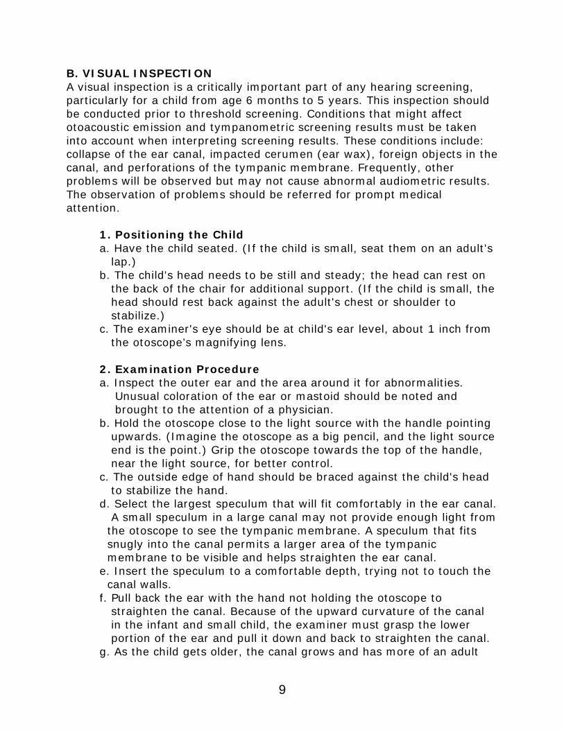
B. VISUAL INSPECTION A visual inspection is a critically important part of any hearing screening, particularly for a child from age 6 months to 5 years. This inspection should be conducted prior to threshold screening. Conditions that might affect otoacoustic emission and tympanometric screening results must be taken into account when interpreting screening results. These conditions include: collapse of the ear canal, impacted cerumen (ear wax), foreign objects in the canal, and perforations of the tympanic membrane. Frequently, other problems will be observed but may not cause abnormal audiometric results. The observation of problems should be referred for prompt medical attention.
1. Positioning the Child a. Have the child seated. (If the child is small, seat them on an adult's lap.) b. The child's head needs to be still and steady; the head can rest on the back of the chair for additional support. (If the child is small, the head should rest back against the adult's chest or shoulder to stabilize.) c. The examiner's eye should be at child's ear level, about 1 inch from the otoscope’s magnifying lens.
2. Examination Procedure a. Inspect the outer ear and the area around it for abnormalities. Unusual coloration of the ear or mastoid should be noted and brought to the attention of a physician. b. Hold the otoscope close to the light source with the handle pointing upwards. (Imagine the otoscope as a big pencil, and the light source end is the point.) Grip the otoscope towards the top of the handle, near the light source, for better control. c. The outside edge of hand should be braced against the child's head to stabilize the hand. d. Select the largest speculum that will fit comfortably in the ear canal. A small speculum in a large canal may not provide enough light from the otoscope to see the tympanic membrane. A speculum that fits snugly into the canal permits a larger area of the tympanic membrane to be visible and helps straighten the ear canal. e. Insert the speculum to a comfortable depth, trying not to touch the canal walls. f. Pull back the ear with the hand not holding the otoscope to straighten the canal. Because of the upward curvature of the canal in the infant and small child, the examiner must grasp the lower portion of the ear and pull it down and back to straighten the canal. g. As the child gets older, the canal grows and has more of an adult
9

angle; therefore, the ear should be pulled up and back to straighten the canal.
3. What We Are Looking For a. A healthy ear will have a tympanic membrane (TM) that is shiny, pearly white, and semitransparent. The degree of transparency will vary from person to person. If impacted cerumen is seen, the child should be referred to a physician. b. Landmarks within the middle ear can often be observed through the TM. For example, the long process of the malleus should extend to the center of the TM and is often visible. c. Tubes are known by various names: bilateral myringotomy tubes (BMTs), pressure equalization tubes (PE tubes), or tympanostomy tubes. Tubes may be made of different colors of plastic or may be metal. They may be seen either in the TM itself or laying in the ear canal. Their presence and positioning in the TM or ear canal should be noted. There should be no drainage from the tube, as this would indicate the presence of middle ear problems.
4. When to Refer Refer the child to a physician if any of the following are noted: a. Structural defects of the ear, head, or neck such as abnormal
positioning of the ear, malformed ear, absence of ear, extremely narrow ear canal, or presence of preauricular pits or tags (unless
physician is previously aware). b. Ear canal abnormalities such as: ♦ impacted wax ♦ presence of foreign object ♦ inflammation or swelling ♦ bleeding ♦ tumor/unknown growth ♦ fungal growth ♦ dermatitis c. Tympanic membrane abnormalities such as: ♦ perforation ♦ drainage/effusion ♦ bleeding ♦ redness ♦ blistering ♦ unknown growth/cholesteatoma
NOTE: NEVER try to remove wax or foreign object from a child's ear canal. ALWAYS refer to the physician.
10
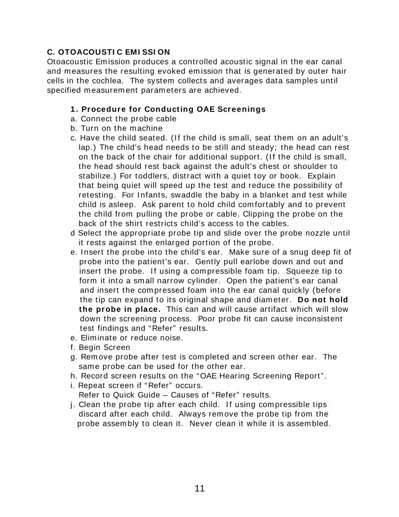
C. OTOACOUSTIC EMISSION Otoacoustic Emission produces a controlled acoustic signal in the ear canal and measures the resulting evoked emission that is generated by outer hair cells in the cochlea. The system collects and averages data samples until specified measurement parameters are achieved. 1. Procedure for Conducting OAE Screenings a. Connect the probe cable b. Turn on the machine
c. Have the child seated. (If the child is small, seat them on an adult's lap.) The child's head needs to be still and steady; the head can rest on the back of the chair for additional support. (If the child is small, the head should rest back against the adult's chest or shoulder to stabilize.) For toddlers, distract with a quiet toy or book. Explain that being quiet will speed up the test and reduce the possibility of retesting. For Infants, swaddle the baby in a blanket and test while child is asleep. Ask parent to hold child comfortably and to prevent the child from pulling the probe or cable. Clipping the probe on the back of the shirt restricts child’s access to the cables.
d Select the appropriate probe tip and slide over the probe nozzle until it rests against the enlarged portion of the probe. e. Insert the probe into the child’s ear. Make sure of a snug deep fit of probe into the patient’s ear. Gently pull earlobe down and out and insert the probe. If using a compressible foam tip. Squeeze tip to
form it into a small narrow cylinder. Open the patient’s ear canal and insert the compressed foam into the ear canal quickly (before the tip can expand to its original shape and diameter. Do not hold the probe in place. This can and will cause artifact which will slow down the screening process. Poor probe fit can cause inconsistent test findings and “Refer” results. e. Eliminate or reduce noise. f. Begin Screen g. Remove probe after test is completed and screen other ear. The same probe can be used for the other ear. h. Record screen results on the “OAE Hearing Screening Report”. i. Repeat screen if “Refer” occurs. Refer to Quick Guide – Causes of “Refer” results. j. Clean the probe tip after each child. If using compressible tips discard after each child. Always remove the probe tip from the probe assembly to clean it. Never clean it while it is assembled.
11
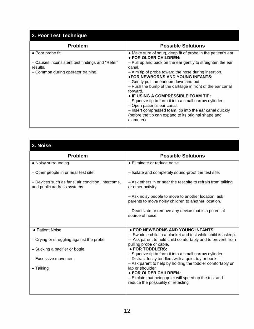
2. Poor Test Technique
Problem Possible Solutions ● Poor probe fit. – Causes inconsistent test findings and "Refer" results. – Common during operator training.
● Make sure of snug, deep fit of probe in the patient's ear. ● FOR OLDER CHILDREN: – Pull up and back on the ear gently to straighten the ear canal. – Aim tip of probe toward the nose during insertion. ●FOR NEWBORNS AND YOUNG INFANTS: – Gently pull the earlobe down and out. – Push the bump of the cartilage in front of the ear canal forward. ● IF USING A COMPRESSIBLE FOAM TIP: – Squeeze tip to form it into a small narrow cylinder. – Open patient's ear canal. – Insert compressed foam, tip into the ear canal quickly (before the tip can expand to its original shape and diameter)
3. Noise
Problem Possible Solutions ● Noisy surrounding. – Other people in or near test site – Devices such as fans, air condition, intercoms, and public address systems
● Eliminate or reduce noise – Isolate and completely sound-proof the test site. – Ask others in or near the test site to refrain from talking or other activity – Ask noisy people to move to another location; ask parents to move noisy children to another location. – Deactivate or remove any device that is a potential source of noise.
● Patient Noise – Crying or struggling against the probe – Sucking a pacifier or bottle – Excessive movement – Talking
● FOR NEWBORNS AND YOUNG INFANTS: – Swaddle child in a blanket and test while child is asleep. – Ask parent to hold child comfortably and to prevent from pulling probe or cable. ● FOR TODDLERS: – Squeeze tip to form it into a small narrow cylinder. – Distract fussy toddlers with a quiet toy or book. – Ask parent to help by holding the toddler comfortably on lap or shoulder ● FOR OLDER CHILDREN : – Explain that being quiet will speed up the test and reduce the possibility of retesting
12
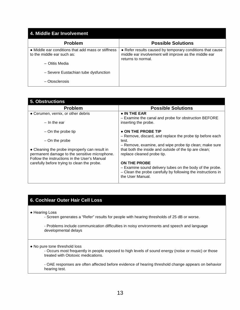
4. Middle Ear Involvement
Problem Possible Solutions ● Middle ear conditions that add mass or stiffness to the middle ear such as:
– Otitis Media – Severe Eustachian tube dysfunction – Otosclerosis
● Refer results caused by temporary conditions that cause middle ear involvement will improve as the middle ear returns to normal.
5. Obstructions
Problem Possible Solutions ● Cerumen, vernix, or other debris
– In the ear – On the probe tip – On the probe
● Cleaning the probe improperly can result in permanent damage to the sensitive microphone. Follow the instructions in the User’s Manual carefully before trying to clean the probe.
● IN THE EAR – Examine the canal and probe for obstruction BEFORE inserting the probe. ● ON THE PROBE TIP – Remove, discard, and replace the probe tip before each test. – Remove, examine, and wipe probe tip clean; make sure that both the inside and outside of the tip are clean; replace cleaned probe tip. ON THE PROBE – Examine sound delivery tubes on the body of the probe. – Clean the probe carefully by following the instructions in the User Manual.
6. Cochlear Outer Hair Cell Loss ● Hearing Loss
- Screen generates a “Refer” results for people with hearing thresholds of 25 dB or worse. - Problems include communication difficulties in noisy environments and speech and language developmental delays
● No pure tone threshold loss
- Occurs most frequently in people exposed to high levels of sound energy (noise or music) or those treated with Ototoxic medications. - OAE responses are often affected before evidence of hearing threshold change appears on behavior hearing test.
13
D. TYMPANOMETRY SCREENING Tympanometry measures the compliance or mobility of the tympanic membrane as a function of varied air pressure in the ear canal. It is not a test of hearing. The addition of tympanometry to the hearing screening protocol complements the overall objectives of a hearing screening program. The American Academy of Pediatrics reports that there is growing evidence of a connection between hearing impairment caused by middle ear disease and delays in the development of speech, language and cognitive skills. Tympanometry is a valuable tool in the detection of medically related conditions of the ear. Tympanometry screening is an essential part of the hearing screening for all children 0 through 3 years of age. The information obtained by conducting tympanometry screening is beneficial in assessing the middle ear system. It is important to remember that tympanometry is a screening process to identify possible disorders and is not to be used as a diagnostic tool.
1. Parameters of Tympanometry Screening Tympanometry is a test that can be used to identify possible disorders of the middle ear. It serves to identify conditions of the ear that may be missed by hearing screening alone. Two components of tympanometry are considered in interpreting results and making referrals. The parameters are physical volume and compliance (mobility) of the middle ear system.
a. Physical Volume The physical volume is the amount of air measured in the space between the probe tip of the tympanometer and the tympanic membrane. It may be called physical volume, volume, or absolute volume. Physical volume is measured in milliliters (ml) or cubic centimeters (cc). The normal range of volume for a particular instrument should be determined by reading the manual and using the values recommended by the manufacturer. In general, physical volumes between 0.3 and 2.0 ml are considered normal ranges for most instruments. There is a large variance in normative values for physical volume measurements. Many adults will have physical volume measurements of greater than 2.0 ml. The size of the ear canal, as well as the amount of wax present, should be taken into account when determining the need for referral.
♦ Volume measurements below 0.3 ml may indicate that the probe is placed against the side of the ear canal or against wax in the ear canal. Occasionally, the ear canal may be completely occluded by wax, and a low volume measurement
14

may indicate the size of the ear canal from the probe tip to the wax.
♦ Volume measurements above 2.0 ml indicate that the cavity being measured is larger than the ear canal volume. If there is a patent (open) PE tube in the tympanic membrane or if there is a perforation of the tympanic membrane, a large physical volume measurement may be obtained. The reason for a large physical measurement should be considered when determining the need for referral.
It is helpful to compare the volume of both ears to check validity of the measurements. Most children will have ear canal volumes that are roughly equal in both ears, and the measurements should fall within the normal range of 0.3 and 2.0 ml.
PHYSICAL VOLUME VALUES CHART * Value Interpretation 0.3 to 2.0 ml - Normal < 0.3 ml - Abnormally small >2.0 ml - Abnormally large
*Check owner's manual for equipment-specific criteria.
b. Compliance (Mobility) of the Middle Ear System A normal middle ear system has a tympanic membrane and attached ossicular chain that vibrate easily, allowing the transmission of sound energy to the inner ear by converting the sound waves to mechanical motion.
♦ In tympanometry, the freedom of movement (mobility or compliance) of the tympanic membrane and ossicular chain is assessed by measuring the amount of energy necessary to move them. On the tympanogram, it is represented by the height of the peak and is expressed in milliliters (ml). ♦ Some conditions of the middle ear cause the mobility of all or part of the middle ear system to be reduced. Other conditions may allow excessive motion. Extremely low or extremely high mobility may indicate a condition that needs further attention. ♦ A compliance peak from 0.2 to 2.0 ml is within the normal range. Check owner's manual for the equipment specific criteria. ♦ A compliance measurement of less than 0.2 ml indicates the middle ear is stiffer than normal.
15

♦ A compliance measurement of greater than 2.0 ml indicates a hyper-flaccid tympanic membrane. A value greater than 2.0 ml may indicate a disarticulated ossicular chain. High compliance measurements are not considered a factor in referral criteria unless hearing loss is present.
COMPLIANCE MEASUREMENTS* Value Interpretation 0.2 to 1.8 ml - Normal < 0.2 ml - Abnormally Low >1.8 ml - Abnormally High
*Check owner's manual for equipment-specific criteria.
2. Reliability of Test Results Under most conditions, tympanometry provides reliable information regarding middle ear function. However, there are factors that affect the reliability and validity of test results. Individuals conducting tympanometry should be aware of these factors in order to obtain accurate information.
a. Tympanometry screening should not be administered to children under the age of 6 months. A high incidence of false negative results (normal tympanometric results found in infants with middle ear effusion) has been obtained from tympanometry conducted on newborn infants. The reason for this is the highly compliant nature of the external auditory canal wall in infants. As such, the normal tympanogram may be a compliance measure of the external auditory canal rather than the tympanic membrane.
b. Middle ear surgeries may include cholesteatoma removal, tympanoplasty, canaloplasty, etc. Tympanometry should not be administered to children who have had middle ear surgery (other than myringotomy and PE tubes) within the past six months unless requested by a physician. Tympanometry can provide valuable data on the patency of tubes shortly after the surgical procedure.
3. Procedure for Conducting Tympanometry
a. Turn on the machine. b. Check the calibration of the tympanometer. Many of the newer
tympanometers will check the calibration automatically each time they are turned on. Older units require you to manually check the calibration by measuring the size of the volume using a specified
16

probe tip. Read your operating manual to determine which procedure to use on your unit. This is only a routine calibration check. It does not take the place of full electroacoustic calibration, which is required annually.
c. Perform visual inspection (refer to Chapter III, Section D for more details). ♦ Wax that completely occludes the ear canal will interfere with the test. If the wax does not completely occlude the canal, a reliable tympanogram can be obtained. ♦ While doing the visual inspection, note the size and shape of the ear canal to determine the appropriate size probe tip.
d. Select the appropriate size probe tip and place on the probe assembly. As a rule, it is better to have a probe tip that is too large than too small.
e. Place against the ear canal for seal. ♦ Most new units obtain a seal rather easily. However, when just
beginning tympanometry screening, you may find this difficult. ♦ Sometimes it is helpful to use a twisting motion to rotate the
probe back and forth to fit into the ear canal. ♦ Always watch the probe, not the tympanometer, while performing
this procedure. f. Hold the probe steady once a seal is obtained. g. Maintain the seal until the test is completed (typically less than 10
seconds). h. Watch the probe/tympanometer signals to assure that a
tympanogram is being obtained. i. Remove the probe after the test is completed. j. Print the results if possible. Some tympanometers automatically
print after each test. It is not necessary to print normal tympanograms.
k. If the tympanogram is different from what was expected, repeat. Tympanograms should also be repeated to verify their reliability, particularly when a medical referral is being made. Even though
the reliability of tympanometry is quite good, repeat the test to check the reliability of the results.
l. Clean the probe tip after each child. j. Use disinfectant or clean with an antiseptic after each use. k. Always remove the probe tip from the probe assembly to clean it.
Never clean it while it is still assembled.
17

4. Special Considerations a. Middle ear function is a dynamic, non-static system. Fluctuations in
negative pressure measurements will often be observed. This is not clinically significant for determining the need for referral. Always consider the medical and the hearing history information.
b. If a flat tympanogram is obtained (compliance of less than 0.2 ml), always repeat the tympanogram. This will minimize inaccurate results based on improper probe placement. Referrals should be made on results that can be replicated.
c. When tympanometry is repeated, physical volume measurements should be monitored to ensure that the probe tip is properly placed, not against the canal wall, or in excessive wax, etc.
d. If a perforation or an open PE tube is present and the eustachian tube is open, an airtight seal may not be obtainable. Do not keep trying to get a probe tip seal.
5. What We Are Looking For
TYMPANOGRAM INTERPRETATION
TYMPANOGRAM COMPLIANCE PHYSICAL VOLUME POSSIBLE CAUSE
Peak is present Compliance within normal limits (0.2 - 1.8ml)
Normal* 0.3 - 2.0 ml
Normal tympanogram
No Peak Low compliance < 0.2 ml
Small <0.3 ml
May suggest blockage of external ear canal with wax or other object, or probe tip against the ear canal
No Peak Low compliance < 0.2 ml
Normal 0.3 - 2.0 ml
May suggest fluid-filled middle ear, otitis media, or retracted tympanic membrane.
No Peak Low comliance < 0.2 ml
Large > 2.0 ml
May suggest perforation or patent ventilation tube.
18
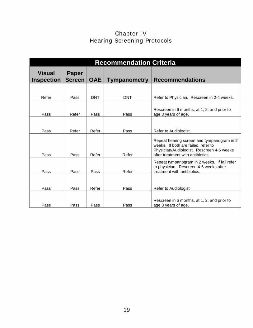
Chapter IV Hearing Screening Protocols
Recommendation Criteria Visual
Inspection Paper Screen OAE Tympanometry Recommendations
Refer Pass DNT DNT Refer to Physician. Rescreen in 2-4 weeks.
Pass Refer Pass Pass Rescreen in 6 months, at 1, 2, and prior to age 3 years of age.
Pass Refer Refer Pass Refer to Audiologist
Pass Pass Refer Refer
Repeat hearing screen and tympanogram in 2 weeks. If both are failed, refer to Physician/Audiologist. Rescreen 4-6 weeks after treatment with antibiotics.
Pass Pass Pass Refer
Repeat tympanogram in 2 weeks. If fail refer to physician. Rescreen 4-6 weeks after treatment with antibiotics.
Pass Pass Refer Pass Refer to Audiologist
Pass Pass Pass Pass Rescreen in 6 months, at 1, 2, and prior to age 3 years of age.
19
Chapter V Impact of Hearing Loss on Infants and Toddlers
A. Impact of Hearing Loss on Infants and Toddlers A moderate, severe, or profound hearing loss has a significant impact on the cognitive, psychological, speech, and language development of the child. A slight or mild bilateral or unilateral hearing loss may also cause significant developmental delay. The impact of hearing loss on speech and language development and on academic achievement has been well-documented. To minimize these debilitating effects, the use of early identification and intervention strategies (both medical and educational) is crucial. The value of early identification and intervention cannot be over-emphasized. An infant-toddler who is deaf or hard of hearing can receive language input through a hearing aid if the infant-toddler has sufficient residual hearing, or through visual and manual language inputs, or both. As soon as the hearing loss is confirmed, the parents of the infant-toddler who is deaf or hard of hearing should be encouraged to get appropriate early intervention for their child. Early intervention gives infants and toddlers the opportunity to use their residual hearing to develop optimal speech and language skills.
B. Otitis Media (OM) results in periods of temporary hearing loss during the critical years of language and speech acquisition and can result in developmental language delays. • OM accounts for 24.5 million visits to doctors’ offices per year. • Approximately 61 to 83% of all children will have otitis media (OM). • Children having persistent OM in the first 3 years of life are more likely to continue to have OM through the first years of school. • The American Academy of Pediatrics(1) and the Agency of Health Care Policy and Research(2) recommend that patients with otitis media persisting more than 3 months be evaluated for hearing loss. 1 Wright P.F., Thompson J & Bess FH (1991) Hearing, speech, and language sequelae of otitis media with effusion. Pediatric Annuals 20(11), 617-18. 2 Stool SE, Berg AO & Berman S (1994) Managing otitis media with effusion in young children: quick reference guide for clinicians. Rockville, MD: Agency for Health Care Policy and Research, Public Health Service, U.S. Department of Health and Human Services, AHCPR publication 94-0623.
20
Frequently Asked Questions 1. How long does an OAE screening take? Test time is very fast – about 10 seconds per ear if the patient is quiet and cooperative; slightly longer for "refer" results. 2. Who should administer the test? Since OAE provides a pass or refer result, any trained staff member, under the supervision of a health-care professional, can easily perform the screening. 3. What patient preparation is required? The ear probe must be placed securely in the ear canal and the patient must be relatively quiet for the few seconds that it takes to perform the test. No other preparation is needed. 4. What does a “refer” result mean? A “refer” result means that the OAEs are absent or small compared to normal data. Assuming good test conditions and technique, possible causes of a refer result are: • Excessive debris in the ear canal such as vernix (newborns) or cerumen • Middle ear fluid or other middle ear abnormalities (e.g. otosclerosis, etc.) • Cochlear hearing loss greater than 25-30 dB HL If a reliable OAE screening cannot be achieved or a refer result persists on repeat screenings despite good test conditions and apparently normal middle ear function, the patient should be referred to an audiologist or otologist for further evaluation. 5. Why do I need OAEs if I have Tympanometry? Tympanometry is a powerful tool for assessing middle ear function. However, it provides no information about cochlear function or hearing. OAEs are affected by both hearing loss and middle ear dysfunction. For children with OME, for example, it is important to monitor their OAEs to verify the return to normal when the middle ear condition appears resolved. Passing OAE screening results are highly correlated with normal hearing and normal middle ear function; whereas normal tympanograms are present in hearing impaired ears as long as the middle ear is healthy. 6. Can I screen a patient who has otitis media? The presence of middle ear fluid almost always blocks transmission of the OAE causing a “refer” result. OAEs are very useful for tracking the resolution of middle ear fluid and the return of hearing to normal. A “pass” result on an OAE screening is highly correlated with normal middle ear function and normal cochlear function (i.e. normal hearing).
21

7. Can I screen a patient who has PE tubes? Yes, most ears with PE tubes in combination with a healthy middle ear and normal cochlear function will achieve a “pass” result. However, in some ears, the added weight of the PE tube in the eardrum can affect the OAE and cause a “refer“ result. In that case, referral for further evaluation may be required.
22


![Kansas Health Statistics Report - KDHE · Kansas Health Statistics Report ... Kansas Trends in Poisoning Morbidity and ... 10th leading cause of death claiming 37,793 lives [1]](https://img.pdfslide.us/doc/110x75/5ae1b93a7f8b9a5b348b6ccf/kansas-health-statistics-report-health-statistics-report-kansas-trends-in.jpg)