Embed Size (px)
Citation preview
Julie V. Philley, MD
University of Texas at Tyler Health Science Center July 28, 2016

I have no disclosures.

Direct inhalation of NTM contaminated soils and aerosols
Micro aspirated NTM-contaminated NTM water from the oropharynx into the lungs
GERD For skin and soft tissue, direct inoculation of
NTM containing soils, water or medications
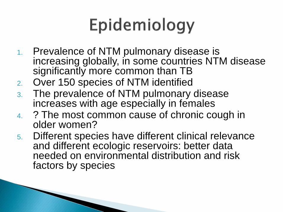
1. Prevalence of NTM pulmonary disease is increasing globally, in some countries NTM disease significantly more common than TB
2. Over 150 species of NTM identified 3. The prevalence of NTM pulmonary disease
increases with age especially in females 4. ? The most common cause of chronic cough in
older women? 5. Different species have different clinical relevance
and different ecologic reservoirs: better data needed on environmental distribution and risk factors by species
Who, what, when and where
do we treat NTM?

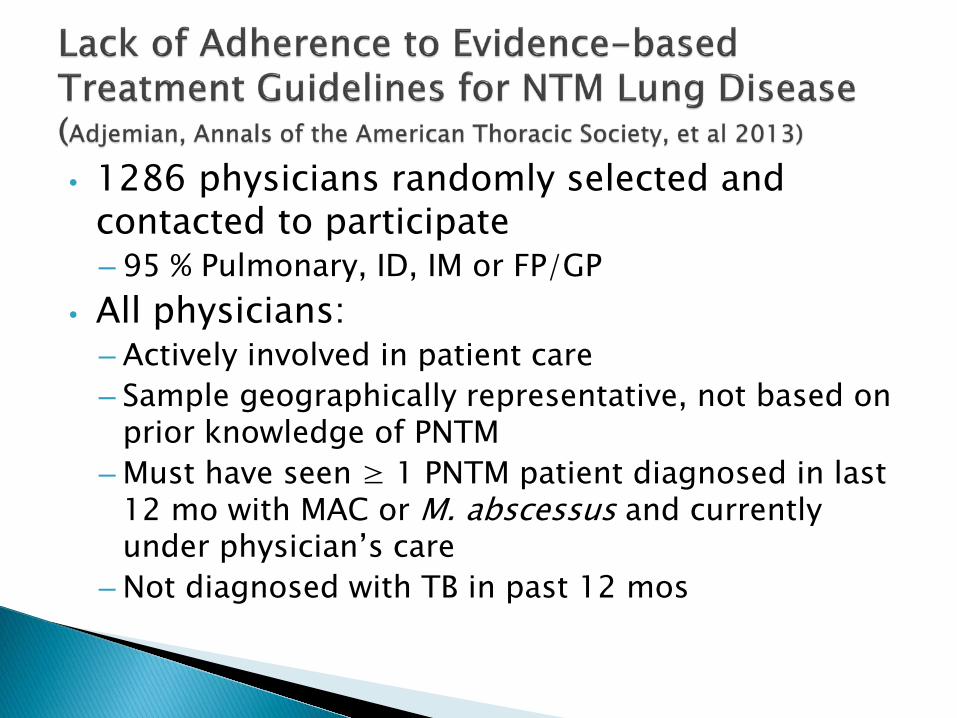
• 1286 physicians randomly selected and contacted to participate – 95 % Pulmonary, ID, IM or FP/GP
• All physicians: – Actively involved in patient care – Sample geographically representative, not based on
prior knowledge of PNTM – Must have seen ≥ 1 PNTM patient diagnosed in last
12 mo with MAC or M. abscessus and currently under physician’s care
– Not diagnosed with TB in past 12 mos
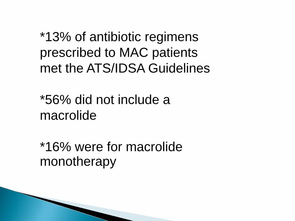
*13% of antibiotic regimens prescribed to MAC patients met the ATS/IDSA Guidelines *56% did not include a macrolide *16% were for macrolide monotherapy
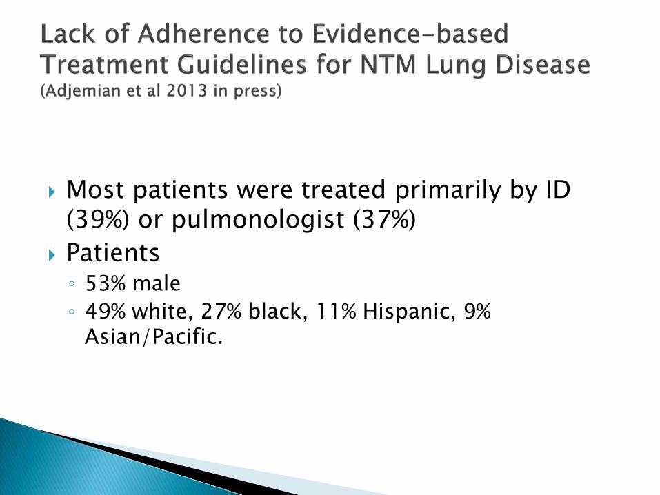
Most patients were treated primarily by ID
(39%) or pulmonologist (37%) Patients ◦ 53% male ◦ 49% white, 27% black, 11% Hispanic, 9%
Asian/Pacific.


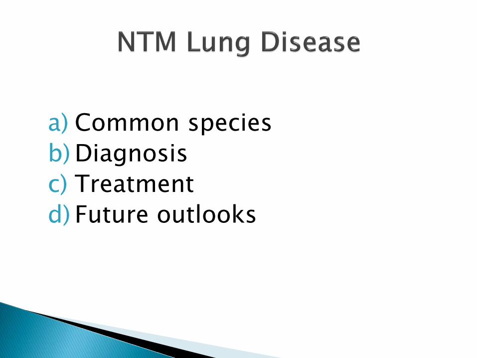
a) Common species b) Diagnosis c) Treatment d) Future outlooks
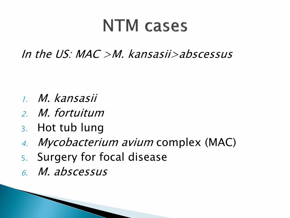
In the US: MAC >M. kansasii>abscessus
1. M. kansasii 2. M. fortuitum 3. Hot tub lung 4. Mycobacterium avium complex (MAC) 5. Surgery for focal disease 6. M. abscessus
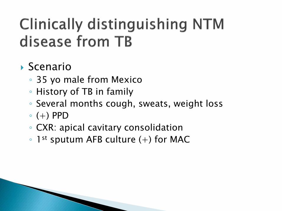
Scenario ◦ 35 yo male from Mexico ◦ History of TB in family ◦ Several months cough, sweats, weight loss ◦ (+) PPD ◦ CXR: apical cavitary consolidation ◦ 1st sputum AFB culture (+) for MAC
Scenario: ◦ Pulmonary MAC disease unusual in a 35 yo male ◦ Multiple risk factors for TB ◦ Clinical presentation typical for TB ◦ Patient subsequently grew multiple (+) cultures for
M. tuberculosis ◦ Empiric therapy for TB ok in this setting, may occur
frequently
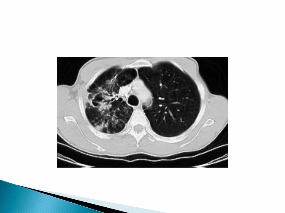
65 yo smoker with COPD seen by his PCP with chronic cough.
Interestingly, a chest CT is available for your review. One sputum had been sent and grew M. kansasii.
Does this patient meet criteria for diagnosis of NTM?
Is M. kansasii a pathogen? What do you do?
Do you think this patient can obtain cure?
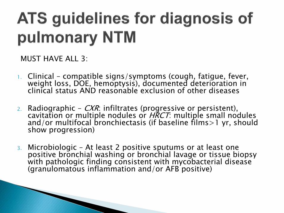
MUST HAVE ALL 3:
1. Clinical – compatible signs/symptoms (cough, fatigue, fever,
weight loss, DOE, hemoptysis), documented deterioration in clinical status AND reasonable exclusion of other diseases
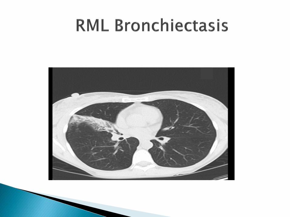
2. Radiographic – CXR: infiltrates (progressive or persistent), cavitation or multiple nodules or HRCT: multiple small nodules and/or multifocal bronchiectasis (if baseline films>1 yr, should show progression)
3. Microbiologic – At least 2 positive sputums or at least one
positive bronchial washing or bronchial lavage or tissue biopsy with pathologic finding consistent with mycobacterial disease (granulomatous inflammation and/or AFB positive)

Does this patient meet criteria for diagnosis of NTM? No. (based on only having one sputum that is positive) Should
you bronch him? Is M. kansasii a pathogen? yes, unless there is no clinical evidence to support the diagnosis What do you do? Obtain more sputum and start him on daily INH 300 mg qd,
Rifampin 600 mg qd, Ethambutol 15 mg/kd/qd and treat for 12 months from 1st negative sputum. (usually 18-24 months) Alternatively, MAC therapy can be used.
Do you think this patient can obtain cure? Yes
76 yo with a history of CAD, HTN, osteoporosis, interstitial lung disease, ulcerative colitis, fibromyalgia, GERD, breast cancer s/p chemotherapy (adriamycin) with complaints of abdominal pain which is attributed to kidney stones. Her PCP orders an abd CT and finds one small patchy area of ground glass with several subcentimeter nodules in both lower lobes. She has no respiratory complaints at the time of her visit.
Sputum cultures are obtained and 1/3 becomes positive for M. fortuitum
Which of her above comorbid conditions is highly associated with
M. fortuitum? What work-up should be done?
Which of her above comorbid conditions is highly associated with M. fortuitum?
GERD/chronic aspiration What work-up could be done? Esophagram +/- ph prob, barium swallow Therapy?
Elevate the HOB, dietary changes, PPI
48 yo with 6 months h/o chronic cough which
began in March 2010. Sought medical attention and was placed on albuterol with minimal relief. An abnormal CXR prompted further evaluation.
ROS: dry cough which is bothersome on a daily basis. Denies fevers, night sweats, nausea, vomiting, joint pains, rashes, dry eyes, dry mouth, chest pain, painful lumps or bumps, conjunctivitis, ulcers, alopecia, weight loss, visual changes, reflux
PMH: HTN PSH: None FH: Mother – asthma Social: smoked 2-3 cigs per day during his freshman year of
college only. Married; 2 children, works as an attorney. 1 small poodle, no feather pillows. No recent travel.
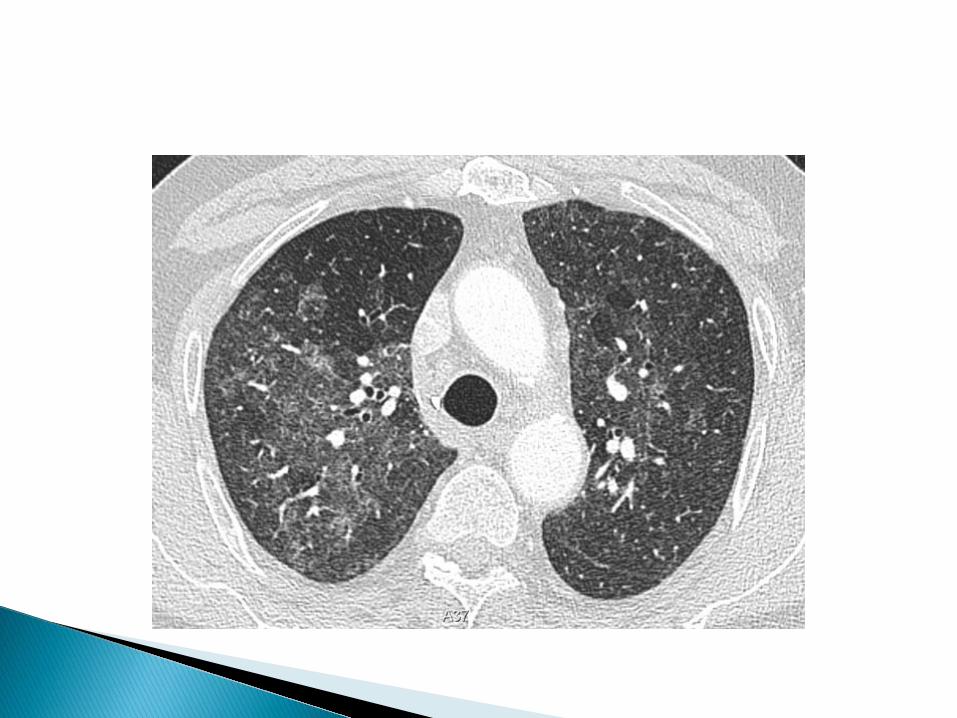
Ultimately, underwent bronchoscopy with TBBX - inflammatory cell infiltrates in alveoli
and interstitial spaces consisting of lymphocytes, plasma cells, activated macrophages and giant cells.
Culture from BAL grew MAC Diagnosis? Does this patient meet criteria for diagnosis of NTM? What do you do?
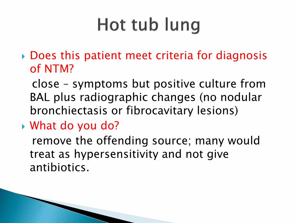
Does this patient meet criteria for diagnosis of NTM?
close – symptoms but positive culture from BAL plus radiographic changes (no nodular bronchiectasis or fibrocavitary lesions)
What do you do? remove the offending source; many would
treat as hypersensitivity and not give antibiotics.
82 yo with complaints of a persistent cough diagnosed in July 2010. He had abnormal imaging and was smear positive for MAC.
Placed on TIW therapy of azi, eth, rif but developed some dizziness so was switched to clari, eth, rifabutin in December 2010. Continued on this therapy TIW but remained smear positive. (over 8 smear positive sputums)
Comes with continued cough, weight loss of 20lb and night sweats
On no airway clearance
What is the treatment for MAC? How do you interpret susceptibility testing?
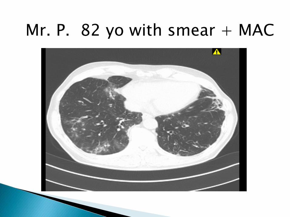
Mr. P. 82 yo with smear + MAC
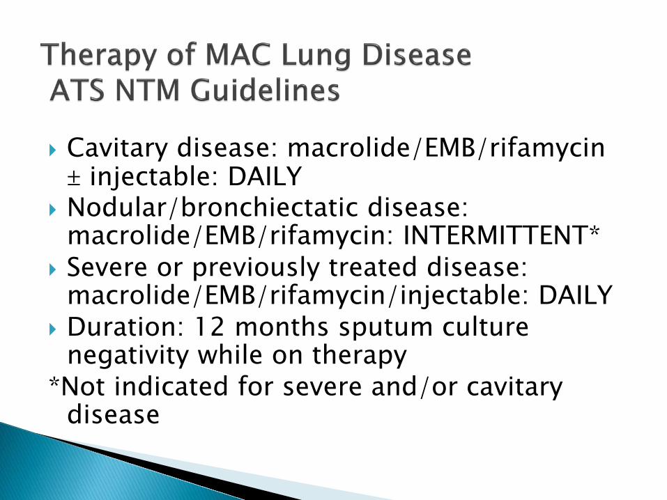
Cavitary disease: macrolide/EMB/rifamycin ± injectable: DAILY
Nodular/bronchiectatic disease: macrolide/EMB/rifamycin: INTERMITTENT*
Severe or previously treated disease: macrolide/EMB/rifamycin/injectable: DAILY
Duration: 12 months sputum culture negativity while on therapy
*Not indicated for severe and/or cavitary disease
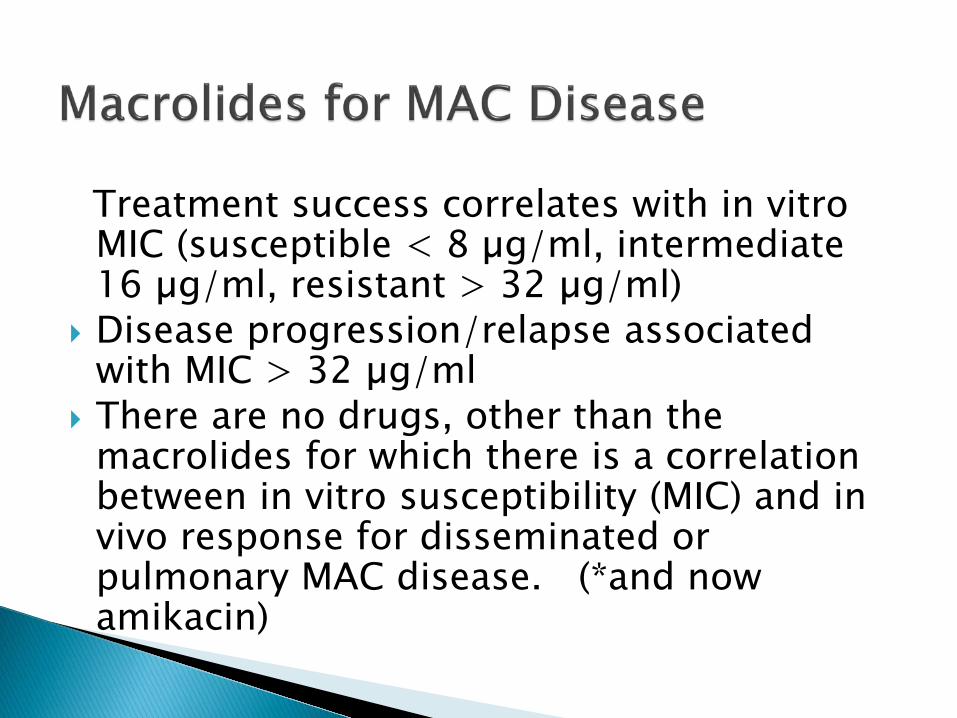
Treatment success correlates with in vitro MIC (susceptible < 8 µg/ml, intermediate 16 µg/ml, resistant > 32 µg/ml)
Disease progression/relapse associated with MIC > 32 µg/ml
There are no drugs, other than the macrolides for which there is a correlation between in vitro susceptibility (MIC) and in vivo response for disseminated or pulmonary MAC disease. (*and now amikacin)
Is this disease treatable?
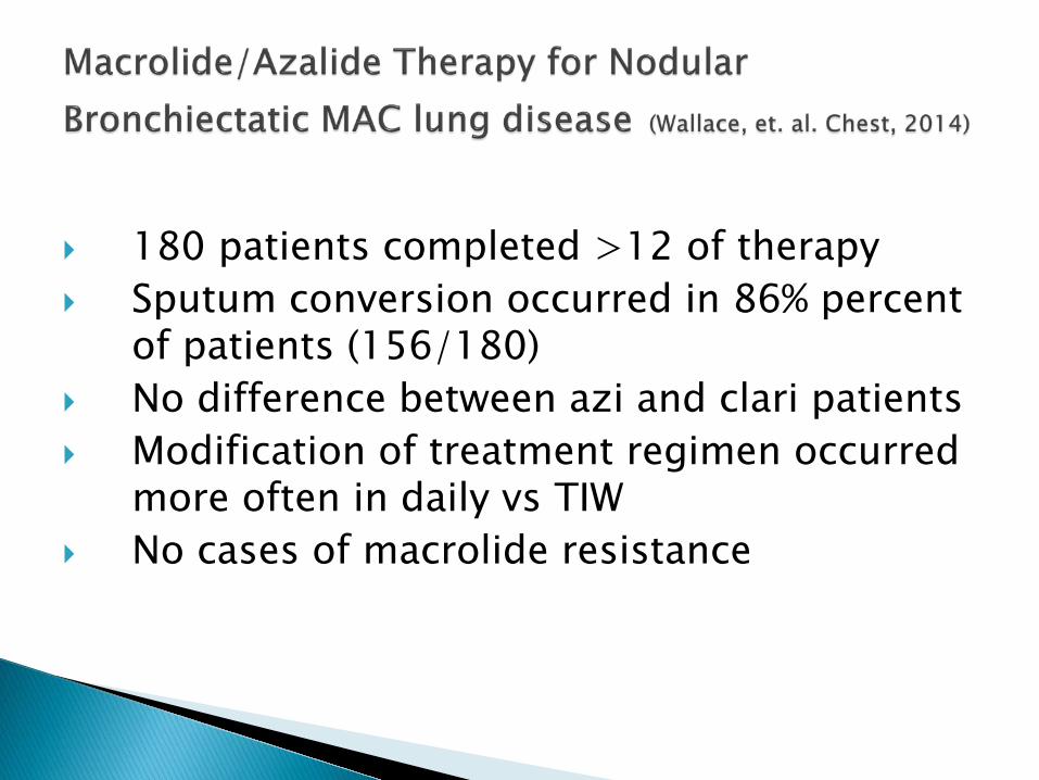
180 patients completed >12 of therapy Sputum conversion occurred in 86% percent
of patients (156/180) No difference between azi and clari patients Modification of treatment regimen occurred
more often in daily vs TIW No cases of macrolide resistance
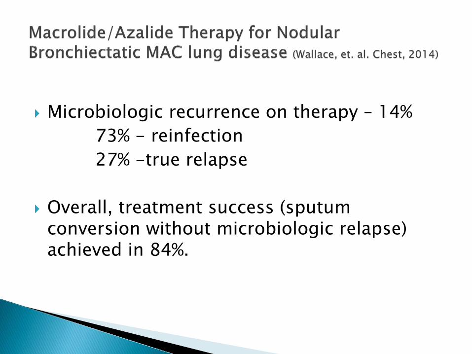
Microbiologic recurrence on therapy – 14% 73% - reinfection 27% -true relapse
Overall, treatment success (sputum
conversion without microbiologic relapse) achieved in 84%.
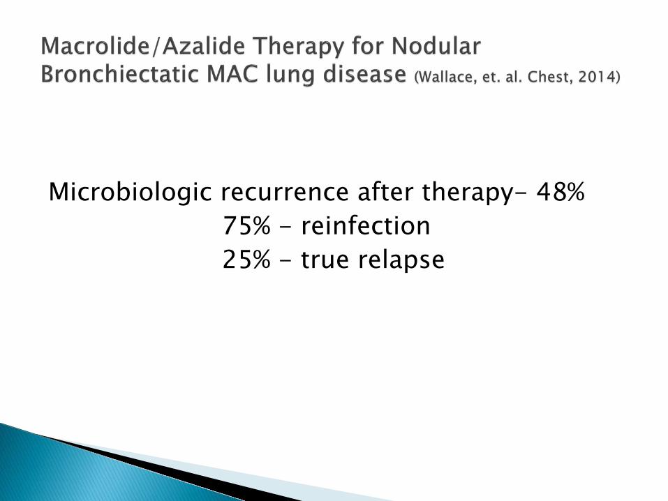
Microbiologic recurrence after therapy- 48% 75% - reinfection 25% - true relapse
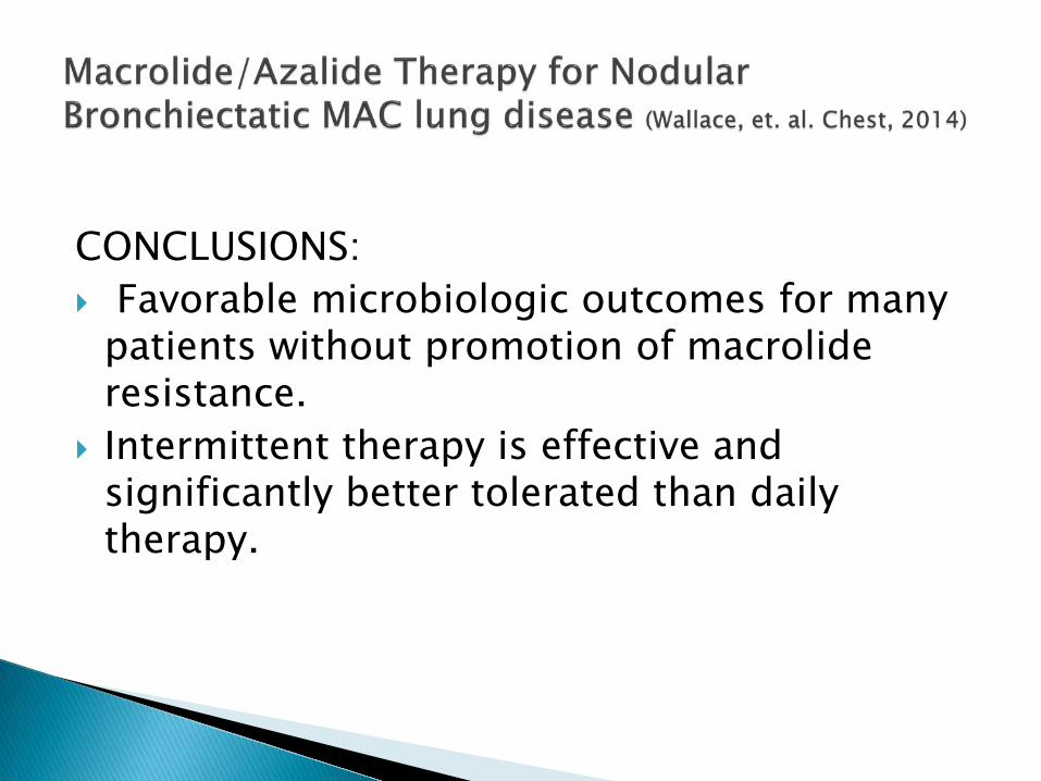
CONCLUSIONS: Favorable microbiologic outcomes for many
patients without promotion of macrolide resistance.
Intermittent therapy is effective and significantly better tolerated than daily therapy.
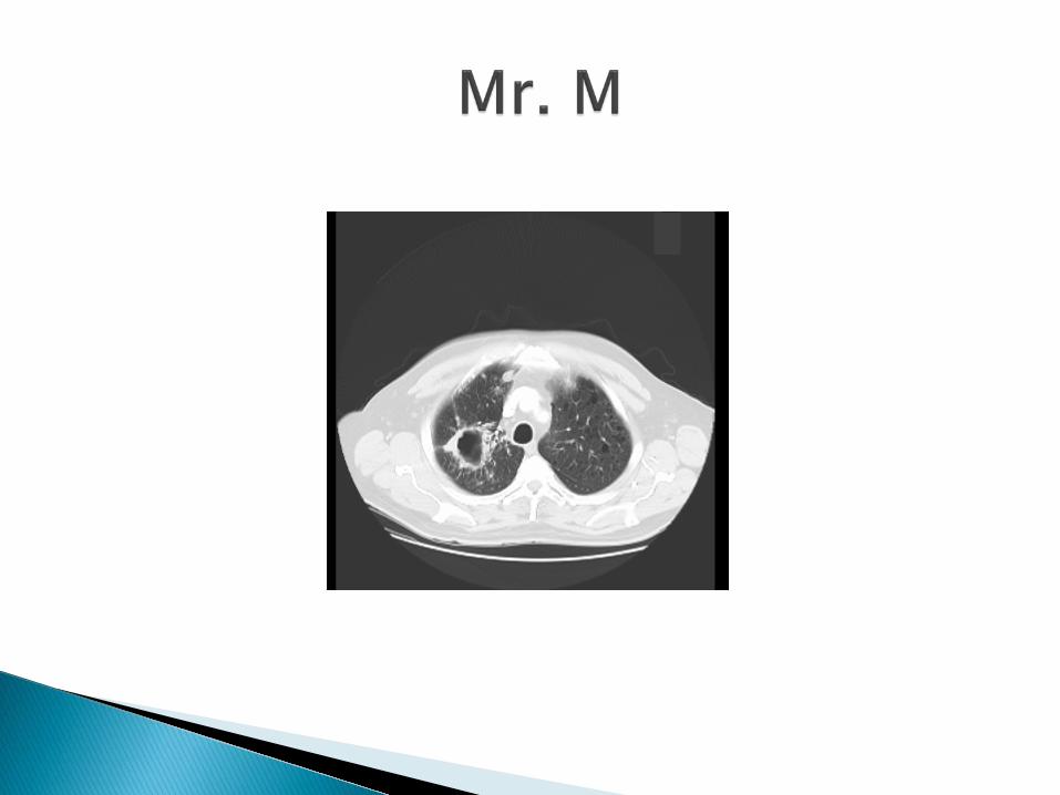
72 yo with a 20 pack year h/o tobacco, quitting in 1980 who in August of 2009, developed wheezing. He was seen by his PCP where an abnormal CXR prompted further work-up.
Diagnosed with MAC in October 2009 by sputum and bronchoscopy culture.
Started 3 drug daily therapy with azithromycin, ethambutol, rifampin
In December, developed severe numbness to
his toes. His sputums remained positive but his pulmonary symptoms disappeared.
Sputum – remained positive for MAC, azi sensitive Alpha-1 WNL Immunoglobulins WNL CF mutations – none CMP, CBC normal B12, TSH, folate, SPEP normal 25-OH D - 16 PFTS: FEV1 3.18 L (85% predicted) normal ratio, DLCO
77% predicted Barium swallow normal Esophagram – small hernia Visual acuity – 20/20 both eyes Audiogram – high frequency hearing loss bilaterally

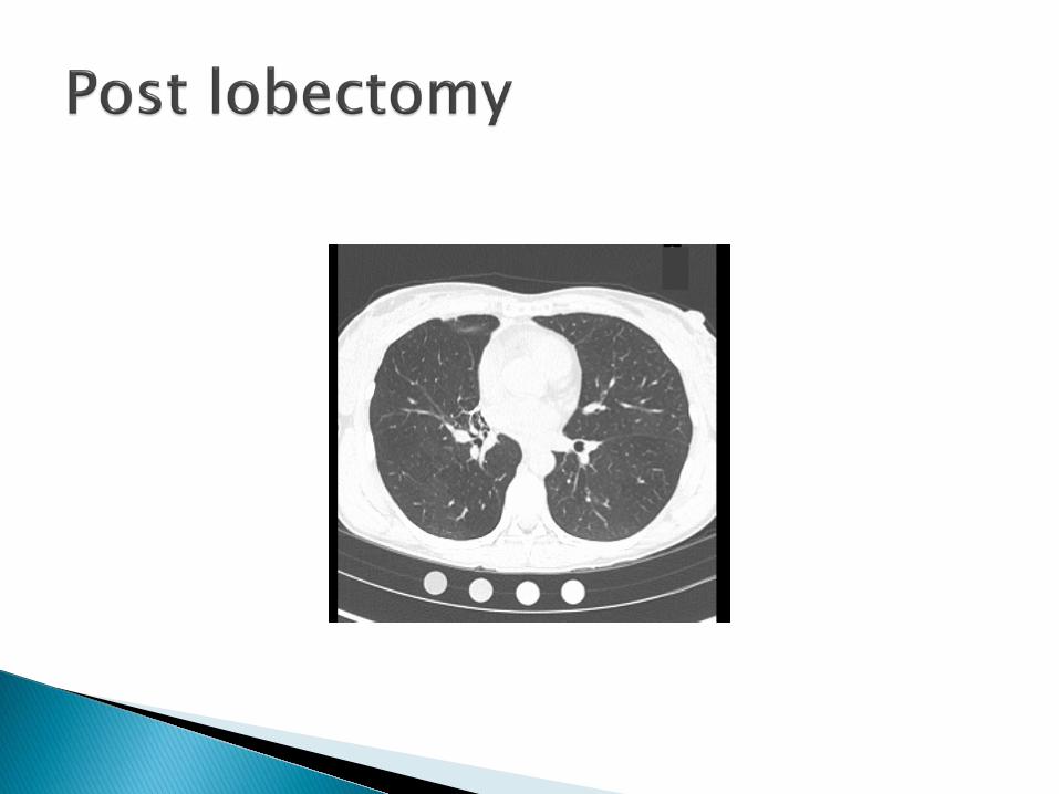
22 patients in Japan who received median of 17 months of antibiotics. All received surgical intervention (i.e. lobectomy, segmentectomy, partial lung resection)
Post-operatively antibiotics continued for 6-35 months post surgery
All patients were alive at 46 months FVC and FEV1 were reduced in both groups but
maintained at 89% and 84% of the preop values MAC disappeared from sputum after surgery in
all patients. Functional capacity was maintained Matanabe, Kobayashi, et al. Early Pulmonary resection for MAC Lung disease treated with macrolides and quinolones The Annals of Thoracic Surgery; 2006; 81: 2026-2030
IV amikacin for 2 months prior to surgery and for at least 2 months post-operatively as well. Weekly CMP, CBC, amikacin levels 30 min after each dose with goal 20-25. Monthly audiogram
Continue azithromycin, rifampin daily. BID acapella GERD lifestyle changes
Mostly for MAC, Abscessus Usually with localized disease V/Q scan Always treat with combination of po and IV
antibiotics before surgery (goal of at least 2 months prior)
Maintain adequate nutritional status Referral to an experienced surgical center
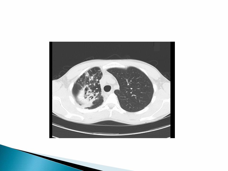
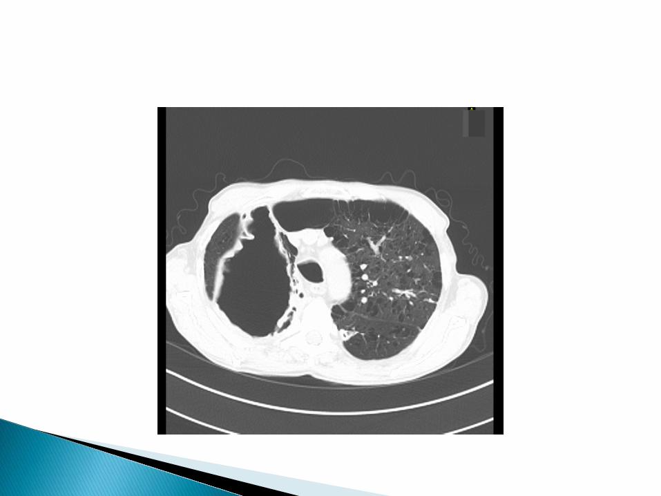
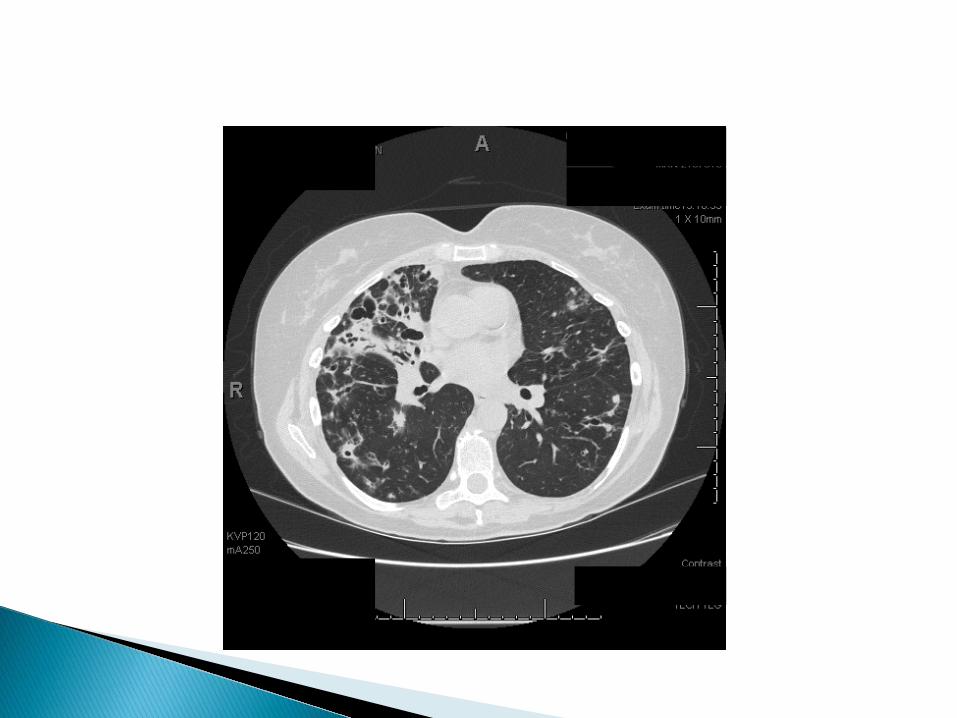
76 yo female with nodular bronchiectasis referred for multiple cultures positive for M. abscessus
She has a history of treated MAC lung disease
5 years ago with three drug therapy. She has a “terrible” cough, 5lb weight loss, no hemoptysis. Major fatigue and occasional night sweats.
There is no predictably or reliably effective medical treatment strategy for M. abscessus lung disease.
Macrolide: value questionable (erm gene),
may be of value as immune modulator Amikacin 10-15 mg/kg 3-7X/week (we
start at 7 mg/kg) Tigecycline 25-50 mg/day Linezolid 300-600 mg/day Alternatives: Imipenem, cefoxitin,
clofazimine
Inhaled amikacin
Bedaquiline?

Japanese Society Meeting - 2012 57


![(b) (6) · From: Nina Perales [mailto:nperales@MALDEF.org] Sent: Thursday, June 15, 2017 7:32 PM To: Halainen, Daniel J. (CIV) < Cc: Tyler, John (CIV) < >; Saltman, Julie](https://img.pdfslide.us/doc/110x75/5e9a42261ad9740f3902564a/b-6-from-nina-perales-mailtonperales-sent-thursday-june-15-2017-732.jpg)