Embed Size (px)
Citation preview
JPET#131565
1
Title page
Ceramide: A Key Signaling Molecule in a Guinea Pig Model of Allergic
Asthmatic Response and Airway Inflammation
Emanuela Masini, Lucia Giannini, Silvia Nistri , Lorenzo Cinci, Rosanna Mastroianni,
Wei Xu, Suzy A.A. Comhair, Dechun Li, Salvatore Cuzzocrea, George M. Matuschak
and Daniela Salvemini.
Department of Preclinical and Clinical Pharmacology University of Florence,
Florence, Italy (E.M., L.G., R.M.); b Anatomy, Histology and Forensic Medicine,
Section of Histology, University of Florence, Florence, Italy (S.N., L.C.);
Pathobiology, and Pulmonary, Critical Care Medicine, Cleveland Clinic, Cleveland
OH 44195, USA (S.A.A.C.); Internal Medicine, Division of Pulmonary, Critical Care
and Sleep Medicine, Saint Louis University, St Louis MO 63110 USA, and
Department of Pharmacological and Physiological Sciences, St Louis University
School of Medicine, St Louis, MO63110, USA (W.X., D.L., G.M.M., D.S.), Clinical and
Experimental Medicine and Pharmacology, School of Medicine, University of
Messina, Italy and IRCCS Centro Neurolesi "Bonino-Pulejo", Messina, Italy (S.C.).
JPET Fast Forward. Published on November 27, 2007 as DOI:10.1124/jpet.107.131565
Copyright 2007 by the American Society for Pharmacology and Experimental Therapeutics.
This article has not been copyedited and formatted. The final version may differ from this version.JPET Fast Forward. Published on November 27, 2007 as DOI: 10.1124/jpet.107.131565
at ASPE
T Journals on February 17, 2019
jpet.aspetjournals.orgD
ownloaded from
JPET#131565
2
Running title page
Running title: ceramide and allergic asthma
Author for correspondence: Prof. Emanuela Masini, Dept. Preclinical & Clinical
Pharmacology, University of Florence. Viale G.Pieraccini 6, I-50139 Florence, Italy.
Phone: +39 055 4271233, fax: +39 055 4271280; [email protected]
Number of text pages: 34 Number of figure: 8 Number of references: 40 Number of words in the abstract: 219 Number of words in the introduction: 745 Number of words in the discussion: 1199 Abbreviations: iNOS inducible nitric oxide synthase
NBT nitro blue tetrazolium
8-OHdG 8-hydroxy-2′-deoxyguanosine
PARP poly(ADP ribose)polymerase
S1P sphingosine-1-phosphate
S1Pr sphingosine-1-phosphate receptor
TdT terminal deoxynucleotidyl transferase
This article has not been copyedited and formatted. The final version may differ from this version.JPET Fast Forward. Published on November 27, 2007 as DOI: 10.1124/jpet.107.131565
at ASPE
T Journals on February 17, 2019
jpet.aspetjournals.orgD
ownloaded from
JPET#131565
3
Abstract
Although mechanisms involved in the pathogenesis of asthma remain unclear, roles
for oxidative/nitrative stress, epithelial cell apoptosis and airway inflammation have
been documented. Ceramide is a sphingolipid with potent proinflammatory and
proapoptotic properties. This study aimed at determining whether increased
formation of ceramide contributes to the development of airway inflammation and
hyperresponsiveness, using a well characterized in vivo model of allergic asthmatic
response and airway inflammation in ovalbumin-sensitized guinea pigs. Aerosol
administration of ovalbumin increased ceramide levels and ceramide synthase
activity in the airway epithelium, associated with respiratory abnormalities, such as
cough, dyspnea and severe bronchoconstriction. These abnormalities correlated with
nitrotyrosine formation in the airway epithelium and oxidative/nitrosative stress,
epithelial cell apoptosis and airway inflammation evident by the infiltration of
neutrophils and eosinophils in lung tissues, mast cell degranulation and release of
prostaglandin D2 and proinflammatory cytokines. Inhibition of de novo ceramide
synthesis with the competitive and reversible inhibitor of ceramide synthase
fumonisin B1 (0.25, 0.5 and 1 mg/kg b.wt.), given i.p. daily for 4 days before allergen
challenge, attenuated nitrotyrosine formation and oxidative/nitrosative stress,
epithelial cell apoptosis and airway inflammation while improving the respiratory and
histopathological abnormalities. These results implicate ceramide in the development
of allergic asthmatic response and airway inflammation. Strategies aimed at reducing
the levels of ceramide and downstream events should yield promising novel anti-
asthmatic agents.
This article has not been copyedited and formatted. The final version may differ from this version.JPET Fast Forward. Published on November 27, 2007 as DOI: 10.1124/jpet.107.131565
at ASPE
T Journals on February 17, 2019
jpet.aspetjournals.orgD
ownloaded from
JPET#131565
4
Introduction
Asthma is defined by airway inflammation and remodelling and bronchial
hyperresponsiveness (Busse and Lemanske, 2001). The role of inflammation in
asthma is well established and neutrophils, eosinophils and mast cells within the lung
have been shown to have pivotal roles for the initiation and maintenance of airway
inflammation (Barnes, 2004; Andreadis et al., 2003). Besides the classical allergen-
dependent pathways, reactive oxygen and nitrogen species produced during airway
inflammation activate these cells to release histamine, prostaglandins, leukotrienes,
proteases and cytokines which exacerbate the response and directly participate to
the pathogenesis of asthma (Andreadis et al., 2003; Comhair et al., 2005). In
experimental models, reactive oxidant species reproduce multiple pathophysiologic
asthmatic hallmarks: bronchoconstriction, β-adrenergic dysfunction, cytokine
production, mucus hypersecretion, microvascular leak and edema, epithelial damage
and sloughing, airway remodeling and hyperresponsiveness (Andreadis et al., 2003;
Comhair et al., 2005). In support of these data, bronchial obstruction in human
asthma is associated with an increased spontaneous and stimulus-induced
production of oxygen free radicals by inflammatory cells in airway lumen (Calhoun et
al., 1992).
Ceramide is a sphingolipid with powerful proapoptotic and proinflammatory
properties and has been implicated in oxidative/nitrative stress (Huwiler et al., 2000;
Pettus et al., 2002; Kolesnick and Fuks, 2003). Ceramide is generated by enzymatic
hydrolysis of sphingomyelin by sphingomyelinases (SMases), the so-called
sphingomyelin pathway. Alternatively, de novo ceramide can be synthesised by
serine palmitosyltransferase and ceramide synthase and it is stimulated by numerous
chemotherapeutics and cytokines, which cause a sustained elevation of ceramide
This article has not been copyedited and formatted. The final version may differ from this version.JPET Fast Forward. Published on November 27, 2007 as DOI: 10.1124/jpet.107.131565
at ASPE
T Journals on February 17, 2019
jpet.aspetjournals.orgD
ownloaded from

JPET#131565
5
levels (Huwiler et al., 2000; Kolesnick, 2002). Steady-state availability of ceramide is
regulated by ceramidases that convert ceramide to sphingosine by catalyzing the
hydrolysis of its amide group (Huwiler et al., 2000; Kolesnick, 2002). The importance
of ceramide activation to inflammation derives from activation of protein kinases and
phosphatases in diverse downstream pathways (Huwiler et al., 2000) as well as from
its generation of additional second messengers such as sphingosine-1-phosphate
(S1P) (Le Stunff et al., 2004). Thus, S1P generated upon phosphorylation of
sphingosine by sphingosine kinase is the natural ligand for a family of G protein-
coupled receptors originally termed endothelial differentiation genes and recently
renamed S1P receptors (S1Pr). Five distinct SIPr’s have been identified, although
selective receptor antagonists have not been reported (Chun et al., 2002). These
receptors couple to different G proteins and elicit a wide array of cellular responses
relevant to airway inflammation (Spiegel et al., 2003). An involvement of S1P in
asthma has been recently reported in animal models (Roviezzo et al., 2007) as well
as humans (Roviezzo et al., 2004). Considering the importance of epithelial
apoptosis in asthma, ceramide is well established as a major proapoptotic mediator
following radiation, chemotherapeutics or exposure to proinflammatory cytokines
including TNF-α, IL-1β , IL-6, and oxidative/nitrosative stressors (Huwiler et al., 2000;
Kolesnick, 2002; Pettus et al., 2002). Pro-apoptotic effects of ceramide involve
multiple mechanisms such as activation of the kinase suppressor of Ras (Zhang et
al., 2002), stimulation of protein phosphatases 1 and 2a (Chalfant et al., 2002),
increased cathepsin d activity (Heinrich et al., 2000), and direct alteration of plasma
and mitochondrial membrane signaling properties by oxidative/nitrosative stress.
(Siskind et al., 2002; Petrache et al., 2005).
This article has not been copyedited and formatted. The final version may differ from this version.JPET Fast Forward. Published on November 27, 2007 as DOI: 10.1124/jpet.107.131565
at ASPE
T Journals on February 17, 2019
jpet.aspetjournals.orgD
ownloaded from
JPET#131565
6
In further support of ceramide as a proinflammatory mediator, reduced
inflammatory responses have been described through pharmacological manipulation
of ceramide synthesis, inhibition of the sphingomyelinase or de novo pathways, and
attenuated synthesis of TNF-α, IL-1β and IL-6 as well as reactive oxygen species
(Won et al., 2004). Furthermore, ceramide has been reported to promote oxidative
stress and enhance expression of endothelial and inducible nitric oxide synthases
(iNOS) (Won et al., 2004), and cyclooxygenase-2 (COX-2) (Newton et al., 2000).
Additionally, ceramide-induced activation of the transcription factor NF-kB and p38
kinase amplify the production of several inflammatory mediators and adhesion
molecules (Won et al., 2004). It is therefore not surprising that both preclinical and
clinical studies support a causal role for ceramide in other conditions characterized
by inflammation, oxidative/nitrosative stress and apoptosis, such as radiation-induced
injury (Kolesnick and Fuks, 2003), sepsis (Delogu et al., 1999), acute lung injury
(Goggel et al., 2004) and emphysema (Petrache et al., 2005).
Collectively, these findings led us to hypothesize the participation of ceramide
as a novel signaling lipid in asthma. Using a well characterized guinea pig model of
allergic asthma-like reaction (Suzuki et al., 2004; Masini et al., 2005), our findings
show that increased levels of ceramide contribute to functional, biochemical and
histological changes associated with allergic airway inflammation and dysfunction,
and consequently support a role of this lipid mediator in asthma.
Methods
Animals. Male Hartley albino guinea pigs were used. They were purchased from a
commercial dealer (Rodentia, Bergamo, Italy) and quarantined for 7 days at 22–24°C
with a 12-h light, 12-h dark cycle before use. The experimental protocol was
This article has not been copyedited and formatted. The final version may differ from this version.JPET Fast Forward. Published on November 27, 2007 as DOI: 10.1124/jpet.107.131565
at ASPE
T Journals on February 17, 2019
jpet.aspetjournals.orgD
ownloaded from
JPET#131565
7
designed in compliance with the recommendations of the European Economic
Community (86/609/CEE) for the care and use of laboratory animals and in
agreement with the Good Laboratory Practice. It was approved by the animal care
committee of the University of Florence (Florence, Italy) where all the in vivo
experiments were carried out. At the end of the treatment, the animals weighed 350–
400 g.
Animal Sensitization and Treatments. The guinea pigs were divided into six
experimental groups with n=10 animals per group.
Group 1. Animals were injected with isotonic saline (5 ml/kg b.wt., i.p., plus 5
ml/kg b.wt. s.c.) and 18 days later received an aerosol of ovalbumin (OVA; Fluka,
Buchs, Switzerland) dissolved in saline (5 mg/ml) and used as negative controls.
They are referred to as naïve, OVA challenged animals.
The remaining guinea pigs were sensitized with 100 mg/kg b.wt. i.p. plus 100 mg/kg
b.wt., s.c. OVA, dissolved in saline (20 mg/ml). After 18 days, they were challenged
with an OVA aerosol (5 mg/ml saline) to verify sensitization by evaluation of allergen-
elicited respiratory changes, as described below. The animals were withdrawn from
antigen exposure at the first signs of respiratory abnormalities. The guinea pigs
reactive to the inhaled antigen were used for the further experiments. By this
protocol, only 3 animal of 50 failed to develop sensitization. Three days later, the
sensitized guinea pigs were divided in the following groups:
Group 2. Animals were treated with a subcutaneous injection (0.5 ml) of PBS
for 4 days prior to undergoing the provocation test with an aerosol of saline alone.
These are referred to as the sensitized, unchallenged group.
This article has not been copyedited and formatted. The final version may differ from this version.JPET Fast Forward. Published on November 27, 2007 as DOI: 10.1124/jpet.107.131565
at ASPE
T Journals on February 17, 2019
jpet.aspetjournals.orgD
ownloaded from
JPET#131565
8
Group 3. Animals were treated with a subcutaneous injection (0.5 ml) of PBS
for 4 days prior to undergoing the provocation test with aerosolized OVA (5 mg/ml
saline). These are referred to as the sensitized, OVA-challenged group.
Group 4-6. Animals were treated with a subcutaneous injection (0.5 ml) of
fumonisin B1 (FB1, Sigma-Aldrich, Milan, Italy), a competitive and reversible inhibitor
of ceramide synthase, dissolved in PBS as vehicle, tested over three daily doses
(0.25, 0.5 and 1 mg/kg b.wt.) for 4 days prior to undergoing the provocation test with
aerosolized OVA. These are referred to as the sensitized, FB1-treated, OVA-
challenged group.
Challenge with Inhaled OVA and Evaluation of Respiratory Activity. Five guinea
pigs from all groups were individually placed in an airtight transparent whole-body
plethysmographic chamber, as previously described (Suzuki et al., 2004; Masini et
al., 2005). The changes in inner pressure in the respiratory chamber induced by
breathing were monitored with a high-sensitivity pressure transducer (Battaglia–
Rangoni, Bologna, Italy) connected to a PC2400A channel polygraph (Battaglia–
Rangoni). Upon breath stabilization, usually occurring within 30–60 s, guinea pigs
were challenged with OVA aerosol for 10 s. Animals from the naïve group were
included in the aerosol challenge to reveal possible breath alterations due to
nonspecific stimulation of the airways by the aerosol droplets. The respiratory activity
of the animals subjected to the different treatments was monitored for 10 min. after
the onset of aerosol administration and classified according to the criteria reported
previously (Masini et al., 2005). Namely, cough was assumed as a transient change
in the pressure (a rapid inspiration followed by a rapid expiration), whereas dyspnea
was assumed as a series of irregular breaths of abnormally elevated frequency
(tachypnea) and amplitude or as repeated gasping. Movements of the guinea pigs
This article has not been copyedited and formatted. The final version may differ from this version.JPET Fast Forward. Published on November 27, 2007 as DOI: 10.1124/jpet.107.131565
at ASPE
T Journals on February 17, 2019
jpet.aspetjournals.orgD
ownloaded from
JPET#131565
9
were visually monitored by two trained observers, who were blinded to group
assignment of the animals. In this way, any motion- and sneezing-related changes in
the inner pressure of the body chamber could also be disregarded. The following
parameters were evaluated: (i) latency time for the first cough stroke (seconds); (ii)
cough severity, the product of cough frequency (cough strokes per minute) and mean
cough amplitude (excess pressure over the normal breath); (iii) latency time for the
onset of dyspnea (seconds).
Measurement of Airway Bronchoconstriction. Anesthesized guinea pigs from
each group (n=5) above were mechanically ventilated by a constant volume method
as reported previously (Masini et al., 2005). Animals were injected with 100 mg/kg
b.wt. sodium pentothal (Abbott, Latina, Italy) to induce anesthesia and abolish natural
respiration. Body temperature was monitored continuously and maintained constant
at 37°C. The trachea was cannulated with a polyethylene tube (inner diameter, 2
mm) and the animals were ventilated with a small-animal respirator (Harvard,
Edenbridge, UK), adjusted to deliver a tidal volume of 10 ml/kg at a rate of 60
strokes/min. Changes in lung resistance to inflation (pressure at the airway opening;
PAO), specifically the lateral pressure of the inlet air tube, were registered by a
pressure transducer connected to a polygraph (Battaglia–Rangoni). After 10-min.
stabilization, the basal inflation pressure was measured. Each animal was then
exposed to an OVA aerosol for 1 min. through the tracheal inlet air pipe. Changes in
inflation pressure, which are directly related to airway resistance, were recorded for
10 min. after the beginning of OVA aerosol and expressed as percentage changes
over the basal values. These animals were also used for collection of
bronchoalveolar lavage fluid, as detailed below.
This article has not been copyedited and formatted. The final version may differ from this version.JPET Fast Forward. Published on November 27, 2007 as DOI: 10.1124/jpet.107.131565
at ASPE
T Journals on February 17, 2019
jpet.aspetjournals.orgD
ownloaded from
JPET#131565
10
Post-mortem Analyses. At the end of the breath recording period, the animals were
removed from the plethysmographic chamber and, 50 min. later (i.e. 1 h. after
aerosol administration), they were killed by decapitation. The gross appearance of
the lungs, liver and kidneys was examined. No macroscopical alterations of these
organs that could be related to a toxic effect of FB1 treatment were observed. Lung
tissue samples from each animal from the middle and the lower lobes were taken for
biochemical and morphological analyses, as described below. Before opening the
thorax, in some animals, bronchoalveolar lavage fluids were obtained by insertion of
a cannula into the trachea and instillation of 3 ml of PBS, pH 7.4. In the animals used
for measurement of airway bronchoconstriction, bronchoalveolar fluid was collected
after three flushes into the bronchial tree and centrifuged at 1,100 g for 30 min. The
cell-free supernatant was collected and its volume was measured and frozen at –
70°C until needed.
Histological and Morphometrical Analyses. Lung tissue samples, 2 from each
animal, were fixed by immersion in Mota fluid (ethanol 50% in water, 0.5% acetic acid
and 15 basic lead acetate) and embedded in paraffin. This fixative solution allows a
rapid infiltration of the tissue, with only minimal artifactual shrinking, thus providing a
tissue morphology which is representative of the lung features in vivo. Lung tissue
sections, 5 µm thick, were used for morphometric analysis, as previously reported
(Masini et al., 2005).
A first series of determinations was carried out on hematoxylin and eosin-
stained sections to evaluate the surface area of alveolar air spaces. In each guinea
pig, determinations were performed on tissue sections cut from the 2 different lung
samples, examined with a ×10 objective. Four randomly chosen microscopical fields
per animal (2 fields per section) were analyzed. At the chosen magnification, each
This article has not been copyedited and formatted. The final version may differ from this version.JPET Fast Forward. Published on November 27, 2007 as DOI: 10.1124/jpet.107.131565
at ASPE
T Journals on February 17, 2019
jpet.aspetjournals.orgD
ownloaded from
JPET#131565
11
field corresponds to a tissue area of 570,224 µm2 that includes an average of 300
alveolar profiles. The same tissue sections were used to evaluate the surface area of
bronchial lumina, selected by: (i) histological appearance of small-sized, muscular
bronchi; (ii) transverse or slightly oblique cross section. In each guinea pig,
measurements were carried out on 4–6 randomly chosen bronchi from the tissue
sections cut from the 2 different lung samples, examined with a ×20 objective. Digital
images of the microscopical fields to be analyzed were recorded and surface area
measurements were carried out using the Scion Image Beta 4.0.2 image analysis
program (Scion Corp., Frederick, MD) upon the appropriate threshold to include only
blank, tissue-free air spaces. The mean values (±S.E.M.) of alveolar and bronchial
lumenal areas were then calculated for each experimental group.
A second series of determinations was carried out on sections stained with
Astra blue (Fluka, Buchs, Switzerland) to evaluate the optical density of lung mast
cells, which is related to the content of secretory granules. In each guinea pig,
determinations were performed on tissue sections cut from the 2 different lung
samples, according to the method described previously for similar purposes (Masini
et al., 2005). The mast cells were viewed by the same image analysis device
described above, using a ×100 oil immersion objective. Measurements of optical
density were carried out on selected mast cell profiles using the previously reported
program. In each animal, thirty different mast cells, fifteen from each lung sample,
were analyzed and the mean optical density value (±S.E.M.) was then calculated for
the entire experimental group.
A third series of determinations was carried out on sections immunostained
with anti-major basic protein (MBP), a marker for eosinophils. Sections were treated
with 0.1% trypsin for 10 min to retrieve antigen and then with 0.3% H2O2 (v/v) in 60%
This article has not been copyedited and formatted. The final version may differ from this version.JPET Fast Forward. Published on November 27, 2007 as DOI: 10.1124/jpet.107.131565
at ASPE
T Journals on February 17, 2019
jpet.aspetjournals.orgD
ownloaded from
JPET#131565
12
methanol (v/v) to quench endogenous peroxidase, and, finally, incubated overnight
with mouse monoclonal anti-human eosinophil MBP antibodies (clone BMK13,
Chemicon, Temecula, CA; 1:50 in PBS). Immune reaction was revealed by the
indirect immunoperoxidase method (Vectastain Elite kit, Vector, Burlingame, CA),
using 3,3′-diaminobenzidine as chromogen. As negative controls, sections incubated
with only the primary or the secondary antisera were used. In each guinea pig, the
number of MBP-positive eosinophils was counted in ten randomly chosen
microscopical fields at a ×200 final magnification (test area: 72,346 µm2). Values
obtained from two different observers were averaged.
Immunohistochemical Analysis of Ceramide and Nitrotyrosine. Lung tissues
were fixed in paraformaldehyde (4% wt/vol) in 0.1 M PBS, pH 7.4, for 2 h. and
processed to paraffin sections. After deparaffinization, the slides were treated in 2%
citric acid buffer (pH 6.8) for 5 min. in microwave oven to retrieve antigens, blocked in
10% goat serum in PBS, and incubated with monoclonal anti-ceramide (Sigma; 1:50)
or anti-nitrotyrosine antibodies (Upstate Biotechnology, Lake Placid, NY; 1:100) at
4°C overnight. Immune reaction was revealed by goat anti-mouse IgG conjugated
with biotin (Vector Lab, Burlingame, CA; 1:200 dilution) followed by incubation with
ABC complex (Vector Lab; 1:200 dilution), according to the manufacturer’s
instructions. Negative controls were carried out by omitting the primary or the
secondary antibodies. The sections were counterstained with hematoxylin before
mounting.
This article has not been copyedited and formatted. The final version may differ from this version.JPET Fast Forward. Published on November 27, 2007 as DOI: 10.1124/jpet.107.131565
at ASPE
T Journals on February 17, 2019
jpet.aspetjournals.orgD
ownloaded from
JPET#131565
13
Ceramide Synthase Activity Assay
About 100 mg of lung tissue homogenates were incubated with [3H]-palmytic acid
(Amersham, Buckinghamshire, UK; 2.5 µCi/ml) for 1 h. Lipids were extracted with
ice-cold methanol containing 2% acetic acid and 5% chloroform and resolved using
thin-layer chromatography. Lipids co-migrating with standards were scraped and
quantified by lipid scintillation counting. Ceramide was measured on the lipidic
extracts using the diacyl-glycerol kinase assay. Lung ceramide content was
expressed as d.p.m. per mg of proteins.
DNA Nicking Assay for Apoptosis in Airway Epithelial Cells. Lung tissue
specimens from guinea pigs were evaluated for cell death by the in situ apoptosis
detection Kit AP (Boehringer Mannheim, Indianapolis, IN), based on the TdT-
mediated dUTP nick end labeling (TUNEL) assay. Apoptotic cells in lung tissue
sections were visualized by labeling DNA strand breaks by terminal deoxynucleotidyl
transferase (TdT), which catalyzes polymerization of labeled nucleotides to free 3’-
OH DNA ends in a template-independent manner. The detection of the incorporated
fluorescein was carried out by an anti-fluorescein antibody conjugated with alkaline
phosphatase, which is converted by Vector Red Alkaline Phosphatase Substrate K
(Vector Laboratories, Burlingame, CA) or by NBT/BCIP (Roche Diagnostics Cor.,
Indianapolis, IN). From each biopsy, at least 4 bronchiolar profiles were evaluated
under a light microscope at a x20 magnification for TUNEL-positive cells. The
percentage is calculated as the number of positive cells/ total number of bronchial
epithelial cells.
This article has not been copyedited and formatted. The final version may differ from this version.JPET Fast Forward. Published on November 27, 2007 as DOI: 10.1124/jpet.107.131565
at ASPE
T Journals on February 17, 2019
jpet.aspetjournals.orgD
ownloaded from
JPET#131565
14
Determination of Caspase-3 Activity. The activity of caspase-3 was determined
using the Ac-Asp-Glu-Val-Asp-AMC (Ac-DEVD-AMC; Bachem) fluorescent substrate,
according to Stennicke and Salvesen (1997). Lung tissue samples were
homogenized with 10 mM N-2-hydroxyethylpiperazine-N′-2-ethanesulfonic acid
(Hepes, pH 7.4), containing 0.5% 3-[(3-cholamidopropyl)dimethylammonio]-1-
propane-sulfonate (CHAPS), 42 mM KCl, 5 mM MgCl2, 1 mM dithiothreitol (DTT), 1
mM phenylmethylsulfonyl fluoride (PMSF), 2 µg/ml leupeptin, and 1 µg/ml pepstatin
A. The homogenate was then centrifuged at 10,000 g for 10 min. The supernatants
(containing 250 µg total protein) were incubated with 40 µM of AC-DEVD-AMC for 60
min. at 37°C. Substrate cleavage was monitored fluorometrically (Spectrofluo JY3 D,
Jobin Yvon, Paris, France) at 380 nm excitation and 460 nm emission wave lengths.
Data are expressed as arbitrary units/mg proteins. One unit of enzyme activity is
defined as the amount of enzyme required to liberate 40 µmol of Ac-DEVD-AMC
upon 60 min. at 37°C.
Determination of Lung Tissue Myeloperoxidase (MPO) Activity. MPO activity, an
indicator of leukocyte accumulation, was determined as previously described (Masini
et al., 2005). Lung tissue samples of about 100 mg were homogenized in a solution
containing 0.5% hexadecyltrimethylammonium bromide dissolved in 10 mM
potassium phosphate buffer (pH 7) and then centrifuged for 30 min. at 20,000 g at
4°C. An aliquot of the supernatant was then allowed to react with a solution of tetra-
methylbenzidine (Sigma; 1.6 mM) and 0.1 mM H2O2. The rate of change in
absorbance was measured spectrophotometrically at 650 nm wave length.
Myeloperoxidase activity was defined as the quantity of enzyme degrading 1 µmol of
hydrogen peroxide per min. at 37°C (expressed in mU/mg wet tissue).
This article has not been copyedited and formatted. The final version may differ from this version.JPET Fast Forward. Published on November 27, 2007 as DOI: 10.1124/jpet.107.131565
at ASPE
T Journals on February 17, 2019
jpet.aspetjournals.orgD
ownloaded from

JPET#131565
15
Determination of Lung Tissue Malonyldialdehyde (MDA) Production. This was
determined by measurement of the chromogen obtained from the reaction of MDA
with 2-thiobarbituric acid (Masini et al., 2005). About 100 mg of lung tissue were
homogenized with 1 ml of 50 mM Tris-HCl buffer containing 180 mM KCl and 10 mM
EDTA, final pH 7.4. Then, 0.5 ml of 2-thiobarbituric acid (Sigma; 1% w/v) in 50 mM
NaOH and 0.5 ml of HCl (25% w/v in water) were added to 0.5 ml of sample. The
mixture was placed in test tubes, sealed with screw caps, and heated in boiling water
for 10 min. After cooling, the chromogen was extracted in 3 ml of 1-butanol and the
organic phase was separated by centrifugation at 2000 g for 10 min. The absorbance
of the organic phase was read spectrophotometrically at 532 nm wave length. The
values are expressed as nmol of thiobarbituric acid-reactive substances (MDA
equivalents) per mg of protein, using a standard curve of 1,1,3,3-
tetramethoxypropane (Sigma).
Determination of 8-hydroxy-2′-deoxyguanosine. DNA isolation was performed
according to Masini et al. (2005). Lung samples were homogenized in 1 ml of 10 mM
PBS, pH 7.4, sonicated on ice for 1 min., added with 1 ml of 10 mM Tris-HCl buffer,
pH 8, containing 10 mM EDTA, 10 mM NaCl, 0.5% SDS, and incubated for 1 h. at 37
°C with 20 µg/ml RNAse (Sigma), and incubated overnight at 37°C under argon in the
presence of 100 µg/ml proteinase K (Sigma). The mixture was extracted with
chloroform/isoamyl alcohol (10/2 v/v). DNA was precipitated from the aqueous phase
with 0.2 vol. of 10 M ammonium acetate, solubilized in 200 µl of 20 mM acetate
buffer, pH 5.3, and denaturated at 90° C for 3 min. The extract was then
supplemented with 10 IU of P1 nuclease (Sigma) in 10 µl and incubated for 1 h, at
37°C with 5 IU of alkaline phosphatase (Sigma) in 0.4 M phosphate buffer, pH 8.8. All
the procedures were performed in the dark under argon. The mixture was filtered by
This article has not been copyedited and formatted. The final version may differ from this version.JPET Fast Forward. Published on November 27, 2007 as DOI: 10.1124/jpet.107.131565
at ASPE
T Journals on February 17, 2019
jpet.aspetjournals.orgD
ownloaded from
JPET#131565
16
an Amicon Micropure–EZ filter (Amicon, MA) and 50 µl of each sample was used for
8-hydroxy-2′-deoxyguanosine (8-OHdG) determination using a Bioxytech EIA kit
(Oxis, Portland, OR), following the instructions provided by the manufacturer. The
values are expressed as ng of 8-OHdG per mg of protein.
Measurement of MnSOD Activity. Lung samples were homogenized with 10 mM
PBS, pH 7.4, sonicated on ice for 1 min. and centrifuged at 100 g for 10 min. The
assay of MnSOD activity was performed on the supernatants by the method
described by Masini et al., (2005). The assay is based on SOD-induced inhibition of
the conversion of nitro blue tetrazolium (NBT) into a blue tetrazolium salt mediated by
superoxide radicals generated by xanthine oxidase. The reaction was performed in
sodium carbonate buffer, 50 mM, pH 10.1, containing 0.1 mM EDTA, 25 µM NBT
(Sigma), 0.1 mM xanthine, and 2 nM xanthine oxidase (Boehringer). The rate of
reduction of NBT was monitored with a Perkin Elmer spectrophotometer set at 560
nm wave length. The amount required to inhibit the rate of reduction of NBT by 50%
was defined as 1 unit of SOD activity. Specific MnSOD activity was calculated by
inhibiting total SOD activity preincubating the sample for 30 min. with 2 mM NaCN.
Values are expressed as mU per mg of proteins.
Measurement of PARP Activity. PARP-1 activity was measured as described
previously (Suzuki et al., 2004). Lung tissues were homogenised in 50 mM Tris HCI,
pH 8, 4°C, containing 0.1% NP-40, 200 mM KCI, 2 mM MgCl2, 50 µM ZnCl2, 2 mM
DTT and protease inhibitors (1 mM PMSF, 5 µl/ml leupeptin and antipain). Samples
were then centrifuged and 10 µl of each supernatant were incubated for 5min at 25°C
with 2 µl of [3H]NAD+ (Amersham; specific activity 25 Ci/nmol) in 50 mM Tris HCI, pH
This article has not been copyedited and formatted. The final version may differ from this version.JPET Fast Forward. Published on November 27, 2007 as DOI: 10.1124/jpet.107.131565
at ASPE
T Journals on February 17, 2019
jpet.aspetjournals.orgD
ownloaded from
JPET#131565
17
8, containing 20 mM MgCl2, 1 mM DTT and 20 µM NAD+, in the absence or
presence of activated calf thymus DNA, in the final volume of 100 µl. The reaction
was stopped by the addition of 5% trichloroacetic acid. Samples were filtered and the
radioactivity in the acid-insoluble fraction was counted by a Beckman LS1801 liquid
scintillation spectrometer. PARP activity estimated without activated DNA in the
mixture was assigned as "endogenous" activity. Activity estimated in the presence of
activated DNA in the assay mixture was assigned as "total" activity of PARP. Ratio
between endogenous and total activity was considered as the measure of PARP
activity in the tissues.
Determination of PGD2 and TNF-α in Bronchoalveolar Lavage Fluid. Production
of PGD2, the major cyclooxygenase product generated by activated mast cells during
allergic response, and TNF-α measured using a commercial enzyme-linked
immunosorbent assay (ELISA) kits (Cayman Chemical, Ann Arbor, MI), following the
protocol provided by the manufacturer. Results are expressed as ng of PGD2 per ml
of bronchoalveolar lavage fluid.
Statistical Analysis. Data are expressed as mean ± S.E.M. Statistical analysis was
performed by one-way ANOVA, followed by the Student–Newman–Keuls multiple
comparison post hoc test; p<0.05 was considered significant. Calculations were done
using a GraphPad Prism 2.0 statistical program (GraphPad Software, San Diego,
CA).
This article has not been copyedited and formatted. The final version may differ from this version.JPET Fast Forward. Published on November 27, 2007 as DOI: 10.1124/jpet.107.131565
at ASPE
T Journals on February 17, 2019
jpet.aspetjournals.orgD
ownloaded from
JPET#131565
18
Results
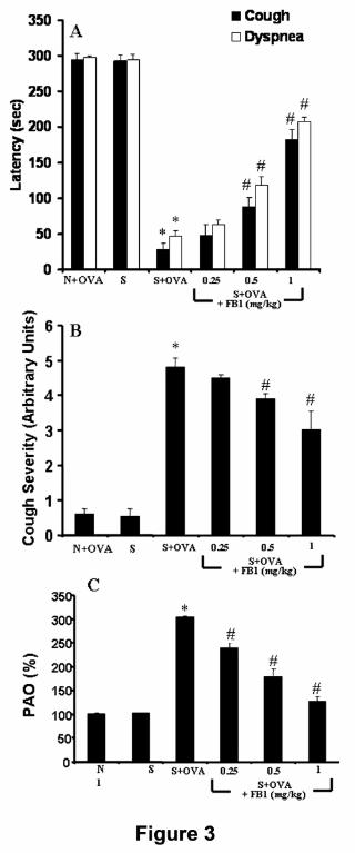
Ceramide Contributes to Respiratory Abnormalities and Hyperresponsiveness
in Actively Sensitized Guinea Pigs. As detected by immunohistochemistry,
aerosol administration of OVA increased lung ceramide levels in sensitized but not in
naïve guinea-pigs (Fig. 1 A,B). Staining for ceramide was stronger in the airway
epithelium (Fig. 1B). Increased ceramide was paralleled by up-regulation of ceramide
synthase activity (Fig. 2) and was associated with marked abnormalities in the
respiratory pattern, consisting of a significant reduction of cough latency and onset of
dyspnea (Fig. 3A) and a significant increase in cough severity score (Fig. 3B).
Similarly, experiments with mechanically ventilated animals revealed that, in
sensitized but not naïve animals, OVA challenge induced a prompt, clear-cut
increase in inflation pressure (PAO), indicating the occurrence of an immediate
asthmatic response (Fig. 3C). Such functional abnormalities were accompanied by
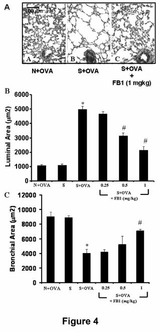
histopathological abnormalities. A representative micrograph of lung tissue is shown
in Fig. 4A. Indeed, macroscopic examination of the lungs showed prominent
changes consisting mainly of marked swelling of the pulmonary lobes due to air
accumulation. Sectioning of trachea or of main bronchi did not cause lung deflation,
thus indicating that obstruction of peripheral bronchi had occurred. Light microscopy
of lung tissue showed that the intrapulmonary bronchioli and respiratory air spaces of
naïve guinea pigs had a normal appearance. In particular, intrapulmonary bronchioli
showed open lumina, with bronchial mucosa forming short folds, and alveolar air
spaces were evenly small sized (Fig. 4A, panel A). On the other hand, in the
sensitized, vehicle-treated guinea pigs challenged with OVA we observed a reduction
of the lumen of intrapulmonary bronchi and, in large areas of the lung parenchyma, a
This article has not been copyedited and formatted. The final version may differ from this version.JPET Fast Forward. Published on November 27, 2007 as DOI: 10.1124/jpet.107.131565
at ASPE
T Journals on February 17, 2019
jpet.aspetjournals.orgD
ownloaded from
JPET#131565
19
marked dilation of the alveolar air spaces due to accumulation of entrapped air (Fig.
4A, panel B). Furthermore and as shown by morphometric analysis, the luminal area
of the alveolar air spaces was significantly increased whereas the bronchiolar area
was significantly reduced post-antigen challenge in sensitized animals (Fig. 4B, panel
C).
Inhibition of de novo ceramide synthesis with fumonisin B1 (FB1, 1 mg/kg), a
competitive and reversible inhibitor of ceramide synthase (Petrache et al., 2005),
attenuated the formation of ceramide as measured by immunohistochemistry (Fig.
1C). In keeping with these results, OVA challenge in sensitized guinea pigs
increased the enzymatic activity of ceramide synthase and FB1 pretreatment
significantly reduced this phenomenon (Fig. 2) FB1 also improved in a dose-
dependent manner (0.25-1 mg/kg) the respiratory abnormalities (Fig. 3A,B),
attenuated hyperresponsiveness (Fig. 3C) and improved overall histopathological
abnormalities (Fig. 4 A-C). Thus, as can be seen in Fig. 3A (panel C), the
intrapulmonary bronchi did not show appreciable signs of constriction and the
alveolar air spaces were not dilated. Morphometric analysis confirmed the visual
observations that FB1 prevented in a dose-dependent manner (0.25-1 mg/kg)
bronchiolar constriction and pulmonary air space inflation (Fig. 4 C,D).
Ceramide Inhibition Attenuates Oxidative and Nitrosative Stress. Ceramide
increased concurrently with markers of oxidative and nitrosative stress as evidenced
by the appearance in lung tissues of nitrotyrosine, an indicator of the formation of
nitrating oxidants (Fig. 1E), and by the increased formation of MDA (Fig. 5A), a well
known by-product of oxidative-stress induced lipid peroxidation of cell membranes.
Furthermore, OVA challenge led to enzymatic inactivation of MnSOD (Fig. 5B),
This article has not been copyedited and formatted. The final version may differ from this version.JPET Fast Forward. Published on November 27, 2007 as DOI: 10.1124/jpet.107.131565
at ASPE
T Journals on February 17, 2019
jpet.aspetjournals.orgD
ownloaded from
JPET#131565
20
increased the levels of 8-OHdG (Fig. 5C), a marker of oxidative DNA damage, and
increased PARP activity (Fig. 5D). FB1 (1 mg/kg) attenuated nitrotyrosine formation
(Fig. 1F) and dose-dependently (0.25-1 mg/kg) restored the enzymatic activity of
MnSOD, and decreased oxidative DNA damage, PARP activity and MDA formation
(Fig. 5A-D).
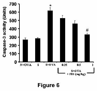
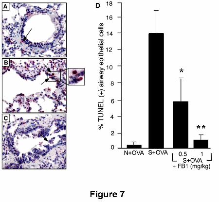
Ceramide inhibition attenuates apoptosis. OVA challenge in sensitized animals
increases the activity of caspase-3 (Fig. 6) supporting the presence of apoptosis.
Indeed, no TUNEL-positive cells were detected in OVA-challenged naïve animals
(Fig. 7A), whereas OVA challenge in sensitized guinea pigs led to the appearance of
several airway epithelial cells undergoing apoptosis (Fig. 7B,D). FB1 attenuated in a
dose-dependent fashion (0.25-1 mg/kg) caspase-3 activation (Fig. 6) and significantly
reduced epithelial cell apoptosis (0.5-1 mg/kg) (Fig. 7 A,B).
Ceramide inhibition attenuates inflammation. When compared to naïve and OVA
challenged animals, the increase in ceramide observed in sensitized animals after
OVA challenge was associated with a clear-cut inflammatory response characterized
by mast cell degranulation (Fig. 8A), and neutrophilic and eosinophilic infiltration in
lung tissue, as measured by increases in MPO levels and MBP-positive cells,
respectively (Fig. 8B,C). Inflammatory cells were mostly located around the bronchi
and within the stromal septa. Antigen challenge also led to increased levels of PGD2,
the major mast cell-derived prostaglandin, in bronchoalveolar lavage fluids as well as
of TNF-α, a proinflammatory cytokine (Fig. 8D). The above signs of inflammatory
response were consistently reduced by FB1 in a dose-dependent fashion (0.25-1
mg/kg) (Fig. 8A-D).
This article has not been copyedited and formatted. The final version may differ from this version.JPET Fast Forward. Published on November 27, 2007 as DOI: 10.1124/jpet.107.131565
at ASPE
T Journals on February 17, 2019
jpet.aspetjournals.orgD
ownloaded from

JPET#131565
21
Discussion
Asthma is a chronic inflammatory disease of the airways affecting 6-7% in the
population and accounting for more than $ 6.2 billion in direct and indirect costs in
the United States alone (Busse and Lemanske, 2001). The pathophysiology of this
disease is complex and not completely clarified, thus being the object of intensive
research efforts. The results of our studies implicate ceramide as a key signalling
mediator in biochemical events culminating in acute asthmatic response and airway
inflammation and, ultimately, respiratory dysfunction.
Antigen challenge in sensitized but not naïve guinea-pigs increased ceramide
synthase activity and the amount of immunoreactive ceramide in lung tissues,
preferentially within the epithelium, reduced the latency for cough and dyspnea, and
significantly increased cough severity. This increase in ceramide was associated with
the appearance in lung tissues of nitrotyrosine, an indicator of the formation of
nitrating oxidants, and by increased formation of MDA, a by-product of oxidative
stress-induced lipid peroxidation of cell membranes. Systemic administration of the
ceramide synthase inhibitor FB1 attenuated the formation of ceramide and
significantly reduced the development of oxidative/nitrosative stress and respiratory
abnormalities. These data provide a link between ceramide, oxidative/nitrosative
stress and acute asthmatic response.
Oxidative and nitrosative stress is a key feature of asthma and as such, the
prominent roles of superoxide, nitric oxide and their reaction product peroxynitrite are
well recognized (Comhair et al., 2005). Importantly, we and others have
demonstrated an inverse correlation between production of these reactive species
and FEV1, an index of bronchial obstruction, in asthmatics (Calhoun et al., 1992;
This article has not been copyedited and formatted. The final version may differ from this version.JPET Fast Forward. Published on November 27, 2007 as DOI: 10.1124/jpet.107.131565
at ASPE
T Journals on February 17, 2019
jpet.aspetjournals.orgD
ownloaded from

JPET#131565
22
Comhair et al., 2005). A key signaling pathway that favors accumulation of
superoxide and thus peroxynitrite is enzymatic inactivation of MnSOD (Radi et al.,
2001) that normally degrades superoxide. MnSOD is itself a target for tyrosine
nitration and oxidation (Yamakura et al., 1998) that lead to loss of enzyme function,
subsequent oxidative damage of mitochondrial proteins and initiation of apoptosis.
Under severe oxidative and nitrosative stress, DNA damage causes over-activation
of the nuclear enzyme PARP, a critical intracellular mechanism of cell death through
both necrotic and apoptotic pathways (Schreiber et al., 2006). Here we have shown
that FB1 blocked the inactivation of MnSOD and prevented oxidative DNA damage,
PARP and caspase-3 activation and epithelial cell apoptosis. The importance of the
reduction of epithelial cell apoptosis by FB1 is underscored by the fact that apoptosis
has been intimately linked to airway remodeling and hyperresponsiveness in
asthmatics (Comhair et al., 2005). Collectively, our results suggest that ceramide
induces oxidative/nitrative stress, resulting in MnSOD inactivation and triggering
epithelial cell apoptosis, presumably via PARP and caspase activation. Ceramide
has been reported to stimulate formation of reactive oxygen/nitrogen species and to
also induce the expression of the inducible nitric oxide synthase (Won et al., 2004).
Interestingly, whereas ceramide promotes generation of these species, superoxide
and nitric oxide (and presumably peroxynitrite) increase steady-state concentrations
of ceramide by activating SMases and by increasing the degradation of ceramidases,
the enzymes responsible for ceramide catabolism (Huwiler et al., 2000; Pautz et al.,
2002). These events contribute to the overall increase in the levels of ceramide and
thus ceramide-mediated damage. Although the mechanisms by which these reactive
oxygen/nitrogen species enhance the degradation of ceramidases are not
understood, one potential mechanism may be nitration, thus facilitating their faster
This article has not been copyedited and formatted. The final version may differ from this version.JPET Fast Forward. Published on November 27, 2007 as DOI: 10.1124/jpet.107.131565
at ASPE
T Journals on February 17, 2019
jpet.aspetjournals.orgD
ownloaded from
JPET#131565
23
degradation by the ubiquitin/proteasome pathway (Souza et al., 2000). In endothelial
cells, co-stimulation with nitric oxide and superoxide synergistically increase
ceramide production and apoptosis (Pettus et al., 2002), which may contribute to an
overall increase in tissue levels of ceramide and thus ceramide-mediated damage.
Our results suggest that the protective effects of FB1 may be secondary to
attenuation of the inflammatory response, as evidenced by reductions of mast cell
activation and degranulation, neutrophil and eosinophil infiltration, and release of
TNF-α. The critical roles of neutrophils, eosinophils, and proinflammatory cytokines in
the pathogenesis of asthma are known. Inhibition of neutrophil and eosinophil
infiltration attenuates inflammation associated with late phase responses and airway
hyperresponsiveness while mast cell mediators including histamine, PGD2 and TNF-
α evoke bronchoconstriction, smooth muscle cell proliferation and recruitment of
inflammatory cells (Busse and Lemanske, 2001). Ceramide modulation of these
inflammatory response through oxidative/nitrosative stress may occur upon several
mechanisms. First, superoxide and peroxynitrite induce endothelial cell damage and
increased microvascular permeability (Haglind et al., 1994), activate redox-sensitive
NF-κB and AP-1 that promote the expression of genes encoding for proinflammatory
and pro-nociceptive cytokines, such as IL-1β, TNF-α and IL-6 (Ndegele et al., 2005);
as well as for adhesion molecules involved in neutrophil and eosinophil recruitment to
inflammatory sites (Salvemini et al., 1999). Second, superoxide/peroxynitrite promote
mast cell degranulation and histamine release (Masini et al., 2005). Third,
superoxide/peroxynitrite activate cyclo-oxygenase enzymes leading to increased
production of prostaglandins. Fourth, therapeutic reduction of oxidative/nitrative
stress with superoxide dismutase mimetics and peroxynitrite decomposition catalysts
is anti-inflammatory and tissue protective (Salvemini et al., 1999).
This article has not been copyedited and formatted. The final version may differ from this version.JPET Fast Forward. Published on November 27, 2007 as DOI: 10.1124/jpet.107.131565
at ASPE
T Journals on February 17, 2019
jpet.aspetjournals.orgD
ownloaded from
JPET#131565
24
In summary, our results have revealed that formation of ceramide in the lung
plays a critical role in the development of allergic asthma through at least three
biochemical pathways: (1) oxidative and nitrosative stress; (2) apoptosis of airway
epithelial cells; and (3) local tissue inflammation. Our study does not provide direct
evidence that some of the observed effects could be ascribed to the downstream
ceramide metabolite S1P, although this possibility is not to be ruled out. S1P is
stored in mast cells, platelets, endothelial and epithelial cells (Jolly et al., 2002) and
is secreted as an autocrine mediator into the extracellular environment. S1P
activates monocytes, endothelial cells, mast cells and smooth muscle cells, mediates
eosinophil and lymphocyte infiltration and promote airway remodelling and
hyperresponsiveness (Lee et al., 1999; Ammit et al., 2001; Roviezzo et al., 2007). In
this context, S1P levels increase in the airways of asthmatic patients after allergen
challenge and correlate with eosinophil number and protein concentration in their
bronchoalveolar lavage fluids (Roviezzo et al., 2004). Multiple S1P receptors have
been identified in the lung, including S1Pr1, and S1Pr3 (Zhang et al., 1999). Recent
studies in S1Pr3 deficient mice suggested that S1Pr3 receptors expressed by the
bronchial epithelium are responsible for S1P-mediated pulmonary leakage (Gonn et
al., 2005). Knowledge of the relative contribution of each S1P receptor through the
development of selective inhibitors will be critical in understanding its importance in
asthma. Nevertheless, therapeutic strategies aimed at lowering pathologically
elevated concentrations of ceramide in asthma could prevent the formation of both
ceramide and S1P, possibly providing a more favourable endpoint than simply
blocking one of the S1P receptors.
Our data suggest that increased formation of ceramide after antigen challenge
promotes oxidative/nitrative stress and enzymatic inactivation of MnSOD that trigger
This article has not been copyedited and formatted. The final version may differ from this version.JPET Fast Forward. Published on November 27, 2007 as DOI: 10.1124/jpet.107.131565
at ASPE
T Journals on February 17, 2019
jpet.aspetjournals.orgD
ownloaded from
JPET#131565
25
apoptosis and loss of airway epithelial cells. Furthermore, ceramide contributes to the
lung inflammatory response by degranulating mast cells, recruiting
neutrophils/eosinophils and increasing formation of cytokines. In this paradigm, it is
important to consider that reciprocal relationships exist between the inflammatory
response, oxidative stress and activation of the ceramide pathway which may sparkle
a vicious cycle that amplifies the disease. In turn, cytokines can stimulate the
formation of reactive oxidative/nitrosative species and activate the de novo ceramide
synthesis pathway (Kolesnik, 2002). The current findings suggest that strategies
aimed at reducing in situ levels of ceramide could interrupt this vicious cycle and be a
novel, promising anti-asthmatic therapeutic strategy.
Ackowledgements.
We would like to thank Dr A. J. Lechner, Department of Pharmacological and
Physiological Sciences, Saint Louis University, School of Medicine and Prof. D. Bani,
Dept. Anatomy, Histology & Forensic Medicine, University of Florence School of
Medicine, Florence, Italy, for their invaluable inputs.
This article has not been copyedited and formatted. The final version may differ from this version.JPET Fast Forward. Published on November 27, 2007 as DOI: 10.1124/jpet.107.131565
at ASPE
T Journals on February 17, 2019
jpet.aspetjournals.orgD
ownloaded from
JPET#131565
26
References
Ammitt AJ, Hastie AT, Edsal LC, Hoffman RK, Amrani Y, Krymskaya VP, Kane SA,
Peters SP, Penn RB, Spiegel S and Panettieri RA, J (2001) Sphingosyne-1-
phosphate modulates human airway smooth muscle cell functions that promote
inflammation and airway remodeling in asthma. FASEB J 15:1212-1214.
Andreadis AA, Hazen SL, Comhair SA and Erzurum SC (2003) Oxidative and
nitrosative events in asthma. Free Radic Biol Med 35:213-225.
Barnes PJ (2004) New drugs for asthma. Nature reviews 3:831-844.
Busse WW and Lemanske RF, Jr (2001) Asthma. The New England journal of
medicine 344:350-362.
Calhoun WJ, Reed HE, Moest DR and Stevens CA (1992) Enhanced superoxide
production by alveolar macrophages and air-space cells, airway inflammation,
and alveolar macrophage density changes after segmental antigen
bronchoprovocation in allergic subjects. The American review of respiratory
disease 145:317-325.
Chalfant CE, Rathman K, Pinkerman RL, Wood RE, Obeid LM, Ogretmen B and
Hannun YA (2002) De novo ceramide regulates the alternative splicing of
caspase 9 and bcl-x in a549 lung adenocarcinoma cells. Dependence on protein
phosphatase-1. J Biol Chem 277:12587-12595.
Chun J, Goetzl EJ, Hla T, Igarashi Y, Lynch KR, Moolenaar W, Pyne S and Tigyi G
(2002) International union of pharmacology. Xxxiv. Lysophospholipid receptor
nomenclature. Pharmacol Rev 54:265-269.
Comhair SA, Xu W, Ghosh S, Thunnissen FB, Almasan A, Calhoun WJ, Janocha AJ,
Zheng L, Hazen SL and Erzurum SC (2005) Superoxide dismutase inactivation in
This article has not been copyedited and formatted. The final version may differ from this version.JPET Fast Forward. Published on November 27, 2007 as DOI: 10.1124/jpet.107.131565
at ASPE
T Journals on February 17, 2019
jpet.aspetjournals.orgD
ownloaded from
JPET#131565
27
pathophysiology of asthmatic airway remodeling and reactivity. Am J Pathol
166:663-674.
Delogu G, Famularo G, Amati F, Signore L, Antonucci A, Trinchieri V, Di Marzio L
and Cifone MG (1999) Ceramide concentrations in septic patients: A possible
marker of multiple organ dysfunction syndrome. Crit Care Med 27:2413-2417.
Goggel R, Winoto-Morbach S, Vielhaber G, Imai Y, Lindner K, Brade L, Brade H,
Ehlers S, Slutsky AS, Schutze S, Gulbins E and Uhlig S (2004) Paf-mediated
pulmonary edema: A new role for acid sphingomyelinase and ceramide. Nat Med
10:155-160.
Gon Y, Wood MR, Kiosses WB, Jo E, Sanna MG, Chun J and Rosen H (2005) S1p3
receptor-induced reorganization of epithelial tight junctions compromises lung
barrier integrity and is potentiated by tnf. Proc Natl Acad Sci U S A 102:9270-
9275.
Haglind E, Xia G and Rylander R (1994) Effects of antioxidants and paf receptor
antagonist in intestinal shock in the rat. Circ Shock 42:83-91.
Heinrich M, Wickel M, Winoto-Morbach S, Schneider-Brachert W, Weber T, Brunner
J, Saftig P, Peters C, Kronke M and Schutze S (2000) Ceramide as an activator
lipid of cathepsin d. Adv Exp Med Biol 477:305-315.
Huwiler A, Kolter T, Pfeilschifter J and Sandhoff K (2000) Physiology and
pathophysiology of sphingolipid metabolism and signaling. Biochim Biophys Acta
1485:63-99.
Jolly PS, Rosenfeldt HM, Milstien S and Spiegel S (2002) The roles of sphingosine-1-
phosphate in asthma. Mol Immunol 38:1239-1245.
Kolesnick R and Golde DW (1994) The sphingomyelin pathway in tumor necrosis
factor and interleukin-1 signaling. Cell 77:325-328.
This article has not been copyedited and formatted. The final version may differ from this version.JPET Fast Forward. Published on November 27, 2007 as DOI: 10.1124/jpet.107.131565
at ASPE
T Journals on February 17, 2019
jpet.aspetjournals.orgD
ownloaded from
JPET#131565
28
Kolesnick R (2002) The therapeutic potential of modulating the
ceramide/sphingomyelin pathway. J Clin Invest 110:3-8.
Kolesnick R and Fuks Z (2003) Radiation and ceramide-induced apoptosis.
Oncogene 22:5897-5906.
Lee MJ, Thangada S, Claffey KP, Ancellin N, Liu CH, Kluk M, Volpi M, Sha'afi RI and
Hla T (1999) Vascular endothelial cell adherens junction assembly and
morphogenesis induced by sphingosine-1-phosphate. Cell 99:301-312.
Le Stunff H, Milstien S and Spiegel S (2004) Generation and metabolism of bioactive
sphingosine-1-phosphate. Journal of cellular biochemistry 92:882-899.
Masini E, Bani D, Vannacci A, Pierpaoli S, Mannaioni PF, Comhair SA, Xu W,
Muscoli C, Erzurum SC and Salvemini D (2005) Reduction of antigen-induced
respiratory abnormalities and airway inflammation in sensitized guinea pigs by a
superoxide dismutase mimetic. Free Radic Biol Med 39:520-531.
Ndengele MM, Muscoli C, Wang ZQ, Doyle TM, Matuschak GM and Salvemini D
(2005) Superoxide potentiates nf-kappab activation and modulates endotoxin-
induced cytokine production in alveolar macrophages. Shock 23:186-193.
Newton R, Hart L, Chung KF and Barnes PJ (2000) Ceramide induction of cox-2 and
pge(2) in pulmonary a549 cells does not involve activation of nf-kappab. Biochem
Biophys Res Commun 277:675-679.
Pautz A, Franzen R, Dorsch S, Boddinghaus B, Briner VA, Pfeilschifter J and Huwiler
A (2002) Cross-talk between nitric oxide and superoxide determines ceramide
formation and apoptosis in glomerular cells. Kidney Int 61:790-796.
Petrache I, Natarajan V, Zhen L, Medler TR, Richter AT, Cho C, Hubbard WC,
Berdyshev EV and Tuder RM (2005) Ceramide upregulation causes pulmonary
cell apoptosis and emphysema-like disease in mice. Nat Med 11:491-498.
This article has not been copyedited and formatted. The final version may differ from this version.JPET Fast Forward. Published on November 27, 2007 as DOI: 10.1124/jpet.107.131565
at ASPE
T Journals on February 17, 2019
jpet.aspetjournals.orgD
ownloaded from

JPET#131565
29
Pettus BJ, Chalfant CE and Hannun YA (2002) Ceramide in apoptosis: An overview
and current perspectives. Biochim Biophys Acta 1585:114-125.
Radi R, Peluffo G, Alvarez MN, Naviliat M and Cayota A (2001) Unraveling
peroxynitrite formation in biological systems. Free Radic Biol Med 30:463-488.
Roviezzo F, Del Galdo F, Abbate G, Bucci M, D'Agostino B, Antunes E, De Dominicis
G, Parente L, Rossi F, Cirino G and De Palma R (2004) Human eosinophil
chemotaxis and selective in vivo recruitment by sphingosine 1-phosphate. Proc
Natl Acad Sci U S A 101:11170-11175.
Roviezzo F, Di Lorenzo A, Bucci M, Brancaleone V, Vellecco V, De Nardo M, Orlotti
D, De Palma R, Rossi F, D'Agostino B and Cirino G (2007) Sphingosine-1-
phosphate/sphingosine kinase pathway is involved in mouse airway hyper-
responsiveness. Am J Respir Cell Mol Biol 36: 757-762.
Salvemini D, Riley DP, Lennon PJ, Wang ZQ, Currie MG, Macarthur H and Misko TP
(1999) Protective effects of a superoxide dismutase mimetic and peroxynitrite
decomposition catalysts in endotoxin-induced intestinal damage. Br J Pharmacol
127:685-692.
Schreiber V, Dantzer F, Ame JC and de Murcia G (2006) Poly(adp-ribose): Novel
functions for an old molecule. Nat Rev Mol Cell Biol 7:517-528.
Siskind LJ, Kolesnick RN and Colombini M (2002) Ceramide channels increase the
permeability of the mitochondrial outer membrane to small proteins. J Biol Chem
277:26796-26803.
Souza JM, Choi I, Chen Q, Weisse M, Daikhin E, Yudkoff M, Obin M, Ara J, Horwitz J
and Ischiropoulos H (2000) Proteolytic degradation of tyrosine nitrated proteins.
Arch Biochem Biophys 380:360-366.
This article has not been copyedited and formatted. The final version may differ from this version.JPET Fast Forward. Published on November 27, 2007 as DOI: 10.1124/jpet.107.131565
at ASPE
T Journals on February 17, 2019
jpet.aspetjournals.orgD
ownloaded from
JPET#131565
30
Spiegel S and Milstien S (2003) Sphingosine-1-phosphate: An enigmatic signalling
lipid. Nat Rev Mol Cell Biol 4:397-407.
Stennicke HR and Salvesen GS (1997) Biochemical characteristics of caspases-3, -
6, -7, and -8. J Biol Chem 272:25719-25723.
Suzuki Y, Masini E, Mazzocca C, Cuzzocrea S, Ciampa A, Suzuki H, and Bani D
(2004) Inhibition of poly(adp-ribose) polymerase prevents allergen-induced
asthma-like reaction in sensitized guinea pigs. The Journal of pharmacology and
experimental therapeutics 311:1241-1248.
Won JS, Im YB, Khan M, Singh AK and Singh I (2004) The role of neutral
sphingomyelinase produced ceramide in lipopolysaccharide-mediated expression
of inducible nitric oxide synthase. J Neurochem 88:583-593.
Yamakura F, Taka H, Fujimura T and Murayama K (1998) Inactivation of human
manganese-superoxide dismutase by peroxynitrite is caused by exclusive
nitration of tyrosine 34 to 3-nitrotyrosine. J Biol Chem 273:14085-14089.
Zhang G, Contos JJ, Weiner JA, Fukushima N and Chun J (1999) Comparative
analysis of three murine g-protein coupled receptors activated by sphingosine-1-
phosphate. Gene 227:89-99.
Zhang YH, Vasko MR and Nicol GD (2002) Ceramide, a putative second messenger
for nerve growth factor, modulates the ttx-resistant na(+) current and delayed
rectifier k(+) current in rat sensory neurons. J Physiol 544:385-402.
This article has not been copyedited and formatted. The final version may differ from this version.JPET Fast Forward. Published on November 27, 2007 as DOI: 10.1124/jpet.107.131565
at ASPE
T Journals on February 17, 2019
jpet.aspetjournals.orgD
ownloaded from

JPET#131565
31
Footnotes
Supported by St Louis University Seed Funds, PPG PO1 081064, IRCCS Centro
Neurolesi "Bonino-Pulejo", Messina, Italy and by funds from the University of
Florence, Florence, Italy.
This article has not been copyedited and formatted. The final version may differ from this version.JPET Fast Forward. Published on November 27, 2007 as DOI: 10.1124/jpet.107.131565
at ASPE
T Journals on February 17, 2019
jpet.aspetjournals.orgD
ownloaded from

JPET#131565
32
Legends for figures
Figure 1. When compared to OVA-challenged naïve animals (A,D), OVA challenge
in sensitized guinea-pigs pre-treated with PBS leads to formation of ceramide and
nitrotyrosine, as detected by immunohistochemistry (B,E). Pre-treatment with FB1 (1
mg/kg) before OVA challenge blocks ceramide staining (C,F). Images are
representative of at least 3 different experiments.
Figure 2. Ceramide synthase activity in lung tissue homogenates is up-regulated in
OVA-challenged, sensitized guinea-pigs (S+OVA) as compared with OVA-challenged
naïve animals (N+OVA). FB1 pre-treatment of OVA-challenged, sensitized animals
significantly reduces the enzyme activity. Values are mean ± S.E.M. of 4 different
experiments. *p<0.01 vs. N+OVA; **p<0.001 vs. S+OVA.
Figure 3. When compared to OVA-challenged naïve (N+OVA) or in sensitized and
not challenged (S) animals, OVA challenge in sensitized guinea-pigs (S+OVA) led to
a reduction of the latency time for cough and dyspnea (A) and to an increase in
cough severity (B). As shown in (C), FB1 also inhibited in a dose-dependent manner
(0.25-1 mg/kg) OVA-induced bronchoconstriction in sensitized, mechanically
ventilated guinea pigs, evaluated 10 min. after OVA aerosol. Pressure at the airway
opening (PAO) is expressed as percent change over basal inflation pressure before
OVA challenge, assumed as 100%. Data are expressed as mean ± S.E.M. of 5
animals per experimental group. *p<0.01 vs OVA-challenged naïve animals; #
p<0.01 vs OVA-challenged, sensitized animals in the absence of FB1 or not
challenged, sensitized animals.
This article has not been copyedited and formatted. The final version may differ from this version.JPET Fast Forward. Published on November 27, 2007 as DOI: 10.1124/jpet.107.131565
at ASPE
T Journals on February 17, 2019
jpet.aspetjournals.orgD
ownloaded from
JPET#131565
33
Figure 4. Upper panels (A). Representative lung tissue micrographs from OVA-
challenged, naïve guinea pigs (panel A), OVA-challenged, sensitized guinea pigs
pre-treated with PBS (panel B) and OVA challenged, sensitized guinea pigs pre-
treated with the highest dose of FB1 (1 mg/kg)(panel C). Compared with A, a marked
dilation of respiratory air spaces and reduction of bronchiolar lumen is seen in B.
These alterations are not present in the FB1 treated group (C). Hematoxylin and
eosin staining. Lower panels. Morphometric analysis of lumenal area of respiratory
air spaces (B) and bronchioli (C). FB1 dose-dependently reduces the lung tissue
abnormalities induced by OVA challenge in sensitised guinea pigs. *p<0.001 vs naïve
animals; #p<0.001 vs OVA-challenged, sensitized animals.
Figure 5. When compared to OVA-challenged naïve (N+OVA) or sensitized and not
challenged (S) animals, OVA challenge in sensitized guinea-pigs (S+OVA) leads to
increased formation of MDA (A), inactivation of MnSOD (B), enhaced generation of
8-OHdG (C) and increased PARP activity (D). These biochemical changes are
reduced in a dose-dependent fashion by FB1 (0.25-1 mg/kg). Data are expressed as
mean ± S.E.M. of 5 animals per experimental group. *p<0.01 vs OVA-challenged
naïve animals; # p<0.05 vs OVA-challenged sensitized animals in the absence of
FB1 or non-OVA challenged sensitized animals.
Figure 6. When compared to OVA-challenged naïve (N+OVA) or in sensitized and
not challenged (S) animals, OVA challenge in sensitized guinea-pigs (S+OVA)
increased caspase-3 activity and this was blocked in a dose-dependent fashion by
FB1 (0.25-1 mg/kg). Data are expressed as mean ± S.E.M. of 5 animals per
experimental group. *p<0.01 vs OVA-challenged, naïve animals; # p<0.05 vs OVA-
This article has not been copyedited and formatted. The final version may differ from this version.JPET Fast Forward. Published on November 27, 2007 as DOI: 10.1124/jpet.107.131565
at ASPE
T Journals on February 17, 2019
jpet.aspetjournals.orgD
ownloaded from
JPET#131565
34
challenged, sensitized animals in the absence of FB1 or sensitized, not challenged
animals.
Figure 7. Immunohistochemical analysis of apoptosis in airway epithelial cells.
Increased numbers of TUNEL-positive epithelial cells can be seen (red nuclei) in
OVA-challenged sensitized animals (B) when compared to OVA-challenged naïve
animals (A). FB1 (0.5-1mg/kg) leads to a reduction of the amount of TUNEL-positive
cells (C). The histogram (D) shows the mean ± S.E.M. of percent TUNEL-positive
epithelial cells.*p<0.05; **p<0.01.
Figure 8. When compared to OVA-challenged naïve (N+OVA) or sensitized and not
challenged (S) animals, OVA challenge in sensitized guinea-pigs (S+OVA) causes
mast cell degranulation (A), promotes lung tissue infiltration by neutrophils (B;
evaluated by MPO in tissue homogenates) and eosinophils (C; evaluated as MBP-
positive cells in lung tissue sections) and increased levels of PGD2 and TNF-α (D;
measured in BAL fluids). These events are blocked in a dose-dependent fashion by
FB1 (0.25-1 mg/kg). Data are expressed as mean ± SEM of 5 animals per
experimental group. *p<0.01 vs OVA-challenged naïve animals; # p<0.05 vs OVA-
challenged sensitized animals in the absence of FB1 or not challenged, sensitized
animals.
This article has not been copyedited and formatted. The final version may differ from this version.JPET Fast Forward. Published on November 27, 2007 as DOI: 10.1124/jpet.107.131565
at ASPE
T Journals on February 17, 2019
jpet.aspetjournals.orgD
ownloaded from

This article has not been copyedited and formatted. The final version may differ from this version.JPET Fast Forward. Published on November 27, 2007 as DOI: 10.1124/jpet.107.131565
at ASPE
T Journals on February 17, 2019
jpet.aspetjournals.orgD
ownloaded from

This article has not been copyedited and formatted. The final version may differ from this version.JPET Fast Forward. Published on November 27, 2007 as DOI: 10.1124/jpet.107.131565
at ASPE
T Journals on February 17, 2019
jpet.aspetjournals.orgD
ownloaded from
This article has not been copyedited and formatted. The final version may differ from this version.JPET Fast Forward. Published on November 27, 2007 as DOI: 10.1124/jpet.107.131565
at ASPE
T Journals on February 17, 2019
jpet.aspetjournals.orgD
ownloaded from
This article has not been copyedited and formatted. The final version may differ from this version.JPET Fast Forward. Published on November 27, 2007 as DOI: 10.1124/jpet.107.131565
at ASPE
T Journals on February 17, 2019
jpet.aspetjournals.orgD
ownloaded from

This article has not been copyedited and formatted. The final version may differ from this version.JPET Fast Forward. Published on November 27, 2007 as DOI: 10.1124/jpet.107.131565
at ASPE
T Journals on February 17, 2019
jpet.aspetjournals.orgD
ownloaded from
This article has not been copyedited and formatted. The final version may differ from this version.JPET Fast Forward. Published on November 27, 2007 as DOI: 10.1124/jpet.107.131565
at ASPE
T Journals on February 17, 2019
jpet.aspetjournals.orgD
ownloaded from
This article has not been copyedited and formatted. The final version may differ from this version.JPET Fast Forward. Published on November 27, 2007 as DOI: 10.1124/jpet.107.131565
at ASPE
T Journals on February 17, 2019
jpet.aspetjournals.orgD
ownloaded from

This article has not been copyedited and formatted. The final version may differ from this version.JPET Fast Forward. Published on November 27, 2007 as DOI: 10.1124/jpet.107.131565
at ASPE
T Journals on February 17, 2019
jpet.aspetjournals.orgD
ownloaded from