Embed Size (px)
Citation preview
JPET#65128
1
Involvement of capsaicin sensitive afferent nerves
and CCK2/gastrin receptors in gastroprotection and adaptation
of gastric mucosa to Helicobacter pylori-lipopolysaccharide (Hp-LPS)
TOMASZ BRZOZOWSKI*, PETER C. KONTUREK**, ANTHONY P. MORAN+,
ROBERT PAJDO*, SLAWOMIR KWIECIEN*, STANISLAW J. KONTUREK*,
ZBIGNIEW SLIWOWSKI*, DANUTA DROZDOWICZ*, WIESLAW. W. PAWLIK*,
ECKHART. G. HAHN**
*Department of Physiology, Jagiellonian University, School of Medicine, Cracow, Poland,
**Department of Medicine I, University of Erlangen-Nuremberg, Erlangen, Germany &
+Department of Microbiology, National University of Ireland, Galway, Ireland
JPET Fast Forward. Published on March 15, 2004 as DOI:10.1124/jpet.104.065128
Copyright 2004 by the American Society for Pharmacology and Experimental Therapeutics.
This article has not been copyedited and formatted. The final version may differ from this version.JPET Fast Forward. Published on March 15, 2004 as DOI: 10.1124/jpet.104.065128
at ASPE
T Journals on February 26, 2019
jpet.aspetjournals.orgD
ownloaded from
JPET#65128
2
Running title: sensory nerves and CCK receptors in adaptation to Hp-LPS
Address correspondence to:
Prof. Dr. T. Brzozowski
Department of Physiology
Jagiellonian University School of Medicine
16 Grzegorzecka Str.,
31-531 Cracow, Poland
E-mail: [email protected]
Document statistics:text...................... pages 20 (page 3 - page 24)
figures................ 7 figs
references.............40 refs (page 25 - page 30)
Number of words: abstract ..................250
introduction............662
discussion..............1433
running title........... 56 characters
Abbreviations:
Helicobacter pylori -------------------Hp
Lipopolysaccharide --------------------LPS
Gastric blood flow ---------------------GBF
Tumor necrosis factor alpha----------TNF-α
Interleukin-1 beta ----------------------IL-1β
Aspirin-----------------------------------ASA
This article has not been copyedited and formatted. The final version may differ from this version.JPET Fast Forward. Published on March 15, 2004 as DOI: 10.1124/jpet.104.065128
at ASPE
T Journals on February 26, 2019
jpet.aspetjournals.orgD
ownloaded from
JPET#65128
3
ABSTRACT
Lipopolysaccharide (LPS) is one of the virulence factors in the Helicobacter pylori (Hp)-
infected stomach, but it remains unknown whether single and prolonged pretreatment with
Hp-LPS can affect the course of gastric damage induced by aspirin (ASA). We compared the
effects of Hp-LPS with those induced by LPSs isolated from intestinal Bacteroides fragilis,
Yersinia enterocolitica and Campylobacter jejuni applied for 4 days on acute ASA-induced
gastric lesions in rats. The area of ASA-induced gastric lesions, the gastric blood flow
(GBF), the expression of mRNA and protein of leptin and plasma leptin, gastrin, interleukin-
(IL-)1β and tumor necrosis factor- (TNF-) α levels were examined. Single (once) or repeated
(5 times) intraperitoneal (i.p.) injections of Hp-LPS (1 mg/kg) or intestinal LPSs failed to
produce macroscopic gastric damage and did not affect the GBF when compared with
vehicle. Hp-LPS injected repeatedly suppressed the gastric acid secretion, upregulated leptin
mRNA, and protein and increased plasma leptin and gastrin levels. Hp-LPS reduced
significantly the ASA-induced gastric damage and the accompanying fall in the GBF, and
these effects were significantly attenuated by capsaicin-denervation and selective antagonism
of cholecystokinin-B (CCK2) receptors by RPR-102681, but not by loxiglumide, an
antagonist of CCK1 receptors. We conclude that 1) daily application of Hp-LPS enhances
gastric mucosal resistance against ASA damage due to the increase of GBF, expression and
release of leptin and gastrin, exerting the trophic and gastroprotective effects, and 2) this
enhanced resistance to ASA damage in Hp-LPS adapted stomach is mediated by the sensory
afferents and specific CCK2/gastrin receptors.
This article has not been copyedited and formatted. The final version may differ from this version.JPET Fast Forward. Published on March 15, 2004 as DOI: 10.1124/jpet.104.065128
at ASPE
T Journals on February 26, 2019
jpet.aspetjournals.orgD
ownloaded from
JPET#65128
4
Introduction
Helicobacter pylori (Hp) is now generally accepted as a major cause of chronic
gastritis and an important risk factor for peptic ulcer disease and gastric cancer (Warren and
Marshal, 1983; Konturek et al., 1999), but it remains unknown whether the gastric mucosa is
capable of adapting to repeated Hp insults and whether such Hp-adaptation might alter the
mucosal resistance to the injurious action of strong irritants.
Various pathogenic factors originating from Hp have been implicated in the damaging
effect of this bacterium on the gastric mucosa, the most important besides ammonia, being
cytotoxins released by Hp-strains expressing the vacuolating cytotoxin A (VacA) and
cytotoxin-associated gene A (CagA) proteins, Hp-derived lipopolysaccharides (Hp-LPS) and
the enhanced generation of reactive oxygen species (Megraud et al., 1992; Figura and
Tabaqchali, 1996; Crabtree, 1996; Moran, 2001a; Moran, 2001b). Hp-LPS exhibits a low
immunological activity and this property has been assumed to play an important role in the
persistency of Hp infection in the human stomach (Moran, 2001a; Moran, 2001b).
Nevertheless, the deleterious action of LPS derived from Hp in the stomach includes an
interaction of this endotoxin with laminin (Valkonen et al., 1994), its influence on the gastric
mucus formation and composition (Slomiany et al., 1992) and expression of proinflammatory
cytokines (Crabtree et al., 1994; Moran, 2001a). Recent evidence suggests, however, that
LPS derived from Escherichia coli applied parenterally, also induces gastroprotective activity
against lesions induced by strong topical irritants such as ethanol (Tepperman and Soper,
1994; Konturek et al., 1998a; Konturek et al., 1998b; Ng et al., 2002) and results in mucosal
adaptation to topical irritants after prolonged administration (Ferraz et al., 1997).
This article has not been copyedited and formatted. The final version may differ from this version.JPET Fast Forward. Published on March 15, 2004 as DOI: 10.1124/jpet.104.065128
at ASPE
T Journals on February 26, 2019
jpet.aspetjournals.orgD
ownloaded from
JPET#65128
5
Leptin is accepted as a protein product of the ob gene acting directly and through the
sensory afferent on central leptin receptors (Ob-R) in the hypothalamus that control food
intake and energy expenditure (Friedman and Halaas, 1998). Recent studies documented that
leptin is present in the plasma of experimental animals, such as mice and rats, as well as in
humans (Barbier et al., 1998; Shalev et al., 1997). Leptin is believed to be secreted mainly by
adipocytes and the placenta, but recent studies have revealed that leptin messenger RNA
(mRNA) and leptin protein can also be detected in the rat gastric oxyntic mucosa, suggesting
that the gastric corpus may be another important source of leptin (Bado et al., 1998;
Brzozowski et al., 1999).
The importance of leptin in the action of bacterial LPS has been supported by
evidence that reduced levels of leptin during starvation increased animal susceptibility to
endotoxic shock (Faggioni et al., 2000). Since parenteral LPS was shown to attenuate
ethanol-induced gastric damage (Tepperman and Soper, 1994; Konturek et al., 1998a;
Brzozowski et al., 2003) the question remains whether leptin, which exhibits gastroprotective
activity in the stomach (Brzozowski et al., 1999), can also contribute to the LPS-induced
protection against mucosal damage induced by aspirin. Finally, the physiological
significance of gut hormones such as leptin and gastrin in adaptation of gastric mucosa,
developed by daily injections of endotoxins such as Hp-LPS, requires an elucidation.
This study was designed to determine the effect of single or repeated parenteral
applications of Hp-LPS on acute gastric lesions induced by intragastric (i.g.) administration
of acidified aspirin (ASA) and accompanying changes in the gastric blood flow (GBF),
gastric secretion, and the gene expression and release of leptin. An attempt was made to
compare the effects of five daily injections with Hp-LPS with those exhibited by different
LPSs isolated from enteric bacteria such as Bacteroides fragilis, Yersinia enterocolitica and
This article has not been copyedited and formatted. The final version may differ from this version.JPET Fast Forward. Published on March 15, 2004 as DOI: 10.1124/jpet.104.065128
at ASPE
T Journals on February 26, 2019
jpet.aspetjournals.orgD
ownloaded from
JPET#65128
6
Campylobacter jejuni on gastric acid secretion and ASA-induced gastric damage.
Furthermore, we attempted to compare the effects of Hp-LPS with those of exogenous leptin,
CCK and peptone meal, a potent releaser of both CCK and leptin, and to examine the
involvement of specific CCK1 (for CCK) and CCK2 (for gastrin) receptors, sensory nerve
activity, proinflammatory cytokines such as interleukin- (IL-) 1β and tumor necrosis factor-
(TNF-) α in gastric mucosal integrity and possible gastric mucosal adaptation afforded by
Hp-LPS.
Methods
Three major series (A, B & C) consisting of 200 male Wistar rats weighing 180-220 g
were used. All procedures have been carried out in accordance with the Declaration of
Helsinki and were accepted by Local Ethical Committee at the Jagiellonian University. Acute
gastric lesions were induced by an i.g. application of acidified ASA (150 mg/kg in 0.15 N
HCl) in a volume of 1.5 ml by means of a metal orogastric tube (series A). In series B, gastric
mucosa was subjected to single or repeated exposures to vehicle (saline) or LPS isolated from
Hp (Moran, 1992). Series C was designed to determine the effect of single and repeated
parenteral injections of LPS derived from the intestinal bacteria B. fragilis, Y. enterocolitica
and C. jejuni. B. fragilis NCTC 9343 was obtained from the National Collection of Type
Cultures (London, UK), C. jejuni ATCC 43431 was purchased from the American Type
Culture Collection (Manassas, Virginia, USA) and Y. enterocolitica IY-9 was a clinical
isolate originating from a human with diarrheal disease.
Induction of gastric adaptation to Hp-LPS. Gastric adaptation was achieved by
daily parenteral administration of Hp-LPS or vehicle (saline) to normally fed rats for the
This article has not been copyedited and formatted. The final version may differ from this version.JPET Fast Forward. Published on March 15, 2004 as DOI: 10.1124/jpet.104.065128
at ASPE
T Journals on February 26, 2019
jpet.aspetjournals.orgD
ownloaded from

JPET#65128
7
entire period of the study. The parenteral route of LPS administration was chosen based on
our previous observations (Konturek et al., 1998a, Brzozowski et al., 2003) that gastric
mucosa directly exposed to LPS applied in a dose of 1 mg/kg (i.g.), failed to adapt to this
endotoxin and such LPS applied i.g. also failed to influence the mucosal lesions induced by
strong irritants (e.g., ethanol, ASA and stress). Since LPS produced by the bacteria
contaminating the gastrointestinal lumen and adherent to mucosal cells under the mucus layer
covering the surface epithelium, may penetrate the mucosa and reach the general circulation,
we decided to employ parenteral injection rather than the intragastric route as the route of
bacterial LPS administration in order to mimic the fate of this systemic LPS. Our preliminary
observation with intragastric daily application of Hp-LPS at a dose of 10 mg/kg also exerted
gastroprotection against ASA-induced gastric damage, but such an investigation required
large doses of Hp-LPS which were not available to us, and therefore, only parenteral
administration of this LPS was employed in the present study. The animals received Hp-LPS
once and, for comparison, LPS isolated from intestinal bacteria (1 mg/kg) by intraperitoneal
route (i.p.), or were treated repeatedly by the same route with Hp-LPS and that of other
intestinal bacteria for four consecutive days as described in detail in our previous studies with
ASA-induced gastric adaptation (Konturek et al., 1994; Brzozowski et al., 1995). Rats with
single or repeated daily (5 times) injections of Hp-LPS were sacrificed, the stomach was
quickly removed, opened along the greater curvature, and the gastric mucosa was
photographed to subsequently measure the area of gastric lesions by two observers using
planimetry (Morphomat, Carl Zeiss, Berlin, Germany). The gastric mucosa of separate
overnight-fasted rats treated repeatedly with vehicle, Hp-LPS, or intestinal bacterial LPS was
then challenged 120 min after the last dose of vehicle, Hp-LPS or intestinal bacterial LPS
with acidified ASA applied i.g. in a volume of 1.5 ml.
This article has not been copyedited and formatted. The final version may differ from this version.JPET Fast Forward. Published on March 15, 2004 as DOI: 10.1124/jpet.104.065128
at ASPE
T Journals on February 26, 2019
jpet.aspetjournals.orgD
ownloaded from
JPET#65128
8
At 3 h upon the ASA application, the animals were lightly anesthetized with ether,
their abdomen was opened by the midline incision and the stomach was exposed for the
measurement of GBF by means of the H2-gas clearance technique (Konturek et al., 1994;
Konturek et al., 2001a). The measurements were made in three areas of the oxyntic mucosa
and the mean values of the measurements were calculated and expressed as percent changes
of those recorded in the vehicle (saline)-treated animals.
The alterations of gastric secretions in rats treated with the vehicle (saline), Hp-LPS or
LPS derived from intestinal bacteria applied once or given repeatedly, were tested in a
separate group of 60 fasted rats, surgically equipped with chronic gastric fistulas as described
in our earlier studies (Brzozowski et al., 2000a). The rats that have been treated once with
Hp-LPS or injected repeatedly (5 times) with Hp-LPS or LPS isolated from intestinal bacteria,
were placed at day 4 in individual Bollman cages (to which the animals were well-
conditioned) to prevent coprophagy and to maintain the necessary restraint. In addition, the
effect of five daily injections with Hp-LPS with or without loxiglumide, an inhibitor of CCK1
receptors, and N-(metoxy-3 phenyl) N-(N-methyl N-phenyl-carbamylmethyl)
carbamoylmethyl]-3 ureido}-3 phenyl}-2 propronique (RPR-102681), the selective CCK2
receptor antagonist, was determined (Konturek et al., 1995; Brzozowski et al., 2000b). Each
fistula was then opened, and the stomach rinsed gently with 5-8 ml of tap water at 37oC.
Basal gastric secretion was collected for 120 min, during which time all animals received
saline at a rate of 4 ml/h subcutaneously. The gastric juice was collected every 30 min, the
volume was measured, and then the acid concentration and output were determined and
expressed as the output per 30 min as described previously (Brzozowski et al., 2000a).
This article has not been copyedited and formatted. The final version may differ from this version.JPET Fast Forward. Published on March 15, 2004 as DOI: 10.1124/jpet.104.065128
at ASPE
T Journals on February 26, 2019
jpet.aspetjournals.orgD
ownloaded from
JPET#65128
9
Experimental groups and treatments. Vehicle or Hp-LPS (dose of 1 mg/kg i.p.)
were given once or were administered at the same dose for four consecutive days. After five
daily injections with Hp-LPS or vehicle, the gastric mucosa was challenged with acidified
ASA. The protective effect of Hp-LPS applied i.p. 2 h prior to ASA was compared with
known gastroprotective agents (Brzozowski et al., 1999) such as those of leptin and CCK
administered i.p. (dose of 10 µg/kg) or 8% peptone meal applied i.g. in a volume of 1 ml per
rat.
The following groups of rats were used: 1) vehicle (1 ml of saline i.p.) followed 120
min later by acidified ASA (150 mg/kg i.g.); 2) Hp-LPS (1 mg/kg i.p.) followed 120 min later
by ASA; 3) B. fragilis-LPS, Y. enterocolitica-LPS and C. jejuni-LPS (1 mg/kg i.p.) followed
120 min later by ASA; 4) leptin (10 µg/kg i.p.) and CCK-8 (10 µg/kg i.p.) followed 120 min
later by ASA; 5) 8% peptone meal (1 ml/rat i.g.) followed 120 min later by ASA; 6) vehicle
(saline) or Hp-LPS (1 mg/kg i.p.) administered daily for 4 days with or without the challenge
with ASA applied at day 4; and 7) B. fragilis-LPS, Y. enterocolitica-LPS and C. jejuni-LPS
(1 mg/kg i.p.) administered daily for 4 days with or without the challenge with ASA applied
at day 4.
Effect of suppression of CCK1 and CCK2 receptors and sensory nerves on
gastroprotection and adaptation induced by Hp-LPS. To check whether gastrin/CCK is
involved in the action of bacterial LPS on mucosal integrity, separate subgroups of rats were
used and the effects of inhibition of CCK1 and CCK2 receptors with loxiglumide and RPR-
102681, respectively, on the protection and adaptation induced by LPS were examined. RPR-
102681 is a novel, non-peptide selective antagonist of the CCK2/gastrin receptor, which has
been shown to display nanomolar affinity of about 2000-fold greater selectivity for CCK2 than
This article has not been copyedited and formatted. The final version may differ from this version.JPET Fast Forward. Published on March 15, 2004 as DOI: 10.1124/jpet.104.065128
at ASPE
T Journals on February 26, 2019
jpet.aspetjournals.orgD
ownloaded from
JPET#65128
10
CCK1 receptors (Bohme et al., 1997). [Loxiglumide was a generous gift of Dr. Rovati
(Milan, Italy), and RPR-102681 was purchased from Rhone-Poulenc, Rorer S.A., (Vitry-sur-
Seine, France).] In subsequent studies, two series of experiments were carried out.
Series (i) was used to examine the effect of Hp-LPS applied (i.p.) against the mucosal
lesions induced by ASA in rats without or with the blockade of CCK1 receptors with
loxiglumide (30 mg/kg i.p.) or CCK2 receptors with RPR-102681 (30 mg/kg i.p.)
(Brzozowski et al., 2000b). The dose of loxiglumide and RPR-102681 was selected on the
basis of our previous studies in rats which showed that loxiglumide attenuated the
gastroprotective and secretory effects of CCK by having no influence on gastroprotection and
secretory activity of gastrin and leptin, whereas RPR-102681 reversed the leptin-induced
gastroprotection without significant effect on that induced by CCK (Konturek et al. 1995,
Brzozowski et al., 2000b).
The involvement of sensory nerves (series ii) in gastroprotection and adaptation by
Hp-LPS, was studied in rats with or without deactivation of afferent nerves with a neurotoxic
dose of capsaicin as described previously (Brzozowski et al., 1996). For this purpose the
animals were pretreated with capsaicin (Sigma Co., St. Louis, MO) injected subcutaneously
(s.c.) for three consecutive days at respective doses of 25, 50 and 50 mg/kg (total of 125
mg/kg) about 2 weeks before the experiment. All injections of capsaicin were performed
under ether anesthesia to counteract the respiratory impairment associated with injection of
this agent. Control rats received vehicle injections. All animals pretreated with capsaicin
showed a negative wiping movement test, thereby confirming the functional denervation of
the capsaicin-sensitive afferent fibers and the loss of corneal reflex.
The following groups of rats, each consisting of 6 to 8 animals, that had been exposed
to Hp-LPS applied once or administered 5 times were used: 1) vehicle (saline) or Hp-LPS (1
This article has not been copyedited and formatted. The final version may differ from this version.JPET Fast Forward. Published on March 15, 2004 as DOI: 10.1124/jpet.104.065128
at ASPE
T Journals on February 26, 2019
jpet.aspetjournals.orgD
ownloaded from
JPET#65128
11
mg/kg i.p.) applied once or given 5 times and followed 2 h later by acidified ASA without or
with capsaicin-denervation; 2) loxiglumide or RPR-102681 (30 mg/kg i.p.) followed 60 min
later by vehicle applied once or given 5 times, and then followed 2 h later by acidified ASA;
3) loxiglumide and RPR-102681 (30 mg/kg i.p.) followed 60 min later by Hp-LPS (1 mg/kg
i.p.) applied once or given 5 times and then finally followed 2 h later by acidified ASA.
Subsequently, rats were anesthetized, the GBF was measured and the area of gastric lesions
was determined by planimetry in a similar manner to that mentioned above.
Determination of plasma leptin levels by radioimmunoassay (RIA) and plasma
IL-1β and TNF-α by ELISA. At the termination of some experiments after treatment with
vehicle or Hp-LPS, leptin, CCK-8 and 8% peptone meal administration followed 2 h later by
acidified ASA, the rats were anesthetized with ether and the blood samples (about 3 ml) were
taken from the vena cava for the measurement of plasma leptin by RIA as described
previously (Brzozowski et al., 1999) and determination of plasma IL-1β and TNF-α levels by
ELISA (Endogen Inc., Cambridge, MA, USA) as described earlier (Brzozowski et al., 2000a).
For comparison, intact rats that had been fasted overnight and only given i.p. vehicle saline
were also anaesthetized with ether, and blood samples were collected for the determination of
control values of leptin, IL-1β and TNF-α in plasma. For determination of plasma leptin,
blood samples collected in heparin-coated polypropylene tubes were centrifuged at 3000 g for
20 min at 4oC, and the clear supernatant plasma was then stored at -80°C until analysis.
Measurement of plasma leptin was by using a RIA-kit for rat leptin from Linco Research Inc.
(St. Charles, Missouri, USA). Briefly, this RIA involved the competition of a rat leptin
sample with 125I-rat leptin tracer for binding to a specific rabbit anti-leptin polyclonal
This article has not been copyedited and formatted. The final version may differ from this version.JPET Fast Forward. Published on March 15, 2004 as DOI: 10.1124/jpet.104.065128
at ASPE
T Journals on February 26, 2019
jpet.aspetjournals.orgD
ownloaded from
JPET#65128
12
antibody. The limit of assay sensitivity was 0.5 ng/ml; the intra-assay variation was less than
7% and the interassay variation less than 9%.
Reverse-transcriptase-polymerase chain reaction (RT-PCR) for detection of
mRNA of leptin. Stomachs were removed from rats treated with vehicle (control) and those
treated with Hp-LPS, with or without i.g. application of ASA for the determination of leptin
mRNA expression by RT-PCR with specific primers (Brzozowski et al., 2000b). Gastric
mucosa was scraped off from the oxyntic gland area using a slide glass and immediately snap
frozen in liquid nitrogen and stored at -80°C until analysis. Total RNA was extracted from
mucosal samples by a guanidinium isothiocyanate/phenol chloroform method using a kit
from Stratagene (Heidelberg, Germany). Aliquoted RNA samples were stored at -80°C until
analysis.
Single-stranded cDNA was generated from 5 µg of total cellular RNA using
StrataScriptTM reverse transcriptase and oligo-(dT)-primers (Stratagene, Heidelberg,
Germany). The nucleotide sequences of the primers for leptin and β-actin were based on
published cDNA-encoding leptin and β-actin, respectively (Bado et al., 1998; Brzozowski et
al., 1999). The sense primer for leptin was CTG CTC AAA GCC ACC ACC TCT G, and the
antisense primer was CCT GTG GCT TTG GTC CTA TCT G. The sense primer for β-actin
was TTG TAA CCA ACT GGG ACG ATA TGG, and for antisense was GAT CTT GAT CTT
CAT GGT GCT AGG. The primers were synthesized by GIBCO BRL/Life Technologies
(Eggenstein, Germany).
This article has not been copyedited and formatted. The final version may differ from this version.JPET Fast Forward. Published on March 15, 2004 as DOI: 10.1124/jpet.104.065128
at ASPE
T Journals on February 26, 2019
jpet.aspetjournals.orgD
ownloaded from
JPET#65128
13
Polymerase chain reaction (PCR) products were detected by electrophoresis on a 1.5%
agarose gel containing ethidium bromide. Location of predicted products was confirmed by
using a DNA 100-bp ladder (GIBCO, Eggenstein, Germany) as a standard size marker.
Protein extraction and analysis of leptin expression in the gastric mucosa by
Western blotting
Shock frozen tissue from rat stomach was homogenized in lysis buffer (100 mmol
Tris-HCl, pH 7.4, 15% glycerol, 2mmol EDTA, 2% sodium dodecyl sulfate (SDS), 100 mmol
D,L-dithiothreitol (DDT) by the addition of 1:20 dilution of aprotinin and 1:50 dilution of
100 mmol phenylmethylsulfonyl floride (PMSF). Insoluble material was removed by
centrifugation at 12000g for 15 min. Approximately 100 µg of cellular protein extract were
loaded into a well, separated electrophoretically through a 13.5% SDS-polyacrylamide gel
and transferred onto Sequi-Blot ™ PVDF membrane (BioRad, USA) by electroblotting.
Skim fast milk powder (5% w/v) in TBS-Tween-20 buffer (137 mmol NaCl, 20 mmol Tris-
HCl, pH7.4, 0.1% Tween-20) was used to block filters for at least 1 h at room temperature.
As primary antibody 1:500 dilution of specific goat polyclonal antiserum against leptin
(Santa Cruz, USA) or 1:1000 dilution of rabbit polyclonal anti-β-actin (Sigma Aldrich,
Germany) antiserum was added to the membrane, followed by an anti-goat or anti-rabbit IgG
horseradish peroxidase-conjugated secondary antibody (1:2000, Santa Cruz, USA).
Incubation of primary antibody was followed by 3 washes with TBS-Tween-20 buffer for 10
min. Incubation of the secondary antibody was followed by 4 washes for 10 min. Non-
isotopic visualization of immunocomplexes was achieved by chemiluminescence using BM
Chemiluminescence Blotting Substrate (Boehringer, Mannheim, Germany). Thereafter, the
developed membrane was exposed to an X-ray film (Kodak, Wiesbaden, Germany).
This article has not been copyedited and formatted. The final version may differ from this version.JPET Fast Forward. Published on March 15, 2004 as DOI: 10.1124/jpet.104.065128
at ASPE
T Journals on February 26, 2019
jpet.aspetjournals.orgD
ownloaded from
JPET#65128
14
Statistical analysis. Results are expressed as means ± SEM. Statistical analysis was
done using analysis of variance and a two way ANOVA test with post hoc Tukey HSD test.
Differences of p<0.05 were considered as significant.
This article has not been copyedited and formatted. The final version may differ from this version.JPET Fast Forward. Published on March 15, 2004 as DOI: 10.1124/jpet.104.065128
at ASPE
T Journals on February 26, 2019
jpet.aspetjournals.orgD
ownloaded from
JPET#65128
15
Results
Effect of single and five daily injections of Hp-LPS and bacterial LPSs on gastric
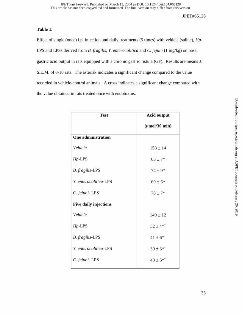
acid secretion. Table 1 shows the effects of vehicle, Hp-LPS and LPSs derived from
intestinal bacteria applied once or as 5 daily injections on gastric acid secretion in conscious
rats with chronic GF. The basal gastric acid output in rats treated with vehicle (saline)
reached the value of 158 ± 14 µmol/30 min. When Hp-LPS was applied once at a dose of 1
mg/kg i.p., the gastric acid output was significantly reduced compared to that obtained in
vehicle control animals (Table 1). At such a dose, Hp-LPS significantly reduced the volume
of gastric juice (2.0 ± 0.1 ml/30 min) and the gastric H+ concentration (32.5 ± 4 µmol/ml) as
compared with those in vehicle-control animals (volume of gastric juice, 2.8 ± 0.3 ml/30 min;
gastric H+ concentration, 56.4 ± 8 µmol/ml). In rats injected daily (5 times) with Hp-LPS (1
mg/kg i.p.), the gastric acid secretion was significantly more suppressed than after a single
application of LPS. In Hp-LPS injected five times, the volume of gastric juice (1.5 ± 0.4
ml/30 min) and the gastric H+ concentration (21.4 ± 2 µmol/ml) were significantly lower as
compared with those in animals once injected with Hp-LPS (volume of gastric juice, 2.0 ±
0.3 ml/30 min and the gastric H+ concentration, 32.5 ± 4 µmol/ml). For comparison, the
single parenteral application of LPSs derived from B. fragilis, Y. enterocolitica and C. jejuni
resulted in a similar decrease in the gastric acid outputs compared to that recorded in Hp-LPS
treated animals (Table 1). Five times daily injections of LPSs derived from B. fragilis, Y.
enterocolitica and C. jejuni caused significantly stronger reduction in the gastric acid outputs
than that observed with a single dose and with an extent similar to that obtained in rats
treated repeatedly with Hp-LPS. Administration of loxiglumide, a CCK1 receptor antagonist
or RPR-102681, a CCK2 receptor antagonist, failed to significantly influence basal gastric
This article has not been copyedited and formatted. The final version may differ from this version.JPET Fast Forward. Published on March 15, 2004 as DOI: 10.1124/jpet.104.065128
at ASPE
T Journals on February 26, 2019
jpet.aspetjournals.orgD
ownloaded from
JPET#65128
16
acid output as compared to that in vehicle-treated animals. Following the five daily injections
with Hp-LPS, a significant decrease in the gastric acid output was observed (158 ± 14
µmol/30 min in vehicle-control vs 32 ± 4 µmol/30 min in Hp-LPS treated). The reduction in
the acid output induced by Hp-LPS applied 5 times was not influenced significantly by
loxiglumide (gastric acid output, 32 ± 4 µmol/30 min in Hp-LPS treated vs 38 ± 5 µmol/30
min in loxiglumide plus Hp-LPS applied 5 times). Administration of RPR-102681 reversed
the attenuation of the gastric acid output induced by five daily injections of Hp-LPS (32 ± 4
µmol/30 min in Hp-LPS vs 114 ± 8 µmol/30 min in RPR-102681 plus Hp-LPS applied 5
times).
Effect of Hp-LPS and bacterial LPSs applied once or five times daily on ASA-
induced gastric lesions with accompanying alterations in GBF, plasma leptin and
gastrin levels. As shown in Fig. 1, the parenteral application of Hp-LPS (1 mg/kg i.p.)
produced negligible macroscopic injury in the stomach when applied once or injected daily 5
times, and failed to alter significantly the GBF when compared to that recorded in vehicle-
control rats. Similarly, single or repeated injections of LPSs from B. fragilis, Y.
enterocolitica and C. jejuni produced only small gastric mucosal lesions and failed to
influence GBF compared to vehicle treatment. In vehicle-pretreated rats, acidified ASA
resulted in typical multiple gastric lesions and a significant fall in the GBF by about 30%,
compared to the respective value recorded in animals pretreated with vehicle alone without
ASA (Fig. 1). Five daily injections with each endotoxin significantly reduced ASA-induced
gastric damage and accompanying fall in the GBF as compared with those in vehicle-
pretreated rats exposed to ASA. Representative gross macroscopic evidence of the ASA-
induced gastric damage and the reduction in these lesions in the animal stomach without and
This article has not been copyedited and formatted. The final version may differ from this version.JPET Fast Forward. Published on March 15, 2004 as DOI: 10.1124/jpet.104.065128
at ASPE
T Journals on February 26, 2019
jpet.aspetjournals.orgD
ownloaded from
JPET#65128
17
with the daily injections of Hp-LPS is presented in Fig. 2A-D. Compared with the intact
gastric mucosa, the exposure of gastric mucosa to ASA in the rat treated 5 times with vehicle,
resulted in a multiple gastric lesions localized mainly in the oxyntic mucosa (Fig. 2A and B).
In contrast, the repeated treatment with Hp-LPS alone produced only a few gross gastric
mucosal lesions (Fig. 2C). In rats injected daily 5 times with Hp-LPS, and then subsequently
exposed to ASA, there was a significant attenuation of the gastric mucosal injury as
compared to those treated 5 times with vehicle and then exposed to ASA (Fig. 2D).
Hp-LPS applied in a single dose of 1 mg/kg i.p. reduced significantly the ASA-
induced gastric lesions, and these protective effects were accompanied by a significant rise in
GBF and an elevation of plasma immunoreactive leptin and gastrin levels (Fig. 3). In intact
animals without ASA, the plasma gastrin concentration averaged 52 ± 5 pmol/l and plasma
leptin reached a value of 0.65 ± 0.04 ng/ml; these values remained relatively unchanged in
animals treated with vehicle (Fig. 3). In rats injected once with Hp-LPS, both plasma leptin
and gastrin concentrations showed a several fold increase, being significantly higher than
those in vehicle-treated animals. In rats injected daily with Hp-LPS, the plasma leptin and
gastrin concentrations gave a further significant rise compared with that recorded in vehicle-
treated animals or those exposed to single treatment with this endotoxin (Fig. 3). The ASA-
damage was significantly attenuated in the gastric mucosa of rats exposed to single or
repeated (5 times) injections of Hp-LPS, and these effects were accompanied by a significant
elevation of plasma gastrin and leptin increments (Fig. 3).
The single parenteral application of B. fragilis-LPS, Y. enterocolitica-LPS and C.
jejuni-LPS applied i.p. at a dose of 1 mg/kg, also resulted in the attenuation of ASA-induced
gastric damage and raised significantly the GBF (Table 2). As shown in Fig. 1, the repeated
parenteral application of these LPSs also reduced significantly the lesions induced by
This article has not been copyedited and formatted. The final version may differ from this version.JPET Fast Forward. Published on March 15, 2004 as DOI: 10.1124/jpet.104.065128
at ASPE
T Journals on February 26, 2019
jpet.aspetjournals.orgD
ownloaded from
JPET#65128
18
acidified ASA. The protective effects against ASA-induced gastric lesions of these
endotoxins injected repeatedly were accompanied by a significant rise in GBF, compared
with the respective values obtained in gastric mucosa exposed to ASA alone (Fig. 1).
Fig. 4 shows the results of parenteral administration of leptin, CCK-8, Hp-LPS and
8% peptone meal on the mean area of ASA-induced gastric lesions and the accompanying
changes in the GBF and plasma leptin levels. Exogenous leptin and CCK-8, both given in a
single dose of 10 µg/kg i.p., or i.g. application of 8% peptone meal, that increased the plasma
leptin levels by 2-3 folds and raised significantly GBF, resulted in a significant attenuation of
gastric lesions induced by ASA, with an extent similar to those achieved with single
parenteral injection of Hp-LPS.
Effect of pretreatment with loxiglumide and RPR-102681 and deactivation of
sensory nerves on the ASA-induced gastric lesions in rats. As shown in Fig. 5, the i.g.
application of acidified ASA produced similar gastric lesions and a similar fall in GBF to
those presented in Figs. 1 and 2. The area of these lesions and the accompanying fall in GBF
were significantly reduced in rats injected once with Hp-LPS or daily (5 times) with this
endotoxin. Suppression of CCK1 receptors with loxiglumide by itself failed to influence
significantly the ASA-induced gastric damage, and the accompanying fall in GBF.
Loxiglumide also failed to affect the reduction in the area of ASA-induced gastric damage
and the accompanying increase in GBF attained with Hp-LPS applied once or administered
repeatedly. Pretreatment with RPR-102681, to suppress specific CCK2 receptors, which by
itself also failed to influence the ASA-induced gastric damage, abolished almost completely
the decrease in the area of these lesions and the accompanying rise in the GBF evoked by
single or repeated treatment with this endotoxin.
This article has not been copyedited and formatted. The final version may differ from this version.JPET Fast Forward. Published on March 15, 2004 as DOI: 10.1124/jpet.104.065128
at ASPE
T Journals on February 26, 2019
jpet.aspetjournals.orgD
ownloaded from
JPET#65128
19
Hp-LPS applied once or injected daily (5 times) reduced significantly the area of
ASA-induced gastric lesions and raised significantly the GBF compared to those recorded in
rats treated with ASA without endotoxin administration (Fig. 6). Capsaicin-denervation
failed to enhance the area of ASA-induced gastric damage and failed to influence
significantly GBF, but resulted in almost complete elimination of the protective and
hyperemic effects induced by Hp-LPS injected once or daily (5 times) (Fig. 6).
Effect of single and repetitive treatment with Hp-LPS on plasma IL-1β and
TNF-α levels. As shown in Table 3, the plasma levels of both proinflammatory cytokines
(IL-1β and TNF-α) in the intact animals were negligible, but they were significantly
increased in Hp-LPS treated animals, and further dramatically raised in rats exposed to
acidified ASA that caused widespread acute gastric mucosal lesions. In rats injected once or
daily with Hp-LPS and later exposed to ASA, a significant decrease in plasma IL-1β and
TNF-α levels was recorded, although the levels in plasma of these cytokines reached
significantly higher values than those obtained in intact gastric mucosa.
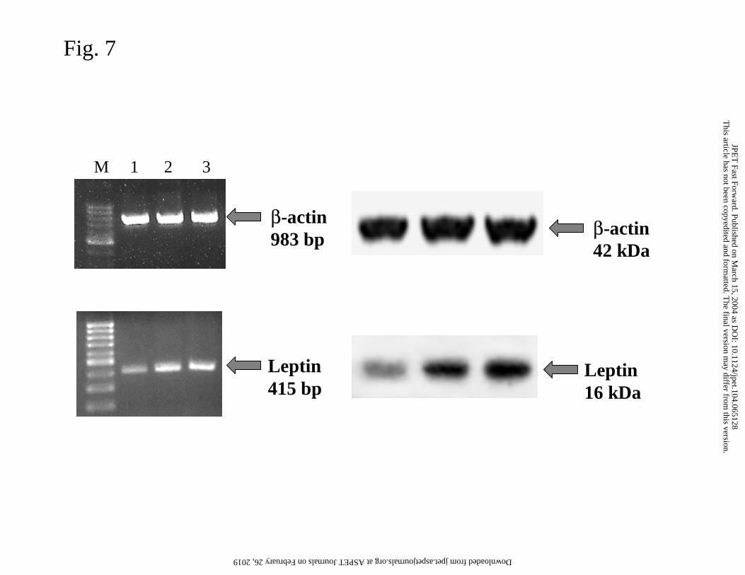
Determination of leptin mRNA and protein by RT-PCR and Western Blotting in
the gastric mucosa of rats treated once or repeatedly with Hp-LPS. The internal control
with the β-actin mRNA and protein showed intense signals in all the samples tested,
indicating a high integrity of RNA that was isolated from the gastric mucosa of vehicle-
control rats, as well as from those injected once or daily with Hp-LPS (Fig. 7, left and right
panels). Expression of leptin mRNA was detectable in intact mucosa not exposed to Hp-LPS
and in that treated with vehicle or Hp-LPS injected once or given daily (Fig. 7, left panel).
This article has not been copyedited and formatted. The final version may differ from this version.JPET Fast Forward. Published on March 15, 2004 as DOI: 10.1124/jpet.104.065128
at ASPE
T Journals on February 26, 2019
jpet.aspetjournals.orgD
ownloaded from
JPET#65128
20
No trace of leptin was recorded in rats serving as the negative control (saline) and this result
has been omitted for the sake of clarity. In rats injected daily with Hp-LPS, the strong signal
for leptin mRNA was greater than that in the vehicle-treated gastric mucosa. A weak signal
for leptin protein was detected in the vehicle-treated gastric mucosa (Fig. 7, right panel). In
contrast, an increased expression of leptin protein occured in gastric mucosa of rats treated
with Hp-LPS injected once or daily five times.
Discussion
The present study demonstrates that gastric mucosa can adapt in a relatively short
period to multiple parenteral administration of Hp-LPS and shows for the first time that this
adaptation enhances the mucosal resistance to subsequent acid-dependent gastric mucosal
lesions, induced by acidified ASA. It is noteworthy that repeated treatment with LPS derived
from other intestinal bacteria mimicked the protective action of Hp-LPS against ASA-
induced gastric damage, suggesting that adaptive efficacy of gastric mucosa to endotoxins of
different bacterial origin is not specifically related to Hp, and that it could contribute to
strengthening the mucosal integrity resulting in attenuation of the damage produced by acid-
dependent ulcerogens such as ASA. Furthermore, we found that both single and repeated
injections of Hp-LPS produced a marked rise in plasma hormones such as gastrin and leptin,
that have been proved to exert the gastroprotective and ulcer healing activity (Konturek et al.,
1995; Bado et al., 1998; Brzozowski et al., 2000b; Konturek et al., 2001a). The protective
effects of Hp-LPS were accompanied by a rise in plasma gastrin and were significantly
attenuated by pretreatment with RPR-102681, a highly specific inhibitor of mucosal gastrin
receptors (CCK2), but not influenced by loxiglumide, an antagonist of receptors for CCK
(CCK1), whose plasma level was not affected by Hp-LPS (unpublished observation). These
This article has not been copyedited and formatted. The final version may differ from this version.JPET Fast Forward. Published on March 15, 2004 as DOI: 10.1124/jpet.104.065128
at ASPE
T Journals on February 26, 2019
jpet.aspetjournals.orgD
ownloaded from
JPET#65128
21
results emphasize the importance of gastrin and CCK2, rather than CCK and CCK1, receptors
in the protective and adaptive response of Hp-LPS. Both the protection and adaptation to
Hp-LPS in ASA-injured mucosa were accompanied by upregulation of leptin at the level of
both mRNA and protein and subsequent release of leptin, indicating that this hormone,
indeed, contributes to Hp-LPS-induced attenuation of ASA-induced gastric damage. Also, in
rats with intact gastric mucosa injected with Hp-LPS, a marked increase in plasma leptin was
observed. Thus, this study shows for the first time, an increase in expression of leptin at the
levels of mRNA and protein with subsequent plasma release of this peptide occurs in rats
injected once or daily with Hp-LPS thereby emphasizing that leptin, which has been shown
previously to exert a protective effect against gastric mucosal injury by strong irritants (Bado
et al., 1998; Brzozowski et al., 1999), might contribute to the enhanced resistance of gastric
mucosa of rats treated with Hp-LPS against damage induced by acidified ASA. This notion
is in agreement with the original finding of Tepperman et al. (1994) who showed protection
of the rat gastric mucosa against ethanol lesions after parenteral administration of E.coli-LPS.
The present study is also consistent with the evidence of Ferraz et al. (1997), and our own
recent observation (Brzozowski et al., 2003), that animals treated repeatedly with E.coli- or
Hp-derived LPS developed mucosal tolerance to these endotoxins, and that this adaptive
response enhanced gastric mucosal resistance to ethanol-induced gastric damage. Our data
also agrees with the observations by Sugiyama et al. (2001) who demonstrated that the extent
of ethanol-induced damage of gastric mucosa was greatly limited in an experimental model of
Hp-infection in the stomach of Mongolian gerbils. These authors concluded that Hp-
infection, possibly due to release of endotoxin and the mild irritant effects of these cytotoxins,
exhibits an apparent paradoxical (protective) effect on gastric mucosal integrity by enhancing
the resistance of this mucosa to damage induced by necrotizing irritants (Sugiyama et al.,
This article has not been copyedited and formatted. The final version may differ from this version.JPET Fast Forward. Published on March 15, 2004 as DOI: 10.1124/jpet.104.065128
at ASPE
T Journals on February 26, 2019
jpet.aspetjournals.orgD
ownloaded from
JPET#65128
22
2001). This “protective” action of Hp infection against ethanol lesions has been attributed to
the increased generation of prostaglandin E2 derived from cyclooxygenase-2 (COX-2)
overexpression in Hp-infected stomach and has been confirmed recently in a model using
daily treatment with LPS (Konturek et al., 2001b).
It is known that LPS produces several neuroendocrine effects, and some of these
effects are believed to be mediated through cytokines and hormones, for instance leptin and
prolactin, and that this process involves the activation of peripheral autonomic nerves such as
efferent and afferent vagal nerves (Mastronardi et al., 2001). This prompted our study to
determine the role of leptin, neuropeptides released from sensory afferents, and gastric
hormones such as gastrin in the mechanism of enhancing the resistance of gastric mucosa to
ASA damage as induced by repetitive treatment with Hp-LPS. In a hamster model of Gram-
negative bacterial infection, systemic leptin was increased after prolonged administration of
LPS, and this was considered to enhance host response to endotoxemia (Grunfeld et al.,
1996). Turrin et al. (2001) have shown that LPS applied i.p. activated cytokine production in
the brain and at the periphery including in adipose tissue, liver and spleen. In another study,
LPS-induced leptin release was mediated through IL-1β because a soluble IL-1β receptor
antagonist completely blocked the LPS-induced increase in the leptin levels (Francis et al.,
1999). Thus, we can conclude that Hp-LPS induced protection and adaptation resulting in
limitation of ASA damage may depend upon leptin expression and release, and, as shown in
this study, could also be mediated by neuropeptides released from sensory afferent nerves.
The latter is supported by our present observation that the capsaicin-induced functional
ablation of sensory nerves abolished the protective and hyperemic effects of single and
repeated administration of Hp-LPS. It is suggested that endotoxins such as Hp-LPS can
affect sensory afferent nerves that in turn may activate the brain-gut axis resulting in
This article has not been copyedited and formatted. The final version may differ from this version.JPET Fast Forward. Published on March 15, 2004 as DOI: 10.1124/jpet.104.065128
at ASPE
T Journals on February 26, 2019
jpet.aspetjournals.orgD
ownloaded from
JPET#65128
23
limitation of ASA-induced gastric damage. This conclusion agrees with the observation by
Hua et al. (1996) that endotoxin treatment enhanced release of vasoactive CGRP from the
primary sensory afferents due to sensitizing their terminals. The mechanism by which
enhancement in the plasma IL-1β and TNF-α induced by ASA was reduced in rats treated
repeatedly with Hp-derived endotoxin remains to be elucidated, but it could be due to the
suppressive action on these cytokines of prostaglandins, nitric oxide (NO) and heat shock
proteins (HSP) released via overexpression of COX-2, inducible NO synthase (iNOS) and
HSP70 mRNA as reported recently (Brzozowski et al., 2003).
The major finding of the present study is that Hp-LPS is capable of inhibiting gastric
acid secretion, while showing a significant rise in plasma gastrin level. The importance of
gastrin in the observed protection and hyperemia appears to be of particular significance as
the antagonism of receptors for gastrin (CCK2) with RPR-102681 completely reversed the
inhibition of gastric secretion, gastroprotection and adaptation of gastric mucosa, and the rise
in GBF afforded by Hp-LPS, whereas the blockade of CCK1 receptors by loxiglumide
eliminated the action of CCK, was ineffective.
It is now becoming evident that non-steroidal anti-inflammatory drugs (NSAIDs) such
as ASA may influence the pathogenic effects of Hp as a result of possible direct interaction
with this microorganism (Wang et al., 2003). However, this was not the case in our study,
because the single or repeated parenteral injections with Hp-derived endotoxin actually
increased mucosal resistance to the damaging effect of ASA in animals treated repeatedly
with this endotoxin. The major drawback of this study is that only parenteral administration
of LPS but not its intragastric administration was employed. Although our previous studies
documented that LPS administered intragastrically in a relatively small dose (1 mg/kg) failed
to influence the lesions provoked by acidified ASA, our preliminary observations with a large
This article has not been copyedited and formatted. The final version may differ from this version.JPET Fast Forward. Published on March 15, 2004 as DOI: 10.1124/jpet.104.065128
at ASPE
T Journals on February 26, 2019
jpet.aspetjournals.orgD
ownloaded from
JPET#65128
24
intragastric dose of LPS (10 mg/kg i.g.) was found effective. This difference required further
explanation but it could be that small Hp-LPS doses given into the stomach lumen may not be
able to penetrate the thick mucus covering of the surface epithelium to activate the mucosa
protective mechanism. However, when Hp remains under the mucus layer and is in direct
contact with the epithelial cells, it could activate the protective mechanism by local release of
its endotoxins such as LPS. Further studies are needed to determine whether Hp-LPS present
in the gastric lumen can reach the gastric circulation sufficiently to mimic the changes
observed after repeated parenteral Hp-LPS injections.
Our results with antisecretory effects of Hp-LPS and other non-Hp related LPSs agree
with those of Uehara et al. (1990) and Konturek et al. (2001) that peripheral and central
applications of LPS derived from E.coli to rats produces profound dose-dependent inhibition
of gastric acid output. Since ASA damage depends upon gastric acidity it is reasonable to
assume that suppression of gastric acid secretion by this endotoxin could contribute to the
limitation of this damage. As shown in the present study, a marked increase in plasma gastrin
in rats treated repeatedly with Hp-LPS, which is known to exert gastroprotective influence on
the gastric mucosa, could result from hypochlorhydria and could contribute to the greater
tolerance of this mucosa to ASA-induced gastric damage. This was confirmed in the present
study by blocking the receptors for gastrin (CCK2) which resulted in attenuation of the
protective effects of Hp-LPS and the related rise in the plasma gastrin levels.
This article has not been copyedited and formatted. The final version may differ from this version.JPET Fast Forward. Published on March 15, 2004 as DOI: 10.1124/jpet.104.065128
at ASPE
T Journals on February 26, 2019
jpet.aspetjournals.orgD
ownloaded from
JPET#65128
25
References
Bado A, Levasseur S, Attoub S, Kermorgant S, Laigneau JP, Bortoluzzi MN, Moizo L, Lehy
T, Guerre-Millo M, Le Marchand-Brustel Y, Lewin MJ (1998) The stomach is a source
of leptin. Nature 394(6695):790-793.
Barbier M, Cherbut C, Aube AC, Blottiere HM, Galmiche JP (1998) Elevated plasma leptin
concentrations in early stages of experimental intestinal inflammation in rats. Gut
43:783-790.
Bohme GA, Bertrand P, Piot O (1997) Pharmacological properties of RPR 102681, a novel
CCKB antagonist. Soc Neurosci 23:2409.
Brzozowski T, Konturek PC, Konturek SJ, Drozdowicz D, Kwiecień S, Pajdo R, Bielanski W,
Hahn EG (2000a) Role of gastric acid secretion in progression of acute gastric erosions
induced by ischemia-reperfusion into gastric ulcers. Eur J Pharmacol 398:147-158.
Brzozowski T, Konturek PC, Konturek SJ, Ernst H, Stachura J, Hahn EG (1995) Gastric
adaptation to injury by repeated doses of aspirin strengthens mucosal defense against
subsequent exposure to various strong irritants in rats. Gut 37:749-757.
Brzozowski T, Konturek PC, Konturek SJ, Pajdo R, Duda A, Pierzchalski P, Bielanski W,
Hahn EG (1999) Leptin in gastroprotection induced by cholecystokinin or by a meal.
Role of vagal and sensory nerves and nitric oxide. Eur J Pharmacol 374:263-276.
Brzozowski T, Konturek PC, Konturek SJ, Pierzchalski P, Bielanski W, Pajdo R, Drozdowicz
D, Kwiecień S, Hahn EG (2000b) Central leptin and cholecystokinin in gastroprotection
against ethanol-induced damage. Digestion 62:126-142.
Brzozowski T, Konturek PC, Moran AP, Kwiecień S, Pajdo R, Konturek SJ, Drozdowicz D,
Ptak A, Pawlik WW, Hahn EG (2003) Enhanced resistance of gastric mucosa to
This article has not been copyedited and formatted. The final version may differ from this version.JPET Fast Forward. Published on March 15, 2004 as DOI: 10.1124/jpet.104.065128
at ASPE
T Journals on February 26, 2019
jpet.aspetjournals.orgD
ownloaded from
JPET#65128
26
damaging agents in the rat stomach adapted to Helicobacter pylori lipopolysaccharide.
Digestion 67:195-208.
Brzozowski T, Konturek SJ, Sliwowski Z, Pytko-Polonczyk J, Szlachcic A, Drozdowicz D
(1996) Role of capsaicin sensitive afferent nerves in gastroprotection against acid-
independent and acid-dependent ulcerogens. Digestion 57:424-432.
Crabtree JE (1996) Gastric mucosal inflammatory responses to Helicobacter pylori. Aliment
Pharmacol Ther 10(suppl 1):29-37.
Crabtree JE, Perry S, Moran A, Peichl P, Tompkins DS, Lindley IJD (1994) Neutrophil IL-8
secretion induced by Helicobacter pylori. Am J Gastroenterol 89:1337.
Faggioni R, Moser A, Feingold KR, Grunfeld C (2000) Reduced leptin levels in starvation
increase susceptibility to endotoxic shock. Am J Pathol 156:1781-1787.
Ferraz JG, Sharkey KA, Reuter BK, Asfaha S, Tigley AW, Brown ML, McKnight W, Wallace
JL (1997) Induction of cyclooxygenase 1 and 2 in the rat stomach during endotoxemia.
Role in resistance to damage. Gastroenterology 113:195-204.
Figura N, Tabaqchali S (1996) Bacterial pathogenic factors. Curr Opin Gastroenterol 12
(suppl):11-15.
Francis J, Mohan-Kumar PS, Mohan-Kumar SM, Quadri SK (1999) Systemic administration
of lipopolysaccharide increases plasma leptin levels: blockade by soluble interleukin-1
receptor. Endocrine 10:291-215.
Friedman JM, Halaas JL (1998) Leptin and the regulation of body weight in mammals.
Nature 395:763-770.
This article has not been copyedited and formatted. The final version may differ from this version.JPET Fast Forward. Published on March 15, 2004 as DOI: 10.1124/jpet.104.065128
at ASPE
T Journals on February 26, 2019
jpet.aspetjournals.orgD
ownloaded from
JPET#65128
27
Grunfeld C, Zhao C, Fuller J, Pollack A, Moser A, Friedman J, Feingold KR (1996)
Endotoxin and cytokines induce expression of leptin, the ob gene product, in hamsters. J
Clin Invest 97:2152-2157.
Hua XY, Chen P, Fox A, Myers RR (1996) Involvement of cytokines in lipopolysaccharide-
induced facilitation of CGRP release from capsaicin-sensitive nerves in the trachea:
studies with interleukin-1β and tumor necrosis factor-α. J Neurosci 16:4742-4748.
Konturek PC, Bielanski W, Konturek SJ, Hahn EG (1999) Helicobacter pylori associated
gastric pathology. J Physiol Pharmacol 50:695-710.
Konturek PC, Brzozowski T, Konturek SJ, Kwiecien S, Dembinski A, Hahn EG (2001a)
Influence of bacterial lipopolysaccharide on healing of chronic experimental ulcer in rat.
Scand J Gastroenterol 36:1239-1247.
Konturek PC, Brzozowski T, Konturek SJ, Taut A, Kwiecien S, Pajdo R, Sliwowski Z, Hahn
EG (1998a) Bacterial lipopolysaccharide protects gastric mucosa against acute injury in
rats by activation of genes for cyclooxygenases and endogenous prostaglandins.
Digestion 59:284-297.
Konturek PC, Brzozowski T, Meixner H, Ptak A, Hahn EG, Konturek SJ (2001b) Central
and peripheral neural aspects of gastroprotective and ulcer healing effects of
lipopolysaccarides. J Physiol Pharmacol 52:611-623.
Konturek PC, Brzozowski T, Sliwowski Z, Pajdo R, Stachura J, Hahn EG, Konturek SJ
(1998b) Involvement of nitric oxide and prostaglandins in gastroprotection induced by
bacterial lipopolysaccharide. Scand J Gastroenterol 33:691-700.
This article has not been copyedited and formatted. The final version may differ from this version.JPET Fast Forward. Published on March 15, 2004 as DOI: 10.1124/jpet.104.065128
at ASPE
T Journals on February 26, 2019
jpet.aspetjournals.orgD
ownloaded from
JPET#65128
28
Konturek SJ, Brzozowski T, Pytko-Polonczyk J, Drozdowicz D (1995) Comparison of
cholecystokinin, pentagastrin and duodenal oleate in gastroprotection in rats. Scand J
Gastroenterol 30:620-630.
Konturek SJ, Brzozowski T, Stachura J, Dembinski A, Majka J (1994) Role of gastric blood
flow, neutrophil infiltration and mucosal cell proliferation in gastric adaptation to aspirin
in the rat. Gut 35:1189-1196.
Mastronardi CA, Yu WH, Srivastava VK, Dees WL, McCann SM (2001)
Lipopolysaccharide-induced leptin release is neurally controlled. Proc Natl Acad Sci
USA 98:14720-14725.
Megraud F, Neman SV, Brugmann D (1992) Further evidence of toxic effects of ammonia
produced by Helicobacter pylori in human epithelial cells. Infect Immun 60:1859-1862.
Moran AP (2001a) Helicobacter pylori lipopolysaccharides, in Helicobacter pylori:
Molecular and Cellular Biology (Achtman M & Suerbaum S eds) pp 201-726, Horizon
Scientific Press, Wymondham, England.
Moran AP (2001b) Molecular structure, biosynthesis and pathogenic roles of Helicobacter
pylori lipopolysaccharides, in Helicobacter pylori: Physiology and Genetics (Mobley H,
Mendz G, Hazell S eds), pp 81-95, American Society for Microbiology, Washington,
D.C.
Moran AP, Helander IM, Kosunen TU (1992) Compositional analysis of Helicobacter pylori
rough-form lipopolysaccharides. J Bacteriol 174:1370-1377.
Ng CJ, Chen JC, Chiu DF, Chen MF, Chen HM (2002) Role of prostacyclin on
microcirculation in endotoxin-induced gastroprotection in rats: a microdialysis study.
Shock 17:334-338.
This article has not been copyedited and formatted. The final version may differ from this version.JPET Fast Forward. Published on March 15, 2004 as DOI: 10.1124/jpet.104.065128
at ASPE
T Journals on February 26, 2019
jpet.aspetjournals.orgD
ownloaded from
JPET#65128
29
Shalev A, Vosmeer S, Keller U (1997) Absence of short-term effects of glucagon-like
peptide-1 and hyperglycemia on plasma leptin levels in man. Metabolism 46:723-725.
Slomiany BL, Liau YH, Lopez RA, Piotrowski J, Czajkowski A, Slomiany A (1992) Effect
of Helicobacter pylori on the synthesis of sulfated gastric mucin. Biochem Int 27:687-
697.
Sugiyama A, Ikeno T, Ishida K, Maruta F, Murakami M, Sato T, Saito H, Ishizone S,
Kawasaki S, Ota H, Katsuyama T (2001) Paradoxical role of Helicobacter infection.
Protective effect against ethanol-induced gastric mucosal injury in Mongolian gerbils.
Dig Dis Sci 46:2433-2439.
Tepperman BL, Soper BD (1994) Nitric oxide synthase induction and cytoprotection of rat
gastric mucosa from injury by ethanol. Can J Physiol Pharmacol 72:1308-1312.
Turrin NP, Gayle D, Ilyin SE, Flynn MC, Langhans W, Schwartz GJ, Plata-Salaman CR
(2001) Pro-inflammatory and anti-inflammatory cytokine mRNA induction in the
periphery and brain following intraperitoneal administration of bacterial
llipopolysaccharide. Brain Res Bull 54:443-453.
Uehara A, Okumura T, Okamura K, Takasugi Y, Namiki M (1990) Lipopolysaccharide-
induced inhibition of gastric acid and pepsin secretion in rats. Eur J Pharmacol
181:141-145.
Valkonen KH, Wadström T, Moran AP (1994) Interaction of lipopolysaccharides of
Helicobacter pylori with basement membrane protein laminin. Infect Immun 62:3640-
3648.
This article has not been copyedited and formatted. The final version may differ from this version.JPET Fast Forward. Published on March 15, 2004 as DOI: 10.1124/jpet.104.065128
at ASPE
T Journals on February 26, 2019
jpet.aspetjournals.orgD
ownloaded from
JPET#65128
30
Wang WH, Wong WM, Dailidiene D, Berg DE, Gu Q, Lai KC, Lam SK, Wong BC (2003)
Aspirin inhibits the growth of Helicobacter pylori and enhances its suceptibility to
antimicrobial agents. Gut 52:490-5.
Warren JR, Marshall BJ (1983) Unidentified curved bacilli on gastric epithelium in active
chronic gastritis. Lancet 1:1273.
This article has not been copyedited and formatted. The final version may differ from this version.JPET Fast Forward. Published on March 15, 2004 as DOI: 10.1124/jpet.104.065128
at ASPE
T Journals on February 26, 2019
jpet.aspetjournals.orgD
ownloaded from
JPET#65128
31
Legends for Figures
Fig. 1. Effect of single (once) and repeated (5 times) administrations of Hp-LPS and LPS of
intestinal bacteria such as C. jejuni, Y. enterocolitica and B. fragilis given intraperitoneally
(i.p.) at a dose of 1 mg/kg on the area of ASA-induced gastric lesions and the alterations in
gastric blood flow (GBF). Data represents the mean ± SEM of 8-10 rats. The asterisk
indicates a significant change compared with the value obtained with rats treated with vehicle
and various LPSs. The cross indicates a significant change compared with the value obtained
in ASA-treated rats.
Fig. 2A,B,C,D. Representative photomicrographs showing the gross appearance of intact rat
gastric mucosa (A), the gastric mucosa exposed to acidified aspirin (ASA; 150 mg/kg in 0.2
N HCl i.g.) (B) or treated 5 times with Hp-LPS (1 mg/kg i.p.) (C) or that treated repeatedly (5
times) with Hp-LPS and then exposed to ASA (D). Please note: ASA produced gross gastric
mucosal lesions localized predominantly in the oxyntic mucosa (arrows) as compared with
intact stomach (B vs. A). Repeated treatment with Hp-LPS, which by itself induced only few
mucosal lesions (C), produced a marked attenuation of the ASA-induced gastric injury (D vs.
B).
Fig. 3. Effect of single (once) and repeated (5 times) administrations of Hp-LPS given
intraperitoneally (i.p.) on the area of gastric lesions induced by acidified aspirin (ASA; 150
mg/kg i.g.) and alterations in plasma gastrin and leptin levels. Data represents the mean ±
SEM of 8-10 rats. The asterisk indicates significant change compared with the value
obtained with vehicle (saline) control. The asterisk and cross indicate a significant change
compared to the respective values in animals treated with Hp-LPS once.
Fig. 4. Mean area of ASA-induced gastric lesions and accompanying changes in gastric
blood flow (GBF) and leptin levels in plasma of rats treated with vehicle (saline), exogenous
This article has not been copyedited and formatted. The final version may differ from this version.JPET Fast Forward. Published on March 15, 2004 as DOI: 10.1124/jpet.104.065128
at ASPE
T Journals on February 26, 2019
jpet.aspetjournals.orgD
ownloaded from
JPET#65128
32
leptin and CCK-8 applied i.p. at a dose of 10 µg/kg, 8% peptone meal or with Hp-LPS (1
mg/kg i.p.) Data represents the mean ± SEM of 8-10 rats. The asterisk indicates a
significant change compared with the value obtained with vehicle (control).
Fig. 5. Mean area of ASA-induced gastric lesions and gastric blood flow (GBF) in rats
treated with vehicle or Hp-LPS (1 mg/kg i.p.) applied once or administered 5 times without or
with pretreatment with loxiglumide (30 mg/kg i.p.) or RPR 102681 (10 mg/kg i.p.). Data
represents the mean ± SEM of 8-10 rats. The asterisk indicates a significant change
compared with the value obtained in control (vehicle) rats. The asterisk and cross indicates a
significant change compared to the values obtained with Hp-LPS applied once. A single
cross indicates a significant change compared with the values obtained with Hp-LPS applied
once or given 5 times.
Fig. 6. Mean area of ASA-induced gastric lesions and gastric blood flow (GBF) in rats
treated with vehicle or Hp-LPS (1 mg/kg i.p.) applied once or administered 5 times without or
with the pretreatment with capsaicin to induce functional ablation of sensory nerves. Data
represents the mean ± SEM of 8-10 rats. The asterisk indicates a significant change
compared to the value obtained with vehicle (control). The asterisk and cross indicates a
significant change compared to the value obtained with Hp-LPS applied once. A single cross
indicates a significant change compared to the values obtained with Hp-LPS applied once or
given 5 times.
Fig. 7. Expression of leptin and β-actin mRNA and protein determined by RT-PCR (left
panel) and Western Blotting (right panel) in the gastric mucosa of rats injected with vehicle
(line 1), Hp-LPS (1 mg/kg i.p.) injected once (line 2) and Hp-LPS injected 5 times (line 3).
M= DNA marker (Gibco 100 bp ladder).
This article has not been copyedited and formatted. The final version may differ from this version.JPET Fast Forward. Published on March 15, 2004 as DOI: 10.1124/jpet.104.065128
at ASPE
T Journals on February 26, 2019
jpet.aspetjournals.orgD
ownloaded from
JPET#65128
33
Table 1.
Effect of single (once) i.p. injection and daily treatments (5 times) with vehicle (saline), Hp-
LPS and LPSs derived from B. fragilis, Y. enterocolitica and C. jejuni (1 mg/kg) on basal
gastric acid output in rats equipped with a chronic gastric fistula (GF). Results are means ±
S.E.M. of 8-10 rats. The asterisk indicates a significant change compared to the value
recorded in vehicle-control animals. A cross indicates a significant change compared with
the value obtained in rats treated once with endotoxins.
Test Acid output
(µmol/30 min)
One administration
Vehicle 158 ± 14
Hp-LPS 65 ± 7*
B. fragilis-LPS 74 ± 9*
Y. enterocolitica-LPS 69 ± 6*
C. jejuni- LPS 78 ± 7*
Five daily injections
Vehicle 149 ± 12
Hp-LPS 32 ± 4*+
B. fragilis-LPS 41 ± 6*+
Y. enterocolitica-LPS 39 ± 3*+
C. jejuni- LPS 48 ± 5*+
This article has not been copyedited and formatted. The final version may differ from this version.JPET Fast Forward. Published on March 15, 2004 as DOI: 10.1124/jpet.104.065128
at ASPE
T Journals on February 26, 2019
jpet.aspetjournals.orgD
ownloaded from
JPET#65128
34
This article has not been copyedited and formatted. The final version may differ from this version.JPET Fast Forward. Published on March 15, 2004 as DOI: 10.1124/jpet.104.065128
at ASPE
T Journals on February 26, 2019
jpet.aspetjournals.orgD
ownloaded from
JPET#65128
35
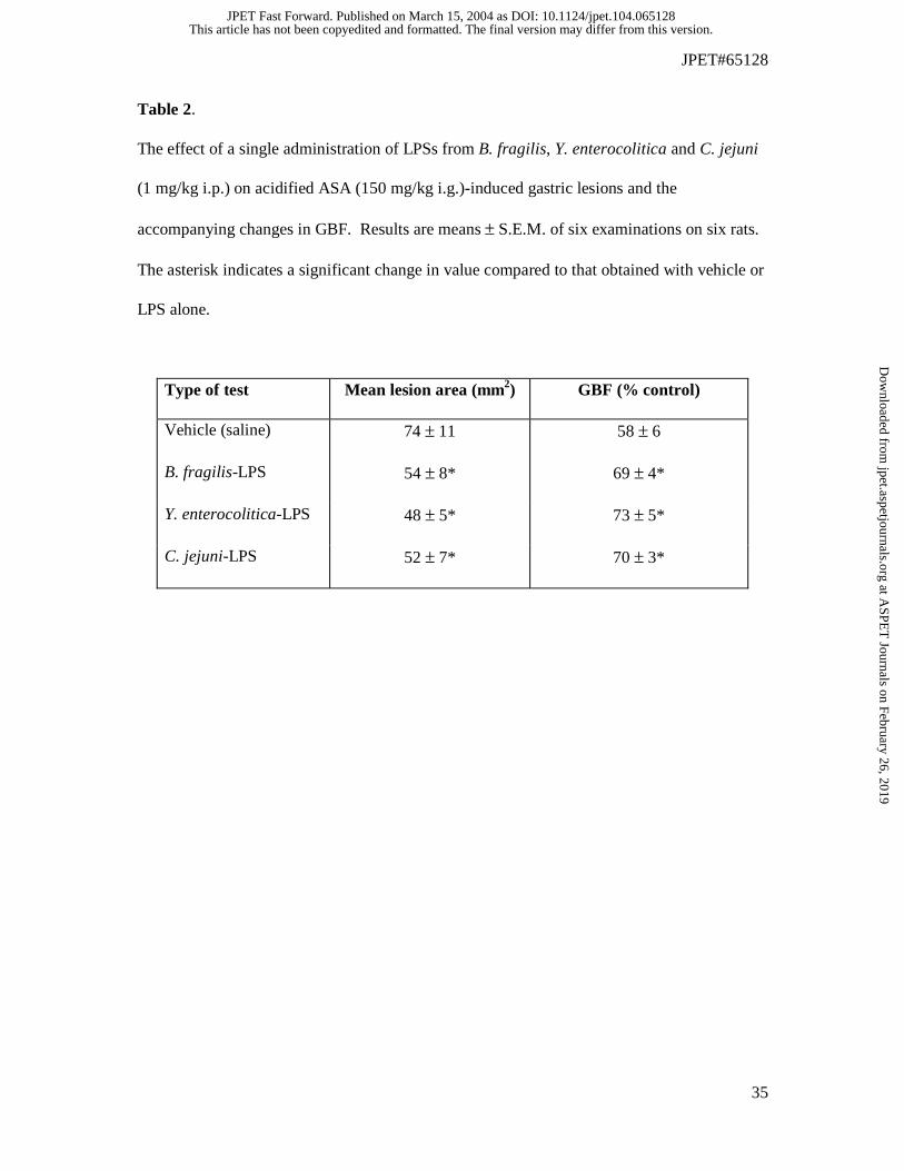
Table 2.
The effect of a single administration of LPSs from B. fragilis, Y. enterocolitica and C. jejuni
(1 mg/kg i.p.) on acidified ASA (150 mg/kg i.g.)-induced gastric lesions and the
accompanying changes in GBF. Results are means ± S.E.M. of six examinations on six rats.
The asterisk indicates a significant change in value compared to that obtained with vehicle or
LPS alone.
Type of test Mean lesion area (mm2) GBF (% control)
Vehicle (saline) 74 ± 11 58 ± 6
B. fragilis-LPS 54 ± 8* 69 ± 4*
Y. enterocolitica-LPS 48 ± 5* 73 ± 5*
C. jejuni-LPS 52 ± 7* 70 ± 3*
This article has not been copyedited and formatted. The final version may differ from this version.JPET Fast Forward. Published on March 15, 2004 as DOI: 10.1124/jpet.104.065128
at ASPE
T Journals on February 26, 2019
jpet.aspetjournals.orgD
ownloaded from
JPET#65128
36
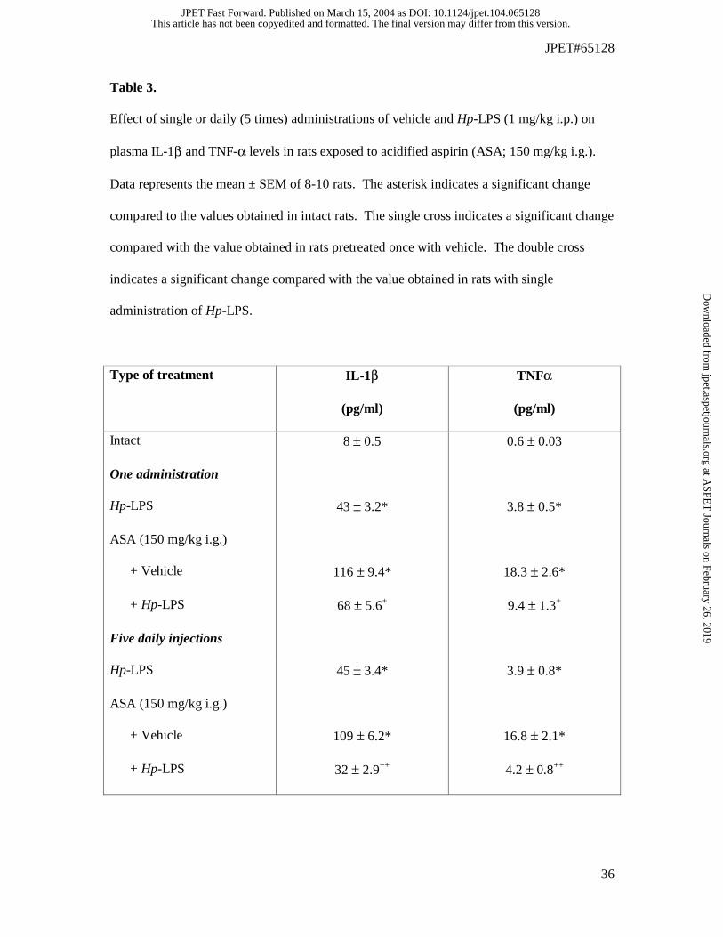
Table 3.
Effect of single or daily (5 times) administrations of vehicle and Hp-LPS (1 mg/kg i.p.) on
plasma IL-1β and TNF-α levels in rats exposed to acidified aspirin (ASA; 150 mg/kg i.g.).
Data represents the mean ± SEM of 8-10 rats. The asterisk indicates a significant change
compared to the values obtained in intact rats. The single cross indicates a significant change
compared with the value obtained in rats pretreated once with vehicle. The double cross
indicates a significant change compared with the value obtained in rats with single
administration of Hp-LPS.
Type of treatment IL-1β
(pg/ml)
TNFα
(pg/ml)
Intact 8 ± 0.5 0.6 ± 0.03
One administration
Hp-LPS 43 ± 3.2* 3.8 ± 0.5*
ASA (150 mg/kg i.g.)
+ Vehicle 116 ± 9.4* 18.3 ± 2.6*
+ Hp-LPS 68 ± 5.6+ 9.4 ± 1.3+
Five daily injections
Hp-LPS 45 ± 3.4* 3.9 ± 0.8*
ASA (150 mg/kg i.g.)
+ Vehicle 109 ± 6.2* 16.8 ± 2.1*
+ Hp-LPS 32 ± 2.9++ 4.2 ± 0.8++
This article has not been copyedited and formatted. The final version may differ from this version.JPET Fast Forward. Published on March 15, 2004 as DOI: 10.1124/jpet.104.065128
at ASPE
T Journals on February 26, 2019
jpet.aspetjournals.orgD
ownloaded from

JPET#65128
37
This article has not been copyedited and formatted. The final version may differ from this version.JPET Fast Forward. Published on March 15, 2004 as DOI: 10.1124/jpet.104.065128
at ASPE
T Journals on February 26, 2019
jpet.aspetjournals.orgD
ownloaded from
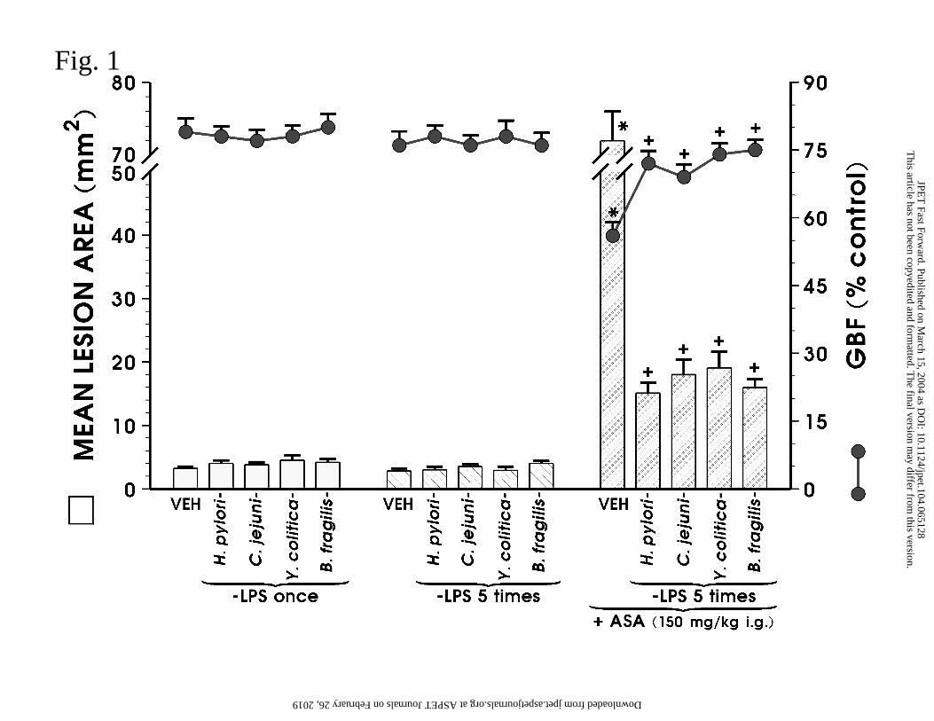
Fig. 1
This article has not been copyedited and form
atted. The final version m
ay differ from this version.
JPET
Fast Forward. Published on M
arch 15, 2004 as DO
I: 10.1124/jpet.104.065128 at ASPET Journals on February 26, 2019 jpet.aspetjournals.org Downloaded from
Fig. 2 A, B, C, D
This article has not been copyedited and form
atted. The final version m
ay differ from this version.
JPET
Fast Forward. Published on M
arch 15, 2004 as DO
I: 10.1124/jpet.104.065128 at ASPET Journals on February 26, 2019 jpet.aspetjournals.org Downloaded from
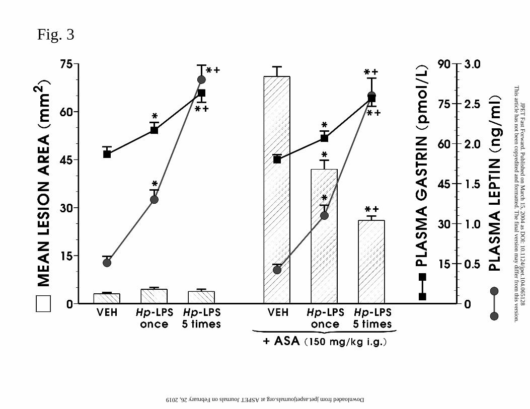
Fig. 3
This article has not been copyedited and form
atted. The final version m
ay differ from this version.
JPET
Fast Forward. Published on M
arch 15, 2004 as DO
I: 10.1124/jpet.104.065128 at ASPET Journals on February 26, 2019 jpet.aspetjournals.org Downloaded from
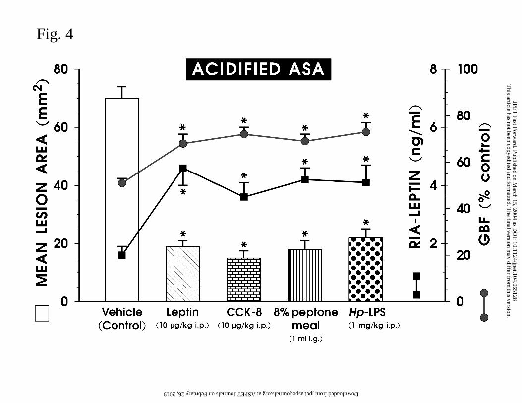
Fig. 4
This article has not been copyedited and form
atted. The final version m
ay differ from this version.
JPET
Fast Forward. Published on M
arch 15, 2004 as DO
I: 10.1124/jpet.104.065128 at ASPET Journals on February 26, 2019 jpet.aspetjournals.org Downloaded from
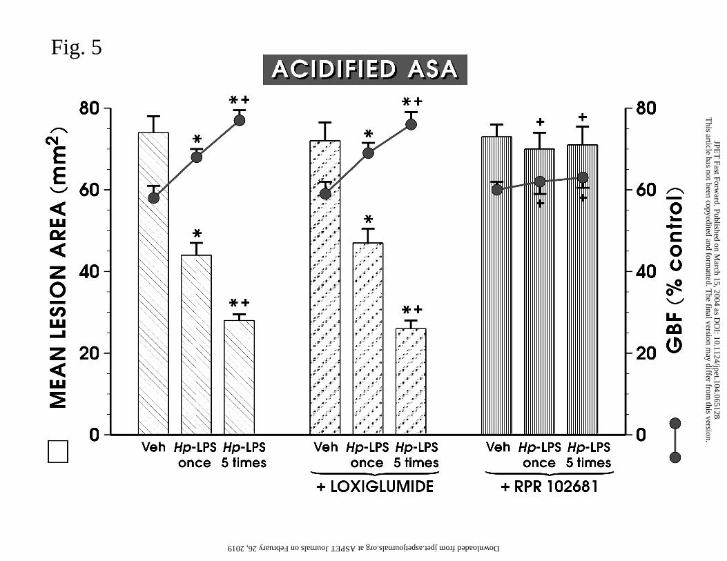
Fig. 5
This article has not been copyedited and form
atted. The final version m
ay differ from this version.
JPET
Fast Forward. Published on M
arch 15, 2004 as DO
I: 10.1124/jpet.104.065128 at ASPET Journals on February 26, 2019 jpet.aspetjournals.org Downloaded from
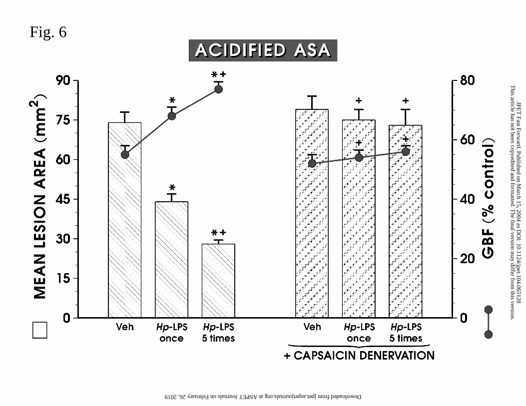
Fig. 6T
his article has not been copyedited and formatted. T
he final version may differ from
this version.JPE
T Fast Forw
ard. Published on March 15, 2004 as D
OI: 10.1124/jpet.104.065128
at ASPET Journals on February 26, 2019 jpet.aspetjournals.org Downloaded from
M 1 2 3
Leptin415 bp
β-actin983 bp
Leptin16 kDa
β-actin42 kDa
Fig. 7
This article has not been copyedited and form
atted. The final version m
ay differ from this version.
JPET
Fast Forward. Published on M
arch 15, 2004 as DO
I: 10.1124/jpet.104.065128 at ASPET Journals on February 26, 2019 jpet.aspetjournals.org Downloaded from


![Capsaicin. Scoville heat unitsExamples 15,000,000– 16,000,000 Pure capsaicin [9]capsaicin [9] 8,600,000–9,100,000 Various capsaicinoids (e.g., homocapsaicin,](https://img.pdfslide.us/doc/110x75/56649ec95503460f94bd6e61/capsaicin-scoville-heat-unitsexamples-15000000-16000000-pure-capsaicin.jpg)