Embed Size (px)
Citation preview

Upregulation of RGS4 expression in a model of neuropathic pain
and insensitivity to morphine
Martine Garnier1*, Paola F. Zaratin1, Giovanna Ficalora1, Maurizio Valente1, Laura
Fontanella1, Man-Hee Rhee2, Kendall J. Blumer2 and Mark A. Scheideler1*
1GlaxoSmithKline Pharmaceuticals, Department of Neurobiology Research, Via Zambeletti,
20021 Baranzate di Bollate, Milan, Italy and Neurology Centre of Excellence for Drug
Discovery, New Frontiers Science Park, Harlow, Essex, United Kingdom CM19 5AW.
2Department of Cell Biology and Physiology, Washington University School of Medicine,
Box 8228, 660 St Euclid Avenue, St Louis, MO, 63110, U.S.A.
Copyright 2002 by the American Society for Pharmacology and Experimental Therapeutics.
JPET Fast Forward. Published on November 25, 2002 as DOI:10.1124/jpet.102.043471This article has not been copyedited and formatted. The final version may differ from this version.JPET Fast Forward. Published on November 25, 2002 as DOI: 10.1124/jpet.102.043471
at ASPE
T Journals on January 4, 2020
jpet.aspetjournals.orgD
ownloaded from

2
Running title: Role of RGS4 in pain and morphine insensitivity
*Corresponding authors:
Mark Scheideler, Ph.D.
MDS Proteomics, Stærmosegaardsvej 6, 5230 Odense, Denmark.
Tel: +45-6557.2056, Fax: +45-6557.2001
Email: [email protected]
Martine Garnier, Ph.D.
GlaxoSmithKline, Psychiatry Centre of Excellence for Drug Discovery,
Via Fleming 4, 37100 Verona, Italy
Tel: +39-045.921.8835, Fax: +39-045.921.8072
E-mail: [email protected]
This article has not been copyedited and formatted. The final version may differ from this version.JPET Fast Forward. Published on November 25, 2002 as DOI: 10.1124/jpet.102.043471
at ASPE
T Journals on January 4, 2020
jpet.aspetjournals.orgD
ownloaded from
3
Number of:
-text pages: 34
-tables: 0
-figures: 6
-references: 37
Number of Words:
-Abstract: 195
-Introduction: 604
-Discussion: 1273
Non-Standard abbreviations: RGS, regulators of G protein signalling; MOR, mu-opioid
receptor: G-protein, GTP binding protein; FSK, forskolin.
Recommended Section assignment: Cellular & Molecular
This article has not been copyedited and formatted. The final version may differ from this version.JPET Fast Forward. Published on November 25, 2002 as DOI: 10.1124/jpet.102.043471
at ASPE
T Journals on January 4, 2020
jpet.aspetjournals.orgD
ownloaded from

4
ABSTRACT
We hypothesized that the upregulated expression of one or more members of the RGS
(regulator of G-protein signalling) family can cause an attenuation of signalling via Gi/Go-
coupled opioid receptors, and thereby play a role in the development of hyperalgesia and
accompanying insensitivity to morphine observed in animal models of neuropathic pain.
Accordingly we examined the mRNA expression of several RGS genes in a rat model of
chronic neuropathic pain induced by partial ligation of the sciatic nerve. During the
development of hyperalgesia, RGS4 was the only isoform examined whose mRNA levels
increased significantly (up to 230%) in the lumbar spinal cord. In situ hybridization studies
confirmed that RGS4 is present in the dorsal horn of the spinal cord where mu-opioid
receptors (MOR) are also expressed. Overexpression of RGS4 in HEK-293 cells stably
expressing MOR predictably attenuated opioid agonist-induced inhibition of adenylyl cyclase.
This inhibitory effect was overcome partially at high agonist concentrations, supporting the
view that morphine insensitivity is promoted by RGS4 overexpression. These studies provide
evidence that the upregulation of RGS4 expression may contribute to changes in pain signal
processing which lead to the development of hyperalgesia, and further effect its modulation
by morphine.
This article has not been copyedited and formatted. The final version may differ from this version.JPET Fast Forward. Published on November 25, 2002 as DOI: 10.1124/jpet.102.043471
at ASPE
T Journals on January 4, 2020
jpet.aspetjournals.orgD
ownloaded from

5
INTRODUCTION
Activation of the mu-opioid receptor (MOR) system leads to anti-nociception under
most pain conditions, and a major site of opioid action is the dorsal horn of the spinal cord,
which receives inputs from primary afferent fibers and integrates the multiple ascending and
descending pathways that contribute to pain modulation (Ossipov et al., 1995). However,
several studies have suggested that morphine is less effective at treating chronic neuropathic
pain, as occurs following nerve injury in humans (Sindrup and Jensen, 1999) and in animal
models (Wegert et al., 1997). Further, the neuronal plasticity which underlies the
development of neuropathic pain syndrome is some instances leads to a reduced morphine
antinociception in drug naïve conditions (Mao et al., 1995; Mayer et al., 1999)
The development of hyperalgesia (increased sensitivity to a repeated painful stimulus)
in neuropathic pain states, and the insensitivity to morphine which has been observed to
accompany it, are thought to share common mechanisms and neuronal substrates (Mayer et
al., 1999). Although the details of these mechanisms are poorly understood, several adaptive
changes in neuronal pathways have been proposed (Dickenson, 1997; Mayer et al., 1999), and
changes in G-protein coupled receptor (GPCR) signalling in pain pathways have been
established (Bohn et al, 1999; Przewlocka et al., 2002).
The coincident regulation of nociception and opioid responsiveness likely involves
members of the RGS (regulator of G protein signalling) family of proteins, many of which are
expressed in the nervous system (Gold et al., 1997) and serve to promote the attenuation of G-
protein signalling by stimulating the ability of Gα subunits to hydrolyze GTP and adopt an
inactive GDP-bound state (Watson et al., 1996). The physiologic importance of the known
RGS isoforms in regulating nociception in rats has been suggested by the effects of specific
This article has not been copyedited and formatted. The final version may differ from this version.JPET Fast Forward. Published on November 25, 2002 as DOI: 10.1124/jpet.102.043471
at ASPE
T Journals on January 4, 2020
jpet.aspetjournals.orgD
ownloaded from

6
antisense oligonucleotides injected into the lateral ventricle of the brain, leading to a
reduction in the expression of various RGS genes (Garzòn et al., 2001). The inhibition of
RGS4, 7, 9, 12, 14 or 16 expression was shown in this study to increase to varying extents the
acute antinociceptive effects of morphine and/or delay the loss of sensistivity to opioids.
RGS isoforms can act upon members of the Gi/o classes of heterotrimeric G proteins
(Ross and Wilkie, 2000) which mediate the physiological effects of opioid receptors. RGS2
and RGS9-2 have been shown to attenuate signalling by MOR expressed heterologously in
frog melanocytes (Rahman et al, 1999). Recombinant RGS4 has been shown to blunt the
inhibitory activity of [Leu]enkephalin, acting via the delta-opioid receptor on cAMP
accumulation in NG108-15 membranes (Hepler et al., 1997).
Although RGS proteins have the potential to modulate nociceptive signalling
pathways, it is unknown whether they are directly involved in physiological adaptations
which lead to hyperalgesia and reductions in morphine antinociception in neuropathic pain
conditions. Defining a role for RGS proteins in the modulation of nociception requires, in
part, demonstrating that their expression, localization or function changes in relevant regions
of the nervous system under conditions in which chronic neuropathic pain occurs.
In this study, we have determined whether the expression of several RGS genes is
regulated in the spinal cord in concert with the development of neuropathic pain induced by
partial ligation of the sciatic nerve. We report that RGS4 mRNA levels are specifically
upregulated in spinal cord when hyperalgesia is fully established, and insensitivity to
morphine is apparent. Moreover, we provide evidence that RGS4 is expressed in the dorsal
horn of the spinal cord, and show that RGS4 overexpression in vitro can attenuate MOR
activity. These results suggest that dynamic regulation of RGS4 expression may contribute to
the changes in pain signalling which occur in neuropathic pain conditions.
This article has not been copyedited and formatted. The final version may differ from this version.JPET Fast Forward. Published on November 25, 2002 as DOI: 10.1124/jpet.102.043471
at ASPE
T Journals on January 4, 2020
jpet.aspetjournals.orgD
ownloaded from

7
MATERIALS AND METHODS
Materials.
Drugs and chemicals used in this study were obtained from the following sources:
morphine-hydrochloride (Salars, Como, Italy); [D-Pen2, D-Pen5]enkephalin (DPDPE), [D-
Ala2,MePhe4Gly-ol5]enkephalin (DAMGO), forskolin, 3-isobutyl-1-methylxantine (IBMX)
and poly-D-lysine hydromide (Sigma Aldrich, Milan, Italy). BRL 52656 (S(-)-2-(1-
pyrrolidinylmethyl)-1-(4-trifluoromethylphenyl)acetyl piperidine hydrochloride) was
synthesized as described by Brooks et al. (1993). Tissue culture plastics were purchased from
Nunc (Milan, Italy), cell culture medium reagents, fetal bovine serum (FBS), hygromycin B,
non essential amino acids (NEAA), FuGENETM 6 transfection reagent, TRIzol RNA
extraction reagent, oligo(dT)12-18 and SuperscriptII reverse transcriptase from GIBCO BRL
Life Technologies (San Giuliano Milanese, Italy) and geneticin (G418 sulfate) from
Calbiochem Inalco (Milan, Italy). Culture plates and reagents used in the luciferase gene-
reporter assay were from Packard Bioscience (Milan, Italy). All PCR reagents were from
Applied Biosystems (Foster City, CA). TaqMan probes and oligonucleotides were
synthesized by Chem Progress (Sesto Ulteriano, Italy).
Sciatic nerve ligation: Measurement of thermal hyperalgesia.
Adult male Sprague-Dawley rats (Charles River, Italy), weighing 275-300g, were
maintained at 22°C on a 12-hour light/dark cycle and provided with a standard diet
(Mucedola, Italy) and water ad libitum. Following a week of acclimatization, the animals
underwent sciatic nerve ligation following the method described by Seltzer (1990). The left
sciatic nerve of anesthetized rats (sodium pentobarbital, 50 mg/kg, i.p) was exposed
surgically at high-thigh level. The dorsum of the nerve was carefully freed from surrounding
This article has not been copyedited and formatted. The final version may differ from this version.JPET Fast Forward. Published on November 25, 2002 as DOI: 10.1124/jpet.102.043471
at ASPE
T Journals on January 4, 2020
jpet.aspetjournals.orgD
ownloaded from

8
connective tissues. A 7-0 silicon-treated silk suture was inserted into the nerve with a 3/8
curved, reversed-cutting mini-needle, tightly ligated so that the dorsal 1/3-1/2 of the nerve
thickness was trapped in the ligature, and the wound was closed with clips. After habituation
to the equipment, baseline withdrawal latencies to a focused, radiant, thermal stimulus
(Plantar Analgesia Instrument, Ugo Basile, Italy) were measured in freely moving rats
(Hargreaves et al., 1988) at different time points following nerve ligation. Thermal
hyperalgesia was indicated if rats exhibited an altered paw posture on the side ipsilateral to
the nerve injury, and a left (ligated)/right paw latency ratio of 80 % or less. All experiments
were performed according to the Institutional Italian Guidelines for Animal Experiments and
the internal GlaxoSmithKline Animal User Committee Regulations.
The effects of morphine (0.03, 0.1 and 0.3 mg/kg, s.c.) or vehicle were evaluated on
thermal hyperalgesia-induced by partial nerve ligation of the rat sciatic nerve 1 day (post-
operative pain) and 7 days (neuropathic pain) after surgery. Ipsilateral and controlateral paw
withdrawal latencies were determined 30 minutes after administration of morphine or vehicle.
Data are expressed as percent of maximal protective effects (%MPE), calculated
according to the following formula: %MPE = [(ipsilateral test latency -ipsilateral vehicle
latency/ contralateral vehicle latency - ipsilateral vehicle latency) x 100]. Statistical analyses
were made using the Student’s t-test (two-tailed, unpaired). Differences with a P-value of
<0.05 were considered statistically significant.
Quantitative RT-PCR analysis of RGS mRNA expression.
RNA was extracted from lumbar spinal cords of three sham-operated rats or three rats
with a tight partial ligation of the sciatic nerve at 24 hours, 3, 7, 14, 21 or 28 days after
surgery. Tissues were homogenized in TRIzol reagent and total RNA was prepared from
This article has not been copyedited and formatted. The final version may differ from this version.JPET Fast Forward. Published on November 25, 2002 as DOI: 10.1124/jpet.102.043471
at ASPE
T Journals on January 4, 2020
jpet.aspetjournals.orgD
ownloaded from

9
pools of three independent extractions followed by treatment with RNase-free DNase
(Promega, Madison, Wisconsin) to remove DNA. First strand cDNA was synthesized using
oligo(dT)12-18 and Superscript-II reverse transcriptase in triplicate reactions from each RNA
sample. Real-time TaqMan PCR (Heid, 1996) was performed in MicroAmp Optical 96-
Well reaction plates containing cDNA from 100ng of total RNA, or, with different amounts
of a pCR3-mycRGS4 plasmid as a standard reference (Heximer et al., 1999). PCR master
mix (20 µl) containing 1X TaqManTM buffer A, 5mM MgCl2, 0.3mM dATP/dCTP/dGTP, 0.6
mM dUTP, AmpErase uracil N-glycosilase (0.01 U), AmpliTaq Gold DNA polymerase (1.25
U), 0.3mM of each primer and 0.2mM of TaqManTM probe was added to each well.
Amplification conditions were 2 min at 50°C, a further 10min at 95°C followed by 45 cycles
at 95°C for 15s, 60°C for 90s. Reactions were performed using an ABI PRISMTM 7700
Sequence Detector (Applied Biosystems, Foster City, California) following the
manufacturer's instructions. The levels of RGS4 mRNA were normalized to the endogenous
reference GAPDH and expressed as the number of copies detected after extrapolation from
pCR3-mycRGS4 plasmid DNA standard curves. Each data point shown is the mean value of
three determinations (each performed in triplicate) on pooled reverse transcribed RNA from
three operated or three sham animals. Primers and probe for rat GAPDH were from Perkin
Elmer (Italy). For RGS sequences, sense, antisense and probe oligonucleotide sequences
(labeled on 5’ end with FAM and on 3’ end with TAMRA) were as follows: RGS4 (U27767),
5'-tgtgcaggcaacaaaagagg-3', 5'-tctgggcttcatcaaaacagg-3' and 5'-
cctggattcttgcaccagagaggagacaa-3'; RGS6 (U32436), 5’-ggccgtccaagatctcaagaag-3’, 5’-
ccaggttgattgcacttggg-3’, 5’-atgtggccaagagggtggaggaaatct-3’; RGS7 (AB024398), 5’-
ggttctggttggcagtggag-3’, 5’-tcgtcctggttccttcacattc-3’, 5’-gaaaaggcctatccgagaggtcccctc-3’;
RGS8 (AB006013), 5’-agccttccgtgccttcctg-3’, 5’-aaagatcctgtgggccttgg-3’, 5’-
This article has not been copyedited and formatted. The final version may differ from this version.JPET Fast Forward. Published on November 25, 2002 as DOI: 10.1124/jpet.102.043471
at ASPE
T Journals on January 4, 2020
jpet.aspetjournals.orgD
ownloaded from

10
gagttctggctggcctgtgaggagttc-3’; RGS9 (AF038006), 5’-gtgcaaggcgatggatcaac-3’, 5’-
tgtaaatgtgggtctgcgcc-3’, 5’-ctgagacacccccaccgctatgtgtt-3’; RGS11 (U32438), 5’-
gctgcccgctggatcaac -3’, 5’-gagcatgtagatgtgcagttgtgc -3’, 5’-agcagaacaatggagtggaccctggag-3’;
RGS12 (U92280), 5’-ttcctgtgcagcaaagccac-3’, 5’-gctgttccttgaacatgtcgg-3’, 5’-
gcccagctggcagacgatattctcaat-3’; RGS14 (U92279), 5’- ttccaacagatcccagccag-3’, 5’-
gcctgtcggtcgatgttcac-3’, 5’-tcaggaggcccacaacatctaccatga-3’; RGS17 (AW140991), 5’-
tttgacaagatgatgaaggctcc-3’, 5’-cttcacaggccagccagaag-3’, 5’-ccttttccgagagttcctccgaacaga-3’;
GAIP (AF068136), 5’-actcccgtgttcgagaaggc-3’, 5’-taggagtcccggtgcatgag-3’, 5’-
gcaagaaccatcaccacacacattcga-3’.
Cloning of rat RGS4 plasmids and riboprobes.
Rat RGS4 single-stranded cDNA was produced by reverse transcription using a gene-
specific primer (5’-gtctgcagaactcttgg-3’, nucleotide 572, gene accession number
NM_017214). This reaction product and the specific primers 5'-
aaaaaaggatccttctggatcagctgtgaggagta-3' and 5'-aaaaaatctagagcagctggaaggat tggtcaggtc-3'
(BamHI and XbaI sites used for subsequent clone are underlined) were used to prepare a
double-stranded RGS4 cDNA (282bp) by PCR. After the PCR product was purified and
cleaved with BamHI and XbaI, it was cloned into pBluescript SKII(+) (Stratagene). The
sequence of the RGS4 cDNA was checked by sequencing and this plasmid was used to
prepare antisense and sense riboprobes by in vitro transcription in the presence of 50 pCi of
[33P]UTP (Dupont NEN), according to the manufacturer's instructions (Ambion).
This article has not been copyedited and formatted. The final version may differ from this version.JPET Fast Forward. Published on November 25, 2002 as DOI: 10.1124/jpet.102.043471
at ASPE
T Journals on January 4, 2020
jpet.aspetjournals.orgD
ownloaded from

11
In situ hybridization and NISSL staining of spinal cord sections.
In situ hybridization was performed in spinal cord sections as described by Golden et
al. (1999). Sections (20µm) were cut using a cryostat (BRIGHT, UK), dried 1-2h, and
postfixed with cold 4% (v/v) paraformaldehyde for 15 min. After washing 3 times with 1X
PBS (pH 7.4 in DEPC-treated DDW), sections were acetylated for 10 min with 0.1 M
triethanolamine (Sigma, MO) containing 0.025% (v/v) acetic anhydride (Sigma, MO),
followed by 3 washes with 1X PBS for 5 min. Sections were sequentially dehydrated with
50%, 75%, 95% and 100% ethanol (v/v), chloroform, 100% and 95% ethanol (v/v), and air-
dried for 30min. Sections were hybridized overnight with labeled probes (1x10⁶ cpm/section)
at 55oC in 20mM Tris-HCl (pH 7.5), 1mM EDTA (pH 8.0), 1X Denhardt’s solution, 0.3 M
NaCl, 50% (v/v) deionized formamide, 20% (w/v) dextran sulfate, 300 µg/ml salmon sperm
RNA, and 150 µg/ml yeast tRNA. After hybridization, samples were washed 4 times with 4X
SSC (1X SSC consists of 30mM sodium citrate, pH 7.4, containing 0.15 M NaCl) for 15min
at 55oC, and then with 2X SSC for 20min at 55oC, 1X RNase buffer (10mM Tris-HCl, pH
8.0, containing 0.5 M NaCl, and 1mM EDTA) for 15min at 37oC. Samples were then treated
with RNase (20µg/ml) for 30min at 37oC followed by washing with 1X RNase buffer for
15min at 37oC, 2X SSC for 20 min at 55oC, 1X SSC for 20min at 55oC, and 0.1X SSC for
20min at 65oC. Samples were sequentially dehydrated with 50%, 70%, 95% and 100%
ethanol (v/v) containing 0.3M sodium acetate, respectively, and air-dried for 3h. Sections
were exposed to NTB2 emulsion (Kodak) for 2 weeks at 4oC. The developed slides were
counter-stained with H & E stains. Images were acquired by dark-field microscopy with a
digital camera. For subsequent NISSL staining of sections, slides were demounted with
xylene for 1 week, dehydrated with ethanol, hydrated with successive washes with ethanol
and deionized H2O, and Cryeyl Violet stain was applied for 1 min. Slides were then rinsed in
This article has not been copyedited and formatted. The final version may differ from this version.JPET Fast Forward. Published on November 25, 2002 as DOI: 10.1124/jpet.102.043471
at ASPE
T Journals on January 4, 2020
jpet.aspetjournals.orgD
ownloaded from

12
95% ethanol (v/v) for 1min, 100% ethanol (v/v) for 1 min, ethanol:xylene (50:50, v/v) for 2
min, and 100% xylene for 2 min. Slides were mounted with permount (Sigma Co., St. Louis)
and imaged by brightfield microscopy and a digital camera.
Opioid receptor cell lines and signalling assays.
Clonal cell lines stably expressing the human mu- (hMOR) and delta (hDOR) opioid
receptor were established in our lab in order to assess opioid receptor signalling activity
employing a gene-reporter assay. Briefly, a human embryonic kidney HEK-293 cell clone (5
x 106 cells) stably expressing a cAMP-responsive MRE/CRE luciferase gene-reporter
construct (Fitzgerald et al., 1999) was transfected with hDOR (Simonin et al., 1994) or
hMOR (Mestek et al, 1995) cDNA subcloned into pcDNA3.1(-)/Hygro (5µg) using the
FuGENE 6 transfection reagent (Roche Molecular Biochemicals, Monza, Italy), as
recommended by the manufacturer. Cloning was done by growth selection using hygromycin
B (400µg/ml). Selected clones expressed a homogenous population of high affinity of
receptors (approximately 1.8 pmol/mg-protein for hDOR and 0.1 pmol/mg-protein for
hMOR) as determined by saturation binding studies using specific radioligands. Clonal cell
lines stably expressing the human kappa-opioid receptor (hKOR) (approximately 2.8
pmol/mg protein of receptor binding sites) were generated by stable expression in HEK-293
cells of a hKOR cDNA following its integration into a pcDNA expression vector
(GlaxoSmithKline, Harlow, UK). Gene-reporter assays in hKOR expressing HEK293 cells
were performed following transient transfection of a cAMP-responsive MRE/CRE luciferase
gene-reporter construct.
Cells stably expressing hDOR or hMOR were maintained in poly-D-lysine
hydromide-coated plastic cultureware in E-MEM medium supplemented with 10% (v/v) fetal
This article has not been copyedited and formatted. The final version may differ from this version.JPET Fast Forward. Published on November 25, 2002 as DOI: 10.1124/jpet.102.043471
at ASPE
T Journals on January 4, 2020
jpet.aspetjournals.orgD
ownloaded from

13
bovine serum, 2 mM L-glutamine, 1% NEEA, 400µg/ml geneticin and 400µg/ml hygromycin
B, at 37°C in a humidified atmosphere with 5% CO2. Cells expressing hKOR expressing
cells were cultured as above in the absence of hygromycin B.
Opioid receptor signalling assays were performed using the luciferase gene reporter
assay, as previously described by Himmler et al. (1993). Control cells (1 x 10⁴/well) and
transfected cells (5 x 10⁴/well) were plated in 70µl of culture medium without phenol red in
poly-D-lysine hydrobromide-coated 96-well CulturPlateTM. Twenty-four hours after seeding,
cells were incubated for 30min in the presence of 0.5 mM phosphodiesterase inhibitor IBMX.
The inhibitory activity of opioid receptor agonists on forskolin (FSK)-stimulated luciferase
expression was then assessed. Forskolin (300nM) and drugs at various concentrations were
added as 10-fold concentrated solutions to yield a final well volume of 100µl. Luciferase
expression was measured after 4h of incubation by adding of 100µl reconstituted LucLite
reagent and measuring chemiluminescence using a TopCount Microplate scintillation and
luminescence counter (Packard Bioscience, Milan, Italy). Data (CPS) are expressed as % of
the value obtained following treatment FSK alone. Each assay point is the mean of triplicate
determinations. Data were analyzed by non-linear fitting analysis using the software GraFit
(Eriacus Software Limited, Horley, UK) and results expressed as EC50 values.
RGS4 overexpression.
RGS4 overexpression in hMOR and hDOR cells was performed by transient
transfection using a pCR3-mycRGS4 expression construct that directs expression of rat RGS4
functionally tagged with 3 copies of the c-Myc epitope at the C-termimus (Heximer et al,
1999). For hKOR cells, cells were co-transfected with pCR3-mycRGS4 and a CRE/MRE-
luciferase gene-reporter construct (Fitzgerald et al, 1999) to allow measurement of receptor
This article has not been copyedited and formatted. The final version may differ from this version.JPET Fast Forward. Published on November 25, 2002 as DOI: 10.1124/jpet.102.043471
at ASPE
T Journals on January 4, 2020
jpet.aspetjournals.orgD
ownloaded from

14
signalling using the gene-reporter assay. Cells (4 x 10⁶ cells in a 100mm culture dish) were
seeded in 10ml of medium and subsequently incubated for 24h in the presence of 800µl of
transfection mix containing various amounts of DNA, and DNA:FuGENE (µg/µl) ratios as
indicated in figure legends of the experiments described. Cells were recovered by gentle
trypsinization and used for receptor gene-reporter signalling studies. Overexpression of
RGS4-myc was checked by immunoblotting analysis. Briefly, cells (106 cells/100µl) were
recovered in Laemmli lysis buffer. Following 3min of boiling, equal amounts of protein were
resolved by electrophoresis using 12% SDS-polyacrylamide gels and subsequently blotted
onto Hybond ECL membranes (Amersham Pharmacia Biotech, Little Chalfont, England).
Blots were incubated for 2h in a 1:500 dilution of anti-c-Myc mouse monoclonal antibody
(Santa Cruz Biotech, Santa Cruz, USA.) followed by 1h incubation with a 1:7000 dilution of
horseradish peroxidase conjugated secondary antibody (Sigma). After washing for 2h in 50
mM Tris-HCl buffer, pH 7.4, containing 0.15M NaCl and 0.05% (w/v) Tween 20, RGS4-
myc was detected on hyperfilm ECL by enhanced chemiluminescence (Amersham Pharmacia
Biotech, Little Chalfont, England).
This article has not been copyedited and formatted. The final version may differ from this version.JPET Fast Forward. Published on November 25, 2002 as DOI: 10.1124/jpet.102.043471
at ASPE
T Journals on January 4, 2020
jpet.aspetjournals.orgD
ownloaded from

15
RESULTS Development of hyperalgesia and reduced morphine antinociception following
partial sciatic nerve ligation.
With the aim to establish that hyperalgesia was associated with reduced
responsiveness to subsequent opioid treatment we initially determined the effect of
morphine in a rat model of neuropathic pain established in our laboratory. Following
partial ligation of the rat sciatic nerve (Seltzer et al., 1990), thermal hyperalgesia was
fully established 3 days after ligation, and was maximal 7-14 days after surgery
(Figure 1A). We compared the nociceptive effect of morphine (0.03, 0.1, and 0.3
mg/kg, s.c.) on thermal hyperalgesia using the plantar test 1 day after ligation, at
which time the animals exhibit a post-operative pain not related to neuropathic
hyperalgesia, with the effect of morphine 7 days after surgical ligation of the sciatic
nerve, at which time neuropathic pain is established (Figure 1A). The antinociceptive
effects of morphine (0.03 and 0.1 mg/kg, s.c.) were significantly reduced by 27% and
57%, respectively, 7 days after surgery when hyperalgesia is maximal relative to 1 day
after surgery (Figure 1B). No changes in the antinociceptive effects of morphine were
observed in sham-operated rats (Figure 1C). This apparent reduction of morphine
antinociception was completely overcome when a higher morphine dose (0.3 mg/kg,
s.c.) was used, demonstrating changes in the effective morphine doseage. These data,
showing that hyperalgesia induced a reduction of morphine antinociception in the rat
partial sciatic nerve ligation model of neuropathic pain, has been previously reported
as well in the chronic nerve constriction injury (CCI) model (Mao et al., 1995).
This article has not been copyedited and formatted. The final version may differ from this version.JPET Fast Forward. Published on November 25, 2002 as DOI: 10.1124/jpet.102.043471
at ASPE
T Journals on January 4, 2020
jpet.aspetjournals.orgD
ownloaded from
16
Changes in RGS expression in spinal cord following partial nerve ligation.
To investigate whether RGS proteins are upregulated specifically in the spinal cord in
conditions of injury, we assessed the spinal expression levels of nine members of the RGS
family, including RGS9 which had been implicated previously in the regulation of acute
morphine activity (Garzòn et al, 2001). As shown in Figure 2, the mRNA levels of GAIP,
RGS6, RGS11 and RGS17 were invariant in rats 7 days following after surgery, relative to
sham-operated control animals. In contrast, the expression level of three RGS genes were
significantly decreased by 50% (RGS9 and RGS12) and 75% (RGS7) following nerve
ligation. RGS8 and RGS14 were not expressed at detectable levels in the spinal cords of
control or ligated rats. Therefore, among the 10 RGS members studied, only RGS4 showed a
significant 2-fold increase in neuropathic rats, and several RGS species were shown to exhibit
significant decreases in expression under these conditions (RGS9, RGS12, RGS7). Although
the reduced expression of RGS7, 9 and 12 could also contribute to the reduced analgesia of
morphine by regulating pronociceptive receptors, we focused on RGS4, with the view that an
increased in RGS that would normally regulate antinociceptive MOR could result in reduced
MOR antinociception activity.
Spinal RGS4 expression coincides with the development of hyperalgesia.
In order to correlate changes in RGS upregulation with the development of
hyperalgesia, RGS4 mRNA levels were measured at various times following nerve ligation
by performing quantitative TaqMan RT-PCR analysis. As shown in Figure 3, RGS4
mRNA expression was upregulated in response to sciatic nerve ligation whereas spinal levels
of RGS4 mRNA in control (sham-operated) animals were invariant 3 and 7 days after
This article has not been copyedited and formatted. The final version may differ from this version.JPET Fast Forward. Published on November 25, 2002 as DOI: 10.1124/jpet.102.043471
at ASPE
T Journals on January 4, 2020
jpet.aspetjournals.orgD
ownloaded from

17
surgery. In contrast, spinal RGS4 mRNA levels 3 and 7 days after ligation were about 2-fold
higher than in sham operated animals at the same time points. Fourteen days after ligation,
spinal RGS4 mRNA levels in nerve-ligated animals were similar to sham-operated control
levels.
Expression of RGS4 in the dorsal horn of the spinal cord.
RGS4 and MOR expression in the spinal cord would be expected to overlap if the
upregulation of RGS4 mRNA expression in the spinal cord as a result of nerve injury directly
plays a role in the loss of sensitivity to morphine in neuropathic pain conditions. We
attempted to detemine the location of RGS4 expression via in situ hybridization experiments
using RGS4 specific probes (Figure 4A). Although the RGS4 in situ hybridization signal in
spinal cord was weaker than in rat brain (data not shown), RGS4 mRNA could be detected
throughout much of the dorsal horn of the spinal cord of control rats. Experiments using a
RGS4 sense probe confirmed that the signal observed in the dorsal horn was specific (Figure
4B). Therefore, it is likely that the upregulation of RGS4 mRNA expression following sciatic
nerve ligation that we observed occurred in the dorsal horn of the spinal cord. However, it
should be noted that at the level of resolution provided by this technique we were unable to
detect changes in the level or sites of RGS4 mRNA expression in spinal cords of neuropathic
rats (data not shown). As rat MOR mRNA is known to be expressed most abundantly in
laminae I and II of the dorsal horn (Peckys and Landwehrmeyer, 1999), these results point to
an overlap in RGS4 and MOR expression. Therefore, an upregulation of RGS4 mRNA
following sciatic nerve ligation could have the potential to regulate MOR signalling,
contributing to the attenuation of morphine anti-nociception.
This article has not been copyedited and formatted. The final version may differ from this version.JPET Fast Forward. Published on November 25, 2002 as DOI: 10.1124/jpet.102.043471
at ASPE
T Journals on January 4, 2020
jpet.aspetjournals.orgD
ownloaded from

18
Attenuation of mu-opioid receptor signalling by RGS4.
To establish a mechanistic basis for our results associating RGS4 up-regulation with
reduced sensitivity to morphine, we further investigated whether RGS4 can affect MOR
signalling at the cellular level. Studies were performed to evaluate the effects of RGS4
overexpression on the ability of MOR agonists to inhibit adenylyl cyclase activity. We used a
previously characterized transcriptional reporter cell line that derives from HEK-293 cells and
stably co-expresses hMOR (100 fmol/mg-protein) and a cAMP-responsive CRE-luciferase
gene-reporter construct (Fitzgerald et al, 1999).
As shown in Figure 5, RGS4 overexpression reduced the inhibitory activity of
DAMGO on luciferase expression. As a result, the inhibition of luciferase expression at the
highest concentration of DAMGO tested (300 nM) was reduced from 69.4 ± 3.6% (n=4) at
300 nM to 50 ± 4.5%. Changes in DAMGO potency (EC50 = 17 ± 1 nM) were not measured
in these experiments. Similar profiles of RGS4 activity was observed when morphine was
used in the assay (data not shown). Therefore, under the conditions employed, RGS4
overexpression attentuated but did not completely block MOR signalling. This result is
consistent with the hypothesis that RGS4 upregulation could contribute to the reduced activity
of morphine in the sciatic nerve ligation model of chronic neuropathic pain.
We further examined whether RGS4 expression exerts an effect similar to morphine
insensitivity by comparing the extent of RGS4 inhibitory activity on DAMGO and morphine
action at submaximal vs. saturating concentrations of these agonists (Figure 6). The extent of
RGS4 inhibitory activity was calculated by the equation 100-[100 x IRGS4/Icontrol] where IRGS4
and Icontrol represent the extent of DAMGO-induced inhibition of FSK-stimulated luciferase
expression in transfected and in control cells, respectively. At the highest level of RGS4-myc
This article has not been copyedited and formatted. The final version may differ from this version.JPET Fast Forward. Published on November 25, 2002 as DOI: 10.1124/jpet.102.043471
at ASPE
T Journals on January 4, 2020
jpet.aspetjournals.orgD
ownloaded from

19
expression, the inhibitory activities of DAMGO and morphine at submaximal concentrations
(30 nM DAMGO, 100 nM morphine) were reduced by 36% ± 1 and 48% ± 3, respectively,
relative to the response of control cells. When higher DAMGO (300 nM) and morphine
(1000 nM) concentrations were used in these experiments the inhibitory activity of agonists
on luciferase expression was only reduced by 26% ± 2 (DAMGO) and 17% ± 2 (morphine) in
the presence of RGS4-myc. Therefore, by heterologously expressing RGS4 together with
MOR in HEK-293 cells, we have been able to demonstrate that RGS4 can reduce Gαi-
mediated MOR signalling and that this inhibitory effect of RGS4 can be partially overcome
by increasing concentrations of opioid agonists.
The ability of RGS4 overexpression to modulate receptor signalling is not specific to
MOR and can lead to an attenuation of Gi-signalling by MOR, DOR and KOR opioid
receptor subtypes. Activity of the delta-opioid agonist DPDPE towards FSK-stimulated
luciferase expression was reduced from 69.5% ± 5.2 (control cells) to 47.3 ± 7.2 (n=3)
following transfection of RGS4-myc plasmid (3µg) together with hDOR. Similarly, the
overexpression of RGS4 in cells expressing hKOR resulted in a near complete loss in the
ability of the selective KOR agonist BRL 52656 to inhibit activity, as compared to the 58% ±
7.6 (n=5) inhibition observed in control cells.
This article has not been copyedited and formatted. The final version may differ from this version.JPET Fast Forward. Published on November 25, 2002 as DOI: 10.1124/jpet.102.043471
at ASPE
T Journals on January 4, 2020
jpet.aspetjournals.orgD
ownloaded from

20
DISCUSSION
This study has provided evidence suggesting that RGS4 overexpression is associated
with the development of hyperalgesia, and may further play a role in the altered response to
morphine which can accompany the development of neuropathic pain by regulating G-protein
coupled receptor signalling pathways involved in the control of nociception. Following
partial ligation of the rat sciatic nerve, we showed that both thermal hyperalgesia and reduced
sensitivity to morphine develops, and is associated with changes in RGS4 gene expression in
the lumbar spinal cord. Further, we demonstrated that RGS4 is expressed in the dorsal horn
of the rat spinal cord, and that RGS4 overexpression in cell culture strongly attenuates
signalling by all subtypes of opioid receptor including the mu-opioid receptor subtype which
mediates the actions of morphine. Therefore, the upregulation of RGS4 in lumbar spinal cord
which occurs in response to nerve injury may potentially be part of a feedback mechanism to
control nociceptive signalling events, including those modulated by mu-opioid receptors.
We were unable to demonstrate that RGS4 mRNA upregulation corresponded to an
increase in RGS4 protein, because of the low levels detected and lack of specificity of RGS4
antibodies. Previous studies employing cell membrane fractions and recombinant RGS4
protein have suggested that RGS4 can affect opioid receptor signalling (Hepler et al., 1997)
although these results were not reproduced in a co-expression system employing frog
melanocytes (Potenza et al, 1999). Our findings indicate that RGS4 overexpression in HEK-
293 cells can blunt Gαi-mediated MOR signalling, a result which is consistent with the
previously reported inhibition of dopamine D2 and serotonin 5-HT1B receptor activity by
overexpressed RGS4 (Yan et al., 1997; Leone et al., 2000). RGS4 could additionally affect
GIRK channel currents through Gβγ-mediated mechanism, as well as cAMP signalling
This article has not been copyedited and formatted. The final version may differ from this version.JPET Fast Forward. Published on November 25, 2002 as DOI: 10.1124/jpet.102.043471
at ASPE
T Journals on January 4, 2020
jpet.aspetjournals.orgD
ownloaded from

21
responses, as shown in several Gαi-coupled receptor systems (Keren-Raifman et al., 2001;
Inanobe et al., 2001). As the RGS domain of RGS4 and the Gαi proteins are required for
RGS modulation of GIRK and RGS modulation of GTPase activity, it is likely that spinal
RGS4 upregulation could completely block MOR signalling by affecting both Gα− and Gβγ−
mediated MOR signalling pathways.
. Increases in RGS4 expression relative to that of the mu-opioid receptor, causing a
change in the available pool of active (G-protein complexed) receptor, may lead in vivo to the
observed losses in morphine antinociception. In support of this view, a study by Sora et al.
(2001) has shown that changes in mu-opioid receptor reserve in vivo can effect morphine
dose-response relationships. The extent of RGS4-mediated inhibition of Gαi-mediated MOR
signalling in vitro can be partially overcome by saturating concentrations of agonist. This
suggests that higher opioid concentrations would be needed in vivo to counteract RGS4
negative regulation and produce an anti-hyperalgesic effect. Indeed, reductions in rat
neuropathic pain at 7-days post-ligation required the use of elevated morphine doses. The
timing of RGS4 mRNA upregulation following sciatic nerve ligation suggests its involvement
in the initiation rather than the maintainence of hyperalgesia and/or development of
insensitivity to morphine. The increased expression of RGS4 mRNA we observed occurs
within 7 days following partial nerve ligation, by which time neuropathic pain is fully
established and is associated with a reduced activity of morphine.
Studies by Nakagawa et al. (2001) showing that agonist activation of MOR and KOR
results in a transient upregulation of RGS4 mRNA in PC12 cells with a time-course profile
that parallels that of opioid receptor desensitization, suggesting that RGS4 can also be
dynamically regulated by opioid receptors and act as a negative feed-back regulator of opioid
activity. Although RGS4 mRNA expression could be regulated by a variety of other
This article has not been copyedited and formatted. The final version may differ from this version.JPET Fast Forward. Published on November 25, 2002 as DOI: 10.1124/jpet.102.043471
at ASPE
T Journals on January 4, 2020
jpet.aspetjournals.orgD
ownloaded from

22
pharmacological systems in our model, it is possible that RGS4 upregulation results from the
activation of antinociceptive opioid receptor systems in response to nerve injury and
contributes to the development of a reduced functional activity.
Two weeks after ligation the relative levels of RGS4 mRNA were similar to that in
control sham animals, even though hyperalgesia was still observed, suggesting that other
regulatory mechanisms will have likely been invoked to maintain the observed hyperalgesia.
RGS4 upregulation could have broader effects on the regulation of nociception in the dorsal
horn of the spinal cord beyond the modulation of opioid responsiveness. In addition to its
ability to attenuate signalling by receptors coupled to Gi/Go, including anti-nociceptive
MOR, RGS4 may inhibit signalling by receptors coupled to Gq, such as group I metabotropic
glutamate receptors (Saugstad et al., 1998) and 5-HT receptors (Leone et al., 2000), that have
pro-nociceptive effects.
The regulation of both pro- and anti-nociceptive pathways involved in the
establishment of neuropathic pain and consequent reduced sensitivity to morphine may
require orchestrated changes in the activity of several different RGS family members. Studies
from Garzòn et al. (2001) using oligonucleotide antisense probes suggest that blocking the
expression of RGS4, RGS7, RGS9 or RGS12 increases the duration of acute morphine
antinociception. Conversely, it would be expected that a reduction in morphine
antinociception would be accompanied with an increase in RGS expression. However, it was
our finding that RGS4 was the only family member shown to be up-regulated in vivo among
the 10 RGS members studied, and that RGS7, RGS9 and RGS12 appeared to be down-
regulated in these studies. These RGSs may constitutively regulate anti-nociceptive receptors
whose reduced expression and activity would contribute to increased hyperalgesia and
neuropathic pain.
This article has not been copyedited and formatted. The final version may differ from this version.JPET Fast Forward. Published on November 25, 2002 as DOI: 10.1124/jpet.102.043471
at ASPE
T Journals on January 4, 2020
jpet.aspetjournals.orgD
ownloaded from
23
We suggest two models in which RGS4 upregulation could contribute to the induction
of hyperalgesia and reduced morphine antinociception. Firstly, RGS4 upregulation in
nociceptive-specific neurons could sensitize postsynaptic responses to excitatory signals
triggered by enhanced activity of C-fibers or excitatory interneurons. Postsynaptic
sensitization could be due to direct inhibition of MOR signalling, thereby relieving mu-opioid
mediated inhibition of NMDA receptor activation, for example. Secondly, RGS4
upregulation could exert its effects presynaptically in C-fibers or excitatory interneurons. In
this model, RGS4 would attenuate signalling by GPCRs including MOR that otherwise
inhibit glutamate release; thus, upregulation of RGS4 would facilitate glutamate release.
Either mechanism could contribute to long-lasting adaptive changes, possibly coupling the
induction and/or maintenance of hyperalgesia with morphine insensitivity. This is consistent
with hypotheses that plastic changes in the sensory and central nervous systems are closely
associated with and are of critical importance to the development and maintenance of
pathological pain states (Mayer et al., 1999).
It will be important to additionally determine which receptors involved in nociception
besides MOR can be targeted by RGS4 inhibition. In this regard, recombinant RGS4 has
been shown to preferentially attenuate signalling by muscarinic receptors relative to CCK
receptors (Xu et al., 1999) in a perfused cell system, and we have found that in HEK-293 cells
RGS4 is able to attenuate signalling by kappa- and delta-opioid receptors. The receptor
targeting of RGS4 overexpression may both directly (as suggested in this study) or indirectly
regulate MOR function. For instance, serotonin infusion in spinal cord can accelerate the
development of morphine insensitivity by reducing the number of MOR binding sites (Li et
al., 2001).
This article has not been copyedited and formatted. The final version may differ from this version.JPET Fast Forward. Published on November 25, 2002 as DOI: 10.1124/jpet.102.043471
at ASPE
T Journals on January 4, 2020
jpet.aspetjournals.orgD
ownloaded from

24
In conclusion, RGS proteins are beginning to emerge as important factors in a host of
pathological conditions affecting the nervous system. These include anxiety and male
aggression in mice lacking RGS2 (Olivera-do-Santos et al., 2000) and overexpression of
RGS4 in certain populations of human schizophrenics (Mirnics et al., 2001). Our studies
suggest that a family of RGS genes expressed in spinal cord may participate in the regulation
of nociceptive GPCR signalling and adaptative changes of the nervous system which occur in
association with hyperalgesia and accompanying morphine tolerance. Elucidation of these
mechanisms may open new therapeutic directions by leading to the identification of small
molecules that modulate the activities of these RGS proteins and enhance the efficacy of
morphine in neuropathic pain.
This article has not been copyedited and formatted. The final version may differ from this version.JPET Fast Forward. Published on November 25, 2002 as DOI: 10.1124/jpet.102.043471
at ASPE
T Journals on January 4, 2020
jpet.aspetjournals.orgD
ownloaded from

25
REFERENCES Bohn LM, Lefkowitz RJ, Gainetdinov RR, Peppel K, Caron MG and Lin FT (1999)
Enhanced morphine analgesia in mice lacking beta-arrestin 2.
Science 286: 2495-2498.
Brooks DP, Giardina G, Gellai M, Dondio M, Edwards RM, Petrone G, DePalma PD,
Sbacchi M, Jugus M and Misiano P (1993) Opiate receptors within the blood-brain barrier
mediate kappa agonist-induced water diuresis. J. Pharmacol. Exp. Ther. 266: 164-171.
Dickenson AH (1997) Plasticity: implications for opioid and other pharmacological
interventions in specific pain states. Behavioral and Brain Res. 20: 392-403.
Fitzgerald LR, Mannan IJ, Dytko GM, Wu HL and Nambi P (1999) Measurement of
responses from Gi-, Gs-, or Gq-coupled receptors by a multiple response element/cAMP
response element-directed reporter assay. Anal Biochem 275: 54–61.
Garzòn J, Rodriguez-Diaz M, Lòpez-Fando A and Sanchez-Blasquez P (2001) RGS9 proteins
facilitate acute tolerance to mu-opioid effects. Eur J Neurosci 13: 801-811.
Gold SJ, Ni YG, Dohlman HG and Nestler EJ (1997) Regulators of G-protein signalling
(RGS) proteins: region-specific expression of nine subtypes in rat brain. J Neurosci 17: 8024-
8037.
This article has not been copyedited and formatted. The final version may differ from this version.JPET Fast Forward. Published on November 25, 2002 as DOI: 10.1124/jpet.102.043471
at ASPE
T Journals on January 4, 2020
jpet.aspetjournals.orgD
ownloaded from

26
Golden JP, DeMaro JA, Osborne PA, Milbrandt J and Johnson EM (1999) Expression of
neurturin, GDNF, and GDNF family-receptor mRNA in the developing and mature mouse.
Exp Neurol 158: 504-28.
Hargreaves K, Dubner R, Brown F, Flores C and Jons J (1988) A new and sensitive method
for measuring thermal nociception in cutaneous hyperalgesia. Pain 32: 77-88.
Heid CA, Stevens J, Livak KJ and Williams PM (1996) Real time quantitative PCR. Genome
Res 6: 986-994.
Hepler JR, Berman DM, Gilman AG and Kozasa T (1997) RGS4 and GAIP are GTPase-
activating proteins for Gq alpha and block activation of phospholipase C beta by gamma-thio-
GTP-Gq alpha. Proc Natl Acad Sci USA 94: 428-432.
Heximer SP, Srinivasa SP, Bernstein LS, Bernard JL, Linder ME, Hepler JR, Blumer KJ
(1999) G protein selectivity is a determinant of RGS2 function. J Biol Chem 274: 34253-
43259.
Himmler A, Stratowa C and Czernilofsky AP (1993) Functional testing of human dopamine
D1 and D5 receptors expressed in a stable cAMP-responsive luciferase reporter cell lines. J
Receptor Res 13: 79-94.
This article has not been copyedited and formatted. The final version may differ from this version.JPET Fast Forward. Published on November 25, 2002 as DOI: 10.1124/jpet.102.043471
at ASPE
T Journals on January 4, 2020
jpet.aspetjournals.orgD
ownloaded from

27
Iananobe A, Fujita S, Makino, Y, Matsushita K, Ishii M, Chachin M and Kurachi Y (2001)
Interaction between the RGS domain of RGS4 with G protein alpha subunits mediates the
voltage-dependent relaxation of the G protein-gated potassium channel. J Physiol 535: 133-
143.
Keren-Raifman T, Bera AK, Zveig D, Peleg S, Witherow DS, Slepak VZ and Dascal N
(2001) Expression levels of RGS7 and RGS4 proteins determine the mode of regulation of the
G protein-activated K(+) channel and control regulation of RGS7 by G beta 5. FEBS Lett 492:
20-28.
Leone AM, Errico M, Lin SL and Cowen DS (2000) Activation of extracellular signal-
regulated kinase (ERK) and Akt by human serotonin 5-HT1B receptors in transfected BE(2)-
C neuroblastoma cells is inhibited by RGS4. J Neurochem 75: 934-938.
Li JY, Wong CH, Huang EY, Lin YC, Chen YL, Tan PP and Chen JC (2001) Modulations of
spinal serotonin activity affect the development of morphine tolerance. Anesth Analg 92:
1563-8.
Mao J, Price MD and Mayer DJ (1995) Experimental mononeuropathy reduces the
antinociceptive effects of morphine: implications for common intracellular mechanisms
involved in morphine tolerance and neuropathic pain. Pain 61: 353-364.
Mayer DJ, Mao J, Holt J and Price DD (1999) Cellular mechanisms of neuropathic pain,
morphine tolerance, and their interactions. Proc Natl Acad Sci U S A 96: 7731-7736.
This article has not been copyedited and formatted. The final version may differ from this version.JPET Fast Forward. Published on November 25, 2002 as DOI: 10.1124/jpet.102.043471
at ASPE
T Journals on January 4, 2020
jpet.aspetjournals.orgD
ownloaded from

28
Mestek A., Hurley JH, Bye LS, Campbell AD, Chen Y, Tian M, Liu J, Schulman H and Yu L
(1995) the human mu opioid receptor: modulation of functional desensitization by
calcium/calmodulin-dependent rotein kinase and protein kinase C. J Neurosci 15: 2396-2406.
Mirnics K, Middleton FA, Stanwood GD, Lewis DA and Levitt P (2001) Disease-specific
changes in regulator of G-protein signalling 4 (RGS4) expression in schizophrenia. Mol
Psychiatry 6: 293-301.
Nakagawa T, Minami M and Satoh M (2001) Up-regulation of RGS4 mRNA by opioid
receptor agonists in PC12 cells expressing cloned mu- or kappa-opioid receptors. Eur J
Pharmacol 433: 29-36.
Oliveira-Dos Santos AJ, Matsumoto G, Snow BE, Bai D, Houston FP, Whishaw IQ,
Mariathasan S, Sasaki T, Wakeham A, Ohashi PS, Roder JC, Barnes CA, Siderovski DP and
Penninger JM (2000) Regulation of T cell activation, anxiety, and male aggression by RGS2.
Proc Natl Acad Sci U S A 97: 12272-12277.
Ossipov MH, Lopez Y, Nichols ML, Bian D and Porreca F (1995) The loss of antinociceptive
efficacy of spinal morphine in rats with nerve ligation injury is prevented by reducing spinal
afferent drive. Neurosci Lett 199: 87-90.
This article has not been copyedited and formatted. The final version may differ from this version.JPET Fast Forward. Published on November 25, 2002 as DOI: 10.1124/jpet.102.043471
at ASPE
T Journals on January 4, 2020
jpet.aspetjournals.orgD
ownloaded from

29
Peckys D and Landwehrmeyer GB (1999) Expression of mu, kappa, and delta opioid receptor
messenger RNA in the human CNS: a 33P in situ hybridization study. Neuroscience 88: 1093-
1097.
Potenza MN, Gold SJ, Roby-Shemkowitz A, Lerner MR and Nestler EJ (1999)
Effects of regulators of G protein-signalling proteins on the functional response of the mu-
opioid receptor in a melanophore-based assay. J Pharmacol Exp Ther 291: 482-91.
Przewlocka B, Sieja A, Starowicz K, Maj M, Bilecki W, Przewlocki R. (2002) Knockdown of
spinal opioid receptors by antisense targeting beta-arrestin reduces morphine tolerance and
allodynia in rat. Neurosci. Letters 325:107-110.
Rahman Z, Gold SJ, Potenza MN, Cowan CW, Ni YG, He W, Wensel TG and Nestler EJ
(1999) Cloning and characterization of RGS9-2: a striatal-enriched alternatively spliced
product of the RGS9 gene. J Neurosci 19: 2016-2026.
Ross EM and Wilkie TM (2000) GTPase-activating proteins for heterotrimeric G proteins:
regulators of G protein signaling (RGS) and RGS-like proteins. Annu Rev Biochem 69: 795-
827.
Saugstad JA, Marino MJ. Folk JA, Hepler JR and Conn PJ (1998) RGS4 inhibits signalling
by group I metabotropic glutamate receptors. J Neurosci 18: 905-13.
This article has not been copyedited and formatted. The final version may differ from this version.JPET Fast Forward. Published on November 25, 2002 as DOI: 10.1124/jpet.102.043471
at ASPE
T Journals on January 4, 2020
jpet.aspetjournals.orgD
ownloaded from

30
Seltzer Z, Dubner R and Shir Y (1990) A novel behavioral model of neuropathic pain
disorders produced in rats by partial sciatic nerve injury. Pain 43:205-210.
Simonin F, Befort K, Gaveriaux RC, Matthes H, Nappey V, Lannes B, Micheletti G and
Kieffer B (1994) The human delta-opioid receptor: genomic organization, cDNA cloning,
functional expression, and distribution in human brain. Mol. Pharmacol 46: 1015–1021.
Sindrup SH and Jensen TS (1999) Efficacy of pharmacological treatments of neuropathic
pain: an update and effect related to mechanism of drug action. Pain 83: 389-400.
Sora I, Elmer G, Funada M, Pieper J, Li XF, Hall FS and Uhl G (2001) Mu opiate receptor
gene dose effects on different morphine actions: evidence for differential in vivo mu receptor
reserve. Neuropsychopharmacol 25: 41-54.
Watson N, Linder ME, Druey KM, Kehrl JH, Blumer KJ. (1996) RGS family members:
GTPase-activating proteins for heterotrimeric G-protein alpha-subunits. Nature 383: 172-175.
Wegert S, Ossipov MH, Nichols ML, Bian D, Vanderah TW, Malan TP and Porreca F (1997)
Differential activities of intrathecal MK-801 or morphine to alter responses to thermal and
mechanical stimuli in normal or nerve-injured rats. Pain 71: 57-64.
Xu X, Zeng W, Popov S, Berman DM, Davignon I, Yu K, Yowe D, Offermanns S, Muallem
S and Wilkie TM (1999) RGS proteins determine signaling specificity of Gq-coupled
receptors. J Biol Chem 274: 3549-3556.
This article has not been copyedited and formatted. The final version may differ from this version.JPET Fast Forward. Published on November 25, 2002 as DOI: 10.1124/jpet.102.043471
at ASPE
T Journals on January 4, 2020
jpet.aspetjournals.orgD
ownloaded from

31
Yan Y, Chi PP and Bourne HR (1997) RGS4 inhibits Gq-mediated activation of mitogen-
activated protein kinase and phosphoinositide synthesis. J Biol Chem 272: 11924-11027.
This article has not been copyedited and formatted. The final version may differ from this version.JPET Fast Forward. Published on November 25, 2002 as DOI: 10.1124/jpet.102.043471
at ASPE
T Journals on January 4, 2020
jpet.aspetjournals.orgD
ownloaded from
32
LEGENDS OF FIGURES
Figure 1: Morphine tolerance following hyperalgesia.
A) Time-course of hyperalgesia in the neuropathic rat model. Hyperalgesia was measured
using the Plantar Test in sham-operated animals and in rats with partial ligation of the sciatic
nerve (Seltzer model) at different times after surgery. Paw withdrawal latencies are reported
and expressed as mean values ± SEM from 4 animals. Vertical lines show S.E.M.. ** P <
0.01 and * P < 0.05 represent significant differences compared to sham animals. B) Effect of
morphine on thermal hyperalgesia in neuropathic rats. Antinociceptive effects of morphine
on thermal hyperalgesia induced by partial rat sciatic nerve ligation measured 1 and 7 days
after surgery. C) Effect of morphine on thermal hyperalgesia in sham-operated rats.
Antinociceptive effects of morphine on thermal hyperalgesia induced by partial rat sciatic
nerve ligation measured 1 and 7 days after surgery. Morphine was administered
subcutaneously 30 minutes before thermal hyperalgesia measurements. Results are means ±
S.E.M of 10 animals. * P < 0.05 compared with 1 day values after injury.
Figure 2: RGS mRNA expression in rat spinal cord following partial nerve ligation.
The mRNA level of 10 RGS genes were measured by means of quantitative TaqMan PCR
in the lumbar spinal cord (pool from three animals) of rats 7 days after tight ligation of the
sciatic nerve, and in sham-operated animals. Data were normalized to the GAPDH reference
gene and are expressed as mRNA copy numbers (triplicate determinations) in ligated animals
relative to sham operated animals.
This article has not been copyedited and formatted. The final version may differ from this version.JPET Fast Forward. Published on November 25, 2002 as DOI: 10.1124/jpet.102.043471
at ASPE
T Journals on January 4, 2020
jpet.aspetjournals.orgD
ownloaded from

33
Figure 3: RGS4 mRNA expression in rat spinal cord following partial nerve ligation.
RGS4 mRNA levels were measured by means of quantitative TaqMan PCR in the lumbar
spinal cord (pool from three animals) of sham-operated animals and of rats with a tight
ligation of the sciatic nerve, at different times after surgery. Data are expressed as mRNA
copy number detected after normalization to the GAPDH reference gene and are means ±
SEM from triplicate determinations. Statistical differences in RGS4 mRNA copy number in
ligated vs sham animals are indicated as * (p<0.05) and ** (p<0.01).
Figure 4: RGS4 expression in rat spinal cord by in situ hybridization.
RGS4 antisense (panel A) and sense (panel B) probes were prepared as described in
"Materials & Methods". RGS4 mRNA was detected in superficial layers of the dorsal horn of
lumbar spinal cord prepared from sham operated controls (panel A). Preservation of the
lamellar structure of spinal cord sections was demonstrated by NISSL staining (panel C).
Figure 5: Inhibition of mu-opioid receptor signalling following RGS4 overexpression.
DAMGO-induced inhibition of FSK-stimulated cAMP accumulation by MOR stably
expressed in HEK-293 cells was determined using a luciferase gene-reporter assay. Data are
presented as chemilumiscence intensities relative to FSK levels as a function of DAMGO
concentration (nM) in control cells (l) and in cells transiently transfected (106 cells) with 1µg
(p) or 3µg (r) of pCR3-mycRGS4 plasmid, using 3µl FuGENE /µg DNA. Data points
are means ± SEM from triplicate determinations from a representative experiment.
This article has not been copyedited and formatted. The final version may differ from this version.JPET Fast Forward. Published on November 25, 2002 as DOI: 10.1124/jpet.102.043471
at ASPE
T Journals on January 4, 2020
jpet.aspetjournals.orgD
ownloaded from
34
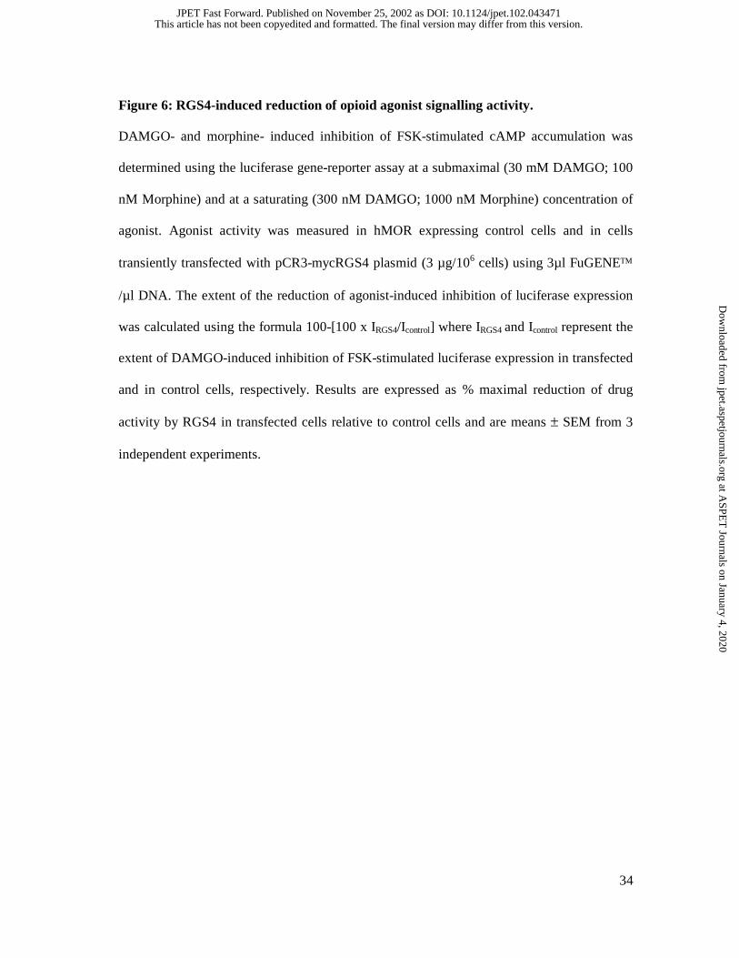
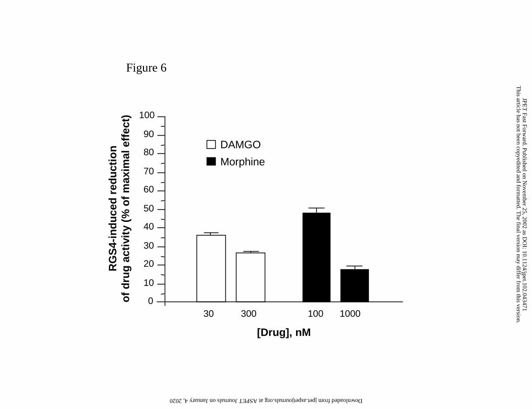
Figure 6: RGS4-induced reduction of opioid agonist signalling activity.
DAMGO- and morphine- induced inhibition of FSK-stimulated cAMP accumulation was
determined using the luciferase gene-reporter assay at a submaximal (30 mM DAMGO; 100
nM Morphine) and at a saturating (300 nM DAMGO; 1000 nM Morphine) concentration of
agonist. Agonist activity was measured in hMOR expressing control cells and in cells
transiently transfected with pCR3-mycRGS4 plasmid (3 µg/106 cells) using 3µl FuGENE
/µl DNA. The extent of the reduction of agonist-induced inhibition of luciferase expression
was calculated using the formula 100-[100 x IRGS4/Icontrol] where IRGS4 and Icontrol represent the
extent of DAMGO-induced inhibition of FSK-stimulated luciferase expression in transfected
and in control cells, respectively. Results are expressed as % maximal reduction of drug
activity by RGS4 in transfected cells relative to control cells and are means ± SEM from 3
independent experiments.
This article has not been copyedited and formatted. The final version may differ from this version.JPET Fast Forward. Published on November 25, 2002 as DOI: 10.1124/jpet.102.043471
at ASPE
T Journals on January 4, 2020
jpet.aspetjournals.orgD
ownloaded from

Figure 1A
Sham
Nerve-injured
6
7
8
9
10
11
12
13
basal 3 7 14 21
Time post-surgery (days)
Paw
wit
hd
raw
al la
ten
cy (
s)
*
***
**
This article has not been copyedited and form
atted. The final version m
ay differ from this version.
JPET
Fast Forward. Published on N
ovember 25, 2002 as D
OI: 10.1124/jpet.102.043471
at ASPET Journals on January 4, 2020 jpet.aspetjournals.org Downloaded from

Figure 1B
1 day after surgery
7 days after surgery
Morphine (mg/kg)-40
0
40
80
120
160
0.03 0.1 0.3
% M
PE
**
This article has not been copyedited and form
atted. The final version m
ay differ from this version.
JPET
Fast Forward. Published on N
ovember 25, 2002 as D
OI: 10.1124/jpet.102.043471
at ASPET Journals on January 4, 2020 jpet.aspetjournals.org Downloaded from

-40
0
40
80
120
0.03 0.1 0.3
1 day after surgery
7 days after surgery
Morphine (mg/kg)
% M
PE
Figure 1C
This article has not been copyedited and form
atted. The final version m
ay differ from this version.
JPET
Fast Forward. Published on N
ovember 25, 2002 as D
OI: 10.1124/jpet.102.043471
at ASPET Journals on January 4, 2020 jpet.aspetjournals.org Downloaded from

RGS members
RG
S m
RN
Aco
py
nu
mb
er(r
elat
ive
to s
ham
)
0.0
0.4
0.8
1.2
1.6
2.0
N.D.
GAIPRGS4
RGS6
RGS7
RGS8
RGS9
RGS11
RGS12
RGS14
RGS17
N.D.
Figure 2
This article has not been copyedited and form
atted. The final version m
ay differ from this version.
JPET
Fast Forward. Published on N
ovember 25, 2002 as D
OI: 10.1124/jpet.102.043471
at ASPET Journals on January 4, 2020 jpet.aspetjournals.org Downloaded from

Figure 3
Sham
Ligated
7 143 210
5
10
15
20
25
30
35
RG
S4
mR
NA
(co
py
nu
mb
er x
106)
Time after surgery (days)
* *
This article has not been copyedited and form
atted. The final version m
ay differ from this version.
JPET
Fast Forward. Published on N
ovember 25, 2002 as D
OI: 10.1124/jpet.102.043471
at ASPET Journals on January 4, 2020 jpet.aspetjournals.org Downloaded from

Figure 4
This article has not been copyedited and form
atted. The final version m
ay differ from this version.
JPET
Fast Forward. Published on N
ovember 25, 2002 as D
OI: 10.1124/jpet.102.043471
at ASPET Journals on January 4, 2020 jpet.aspetjournals.org Downloaded from

CP
S, %
FS
K
[DAMGO], nM
1 10 1 10 220
30
40
50
60
70
80
90
100
110
Figure 5
This article has not been copyedited and form
atted. The final version m
ay differ from this version.
JPET
Fast Forward. Published on N
ovember 25, 2002 as D
OI: 10.1124/jpet.102.043471
at ASPET Journals on January 4, 2020 jpet.aspetjournals.org Downloaded from
0
10
20
30
40
50
60
70
80
90
100
100 1000
DAMGO
Morphine
30 300
[Drug], nM
RG
S4-
ind
uce
d r
edu
ctio
no
f d
rug
act
ivit
y (%
of
max
imal
eff
ect)
Figure 6
This article has not been copyedited and form
atted. The final version m
ay differ from this version.
JPET
Fast Forward. Published on N
ovember 25, 2002 as D
OI: 10.1124/jpet.102.043471
at ASPET Journals on January 4, 2020 jpet.aspetjournals.org Downloaded from