Embed Size (px)
Citation preview


Journal of the Arab Board of Health Specializations
General SupervisorPresident of the Higher Council of the Arab Board of Health Specializations
Faisal Radi Al-Moussawi, MD.
Editor-in-ChiefSecretary General of the Arab Board of Health Specializations
Mohammad Hisham Al-Sibai, MD.
Co-EditorSamir Al-Dalati, MD.
Editorial BoardSHAWQI SALEH, MD (JORDAN)SALAH FADELALLA, MD (SUDAN)
EHTUISH FARAG EHTUISH, MD (LIBYA)FALEH ALBAYATI, MD (IRAQ)FAISAL AL-NASIR, MD (BAHRAIN)HASAN ZAHER HASAN, MD (EGYPT)
IBRAHIM BANI HANI, MD (JORDAN)ABDUL WAHAB FOUZAN, MD (KUWAIT)NASSER LOZA, MD (EGYPT)JAMAL BLEIK, MD (LEBANON)
MAZEN AL-KHABOURI, MD (SULTANATE OF OMAN)MOHAMMAD SALEH ELARABI, MD (LIBYA)AGHIAD AL-KUTOUBI, MD (LEBANON)ABDULLAH MOHAMMED ALSAKKA, MD (SAUDI ARABIA)
SALIH AL-MOHSEN, MD (SAUDI ARABIA)GHAZI ZAATARI, MD (LEBANON)MARIO PIANESI, MD (ITALY)SALWA AL-SHEIKH, MD (SYRIA)
ALY ELYAN, MD (EGYPT)ROBERT F. HARRISON, MD (IRELAND)OMAR DARDIRI, MD (SUDAN)GHASSAN HAMADEH, MD (LEBANON)
Editorial AssistantsLama Al-Trabulsi Lina Al-Kallas Lina Jeroudi
Advisory BoardMAHMOUD BOZO, MDRAYDAH ALKHANI, MDMANSOUR AL-NOZHA, MDISSAM ANJAK, MD
MOHAMMED H. ALKAFF, MDSALAH QARIOUH, MDSAMIR SROUR, MDHYAM BASHOUR, MDABDULLAH SAADEH, MDYASER ISKANDER, MDABDUL KARIM ALOMARI, MDSUHAILA GHULOUM, MDMOHSEH G. NAOUM, MDAHMED EL-SHEIKHLY, MDABDUL HADI ALBRIEZAT, MDMICHEAL GHOSSAIN, MD
The Journal of the Arab Board of Health Specializations is a Medical Journal, Issued quarterly, encompassing all medical specializations. It will strive to publish researches of the Arab physicians in order to strengthen the
communication and exchange of scientific and medical information within the Arab Countries. Besides, the Journal publishes selected important medical abstracts which have recently been accepted for
publication elsewhere, along with their Arabic translation to facilitate communication. The Journal will also publish the activities and news of the Arab Board of Health Specializations.
Correspondence to: Journal of the Arab Board of Health SpecializationsThe Arab Board of Health Specializations
P.O. Box 7669, Damascus, Syria.Tel: +963-11-6119741/6119740 Fax: +963-11-6119739/6119259.
E-mail: [email protected]

Requirements for Authors Submitting Manuscriptsto the Journal of the Arab Board of Health Specializations
arab-board

Journal of the Arab Board of Health SpecializationsA Medical Journal Encompassing all Health Specializations
Issued Quarterly
LETTER FROM THE EDITOR
OR
IGIN
AL
ART
ICLE
S
Pseudoexfoliation Syndrome: Prevalence and itsAssociations With some Parameters
متالزمة التقشر الكاذب: انتشارها وعالقاتها مع بعض المشعرات
Zuhair Al Jaffal, et al. (Syria). ...............................................................................................................P 2
Thrombolysis in Yemeni Patients with Acute Coronary Syndrome, Facts and Prognosis. Data from GulfRace (Gulf Registry of Acute Coronary Events)– Phase I
انحالل الخثرة عند المرضى اليمنيين المصابين بالمتالزمة اإلكليلية الحادة، الحقائق واإلنذارGULFRACE بيانات سجل الخليج للحوادث اإلكليلية الحادة- المرحلة األولى
Abd Nasser Munibari, et al. (Yemen). ...................................................................................................P 9
Epidemiological Characteristics of Chronic Renal Failure Patients in Southern Provinces of Iraq 2012
الخصائص الوبائية لمرضى القصور الكلوي المزمن في محافظات العراق الجنوبية لعام 2012
Alaa Abdullatif Alaugili, et al. (Iraq)....................................................................................................P 15
Study of Cryptosporidium Infection Among Children Attending the Pediatric Hospital in Damascus
دراسة الخمج بخفيات األبواغ عند األطفال المراجعين لمشفى األطفال في مدينة دمشق
Elen Ibrahim, et al. (Syria). .................................................................................................................P 25
Mohammad Hisham Al-Sibai, MDEditor-in-Chief, Secretary General of the Arab Board of Health Specializations...........................P 1
CONTENTS
JABHS Vol. 16, No. 4, 2015

Journal of the Arab Board of Health SpecializationsA Medical Journal Encompassing all Health Specializations
Issued Quarterly
SELECTED ABSTRACTS
CONTENTS
JABHS Vol. 16, No. 4, 2015
....................................................................................................................................P 45
MEDICAL CASEAchalasia With Megaesophagus
P 43 ......................................................................................... حالة تعذر ارتخاء ومريء عرطل
CASE
REPORTS
Atypical Presentations of Acute Appendicitis
تظاهرات غير وصفية اللتهاب الزائدة الحادAmmar Shehadeh, et al. (UAE). .....................................................................................P 35
A Case of Angiomyolipoma of the Testis
حالة ورم عضلي شحمي وعائي في الخصيةYasin Idweini. (Jordan). ..................................................................................................P 40
The Results of Kidney Transplantation in RecipientsOperated by DJ-stented Versus Non-Stented Ureteroneocystostomy
نتائج زرع الكلية عند المرضى المجرى لهمزرع الحالب مع ستنت حالبي أو بدونه
Assem Nasser, et al. (Syria).............................................................................................P 30
ORIGINAL
ARTICLES

1
Journal of the Arab Board of Health Specializations Vol.16, No.4, 2015
Letter from the Editor
Clinical practice guidelines
Guidelines are designed to support the decision-making processes in patient care. The content of a guideline is based on a systematic review of clinical evidence- the main source for evidence-based care.
The movement towards evidence-based healthcare has been gaining ground quickly over the past few years, motivated by clinicians, politicians and management concerned about quality, consistency and costs. Clinical practice guidelines based on standardised best practice, have been shown to be capable of supporting improvements in quality and consistency in healthcare. Many have been developed, though the process is time- and resource-consuming. Many have been disseminated, though largely in the relatively difficult to use format of narrative text. As yet they have not had a major impact on medical practice, but their importance is growing.
Purposes of guidelines
- To describe appropriate care based on the best available scientific evidence and broad consensus. - To reduce inappropriate variation in practice.- To provide a more rational basis for referral. - To provide a focus for continuing education. - To promote efficient use of resources. - To act as focus for quality control, including audit.- To highlight shortcomings of existing literature and suggest appropriate future research.
Guidelines and protocols
Clinical protocols can be seen as more specific than guidelines, defined in greater detail. Protocols provide “a comprehensive set of rigid criteria outlining the management steps for a single clinical condition or aspects of organization”
Computerized guidelines
Computerized guidelines encode evidence-based recommendations for and can automatically generate recommendations about what medical procedures to perform tailored for an individual patient. Computerized guidelines offer benefits over and above those offered by paper-based guidelines:
- They offer a readily accessible reference, providing selective access to guideline knowledge. - They help reveal errors in the content of a guideline.- They help improve the clarity of a guideline, e.g. in decision criteria and clinical recommendations. - They help offer better descriptions of patient states. - They can automatically propose timely, patient-specific decision support and reminders.
Professor M. Hisham Al-SibaiEditor-in-chief
Secretary General of the Arab Board of Health Specializations

2
Journal of the Arab Board of Health Specializations Vol.16, No.4, 2015
Original Article
PSEUDOEXFOLIATION SYNDROME: PREVALENCE AND ITSASSOCIATION WITH SOME PARAMETERS
متالزمة التقشر الكاذب: انتشارها وعالقاتها مع بعض المشعراتZuhair Al Jaffal, MD; Ammar Kayyali, MD (CIS)
د. زهير اجلّفال، د. عمار كيالي
ABSTRACT
Objective: Pseudoexfoliation (PXF) is a recognized risk factor for developing cataract, glaucoma and lens dislocation. PXF is also associated with increased risk of complications during cataract surgery due to poor mydriasis and zonular weakness. The aim of this study
is to report the prevalence of pseudoexfoliation among Syrians attending the ophthalmology clinic of Aleppo University Hospital.
Methods: A prospective study conducted in the period from September 2014 to June 2015. A total of 500 patients aged 40 years or older attending the general ophthalmic clinic of Aleppo University Hospital in separated
ملخص البحث
هدف البحث: تعتبر متالزمة التقشر الكاذب عامل خطورة معروفًا لتطور الساد والزرق وخلع العدسة. كما تترافق هذه المتالزمة مع زيادة احتمالية االختالطات خالل جراحة الساد بسبب ضعف التوسع الحدقي وضعف ألياف النطيقة العدسية. تهدف هذه الدراسة إلى تحديد شيوع متالزمة التقشر الكاذب
عند المرضى المراجعين للعيادة العينية في مستشفى حلب الجامعي.طرق البحث: تمت هذه الدراسة المستقبلية في الفترة الممتدة بين أيلول 2014 وحزيران 2015، حيث تم فحص 500 مريضًا تتجاوز أعمارهم الـ 40 عامًا من مراجعي العيادة العينية في مستشفى حلب الجامعي في أيام منفصلة من قبل نفس الطبيب. تم اجراء فحص مفصل لكل المرضى يتضمن القصة السريرية والعينية والفحص على جهاز المصباح الشقي وقياس الضغط داخل المقلة وتنظير زاوية البيت األمامي وتنظير قعر العين بعد توسيع الحدقة. صنف المرضى الذين لديهم مواد قشرية دقيقة على المحفظة األمامية للعدسة و/ أو حافة الحدقة في إحدى العينين أو كلتيهما كمصابين بمتالزمة التقشر
الكاذب.النتائج: ُشخصت المتالزمة عند 8 مرضى من أصل 500 مريضًا مضمنين في الدراسة )1.6%(. كما شخصت المتالزمة في 10 عيون من أصل 870 عينًا تم فحصها )1.14%(. كان الشيوع المصحح بالنسبة للعمر لمتالزمة التقشر الكاذب عند المرضى بعمر أكثر أو يساوي 40 عامًا %0.97 )بفواصل ثقة 95%:0.28-1.67( عندما حسب باستخدام الجمهرة السورية كمعيار؛ وكان الشيوع المصحح للعمر لمتالزمة التقشر الكاذب بعمر أكثر أو يساوي 40 عامًا 1.34% )بفواصل ثقة 95%:0.38-2.30( عندما حسب باستخدام الجمهرة العالمية لمنظمة الصحة العالمية WHO. بلغ متوسط العمر للمرضى المصابين بمتالزمة التقشر الكاذب 71.50 سنة )بانحراف معياري 7.91 والمدى 58-83(. كانت متالزمة التقشر الكاذب ثنائية الجانب في 25% من الحاالت. لوحظ ترافق هام لمتالزمة التقشر الكاذب مع الساد والزرق ونقص السمع. حيث لوحظ الساد عند 87.5% والزرق عند %37.5
ونقص السمع عند 37.5% من المصابين بالمتالزمة.االستنتاجات: تعتبر متالزمة التقشر الكاذب مرضًا مهمًا عند المرضى كبار السن في سوريا كونها تترافق بزيادة حدوث الساد والزرق ونقص السمع.
*Zuhair Al Jaffal, MD; Chief Resident of Department of Ophthalmology at Aleppo University Hospital, Aleppo, Syria. E-mail:[email protected].*Ammar Kayyali, MD; CIS, Chief of Department of Ophthalmology at Aleppo University Hospital, Aleppo, Syria.
مو�صوع �أ�صيل

3
Journal of the Arab Board of Health Specializations Vol.16, No.4, 2015
days were included in this study and examined by the same ophthalmologist. A detailed evaluation including ophthalmic and general history, slit lamp biomicroscopy, intraocular pressure measurement, gonioscopy and dilated eye examination were performed. Patients with pseudoexfoliative material on the anterior lens surface and ⁄ or the pupillary margin in either or both eyes were labeled as having PXF.
Results: Out of the 500 patients included, eight (1.6%) patients had PXF. Out of 870 eyes included, ten (1.14%) eyes had PXF. The age-adjusted prevalence of PXF in those ≥40 years was 0.97% (95% CI: 0.28-1.67) when calculated using the Syrian population as a standard, and 1.34% (95% CI: 0.38-2.30) when calculated using the WHO world population as a standard. Mean age of PXF group was 71.50 years (SD 7.91, range 58-83 years). PXF was bilateral in 25% of cases. It was significantly associated with cataract, glaucoma and hearing loss. Of the PXF patients, 87.5% had cataract, 37.5% had glaucoma and 37.5% had hearing loss.
Conclusions: Pseudoexfoliation appears to be an important disorder in older individuals in Syria because it is associated with increased risk for developing glaucoma, cataract and hearing loss.
INTRODUCTION
Pseudoexfoliation syndrome (PXF) was first reported by Lindberg in 1917 in a Finnish population.1 It is characterized by the deposition of a distinctive fibrillar material in the anterior segment of the eye,2 Figure 1. Pseudoexfoliation syndrome is frequently associated with open angle glaucoma, known as pseudoexfoliation glaucoma, which is the most common identifiable form of secondary open angle glaucoma worldwide.2 Pseudoexfoliation is a known risk factor for developing cataracts.3 Complicating factors such as poor mydriasis, zonular weakness, corneal endothelial dysfunction, higher rate of vitreous loss, capsular phimosis, and opacification have all been reported after cataract surgery.4,5
Pseudoexfoliation is considered to be a systemic disorder. Pseudoexfoliative material has been reported in lungs, skin, liver, heart, kidney, gallbladder, blood vessels, extra ocular muscles and meninges.6 An
association between PXF and sensorineural deafness has been reported.7-11
Pseudoexfoliation is rarely seen before the age of 40, and its prevalence increases markedly with age.12 Although it occurs in virtually every area of the world, a considerable racial variation exists. Epidemiological studies of PXF have been done in some areas in the Middle East, but there is no data available on prevalence of PXF in Syria.
Aleppo University Hospital (AUH) is the most populous governorate medical center of Aleppo. The aim of this hospital-based study is to estimate the prevalence of PXF, provide a descriptive analysis and assess whether PXF is associated with cataract, glaucoma, hearing loss, diabetes mellitus and systemic arterial hypertension.
METHODS
A total of 500 patients aged 40 years or older attending the general ophthalmic clinic of Aleppo University Hospital in the period from September 2014 to June 2015 in separated days were included in this study and examined by the same ophthalmologist. The study protocol was approved by the medical ethics committee of Aleppo University.
Relevant details in medical and ocular history were obtained from each patient including; history of systemic arterial hypertension, diabetes mellitus, hearing loss, intraocular surgery, visual problems, amblyopia and use of corrective glasses and/or contact lenses. All patients underwent complete ocular examination conducted by a senior experienced ophthalmologist included slit lamp biomicroscopy, intraocular pressure measurement, gonioscopy, and dilated fundus examination.
Pseudoexfoliation was diagnosed clinically by the presence of typical pseudoexfoliation material (PXM) at the pupil border on undilated examination, on anterior lens capsule on dilated examination, or on the trabecular meshwork on gonioscopy, with or without Sampaolesi’s line and pigment deposition in angle and/or corneal endothelium.2

4
Journal of the Arab Board of Health Specializations Vol.16, No.4, 2015
The diagnosis of cataract was based on clinical biomicroscopy and classified into cortical, nuclear, and posterior subcapsular cataract.13
Glaucoma was defined according to the criteria of the International Society of Geographical and Epidemiological Ophthalmology (ISGEO) classification scheme.14 In that definition, criteria for a category 1 diagnosis (structural and functional evidence) were a vertical cup/disc diameter ratio (VCDR) or an inter-eye asymmetry in the VCDR of ≥97.5th percentile for the normal population, or a neuroretinal rim width reduced to ≤0.1 VCDR (between 11 to 1 o’clock or 5 to 7 o’clock), in addition to a definite visual field defect consistent with glaucoma. Criteria for the category 2 diagnosis (advanced structural damage with unproven visual field loss) were a VCDR or a VCDR asymmetry ≥99.5th percentile for the normal population. Criteria for a category 3 diagnosis (for eyes the optic nerve head of which could not be examined or for which a visual field examination was not possible) were a visual acuity <3/60 combined with either an intraocular pressure >99.5th percentile, or definite glaucoma medical records such as filtering surgery history.14
Any eye with previous cataract surgery, history of severe ocular trauma, uveitis, corneal scars, and any other ocular pathology that could have led to inability of examining the anterior capsule was excluded. Any patient whose both eyes met exclusion criteria was excluded from the study.
The statistical analysis was performed using the Statistical Program for the Social Sciences Version 19.0 (SPSS, Inc, Chicago, IL, USA). Means, standard deviations (SDs) and 95% confidence intervals (CIs) were obtained. A p-value of <0.05, measured by Pearson’s Chi-square test, was considered to indicate statistical significance.
Patients were divided into two groups; PXF and non-PXF. For each group the sex distribution, mean age and standard deviation (SD) were calculated. The total number of eyes studied was 870. The PXF group included 10 eyes and the non-PXF group included 860 eyes. Frequencies of cataract, glaucoma, hearing loss,
diabetes mellitus and systemic arterial hypertension in both groups were estimated.
RESULTS
Out of 500 patients enrolled, 8 were diagnosed with PXF. Thus the prevalence of PXF in those ≥40 years was 1.6%. The age-adjusted prevalence of PXF in those ≥40 years was 0.97% (95% CI: 0.28-1.67) when calculated using the Syrian population as a standard, and 1.34 (95% CI: 0.38-2.30) when calculated using the WHO world population as a standard Table 1. Out of 870 eyes included, ten (1.14%) eyes had PXF.
Table 1. Age distribution and age adjusted prevalence of PXF in study group.
Of the 8 PXF patient included, 2 (25%) were females and 6 (75%) were males. The association between PXF and gender was not statistically significant (p=0.288).
Mean age of PXF group was 71.50 years (SD 7.91, range 58-83 years). Mean age of non-PXF group was 59.76 years (SD 10.97, range 40-94 years). Prevalence of PXF increased with age and was highest among subjects aged >60 years.
Unilateral PXF was noted in 75% of the PXF group and bilateral PXF was noted in 25%.
Low CI High CI
40-44 40 (0) 0.00 0.00 9.22 45-49 46 (0) 0.00 0.00 8.02 50-54 62 (0) 0.00 0.00 5.95 55-59 81 (1) 1.23 0.03 6.88 60-64 96 (0) 0.00 0.00 3.84 65-69 84 (2) 2.38 0.29 8.60 70-74 38 (1) 2.63 0.07 14.66 75-79 21 (3) 14.29 2.95 41.75 80-84 15 (1) 6.67 0.17 37.14
>85 17 (0) 0.00 0.00 21.70500 (8)
Crude 1.60 0.69 3.15
Adjusted (Syrian) 0.97 0.28 1.67
Adjusted (WHO World) 1.34 0.38 2.30

5
Journal of the Arab Board of Health Specializations Vol.16, No.4, 2015
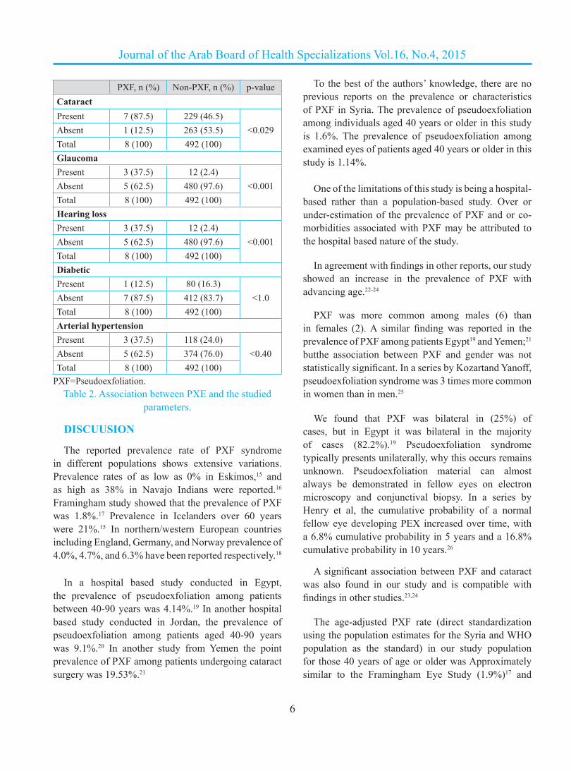
Cataract was found in 87.5% of eyes with PXF, but in only 46.5% of non-PXF eyes (p<0.029), indicating a strong association between cataract and PXF, Table 2, Figure 2.
Glaucoma was found in 37.50% of eyes in the PXF group, but in only 2.4% of eyes in the non-PXF group. The association between PXF and glaucoma was statistically significant (p<0.001) Table 2, Figure 2.
Hearing loss was documented in 37.5% of PXF
patients and in only 2.4% of non-PXF. This association between PXF and hearing loss was statistically significant (p<0.001), Table 2, Figure 2. Diabetic was found in 12.50% of patients in the PXF group, and in 16.3% of patients in the non-PXF group. The association between PXF and diabetes mellituswas statistically insignificant (p<1.0). Systemic arterial hypertension was found in 37.5% of PXF group, and in 24.0% non- PXF group. The association between PXF and Arterial hypertension was statistically insignificant (p<0.40), Table 2, Figure 2.
Figure 1. An eye with pseudoexfoliation showing the dandruff like pseudoexfoliationmaterial on the surface of the lens capsule.
Figure 2. Prevalence of cataract, glaucoma, hearing loss, diabetes mellitus, systemic hypertension among PXF vs non-PXF group.
PXF=Pseudoexfoliation.
6
Journal of the Arab Board of Health Specializations Vol.16, No.4, 2015
DISCUUSION
The reported prevalence rate of PXF syndrome in different populations shows extensive variations. Prevalence rates of as low as 0% in Eskimos,15 and as high as 38% in Navajo Indians were reported.16 Framingham study showed that the prevalence of PXF was 1.8%.17 Prevalence in Icelanders over 60 years were 21%.15 In northern/western European countries including England, Germany, and Norway prevalence of 4.0%, 4.7%, and 6.3% have been reported respectively.18
In a hospital based study conducted in Egypt, the prevalence of pseudoexfoliation among patients between 40-90 years was 4.14%.19 In another hospital based study conducted in Jordan, the prevalence of pseudoexfoliation among patients aged 40-90 years was 9.1%.20 In another study from Yemen the point prevalence of PXF among patients undergoing cataract surgery was 19.53%.21
To the best of the authors’ knowledge, there are no previous reports on the prevalence or characteristics of PXF in Syria. The prevalence of pseudoexfoliation among individuals aged 40 years or older in this study is 1.6%. The prevalence of pseudoexfoliation among examined eyes of patients aged 40 years or older in this study is 1.14%.
One of the limitations of this study is being a hospital-
based rather than a population-based study. Over or under-estimation of the prevalence of PXF and or co-morbidities associated with PXF may be attributed to the hospital based nature of the study.
In agreement with findings in other reports, our study showed an increase in the prevalence of PXF with advancing age.22-24
PXF was more common among males (6) than in females (2). A similar finding was reported in the prevalence of PXF among patients Egypt19 and Yemen;21 butthe association between PXF and gender was not statistically significant. In a series by Kozartand Yanoff, pseudoexfoliation syndrome was 3 times more common in women than in men.25
We found that PXF was bilateral in (25%) of cases, but in Egypt it was bilateral in the majority of cases (82.2%).19 Pseudoexfoliation syndrome typically presents unilaterally, why this occurs remains unknown. Pseudoexfoliation material can almost always be demonstrated in fellow eyes on electron microscopy and conjunctival biopsy. In a series by Henry et al, the cumulative probability of a normal fellow eye developing PEX increased over time, with a 6.8% cumulative probability in 5 years and a 16.8% cumulative probability in 10 years.26
A significant association between PXF and cataract was also found in our study and is compatible with findings in other studies.23,24
The age-adjusted PXF rate (direct standardization using the population estimates for the Syria and WHO population as the standard) in our study population for those 40 years of age or older was Approximately similar to the Framingham Eye Study (1.9%)17 and
p-valueNon-PXF, n (%)PXF, n (%)Cataract
<0.029229 (46.5)7 (87.5)Present263 (53.5)1 (12.5)Absent492 (100)8 (100)Total
Glaucoma
<0.00112 (2.4)3 (37.5)Present
480 (97.6)5 (62.5)Absent492 (100)8 (100)Total
Hearing loss
<0.00112 (2.4)3 (37.5)Present
480 (97.6)5 (62.5)Absent492 (100)8 (100)Total
Diabetic
<1.080 (16.3)1 (12.5)Present412 (83.7)7 (87.5)Absent492 (100)8 (100)Total
Arterial hypertension
<0.40118 (24.0)3 (37.5)Present374 (76.0)5 (62.5)Absent492 (100)8 (100)Total
PXF=Pseudoexfoliation.Table 2. Association between PXE and the studied
parameters.

7
Journal of the Arab Board of Health Specializations Vol.16, No.4, 2015
Visual Impairment Study (0.98%);27 and lower than the rates the age-standardized rates of PXF reported in south India (3.01%) reported by Thomas et al,28 in the Chennai study (4.9%)23 and Blue Mountains Eye Study (2.3%).22 However, the age-specific standardized PXF rates in other population-based studies, one from southern India,29 central Iran30 and from Crete (Greece)31-32 were high (7.6%, 9.4% and 16.1% respectively) in comparison to those in our study.
A strong relationship between glaucoma and PXF is known.26 Subjects with PXF had a two to threefold increased risk for glaucoma according to the Blue Mountains Eye Study.22 Other studies have demonstrated that eyes with PXF had higher mean IOP than eyes without PXF.13,27 Moreover, Topouzis F et al. reported increased likelihood of glaucoma at the same IOP in subjects with PXF.33 Our study is consistent with the above as we found an increased risk for glaucoma in patients with PXF. Our finding that 37.50% of eyes with PXF had glaucoma reflects a comparable proportion to that reported by Al-Bdour et al. in Jordan,20 and Tarek A Shazly et al. in Egypt,19 yet higher that found by the Blue Mountains Eye Study, which found incidences of 14.2%.22 This finding may reflect an overestimation, which is one of the limitations of hospital-based studies.
One of the limitations of our study is that hearing loss was assessed via detailed history taking. Some of the patients had audiometry documented sensorineural hearing loss, while others didn’t undergo audiometric studies. Systemic hypertension was diagnosis with only one record or via detailed medical history and hypertensive retinopathy in funduscopy which aren’t enough to make the diagnosis.
In our study hearing loss was found in 37.5% of PXF patients. This association between PXF and hearing loss was statistically significant. Cahill et al. reported that a large proportion of patients with PXF have sensorineural hearing loss in comparison to age-matched controls, regardless of whether or not there is associated glaucoma.7 This was confirmed by other studies from different Saudi Arabia, Canada, Turkey and Iran.8-11 Furthermore, in a recent study Turgut et al. reported high prevalence of asymptomatic vestibular dysfunction among patients with PXF.34
Although the material reported here has many limitations, it adds some new information on the prevalence and characteristics of PXF in a region where data on PXF are scarce.
CONCLUSIONS
In conclusion we found the prevalence of PXF among Syrians individuals attending ophthalmology clinic aged 40 years or older to be 1.6%. This rate is similar to other studies conducted in Visual Impairment Study (0.98%), but lower than other studies conducted in other Middle Eastern countries in Egypt (4.14%) and Jordan (9.1%).Population-based study should be performed to plan and tackle its complications, namely, cataract and glaucoma.Ophthalmologists in Syria should focus on the detection of PXF, especially considering the risks for operative complications related to PXF and the higher prevalence of glaucoma among PXF patients.
REFERENCES
1. Lindberg JG. Kliniskaundersokningar over eringenav-pupillarrandenochgenomlysbarhetenav iris vid fall avaldersstarrsamt i normalaogon hos gamlapersoner. En [Clinical studies of depigmentation of the pupillary margin and transillumination of the iris in cases of senile cataract and also in normal eyes in the aged] [Thesis]. Helsinki, Finland: Helsinki University; 1917.
2. Ritch R. Exfoliation syndrome: The most common identifiable cause of open-angle glaucoma. Trans Am Ophthalmol Soc 1994;92:845-944.
3. Hirvela H, Luukinen H, Laatikainen L. Prevalence and risk factors of lens opacities in the elderly in Finland.Ophthalmology 1995;102:108-17.
4. Alfaite M, Leite E, Mira J, et al. Prevalence and surgical complications of pseudoexfoliation syndrome in Portuguese patients with senile cataract. J Cataract Refract Surg 1996;22:972-6.
5. Lumme P, Laatikainen L. Exfoliation syndrome and cataract extraction. Am J Ophthalmol 1993;116:51-5.
6. Schlotzer-Schrehardt UM, Koca MR, Naumann GO, et al. Pseudoexfoliation syndrome. Ocular manifestation of a systemic disorder?Arch Ophthalmol 1992;110(12):1752-6.

8
Journal of the Arab Board of Health Specializations Vol.16, No.4, 2015
7. Cahill M, Early A, Stack S, et al. Pseudoexfoliation and sensorineural hearing loss. Eye (Lond) 2002;16(3):261-6.
8. Shaban RI, Asfour WM. Ocular pseudoexfoliation associated with hearing loss. Saudi Med J 2004;25(9):1254-7.
9. Turacli ME, Ozdemir FA, Tekeli O, et al. Sensorineural hearing loss in pseudoexfoliation. Can J Ophthalmol 2007;42(1):56-9.
10. Detorakis ET, Chrysochoou F, Paliobei V, et al. Evaluation of the acoustic function in pseudoexfoliation syndrome and exfoliation glaucoma: audiometric and tympanometric findings. Eur J Ophthalmol 2008;18(1):71-6.
11. Yazdani S, Tousi A, Pakravan M, et al. Sensorineural hearing loss in pseudoexfoliation syndrome. Ophthalmology 2008;115(3):425-9.
12. Aasved H. Mass screening for fibrill opathiaepitheliocapsularis, so-called senile exfoliation or pseudoexfoliation of the anterior lens capsule. Acta Ophthalmol (Copenh) 1971;49(2):334-43.
13. Sperduto RD, Hiller R. The prevalence of nuclear, cortical, and posterior subcapsular lens opacities in a general population sample. Ophthalmology 1984 Jul;91(7):815-8.
14. Foster PJ, Buhrmann R, Quigley HA, et al. The definition and classification of glaucoma in prevalence surveys. Br J Ophthalmol 2002;86(2):238-42.
15. Forsius H. Prevalence of pseudoexfoliation of the lens in Finns, Lapps, Icelanders, Eskimos and Russians. Trans Ophthalmol Soc UK 1979;99:296-8.
16. Faulkener HW. Pseudoexfoliation of the lens among Navajo Indians. Am J Ophthalmol 1971;72:206-7.
17. Hiller R, Sperduto RD, Krueger DE. Pseudoexfoliation, intraocular pressure, and senile lens changes in a population based survey. Arch Ophthalmol 1982;100:1080-2.
18. Aasved H. Prevalence of fibrillo pathiaepithelio capsularis [pseudoexfoliation] and capsular glaucoma. Trans Ophthalmol Soc UK 1979;99:293-5.
19. Shazly TA, Farrag AN, Kamel A, et al. Prevalence of pseudoexfoliation syndrome and pseudoexfoliation glaucoma in upper Egypt. BMC Ophthalmology 2011;11:18.
20. Al-Bdour MD, Al-Till MI, Idrees GM, et al. Pseudoexfoliation syndrome at Jordan University
Hospital. Acta Ophthalmol 2008;86(7):755-7.21. Al-Shaer M, Bamashmus M, Al-Barrag A. Point
prevalence of pseudoexfoliation syndrome in patients scheduled for cataract surgery in eye camps in Yemen. Middle East Afr J Ophthalmol 2010;17(1):74-7.
22. Mitchell P, Wang JJ, Hourihan F. The relationship between glaucoma and pseudoexfoliation: The Blue Mountains Eye Study. Arch Ophthalmol 1999;117:1319-24.
23. Arvind H, Raju P, Paul PG, et al. Pseudoexfoliation in South India. Br J Ophthalmol 2003;87:1321-3.
24. Tiliksew T, Kefyalew R. Prevalence of pseudoexfoliation in Ethiopian patients scheduled for cataract surgery. Acta Ophthalmol Scand 2004;82:254-8.
25. Kozart DM, Yanoff M. Intraocular pressure status in 100 consecutive patients with exfoliation syndrome.Ophthalmology 1982 Mar;89(3):214-8.
26. Henry JC, Krupin T, Schmitt M, et al. Longterm follow-up of pseudoexfoliation and the development of elevated intraocular pressure. Ophthalmology 1987;94:545-52.
27. McCarthy CA, Taylor HR. Pseudoexfoliation syndrome in Australian adults. Am J Ophthalmol 2000;129:629-33.
28. Thomas R, Nirmalan PK, Krishnaiah S. Pseudoexfoliation in southern India: the Andhra Pradesh Eye Disease Study. Invest Ophthalmol Vis Sci 2005;46(4):1170-6.
29. Krishnadas R, Nirmalan PK, Ramakrishnan R, et al. Pseudoexfoliation in a rural population of southern India: the Aravind Comprehensive Eye Survey. Am J Ophthalmol 2003;135:830-7.
30. Nouri-Mahdavi K, Nosrat N, Sahebghalam R, et al. Pseudoexfoliation syndrome in central Iran: a population-based survey. Acta Ophthalmol Scand 1999;77:581-4.
31. Kozobolis VP, Papatzanaki M, Vlachonikolis IG, et al. Epidemiology of pseudoexfoliation in the island of Crete (Greece). Acta Ophthalmol Scand 1997;75(6):726-9.
32. Kozobolis VP, Detorakis ET, Tsilimbaris M, et al. Crete, Greece glaucoma study. J Glaucoma 2000;9(2):143-9.
33. Topouzis F, Harris A, Wilson MR, et al. Increased likelihood of glaucoma at the same screening intraocular pressure in subjects with pseudoexfoliation: the Thessaloniki Eye Study. Am J Ophthalmol 2009;148(4):606-13.
34. Turgut B, Alpay HC, Kaya MK, et al. The evaluation of vestibular functions in patients with pseudoexfoliation syndrome. Eur Arch Otorhinolaryngol 2010;267(4):523-7.

9
Journal of the Arab Board of Health Specializations Vol.16, No.4, 2015
Original Article
THROMBOLYSIS IN YEMENI PATIENTS WITH ACUTE CORONARY SYNDROME, FACTS AND PROGNOSIS. DATA FROM GULFRACE (GULF
REGISTRY OF ACUTE CORONARY EVENTS)– PHASE Iانحالل الخثرة عند المرضى اليمنيين المصابين بالمتالزمة اإلكليلية الحادة، الحقائق واإلنذار.
GULFRACE بيانات سجل الخليج للحوادث اإلكليلية الحادة- المرحلة األولىAbd Nasser Munibari, MD; Ahmed Al-Motarreb, MD; Ahmed Alansi, MD
د. عبد الناصر منيباري، د. أحمد املترب، د. أحمد العنسي
ABSTRACT
Objective: Rapid coronary revascularization after
ACS with ST elevation acute myocardial infarction (STEMI) is a cornerstone in management. Yemen where no primary Percutaneous Coronary Intervention
ملخص البحث
هدف البحث: تعتبر االستعادة السريعة لتروية الشرايين التاجية بعد حدوث المتالزمة اإلكليلية الحادة ACS واحتشاء العضلة القلبية مع ارتفاع القطعة ST حجر الزاوية في المعالجة. ونظراً لعدم وجود برنامج للتدخل األولي للشرايين التاجية عبر الجلد في اليمن فإن استخدام المعالجات الحالة للخثرة هي الوسيلة األساسية الستعادة التروية اإلكليلية. تهدف هذه الدراسة إلى تسليط الضوء على حجم مشكلة المتالزمة اإلكليلية الحادة عند المرضى اليمنيين،
عوامل الخطورة المرافقة، معدالت استخدام حاالت الخثرة، معدالت المراضة والوفيات لدى هؤالء المرضى.طرق البحث: تمثل دراسة Gulf Race دراسة مسحية استباقية متعددة المراكز ومتعددة البلدان للمرضى المقبولين في المشفى بتشخيص نهائي بوجود
المتالزمة اإلكليلية الحادة في 6 دول عربية في منطقة الخليج والجزيرة العربية خالل مدة 6 أشهر. النتائج: وجد 1054 من المرضى اليمنيين في دراسة Gulf Race بحاالت المتالزمة اإلكليلية الحادة، خضع 218 فقط للمعالجة الحالة للخثرة من أصل 750 مريضاً بحالة احتشاء عضلة قلبية مع ارتفاع القطعة ST أو حصار الحزمة الكهربائية اليسرى القلب جديد المنشأ. شكل هؤالء المرضى نسبة 41% فقط من مجمل الحاالت المرشحة لتطبيق المعالجة الحالة للخثرة (531 حالة). بلغ متوسط أعمار المرضى 55.9±11.01 سنة وكان غالبيتهم من الذكور. أورد وجود تدخين عند 127 مريضاً (58.3%)، مضغ القات عند 163 مريضاً (74.3%) بينما سجل وجود فرط في التوتر الشرياني في 57 حالة (26.1%) وداء سكري من النمط الثاني عند 54 مريضاً (24.8%). أورد حدوث قصور القلب االحتقاني بعد إجراء المعالجة بحاالت الخثرة عند
مرضى المتالزمة اإلكليلية الحادة في 11.5% من الحاالت مع حدوث وفيات عند 14 مريضاً (%6.4).االستنتاجات: تعتبرنسبة حدوث المتالزمة اإلكليلية الحادة في اليمن واحدة من أعلى النسب المالحظة في المنطقة. إن المعدالت المنخفضة لتطبيق المعالجة بحاالت الخثرة كخط أول في المعالجة إلعادة التروية ووصول المريض المتأخر للمشفى تساهم في ضياع فرصة ذهبية الستعادة التروية التاجية وهو ما يزيد من معدالت المراضة والوفيات. يجب العمل على زيادة برامج التوعية عند المجتمع ولدى األطباء لتحسين تدبير حاالت المتالزمة اإلكليلية
الحادة.
*Abd Nasser Munibari, MD; Faculty of Medicine and Health Sciences, Department of Internal Medicine, P. O. Box 19221, Hadda Office, Sana'a University, Sana'a, Yemen. E-mail: [email protected].*Ahmed Al-Motarreb, MD; Cardiac Center, Al-Thawra Modern Teaching General Hospital-Sana'a, Sana'a.*Ahmed Alansi, MD; Cardiac Center, Al-Thawra Modern Teaching General Hospital-Sana'a, Sana'a.
مو�صوع �أ�صيل

10
Journal of the Arab Board of Health Specializations Vol.16, No.4, 2015
available, utilizing thrombolytic therapy is the main tool for coronary reperfusion. The major objective was to highlight the magnitude of ACS among Yemeni patients, predisposing risk factors to ACS, the rate of use of thrombolytic therapy, the morbidity and mortality among those patients.
Methods: Gulf Race I is a prospective, multinational, multicenter survey of patients hospitalized with the final diagnosis of ACS in six Arabian Peninsula/Gulf countries over a period of six month.
Results: 1054 Yemeni patients with ACS participated in the Gulf Race I, only 218 patients had received thrombolytic therapy out of 750 cases of STEMI or newly developed LBBB. Those 218 patients represent only 41% of all the cases (531 cases) eligible for thrombolytic therapy. The mean age 55.9±11.01 years and were mainly males. Streptokinase was commonly thrombolytic used (95.4%). Smoking reported in 127 patient (58.3%), khat chewing in 163 patient (74.3%) while arterial hypertension in 57 cases (26.1%) and diabetes mellitus type II in 54 patients (24.8%). The mean door to needle in those patient was 59.1 minutes. Heart failure in ACS group after thrombolytic therapy was reported in 11.5% of the patients and death in 14 patients 6.4%.
Conclusions: ACS among Yemeni is one of the highest in the area, in spite of low rate of using thrombolytic therapy as a first line of revascularization in Yemen, still the time of presentation of the patient with ACS to the hospitals is late. Missing the golden hours for thrombolytic therapy in Yemeni patients with ACS is associated with high rate of morbidity and mortality. Community and physician awareness programs are needed for better management of ACS.
INTRODUCTION
The instant reestablishment of coronary blood flow is the major goal in the treatment of (STEMI). Myocardial reperfusion in STEMI is essential to myocardial salvage and improved outcomes.1 Reperfusion with restoration of antegrade epicardial flow with either thrombolysis or primary percutaneous coronary intervention (PCI) reduces mortality in (STEMI).2
A meta-analysis comprising 23 studies, indicates
that primary PCI is superior to thrombolysis in reducing death nonfatal reinfarction, and stroke at short -and long-term follow-ups. The result has led to the recommendation of primary PCI as the first-choice reperfusion therapy.2 Countries with no facilities to establish a primary PCI programs, thrombolytic therapy remains the reperfusion method of choice. The benefits of thrombolytic therapy in patients with AMI are well established in the meta-analyses by Yusuf et al3, and by the Fibrinolytic Therapy Trial lists (FTT) Collaborative Group who showed that thrombolytic therapy decreases mortality at 35 days by 1.9%.4
Yemen is one of the countries participated in (Gulf Race -I), a limited resources country with no program of primary PCI yet, exposure of the Gulf Race I data regarding Yemeni patient with ACS aiming to highlight the magnitude of ACS among Yemeni patients.
METHODS
Gulf Race is a prospective, multinational, multicenter survey of consecutive patients hospitalized with the final diagnosis of ACS in six Arabian Peninsula/Gulf countries over a period of six months. An attempt was made to include everyone with the final diagnosis of ACS, and there were no exclusion criteria. The study received ethical approval from the institutional ethical bodies in all participating countries. Over a six month period which represents phase I Gulf Race. This registry is a descriptive study of 65 medical centers who confirmed their participation and enrolled patients according to the survey inclusion criteria. Over all participants’ numbers from all Gulf States were 8176 patients out of them there were 1054 Yemeni patients with ACS pointing out the cases of ST Elevation Acute Myocardial Infarction (STEMI) or newly developed Left Bundle Branch Block (LBBB) who are eligible for thrombolytic therapy.
Diagnosis of the different types of ACS and definitions of data variables were based on the American College of Cardiology (ACC) clinical data standards, published in December 2001.5-7 These definitions are based on clinical presentations, electrocardiogram

11
Journal of the Arab Board of Health Specializations Vol.16, No.4, 2015
(ECG) findings and cardiac biomarkers. Data collected included patients’ demographics, past medical history, provisional diagnosis on admission and final discharge diagnosis, clinical features at hospital presentation, ECG findings, laboratory investigations, early in-hospital (administered within 24 hours of admission) and discharge medications, use of cardiac procedures and interventions, in-hospital outcomes and in-hospital mortality. All management decisions were at the discretion of the treating physician.8 Data collected were subjected to statistical analysis Continuous variables are summarized as median and inter-quartile ranges and compared using the Wilcoxon rank sum test. Categorical variables are summarized as percentages and compared using χ2 tests. Step-wise, multivariable logistic regression was used to identify independent predictors of in-hospital morbidity and mortality, the estimated odds ratio (OR) against age as a continuous variable. All associations from the logistic regression models are quantified as OR with 95% confidence intervals. Analyses were performed with SPSS version 20 statistical package (IBM Corporation 1 New Orchard Road Armonk, NewYork10504-1722 United States).
RESULTS
Out of 1054 Yemeni patients with ACS participated in the Gulf Race Phase I over a six month period, only 218 patients had received thrombolytic therapy out of 750 cases of STEMI or newly developed LBBB. Those 218 patients represent only 28% on all cases of STEMI or new LBBB eligible for thrombolytic therapy 264 patients (patients presented 12 hours or less from the start of symptoms). Out of those 10 were subjected to ad-hoc primary PCI by visiting teams. 218 patients received thrombolytic therapy, 36 patients did not receive any reperfusion therapy (13.6%) of all eligible cases of thrombolytic therapy, this percentage represent the shortfall of reperfusion therapy, Figure 1. The mean age was 55.97±11.01 years, with a male predominance (82.6%) in contrast with female patients, Table 1.
Streptokinase was the most commonly used preparation among the thrombolytic drugs (95.4%) followed by Reteplase (4.1%) and t-PA (0.5%) respectively, Table 2. The clinical presentation shows
prominent smoking habit in 127 patient (58.3%), khat chewing in 163 patient (74.3%) while arterial hypertension was documented in 57 cases (26.1%), diabetes mellitus type II in 54 patients (24.8%), hyperlipidemia mia in 22 patients (10.1%) and family history of IHD was recorded in 34 cases (17%).
Patient baseline characteristics STEMI patients with thrombolytic therapy
No. (%) 218 (28%)Age (Mean±SD) years 55.97±11.01
Male gender 180 (82.6%)Diabetes mellitus 54 (24.8%)
Hypertension 57 (26.1%)Hyperlipidemia 22 (10.1%)Smoking habits 127 (57.3%)Khat chewing 163 (74.3%)
Family history of IHD 34 (17%)Prior PCI 7 (3.2%)Old MI 19 (8.7%)
Prior CABG 3 (1.7%)AMI=acute myocardial infarction, CABG=coronary artery bypass
grafting, IHD=ischemic heart disease.PCI=percutaneous coronary intervention.
Table 1. Baseline clinical characteristics of STEMI patients who received thrombolytic therapy.
Patients with history of previous MI was recorded in 19 patients (8.7%) while 7 patients had PCI prior to episodes of ACS and only three (1.7%) had CABG in the past. Location of ST segment elevation in ECG was predominantly in inferior leads (29.8%) followed by extensive anterior leads (24.3%) and anterioseptal leads (18.3%), Table 2. The mean door to needle in those patient was 59.1 minutes (±102.6 SD).
Congestive heart failure was the most common sequel in ACS group after thrombolytic therapy (11.5%), while cardiogenic shock was documented in 9.2%, tachyarrhythmia were noted in 6% and major bleeding in one patient (0.5%). Mortality in 14 patients 6.4%.
DISCUSSION
Whereas the medical and technological improvement in the last 3 decades has enhanced clinical outcomes in

12
Journal of the Arab Board of Health Specializations Vol.16, No.4, 2015
Site of AMI Frequency %
Anterior+inferior 7 3.2Antero-lateral 19 8.7Antero-septal 40 18.3
Extensive anterior 53 24.3High lateral 2 1
Inferior 65 29.8Inferior+RV 9 4.1Infero lateral 12 5.5
Inferior-posterior 9 4.1Lateral 2 0.9
Table 2. Location of AMI in patients received thrombolytic therapy.
Thrombolytic agent Frequency %Streptokinase 208 95.4
Tissue Plasminogen Activator (tPA) 1 0.5Reteplase 9 4.1
Table 3. Type of thrombolytic agent used.
patients presenting with acute STEMI, residual morbidity and mortality are still high. Randomized controlled trials of thrombolytic therapy have demonstrated the benefit of initiating treatment as early as possible after the onset of STEMI symptoms.9,10 This is the first data
from Yemen regarding reperfusion after ACS over 6 month period, Yemen represents a low income country in middle east.11 Data from GulfRace I indicates that nearly one-third of patients with STEMI in the Arabian Gulf presented to the hospital 12 hours after symptom onset. Morbidity and mortality related to this delay in coronary reperfusion among those patients.12
Yemen data analysis is consistent with data of the Gulf-Race phase I13 as well as previous reports from other registries. The delayed presentation rate (>12 hours) 8.7%14,15 was reported in a supplementary study of the National Registry of Myocardial Infarction (NRMI). The GRACE registry indicated that only 70.5% of the cases had presented within 12 hours of symptom onset.16
Cohen et al reported that 40% of acute STEMI patients presented >12 hours after symptoms onset, female gender and older age were independent predictors of late presentation.17 Yemen data shows the highest rate of late presentation, nearly 66% of patients presented late. This could be related to the socioeconomic status and literacy rate (65.3%)18 may play an important role in late presentation. Advocacy programs directed toward medical and patient population may have impact the early presentation
Figure 1. Thrombolytic therapy and missed opportunity for reperfusion.

13
Journal of the Arab Board of Health Specializations Vol.16, No.4, 2015
rate. The high reperfusion shortfall rate in Yemen data 13.6% may reflect the lack of organization of health facility, the lack of national treatment protocols and scarcity of training of medical teams. Many limitations were noted in our study. Although our cohort included nearly consecutive patients with suspected ACS, only 85% of the cases were included. Nearly 60% of the study cohort had ejection fraction assessment, and only a minority of patients underwent coronary angiography. The socioeconomic status of patients and their literacy rate were not included. In addition, there were no information about the credentials of treating physicians and the impact of specialized training on shortfall rate.
CONCLUSIONS
GulfRace I is an awakening call for practicing cardiologists and health care providers in the Gulf.
Exposure of practice: doing well with certain aspects of care, but doing poorly with other aspects.
There is a large gap between what is known and what is done in daily practice. Cardiologists in the area needed to get together to breach this gap. Prevalence of ACS among Yemeni patients is one of the highest in the area in spite of low rate of using thrombolytic therapy as a first line of revascularization in Yemen. Still the time of presentation of the patient with ACS to the hospitalsis late. Missing the golden hours for thrombolytic therapy in Yemeni patients with ACS is associated with high rate of morbidity and mortality. Community and physician awareness programs are needed to increase the utilization of better management of ACS.
REFERENCES
1. Antman EM, Hand M, Armstrong PW, et al. 2007 focused update of the ACC/AHA 2004 guidelines for the management of patients with ST-elevation myocardial infarction: a report of the American College of Cardiology/American Heart Association Task Force on Practice Guidelines. J Am Coll Cardiol 2008;51:210-47.
2. Nielsen PH, Busk M, Mortensen LS, et al. Primary
angioplasty versus fibrinolysis in acute myocardial infarction. Long-term follow-up in the danish Acute Myocardial Infarction 2 Trial. Circulation 2010;121:1484-91.
3. Yusuf S, Collins R, Peto R, et al. Intravenous and intracoronary fibrinolytic therapy in acute myocardial infarction: overview of results on mortality, reinfarction and side-effects from 33 randomized controlled trials. Eur Heart J 1985;6:556-85.
4. Fibrinolytic Therapy Trialists’ (FTT) Collaborative Group. Indications for fibrinolytic therapy in suspected acute myocardial infarction: collaborative overview of early mortality and major morbidity results from all randomized trials of more than 1000 patients. Lancet 1994;343:311-22.
5. Cannon CP, Battler A. American College of Cardiology key data elements and definitions for measuring the clinical management and outcomes of patients with acute coronary syndromes. A report of the American College of Cardiology Task Force on Clinical Data Standards (Acute Coronary Syndromes Writing Committee). J Am Coll Cardiol 2001;38(7):2114-30.
6. Gara PT, Kushner FG. ACCF/AHA Guideline for the Management of ST-Elevation Myocardial Infarction. A Report of the American College of Cardiology Foundation/American Heart Association Task Force on Practice Guidelines. J Am Coll Cardiol 2013;61(4):e78-e140.
7. Lam S, Lee SW, Cham K, et al. A 10-year review of thrombolytic therapy in patients with ST-segment elevation myocardial infarction in A university hospital in Hong Kong: Intracranial bleeding and other outcomes. J Am Coll Cardiol 2012;59(13s1):E461.
8. Zubaid M, Rashed WA. Management and outcomes of Middle Eastern patients admitted with acute coronary syndromes in the Gulf Registry of Acute Coronary Events (GulfRace). Acta Cardiologica 2009;64:439-46.
9. Moyer P, Ornato JP, Brady WJ Jr, et al. Development of systems of care for ST-elevation myocardial infarction patients: the emergency medical services and emergency department perspective. Circulation 2007;116:e43-8.
10. Huber K, De Caterina R, Kristensen SD, et al. Pre-hospital reperfusion therapy: a strategy to improve therapeutic outcome in patients with ST-elevation myocardial infarction. Eur Heart J 2005;26:2063-74.
11. Library of Congress. Federal Research Division Country

14
Journal of the Arab Board of Health Specializations Vol.16, No.4, 2015
Profile: Yemen, August 2008. http://lcweb2.loc.gov/frd/cs/profiles/Yemen.pdf
12. Zubaid M, Rashed WA. Clinical presentation and outcomes of acute coronary syndromes in the Gulf Registry of Acute Coronary Events (GulfRace). Saudi Med J 2008;29:251-5.
13. Al-Mallah MH, Alsheikh-Ali AA. Missed opportunities in the management of ST-segment elevation myocardial infarction in the Arab Middle East. Patient and physician impediments. Clin Cardiol 2010;33:565-71.
14. Ting HH, Bradley EH, Wang Y, et al. Delay in presentation and reperfusion therapy in ST-elevation myocardial infarction. Am J Med 2008;121:316-23.
15. Ting HH, Bradley EH, Wang Y, et al. Factors associated
with longer time from symptom onset to hospital presentation for patients with ST-elevation myocardial infarction. Arch Intern Med 2008;168:959-68.
16. Eagle KA, Goodman SG, Avezum A, et al. Practice variation and missed opportunities for reperfusion in ST-segment-elevation myocardial infarction: findings from the Global Registry of Acute Coronary Events (GRACE). Lancet 2002;359:373-7.
17. Cohen M, Gensini GF, Maritz F, et al. The role of gender and other factors as predictors of not receiving reperfusion therapy and of outcome in STEMI. J Thromb 2005;19:155-61.
18. The Unicef organization fact sheets website: http://www.unicef.org/infobycountry/yemen_statistics.html.

15
Journal of the Arab Board of Health Specializations Vol.16, No.4, 2015
Original Article
ABSTRACT
Objective: This study aimed to assess the burden and develop a baseline data on ESRD in the southern province through estimation the prevalence, incidence,
EPIDEMIOLOGICAL CHARACTERISTICS OF CHRONIC RENAL FAILURE PATIENTS IN SOUTHERN PROVINCES OF IRAQ 2012
الخصائص الوبائية لمرضى القصور الكلوي المزمن في محافظات العراق الجنوبية لعام 2012 Alaa Abdullatif Alaugili, MBChB, Ph.D; Faris Hasan Alami, MBChB, Ph.D
د. عالء عبد اللطيف مزعل العقيلي، د. فارس حسن الالمي
ملخص البحث
النهائية هدف البحث: تهدف هذه الدراسة إلى تقييم عبىء حاالت القصور الكلوي المزمن وتطوير قاعدة بيانات حول األمراض الكلوية بالمراحل ESRD في المحافظات الجنوبية من العراق من خالل تقييم معدالت االنتشار، معدل الحدوث ومعدل إماتة الحالة ومعدل الوفيات المرتبط بالسبب لحاالت
األمراض الكلوية بالمراحل النهائية، وتقييم الموارد البشرية والبنى التحتية ومشعرات نوعية بعض الخدمات المقدمة.طرق البحث: تضمنت هذه الداسة المقطعية المستعرضة 243 من المرضى الموضوعين على التحال الدموي )الديلزة( في المحافظات الجنوبية للعراق )البصرة، ميسان وذي قار( خالل الفترة بين 1 كانون الثاني وحتى 30 تموز لعام 2012. تم جمع البيانات باستخدام نموذج استبيان تم ملؤه عبر المقابلة المباشرة مع المرضى واألطباء المعالجين ومراجعة سجالت كل حالة. تم تقييم مشعرات المراضة والوفيات من خالل مراجعة سجالت وحدة التحال الدموي
حتى نهاية عام 2012. النتائج: شملت الدراسة 243 مريضًا )45% منهم من البصرة، 22% من ميسان والبقية من ذي قار(. كانت نسبة الذكور 56.8% أعلى بقليل من اإلناث 43.2%، كما أن ثلثي الحاالت كانت دون سن 60 سنة و64% من المرضى كانوا من سكان الحضر. بلغ انتشار وحدوث األمراض الكلوية بالمراحل النهائية في المحافظات الجنوبية 95 و78.8 لكل مليون نسمة على الترتيب. بلغ معدل الوفيات المرتبط بالحالة 61.3 لكل مليون نسمة فيما بلغ معدل إماتة الحالة 62.2%. تم إجراء زرع كلية عند 4.1% من مجمل مرضى التحال الدموي خالل عام 2012. لوحظ ارتباط هام لنسبة الوفاة بسبب المراحل النهائية لألمراض الكلوية مع ازدياد العمر )p=0.000(، الحالة االجتماعية وحالة العمل والمحافظة التي ينحدر منها المريض )p=0.01(، دون وجود ارتباط هام مع الجنس، التعليم والسكن )p>0.05(. بلغت نسبة وحدات الديلزة إلى تعداد السكان 0.6 لكل مليون نسمة، بينما بلغت نسبة أجهزة الديلزة إلى تعداد السكان 8.6 لكل مليون نسمة، نسبة المرضى لكل جهاز ديلزة واحد 11.4 فيما بلغت نسبة المرضى لكل كادر طبي واحد 10.2. خضع 13.6% فقط من المرضى لثالث جلسات تحال أسبوعيًا، في حين أن غالبية الحاالت )62.1%( خضعت لجلستين فقط في األسبوع، و24.3% من
المرضى خضعوا لجلسة واحدة أسبوعيًا فقط، بلغ معدل مدة جلسات التحال الدموي 5.1 ساعة أسبوعيًا.االستنتاجات: على الرغم من تشابه معدالت الحدوث واالنتشار لألمراض الكلوية بالمراحل النهائية مع المعدالت المالحظة في البلدان المجاورة، إال أن معدالت الوفيات المالحظة كانت أعلى بكثير مع وجود ضعف كبير في مشعرات البنى التحتية وهو ما يعكس سوء الخدمات الصحية المقدمة للمرضى.
*Alaa Abdullatif Alaugili, MBChB. PhD; Community Medicine, Public Health of Basrah, Al-Moni Hospital, Iraq. E-mail: [email protected]*Faris Hasan Alami, MBChB, Ms PhD, FPH; Assistant Professor, Department of Community Medicine and Family Medicine, Baghdad, Iraq.
case fatality rate and cause specific mortality rate of end stage renal disease ESRD, assess the human resources and logistics infrastructure, and assess some services quality indicators.
Methods: A cross sectional study was done on 243
مو�صوع �أ�صيل

16
Journal of the Arab Board of Health Specializations Vol.16, No.4, 2015
hemodialysis patients at hemodialysis units of southern provinces of Iraq (Basra, Missan and Thiqar) during the period from 1 January to 30 July 2012 were included. Data collection was carried out using a questionnaire filled through direct interview with the patients and treating physicians and reviewing their case records. Morbidity and mortality indicators were estimated through reviewing the hemodialysis unit’s registries on the end of 2012.
Results: The total number of cases was 243. Around 45% of them were from Basra, 22% were from Missan and the remaining was from Thiqar. Males are 56.8% slightly more than females 43.2%. Around two thirds of the cases were below 60 years, and 64% of the cases were of urban residence. The prevalence and incidence of ESRD in Southern provinces were 98.5 pmp, and 78.8 pmp, respectively. The cause specific mortality rate was 61.3 pmp, and the case fatality ratio was 62.2%. Only 4.1% of HD patients had transplantation during 2012. Death due to ESRD was significantly increased with increasing age (p=0.000), marital status, employment status and with the province (p=0.01), while no significant association was found with gender, education, and residence (p>0.05). The HD unit to population ratio was 0.6 pmp, HD machine to population ratio was 8.6 pmp, patients to machine ratio was 11.4 and medical staff to patient ratio was 10.2. Only 13.6% had three HD session per week, the majority of the cases (62.1%) had two HD sessions per week, and 24.3% one HD session per week and the average duration of HD session was 5.1 hours (±1.3).
Conculusions : We concluded that while the incidence and prevalence of ESRD were almost comparable to neighboring countries, but the extremely high case fatality rate and poor infrastructure indicators are reflecting the poor delivered services.
INTRODUCTION
Globally, there is noticeable increase in mortality and morbidity of end stage renal disease, as a consequence to increasing morbidity of chronic Non Communicable Diseases (NCDs). In Iraq, little is known about the prevalence of ESRD, little is known about the burden of the disease and the capacity of the health system to deal with the problem.
There was shortage in the demand number of HD beside the seriousness of the disease and its immense social and economic impact were the justifications behind conducting, studies were done in Baghdad on 2012 (1.4 pmp)1 of hemodialysis unit to population ratio. Other countries like Jordan had 72 Hemodialysis Units in 2010, making Hemodialysis Units: Population Ratio 12: 1.000.000.2 In Saudi Arabia there was 177 Hemodialysis Units in 2010, making Hemodialysis Units: Population Ratio of 7:1000000.3 In Iran there are 305 Hemodialysis Units in 2006, making Hemodialysis Units: Population Ratio 4.24:1000000.4 Similarly in Turkey there were 754 Hemodialysis Units in 2008, making Hemodialysis Units: Population Ratio of 10.41:1.000.000.5
The average recommended HD duration of 12 hours/ week.6 In Iran, the frequency of three sessions per week was 60%4 in Jordan it was 49%.2 Globally, despite ongoing technical care, improvements in both dialysis and overall patient care, the annual mortality rate of patients with ESRD managed with thrice weekly HD remains high (10-22%).7-8
METHODS
A cross sectional study was conducted in the three hemodialysis units of the major hospitals in the three southern provinces in Iraq (Basra, Missan and Thiqar) and all the patients with ESRD on regular hemodialysis during first six months 2012 were included.
Hemodialysis related variables includes: first form determine the number of the sessions per hour in a week of each patient in the dialysis unit. Its recommended 12 hour per week.6 The second form the investigator was enrolled the patient's admission during 2012 and before 2012 to determine incidence and prevalence in each province using the total population of each province obtained from the statistics departments in the province HD unit. In addition enrolled the outcome of ESRD in 2012 either done renal transplantation or deceased cases to (determine the case fatality rate and cause specific mortality rate this depend on total population). Patients who had HD in the first half of year were followed up in the second half of the same year to determine the characteristics of the deceased cases

17
Journal of the Arab Board of Health Specializations Vol.16, No.4, 2015
Characteristics Categories Count %
GenderMale 138 56.8
Female 105 43.2
Age interval40> 70 28.8
40-59 85 35.060+ 88 36.2
Mean age Mean ±sd 53±8
ResidenceRural 88 36.2Urban 155 63.8
Occupation
Employed 42 17.3Non-employed 85 35.0
House wife 80 32.9Retired 36 14.8
Marital status
Single 29 11.9Married 167 68.7Divorced 16 6.6Widow 31 12.8
Education
Low education 182 74.9 Intermediate
Education41 16.9
Higher Education 20 8.2
ExerciseYes 32 13.2No 211 86.8
Alcohol
Regular drinker 6 2.5Occasional drinker 11 4.5
Former drinker 11 4.5Never drinker 215 88.5
SmokingCurrent smoker 6 2.5
Ex smoker 63 25.9Never smoker 174 71.6
Table 1. Distribution of study group by basic demographic characteristics.
and compare a lives with deceased cases. The third form includes questions on the number of HD units, machines, medical and paramedical staff in each province to assess human resources and logistics infrastructure.
Statistical analysis data were analyzed using “Statistical Package for Social Science’’(SPSS) software version 17. Appropriate tables were used for presentation of the data. Chi square test was used for assessment of the association between categorical data. ANOVA and Tukey test were applied to reveal the significance differences in average duration of HD sessions between the three provinces. P<0.05 was considered statistically significant.
RESULTS
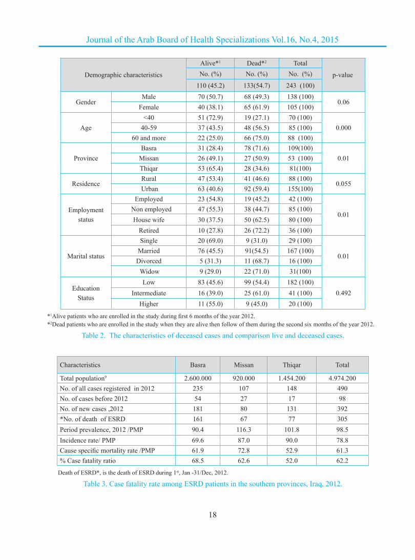
The total number of cases was 243 during the first six months of 2012 were identified in the three southern provinces. The distribution of the study group by basic demographic characteristics (Table 1). Males are slightly more than females. Although the major proportion aged 60+ (36.2%), still around two thirds of the cases were below this age and the mean age 53±8 years. Around 63.8% of the cases were of urban residence. Regarding the occupation, while housewives constituted about 32.9% of the cases, the currently employed group represented only 17.3%. Married patients represented the majority (68.7%). Around 74.9% of the cases were of low education (illiterate or primary school graduates).
As mentioned during the first six months of 2012, 243 cases were identified in the three provinces. This group was followed for the whole 2012, (up to Dec/31st), 133 (54.7%) had deceased. A comparison was made between deceased and alive patients by different demographic characteristics; the results are presented in Table 2. The proportion of deceased patients among the female group was 61.9% compared to 49.3% among the males. The difference is not statistically significant (p=0.06). The proportion of dead patients was significantly increasing with increasing age reaching the maximum among those aged more than 60 years (p=0.000). The proportion of deceased cases was significantly higher in Basra (71.6%) as compared to Missan (50.9%) and Thiqar (34.6%) (p=0.01). The proportion of deaths among urban residents was 59.9% compared to 46.6% among
rural residents (p=0.055). Regarding employment, the highest proportion of death was among retired group (72.2%) followed by the housewives (62.5%) (p=0.01). Similarly the highest proportion of death was seen among the widowed (71%) and divorced (68.7%) (p=0.01). No statistical significant association was formed between education status and fatal outcome (p=0.492).
Regarding the morbidity and mortality indicators, all data of 2012 were used. The period prevalence was
18
Journal of the Arab Board of Health Specializations Vol.16, No.4, 2015
Demographic characteristics
Alive*1 Dead*2 Total
p-value No. (%) No. (%) No. (%)
110 (45.2) 133(54.7) 243 (100)
GenderMale 70 (50.7) 68 (49.3) 138 (100)
0.06Female 40 (38.1) 65 (61.9) 105 (100)
Age<40 51 (72.9) 19 (27.1) 70 (100)
0.00040-59 37 (43.5) 48 (56.5) 85 (100)60 and more 22 (25.0) 66 (75.0) 88 (100)
ProvinceBasra 31 (28.4) 78 (71.6) 109(100)
0.01Missan 26 (49.1) 27 (50.9) 53 (100)Thiqar 53 (65.4) 28 (34.6) 81(100)
ResidenceRural 47 (53.4) 41 (46.6) 88 (100)
0.055 Urban 63 (40.6) 92 (59.4) 155(100)
Employmentstatus
Employed 23 (54.8) 19 (45.2) 42 (100)
0.01 Non employed 47 (55.3) 38 (44.7) 85 (100)
House wife 30 (37.5) 50 (62.5) 80 (100)Retired 10 (27.8) 26 (72.2) 36 (100)
Marital status
Single 20 (69.0) 9 (31.0) 29 (100)
0.01Married 76 (45.5) 91(54.5) 167 (100)Divorced 5 (31.3) 11 (68.7) 16 (100) Widow 9 (29.0) 22 (71.0) 31(100)
EducationStatus
Low 83 (45.6) 99 (54.4) 182 (100)0.492Intermediate 16 (39.0) 25 (61.0) 41 (100)
Higher 11 (55.0) 9 (45.0) 20 (100)
*1Alive patients who are enrolled in the study during first 6 months of the year 2012.*2Dead patients who are enrolled in the study when they are alive then follow of them during the second six months of the year 2012.
Table 2. The characteristics of deceased cases and comparison live and deceased cases.
Characteristics Basra Missan Thiqar Total
Total population9 2.600.000 920.000 1.454.200 4.974.200 No. of all cases registered in 2012 235 107 148 490No. of cases before 2012 54 27 17 98No. of new cases ,2012 181 80 131 392*No. of death of ESRD 161 67 77 305Period prevalence, 2012 /PMP 90.4 116.3 101.8 98.5Incidence rate/ PMP 69.6 87.0 90.0 78.8Cause specific mortality rate /PMP 61.9 72.8 52.9 61.3% Case fatality ratio 68.5 62.6 52.0 62.2
Death of ESRD*, is the death of ESRD during 1st, Jan -31/Dec, 2012.
Table 3. Case fatality rate among ESRD patients in the southern provinces, Iraq, 2012.

19
Journal of the Arab Board of Health Specializations Vol.16, No.4, 2015
Characteristics Basra Missan Thiqar Total
Total population 2.600.000 920.000 1.454.200 4.974.2009
No of new cases in 2012 181 80 131 392(%) Transplant count 6 (3.3%) 3 (3.8%) 7 (5.3%) 16 (4.1%)
Transplant pmp 2.3 3.2 4.8 3.2
Table 4. Distribution of the ESRD outcome who had a renal transplant during 2012.
Frequency of dialysisProvinces
TotalBasra Missan Thiqar
Count (%) Count (%) Count (%) Count (%)Once a week 28 (25.7) 14 (26.4) 17 (21.0) 59 (24.3)Twice a week 69 (63.3) 35 (66.0) 47 (58.0) 151 (62.1)
Thrice a week 12 (11.0) 4 (7.5) 17 (21.0) 33 (13.6)Total 109 (100.0) 53 (100) 81 (100) 243 (100.0)
Table 6. Distribution of the study group by frequency of hemodialysis/week and provinces.
(98.5 pmp), and the highest was in Missan (116.3 pmp). The incidence of ESRD was (78.8 pmp), the highest was in Thiqar (90 pmp). The mortality indicators showed that ESRD specific mortality rate was (61.3 pmp), the highest was in Missan (72.8 pmp). The case fatality ratio was 62.2%; the highest was in Basra (68.5%), Table 3.
The proportion of transplants to incident cases was highest in Thiqar (5.3%) and lowest in Basra (3.3%) and in general in the southern provinces of Iraq 4.1% had done renal transplantation, Table 4.
The average weekly duration of HD sessions hours (Table 5-A). In the three provinces was 5.1±1.34 the longest session was in Thiqar (6±1.7hr), and the shortest was in Basra 3.8±1.15 hrs. ANOVA and Tukey test were applied and revealed a statistically significant difference in average duration of HD sessions between Basra and Missan, Basra and Thiqar (p=0.000), but no significant difference between Missan and Thiqar (p=0. 41), Table 5-B.
The frequency of dialysis was reviewed in Table 6, and it was found that only 13.6% received dialysis three times a week. This proportion was higher in Thiqar (21.0%) and lowest in Missan (7.5%). The majority of the cases (62.1%) had two HD sessions per week, the lowest being in Thiqar (58.0%), Table 6.
Measurement Basra Missan Thiqar TotalMean 3.798 5.66 6.000 5.1Standard deviation 1.152 1.223 1.7 1.348Lower bound 3.553 5.309 5.614 4.825Upper bound 4.043 6.012 6.386 5.48
Table 5-A. Average (±Standard deviation) duration of hemodialysis session in the three provinces.
Provinces Mean
Difference Stander
ErrorSig
Basra Missan -1.8622* .21718 .000
Basra-Thiqar -2.2018* .23164 .000
Missan-Thiqar -.3396 .26451 .406
Table 5-B. Tukey test results.
Although there was a clear difference in the total population in the three provinces, there is one single unit in each province. The overall unit per population ratio was 0.6 pmp. As expected, the highest was in Missan (1.1 pmp), and the lowest was in Basra (0.4 pmp). The total number of HD machines was 43, and the average machine per population was 8.6 pmp; the highest was in Missan (9.78 pmp), and the lowest was in Thiqar (6.87 pmp). The average patients to machine ratio was 11.4, the highest was in Thiqar (14.8), and the lowest was in Basra (9.8) as seen in Table 7.

20
Journal of the Arab Board of Health Specializations Vol.16, No.4, 2015
Characteristics Basra Missan Thiqar Total
Number units 1 1 1 3Number of population 2.600.000 920.000 1.454.200 4.974.2009
Total patients number 235 107 148 490Number of units pmp 0.4 1.1 0.7 0.6Number of machines 24 9 10 43Number of machines pmp 9.23 9.78 6.87 8.6Ratio of patients to machines 9.8 11.8 14.8 11.4
Table 7. Hemodialysis units and hemodialysis machines to population and patient ratios in the southern provinces, 2012.
Regarding human resources, it was found that the total staff running the three units was 48. The average ratio of patients to medical staff was 10.2:1, the highest was in Thiqar (12.3:1), and the lowest was in Missan (7.1:1). Table 8.
Characteristics Basra Missan Thiqar Total
Number ofmedical staff
21 15 12 48
Total patientsnumber
235 107 148 490
Ratio of patientsto medical staff
11.2 7.1 12.3 10.2
Table 8. Ratios of patients to medical staff in southern province of Iraq, 2012.
DISCUSUON End Stage Renal Disease (ESRD) has assume
epidemic proportion worldwide hence its being regarded as the major public health challenge mean while data on incidence and prevalence of ESRD are available in developed countries because of reliability and functional renal registries, they are either unavailable or unreliable in developing countries.
SociodemographicGender: The sociodemographic characteristics of
HD patients show slight male preponderance. This is similar to another study in Baghdad in 2008 with male preponderance,10 as the males have a higher risk for
the two main risk factors of ESTD (Hypertension and Diabetes).11
Age: Mean age of HD patients in this study was 53.8 years, in study conduct in Baghdad 2009 the mean age was 46 year,10 in Al Anbar province west of Iraq 2009 the mean age was 48 year,12 less than 30% of HD cases were in the age group <40 years, and then the proportion increase with increasing age, then age group 60 year and above it reach 36.2%, ESRD is more common among elderly persons than other age groups,13 this is mostly attributed to the increased incidence of risk factors for ESRD particularly hypertension and diabetes mellitus.11,14,15,16
Marital status: High proportion of patients in this study were married and widows, this similar study in Iran17 while the majority of the females were found to be housewives, this is consistent with employment status of females in this part of Iraq.18
Educational status: Around three quarters of HD cases were of low education the illiteracy is widely spread in south of Iraq (39%) so the figure is close to the general population figures and only 22% of adult population in Iraq has never attended school and more than 9% have secondary school.18
Residence: most patients in the current study (63.2%) were from urban areas. This is almost compatible with the residence distribution of the population in the southern provinces of Iraq by ministry of planning in 2008, they found 64.2% of the populations are urban.18
21
Journal of the Arab Board of Health Specializations Vol.16, No.4, 2015
Smoking habits: In contrast to another study by yacoub et al (2010)19 most of our HD patients were non smoker.Variation in definition current smoker may be responsible for such variation. The current figure of smoker is much less than that of general population prevalence of smoking 21.9% with proportion among males was six folds than females.11
Period prevalenceThe period prevalence in the current study was 98.5
pmp; the highest was in Missan (116.3 pmp). In Al-Anbar province west of Iraq, 2009, the prevalence was 141 pmp,12 while the period prevalence of Hemodialysis in Baghdad, 2009 was 64 pmp.10 Other study in Baghdad 2012 point prevalence was 84 pmp and in Ninwa 95 pmp.1 The prevalence of ESRD in Jordan was 421, and 456 pmp in 2008 and 2010 respectively.2-20 In Saudi Arabia the prevalence was 434 and 498 pmp in 2007 and 2010, respectively.3-21 In Turkey, the point prevalence (pmp), in 2004; (444), 2005; (491), 2007; (709), 2008; (756).5-22 In Malaysia the prevalence was 747, 812 pmp in 2009 and 2010 respectively.23,24 In Egypt the prevalence was 483 pmp in 2008.25 In the United Kingdom the prevalence was 293 in 2005, 311 in 2006, 323 in 2007, 342 in 2008 and lastly 354 pmp in 2009.26
On comparing the population pyramid in Iraq with other populations, it is reported that around 43.1% of Iraqi populations are below the age of 15 years,27 compared to 15.3% in Europe, 19.6% in North America, 26.8% in South America, 40.3% in Africa, 25.1% in Asia, and at global level, the world population under 15 years old represent 26.3% of total population.28 Since ESRD is mostly an age related disorder as the main risk factors are age related. We can anticipate a higher prevalence of ESRD in population with higher proportion of elderly people. Another factor that may explain the lower prevalence in our society is the tendency in western societies to provide early renal transplant therapy (RRT) for patients with ESRD, and thus increasing the number of patients receiving HD, meanwhile the availability of renal replacement therapies is limited in low and middle income countries. Most patients around the world with chronic kidney disease will die from kidney failure without receiving dialysis or transplantation.29
In western countries, an increase in the prevalence of patients on RRT has been observed during the recent past, this result from a decreased mortality rate on the one hand and an increased in the incidence rate on the other.30 Another reason for this low prevalence is the poor services delivered to the ESRD patients in Iraq that can lead to high case fatality rate in southern region. The service delivered indicators showed a high Patient: Machines Ratio, low Hemodialysis Units: Population Ratio and low Hemodialysis Machines: Population Ratio and short duration of HD, as compared to some neighboring countries like Jordan, Saudi Arabia and Turkey. The Hemodialysis Unit: Population Ratio in Jordan,2 Saudi Arabia3 and Turkey was. 12:1000000, 7:1000000, 11:1000000 respectively, Hemodialysis Machines: Population Ratio was 124:1000000, 177: 1000000, 202:1000000 respectively.22
Incidence The incidence of ESRD was 78.8 pmp, the
highest was in Thiqar (90 pmp). The incidence of RRT in some countries of the developing world: in Egypt 190 PMP, in Saudi Arabia 130 PMP, Pakistan 40 PMP, India 100 PMP, Argentine 120 PMP, Venezuela 120 PMP, and Mexico 340 PMP.31 The incidence of renal failure is increasing all over the world, in UK 93 new patients per millions were dialyzed in 2001. In USA, 336 new patients per millions are added each year.32 In Yemen, in 2002, an incidence of 64 per million per year was reported.33 So lower incidence was reported in the current study as compared to Egypt, S. Arabia, India, Argentine, Venezuela, USA and Mexico but higher than that of Pakistan and Yemen. Under diagnosis, possibility of early death shortly after diagnosis while they are receiving peritoneal dialysis is possibility.
Case Fatality Ratio (CFR): The case fatality ratio within 2012 was 62.2%, the highest was in Basra 68.5%.age is significance with deceased cases ESRD. Published reports demonstrated that the case fatality ratio in USA is 22.8%, in Canada it was 17.9%, in Western Europe it was 10.4 %.34 In Saudi Arabia during the year 1997 to 2000 the death rate per annum was varied from 3.5 to 19.5.35 In 2004 in Taiwan CFR was 5.18 within two years, and the cause mortality rate was 20.3 per 1000 person year.36 The influencing factors for variation in death rate are: age at which patients are inducted

22
Journal of the Arab Board of Health Specializations Vol.16, No.4, 2015
for maintenance hemodialysis program, associated co-morbid condition.34 late arrival with complications at time of initiation of HD,37 and compliance with HD.38
The high mortality in south of Iraq is also due to the poor delivered services as indicated by the high Patient: Machines Ratio, low Hemodialysis Units: Population Ratio and low Hemodialysis Machines: Population Ratio.
Infrastructure of HD units
1. Human resources: Human resources working in HD units are unequally distributed throughout Iraqi southern provinces. The current ratio of all medical staff (including the specialized and non-specialized) in south of Iraq is far from global ratio. This is another discrepancy that may lead to poor quality of service offered to the ESRD patients. In Jordan 2008, the total number of Nephrologists caring of ESRD patients was 51 with the average of 52 patients for each Nephrologist.20 In Saudi Arabia the total number of Consultant Nephrologists, Nephrology Specialists, General Practitioner and Nurses caring of ESRD patients was 172, 278, 246 and 3239 respectively in 2010.3 Also in Turkey the number of Specialist Physicians, General Practitioner and Nurses caring for ESRD patients in 2008 was 733, 1051, and 4393, respectively.22
2. Hemodialysis unit and machinery resources: The total number of Hemodialysis Centers in the south of the Iraqi was three making very low ratio (0.6 pmp) as compared with Baghdad in 2012 (1.4 pmp).1 Other countries like Jordan had 72 Hemodialysis Units in 2010, making Hemodialysis Units: Population Ratio 12: 1.000.000.2 In Saudi Arabia there was 177 Hemodialysis Units in 2010, making Hemodialysis Units: Population Ratio of 7:1000000.3 In Iran there are 305 Hemodialysis Units in 2006, making Hemodialysis Units: Population Ratio 4.24:1000000.4 Similarly in Turkey there were 754 Hemodialysis Units in 2008, making Hemodialysis Units: Population Ratio of 10.41:1.000.000.5 This low ratio in southern provinces in Iraq is one of the causes behind the high case fatality rate and low prevalence.
The total number of HD machines in southern provinces of Iraq was 43 and the number of machines
per million populations was 8.6. In Baghdad city, 2012, the ratio was 27.25 machines /pmp, and in Ninawa it was 14.12 machines/pmp.1 The ratio of patients to machines in south region was 11.3. In Bagdad, 2012 this ratio was 3.09 patients: machine ratio, and in Ninawa it was 6.7 patients: machine ratio.1
Frequency and duration of HD: In southern provinces of Iraq only 13.6% of the patients had three sessions per week while 62.1% had twice per week and 24.3 had once per week. In Iran, the frequency of three sessions per week was 60%4; in Jordan it was 49%.2 Globally, despite ongoing technical care, improvements in both dialysis and overall patient care, the annual mortality rate of patients with ESRD managed with thrice weekly HD remains high (10-22%).7,8 The low proportion of thrice weekly dialysis in the current study is one of the areas of poor services provided to HD cases and hence the high mortality. As compared to the recommended average HD duration of 12 hours/week.6 The mean duration of hemodialysis session was (5.1±1.3 hrs/week). In Baghdad, the average duration was 6.4 hours.10 In Jordan, the mean duration was 9.6 hrs/week.39 It is clear that the duration is far from the adequate adding the indicator of poor delivered services that lead to high case fatality ratio.
The average survival of HD patients in the three provinces was 13.3 months. This is compared to 26 months in Baghdad in 200910 and 4.3 years in Jordan, 2008.2
Renal transplantation: During the year 2012, the proportion of transplants to incident cases was only 4.1%. In the Gulf Co-operation Council countries (1994), the estimated incidence of ESRD was 75-120 new cases pmp per year, the proportion of transplants to incident cases was 30%; still it was presumed that about 60% of these patient need transplants (50-100 patients). While the ratio of transplantation per million population in the south of Iraq was 3.2 which is lower than GCC countries (in Bahrain 8 pmp, Kuwait 65 pmp, Oman 38 pmp, Qatar 29 pmp, Saudi Arabia 21 pmp ,and UAE 17 pmp)40 and lower than the USA 2010 figure of 50 pmp.41 The renal transplantation rate depends on many economic and social factors.42

23
Journal of the Arab Board of Health Specializations Vol.16, No.4, 2015
CONCLUSIONS
Males had slightly higher proportion than females and around two thirds of the patients were below the age of 60 years. The prevalence and incidence of ESRD in Southern provinces are 98.5 pmp, and 78.8 pmp, respectively. The outcomes of ESRD the cause specific mortality rate was 61.3 pmp, and the case fatality ratio were 62.2%, Only 4.1% of HD patients in southern provinces of Iraq had renal transplant annually. Death due to ESRD high proportion with increasing age and high proportions with marital status, employment status and with provinces. The HD unit to population ratio was 0.6 pmp. HD machine to population ratio was 8.6 pmp, patients to machine ratio was 11.4:1 and medical staff to patient ratio was 10.2:1. All were below the international and regional figures. Average duration of HD session was 5.1 hours per week; less than half of the recommended duration, so we recommend that the extremely high case fatality ratio and low quality services should be addressed and approached the policy maker to ensure strong political commitment towards ESRD patients. Secondly the number of HD units, machines and the medical staff should be increased to meet the patient’s needs, and ensure 12 hours weekly HD. Thirdly Develop Standardized Operation Procedures (SOPs) in the HD units and ensure strict implementations of these SOPs. Fourthly early detection of all patients at risk of impaired renal function through screening to minimize ESRD. Fifthly enhance renal transplantation in southern part of Iraq. Seventhly provision of adequate number of qualified medical and health personnel running HD unit. Lastly developing a national registry of End stage renal Disease.
REFERENCES
1. Al-lami F. Infrastructure of hemodialysis units in Iraq: Situation analysis. Thesis: Board, Ministry of higher education 2012. p. 71.
2. The Hashemite Kingdom of Jordan/MOH/Non- Communicable Disease Directorate, National Registry of End Stage Renal Disease; Bi-Annual Report 2009 -2010, available at Web Site of the Ministry of Health: www.moh. gov.jo .[accessed Jun 2012].
3. Shaheen F, Souqiyyeh M, Al-Sawailem A. Renal replacement therapy organ donation and transplantation
in the kingdom of Saudi Arabia, Annual Report 2010.4. Aghighi M, Heidary R, Zamyadi M. et al. Dialysis in
Iran. Iran J Kidney Dis 2008;2(1):11-5.5. Turkish Society of Nephrology, Registry of the Nephrology,
Dialysis and Transplantation in Turkey Registry 2009.6. Arogundade F, Sanusi A, Hassan M, et al. The pattern,
clinical characteristics an outcome of ESRD in Ile-Ife, Nigeria: is there a change in trend?. Afr Health Sci 2011;11(4):594-601.
7. Rayner C, Pisoni RL, Bommer J, et al. Mortality and hospital in hemodialysis patients in five European countries: Result from the Dialysis Outcomes and Practice Patterns Study (DOPPS). Nephrol Dial Transplant 2004;19(1):108-20.
8. Ganesh K, Hulbert-Shearon T, Port FK, et al. Mortality differences by dialysis modality among incident ESRD patients with and without coronary artery disease. J Am Soc Nephrol 2003;14(2):415-24.
9. Ministry of Iraqi Planning. Central organization for statistics and information technology 2012.
10. Al-Saedy AJ, Al-Kahichy H. The current status of hemodialysis in Baghdad. Saudi J Kidney Dis Transpl 2011;22(2):363-7.
11. WHO Chronic Non Communicable Diseases Risk Factors Survey In Iraq: Ministry of Health, Directorate of Public Health and Primary Health Care and Ministry of Planning and Development Cooperation 2006. p.1-130.
12. Awad S. Chronic renal failure in Al Anbar of Iraq. Saudi J Kidney Dis Transpl 2011;22(6):1280-4.
13. Ahmed AK, Brown SH, Abedelhafiz AH. Chronic kidney disease in older people disease or dilemma. Saudi J Kidney Dis Transpl 2010;21(5):855-41.
14. McEniey M, Wilkinsonlan B, Avolio P. Age, hypertension and arterial function. Clin Experimental Pharmacol Physiol 2007;34(7):665-71.
15. Kesavadev D, Short R, Nair S. Diabetes in old age: An emerging epidemic. J Assocphys India 2003;51:1083-94.
16. Centers for Disease Control and Prevention. National diabetes fact sheet: national estimates and general information on diabetes and pre diabetes in the United States, 2011. Atlanta, GA: U.S. Department of Health and Human Services, Centers for Disease Control and Prevention. Available at www.cdc. gov/diabetes /pubs/ references11.htm. [Accessed Aug 2012].

24
Journal of the Arab Board of Health Specializations Vol.16, No.4, 2015
17. Malekmakan L, Haghpanah S, Pakfetrat M, et al. Cause of chronic renal failure among Iranian hemodialysis patients. Saudi J kidney Dis Transpl 2009;20(3):501-4.
18. Baban AG, Pollus SM. National development plan for the Years 2010-2014. Republic of Iraq, Ministry of Planning, Baghdad. p. 1-186.
19. Yacoub R, Habib H, Lahdo A, et al. Association between smoking and chronic kidney: as case control study. BMC Public Health 2010;(10):1-703.
20. The Hashemite Kingdom of Jordan/MOH/Non-Communicable Diseases Directorate, National Registry of End Stage Renal Disease; Annual Report 2008.available at Web Site of ministry of health: wwww.moh.gov.jo. [accessed Jun 2012].
21. Shaheen F, Souqiyyeh M, Al-Sawailem A. Renal replacement therapy organ donation and transplantation in the kingdom of Saudi Arabia, Annual Report 2007.
22. Turkish Society of Nephrology, Registry of the Nephrology, Dialysis and Transplantation in Turkey Registry 2004.
23. Malaysian Society of Nephrology, 17th Report of the Malaysian Dialysis & Transplant Registry 2009, available at http://www.msn.org.my/nrr, ISSN 1675-8862. Accessed Jun 2012.
24. Malaysian Society of Nephrology, 17th Report of the Malaysian Dialysis & Transplant Registry 2010, available at http://www.msn.org.my/nrr, ISSN 1675-8862. Accessed Jun 2012.
25. Afifi A. The Egyptian Renal Registry, 9th, Annual Report 2008.
26. The Renal Association, 2010 UK Renal Registry, 13th Annual Report, p. 42. www.renalreg.org.
27. United Nations Country Team, Iraq, 2010. United Nations Development Assistance Frame for Iraq 2011-2014, p. 10.
28. World stat info, available at http://en.worldstat.info. [accessed at 19-8-2012].
29. James MT, Hemmelgarn BR, Tonelli M. Early recognition and prevention of chronic kidney disease. Lancet 2010;375(9722):1296-309.
30. Haghighi A, Broumand B, Amico Ma, et al. The
epidemiology of ESRD in Iran an international perspective. Nephrol Dial Transplant 2002;(17):28-32.
31. Barsoum R. Over view of the end stage renal disease in developing country. Artif Organs 2002:26(9):737-46.
32. Jeremy MA, Julie M, Edwina B. Oxford Handbook of dialysis, 2nd ed. Oxford University Press, New York, United State; 2004. p. 96-100.
33. Al-Rohani M. Renal failure in Yemen. Transplant Proc 2004;36(6):1777-9.
34. Mendelssohn D, Kriger F, Winchester J. A comparison of dialysis in the US and Canada. Contemp Dial Nephrol 1993;14:27-30.
35. Subramanian T, Jamal A, Shah Y. Hemodialysis utilization in a single in center dialysis unit, in the Kingdom of Saudi Arabia. Saudi J Kidney Transplant 2001;12(1):64-74.
36. Hwang SJ, Lin MY, Chen HC, et al. Increased risk of mortality in the elderly population with late-stage chronic kidney disease: a cohort study in Taiwan. Transplant 2008;23(10):3192-8.
37. Sesso R, Belasco AG, Ajzen H. Diagnosis of chronic renal failure. Braz J Med Biol Res 1996;29:1473-8.
38. Beley AJ, Hylander B, Sudo H. An international study of patient compliance with hemodialysis. JAMA 1999;281(13):1211-3.
39. Tayyem R, Mrayyan M, Heath D, et al. Assessment of the nutritional status among ESRD patients in Jordanian Hospitals. J Ren Nutr 2008;18(3):281-7.
40. Shaheen A, Salmeen M, Al-Muzairy A, et al. Renal transplantation in Gulf Co-operation Council Countries. Saudi J Kidney Dis Transplant1995;6(1):5-8.
41. National Kidney and Urologic Diseases Information Clearinghouse (NKUDIC) United States Renal Data System’s (2010 ) Annual Data Report and 2011 Annual Data Report. Available at http://kidney.niddk.nih.gov/kudiseases/pubs/kustats/ [accessed Jun 2012].
42. Abecassis M, Bartlett T, Collins J, et al. Kidney transplantation as primary therapy for ESRD: a National Kidney Foundation/Kidney disease outcomes quality initiative (NKF/KDOQITM) conference. Clin J Am Soc Nephrol 2008;3(2):471-80.

25
Journal of the Arab Board of Health Specializations Vol.16, No.4, 2015
Original Article
ملخص البحث
المقيمين األطفال عند األبواغ بخفيات الخمج دراسة البحث: هدف والمراجعين لمشفى األطفال بمدينة دمشق ومعرفة نسبة اإلصابة بها.
طرق البحث: تم جمع 146 عينة براز من األطفال المقيمين والمراجعين لمشفى األطفال الجامعي بمدينة دمشق دون الخمس سنوات من العمر وذلك في الفترة الواقعة بين شهر آب 2013 حتى شهر كانون الثاني 2014. تم تسجيل البيانات المتعلقة بتاريخ أخذ العينة، عمر المريض، جنسه واألعراض استبيان خاص. سابق ضمن استشفاء أو أدوية تناول وقصة السريرية
تم فحص العينات عيانيًا ومجهريًا بالفحص الندي مع إجراء لطاخة براز المتكيسة البيوض وجود لتحري المعدلة نلسون تسيل- بطريقة ملونة
لخفيات األبواغ.النتائج: بلغ عدد العينات اإليجابية 6 عينات من أصل 146 عينة والمراجعين المقيمين األطفال عند %4.1 االنتشار نسبة بلغت حيث
لمشفى األطفال بمدينة دمشق. االستنتاجات: لقد أظهرت دراستنا أن نسبة انتشار خفيات األبواغ تبلغ أنه ال يمكن تشخيص العلم 4.1% عند األطفال وهي نسبة عالية مع الخفيات بالفحص الندي مما يستوجب إدخال تحري خفيات األبواغ ضمن
STUDY OF CRYPTOSPORIDIUM INFECTION AMONG CHILDREN ATTENDING THE PEDIATRIC HOSPITAL IN DAMASCUS
دراسة الخمج بخفيات األبواغ عند األطفال المراجعين لمشفى األطفال في مدينة دمشق Elen Ibrahim, MD; Morshed Kassouha; Abeer Al-kafri, MD
د.ايلني ابراهيم، د.مرشد كاسوحة، د. عبير الكفري
ABSTRACT Objective: The aim of this study was to estimate the prevalence of cryptosporidium among children attending the
Pediatric Hospital in Damascus. Methods: 146 stool samples were collected from children attending the Pediatric Hospital in Damascus. Children
were aged under 5 years. The study was conducted during the period from August 2013 to December 2014. Information about date of the sample collection, age, gender, symptoms, history of medications and previous hospitalization was taken. Samples were examined visually and microscopically by wet mount. Fecal smears were prepared and stained by modified Zeihl-Neelsen technique to detect the presence of cryptosporidium oocysts.
Results: Six samples out of 146 were positive which results in 4.1% infection rate among children attending the Pediatric Hospital in Damascus city.
Conclusions: Our study showed that the prevalence of cryptosporidium among children is 4.1% which is considerable compared to other parasites. We suggest routine stool examination for cryptosporidium by modified Zeihl-Neelsen technique or other available methods and the necessary of improving the sewage system, providing better water resources and the importance of health education to reduce the infection.
*Elen Ibrahim, MD; Damascus University, Damascus, Syria. E-mail: [email protected]*Morshed Kassouha; Master degree in Veterinarian Medicine at Hama University, Syria. *Abeer Al-kafri, MD; Assistant Professor in Damascus University, Arab International University (AIU), Damascus, Syria.
مو�صوع �أ�صيل

26
Journal of the Arab Board of Health Specializations Vol.16, No.4, 2015
الفحص الروتيني للبراز وذلك باستخدام تلوين تسيل- نلسون المعدل أو غيرها من الطرق التشخيصية األخرى المتوافرة مع أهمية تحسين الصرف الصحي ومصادر المياه وتطوير برامج التوعية الصحية للعائلة وذلك للحد
من اإلصابة.
مقدمة
الظهارية الخاليا تصيب طفيلية بوائغ هي األبواغ خفيات االقتصاد على مؤثرة اإلنسان فيها بما الفقاريات من للعديد المخاطية العالم.1 حول والحيوانات األشخاص لماليين العامة والصحة أول من اكتشف خفيات األبواغ هو تيزر Tyzzer عام 1912م واعتبرها العشرين القرن من الثمانينيات وفي 1976م. عام لإلنسان ممرضة أصبحت تعد من العوامل المسببة إلنتان شديد مهدد للحياة عند المرضى مثبطي المناعة خاصة مرضى اإليدز. إن الخمج بخفيات األبواغ محدد
لذاته عند أسوياء المناعة خاصة األطفال.
تشمل اإلصابة بخفيات األبواغ األطفال والبالغين لكنها تعد المسبب األساسي لحدوث اإلسهال عند األطفال تحت عمر الخمس سنوات في قد يحدث أو المصابون ال عرضيين يكون قد والمتقدمة. النامية الدول لديهم اسهال مزمن أو حاد اعتمادًا على العمر وحالة الجهاز المناعي.2
يتظاهر اإلنتان بخفيات األبواغ في األشخاص أسوياء المناعة باسهال حاد محدد لذاته، أما عند المرضى مثبطي المناعة )تحت تأثير األدوية أو اإليدز أو وجود انتان حالي كالحصبة أو الجدري( فتكون األعراض الشوارد فقدان إلى مؤدية مزمنة تصبح وقد شدة أكثر لديهم السريرية
ونقص الوزن وحتى الوفاة أيضًا.2،1
كشف أكثر من 20 نوعًا من خفيات األبواغ حتى اآلن وتعد خفيات C. Parvum وخفيات األبواغ الصغيرة C. Hominis األبواغ البشريةالنامية. يتوضع طفيلي الدول للبشر خصوصًا في أكثر األنواع إصابة خفيات األبواغ داخل الخلية لكن خارج الغشاء الهيولي للخاليا الظهارية البطانية ألمعاء العديد من األسوياء وقد يوجد في مخاطيات خارج األمعاء مثل الطرق التنفسية أو المرارة خصوصًا عند المرضى مثبطي المناعة.1
Oocyst المتكيسة البيوض هو األبواغ لخفيات المعدي الشكل إن التي يتم طرحها مع البراز و تقيس بين )4-6 مكرون( ذات شكل دائري طبقات ثالث من يتكون نانومتر 49 يقيس سميك وجدار بيضوي أو trilaminar وتحوي في داخلها على أربع بوائغ sporozoites متحركة كما تحوي على أجسام متبقية residual bodies تلعب دورًا في تأمين التغذية والطاقة لألبواغ.3 ال يمكن بسهولة تمييز البيوض المتكيسة بالبراز
طرق إلى اللجوء يتم ما غالبًا لذلك ،wet mount 4الندي بالفحص التكثيف أو تلوين لطاخة براز.
يتم عادة تشخيص خفيات األبواغ عن طريق الفحص المجهري المعدلة نلسون تسيل طريقة باستخدام ملونة براز للطاخة الطريقة تعتبر زالت ما إذ )mZN( modified Zeihl-Neelsenالرغم من النامية على الدول الطبية خصوصًا في المخابر الذهبية في المقايسة أو المناعي االستشراب مثل أخرى تشخيصية طرق وجود وتضخيم المباشر وغير المباشر المناعي التألق أو االنزيمية المناعية polymerase البوليميراز سلسلة تفاعل باستخدام النووي الحمض chain reaction (PCR) وذلك بسبب كلفتها القليلة وحساسيتها الجيدة
مقارنة مع غيرها من الطرق.5-1
المتكيسة، البيوض تناول طريق عن اإلنسان عند اإلصابة تحدث ويختلف عدد البيوض المتكيسة المسببة لإلصابة من مرجع آلخر، لكن معظم المراجع تشير إلى أن الحد األدنى من البيوض المتكيسة هو 10 بيوض متكيسة لتسبب اإلصابة عند اإلنسان سوي المناعة، ومن الجدير ذكره أن اإلنسان والحيوانات المصابة تطرح ما يقارب )710 - 1110(
بيضة متكيسة في كل غرام من البراز.6
كشفت دراسة سريرية7 ووبائية ضخمة في مطلع 2013 ُأجريت على 22500 طفاًل من أفريقيا وآسيا -بشكل غير متوقَّع- أنَّ طفيلي خفيات األبواغ هو واحد من أربعة ُمْمِرضات مسؤولة عن الّتسبب في أكثر حاالت تقديرات -وفق اإلسهال أن كما ع. والرُّضَّ األطفال لدى الحاّد اإلسهال ماليين 8 10.5%من مسؤول عن -WHO العالمية الصحة منظمة
حالة وفيات تقريبًا لدى األطفال تحت سن الخامسة كل عام.8
تختلف نسبة االنتشار عالميًا إضافة إلى اختالفها ضمن الدولة نفسها الكشف ضمن دخالها واإ وحساسيتها التشخيص طرق الختالف نظرًا الروتيني أم ال وتعتبر نسبة اإلصابة عن طريق كشف البيوض المتكسية بالبراز بشكل عام أقل من نسبة اإلصابة المكتشفة بالطرق المصلية.1,5 إن نسبة االنتشار تتراوح وسطيًا بين 2.1% في الدول المتطورة و8.5% في الدول النامية وتعتبر مسؤولة عن 30-50% من وفيات األطفال تحت عمر الخمس سنوات في هذه الدول. لقد تم دراسة االنتشار عند األطفال تونس. فلسطين، ليبيا، العراق، األردن، مثل مصر، العربية الدول في وقد تراوحت النسبة بين 1-43% )وسطيًا 8.7%( عند األطفال أسوياء )وسطيًا %82-1 بين النسبة وتراوحت باإلسهال المصابين المناعة المناعة من األطفال والبالغين مع مالحظة زيادة 41%( عند مضعفي جرى أنه بالذكر الجدير ومن المدن9 سكان عن البادية سكان إصابة
تشخيص خفيات األبواغ في سورية ألول مرة في عام 1991م.10

27
Journal of the Arab Board of Health Specializations Vol.16, No.4, 2015
طرق البحث
تم جمع 146 عينة براز من األطفال المقيمين والمراجعين لمستشفى األطفال الجامعي في مدينة دمشق والذين تتراوح أعمارهم بين 40 يومًا وأقل من خمس سنوات وذلك في الفترة الواقعة ما بين شهر أب 2013 حتى شهر كانون الثاني 2014. وتم تسجيل البيانات المتعلقة بتاريخ أخذ العينة وعمر المريض وجنسه واألعراض السريرية وقصة تناول أدوية أو استشفاء سابق. ومضادات المعوية المطهرات يتناولون الذين األطفال استبعاد تم الحواضن. شعبة أطفال جانب إلى الشرجية والرحضات اإلسهال أجري فحص كل العينات عيانيًا لتحديد اللون والقوام ومجهريًا باستخدام أو طفيليات وجود لتحري الفيزيولوجي والمصل باليود الندي الفحص بيوض ثم تم تحضير لطاخة براز وتلوينها باستخدام تلوين تسيل- نلسون 11 وذلك بعد أخذ جزء من العينة واستحالبها بالمصل
)mZN) المعدلتثبت وبعدها جفافها انتظار ثم الزجاجية الشريحة على الفيزيولوجي الكثيف بالفوكسين الشريحة تغمر ذلك وبعد دقائق 5 لمدة بالفورمول الحامضي )حمض اللون لمدة 4 دقائق ثم تغسل ونضيف بعدها مزيل الكلور بتركيز 1%( لمدة دقيقة مع الغسل ثم يضاف زرقة الميتيلين أو أخضر المالشيت كملون تباين لمدة 3 دقائق ثم تغسل وتجفف. تفحص .(X100( ثم العدسة الغاطسة (X40( الشريحة باستخدام العدسة الجافة
تظهر البيوض المتكيسة لخفيات األبواغ بشكل دائري إلى بيضوي قلياًل حدود مع األحمر إلى مائل زهري لون ذات مكرون 6-4 بين تقيس واضحة وتلون غير متجانس مع وجود حبيبات داكنة اللون داخلها وقد
يشاهد حولها هالة بيضاء بسبب حدوث االنكماش، الشكل 1.
تم تحديد العينات السلبية وااليجابية وفق التلوين باستخدام طريقة تسيل .PCR نلسون المعدلة، وتم تأكيد العينات اإليجابية بفحصها بالـ
النتائج
يومًا وخمس الـ 40 بين عمر تم جمع 146 عينة برازية لألطفال سنوات وتوزعت العينات بين الذكور واإلناث إذ شملت الدراسة 89 ذكرًا عينات 109 بين العينات توزعت .)%39.1( أنثى و57 )%60.9)إسهالية مع 37 عينة غير إسهالية وقد وجد لدينا بالتلوين المقاوم للحمض %4.1 بنسبة أي األبواغ لخفيات المتكيسة البيوض تحوي عينات 6)الجدول 1(، توزعت العينات اإليجابية بين الذكور واإلناث إذ وجد لدينا 4 عينات عند الذكور مقابل 2 عند اإلناث وتوزعت العينات حسب العمر نسبة مع شهرًا 30 اإلصابة عمر متوسط بلغ إذ ،)2 )الجدول وفق إصابة 5.4% لعمر سنتين وأقل و4.1% لعمر بين السنتين و4 سنوات.
شوهدت 5 عينات إيجابية خفيات األبواغ ضمن العينات اإلسهالية بنسبة اإلسهالية غير الحاالت ضمن شوهدت واحدة عينة مقابل )%4.6)
.)%2.7)
شوهدت طفيليات أخرى أيضًا ضمن الفحص الندي للعينات البرازية تحوي التي اإليجابية العينات نسبة كانت إذ 3 الجدول توزعت حسب طفيليات بغض النظر عن خفيات األبواغ 2.7% ويشغل طفيلي الجيارديا مع الطفيليات لهذه مشاركة وجود إلى إضافة ،%5.4 األكبر النسبة خفيات األبواغ كما يوضح الجدول 4، ولم تشاهد بيوض ألي ديدان أبدًا. بلغ عدد العينات اإليجابية في فصل الصيف عينتين فقط بينما شوهدت
4 في فصل الشتاء.
عدد العينات السلبية عدد العينات االيجابية عدد العينات الكلي
140 6 146
%95.9 %4.1 %100
الجدول 1. عدد العينات الكلي وتوزعها بين االيجابي والسلبي.
4-5 سنوات 2 -4 سنوات 40 يوم -2 سنة الفئة العمرية
9 63 74 عدد العينات
0 2 4 عدد العينات اإليجابية
الجدول 2. توزع العينات بالنسبة للفئات العمرية. الشكل 1. البيوض المتكيسة بعد التلوين باستخدام mZN (الباحثة(.

28
Journal of the Arab Board of Health Specializations Vol.16, No.4, 2015
خفيات األبواغ +جيارديا
خفيات األبواغ جيارديا المتحول
الزحاري المتحول الكولوني
عدد العينات الكلي
2 6 8 2 4 146
%1.3 %4.1 %5.4 %1.3 %2.7 %100
الجدول 3. توزع الطفيليات المشاهدة بالنسبة للعدد الكلي للعينات.
طفيليات أخرى خفيات األبواغ عدد العينات ايجابية الطفيليات
14 6 20
%70 %30 %100
الجدول 4. توزع خفيات األبواغ والطفيليات األخرى في العينات إيجابية الطفيليات ونسبها.
المناقشة
البشر عند لإلسهال المسببة الطفيليات أحد األبواغ خفيات ُتعد وتشخيصها يعتمد على طريقة خاصة بتلوين عينة البراز وألن كشفها ال يتم بفحص البراز الندي روتينيًا فإن الدراسات حولها قليلة جدًا في بلدنا لذا كان هدف دراستنا هو معرفة نسبة انتشار خفيات األبواغ عند األطفال تحت عمر الخمس سنوات المقيمين والمراجعين لمشفى األطفال الجامعي
في دمشق.
لقد أظهرت دراستنا نسبة انتشار بلغت 4.1% ضمن 146 عينة برازية بالعينات العينات االسهالية 4.6% مقارنة نسبة اإلصابة في مع زيادة بحدوث األبواغ خفيات ارتباط إلى يوجه مما %2.7 االسهالية غير نسبة هامة من االسهاالت عند األطفال ولم يشاهد أي اختالف بين نسبة اإلصابة بين الذكور واإلناث )p<0.5(. كانت نسبة اإلصابة األكبر تحت عمر السنتين قد يعود ذلك أن هذه السن هي بداية سن المشي مع األطفال واالحتكاك مع البيئة واألطفال اآلخرين مما يزيد فرص العدوى. لقد بلغت اإلصابات الطفيلية ضمن العينات نسبة 2.7% ولم تشاهد أي بيوض ألي ديدان أبدًا مما يدل على تحسن الظروف الصحية والمعيشية وزيادة الثقافة الصحية إلى جانب تحسين الصرف الصحي ومصادر المياه األمر الذي أدى إلى تراجع نسبة اإلصابة الطفيلية. لقد شوهدت نسبة المشاركة األكبر بين طفيلي خفيات األبواغ مع الجيارديا 1.3% مقارنة مع باقي الطفيليات وقد شكلت الجيارديا النسبة األكبر من اإلصابات الطفيلية 5.4% األمر
الذي يستدعي التفكير بوجود تلوث في مصادر المياه والتي تعتبر عاماًل هامًا في انتشار هذين الطفيليان معًا، كما أن زيادة نسبة اإليجابيات في فصل الشتاء مقارنة مع فصل الصيف يوجه إلى هذا االحتمال أيضًا حيث العالية المشاهدة أن خفيات األبواغ ال تتحمل التجفاف ودرجات الحرارة
بالصيف.
عند مقارنة دراستنا مع دراسات أخرى وجد أن نسبتنا أقل بكثير من النسبة المشاهدة في الدراسة التي أجريت في دمشق12 عام 1999م من قبل اسماعيل والتي كانت نسبة االنتشار فيها 24.09% وقد يعود هذا الظروف وتحسن الصحي الوعي زيادة إلى انتشارها نسبة في التراجع وريفها دمشق مدينة في الصحي الصرف تحسن مع للسكان المعيشية الصحي سنة للصرف إنشاء محطة عدرا بعد الفترة خصوصًا تلك منذ 2000 -2001، كما لم يشاهد أي فرق في اإلصابة بين الجنسيين مع ذروة لإلصابة بعمر تحت الخمس سنوات بنسبة 71.7% خصوصًا بعمر 10-15 شهرًا، وشوهدت المشاركة مع الجيارديا كما في دراستنا بسبب بالصرف الخاصة المياه معالجة طرق لكافة الجيارديا طفيلي مقاومة
الصحي وكانت النسبة 13.2% وقد يرجع ذلك لكبر حجم العينة.
المشاهدة في مصر النسبة النسبة في دراستنا أقل أيضًا من وكانت إذ بلغت 15.3%13 مع نسبة إصابة أعلى بعمر 1-2 سنة دون وجود المدروسة والمنطقة البيئية بالظروف االختالف إن الجنسين، بين فرق ودرجة الوعي الصحي واختالف مصادر المياه والتي تعد من أهم مصادر هو يكون قد المجهرية وخبرته الفاحص اختالف إلى إضافًة االنتشار
السبب الختالف هذه النسبة.
وفي دراسات مشابهة كالتي أجريت في مدينة أصفهان في إيران بلغت سنوات الخمس عمر تحت كانت اإلصابات ومعظم 14%،4.6 النسبة (89.2%( )وسطيًا عمر 38 شهرًا( مع عدم وجود فرق باإلصابة بين الجنسين. وفي كينيا بلغت النسبة أيضًا 4%15 دون وجود فرق باإلصابة بين الجنسين مع وسطي عمر اإلصابة 36 شهرًا وهي نسب قريبة للنسبة
التي حصلنا عليها في دراستنا.
االستنتاجات
لقد أظهرت دراستنا أن نسبة انتشار خفيات األبواغ تبلغ 4.1% عند المعوية األخرى مما يستوجب الطفيليات األطفال وهي نسبة عالية بين إدخال تحري خفيات األبواغ ضمن الفحص الروتيني للبراز وذلك باستخدام األخرى التشخيصية الطرق من أو غيرها المعدل نلسون تسيل- تلوين المتوافرة مع أهمية تحسين الصرف الصحي ومصادر المياه وتطوير برامج
التوعية الصحية للعائلة وذلك للحد من انتشارها.

29
Journal of the Arab Board of Health Specializations Vol.16, No.4, 2015
REFERENCES
1. Sears LC, Kirkpatrick DB. editors. Principle and practical of clinical parasitology.1st ed. NewYork: John Wiely & sons; 2001. p. 140-55.
2. Warrell D, Cox MT, Filth J. editors. Oxford textbook of medicine: Infection. 5th ed. United Kingdom: Oxford university press; 2012. p.722-32.
3. Xiao L, Fayer R. editors. Cryptosporidium and cryptosporidiosis. 2nd ed. USA: CRC press; 2007. p. 21-27.
4. Casemore DP, Armstrong M, Sands RL. Laboratory diagnosis of cryptosporidiosis. J Clin Pathol 1985; 38(12):1337-41.
5. Vohra P, Sharma M, Chaudhary U. A comprehensive review of diagnostic techniques for detection of cryptosporidium parvum in stool samples. IOSR J Pharmacy 2012;2(5):15-26.
6. Satoskar RA, Simon LG, Hotez JP. editors. Medical parasitology, 1st ed. Landes Bioscience: USA; 2009. p. 214-21.
7. Kotloff KL, Nataro JP, Blackwelder WC, et al. Burden and aetiology of diarrhoeal disease in infants and young children in developing countries (the Global Enteric Multicenter Study, GEMS): a prospective, case-control
study. Lancet 2013;382(9888):209-22.8. Liu LI, Johnson HL, Cousens S, et al. Global, regional,
and national causes of child mortality: an updated systematic analysis for 2010 with time trends since 2000. Lancet 2012;379(9832):2151-61.
9. Ghenghesh SK, El-Mohammady H, Franka E. Cryptosporidium in countries of the Arab world: the past decade (2002-2011). Libyan J Med 2012;7.
10. Ismail MT. La cryptosporidiosis en Syria. Nouvelles Scientifiques 1995; p. 72-6.
11. Goldman E, Green HL. Practical handbook of microbiology, 2nd ed. New York; CRC press: 2009. p. 41-2.
12. Ismail MT, Al kassar BD. Cryptosporidium in children.Damascus university 1999.
13. El-helaly N, Aly MM, Attia SS. Detection of cryptosporidium infection among children with diarrehea. New York Science J 2012;5(7):68-76.
14. Saneian H, Yaghini O, Yaghini A, et al. Infection rate of cryptosporidium parvum among diarrheic children in Isfahan. Iran J Pediatr 2010;20(3):343-7.
15. Gatei W, Wamae C, Mbae C, et al. Cryptosporidiosis: prevalence, genotype analysis and symptoms associated with infections in children in Kenya. Am J Trop Med Hyg 2006;75(1):78-82.

30
Journal of the Arab Board of Health Specializations Vol.16, No.4, 2015
Original Article
THE RESULTS OF KIDNEY TRANSPLANTATION IN RECIPIENTSOPERATED BY DJ-STENTED VERSUS NON-STENTED
URETERONEOCYSTOSTOMYنتائج زرع الكلية عند المرضى المجرى لهم
زرع الحالب مع ستنت حالبي أو بدونهAssem Nasser, MD; Mahdi Dagher, MD; Ammar Raiy, MD
د. عاصم ناصر ، د. مهدي داغر ، د. عمار الراعي
ملخص البحث
هدف البحث: لتبيان جدوى االستعمال الروتيني للستنت الحالبي عند
مرضى زرع الكلية، قمنا بإجراء مقارنة نتائج عملية الزرع بين مجموعتين تقييم تم ،DJ قثطرة بدون والثانية DJ قثطرة مع األولى المرضى من وظيفة الكلية المزروعة بعد مرور عام على العمل الجراحي باإلضافة إلى
ABSTRACT
Objective: To verify if the routine use of DJ-stent in kidney transplantation is worthwhile we performed a comparison between stented versus non-stented ureteroneocystostomy in kidney transplant patients. We have evaluated the mean graft function one-year postoperatively in addition to some clinical and surgical complications.
Methods: This study is performed in two centers of kidney transplantation in our university. From the beginning of January 2010 until the end of December 2013 we had 220 patients (mean age was 39±7 years). All of them were transplanted from living donors (related or non-related), with or without DJ-stenting. The follow up was one-year postoperatively.
Results: No important differences were noted between the two groups (group 1 is DJ-stented, group 2 is non-stented). These results are eligible regarding either graft function one-year postoperatively (creatine clearance=79±15 in group 1 versus 83±11 in group 2, p-value=0.08 or in clinical and surgical complications. Ureteral obstruction was noted in 0.8% versus 5% in group 1 and group 2 respectively, p-value=0.9). Urinary leakage was found in 2% versus 8% in group 1 and 2 respectively, p-value=0.08. Urinary tract infections were documented in 45% of group 1 versus 35% of group 2, p-value =0.08. Gross hematuria was seen in 22% of group 1 versus 17% of group 2, p-value=0.12. Irretitive lower urinary tract symptoms happened in 78% of group 1 versus 16% in group 2, p-value=0.03.
Conclusions: The follow-up demonstrated similar results between two groups, with one exception, regarding irretitive lower urinary tract symptoms which were higher in stented group. We believe that a larger double-blinded prospective study is more capable, if it is performed in the future, to answer the main question in this study: is it worthwhile to use DJ-stent routinely in ureteroneocystostomy in kidney transplantation. In the mean while we do not recommend the routine use of ureteral stent in kidney transplantation, otherwise just in selected cases as compromised vascularity or difficult anstomosis.
*Assem Nasser, MD; Urologist, Transplant Surgeon, Al-Mouassat University Hospital, Damascus, Syria. E-mail:[email protected].*Mahdi Dagher, MD; Urologist, Al-Assad University Hospital, Damascus, Syria.*Ammar Raiy, MD; General and Transplant Surgeon, Professor, Assistant of Surgery, Damascus, Syria.
مو�صوع �أ�صيل

31
Journal of the Arab Board of Health Specializations Vol.16, No.4, 2015
بعض االختالطات السريرية والجراحية.طرق البحث: تم إجراء هذه الدراسة في مركزين جامعيين لزرع الكلية. منذ بداية كانون الثاني 2010 وحتى نهاية كانون األول عام 2013 وكان لدينا 220 مريضًا )العمر الوسطي 39±7 سنوات( جميعهم من متبرعين أحياء )أقارب وغير أقارب( مع أو بدون ستنت حالبي. امتدت فترة المتابعة
حتى عام واحد بعد الجراحة.بين الكلية زرع نتائج في هامة فروقات وجود ُيالحظ لم النتائج: بدون الثانية والمجموعة حالبي ستنت مع )األولى المجموعتين كانت عام مرور بعد الكلوية الوظيفة يخص فيما حالبي(. ستنت المجموعة وفي 15±79 األولى المجموعة في الكرياتنين تصفية والجراحية السريرية االختالطات أما .)0.08=p( 11±83 الثانية مقابل %0.8 األولى المجموعة في الحالبي االنسداد نسبة بلغت فقد
5% في المجموعة الثانية )p=0.09( والتسريب البولي 2% مقابل %8 في المجموعتين األولى والثانية على التوالي p=0.05. تم توثيق اإلنتانات البولية عند 45% من مرضى المجموعة األولى مقابل 35% من مرضى المجموعة الثانية p=0.08 لوحظ تطور بيلة دموية عيانية عند 22% من بينما )0.12=p( الثانية المجموعة من %17 مقابل األولى المجموعة كانت أعراض الطرق البولية السفلية التخريشية أكثر تواردًا في المجموعة
.)0.03=p( األولى 78% مقابل 16% في المجموعة الثانيةاالستنتاجات: أظهرت المتابعة نتائج متشابهة بين مجموعتي الدراسة التخريشية السفلية البولية األعراض توارد معدالت باستثناء المذكورتين والتي كانت أعلى لدى المجموعة التي أجري لها تركيب ستنت حالبي. إن إجراء دراسات مزدوجة التعمية مستقبلية بحجم عينة أكبر قد يكون كفياًل باإلجابة على التساؤل الذي تطرحه هذه الدراسة أال وهو: هل من المجدي وضع ستنت حالبي بشكل روتيني عند جميع مرضى زرع الكلية. وحتى ذلك الحين نوصي بعدم االستخدام الروتيني للستنت الحالبي عند مرضى بتروية انتقائية فقط عند وجود شك الكلية واالقتصار على حاالت زرع
الحالب أو في المفاغرات الحالبية الصعبة.
المقدمة
الجراحي العمل نتائج أن العالم في الكلية زرع مراكز معظم تذكر أصبحت حديثًا أفضل بكثير مما كانت عليه في العقود الماضية.2،1 ويعود هذا التحسن إلى تطور التقنية الجراحية باإلضافة إلى بروتوكوالت التثبيط
المناعي الحديثة.3
الكلية أصبح أكثر قبواًل وانتشارًا كخيار الواضح أيضًا أن زرع ومن البولية االختالطات ُتْعَتبر االنتهائي.4 الكلية قصور لمرضى عالجي كاالنسداد الحالبي والتسريب البولي من أهم االختالطات المالحظة بعد عملية زرع الكلية5 وقد تحمل في طياتها نسبة خطورة مرتفعة تصل في
بعض الدراسات حتى 30%. تم استخدام الستنت الحالبي بشكل وقائي في مراكز كثيرة حول العالم في سبيل التقليل من هذه االختالطات.5 ذكر زرع مرضى عند البولية االختالطات نسبة أن وزمالؤه Sirvastavaالكلية المجرى لهم زرع الحالب مع ستنت كانت 2% بينما بلغت %7.7 عند المرضى بدون تركيب ستنت حالبي.6 ال بد من القول أن االستعمال الوقائي وبشكل روتيني للستنت الحالبي يترافق مع بعض االختالطات مثل البولية والبيلة نتانات الطرق التخريشية واإ السفلية البولية زيادة األعراض الدموية وأحيانًا نسيان الستنت الحالبي داخل الحالب لفترة طويلة.5 وهذا عند جميع روتيني بشكل الحالبي للستنت الوقائي االستعمال يجعل ما مرضى زرع الكلية هو مثار نقاش وجدل.7 اعتمدت بعض المراكز أسلوب استعمال الستنت الحالبي عند جميع مرضى زرع الكلية بينما تقوم مراكز
أخرى بوضع ستنت حالبي في حاالت انتقائية فقط.9،8
التوجه عندنا حديثًا هو تركيب ستنت حالبي في بعض الحاالت إن التي تستدعي ذلك وعدم القيام بهذا اإلجراء روتينيًا. في هذه الدراسة تم مقارنة نتائج زرع الكلية من حيث وظيفة الطعم وبعض االختالطات بعد الزرع عند المرضى المجرى لهم زرع حالب مع تركيب ستنت حالبي أو
بدونه.
طرق البحث
تم إجراء دراسة راجعة للمرضى في مركز زرع الكلية في مستشفيين نهاية وحتى 2010 عام الثاني كانون بداية بين ما متقاربين جامعيين )العمر مريضًا 220 الكّلي المرضى عدد .2013 عام األول كانون خارج بالطريقة المثانة على الحالب زرع تم سنوات(. 7±39 الوسطي المثانة حسب Lich-Gregoire عند جميع المرضى. تم تقسيم المرضى المرضى مجموعة بلغت حالبي(، ستنت بدون أو )مع مجموعتين إلى المرضى( )57% من 125 مريضًا لهم تركيب ستنت حالبي تم الذين 95 الحالبي الستنت مرضاها عند ُيستعمل لم التي الثانية والمجموعة مريضًا )43%(. بلغت نسبة الذكور في المجموعة األولى 70% مقابل 30% لإلناث وفي المجموعة الثانية نسبة 75% للذكور و25% لإلناث
حسب الجدول 1.
المجموعة األولى)مع ستنت حالبي(
المجموعة الثانية)بدون ستنت حالبي(
71 )75%(87 )70%(ذكور
24 )25%(38 )30%(إناث
12595المجموع
الجدول 1. توزع الذكور واإلناث بين مجموعتي الدراسة.

32
Journal of the Arab Board of Health Specializations Vol.16, No.4, 2015
باستعمال الجراحي العمل بعد أسابيع 6 مرور بعد DJ الـ ُسحبت المنظار القاسي Rigid تحت التخدير الموضعي عند جميع المرضى. تم في الفترة األخيرة من الدراسة وضع الستنت الحالبي في حاالت انتقائية فقط مثل مرضى المفاغرات الحالبية الصعبة أو عند الشك بتروية الحالب بين المرضى تقسيم كان ذلك قبل المزروعة. الكلية إدرار تأخر أو المجموعتين حسب المركز إذ اختار الفريق الطبي في المركز األول إجراء زرع الكلية مع ستنت حالبي واتبع الفريق الطبي في المركز الثاني طريقة
زرع الحالب بدون ستنت وذلك تبعًا الختيار الجراح.
تم بعد سحب القثطرة البولية اإلحليلية تخريج المرضى في اليوم السابع تقريبًا. بعد العمل الجراحي تتم متابعة المرضى كالتالي: زيارة إلى عيادة الكلية مرتين باألسبوع خالل الشهر األول، ثم مرة باألسبوع خالل زرع الشهر الثاني والثالث ثم مرة بالشهر حتى نهاية الشهر السادس، ثم مرة
كل شهرين حتى نهاية السنة األولى بعد الجراحة.
يتم في كل زيارة إجراء فحص سريري كامل للمريض وطلب تحاليل مخبرية تشمل تعداد عام صيغة ووظائف كلية وفحص بول وراسب وتم إجراء تصفية الكرياتنين عند جميع المرضى بعد مرور عام على العمل
الجراحي.
النتائج
عند تواردًا أكثر التخريشية السفلية البولية األعراض كانت من مريضًا 98( DJ وضع مع الحالب زرع لهم المجرى المرضى
الثانية المجموعة من مريضًا 15 مقابل (%78( مريضًا 125 أصل مريضًا 57 أصيب .]0.03=p[ ،(%6( DJ عندها ُيستخدم لم التي مقابل %45 بالزرع مثبتة بولية بإنتانات األولى المجموعة من (%35( حالبي ستنت لها يوضع لم التي المجموعة من مريضًا 34
تصفية بحساب تقديرها تم والتي الكلوية الوظيفة كانت ]0.12=p[المجموعة عند ملغ/دل 11±83( المجموعتين عند متقاربًا الكرياتنين األولى مقابل 79±15 ملغ /دل عند المجموعة الثانية ]p=0.08[). كان ُمعّدل االختالطات األخرى من انسداد حالبي وتسريب بولي باإلضافة إلى البيلة الدموية العيانية متقاربًا بين المجموعتين كما هو مبين في الجدول 2. كما يظهر الجدول 3 االختالطات السريرية والشعاعية عند مرضى
زرع الكلية حسب الجنس.
عدد المرضىالمجموعة األولى )مع ستنت حالبي(
المجموعة الثانيةp-value)بدون ستنت حالبي(
-95 )43%(125 )57%(220عدد المرضى
50.09 )5%(1 )0.8%(6انسداد حالبي
80.05 )8%(3 )2%(11تسريب بولي
340.08 )35%(57 )45%(91إنتانات بولية
160.12 )17%(28 )22%(44بيلة دموية عيانية
DJ 11نسيان--
150.03 )16%(98 )78%(113أعراض بولية سفلية تخريشية
وظيفة الطعم بعد عام 790.08±15 ملغ/دل83±11 ملغ/دل-)تصفية الكرياتنين(
الجدول 2. وظيفة الطعم بعد مرور عام وبعض االختالطات الجراحية والسريرية عند مرضى زرع الكلية.
االختالط
عدد المرضى
المجموعة األولى
المجموعة الثانية
إناثذكورإناثذكور
32-61انسداد حالبي
111253تسريب بولي
9126312113إنتانات بولية
44131597بيلة دموية عيانية
DJ 1-1نسيان--
113514778أعراض بولية سفلية تخريشية
الجدول 3. توزع االختالطات الجراحية والسريرية عند مرضى زرع الكلية حسب الجنس.
33
Journal of the Arab Board of Health Specializations Vol.16, No.4, 2015
تم اكتشاف حالة واحدة من DJ منسية بعد مرور 11 شهرًا وكانت متكلسة تم إجراء جلستي تفتيت باألمواج الصادمة من خارج الجسم وأزيلت
بعد ذلك بصعوبة.
المناقشة
لم َيَزل االستخدام الوقائي والروتيني للستنت الحالبي عند مرضى زرع الكلية مثار نقاش وجدل حتى هذا الحين.7 يقّلل استخدام الـ DJ من بعض االختالطات الجراحية كالتسريب البولي أو االنسداد الحالبي بينما يترافق والبيلة البولية واإلنتانات التخريشية السفلية البولية األعراض زيادة مع
الدموية العيانية وكذلك هجرة الستنت أو نسيانه.10،5
بتركيب المرتبطة االختالطات أشيع من البولية االختالطات تعتبر هذه نسبة ألن وجد وقد الكلية،11 زرع مرضى عند الحالبي الستنت اإلنتانات ال تكون أعلى في الفترة ُبَعيد الجراحة فقط، بل هي كذلك حتى
بعد إزالة الستنت الحالبي.14-12
توّضح الكثير من الدراسات وجود اختالف كبير في تواتر اإلنتانات أظهرت فقد .DJ تركيب بدون أو مع الكلية زرع مرضى عند البولية حدوث نسبة زيادة وزمالؤه Ranganathan M أجراها التي الدراسة اإلنتانات البولية بشكل هام عند مرضى الستنت الحالبي مقارنًة بالمرضى
13.)0.02=p ،%49 71% مقابل( DJ الذين لم يوضع لهم
الستنت مرضى من %36 أن وزمالؤه Yasser Ossman ذكر الحالبي تطور لديهم إنتان طرق بولية مقابل 18% من الذين لم يوضع لهم قثطرة 15DJ بينما يذكر Math.Z وزمالؤه عدم وجود فرق هام في DJ نسبة حدوث اإلنتانات البولية عند مرضى زرع الكلية سواء تم وضع
أم لم ُتوضع.16
لوحظ في دراستنا زيادة نسبة األعراض البولية السفلية التخريشية عند
المرضى الذين تم تركيب DJ عندهم )78% مقابل 16% عند المجموعة .]0.03=p[ ،الثانية
ذكرت بعض الدراسات أن إزالة الستنت الحالبي مبكرًا يلعب دورًا إيجابيًا أظهرت الحالبي.20-17 بالستنت المرتبطة االختالطات نسبة تقليل في دراسة Pleass وزمالئه المجراة عام 1997 أن وضع ستنت حالبي بشكل
وقائي لجميع مرضى زرع الكلية قد قلل من حدوث االختالطات البولية.21
في دراستنا لم نجد فارقًا مهمًا في الوظيفة الكلوية عند المجموعتين بعد المجموعة األولى الجراحي )تصفية كرياتين في العمل مرور عام على إن الثانية(. المجموعة عند ملغ/دل 15±79 مقابل ملغ/دل 11±83االرتفاع الطفيف في تصفية الكرياتنين عند مرضى الستنت الحالبي قد يكون مرده إلى أننا استعملنا الـ DJ في اآلونة األخيرة عند مرضى تأخر
وظيفة الطعم وبقية الحاالت الصعبة.
االستنتاجات
ال يوجد فرق مهم في نتائج زرع الكلية عند المرضى الذين تم تركيب من وذلك حالبي. ستنت بحاالت عدم وضع مقارنة DJ حالبي ستنت حيث ومن الجراحي العمل على عام مرور بعد الطعم وظيفة حيث معدل االختالطات السريرية والجراحية كالبيلة الدموية العيانية واإلنتانات البولية وكذلك االنسداد الحالبي والتسريب البولي. كان الفارق الوحيد بين التخريشية السفلية البولية تواتر حدوث األعراض المجموعتين هو زيادة إجراء إن لديهم. DJ قثطرة استعمال تم الذين الكلية عند مرضى زرع دراسات مزدوجة التعمية مستقبلية بحجم عينة أكبر قد يكون كفياًل باإلجابة عن التساؤل الذي تطرحه هذه الدراسة أال وهو: هل من المجدي وضع الكلية. وحتى ذلك ستنت حالبي بشكٍل روتيني عند جميع مرضى زرع الحين توصي هذه الدراسة بعدم االستخدام الروتيني للستنت الحالبي عند مرضى زرع الكلية واالقتصار على حاالت انتقائية فقط مثل وجود شك
بتروية الحالب أو في المفاغرات الحالبية الصعبة.
REFERENCES
1. Van Roijen JH, Kirkels WJ. Long-term graft survival after urological complications of 695 kidney transplantations. J Urol 2001;1884-7.
2. Domingo H, Margarita R, Silvia A, et al. Retrospective analysis of surgical complications following cadaver kidney transplantation in the modern transplant era. Nephrol Dial Transplant 2006;21(10):2908-15.
3. Matheus WE, Reis LO, Ferreira U. Kidney transplant camastomosis, internal or external artery? Urology J 2009;6:4.
4. Ashraf HS, Khan MU, Hussain L, et al. Urological complications in ureteric stenting live related renal transplantation. J Coll Physicians Surg Pak Jan 2011;21(1):34-6.
5. KN Indu, Lakshminarayana G. Is early removal of prophylactic ureteric stents beneficial in live donor renal transplantation. Indian J Nephrol 2012 Jul-Aug;22(4):275-9.
6. Srivastava A, Chudhary H, Sehgal A, et al. Ureteric complication in live related donor renal transplant: impact on graft and patient survival. Indian J Urol

34
Journal of the Arab Board of Health Specializations Vol.16, No.4, 2015
2004;20:11-4.7. Mangus RS, Haag BW. Stented versus nonstented
extravesical ureteroneocystostomy in renal transplantation: a metaanalysis. Am J Transplant 2004 Nov;4(11):1889-96.
8. Wilson CH, Bhatti AA, Rix DA, et al. Routine intraoperative ureteric stenting for kidney transplant recipients. Cochrane Database Syst Rev 2005 Oct 19;(4).
9. Dols LF, Terkivatan T, Kok NF, et al. Use of stenting in living donor kidney transplantation: does it reduce vesicoureteral complications? Transplant Proc 2011 Jun;43(5):1623-6.
10. de Souza RM. Urinary tract infection in the renal transplant patient. Nat Clin Pract Nephrol 2008 May;4(5):252-64.
11. Mongha R, Kumar A. Transplant ureter should be stented routinely. Indian J Urol 2010 Jul-Sep;26(3):450-3.
12. Rajaian S, Kumar S. There is no need to stent the ureterovesic anastomosis in live renal transplants. Indian J Urol 2010 Jul-Sep;26(3):454-6.
13. Ranganathan M, Akbar M, Ilham MA, et al. Infective complications associated with ureteral stents in renal transplant recipients. Transplant Proc 2009 Jan-Feb;41(1):162-4.
14. Ashraf HS, Khan MU, Hussain I, et al. Urological complications in ureteric stenting live related renal transplantation. J ColI Physicians Surg Pak 2011 Jan;21(1):34-6.
15. Osman Y, AIi-EI-Dein B, Shokeir AA, et al.
Routine insertion of ureteral stent in live-donor renal transplantation: Is it worthwhile? Urology 2005;65(5):867-71.
16. Mathe Z, Treckmann JW, Heuer M, et al. Stented ureterovesical anastomosis in renal transplantation: does it influence the rate of urinary tract infections? Eur J Med Res 2010;15:297-302.
17. Sansalone CV, Maione G, Aseni P, et al. Advantages of short-time ureteric stenting for prevention of urological complications in kidney transplantation: an 18-year experience. Transplant Proc 2005 Jul-Aug;37(6):2511-5.
18. Tavakoli A, Surange RS, Pearson RC, et al. Impact of stents on urological complications and health care expenditure in renal transplant recipients: results of a prospective, randomized clinical trial. J Urol 2007 Jun;177(6):2260-4.
19. Derouich A, Hajri M, Pacha K, et al. Impact of the use of double J stents in renal transplantation on incidence of urologic complications and urinary infection. Prog Urol 2002 Dec;12(6):1209-12.
20. Georgiev P, Boni C, Dahm F, et al. Routine stenting reduces urologic complications as compared with stenting “on demand” in adult kidney transplantation. Urology 2007 Nov;70(5):893-7.
21. Pleas HC, Clark KR, Rigg KM, et al. Urological complications after renal transplantation: a prospective randomized trial comparing different techniques of ureteric anastomosis and the use of prophylactic uretric stents. Transplant Proc 1997;27:1091.

35
Journal of the Arab Board of Health Specializations Vol.16, No.4, 2015
Case Report
ATYPICAL PRESENTATIONS OF ACUTE APPENDICITISتظاهرات غير وصفية اللتهاب الزائدة الحاد
Ammar Shehadeh, MD; Mohammed Alaqqad, MD; Packirisamy Kannan, MDد. عمار شحادة، د. محمد العقاد، د. كنان بكريسامي
ABSTRACT
Abdominal pain is a common complaint in children, virtually; every child will have abdominal pain at some point in his life. However, most cases are mild, nonsurgical, functional, and do not need deep investigations. For example; constipation, gastroenteritis and viral syndrome are one of common causes. Nevertheless, serious causes of abdominal pain can be misleading and present with almost same complaint as per the mild causes. Acute appendicitis is the most common surgical cause of abdominal pain in children. Yet; Diagnosis is missed in 25% of cases mainly due to atypical presentation. Here we report two cases of acute appendicitis. The first case presented with a picture of acute gastroenteritis without a clear signs of acute appendicitis. The other one was of a very rare presentation of acute appendicitis with acute urinary retention.
INTRODUCTION
Abdominal pain is a common complaint in children. Every child will have abdominal pain at some point in his life. However, most cases are mild, nonsurgical, functional, and does not need deep investigation. Mesenteric lymphadenitis, gastroenteritis and viral syndromescan mimic acute appendicitis.1 Nevertheless, serious surgical or medical causes of abdominal pain can be misleading and present with almost the same complaint as per the mild causes.
Acute appendicitis is the most common surgical cause of abdominal pain in children.2 It explains 1% of abdominal pain in children visiting ED.3 Nevertheless, Diagnosis of appendicitis is missed in 25% of cases,4 mainly due to atypical presentation.
Here we report two cases of acute appendicitis with
ملخص البحث
يعتبر األلم البطني من الشكاوى الشائعة لدى األطفال، وفي الواقع فإن كل طفل سوف يعاني من ألم بطني في وقت ما من حياته، ولكن في معظم الحاالت يكون األلم خفيفًا، وظيفيًا، غير جراحي، وال يحتاج إلى استقصاءات دقيقة مثل حاالت اإلمساك، التهاب األمعاء واألسباب الفيروسية الشائعة. بالرغم من ذلك فإن الحاالت الخطيرة من األلم البطني يمكن أن تتظاهر بشكل مخاتل ومشابه تمامًا لأللم البطني السليم. يعتبر التهاب الزائدة الحاد أشيع األسباب الجراحية لأللم البطني الحاد عند األطفال ورغم ذلك فإنه ال ينتبه للتشخيص في 25% من الحاالت نتيجة للتظاهر غير الوصفي للحالة. لدينا هنا تقرير عن حالتين من حاالت التهاب الزائدة الحاد، الحالة األولى تظاهرت على شكل التهاب معدة وأمعاء حاد دون أي عالمات واضحة اللتهاب
الزائدة الحاد والثانية هي حالة نادرة تظاهرت باحتباس بولي حاد.
*Ammar Shehadeh, MD; Pediatric Department, Pediatician, Hatta Hospital, Dubai, UAE. E-mail: [email protected].*Mohammed Alaqqad, MD; Surgical Department, General Surgeon, Hatta Hospital, Dubai, UAE. E-mail:[email protected].*Packirisamy Kannan, MD; Head of General Surgery, Senior Consultant, Hatta Hospital, Dubai, UAE. E-mail:[email protected].
تقرير حالة طبية

36
Journal of the Arab Board of Health Specializations Vol.16, No.4, 2015
atypical presentation. The first case was of a 7 years old girl with a long history of recurrent constipation and abdominal pain, presented with vomiting and diarrhea that turned out to be acute appendicitis. Because of the misleading history and examination, the patient reached the stage of perforation before appendicitis to be considered. Later the patient was operated and appendectomy was done.
The second case was of an eight years deaf mute child who presented with acute urinary retention, with persistent pain due UTI. However, ultrasound showed a large swollen appendix that was operated accordingly.
CASE PRESENTATION
The first case:- First presentation: A 7 years old female presented with vomiting for 2 days associated with abdominal pain and diarrhea. The vomiting contained food particles non bilious or bloody stain. The pain was at the periumbilical region and there were loose motion about 5 to 6 times per day. There was low grade fever from the start of the acute symptoms, which was responding to antipyretics. The patient had recurrent history of abdominal pain and constipation. When the patient was examined at the emergency department, she was felt to be well with no sign of dehydration, no respiratory distress, normal vital signs and the abdomen was soft and lax. Her laboratory tests showed leukocytosis (white blood cells 12000) with neutrophilia (87%). Her urine analysis was normal and serum amylase was normal. The X-ray of the abdomen was not significant. Abdominal ultrasound was not available at that time. The patient was discharged on oral rehydration with antiemetic drug.- Second presentation: One day later patient came back with persistent vomiting and abdominal pain. She didn’t pass motion for the last 12 hours and looks sick. Clinically, she was mildly dehydrated, with normal abdominal contour and not distended. Generalized tenderness was elected. Her potassium was low (3.1 mmol/l), white blood cells (5900), neutrophils 71%, CRP 91 mg/dl. The patient was admitted in the pediatric section and started treatment as a gastroenteritis with hypokalemia. But after 8 hours of admission, the patient was persistently febrile, dehydrated and very ill. The abdomen became
distended with guarding all over. Her abdominal X-ray showed multiple air fluid levels with no gas in the lower intestine. Ultrasound was afforded and findings were with impression of dilated large intestinal bowel loops, enlarged mesenteric lymph nodes and free peritoneal fluid. So the ultrasound was suggestive of mesenteric adenitis and that's why, referred to the pediatric surgery hospital with diagnosis of peritonitis.
The patient was admitted there and repeated ultrasound showed signs of ileus due to perforated appendix. Later she was operated and kept for 8 days at the hospital. She was covered with antibiotic. Repeated ultrasound post operatively showed residual inflammatory changes of the jejunal loops and small amount of fluid in lower abdomen.
Histopathology report revealed acute suppurative appendicitis with periappendicitis. Finally the patient was discharged in a good health and on oral antibiotic for 5 days.
The second case: Eight years child presented to the ER with a complaint
of acute abdominal pain and mild fever for two days. He is known to have Jervell and Lange-Nielsen syndrome. He was deaf, mute; most of the history was taken from the mother.
At the first presentation, he was diagnosed as urinary tract infection and sent home with antibiotic and antipyretic medicines.
The second day he came to the outpatient clinic with
persistent symptoms, not responding to the medical treatment. For two days, he was complaining of mild fever and abdominal pain that increased in intensity in the last few hours before presentation. The pain associated with poor appetite. There was no vomiting and he passed motion recently.
It was very difficult to make a conclusion about urinary symptoms, last urine passed, or any change in urine color, as the child was going to toilet independently He cannot communicate verbally and most of the history was taken from the mother.

37
Journal of the Arab Board of Health Specializations Vol.16, No.4, 2015
There were no previous surgeries and no known drug allergies. He was on propranolol for the prolonged QT, which is a part of his Jervell and Lange-Nielsen syndrome.
On examination, he was not pale, afebrile and with normal vital signs, but we observed he was straining and the mother said, he just passed urine. His abdomen was distended below the umbilicus and tender with large palpable bladder. The rest of systemic examination was unremarkable.
Total blood count showed mild leukocytosis. Urinary catheter was inserted for the acute urinary retention and a urine sample was showing a marked pyuria. Abdominal ultrasound was requested. During observation the patient was still in pain even after fixing the urinary catheter. His abdomen was very tender at lower right quadrant. The abdominal ultrasound was done and showed swollen inflamed appendix. Patient was shifted to pediatric surgery and operated.
Therefore, the case was reported to be an acute appendicitis presented with acute urinary retention. A fairly difficult case to diagnose as the history was not clear. No history from the patient could be taken and the presentation was atypical. Presentation was more of an acute urinary tract infection with retention, than of acute appendicitis.
DISCUSSION
Acute abdominal pain is a common complaint in children. It represents 8.1% of pediatric emergency department or outpatient clinic visits.4 Although, most of the cases are functional, physiologic and non-organic, emergency surgical or medical causes should be ruled out in every single case of abdominal pain. The most difficult challenge is to make a timely diagnosis so that treatment can be initiated and morbidity prevented.5
Acute appendicitis is the most common surgical cause of abdominal pain in children;2 it explains 1% of abdominal pain in children visiting ED.4 Nevertheless, diagnosis of appendicitis is missed in 25% of cases.3
In children good history could be difficult. Children
may not be able to describe the pain well, cannot verbalise, or cannot accurately recall symptoms, therefore, in many cases we depend on parents in taking the history, or at least take a shared history from parents and children. Taking history from the parents may not give a clear idea about the real complaint.
The mission will be difficult when you are facing poor history, lack of resources and vague recurrent condition which mimic the diagnosis of appendicitis. Many doctors recorded their experience with missed appendicitis and explained why it was not clear.6
A very recent study shows that the classical presentation of acute appendicitis with ahistory of pain migration to the right lower quadrant, and tenderness and rigidity in the right lower quadrant was present in only 6% of patients with suspected appendicitis,7 while, a combination of the clinical features migration of pain to the right lower quadrant and direct tenderness in the right lower quadrant, was present in only 28% of clinically suspected patients.7 The discriminative power of individual and combined clinical features and laboratory test results for appendicitis is weak in patients with suspected appendicitis.7,8
Appendicitis is not an abdominal pain only, it is signs and symptoms. A lot of doctors spent their lives to make the diagnosis easy for us. For example, Alvarado scores for the diagnosis of acute appendicitis in children, which is shown in Table 1.9
Appendicitis is a serious surgical emergency, and clinical evaluation can miss or falsely diagnose a substantial percentage of cases, as in Kerstin and Schick studies diagnosis of appendicitis is missed in 25% of cases, while 25% of case undergone operation without having the correct diagnosis.3 Hence, the need for added imaging evidence in diagnosing appendicitis. The evidence shows that the routine use of imaging has a positive effect and outcomes in patients with suspected appendicitis.9,10
American College of Radiology has published a consensus document on appropriateness criteria for imaging evaluation f patients with acute pain in the

38
Journal of the Arab Board of Health Specializations Vol.16, No.4, 2015
ScoreSymtpoms1Migration of the pain1Anorxia1Nausea- vomiting
Signs2Tenderness in RIF1Rebound tenderness in RIF1Elevated temperature1Leuckocytosis
1 Shift to the left ofNeutrophils
10 TotalPossible5-6Propable7-8
Highly propable>9
Table 1. Alvarado score.
right lower quadrant. The consensus finds CT the most appropriate for these patients.11 Recently the results of a multicenter diagnostic accuracy study in over 1000 patients with acute abdominal pain, showing that initial ultrasonography in all patients and additional CT in case of negative or inconclusive ultrasonography was the most sensitive imaging strategy to detect urgent disease, while minimizing exposure to radiation.12
High resolution ultrasound is approved with good results, easy done and can be repeated for serial follow up. It has a specificity of 95%, a sensitivity of 80%, and an accuracy of 90%.13
Mesenteric lymph adenitis, Meckel’s diverticulitis, gastroenteritis, kidney disease, PID and a lot of other diseases can mimic the clinical presentation of acute appendicitis. Moreover, it is not possible accurately to distinguish acute mesenteric lymphadenitis from acute appendicitis in children using clinical evaluation alone, ultrasound should be performed in equivocal cases.14
Return back to our patient who had recurrent constipation and her assessment was not helpful on arrival. Clinical alverado score was the key to closely follow up. She had anorexia, vomiting, fever, leucocytosis, and leucocyte left shift. So it was at least 5. The missing points are the rebound or migratory pain and right lower quadrant pain.
The radiologist was not available at that time. And even later when the ultrasound was done and showed the enlarged lymph nodes and the appendix could not be visualized.
This will support our suggestion to always depend on the clinical evaluation, and consider Alvarado score for a help, as imaging may not be available, inconclusive or even misleading.
Regarding our second case, it is a rare, misleading presentation of appendicitis, and it was really difficult to diagnose especially that the child was deaf mute with no reliable history. Similar to this case, a very few cases were reported.
Noble and colleagues reported a similar case in 1990 and reviewed seven paediatric cases reported between 1932 and 1985. Most were male and an appendicular abscess was the cause of the urinary retention. Noble et al highlighted the problems of delayed diagnosis and the need for repeat clinical abdominal examination after bladder decompression to elicit any subtle signs which might be present.15
J M Preece in 2001, has described a case of a 3.5 years old boy, admitted with history of abdominal pain associated with diarrhoea but no urinary symptoms. He developed dysuria and went into acute urinary retention confirmed by an ultrasound scan that also showed moderate hydronephrosis without signs of appendicitis; the child was transferred to the paediatric urologist at a tertiary centre. Cystoscopy suggested a pelvic tumour arising on the right, extending to the midline with extensive involvement of the rectum. A suprapubic catheter was inserted to decompress the bladder. A computed tomography (CT) scan confirmed an enhancing pelvic mass related to the bowel which turned to be acute appendicitis.16
Urinary retention is uncommon in children, especially the young, and the diagnosis of appendicitis must always be considered in such cases.
Perforated appendicitis occurs in 10-20% of cases.3 A substantial risk of perforation within 24 hours of

39
Journal of the Arab Board of Health Specializations Vol.16, No.4, 2015
onset was noted (7.7%), and was found to increase with duration of symptoms, especially if the delay was more than 2 days,17 while perforation was directly related to the duration of symptoms before surgery. The risk was associated more with prehospital delay than with in-hospital delay.18 Therefore, delay in diagnosing or operating acute appendicitis can have disastrous consequences.
CONCLUSIONS
When the patient`s history or clinical findings are equivocal, always follow up the patient with repeated examinations, evaluation, and imaging. As clinical evaluation alone is not reliable enough to rule in or out appendicitis, imaging alone without regarding the clinical background cannot be conclusive. When the patient present with unusual complaint that cannot be explained by a common cause think of uncommon causes like appendicitis.
Acute abdominal pain in children should not be taken lightly and the patient should be closely observed if diagnosis is not clear yet after history, examination and imaging. Missing acute appendicitis is disastrous, so when discharging the patient, we should consider the possibility of having the early signs of appendicitis. The parents should be given a detailed anticipatory guidance, and an easy access to medical care was any doubt or change in the condition.
REFERENCES1. Reynolds SL, Jaffe DM. Diagnosing abdominal pain in
a pediatric emergency department. Pediatr Emerg Care 1992;8:126.
2. Banieghbal B, Lakhoo K. Appendicitis. In: Emmanuel A. Ameh et al, editors. Paediatr Surg Global Help 2011;440.
3. Schick KS, Hoffmann JN. Appendicitis. In: Wichmann M, Caron DC, Maddern NR, editors. Rural surgery springer 2011: p. 217-21.
4. Paris CA, Klein EJ. Abdominal pain in children and the diagnosis of appendicitis. West J Med 2002 Mar;176(2):104-7.
5. Leung AKC, Sigalet DL. Acute abdominal pain in children. Am Fam Physician 2003 Jun;67(11):2321-7.
6. Akhavizadegan H. Missed appendicitis: Mimicking urologic symptoms. Case Rep Urol 2012;2012:571037.
7. Lameris W, Van RA, Go PM, et al. Single and combined diagnostic value of clinical features and laboratory tests in acute appendicitis. Acad Emerg Med 2009 Sep;16(9):835-42.
8. Andersson RE. Meta-analysis of the clinical and laboratory diagnosis of appendicitis. Br J Surg 2004;91:28-37.
9. Alvarado A. A practical score for the early diagnosis of acute appendicitis. Ann Emerg Med 1986;15:557-64.
10. Ng CS, Watson CJE, Palmer CR, et al. Evaluation of early abdominopelvic computed unknown cause: prospective randomised study. BMJ 2002;325:4-7.
11. Rosen MP, Ding A, Blake MA, et al. American College of Radiology Appropriateness Criteria. Right lower quadrant pain suspected. Appendicitis 2010; reassessed October 5, 2011.
12. Lamris W, van Randen A, van Es HW, et al. Imaging strategies for detection of urgent conditions in patients with acute abdominal pain: diagnostic accuracy study. BMJ 2009 Jun 26;338:b2431.
13. Abu-Yousef MM, Bleicher JJ, Maher JW, et al. High-resolution sonography of acute appendicitis. Am J Roentgenol 1987 Jul;149(1):53-8.
14. Toorenvliet B, Vellekoop A, Bakker R, et al. Clinical differentiation between acute appendicitis and acute mesenteric lymphadenitis in children. Eur J Pediatr Surg 2011 Mar;21(2):120-3.
15. Noble J, Culkin DJ, Willis S, et al. Acute urinary retention in a child with appendiceal abscess: diagnostic dilemma. Urology 1990;36:513-5.
16. Preece JM, Beverley DW. Acute urinary retention: an unusual presentation of acute appendicitis in a 3 year old boy. Arch Dis Child 2001;84:269.
17. Lee J, Tashjian DB, Moriarty KP. Missed opportunities in the treatment of pediatric appendicitis. Pediatr Surg Int 2012 Jul;28(7):697-701.
18. Narsule CK, Kahle EJ, Kim DS, et al. Effect of delay in presentation on rate of perforation in children with appendicitis. Am J Emerg Med 2011 Oct;29(8):890-3.

40
Journal of the Arab Board of Health Specializations Vol.16, No.4, 2015
Case Report
ABSTRACT
A case of testicular angiomyolipoma is presented herein. A 35-year old male with a history of one week duration of painful left scrotal mass, with previous diagnosis of epididymitis presented to the emergency room with severe left scrotal pain. His scrotal sonography demonstrated that his left testicle was heterogeneous with evidence of left testicular infarction with multiple abscess formation. Left orchedectomy performed and the histopathologic study reported a tumor composed of mature adipose tissue, tortuous thick walled blood vessels and bundles of smooth muscle, corresponding to testicular angiomyolipoma.This benign tumor is rare and there are very few cases reported on at the scrotal level. Its management should take into account the size of the tumor, the presence of symptoms and factors characteristic of the patient, particularly the
risk for hemorrhage; the majority of symptomatic angiomyolipoma measure around 4 cm. Histopathologic study regularly provides the definitive diagnosis.
INTRODUCTION
The majority of tumors of the testis is derived from germ cells and account for 90-95%. Non-germ testicular tumors constitute just 5% of all testicular neoplasms mostly comprised of malignant lymphoma, Leydig cell tumors and Sertoli cell tumors.1 Angiomyolipoma (AML) is a common benign mesenchymal tumor that is usually found in the kindey.2 AML in tissues other than the kidney is rare and can affect the visceral organs including the kidney, lung,3 liver,4 nasal cavity5 small and large intestine,6 prostate and uterus, as well as the retroperitoneum,7,8 pelvis,9 pancreas,10 skin,11,12 testis,13 and scrotum14,15,16,17 and to our knowledge, very few
A CASE OF ANGIOMYOLIPOMA OF THE TESTISحالة ورم عضلي شحمي وعائي في الخصية
Yasin Idweini, MDد.ياسني دوينع اخلوالدة
ملخص الحالة
سيتم هنا عرض حالة ورم عضلي شحمي وعائي في الخصية عند رجل عمره 35 سنة عانى من كتلة مؤلمة في الخصية اليسرى منذ مدة أسبوع. راجع المريض قسم الطوارىء حيث أظهر تصوير الخصية باألمواج فوق الصوتية وجود كتلة متغايرة في الخصية اليسرى مع تموت وتشكل عدة خراجات. تم إجراء استئصال للخصية حيث أظهر الفحص النسيجي للكتلة وجود نسيج شحمي ناضج مع أوعية دموية متعرجة سميكة الجدران وحزم من العضالت الملساء وهو ما يتوافق مع تشخيص الورم العضلي الشحمي الوعائي في الخصية. يعتبر هذا الورم السليم من الحاالت النادرة الحدوث وقد أوردت حاالت قليلة جدًا في األدب الطبي على مستوى الصفن. يجب األخذ باالعتبار عند تدبير هذه الحالة حجم الورم ووجود األعراض والخصائص المتعلقة بالمريض
وخاصة خطر النزف. معظم األورام العضلية الشحمية الوعائية العرضية تكون بحدود 4 سم والتشخيص النهائي لها يكون عبر الفحص النسيجي.
*Yasin Idweini, MD; PhD. FEBU. Urology Department, Al-Bashir Hospital, Amman, Jordan. E-mail: [email protected]
تقرير حالة طبية

41
Journal of the Arab Board of Health Specializations Vol.16, No.4, 2015
cases have been described in the testis to date. Here we report a case of testicular AML in 32-year old man with detailed findings.
Angiomyolipma is a rare tumor (0.13% to 0.3% in the general population) that mainly involves the kidneys, presents in women over 50 years of age, and is particularly associated with tuberous sclerosis. Originally described by Fischer in 1911, Morgan gave it its present name in 1951. (AML) are mesenchymal tumors of the kidney consisting of varying proportions of vascular, immature smooth muscle and mature fat cells. AML are only rarely malignant, with only 12 cases of malignant transformation reported in the literature, and the vast majority are benign and do not necessitate treatment unless they are very large and at risk of bleeding.
They occur most commonly in the kidney and less occasionally in the liver, bone, retroperitoneum, pelvis and soft tissue. Though most AML occur sporadically, they can also be associated with tuberous sclerosis an autosomal dominant condition characterized by seizures, mental retardation, skin lesions (ash leaf spots) and hamartomas of other areas of the body. Less commonly AML can be associated with von Hippel Lindau, von Recklinghausen, or autosomal dominant polycystic kidney disease.
CASE PRESENTATION
A 35-year old male with a history of one week duration of painful left scrotal mass, with previous diagnosis of epididymitis presented to the emergency room with severe left scrotal pain. His scrotal sonography demonstrated that his left testicle was heterogeneous mainly hypoechoic inechopattern without evidence of blood flow and evidence of left testicular infarction with multiple abscess formation. The patient underwent scrotal exploration which ended by left orchidectomy. His pathologic examination of the mass revealed a tumor composed of mature adipose tissue, tortuous thick walled blood vessels and bundles of smooth muscle.The tumor involving most of the testis with remnant foci of epididymis also the tumor was involving the spermatic cord. Areas of abscess formation with pus,
heamorrhage and granulation tissue formation were seen. Immunhistochemistry of the lesion revealed smooth muscle actin (SMA+) positively highlighted the smooth muscle bundles, and endothelial marker CD34 positively highlighted the vascular spaces, features corresponding with angiomyolipoma. His tumor markers Beta HCG, alphafeto protein, were within normal value, and his abdominal CT scan revealed small Rt renal cortical cyst in lower pole, multiple small sub cm paraaortic L.N were noted with intact pelvi- abdominal solid organs.
DISCUSSION
Testicular AML is not officially listed in the World Health Organization histological typing of male testicular tumors. Therefore, the diagnosis and treatment strategy for testicular AML has not been established. Angiomyolipoma is an uncommon, benign tumor that is rarely located in the scrotum. The first case of AML in the testis has been described by Lane et al in 2004,15 and reported a case of AML diagnosed by microscopic findings without immunohistochemical analysis. The tumor in the present case revealed a tumor composed of mature adipose tissue, tortuous thick walled blood vessels and bundles of smooth muscle, the tumor involving most of the testis with remnant foci of epididymis also tumor involving the spermatic cord, in addition to the superimposed inflammatory process as the presence of areas of abscess formation with pus, hemorrhage and granulation tissue formation. This atypical presentation of this case as emergency case of acute scrotumwith abscess, led us to make the diagnosis by pathologic examination and immunohistochemestry of the lesion which revealed smooth muscle actin (SMA) and endothelial marker CD34 positively highlighted the vascular spaces. In conclusion, pathological and immunohistochemical study strongly indicated benign testicular AML with a non-germ cell origin. Although his abdomino-pelvic CT revealed para-aortic small nodulesit was considered a manifestation of the multicentric nature of AML or benign swelling due to the previous inflammatory changes.
CONCLUSIONS
Angiomyolipoma is an uncommon, benign tumor that

42
Journal of the Arab Board of Health Specializations Vol.16, No.4, 2015
is rarely located in the scrotum. Its management should take into account the size of the tumor the presence of symptoms and factors characteristic of the patient, particularly the risk for hemorrhage; the majority of symptomatic angiomyolipoma measure around 4 cm. Histopathologicstudy regularly provides the definitive diagnosis.
REFERENCES
1. Richie JP, Steele GS. Neoplasms of the testis. In: Walsh PC, Retik AB, Vaughan ED Jr, Wein AJ, editors. Campbell, Urology. 8th ed. Saunders; Philadelphia Pa: 2001; p. 2876-919.
2. Nelson CP, Sanda MG. Contemporary diagnosis and management of renal angiomyolipoma. J Urol 2002;168:1315-25.
3. Marcheix B, Brouchet L, Lamarche Y, et al. Pulmonary angiomyolipoma. Ann Thorac Surg 2006;82(4):1504-6.
4. Tryggvason G, Blondal S, Goldin RD, et al. Epithelioid angiomyolipoma of the liver; case report and review of the literature. APIMS 2004;112(9):612-6.
5. Stodulski D, Stankiewicz C, Rzepko R, et al. Angiomyolipoma of the larynx: case report. Eur Arch Otorhinolaryngol 2007;264(1):89-92.
6. Lin Cy, Chen HY, Jwo SC, et al. Ileal angiomyolipoma as an unusual cause of small-intestinal intussusceptions. J Gastroenterol 2005;40(2):200-3.
7. Tseng CA, Pan YS, Su YC, et al. Extrarenal retroperitoneal
angiomyolipoma: case report and review of the literature. Abdom Imaging 2004;29(6):721-3.
8. Yener O, Ozcelik A. Angiomyolipoma of the right adrenal gland. ISRN Surg 2011;102:74.
9. Gronchi A, Diment J, Colecchia M, et al. Atypical pleomorphic epithelioid angiomyolipoma localized to the pelvis: a case report and review of the literature. Histopathology 2004;44(3):292-5.
10. Gleeson FC, Levy JG, Zhang L, et al. The differential broadens. EUS FNA appearance and cytological findings of pancreatic angiomyolipoma. JOP 2008;9(1):67-70.
11. Mikoshiba Y, Murata H, Ashida A, et al. Case of a cutaneous angiomyolipoma in the ear. J Dermatol 2012;39(9):808-9.
12. Singh k, Pal RR, Kini H, et al. Cutaneous angiomyolipoma. Indian J Pathol Microbiol 2009;52(2);242-3.
13. Saito M, Yuasa T, Nanjo H, et al. A case of testicular angiomyolipoma. Int J Urol 2008;15(2):185-7.
14. Hosseini MM, Geramizadeh B, Shakeri S, et al. Intrascrotal solitary neurofibroma: A case report and review of the literature. Urol Ann 2012;4(2):119-20.
15. Lane TM, Masoud J, Shah N, et al. Angiomyolipoma of the testis. J Urol 2004;171:794.
16. Giulianelli RI, Albanesi L, Attisani F, et al. A case of angiomyolipoma of the spermatic cord and testicle. Arch Ital Urol Androl 2012 Sep;84(3):165-6.
17. Flores-Murcioa FJ, Ávila-Bozaby MP, Aguirre-Quezada DE. Scrotal angiomyolipoma: a clinical case. Rev Mex Urol 2013;73(3):143-5.

43
Journal of the Arab Board of Health Specializations Vol.16, No.4, 2015
Medical Case
Achalasia with Megaesophagus
حالة تعذر ارتخاء ومريء عرطل
A 63-year-old man with a history of hypothyroidism presented with a 10-year history of dysphagia to solid foods and liquids. He had had worsening of his symptoms for 6 months before presentation, with an associated weight loss of 27 kg (60 lb). An anterior-posterior radiograph of the chest obtained on admission while the patient was in the upright position showed a mediastinal structure with internal debris (Panel A). Computed tomographic imaging revealed food extending from the proximal esophagus to the esophagogastric junction (Panel B shows the coronal

44
Journal of the Arab Board of Health Specializations Vol.16, No.4, 2015
scan, Panel C the cross-section view, and Panel D the sagittal view). Esophagogastroduodenoscopy revealed a dilated esophagus with food, and no strictures or masses were identified. The patient received a diagnosis of achalasia causing megaesophagus. Achalasia is caused by impaired relaxation of the lower esophageal sphincter and typically manifests as dysphagia to both solid foods and liquids. The patient underwent esophagectomy with a gastric pull-through procedure, and a jejunal feeding tube was placed. Eight months later, he had good oral intake and the tube
was removed.
مريض عمره 63 سنة لديه قصة سابقة من قصور في الغدة الدرقية، لديه حاليًا عسرة بلع لألطعمة الصلبة والسائلة منذ 10 سنوات. تفاقمت األعراض لديه خالل األشهر الستة الماضية مع حدوث نقص في الوزن بمقدار 27 كغ )Ib 60(. أظهرت الصورة األمامية الخلفية للصدر عند القبول بوضعية B الوقوف وجود بنية منصفية، وقد أظهر التصوير المقطعي المحوسب امتداد الطعام من القسم القريب من المري وحتى الوصل المعدي المريئي )الصورةتظهر المقطع اإلكليلي، الصورة C تظهر المقطع المستعرض والصورة D تظهر المقطع السهمي(. أظهر تنظير المريء والمعدة والعفج توسعًا في المريء ووجود طعام فيه دون وجود تضيقات أو كتل. تم تشخيص الحالة بوجود تعذر ارتخاء في المريء )األكاالزيا( يسبب مريء عرطل. تنتج هذه الحالة عن خلل في ارتخاء المصرة المريئية السفلية وتتظاهر عادة بعسرة بلع للسوائل والجوامد. خضع المريض لعملية خزع للمصرة المريئية مع وضع أنبوب تغذية
في الصائم. لوحظ بعد 8 أشهر تحسن في الحالة والوصول لتناول طبيعي للطعام عبر الفم وتمت إزالة أنبوب التغذية.
............................................................................................................Chinemerem Okwara, M.D.and David Cangemi, M.D.
University of Texas Southwestern Medical Center, Dallas, TXImage for clinical medicine. N Engl J Med 2015; 373:e30.December 17, 2015
Prepared and translated by Samir Aldalati

45
Journal of the Arab Board of Health Specializations Vol.16, No.4, 2015
Pediatrics..............................................................(P46)*Thyroid dysfunction in neonates born to mothers who have undergone hysterosalpingography involving an oil-soluble iodinated contrast medium.*Impact of thrombophilia on the risk of hypoxic-ischemic encephalopathy in term neonates.*Vasculitis as an adverse event following immunization: Systematic literature review.*Relationship between vitamin D receptor (VDR) polymorphisms and the efficacy of recombinant human growth hormone (rhGH) treatment in children with idiopathic short stature.*Axillary, oral, and rectal routes of temperature measurement during treatment of acute Kawasaki disease.Obstetrics And Gynecology.................................(P51)*Timing of umbilical cord clamping among infants born at 22 through 27 weeks’ gestation.*Glyceryl trinitrate for the treatment of preterm labor.*Blood loss in elective cesarean section: is there a difference related to the type of anesthesia?*First trimester prediction of HELLP syndrome.*Accuracy of three-dimensional multislice view Doppler in diagnosis of morbid adherent placenta.Surgery..................................................................(P55)*Contrast-enhanced sonography for the identification of benign and malignant thyroid nodules.*Chronic NSAIDs use increases the risk of a second hip fracture in patients after hip fracture surgery.*Alcohol exposure, injury, and death in trauma patients.*Clinical significance of para-aortic lymph node dissection for advanced or metastatic colorectal cancer in the current era of modern chemotherapy.Cardiovascular Diseases......................................(P59)*The current role of Omega-3 fatty acids in the management of atrial fibrillation.*Clinical benefit of adenosine as an adjunct to reperfusion in ST-elevation myocardial infarction patients.Pulmonary Diseases.............................................(P61)*Role of oxidative stress and serum lipid levels in stable chronic obstructive pulmonary disease.Gastroenterology..................................................(P62)*Hepatitis C virus infection associated with an increased risk
of deep vein thrombosis.*Association between coeliac disease and risk of any malignancy and gastrointestinal malignancy.Neurology..............................................................(P64)*Monthly oral methylprednisolone pulse treatment in progressive multiple sclerosis.*Vestibular function impairment in Alzheimer’s disease.Endocrinology......................................................(P66)*Short-term and long-term effects of dipeptidyl peptidase-4 inhibitors in type 2 diabetes mellitus patients with renal impairment.*Sitagliptin increases acute pancreatitis risk within 2 years of its initiation.Rheumatology And Orthopedics........................(P68)*The efficacy of parathyroid hormone analogues in combination with bisphosphonates for the treatment of osteoporosis.*Effect of antituberculous drugs on serum uric acid and urine uric acid excretion.Hematology And Oncology.................................(P70)*Long-term coffee consumption and risk of gastric cancer.*Targeting the tumor microenvironment to improve natural killer cell-based immunotherapies.Urology And Nephrology....................................(P72)*Increased risk of end-stage renal disease (ESRD) requiring chronic dialysis is associated with use of nonsteroidal anti-inflammatory drugs (NSAIDs).Dermatology.........................................................(P73)*Propranolol therapy for the treatment of genital infantile hemangioma.Otorhinolaryngology...........................................(P74)*Protection from blood aerosol contamination when managing epistaxis.*Management of venous pulsatile tinnitus with normal otoscopic findings.Psychiatry.............................................................(P75)*Reduced brain cannabinoid receptor availability in schizophrenia.*Differentiating the bipolar disorders from borderline personality disorder.Ophthalmology.....................................................(P77)*Connexin43 in retinal injury and disease
Selected Abstracts

46
Journal of the Arab Board of Health Specializations Vol.16, No.4, 2015
Pediatrics
طب األطفال
Thyroid dysfunction in neonates born to mothers who have undergone hysterosalpingography involving an oil-soluble iodinated contrast medium
سوء الوظيفة الدرقية عند مواليد األمهات الخاضعات لتصوير الرحم والبوقين باستخدام مادة ظليلة يودية منحلة بالزيت
Satoh M, et al. Horm Res Paediatr 2015 Sep 25.
Background/Aims: Patients developing neonatal thyroid dysfunction following maternal hysterosalpingography (HSG) involving the use of oil-soluble iodinated contrast medium (ethiodized oil) have been reported. The present study aimed to investigate the frequency and risk factors for neonatal thyroid dysfunction following HSG. Methods: We studied 212 infants born to mothers who had become pregnant after undergoing HSG involving the use of ethiodized oil.Results: Five of the 212 infants tested positive during congenital hypothyroidism screening; this frequency (2.4%) was higher than the recall rate among first congenital hypothyroidism screening results (0.7%) in Tokyo, Japan. Two of the 5 screening-positive infants showed hypothyroidism, and 3 showed hyperthyrotropinemia. The urinary iodine concentrations in 4 out of the 5 screening-positive infants were 1.150, 940, 1.570, and 319 μg/l. The subjects were divided into thyroid dysfunction (n=5) and normal thyroid function (n=207) groups. The median dosage of ethiodized oil in the thyroid dysfunction group was significantly higher than in the normal thyroid function group (20 vs. 8 ml, p=0.033). Conclusion: When infertile women undergo HSG, the dosage of oil-soluble iodinated contrast medium should be as low as possible to minimize the risk of fetal or neonatal thyroid dysfunction.
خلفية وهدف البحث: لقد أورد حدوث سوء في وظيفة الغدة الدرقية عند الوليدين إثر إجراء تصوير الرحم والبوقين HSG باستخدام مادة ظليلة يودية منحلة بالزيت )الزيت اإليثيلي الميودن ethiodized oil(. تهدف هذه الدراسة إلى استقصاء توارد وعوامل الخطورة المرافقة لسوء الوظيفة الدرقية عند الوليدين
بعد إجراء تصوير الرحم والبوقين الظليل HSG عند األم. طرق البحث: تمت دراسة حالة 212 من الرضع المولودين ألمهات حدث لديهن الحمل بعد إجراء تصوير للرحم والبوقين HSG مع استخدام الزيت
.ethiodized oil اإليثيلي الميودنالنتائج: أظهر 5 من أصل 212 من الرضع نتائج إيجابية خالل المسح عن قصور الغدة الدرقية الوالدي عند الوليدين، حيث كان هذا التوارد )%2.4( أعلى من المعدالت المالحظة بنتائج المسح األول عن قصور الغدة الدرقية الوالدي في طوكيو في اليابان )0.7%(. أظهر 2 من هؤالء الرضع الخمسة وجود قصور في الغدة الدرقية، بينما أظهر 3 آخرون فرطًا في الموجهات الدرقية. بلغت تراكيز اليود في البول عند 4 من الرضع الخمسة إيجابيي نتائج المسح القيم التالية: 1.150، 940، 1.570 و319 مكروغرام/ل. تم تقسيم المرضى إلى مجموعة سوء الوظيفة الدرقية )5 حاالت( ومجموعة الوظيفة الدرقية الطبيعية )207 حاالت(. لوحظ أن وسيط جرعة الزيت اإليثيلي الميودن لدى مجموعة سوء الوظيفة الدرقية أعلى وبشكٍل هام من تلك المالحظة
.)0.033=p ،لدى مجموعة الوظيفة الدرقية الطبيعية )20 مقابل 8 ملاالستنتاجات: يالحظ لدى خضوع النساء عديمات الخصوبة إلى تصوير الرحم والبوقين الظليل أن جرعة المادة الظليلة اليودية المنحلة بالزيت يجب أن
تكون بالحد األدنى الممكن وذلك بغية التقليل من خطر حدوث سوء الوظيفة الدرقية عند الوليدين أو الرضع.

47
Journal of the Arab Board of Health Specializations Vol.16, No.4, 2015
Impact of thrombophilia on the risk of hypoxic-ischemicencephalopathy in term neonates
تأثير التأهب للتخثر على خطر اعتالل الدماغي اإلقفاري بنقص التأكسج عند المواليد بتمام الحمل
Abdel Aziz NH, et al. Clin Appl Thromb Hemost 2015 Sep 23.
Background: The incidence of neonatal hypoxic-ischemic encephalopathy (HIE) is reportedly high in countries with limited resources. Its pathogenesis is multifactorial. A role for thrombophilia has been described in different patterns of preterm and full-term perinatal brain injury.Aim: This study aims to identify risk factors associated with neonatal HIE and also to determine the contributions of genetic thrombophilia in the development of neonatal HIE. Methods: Sixty-seven neonates with HIE and 67 controls were enrolled in the study. Clinical history and examination were undertaken. Patients and controls were tested for the presence of factor V G1691A and prothrombin G20210A mutations. In addition, protein S, protein C, and antithrombin III levels were assessed.Results: Parental consanguinity and performing emergency cesarean section (CS) were significant risk factors for neonatal HIE (odds ratio [OR] 6.5, 95% confidence interval [CI] 2.6-15.3, p<0.001, OR 12.6, 95% CI 2.52-63.3, p=0.002, respectively). No significant difference was found regarding maternal age and parity. About 33% of cases and 6% of controls were found to have at least 1 thrombophilic factor (P<0.001). Factor V G1691A mutation significantly increased the risk of neonatal HIE (OR 4.5, 95% CI 1.4-14.5, p=0.012), while prothrombin G 20210A mutation and protein C deficiency were not.Conclusion: Parental consanguinity, emergency CS, and factor V mutation may contribute to the higher risk of developing neonatal HIE.
الحالة المحدودة. تتميز هذه الموارد البلدان ذات التأكسج HIE في الدماغ اإلقفاري بنقص البحث: يورد وجود حدوث مرتفع لحاالت اعتالل خلفية بإمراضية متعددة العوامل. تم تحديد وجود دور للتأهب للتخثر في أنماط مختلفة من أذيات الدماغ في الفترة المحيطة بالوالدة عند المواليد قبل األوان
والمواليد بتمام الحمل.هدف البحث: تهدف هذه الدراسة إلى تحديد عوامل الخطورة المرافقة لحاالت اعتالل الدماغ اإلقفاري بنقص التأكسج HIE عند الوليدين ومدى مساهمة
حالة التأهب الوراثية للتخثر في تطور هذه الحالة. طرق البحث: شملت الدراسة 67 من الوليدين بحالة اعتالل الدماغ اإلقفاري بنقص التأكسج HIE و67 من الشواهد. تم الحصول على القصة السريرية جراء فحص سريري. تم اختبار المرضى والشواهد لوجود طفرات العامل الخامس V G1691A ، طفرة طليعة الترومبين G20210A، باإلضافة إلى واإ
.III ومضاد الترومبين C البروتين ،S مقايسة مستويات البروتينجراء عملية قيصرية إسعافية عوامل خطورة هامة في حدوث اعتالل الدماغ اإلقفاري بنقص التأكسج HIE عند النتائج: شكل وجود قرابة بين األبوين واإ 0.002=p ،63.3-2.52 :%95 0.001، ونسبة األرجحية 12.6، بفواصل ثقة>p ،15.3-2.6 :%95 الوليدين )نسبة األرجحية 6.5، بفواصل ثقةعلى الترتيب(. لم تالحظ فروقات هامة في بالنسبة لعمر األم والوالدات. لوحظ لدى حوالي 33% من الحاالت و6% من الشواهد وجود عامل واحد على األقل من العوامل المؤهبة للتخثر )p<0.001(. زاد وجود طفرة في العامل الخامس V G1691A بشكٍل ملحوظ من خطر حدوث اعتالل الدماغ اإلقفاري بنقص التأكسج HIE عند الوليدين )نسبة األرجحية 4.5، بفواصل ثقة p ،14.5-1.4 :%95=0.012( بينما لم تظهر طفرة طليعة الترومبين
G20210A، وعوز البروتين C زيادة مشابهة في الخطورة.االستنتاجات: يمكن لوجود قرابة بين األبوين، إجراء القيصرية اإلسعافية وطفرة العامل V أن تساهم في وجود خطر أعلى لتطور حالة اعتالل الدماغ
اإلقفاري بنقص التأكسج HIE عند الوليدين.

48
Journal of the Arab Board of Health Specializations Vol.16, No.4, 2015
Vasculitis as an adverse event following immunization: Systematic literature review
التهاب األوعية كتأثير جانبي بعد إجراء التمنيع: مراجعة منهجية في المنشورات الطبية
Bonetto C, et al. Vaccine 2015 Sep 19.
Background: Several types of vasculitis have been observed and reported in temporal association with the administration of various vaccines. A systematic review of current evidence is lacking.Objective: This systematic literature review aimed to assess available evidence and current reporting practice of vasculitides as adverse events following immunization (AEFI). Methods: We reviewed the literature from 1st January 1994 to 30th June 2014. This review comprises randomized controlled trials, observational studies, case series, case reports, reviews and comments regardless of vaccine and target population.Results: The initial search resulted in the identification of 6656 articles. Of these, 157 articles were assessed for eligibility and 75 studies were considered for analysis, including 6 retrospective/observational studies, 2 randomized controlled trials, 7 reviews, 11 case series, 46 case reports and 3 comments. Most of the larger, higher quality studies found no casual association between vaccination and subsequent development of vasculitis, including several studies on Kawasaki disease and Henoch-Schönlein purpura (IgA vasculitis). Smaller case series reported a few cases of vasculitis following BCG and vaccines against influenza and hepatitis. Only 24% of the articles reported using a case definition of vasculitis.Conclusions: Existing literature does not allow establishing a causative link between vaccination and vasculitides. Further investigations were strengthened by the use of standardized case definitions and methods for data collection, analysis and presentation to improve data comparability and interpretation of vasculitis cases following immunization.
يراد حدوثها المزامن إلعطاء العديد من اللقاحات. ولكن ال توجد معطيات مراجعة منهجية خلفية البحث: تمت مالحظة العديد من أنماط التهابات األوعية واإللدالئل المتوافرة حول هذا الموضوع.
هدف البحث: تهدف هذه المراجعة المنهجية إلى تقييم الدالئل المتوافرة حاليًا حول حاالت التهاب األوعية كإحدى التأثيرات غير المرغوبة المالحظة بعد التمنيع.
طرق البحث: تمت مراجعة المنشورات الطبية من تاريخ 1 كانون الثاني 1994 وحتى 30 حزيران 2014. تضمنت هذه المراجعة الدراسات العشوائية اللقاح المطبق والعينة السكانية المضبوطة، دراسات المراقبة، الحاالت المتسلسلة، تقارير الحاالت الطبية، المراجعات والمالحظات بغض النظر عن
المستهدفة. النتائج: تم من خالل البحث األولي تحديد 6656 من المقاالت. تم تقييم 157 منها لبيان أهليتها للمراجعة، و75 دراسة أخذت باالعتبار للتحليل منها 6 دراسات راجعة/مراقبة، دراستين عشوائيتين مضبوطتين، 7 مراجعات، 11 من الحاالت المتسلسلة، 46 من تقارير الحاالت الطبية و3 تعليقات. لم تجد معظم الدراسات الكبيرة ذات النوعية العالية وجود عالقة سببية بين التلقيح والتطور الالحق اللتهابات األوعية ومن ضمنها عدة دراسات حول داء كاوازاكي وفرفرية هينوخ شوينالين )التهاب األوعية IgA(. أظهرت الحاالت المتسلسلة األصغر حاالٍت قليلة من التهاب األوعية إثر إعطاء لقاح BCG ولقاح
اإلنفلونزا والتهاب الكبد. استخدمت 24% من الدراسات الموردة تعريفًا محددًا لحالة التهاب األوعية. االستنتاجات: ال تدعم الدالئل المتوافرة حاليًا في المنشورات الطبية وجود رابٍط سببي بين التلقيح والتهابات األوعية. يجب إجراء المزيد من االستقصاءات وتقويتها عبر اعتماد تعريٍف معياري محدد للحالة وطرق معيارية في جمع البيانات، التحليل والتقديم لتحسين القدرة على مقارنة البيانات وتفسير حاالت
التهاب األوعية التالية للتمنيع.

49
Journal of the Arab Board of Health Specializations Vol.16, No.4, 2015
Relationship between vitamin D receptor (VDR) polymorphisms and the efficacy of recombinant human growth hormone (rhGH) treatment
in children with idiopathic short stature rhGH وفعالية المعالجة بهرمون النمو البشري المأشوب D العالقة بين التعددية الشكلية في مستقبل الفيتامين
في حاالت قصر القامة مجهول السبب عند األطفال
Wang W, et al. Genet Mol Res 2015 Sep 8;14(3):10507-14.
Polymorphisms in the vitamin D receptor (VDR) gene are associated with idiopathic short stature (ISS) in several countries. This study aimed to identify a possible correlation between polymorphisms in the VDR promoter in Chinese children with ISS and the efficacy of the recombinant human growth hormone (rhGH) treatment. Pre-pubertal children with ISS and healthy age- and gender-matched children (N=95 each) were enrolled in this study. Two single nucleotide polymorphisms (SNPs) in the VDR promoter (rs11568820 at the Cdx-2-binding site upstream of exon 1e and rs4516035 at -1012 upstream of exon 1a) were typed. The growth velocity, standard deviation score (SDS) of height for chronological age, height SDS for bone age, predicted adult height, and serum insulin-like growth factor 1 (IGF-1) and IGF-binding protein 3 (IGFBP-3) levels of the ISS patients were determined before and 6 months after rhGH treatment. No significant differences were observed in the genotype frequencies between the ISS cases and controls. After rhGH treatment, the growth velocity of the A/G genotype at the Cdx-2-binding site SNP locus was significantly higher than that of the G/G genotype; the IGF-1 and IGFBP-3 levels were also higher in the treated group than the untreated group. However, these changes were independent of the VDR-promoter genotype. Polymorphisms in the VDR promoter may not result in the pathogenesis of ISS in Chinese children. The A/G genotype showed a significantly higher growth velocity than the G/G genotype, and may represent a short-term marker of growth potential.
تحديد إلى الدراسة تهدف هذه بلدان. السبب في عدة القامة مجهول D (VDR) مع حالة قصر الفيتامين في مورثة مستقبل الشكلية التعددية تترافق االرتباطات المحتملة بين التعدديات الشكلية في محفز مورثة مستقبل الفيتامين D (VDR) عند األطفال الصينيين بحاالت قصر القامة مجهول السبب وفعالية المعالجة بهرمون النمو البشري المأشوب rhGH. تم تضمين األطفال المصابين بحاالت قصر القامة مجهول السبب قبل سن البلوغ مع مجموعة (SNPs) من الشواهد الموافقين لهم من ناحية العمر والجنس (95 في كل مجموعة) في هذه الدراسة. تم تنميط 2 من التعدديات الشكلية مفردة النكليوتيدفي محفز مورثة مستقبل الفيتامين rs11568820) (VDR) D في موقع االرتباط Cdx-2 من اإلكسون 1e، وrs4516035 في الموقع 1012 من اإلكسون 1a). تمت مراقبة سرعة النمو، نقاط االنحراف المعياري للطول بالنسبة للعمر الزمني، نقاط االنحراف المعياري للطول بالنسبة للعمر العظمي، 3 باألنسولين المشبه النمو لعامل الرابط البروتين المصل، مستويات في (IGF-1) 1 باألنسولين المشبه النمو المتوقع، مستويات عامل البالغ طول (IGFBP-3) عند مرضى قصر القامة مجهول السبب قبل وبعد 6 أشهر من المعالجة بهرمون النمو البشري المأشوب rhGH. لم تالحظ فروقات هامة في تواتر النمط الوراثي بين مجموعة قصر القامة مجهول السبب وحاالت الشاهد. لوحظ بعد المعالجة بهرمون النمو البشري المأشوب rhGH أن سرعة النمو في النمط الوراثي A/G في موقع التعددية الشكلية مفردة النكليوتيد (SNPs) في موقع االرتباط Cdx-2 كانت أعلى وبشكٍل هام بالمقارنة مع النمط الوراثي G/G، كما أن مستويات IGF-1 وIGFBP-3 كانت أعلى أيضاً لدى مجموعة المعالجة مقارنًة بمجموعة عدم المعالجة. إال أن هذه التغيرات كانت مستقلة عن النمط الوراثي لمحفز مورثة مستقبل الفيتامين D (VDR). يمكن للتعدديات الشكلية في محفز مستقبل الفيتامين D أن ال تؤدي إلى ،G/G سرعة نمو أعلى وبشكٍل هام بالمقارنة مع النمط الوراثي A/G إمراضية قصر القامة مجهول السبب عند األطفال الصينيين. أظهر النمط الوراثي
وربما يمثل واسماً قصير األمد الحتمالية النمو المتوقعة.
50
Journal of the Arab Board of Health Specializations Vol.16, No.4, 2015
Axillary, oral, and rectal routes of temperature measurement during treatment of acute Kawasaki disease
الطرق اإلبطية، الفموية والمستقيمية في قياس درجة الحرارة خالل معالجة داء كاوازاكي الحاد
Kanegaye JT, et al. Pediatr Infect Dis J 2015 Sep 21.
Background: Important therapeutic decisions are made based on the presence or absence of fever in patients with Kawasaki disease (KD), yet no standard method or threshold exists for temperature measurement during the diagnosis and treatment of these patients. We sought to compare surface and internal (rectal or oral) routes of temperature measurement for the detection of fever as a marker of treatment resistance. Methods: From a randomized, placebo-controlled trial of infliximab as an adjunct to primary intravenous immunoglobulin treatment for acute KD, we collected concurrent (within 5 minutes) axillary and internal temperature measurements and performed receiver-operating characteristic and Bland-Altman analyses. We also determined the ability of surface temperatures to detect treatment resistance defined by internal temperature measurements.Results: Among 452 oral-axillary and 439 rectal-axillary pairs from 159 patients, mean axillary temperatures were 0.25°C and 0.43°C lower than oral and rectal temperatures and had high receiver-operating characteristic areas-under-curves. However, axillary temperatures ≥38.0°C had limited sensitivity to detect fever defined by internal temperatures. Axillary thresholds of 37.5°C and 37.2°C provided maximal sensitivity and specificity to detect oral and rectal temperatures ≥38.0°C, respectively.Conclusions: Axillary temperatures are an insensitive metric for fevers defining treatment resistance. Clinical trials should adopt temperature measurement by the oral or rectal routes for adjudication of treatment resistance in KD.
خلفية البحث: يتم اتخاذ قرارات عالجية هامة بناًء على وجود أو غياب الحمى عند مرضى داء كاوازاكي KD، دون وجود طريقة معيارية محددة أو عتبة في قياس درجة الحرارة خالل مراحل التشخيص والمعالجة في هذا الداء. تهدف هذه الدراسة إلى مقارنة الطرق السطحية والداخلية )عبر المستقيم وعبر
الفم( في قياس درجة حرارة الجسم لكشف وجود الحمى كواسم لوجود مقاومة على العالج. طرق البحث: خالل دراسة عشوائية مضبوطة بمعالجة إرضائية حول استخدام infliximab كمعالجة مساعدة للمعالجة بإعطاء الغلوبيولينات المناعية جراء تحليل عبر الوريد في الحاالت الحادة من داء كاوازاكي تم جمع قياسات الحرارة المتزامنة )خالل مدة 5 دقائق( اإلبطية وقياسات الحرارة الداخلية واإخصائص العمل وتحليل Bland-Altman. كما تم تحديد قدرة قياس الحرارة السطحية في كشف المقاومة العالجية المعرفة عبر درجات الحرارة
الداخلية. النتائج: لوحظ ضمن 452 من أزواج قياسات الحرارة الفموية-اإلبطية و439 من أزواج قياسات الحرارة المستقيمية-اإلبطية عند 159 مريضًا أن متوسط الحرارة اإلبطية المقاسة كان أقل ب 0.25 درجة C و0.43 درجة C من درجة الحرارة المقاسة فمويًا أو مستقيميًا مع وجود قيمة كبيرة للمنطقة تحت منحني خصائص العمل. إال أن درجة الحرارة اإلبطية ≥38 درجة C كان لديها حساسية محدودة في كشف الحمى المعرفة بالقياسات الداخلية. وفرت عتبة القياس 37.5 و37.2 درجة C لدرجة الحرارة اإلبطية القيم األعلى للحساسية والنوعية في كشف قياسات الحرارة الفموية والمستقيمية ≥38
درجة C على الترتيب. االستنتاجات: تعتبر درجة الحرارة اإلبطية قياسًا قليل الحساسية للحمى التي تعرف وجود مقاومة عالجية. يجب على الدراسات السريرية اعتماد الطرق
الفموية أو المستقيمية في قياس درجة الحرارة لتدبير المقاومة العالجية في حاالت داء كاوازاكي.

51
Journal of the Arab Board of Health Specializations Vol.16, No.4, 2015
Obstetrics And Gynecology
التوليد واألمراض النسائيةTiming of umbilical cord clamping among infants born
at 22 through 27 weeks’ gestationتوقيت وضع ملقط الحبل السري عند الرضع المولودين بعمر 22-27 أسبوعًا حمليًا
Backes CH, et al. J Perinatol 2015 Sep 24.
Objective: To investigate the safety, feasibility and efficacy of delayed cord clamping (DCC) compared with immediate cord clamping (ICC) at delivery among infants born at 22 to 27 weeks' gestation. Study design: This was a pilot, randomized, controlled trial in which women in labor with singleton pregnancies at 22 to 27 weeks' gestation were randomly assigned to ICC (cord clamped at 5 to 10 s) or DCC (30 to 45 s).Results: Forty mother-infant pairs were randomized. Infants in the ICC and DCC groups had mean gestational ages (GA) of 24.6 and 24.4 weeks, respectively. No differences were observed between the groups across all available safety measures, although infants in the DCC group had higher admission temperatures than infants in the ICC group (97.4 vs 96.2 °F, p=0.04). During the first 24 h of life, blood pressures were lower in the ICC group than in the DCC group (p<0.05), despite a threefold greater incidence of treatment for hypotension (45% vs 12%, P<0.01). Infants in the ICC group had increased numbers of red blood transfusions (in first 28 days of life) than infants in DCC group (4.1±3.9 vs 2.8±2.2, p=0.04).Conclusion: Among infants born at an average GA of 24 weeks', DCC appears safe, logistically feasible, and offers hematological and circulatory advantages compared with ICC. A more comprehensive appraisal of this practice is needed.
هدف البحث: استقصاء سالمة ومالءمة وفعالية عملية تأخير وضع ملقط الحبل السري DCC بالمقارنة مع الوضع الفوري للملقط ICC عند الوالدة عند الرضع المولودين بعمر 22-27 أسبوعًا حمليًا.
نمط البحث: شملت هذه الدراسة االرتيادية العشوائية المضبوطة مجموعة من النساء بحاالت مخاض بحمل مفرد خالل األسبوع الحملي 22 وحتى 27 تم توزيعهن بشكٍل عشوائي إلى مجموعتين: مجموعة تأخير وضع ملقط الحبل السري DCC )وضع الملقط بعد 30-45 ثانية( ومجموعة الوضع الفوري
للملقط ICC )وضع الملقط بعد 5-10 ثوان(. النتائج: تم توزيع 40 زوجًا من األمهات-الرضع. كان متوسط عمر الحمل لدى مجموعة الوضع الفوري للملقط ICC ومجموعة تأخير وضع ملقط الحبل السري DCC 24.6 و24.4 أسبوعًا على الترتيب. لم تالحظ فروقات بين المجموعتين بالنسبة لجميع مقاييس السالمة المتوافرة، وذلك على الرغم من كون الرضع في مجموعة تأخير وضع ملقط الحبل السري لديهم قيم أعلى للحرارة عند القبول بالمقارنة مع مجموعة الرضع في مجموعة الوضع الفوري للملقط (97.4 مقابل 96.2 درجة p ،F=0.04(. لوحظ خالل الساعات 24 األولى من الحياة أن قيم ضغط الدم كانت أخفض في مجموعة الوضع الفوري للملقط بالمقارنة مع مجموعة تأخير وضع ملقط الحبل السري )p<0.05(، وذلك رغم زيادة تطبيق معالجة حالة هبوط ضغط الدم بثالثة أضعاف )45% مقابل p ،%12<0.01(. لوحظت زيادة في عدد وحدات الدم المنقولة )خالل األيام 28 األولى من الحياة( عند الرضع في مجموعة الوضع الفوري لملقط الحبل
.)0.04=p ،2.2±2.8 السري بالمقارنة مع الرضع في مجموعة تأخير وضع ملقط الحبل السري )4.1±3.9 مقابلاالستنتاجات: لوحظ لدى الرضع المولودين بمتوسط عمر حملي 24 أسبوعًا أن تأخير وضع ملقط الحبل السري يعتبر إجراًء آمنًا ومالئمًا منطقيًا، يعطي
فوائد دموية ودورانية بالمقارنة مع الوضع الفوري لملقط الحبل السري. يجب إجراء المزيد من التقييم الشامل لهذه الممارسة.

52
Journal of the Arab Board of Health Specializations Vol.16, No.4, 2015
Glyceryl trinitrate for the treatment of preterm laborاستخدام glyceryl trinitrate في معالجة حاالت المخاض قبل األوان
Çalışkan Ş, et al. J Turk Ger Gynecol Assoc 2015 Jul 14;16(3):174-8.
Objective: This study was conducted to compare the tocolytic efficacy of glyceryl trinitrate (GTN) with that of magnesium sulfate (MgSO4) and to investigate serum nitric oxide metabolites before and after tocolysis.Material and methods: In total, 48 women between 27 and 34 weeks' gestation with threatened preterm labor and intact membranes were randomly allocated to receive either GTN or MgSO4 tocolysis. Main outcome measures included tocolytic efficacy and maternal side effect(s) of the tocolytic agent. Obstetric and neonatal outcomes as well as pretreatment and posttreatment nitric oxide (NO) metabolites were assessed. Results: Forty-one patients were included into the final analysis. Uterine contraction cessation times were 3.66±1.28 and 6.83±3.47 hours for GTN and MgSO4 groups, respectively. Similarly, maternal side effects were significantly lower in the GTN group than in the MgSO4 group, and no serious maternal side effects were recorded. Serum NO metabolite levels before treatment were significantly lower in the treatment groups than in the controls. Serum nitrite levels were significantly increased after tocolytic treatment both in MgSO4 and GTN groups. Conclusion: GTN effectively delays preterm delivery and reduces neonatal morbidity and mortality with less maternal side effects and seems to be an effective and safe alternative to MgSO4.
هدف البحث: تم إجراء هذه الدراسة لمقارنة الفعالية الحالة للمخاض لدواء GTN) glyceryl trinitrate( مع سلفات المغنيزيوم MgSO4، واستقصاء مستقلبات أوكسيد النتريك في المصل قبل وبعد عملية حل المخاض.
مواد وطرق البحث: باإلجمال تم توزيع 48 من النساء بعمر حمل بين 27 و34 أسبوعًا بحالة تهديد إسقاط مع سالمة األغشية بشكٍل عشوائي للخضوع لمعالجة حالة للمخاض باستخدام GTN أو MgSO4. تضمنت النتائج الرئيسية المقاسة الفعالية الحالة للمخاض والتأثيرات الجانبية المالحظة عند األم
للعامل الحال للمخاض. تم تقييم النتائج التوليدية والنتائج عند الوليدين ومستويات مستقلبات أوكسيد النتريك قبل وبعد المعالجة. GTN النتائج: تم تضمين 41 من النساء في التحليل النهائي. بلغت مدة توقف التقلصات الرحمية 3.66±1.28 و6.83±3.47 ساعة بالنسبة لدواءوMgSO4على الترتيب. وبشكٍل مشابه لوحظ أن التأثيرات الجانبية عند األم كانت أقل وبشكٍل هام لدى مجموعة استخدام GTN بالمقارنة مع مجموعة استخدام MgSO4، دون تسجيل تأثيراٍت جانبية هامة. لوحظ أن مستويات مستقلبات النتروجين في المصل قبل المعالجة كانت أقل وبشكٍل هام في مجموعتي المعالجة بالمقارنة مع مجموعة الشاهد. كما أن مستويات النتريت في المصل ازدادت وبشكٍل هام بعد المعالجة الحالة للمخاض في مجموعتي
MgSO4 وGTN على حد سواء. االستنتاجات: يقوم دواء GTN بتأخير الوالدة الباكرة بشكٍل فعال ويقلل من المراضة والوفيات عند الوليدين وبتأثيراٍت جانبية أقل عند األم، كما يبدو أنه
.MgSO4 يشكل بدياًل فعااًل وآمنًا لدواء
Blood loss in elective cesarean section: is therea difference related to the type of anesthesia?
فقدان الدم خالل العملية القيصرية االنتخابية: هل توجد فروقات متعلقة بنمط التخدير المعتمد؟
Aksoy H, et al. J Turk Ger Gynecol Assoc 2015 Jul 14;16(3):158-63.
Objective: We aimed to compare the effect of general and spinal anesthesia on maternal blood loss in elective cesarean section (CS).

53
Journal of the Arab Board of Health Specializations Vol.16, No.4, 2015
Material and Methods: This was a prospective randomized study and included 418 healthy pregnant women with a term uncomplicated singleton pregnancy between 37 and 41 weeks of gestation. The study participants were randomly divided into two groups: the general anesthesia group and spinal anesthesia group. CSs were all performed using the same surgical technique, and within the groups, the same anesthetic procedures were used (either general or spinal). The primary outcome for this study was operative blood loss. Hemoglobin and hematocrit concentrations were compared between the two groups.Results: The preoperative hemoglobin and hematocrit levels were similar in the both groups (p=0.08 and p=0.239, respectively). Significantly lower operative blood loss was achieved using spinal anesthesia versus general anesthesia during elective CS. The differences between preoperative and postoperative blood values for both the study groups were statistically significant (p<0.001). Conclusion: This study demonstrates that spinal anesthesia is associated with a lower risk of operative blood loss than general anesthesia in low risk patients undergoing elective CS.
القيصرية العمليات الدم عند األم خالل إجراء الشوكي على فقدان العام والتخدير التخدير تأثيرات إلى مقارنة الدراسة البحث: تهدف هذه هدف .CS االنتخابية
مواد وطرق البحث: شملت هذه الدراسة اإلستباقية العشوائية 418 من النساء الحوامل السويات صحيًا بحالة حمل مفرد غير مختلط بعمر 37 و41 أسبوعًا حمليًا. تم تقسيم مريضات البحث بشكٍل عشوائي إلى مجموعتين: األولى مجموعة التخدير العام والثانية مجموعة التخدير الشوكي. تم إجراء جميع العمليات القيصرية باستخدام نفس التقنية الجراحية، وضمن نفس المجموعة تم تطبيق الطرق التخديرية عينها )سواًء تخدير عام أو شوكي(. شملت النتائج
األساسية المقاسة فقدان الدم خالل العملية. تمت مقارنة قيم خضاب الدم والرسابة )الهيماتوكريت( بين المجموعتين. النتائج: لوحظ أن قيم الخضاب الدموي والرسابة قبل الجراحة كانت متشابهة بين المجموعتين )p=0.08 وp=0.239 على الترتيب(. تم الوصول لفقدان أقل للدم خالل العملية باستخدام التخدير الشوكي بالمقارنة مع التخدير العام وذلك خالل عمليات القيصرية االنتخابية. لوحظ أن الفروقات في القيم الدموية
.)0.001>p( قبل وبعد العملية بالنسبة لمجموعتي الدراسة كانت هامة من الناحية اإلحصائيةاالستنتاجات: أظهرت هذه الدراسة أن التخدير الشوكي ترافق مع خطٍر أقل لفقدان الدم خالل العملية مقارنًة بالتخدير العام وذلك عند المريضات ذوات
الخطورة المنخفضة الخاضعات لعملية قيصرية انتخابية.
First trimester prediction of HELLP syndromeالتنبؤ بمتالزمة HELLP خالل الثلث األول من الحمل
Oliveira N, et al. Prenat Diagn 2015 Sep 24.
Objective: To evaluate first trimester maternal characteristics and biomarkers in pregnancies that subsequently develop HELLP (hemolysis, elevated liver enzymes and low platelets) syndrome. Method: Maternal history, biochemical and biophysical parameters were compared between women who developed HELLP, preeclampsia (PE) without HELLP and controls. After determination of significant variables through univariate analysis a first trimester prediction model was obtained by applying logistic regression analysis. Performance of the model was evaluated.Results: Twenty participants with HELLP were compared with 147 patients that developed PE without HELLP and 2810 controls. Women with HELLP were more likely Caucasian, nulliparous and presented a higher MAP when compared with controls. As opposing to women who developed HELLP, women who developed PE without HELLP were more likely of African American origin and presented an even higher first trimester MAP. Enrollment biochemical and biophysical parameters were similar between HELLP and PE or controls. Ethnicity, nulliparity, history of previous PE, history of previous HELLP syndrome and first trimester MAP were primary risk factors. A prediction rule for HELLP syndrome had an area under the curve of 0.80, with 75% sensitivity for 79% specificity.

54
Journal of the Arab Board of Health Specializations Vol.16, No.4, 2015
Conclusion: The majority of pregnancies that develop HELLP syndrome can be predicted in the first trimester. HELLP هدف البحث: تقييم الواسمات الحيوية والخصائص المالحظة عند األم خالل الثلث األول من الحمل في الحاالت التي تتطور لحدوث متالزمة
)والتي تتضمن انحالل دم، ارتفاع خمائر الكبد ونقص تعداد الصفيحات(. ،HELLP طرق البحث: تمت مقارنة السوابق الشخصية والمشعرات الكيميائية الحيوية والفيزيائية الحيوية عند النساء اللواتي طورن حاالت من متالزمةحاالت ما قبل اإلرجاج دون متالزمة HELLP وبين مجموعة من الشواهد. تم بعد تحديد المتغيرات الهامة عبر التحليل وحيد المتغير وضع نموذج تنبؤي
للحالة في الثلث األول من الحمل من خالل تطبيق تحليل التقهقر المنطقي. تم تقييم أداء النموذج المطروح. النتائج: تمت مقارنة 20 حالة من متالزمة HELLP مع 147 حالة ما قبل اإلرجاج دون متالزمة HELLP و2810 من الشواهد. لوحظ أن النساء بحاالت متالزمة HELLP هن أكثر احتمالية لكونهن قوقازيات، من عديمات الوالدة )الخروسات(، مع وجود قيم أعلى لقيم الضغط الشرياني الوسطي MAP بالمقارنة مع حاالت الشاهد. أما النساء بحاالت ما قبل اإلرجاج دون متالزمة HELLP فهن أكثر احتمالية لكونهن من األصل األفريقي األمريكي مع قيم أعلى للضغط الشرياني الوسطي MAP في الثلث األول للحمل. كانت قيم المشعرات الكيميائية والفيزيائية الحيوية متشابهة عند الدخول بين حاالت متالزمة HELLP وحاالت ما قبل اإلرجاج وحاالت الشاهد. يمثل العرق، عدم الوالدة ووجود قصة سابقة لحالة ما قبل اإلرجاج، قصة سابقة لمتالزمة HELLP وقيم الضغط الشرياني الوسطي خالل الثلث األول للحمل عوامل الخطورة األساسية. إن الدور التنبؤي لمتالزمة HELLP تمتع بمنطقة تحت
المنحني تعادل 0.80، بحساسية 75% ونوعية %79. االستنتاجات: يمكن التنبؤ بمعظم حاالت الحمل التي ستطور متالزمة HELLP خالل الثلث األول من الحمل.
Accuracy of three-dimensional multislice view Doppler in diagnosis of morbid adherent placenta
دقة صور الدوبلر ثالثي األبعاد متعدد الشرائح في تشخيص حاالت المشيمة الملتصقة المرضية
Abdel Moniem AM, et al. J Turk Ger Gynecol Assoc 2015 Jul 14;16(3):126-36.
Objective: To detect the accuracy of the three-dimensional multislice view (3D MSV) Doppler in the diagnosis of morbid adherent placenta (MAP).Material and methods: Fifty pregnant women at ≥28 weeks gestation with suspected MAP were included in this prospective study. Two dimensional (2D) trans-abdominal gray-scale ultrasound scan was performed for the subjects to confirm the gestational age, placental location, and findings suggestive of MAP, followed by the 3D power Doppler and then the 3D MSV Doppler to confirm the diagnosis of MAP. Intraoperative findings and histopathology results of removed uteri in cases managed by emergency hysterectomy were compared with preoperative sonographic findings to detect the accuracy of the 3D MSV Doppler in the diagnosis of MAP.Results: The 3D MSV Doppler increased the accuracy and predictive values of the diagnostic criteria of MAP compared with the 3D power Doppler. The sensitivity and negative predictive value (NPV) (79.6% and 82.2%, respectively) of crowded vessels over the peripheral sub-placental zone to detect difficult placental separation and considerable intraoperative blood loss in cases of MAP using the 3D power Doppler was increased to 82.6% and 84%, respectively, using the 3D MSV Doppler. In addition, the sensitivity, specificity, and positive predictive value (PPV) (90.9%, 68.8%, and 47%, respectively) of the disruption of the uterine serosa-bladder interface for the detection of emergency hysterectomy in cases of MAP using the 3D power Doppler was increased to 100%, 71.8%, and 50%, respectively, using the 3D MSV Doppler.Conclusion: The 3D MSV Doppler is a useful adjunctive tool to the 3D power Doppler or color Doppler to refine the diagnosis of MAP.
.MAP 3( في تشخيص حاالت المشيمة الملتصقة المرضيةD MSV( هدف البحث: تحديد دقة صور الدوبلر ثالثي األبعاد متعدد الشرائح .MAP مواد وطرق البحث: شملت هذه الدراسة اإلستباقية 50 من النساء الحوامل بعمر حمل ≥28 أسبوعًا بحالة شك بوجود مشيمة ملتصقة مرضيةتم إجراء مسح باألمواج فوق الصوتية ثنائي البعد عبر البطن )رمادي التدرج( لتأكيد عمر الحمل، توضع المشيمة والموجودات الموجهة لوجود مشيمة
55
Journal of the Arab Board of Health Specializations Vol.16, No.4, 2015
ملتصقة مرضية، يليه إجراء تصوير بالدوبلر ثالثي األبعاد ومن ثم بالدوبلر ثالثي األبعاد متعدد الشرائح (3D MSV) لتأكيد تشخيص المشيمة الملتصقة. تمت مقارنة الموجودات المالحظة خالل الجراحة والنتائج النسيجية التشريحية المرضية لألرحام المستأصلة في الحاالت التي تم تدبيرها عبر استئصال الرحم اإلسعافي مع موجودات التصوير باألمواج فوق الصوتية قبل العملية وذلك لتحديد دقة صور الدوبلر ثالثي األبعاد متعدد الشرائح (3D MSV) في
.MAP تشخيص حاالت المشيمة الملتصقة المرضيةالنتائج: ساعد التصوير بالدوبلر ثالثي األبعاد متعدد الشرائح )3D MSV( في زيادة قيم الدقة والقيم التنبؤية للمعايير التشخيصية لحاالت المشيمة الملتصقة المرضية بالمقارنة مع استخدام الدوبلر ثالثي األبعاد. إن قيمة الحساسية والقيمة التنبؤية السلبية NPV )79.6% و82.2% على الترتيب( لوجود تراص في األوعية الدموية عند المنطقة المحيطية تحت المشيمة في توقع وجود صعوبة في فصل المشيمة وفقدان كبير للدم خالل العملية في حاالت المشيمة الملتصقة المرضية باستخدام الدوبلر ثالثي األبعاد قد ازدادت حتى 82.6% و84% على الترتيب عند استخدام التصوير بالدوبلر ثالثي األبعاد متعدد الشرائح )3D MSV(. باإلضافة لذلك فإن قيم الحساسية، النوعية، القيمة التنبؤية اإليجابية PPV )90.9%، 68.8% و47% على الترتيب( لوجود تمزق في المنطقة الفاصلة بين الطبقة المصلية للرحم والمثانة في تحديد حاالت إجراء استئصال الرحم اإلسعافي لتدبير المشيمة الملتصقة المرضية باستخدام الدوبلر ثالثي األبعاد قد ازدادت حتى 100%، 71.8% و50% على الترتيب عند استخدام التصوير بالدوبلر ثالثي األبعاد متعدد
.)3D MSV( الشرائحاالستنتاجات: يعتبر التصوير بالدوبلر ثالثي األبعاد متعدد الشرائح )3D MSV( أداة مفيدة مساعدة للدوبلر ثالثي األبعاد أو الدوبلر الملون في دعم
تشخيص حاالت المشيمة الملتصقة المرضية.
Surgery
الجراحةContrast-enhanced sonography for the identification
of benign and malignant thyroid nodulesاستخدام التصوير باألمواج فوق الصوتية المعزز بمادة ظليلة لكشف العقيدات الدرقية السليمة والخبيثة
Ma X, et al. J Clin Ultrasound 2015 Sep 24.
Purpose: The use of contrast-enhanced sonography (CEUS) has yielded promising results in the differentiation of thyroid nodules. We conducted this meta-analysis to assess its performance in identifying and distinguishing between benign and malignant thyroid nodules.Methods: PubMed, Medline, Embase, and the Cochrane Library were searched for studies published through the end of December 2013. Sensitivity, specificity, positive and negative likelihood ratios, diagnostic odds ratio, and area under the curve were calculated. Results: A total of 13 studies were included in this meta-analysis. For the diagnosis of malignant thyroid nodules worldwide, the overall mean rates of sensitivity and specificity of CEUS were 90% (95% confidence interval [CI], 88-93%) and 86% (95% CI, 83-89%), respectively. The summary diagnostic odds ratio was 52.83 (95% CI, 21.71-128.55), and the area under the curve for the summary receiver operating characteristic curve was 0.94 (95% CI, 0.90-0.98).Conclusions: This meta-analysis indicates that CEUS may be a valuable supplemental method, with high rates of sensitivity and specificity, to use for identifying and distinguishing between benign and malignant thyroid nodules.
هدف البحث: لقد أعطى استخدام التصوير باألمواج فوق الصوتية المعزز بمادة ظليلة نتائج واعدة على صعيد تمييز العقيدات الدرقية. تم إجراء هذا التحليل النهائي لتقييم أداء هذه الوسيلة في الكشف والتفريق بين العقيدات الدرقية السليمة والخبيثة.
طرق البحث: تم إجراء بحث في قواعد بيانات Embase ،Medline ،PubMed ومكتبة Cochrane عن الدراسات المنشورة حتى نهاية شهر كانون

56
Journal of the Arab Board of Health Specializations Vol.16, No.4, 2015
األول 2013. تم حساب قيم الحساسية، النوعية، نسب االحتمالية اإليجابية والسلبية، نسبة األرجحية التشخيصية والمنطقة تحت المنحني.النتائج: تم باإلجمال تضمين 13 دراسة في هذا التحليل النهائي. بالنسبة لتشخيص العقيدات الدرقية الخبيثة حول العالم فقد بلغت المعدالت الوسطية اإلجمالية للحساسية والنوعية لدى استخدام التصوير باألمواج فوق الصوتية المعزز بمادة ظليلة CEUS 90% )بفواصل ثقة 95%:88-93%( و%86 ،)%128.55-21.71:%95 ثقة )بفواصل 52.83 المجملة التشخيصية األرجحية نسبة بلغت فيما الترتيب. على )%89-83:%95 ثقة )بفواصل
والمنطقة تحت المنحني لخصائص منحني العمل المجملة 0.94 )بفواصل ثقة %95:0.98-0.90(.االستنتاجات: يشير هذا التحليل النهائي إلى أن التصوير باألمواج فوق الصوتية المعزز بمادة ظليلة CEUS قد يمثل طريقة داعمة هامة وقيمة تتميز
بمعدالت عالية من الحساسية النوعية الستخدامه في الكشف والتفريق بين العقيدات الدرقية السليمة والخبيثة.
Chronic NSAIDs use increases the risk of a second hip fracture in patients after hip fracture surgery
االستخدام المزمن لمضادات االلتهاب غير الستيروئيدية وخطر حدوث كسر ثان في الورك عند مرضى كسور الورك المعالجة جراحيًا
Huang KC, et al.
Baltimore Medicine 2015 Sep;94(38):e1566.
Nonsteroidal anti-inflammatory drugs (NSAIDs) control musculoskeletal pain, but they also cause adverse side effects. The aim of this study is to explore the impact of chronic NSAIDs use on the risk of a second hip fracture (SHFx) after hip fracture surgery. This population-based case-cohort study used the Taiwan National Health Insurance Research Database (NHIRD), which contains data from >99% of the population. From a random sample of 1 million enrollees, we identified 34,725 patients ≥40 years who sustained a first hip fracture and underwent hip fracture surgery between 1999 and 2009. Chronic NSAIDs use is defined as taking NSAIDs for at least 14 days a month for at least 3 months. The main outcome measure is an SHFx. Propensity-score matching was used to control for confounding. Our results revealed that chronic NSAIDs use was a significant risk factor for an SHFx in patients after hip fracture surgery and for adverse side effects that might last for 12 months. Compared with the nonchronic-use cohort (n = 29,764), the adjusted hazard ratio of an SHFx was 2.15 (95% CI: 2.07-2.33) for the chronic-use cohort (n = 4961). The 10-year Kaplan-Meier survival analyses showed that chronic NSAIDs use presented a positive year-postsurgery-dependency effect on the risk of an SHFx in all the selected subgroups of patients (all P≤0.011). In conclusion, chronic NSAIDs use increases the risk of an SHFx after hip fracture surgery. Avoiding chronic NSAIDs use must be emphasized in clinical practice.
تساعد مضادات االلتهاب غير الستيروئيدية NSAIDs في السيطرة على اآلالم العضلية الهيكلية، إال أنها تترافق مع تأثيراٍت جانبية سلبية. تهدف هذه الدراسة إلى استقصاء تأثير االستخدام المزمن لمضادات االلتهاب غير الستيروئيدية NSAIDs على حدوث كسٍر ثاٍن في الورك بعد اإلصالح الجراحي لكسر الورك. استخدمت هذه الدراسة السكانية األترابية قاعدة البيانات البحثية للتأمين الصحي الوطني في تايوان NHIRD والذي يتضمن معطيات أكثر من 99% من السكان. تم من خالل عينة عشوائية مكونة من مليون حالة دخول تحديد 34725 مريضًا بأعمار ≥40 سنة عانوا من كسر في الورك تم إصالحه جراحيًا خالل الفترة بين عامي 1999 و2009. عرف االستخدام المزمن لمضادات االلتهاب غير الستيروئيدية NSAIDs بتناول هذه األدوية لمدة 14 يومًا على األقل في الشهر لمدة 3 أشهر على األقل. النتيجة األساسية المقاسة في الدراسة هي حدوث كسر ثان في الورك بعد إصالح الكسر األول جراحيًا. تم استخدام نقاط النزوع المتوافقة لضبط العوامل المربكة. أظهرت النتائج أن االستخدام المزمن لمضادات االلتهاب غير الستيروئيدية شكل عامل خطورة هام لحدوث كسر ثان في الورك عند المرضى بعد إصالح الكسر األول جراحيًا وللتأثيرات الجانبية السلبية التي قد تمتد لمدة 12 شهرًا. لوحظ بالمقارنة مع العينة األترابية دون االستخدام المزمن لمضادات االلتهاب غير الستيروئيدية )وعددهم 29764( أن نسبة الخطورة المعدلة لحدوث كسر ثان في الورك بلغت 2.15 )بفواصل ثقة 95%: 2.07-2.33( لدى العينة األترابية ذات االستخدام المزمن لهذه األدوية )4961(. أظهرت تحليالت

57
Journal of the Arab Board of Health Specializations Vol.16, No.4, 2015
Kaplan-Meier للبقيا لمدة 10 سنوات أن االستخدام المزمن لمضادات االلتهاب غير الستيروئيدية مثل تأثيرًا إيجابيًا لحالة )السنوات-بعد الجراحة-االعتماد( على خطورة تطور كسر ثان في الورك في جميع المجموعات الفرعية التي تم اختيارها )جميع قيم p≤0.011(. يمكن االستنتاج أن االستخدام المزمن لمضادات االلتهاب غير الستيروئيدية يزيد خطر تطور كسر ثاٍن في الورك بعد عملية اإلصالح الجراحي لكسر الورك. يجب التأكيد على تجنب
االستخدام المزمن لهذا األدوية في الممارسة السريرية.
Alcohol exposure, injury, and death in trauma patientsالعالقة بين تناول الكحول ودرجة األذية والوفيات عند مرضى الرضوض
Afshar M, et al. J Trauma Acute Care Surg 2015 Oct;79(4):643-8.
Background: The association of alcohol use with in-hospital trauma deaths remains unclear. This study identifies the association of blood alcohol content (BAC) with in-hospital death accounting for injury severity and mechanism. Methods: This study involves a historical cohort of 46,222 admissions to a statewide trauma center between January 1, 2002, and October 31, 2011. Blood alcohol was evaluated as an ordinal variable: 1 mg/dL to 100 mg/dL as moderate blood alcohol, 101 mg/dL to 230 mg/dL as high blood alcohol, and greater than 230 mg/dL as very high blood alcohol.Results: Blood alcohol was recorded in 44,502 patients (96.3%). Moderate blood alcohol was associated with an increased odds for both penetrating mechanism (odds ratio [OR], 2.22; 95% confidence interval [CI], 2.04-2.42) and severe injury (OR, 1.25; 95% CI, 1.16-1.35). Very high blood alcohol had a decreased odds for penetrating mechanism (OR, 0.75; 95% CI, 0.67-0.85) compared with the undetectable blood alcohol group. An inverse U-shaped association was shown for severe injury and penetrating mechanism by alcohol group (p<0.001). Moderate blood alcohol had an increased odds for in-hospital death (OR, 1.50; 95% CI, 1.25-1.79), and the odds decreased for very high blood alcohol (OR, 0.69; 95% CI, 0.54-0.87). An inverse U-shaped association was also shown for in-hospital death by alcohol group (p<0.001). Model discrimination for in-hospital death had an area under the receiver operating characteristic curve of 0.64 (95% CI, 0.63-0.65). Conclusion: Injury severity and mechanism are strong intermediate outcomes between alcohol and death. Severe injury itself carried the greatest odds for death, and with the moderate BAC group at greatest odds for severe injury and the very high BAC group at the lowest odds for severe injury. The result was a similar inverse-U shaped curve for odds for in-hospital death. Clear associations between blood alcohol and in-hospital death cannot be analyzed without consideration for the different injuries by blood alcohol groups.
خلفية البحث: تبقى العالقة بين تناول الكحول والوفيات ضمن المشفى عند مرضى الرضوض غير واضحة. تقوم هذه الدراسة بتحديد العالقة بين محتوى الكحول في الدم BAC والوفيات ضمن المشفى مع األخذ باالعتبار شدة األذية وآلية حدوثها.
طرق البحث: تضمنت هذه الدراسة مجموعة أترابية مكونة من 46222 حالة قبول في مركز الرضوض المركزي خالل الفترة بين 1 كانون الثاني 2002 و31 من شهر تشرين األول 2011. تم تقييم مستوى الكحول في الدم كمتغير ترتيبي، حيث اعتبرت المستويات بين 1 و100 ملغ/دل كمستوياٍت متوسطة من محتوى الكحول في الدم، والمستويات بين 101 و230 ملغ/دل كمستوياٍت مرتفعة لمحتوى الكحول في الدم والمستويات التي تتعدى 230 ملغ/دل
مستوياٍت مرتفعة جدًا. النتائج: تم تسجيل مستويات الكحول في الدم عند 44502 مريضًا )بنسبة 96.3%(. ارتبطت المستويات المتوسطة من محتوى الكحول في الدم مع زيادة أرجحية اإلصابات النافذة )نسبة األرجحية 2.22، بفواصل ثقة 95%: 2.04-2.42( واألذيات الشديدة )نسبة األرجحية 1.25، بفواصل ثقة %95: 1.16-1.35(. أما المستويات المرتفعة جدًا من الكحول في الدم فقد قللت أرجحية اآللية النافذة لإلصابة )نسبة األرجحية 0.75، بفواصل ثقة %95: 0.67-0.85( بالمقارنة مع مجموعة مستويات الكحول غير القابلة للكشف. لوحظ وجود ارتباط عكسي )منحني ذو الشكل U( بالنسبة لإلصابة الشديدة

58
Journal of the Arab Board of Health Specializations Vol.16, No.4, 2015
والنافذة تبعًا لمجموعة استهالك الكحول )p<0.001(. ارتبطت المستويات المتوسطة من الكحول في الدم مع زيادة أرجحية الوفيات ضمن المشفى )نسبة األرجحية 1.50، بفواصل ثقة 95%: 1.25-1.79(، مع انخفاض هذه األرجحية في حالة المستويات المرتفعة جدًا )نسبة األرجحية 0.69، بفواصل ثقة 95%: 0.54-0.87(. تبين وجود ارتباط عكسي )من الشكل U( بالنسبة للوفيات ضمن المشفى تبعًا لمجموعة استهالك الكحول )p<0.001(. إن النموذج
المصمم للتمييز في حاالت الوفيات ضمن المشفى تمتع بمنطقة تحت منحني العمل تعادل 0.64 )بفواصل ثقة %95: 0.65-0.63(.االستنتاجات: تعتبر شدة وآلية األذية من العوامل الوسيطة بين الكحول وحدوث الوفاة. إن األذية الشديدة تحمل في حد ذاتها األرجحية األعلى للوفاة، كما أن مجموعة المستويات المتوسطة من محتوى الكحول في الدم عندها األرجحية األكبر لألذيات الشديدة، أما مجموعة المستويات العالية جدًا من محتوى الكحول في الدم هي ضمن األرجحية األدنى لألذيات الشديدة. كما أن النتائج كانت تشبه المنحني العكسي ذو الشكل U بالنسبة للوفيات ضمن المشفى. إن االرتباطات الواضحة بين مستوى الكحول في الدم والوفيات ضمن المشفى ال يمكن تحليلها دون األخذ باالعتبار المجموعات المختلفة من األذيات تبعًا
لمجموعات مستويات الكحول في الدم المختلفة.
Clinical significance of para-aortic lymph node dissection for advanced or metastatic colorectal cancer in the current era of modern chemotherapy
األهمية السريرية لتسليخ العقد اللمفاوية حول األبهر في الحاالت المتقدمة أو االنتقالية من سرطان الكولون والمستقيم في الحقبة الحالية من المعالجات الكيميائية الحديثة
Arimoto A, et al.
Dig Surg 2015 Sep 25;32(6):439-44.
Backgroud/aims: Surgical resection is not generally indicated for para-aortic lymph node (PALN) metastasis from colorectal cancer. However, the clinical significance of PALN dissection (PALND) in the current era of modern chemotherapy has not been fully discussed. Methods: Between November 2006 and February 2013, 14 patients underwent PALND for colorectal cancer and were proven as having pathological PALN metastasis. The median follow-up was 33.2 months.Results: Primary location was the right-colon in 2 patients, and the left-colon or rectum in 12 patients. The timing of metastasis was metachronous in 5 patients and synchronous in 9 patients. Eleven patients (79%) received perioperative aggressive modern chemotherapy. Neoadjuvant chemotherapy with targeted drugs was introduced in 9 patients (64%) and 6 patients received adjuvant chemotherapy. Recurrence after PALND occurred in 12 patients (86%). The most common site was the lung in 6 patients (43%). The 1- and 3-year disease-free survivals were 39.3 and 7.9%, respectively. The 3-year overall survival were 41.2%.Conclusion: The recurrence rate after PALND for strictly selected patients was quite high even in the current era of modern chemotherapy. However, some patients achieved long-term survival or could be cured. Therefore, we should re-evaluate the efficacy of PALND in a larger prospective study.
خلفية وهدف البحث: ال يستطب االستئصال الجراحي عادًة في نقائل العقد اللمفاوية حول األبهر لسرطان الكولون والمستقيم. إال أن األهمية السريرية لتسليخ العقد اللمفاوية حول األبهر PALND في الحقبة الحالية من المعالجات الكيميائية الحديثة لم تناقش بالشكل الكافي بعد.
طرق البحث: خالل الفترة بين شهري تشرين الثاني 2006 وشباط 2013 خضع 14 مريضًا لعملية تسليخ للعقد اللمفاوية حول األبهر في سياق حالة سرطان كولون ومستقيم وقد أثبت التشريح المرضي وجود نقائل ورمية للعقد. بلغت فترة المتابعة الوسيطة 33.2 شهرًا.
النتائج: كان الموقع البدئي للورم هو الكولون األيمن عند مريضين، والكولون األيسر أو المستقيم عند 12 مريضًا. إن حدوث النقائل الورمية كان متبدل التوقيت عند 5 مرضى ومزامن للورم البدئي في 12 مريضًا. خضع 11 مريضًا )79%( إلى معالجة كيميائية حديثة مركزة في الفترة المحيطة بالجراحة. تم تطبيق أدوية المعالجة الكيميائية الحديثة المساعدة عند 9 مرضى )64%( بينما خضع 6 مرضى إلى معالجة كيميائية مساعدة. حدث النكس بعد إجراء تسليخ العقد اللمفاوية حول األبهر عند 12 مريضًا )86%(، حيث كان الموقع األكثر شيوعًا للنكس هو الرئة عند 6 مرضى )43%(. بلغت معدالت
البقيا مع التحرر من المرض لمدة سنة و3 سنوات 39.3% و7.9% على الترتيب، فيما بلغ إجمالي البقيا لمدة 3 سنوات %41.2.

59
Journal of the Arab Board of Health Specializations Vol.16, No.4, 2015
االستنتاجات: إن معدالت النكس بعد إجراء تسليخ للعقد اللمفاوية حول األبهر عند المرضى المنتقين بدقة ما يزال مرتفعًا حتى في حقبة تطبيق المعالجات الكيميائية الحديثة. إال أن بعض المرضى يحققون بقيا طويلة األمد أو قد يصلون للشفاء، ولهذا يجب إعادة تقييم فعالية عملية تسليخ العقد اللمفاوية حول
األبهر من خالل دراسات مستقبلية أكبر حجمًا.
Cardiovascular Diseases
األمراض القلبية الوعائية The current role of Omega-3 fatty acids in the management of atrial fibrillation
الدور الحالي للحموض الدسمة omega-3 في تدبير حاالت الرجفان األذيني
Christou GA, et al. Int J Mol Sci 2015 Sep 22;16(9):22870-87.
Background: The main dietary source of omega-3 polyunsaturated fatty acids (n-3 PUFA) is fish, which contains eicosapentaenoic acid (EPA) and docosahexaenoic acid (DHA). In the present manuscript, we aimed to review the current evidence regarding the clinical role of n-3 PUFA in the prevention of atrial fibrillation (AF) and the possible underlying mechanisms.Methods: A literature search based on PubMed listings was performed using «Omega-3 fatty acids» and «atrial fibrilation» as key search terms. Results: n-3 PUFA have been shown to attenuate structural atrial remodeling, prolong atrial effective refractory period through the prevention of reentry and suppress ectopic firing from pulmonary veins. Dietary fish intake has been found to have no effect on the incidence of AF in the majority of studies. Circulating DHA has been consistently reported to be inversely associated with AF risk, whereas EPA has no such effect. The majority of studies investigating the impact of n-3 PUFA supplementation on the incidence of AF following cardiac surgery reported no benefit, though most of them did not use n-3 PUFA pretreatment for adequate duration. Studies using adequate four-week pretreatment with n-3 PUFA before cardioversion of AF showed a reduction of the AF incidence.Conclusions: Although n-3 PUFA have antiarrhythmogenic properties, their clinical efficacy on the prevention of AF is not consistently supported. Further well-designed studies are needed to overcome the limitations of the existing studies and provide robust conclusions.
على حمض يحتوي (n-3 PUFA( حيث omega-3 الالإشباع متعددة الدسمة للحموض األساسي الغذائي المصدر السمك يعتبر البحث: خلفية eicosapentaenoic (EPA) وحمض docosahexaenoic (DHA). تهدف هذه الورقة إلى مراجعة الدالئل المتوافرة حول الدور السريري للحموض
الدسمة متعددة الالإشباع n-3 PUFA( omega-3( في الوقاية من الرجفان األذيني واآلليات الكامنة وراء ذلك. طرق البحث: تم إجراء بحث في المنشورات الطبية في قوائم PubMed باستخدام )الحموض الدسمة متعددة الالإشباع omega-3( و)الرجفان األذيني(
ككلماٍت مفتاحية للبحث. طالة فترة العصيان الفعال النتائج: أظهرت الحموض الدسمة متعددة الالإشباع omega-3 دورًا في تخفيف عملية إعادة البناء البنيوية في األذينات، واإلألذينة عبر منع إعادة الدخول وتثبيط التنشيط المنتبذ من األوردة الرئوية. لم يظهر تناول السمك ضمن الحمية الغذائية تُأثيرًا على حدوث الرجفان األذيني في معظم الدراسات. لقد أورد مرارًا أن مستويات حمض DHA في الدوران ترتبط بعالقٍة عكسية مع خطر تطور الرجفان األذيني، بينما لم يالحظ omega-3 إن معظم الدراسات التي قامت باستقصاء تأثير المعالجة الداعمة بالحموض الدسمة متعددة الالإشباع .EPA هذا التأثير في حالة حمضعلى حدوث الرجفان األذيني بعد الجراحة القلبية لم تظهر أية فائدة، على الرغم أن معظم هذه الدراسات لم تستخدم الحموض الدسمة متعددة الالإشباع omega-3 قبل المعالجة لمدة كافية. إن الدراسات التي طبقت المعالجة بالحموض الدسمة متعددة الالإشباع omega-3 لمدة كافية تمتد ألربعة أسابيع
60
Journal of the Arab Board of Health Specializations Vol.16, No.4, 2015
قبل إجراء تقويم النظم القلبي للرجفان األذيني أظهرت تراجعًا في حدوث الرجفان األذيني. االستنتاجات: على الرغم من وجود خصائص مضادة لالنظميات القلبية للحموض الدسمة متعددة الالإشباع omega-3، إال أن فعاليتها السريرية في الوقاية من الرجفان األذيني لم تثبت بشكٍل متسٍق بين الدراسات. يجب إجراء المزيد من الدراسات عالية النوعية للتغلب على محدوديات الدراسات المتوافرة
حاليًا والوصول إلى استنتاجات حاسمة حول هذا الموضوع.
Clinical benefit of adenosine as an adjunct to reperfusionin ST-elevation myocardial infarction patients
الفائدة السريرية إلعطاء adenosine للمساعدة على إعادة التروية ST عند مرضى احتشاء العضلة القلبية مع ارتفاع القطعة
Bulluck H, et al. Int J Cardiol 2015 Sep 9;202:228-37.
Background: Adenosine administered as an adjunct to reperfusion can reduce coronary no-reflow and limit myocardial infarct (MI) size in ST-segment elevation myocardial infarction (STEMI) patients. Whether adjunctive adenosine therapy can improve clinical outcomes in reperfused STEMI patients is not clear and is investigated in this meta-analysis of 13 randomized controlled trials (RCTs).Methods: We performed an up-to-date search for all RCTs investigating adenosine as an adjunct to reperfusion in STEMI patients. We calculated pooled relative risks using a fixed-effect meta-analysis assessing the impact of adjunctive adenosine therapy on major clinical endpoint including all-cause mortality, non-fatal myocardial infarction, and heart failure. Surrogate markers of reperfusion were also analyzed.Results: 13 RCTs (4273 STEMI patients) were identified and divided into 2 subgroups: intracoronary adenosine versus control (8 RCTs) and intravenous adenosine versus control (5 RCTs). In patients administered intracoronary adenosine, the incidence of heart failure was significantly lower (risk ratio [RR] 0.44 [95% CI 0.25-0.78], P=0.005) and the incidence of coronary no-reflow was reduced (RR for TIMI flow'3 postreperfusion 0.68 [95% CI 0.47-0.99], P=0.04). There was no difference in heart failure incidence in the intravenous adenosine group but most RCTs in this subgroup were from the thrombolysis era. There was no difference in non-fatal MI or all-cause mortality in both subgroups. Conclusion: We find evidence of improved clinical outcome in terms of less heart failure in STEMI patients administered intracoronary adenosine as an adjunct to reperfusion. This finding will need to be confirmed in a large adequately powered prospective RCT.
خلفية البحث: يمكن إلعطاء adenosine كعامل مساعد على إعادة التروية أن يقلل من حالة عدم الجريان في األوعية اإلكليلية والحد من قياس منطقة االحتشاء عند مرضى احتشاء العضلة القلبية مع ارتفاع القطعة STEMI( ST(. إال أنه من غير الواضح دور إعطاء adenosine في تحسين النتائج السريرية عند مرضى إعادة التروية من احتشاء العضلة القلبية مع ارتفاع القطعة STEMI( ST( وهو ما سيتم استقصاؤه في هذا التحليل النهائي الذي
يشمل 13 من الدراسات العشوائية المضبوطة. طرق البحث: تم إجراء بحث عن الدراسات العشوائية المضبوطة التي قامت باستقصاء دور adenosine كعامٍل مساعد في إعادة التروية عند مرضى إعطاء تأثير لتقييم الثابتة للتأثيرات النهائي التحليل باستخدام التراكمية النسبية الخطورة حساب تم .ST القطعة ارتفاع مع القلبية العضلة احتشاء adenosine المساعد على النقاط النهائية األساسية والتي تتضمن: الوفيات بجمل األسباب، احتشاء العضلة القلبية غير القاتل وقصور القلب. كما تم
تحليل الواسمات البديلة إلعادة التروية. النتائج: تم تحديد 13 من الدراسات العشوائية المضبوطة )بمجموع 4273 من مرضى احتشاء العضلة القلبية مع ارتفاع القطعة ST( وتم تقسيمها إلى مجموعتين فرعيتين: دراسات إعطاء adenosine داخل األوعية اإلكليلية مقابل الشاهد )8 دراسات( ودراسات إعطاء adenosine عبر الوريد مقابل

61
Journal of the Arab Board of Health Specializations Vol.16, No.4, 2015
الشاهد )5 دراسات(. لوحظ لدى مرضى إعطاء adenosine داخل األوعية اإلكليلية أن حدوث قصور القلب كان أقل حدوثًا وبشكٍل هام )الخطورة النسبية 0.44 بفواصل ثقة p ،0.78-0.25 :%95=0.005(، كما تراجع حدوث حالة عدم الجريان في األوعية اإلكليلية )الخطورة النسبية للجريان TIMI<3 بعد adenosine 0.04(. لم تالحظ فروقات هامة في حدوث قصور القلب في مجموعة إعطاء=p ،0.99-0.47 :%95 إعادة التروية 0.68، بفواصل ثقةالوريدي، إال أن معظم الدراسات العشوائية المضبوطة لدى هذه المجموعة الفرعية كانت من فترة انحالل الخثرة. لم تالحظ اختالفات في احتشاء العضلة
القلبية غير القاتل والوفيات بمجمل األسباب بين المجموعتين. االستنتاجات: لوحظ وجود دالئل على حدوث تحسن في النتائج السريرية حيث ظهر تراجع في حاالت قصور القلب عند مرضى احتشاء العضلة القلبية مع ارتفاع القطعة ST عند إعطاء adenosine داخل األوعية اإلكليلية بالتزامن مع إعادة التروية. تحتاج هذه الموجودات للتأكيد من خالل دراساٍت
عشوائية مضبوطة مستقبلية كبيرة وموثوقة.
Pulmonary Diseases
األمراض الصدرية
Role of oxidative stress and serum lipid levels in stable chronic obstructive pulmonary disease
دور الشدة التأكسدية ومستويات الشحوم في المصل في الحاالت المستقرة من الداء الرئوي الساد المزمن
Can U, et al.
J Chin Med Assoc 2015 Sep 29.
Bsckground: Chronic obstructive pulmonary disease (COPD) has been associated with increased oxidative stress or reduced antioxidant resources. The main goal of this study was to evaluate the levels of serum ischemia-modified albumin (IMA), oxidized low-density lipoprotein (ox-LDL), total oxidant status (TOS), and total antioxidant status in patients with stable COPD, compared with a control group.Methods: This study was performed on 51 patients with stable COPD (42 men and 9 women; mean age 56.92±3.0 years) and 45 healthy control participants (32 men and 13 women; 54.8±3.8 years). The levels of serum lipids, IMA, total antioxidant status, TOS, and ox-LDL were measured in all participants. Results: The levels of serum IMA, ox-LDL, and TOS were significantly higher in patients with COPD than those in control individuals. There was no difference between the levels of serum total antioxidant status, triglycerides, total cholesterol, and low-density lipoprotein cholesterol (LDL-C) of patients with COPD and those of control individuals. Serum high-density lipoprotein cholesterol levels were significantly lower in patients with COPD than in control individuals.Conclusion: Our study indicated that serum IMA, ox-LDL, and TOS may be increased as a result of chronic hypoxia, inflammation, and oxidative stress in patients with severe and very severe stable COPD. Our findings also revealed that IMA is higher in patients with Global Initiative for Chronic Obstructive Lung Disease Stages II, III, and IV, while TOS and ox-LDL are higher in patients with Global Initiative for Chronic Obstructive Lung Disease Stage IV. Measurements of serum IMA, TOS, and ox-LDL levels may be useful markers in the evaluation of stable COPD.
خلفية البحث: يترافق الداء الرئوي الساد المزمن مع زيادة في الشدة التأكسدية أو تراجع في مصادر مضادات األكسدة. إن الهدف األساسي من هذه الدراسة هو تقييم مستويات األلبومين المعدل بنقص التروية في المصل IMA، البروتين الشحمي منخفض الكثافة المؤكسد ox-LDL، الحالة التأكسدية اإلجمالية TOS وحالة مضادات األكسدة اإلجمالية عند مرضى الحاالت المستقرة من الداء الرئوي الساد المزمن COPD بالمقارنة مع حاالت الشاهد.

62
Journal of the Arab Board of Health Specializations Vol.16, No.4, 2015
طرق البحث: تم إجراء هذه الدراسة على 51 مريضًا من مرضى الحاالت المستقرة من الداء الرئوي الساد المزمن )42 رجاًل و9 نساء، بمتوسط أعمار 56.92±3.0 سنة( و45 من الشواهد األصحاء )32 رجاًل و13 امرأة، بمتوسط أعمار 54.8±3.8 سنة(. تم تحديد مستويات شحوم المصل، األلبومين األكسدة مضادات وحالة TOS اإلجمالية التأكسدية الحالة ،ox-LDL المؤكسد الكثافة منخفض الشحمي البروتين ،IMA التروية بنقص المعدل
اإلجمالية في المصل عند جميع المرضى. النتائج: لوحظ أن المستويات المصلية لكل من IMA، ox-LDL وTOS كانت أعلى وبشكٍل هام عند مرضى الداء الرئوي الساد المزمن بالمقارنة مع مجموعة الشاهد. لم تالحظ فروقات في مستويات وحالة مضادات األكسدة اإلجمالية في المصل، الشحوم الثالثية، الكولسترول الكلي، كولسترول البروتين الشحمي منخفض الكثافة LDL-C بين مرضى الداء الرئوي الساد المزمن وحاالت الشاهد. لوحظ أن مستويات كولسترول البروتين الشحمي عالي الكثافة
HDL-C كانت أخفض عند مرضى الداء الرئوي الساد المزمن بالمقارنة مع حاالت الشاهد.االستنتاجات: تشير هذه الدراسة إلى أن المستويات المصلية من IMA، ox-LDL وTOS قد ترتفع نتيجة حالة نقص األكسجة المزمنة، االلتهاب IMA كما تشير الموجودات إلى أن مستوى .COPD والشدة التأكسدية عند مرضى الحاالت المستقرة الشديدة والشديدة جدًا من الداء الرئوي الساد المزمن TOS من الداء الرئوي الساد المزمن تبعًا لمراحل المبادرة الدولية للداء الرئوي الساد المزمن، أما مستويات IVو III ،II يكون أعلى عند مرضى المراحلوox-LDL فهي تكون أعلى عند مرضى المراحل IV. إن قياس المستويات المصلية لكل من IMA، ox-LDL وTOS يوفر واسماٍت مفيدة في تقييم
الحاالت المستقرة من الداء الرئوي الساد المزمن.
Gastroenterology
األمراض الهضمية
Hepatitis C virus infection associated with an increased risk of deep vein thrombosis
ترافق اإلنتان بفيروس التهاب الكبد C مع زيادة خطر الخثار الوريدي العميق
Wang CC,et al.Medicine (Baltimore) 2015 Sep;94(38):e1585.
The association between the hepatitis C virus (HCV) infection and the risk of myocardial infarction (MI) and stroke has been previously investigated. However, the association between the HCV infection and the risk of venous thromboembolism (VTE) has not been extensively discussed. Using the Longitudinal Health Insurance Database 2000 (LHID2000), we selected 3686 patients with newly diagnosed HCV infection. We randomly selected 14,744 people with no HCV or hepatitis B virus (HBV) infection as comparison group and frequency matched them with patients with HCV infection according to their age, sex, and index year. The incidence density rates and hazard ratios (HRs) of deep vein thrombosis (DVT) and pulmonary embolism (PE) were calculated until the end of 2011. The mean follow-up duration of 5.14 years for the HCV cohort and 5.61 years for the non-HCV cohort, the overall incidence density rates of DVT were 7.92 and 3.51 per 10.000 person-years in the non-HCV group, and the HCV groups, respectively (crude HR = 2.25; 95% confidence interval [CI] = 1.21-4.21). After adjusted for age, sex, and comorbidities, the risk of DVT remained significantly higher in the HCV group than in the non-HCV group (adjusted HR = 1.96; 95% CI = 1.03-3.73). The overall incidence density rates of PE in the HCV and non-HCV groups were not significantly different (crude HR = 2.20; 95% CI = 0.94-5.14). HCV infection is associated with the risk of DVT in a long-term follow-up period.
لقد تم سابقًا استقصاء العالقة بين اإلنتان بفيروس التهاب الكبد C وخطر احتشاء العضلة القلبية والسكتة، إال أن العالقة بين هذا اإلنتان وخطر االنصمام الوريدي الخثري لم تخضع لمناقشة مسهبة. تم من خالل قاعدة بيانات التأمين الصحي الطوالنية LHID2000( 2000( اختيار 3686 مريضًا من

63
Journal of the Arab Board of Health Specializations Vol.16, No.4, 2015
المشخصين حديثًا بإنتان بفيروس التهاب الكبد C. تم اختيار 14744 من األشخاص دون وجود إنتان بفيروس التهاب الكبد C أو فيروس التهاب الكبد B كمجموعة مقارنة )شاهد( مع وجود توافق مع مجموعة المرضى بالنسبة للجنس، العمر ومشعر السنة. تم حساب كثافة الحدوث، نسبة الخطورة للخثار الوريدي العميق DVT والصمة الرئوية PE حتى نهاية عام 2011. بلغ متوسط مدة المتابعة 5.14 سنة لعينة المصابين باإلنتان بفيروس التهاب الكبد C و5.61 سنة لغير المصابين به. بلغت معدالت كثافة الحدوث اإلجمالية للخثار الوريدي العميق 7.92 و3.51 لكل 10000 شخص-سنوات في مجموعة غير المصابين بإنتان بفيروس التهاب الكبد C ومجموعة التهاب الكبد C على الترتيب )نسبة الخطورة الصريحة 2.25، بفواصل ثقة %95: 1.21-4.21(. لوحظ بعد إجراء التعديل نسبًة للعمر، الجنس، حاالت المراضة المرافقة أن خطر الخثار الوريدي العميق DVT بقي أعلى وبشكٍل هام لدى مجموعة اإلنتان بفيروس التهاب الكبد C بالمقارنة مع حاالت عدم وجود إنتان بهذا الفيروس )نسبة الخطورة المعدلة 1.96، بفواصل ثقة %95: 1.03-
3.73(. إن معدالت كثافة الحدوث اإلجمالية للصمة الرئوية لدى مجموعة اإلنتان بفيروس التهاب الكبد C ومجموعة عدم وجود إنتان بهذا الفيروس لم تسجل اختالفاٍت هامة فيما بينها )نسبة الخطورة الصريحة 2.20، بفواصل ثقة 95%: 0.94-5.14(. يترافق اإلنتان بفيروس التهاب الكبد C مع زيادة
خطر الخثار الوريدي العميق خالل فترة المتابعة طويلة األمد.
Association between coeliac disease and risk of any malignancy and gastrointestinal malignancy
العالقة بين الداء الزالقي وخطر الخباثات عمومًا وخباثات السبيل المعدي المعوي بشكٍل خاص
Han Y, et al. Medicine (Baltimore) 2015 Sep;94(38):e1612.
Coeliac disease (CD) is reported to be associated with risk of malignancy; however, this association remains unclear. We aimed to systematically evaluate the association between CD and risk of all malignancies as well as gastrointestinal (GI) malignancy specifically. The PUBMED and EMBASE databases were searched to identify eligible studies from 1960 to March 2015, without restriction. Two reviewers independently performed the study inclusion and data extraction methods. Odds ratios (ORs), risk ratios, or standardized incidence ratios were pooled using either a fixed- or a random-effects model. Sensitivity and subgroup analyses were used to explore sources of heterogeneity. A total of 17 studies were included in this meta-analysis. The pooled OR for risk of all malignancies was 1.25 (95% confidence interval [CI] 1.09-1.44), whereas the pooled OR for risk of GI malignancy was 1.60 (95% CI 1.39-1.84) and suggested an inverse association with CD. Moreover, patients with CD were at a higher risk of esophageal cancer (pooled OR = 3.72, 95% CI 1.90-7.28) and small intestinal carcinoma (pooled OR = 14.41, 95% CI 5.53-37.60), whereas no significant associations were observed for other GI cancers, including gastric, colorectal, liver, and pancreatic cancers. Subgroup analyses also indicated that the results were influenced by the CD diagnostic method, as well as the follow-up time after CD diagnosis.CD was associated with increased risk of all malignancies as well as GI malignancies, including esophageal cancer and small intestinal carcinoma.
أورد سابقًا أن الداء الزالقي يترافق مع زيادة خطر الخباثات، إال أن العالقة بينهما ما تزال غير واضحة. تهدف هذه الدراسة إلى إجراء تقييم منهجي للعالقة بين الداء الزالقي وخطر مجمل الخباثات والخباثات المعدية المعوية بشكٍل خاص. تم البحث في قاعدة بيانات PUBMED وEMBASE لتحديد الدراسات ذات الصلة بدًء من عام 1960 وحتى شهر آذار لعام 2015 دون أي تحديد. قام مراجعان وبشكٍل مستقل بوضع معايير تضمين الدراسات وطرق استخالص المعطيات. تم وضع نسب األرجحية، نسب الخطورة ونسب الحدوث المعيارية باستخدام نموذج التأثيرات الثابتة أو نموذج التأثيرات العشوائية. تم استخدام الحساسية وتحليالت المجموعات الفرعية لتحديد مصدر التغايرية بين الدراسات. تم باإلجمال تضمين 17 دراسة في التحليل النهائي. بلغت نسبة األرجحية التراكمية لمجمل الخباثات 1.25 )بفواصل ثقة 95%: 1.09-1.44(، أما نسبة األرجحية التراكمية لخباثات السبيل المعدي المعوي فبلغت 1.60 )بفواصل ثقة 95%: 1.39-1.84( وتقترح وجود ارتباط عكسي مع الداء الزالقي. عالوًة على ذلك فقد لوحظ أن مرضى الداء الزالقي لديهم خطورة أعلى لسرطان المريء )نسبة األرجحية التراكمية 3.72 بفواصل ثقة 95%: 1.90-7.28( وسرطانة األمعاء الدقيقة )نسبة األرجحية التراكمية 14.41 بفواصل ثقة 95%: 5.53-37.60(، دون وجود ارتباط هام مع سرطانات السبيل المعدي المعوي األخرى ومن ضمنها سرطانات المعدة، الكولون

64
Journal of the Arab Board of Health Specializations Vol.16, No.4, 2015
والمستقيم، الكبد والبنكرياس. أظهر تحليل المجموعات الفرعية أن هذه النتائج تتأثر بطريقة تشخيص الداء الزالقي من جهة ومدة المتابعة بعد وضع تشخيص الداء الزالقي من جهٍة أخرى. ترافق الداء الزالقي مع زيادة خطر مجمل الخباثات ومن ضمنها خباثات السبيل المعدي المعوي وخاصًة سرطان
المريء وسرطانة األمعاء الدقيقة.
Neurology
األمراض العصبية
Monthly oral methylprednisolone pulse treatment in progressive multiple sclerosis
المعالجة النبضية الشهرية ب methylprednisolone الفموي في الحاالت المترقية من التصلب المتعدد
Ratzer R, et al. Mult Scler 2015 Oct 2.
Background: There is a large unmet need for treatments for patients with progressive multiple sclerosis (MS). Phase 2 studies with cerebrospinal fluid (CSF) biomarker outcomes may be well suited for the initial evaluation of efficacious treatments.Objective: To evaluate the effect of monthly oral methylprednisolone pulse treatment on intrathecal inflammation in progressive MS. Methods: In this open-label phase 2A study, 15 primary progressive and 15 secondary progressive MS patients received oral methylprednisolone pulse treatment for 60 weeks. Primary outcome was changes in CSF concentrations of osteopontin. Secondary outcomes were other CSF biomarkers of inflammation, axonal damage and demyelination; clinical scores; magnetic resonance imaging measures of disease activity, magnetization transfer ratio (MTR) and diffusion tensor imaging (DTI); motor evoked potentials; and bone density scans. Results: We found no change in the CSF concentration of osteopontin, but we observed significant improvement in clinical scores, MTR, DTI and some secondary CSF outcome measures. Adverse events were well-known side effects to methylprednisolone.Conclusion: Monthly methylprednisolone pulse treatment was safe, but had no effect on the primary outcome. However, improvements in secondary clinical and MRI outcome measures suggest that this treatment regimen may have a beneficial effect in progressive MS.
خلفية البحث: توجد حاجة ملحة لتطوير عالجات للحاالت المترقية من التصلب المتعدد. تعتبر دراسات الطور الثاني التي تتضمن نتائج واسمات السائل الدماغي الشوكي دراساٍت مناسبة لبدء تقييم المعالجات الفعالة.
هدف البحث: تقييم تأثير المعالجة النبضية الشهرية بدواء methylprednisolone عبر الفم على االلتهاب داخل القراب intrathecal عند مرضى الحاالت المترقية من التصلب المتعدد.
المتعدد التصلب مرضى من مترقية ثانوية حالة و15 مترقية بدئية حالة 15 تضمين 2A الطور من المفتوحة الدراسة هذه في تم البحث: طرق الخاضعين للمعالجة النبضية الشهرية بدواء methylprednisolone عبر الفم لمدة 60 أسبوعًا. شملت النتائج األساسية التغيرات المالحظة في تراكيز المتعلقة الشوكي الدماغي السائل المالحظة في الحيوية األخرى الواسمات الثانوية فشملت النتائج أما الشوكي، الدماغي السائل osteopontin في ،MTR لفعالية الداء، نسبة نقل المغنطة MRI بااللتهاب، األذية العصبونية وزوال الميالين، النقاط السريرية، قياسات التصوير بالرنين المغناطيسي
التصوير التوتري المنتشر DTI، الكمونات المحركة المحرضة ومسح الكثافة العظمية.

65
Journal of the Arab Board of Health Specializations Vol.16, No.4, 2015
النتائج: لم تالحظ تغيرات في تراكيز osteopontin في السائل الدماغي الشوكي، ولكن سجل حدوث تحسن كبير في النقاط السريرية، نسبة نقل المغنطة MTR، التصوير التوتري المنتشر DTI وبعض القياسات الثانوية في السائل الدماغي الشوكي. أما التأثيرات السلبية للعالج فهي التأثيرات الجانبية
.methylprednisolone المعروفة جيدًا لدواءاالستنتاجات: تعتبر المعالجة النبضية الشهرية بدواء methylprednisolone معالجة آمنة، إال أنها ال تؤثر على النتائج األولية للداء. إن التحسن المالحظ في النقاط السريرية الثانوية وقياسات النتائج عبر التصوير بالرنين المغناطيسي يقترح وجود فائدة لهذه المعالجة في الحاالت المترقية من التصلب
المتعدد.
Vestibular function impairment in Alzheimer’s diseaseتراجع الوظيفة الدهليزية في سياق داء الزهايمر
Nakamagoe K, et al.J Alzheimers Dis 2015 Jul 9;47(1):185-96.
Background: Falls and fractures due to impaired balance in patients with Alzheimer’s disease (AD) have an adverse effect on the clinical course of the disease.Objective: To evaluate balance impairment in AD from the viewpoint of vestibular functional impairment.Methods: The subjects were 12 patients with AD, 12 dementia-free elderly adults, and 12 younger adults. Vestibular function was assessed using a stepping test, caloric nystagmus, and a visual suppression (VS) test. Results: The stepping test was abnormal in 9 of the 12 patients in the AD group. An abnormal stepping test was not associated with self-reported dizziness or tendency to fall. Significant VS abnormalities were present in the AD group. The suppression rate of VS was lower in AD patients with either a tendency to fall or constructional apraxia than in AD patients without either. The velocity of the rapid phase of caloric nystagmus before the VS test was similar in the AD group and the elderly control group. Significant abnormalities of both caloric nystagmus and VS were not present in either the elderly or the younger control groups.Conclusion: AD could involve impairments in the vestibular control of balance. The VS test is useful for assessing the tendency to fall in AD. Impairment of VS in AD might arise from cerebral vestibular cortex impairment rather than comorbid peripheral vestibular disorders.
خلفية البحث: تؤثر حاالت السقوط والكسور نتيجة اضطرابات التوازن عند مرضى داء الزهايمر تأثيراٍت سلبية على السير السريري للداء. هدف البحث: تقييم وظيفة التوازن عند مرضى داء الزهايمر من وجهة نظر اضطراب الوظيفة الدهليزية.
طرق البحث: شملت الدراسة 12 من مرضى داء الزهايمر، 12 من المرضى المسنين دون وجود عته و12 من البالغين الشباب. تم تقييم وظيفة الدهليز باستخدام اختبار الخطو، الرأرأة الحرورية واختبار التثبيط البصري.
النتائج: لوحظ وجود شذوذات في اختبار الخطو عند 9 من أصل 12 مريضًا في مجموعة داء الزهايمر. لم ترتبط الشذوذات المالحظة في اختبار الخطو مع حاالت الدوار أو الميل للسقوط. لوحظ وجود شذوذات كبيرة في التثبيط البصري لدى مرضى داء الزهايمر. إن معدل التثبيط في اختبار التثبيط البصري كان أخفض عند مرضى داء الزهايمر مع وجود ميل للسقوط أو وجود تعذر أداء تجسيمي بالمقارنة مع حاالت داء الزهايمر مع عدم وجود هاتين الحالتين. لوحظ أن سرعة الطور السريع من الرأرأة الحرورية قبل اختبار التثبيط البصري كانت متشابهًة لدى مرضى داء الزهايمر والمسنين في مجموعة الشاهد.
لم يتبين وجود شذوذاٍت هامة في الرأرأة الحرورية والتثبيط البصري لدى مجموعة المسنين أو الشواهد األصحاء األصغر. االستنتاجات: يمكن لداء الزهايمر أن يؤدي إلى تراجع في وظيفة الدهليز في ضبط التوازن. يفيد اختبار التثبيط البصري في تقييم الميل للسقوط عند مرضى داء الزهايمر. يمكن للتراجع في التثبيط البصري المالحظ عند مرضى داء الزهايمر أن ينشأ عن تراجع في القشرة المخية الدهليزية وليس نتيجة
اضطرابات دهليزية محيطية.

66
Journal of the Arab Board of Health Specializations Vol.16, No.4, 2015
Endocrinology
أمراض الغدد الصم
Short-term and long-term effects of dipeptidyl peptidase-4 inhibitorsin type 2 diabetes mellitus patients with renal impairment dipeptidyl peptidase-4 التأثيرات قصيرة األمد وطويلة األمد لمثبطات
عند مرضى النمط الثاني للداء السكري مع وجود تراجع كلوي
Li R, et al. Diabetes Metab Res Rev 2015 Oct 3.
Purpose: To assess the short-term and long-term effects of dipeptidyl peptidase-4 (DPP-4) inhibitors in type 2 diabetes mellitus (T2DM) patients with renal impairment (RI).Methods: A meta-analysis of randomized clinical trials (RCTs) of DPP-4 inhibitor interventions in T2DM patients with RI was performed. PubMed, Embase, Cochrane Library and ClinicalTrials.gov were searched through the end of March 2015. RCTs were selected if (1) DPP-4 inhibitors were compared with a placebo or other active-comparators, (2) the treatment duration was ≥12 weeks and (3) data regarding changes in haemoglobin A1c (HbA1c), changes in fasting plasma glucose (FPG) or hypoglycaemia adverse events (AEs) were reported. Results: Compared with the control-group, DPP-4 inhibitors were associated with a greater HbA1c reduction in both the short-term [mean differences (MD)= -0.45, 95% confidence intervals (CI) (-0.57, -0.33), P<0.0001] and long-term [MD= -0.33, 95% CI (-0.63, -0.03), P=0.03] treatments. However, the long-term greater reduction in HbA1c with DPP-4 inhibitor treatment was only significant when the control treatment comprised placebo plus stable background treatment (SBT), but not glipizide plus SBT. DPP-4 inhibitors were associated with a greater FPG reduction [MD = -12.59, 95% CI (-22.01, -3.17), P=0.009] over the short-term; however, this effect was not present over the long-term. Regarding the hypoglycaemia AEs assessment, the long-term treatment data indicated there was no increased risk of hypoglycaemia compared with placebo or active-controlled antidiabetic drugs.Conclusions: The present meta-analysis confirms that DPP-4 inhibitors are effective and equivalent to other agents in T2DM patients with RI.
هدف البحث: تقييم التأثيرات قصيرة األمد وطويلة األمد لمثبطات dipeptidyl peptidase-4 عند مرضى النمط الثاني للداء السكري T2DM مع .RI وجود تراجٍع كلوي
الثاني النمط مرضى عند dipeptidyl peptidase-4 بمثبطات التداخل حول العشوائية السريرية للدراسات نهائي تحليل إجراء تم البحث: طرق وموقع Cochrane ومكتبة Embase ،PubMed بيانات قواعد في البحث تم .RI الكلوية الوظيفة في تراجع مع وجود T2DM السكري للداء ClinicalTrials.gov حتى نهاية شهر آذار لعام 2015. تم اختيار الدراسات السريرية العشوائية عند توفر الشروط التالية: 1- وجود مقارنة بين مثبطات dipeptidyl peptidase-4 مع المعالجة اإلرضائية أو مكونات مقارنة أخرى فعلية، 2- مدة المعالجة ≤12 أسبوعًا، 3- إيراد معطيات حول التغيرات
.AEs أو حوادث نقص سكر الدم FPG التغيرات في مستويات سكر الدم الصيامية ،HbA1c )المسجلة في مستويات الخضاب السكري )الغلوكوزي HbA1c ترافقت مع تراجٍع أكبر في مستويات الخضاب السكري dipeptidyl peptidase-4 النتائج: لوحظ بالمقارنة مع مجموعة الشاهد أن مثبطاتوذلك على المدى القصير )متوسط الفروقات -0.45، بفواصل ثقة 95%: -0.57 وحتى -p ،0.33<0.0001( والمدى البعيد )متوسط الفروقات -0.33، بفواصل ثقة 95%: -0.63 وحتى -p ،0.03=0.03( من المعالجة. إال أن التراجع األكبر المالحظ في قيم الخضاب السكري على المدى البعيد من المعالجة كان هامًا فقط عندما تكونت المعالجة في مجموعة الشاهد من المعالجة اإلرضائية مع المعالجة األساسية الثابتة، دون وجود أهمية للتراجع عند

67
Journal of the Arab Board of Health Specializations Vol.16, No.4, 2015
المقارنة مع استخدام glipizide مع المعالجة األساسية الثابتة. ترافق استخدام مثبطات dipeptidyl peptidase-4 مع تراجٍع أكبر في مستويات سكر الدم الصيامية )متوسط الفروقات -12.59، بفواصل ثقة 95%: -22.01 وحتى -p ،3.17=0.009( على المدى القصير، إال أن هذا التراجع لم يالحظ على المدى البعيد. أما بالنسبة لتقييم حوادث نقص السكر فقد أشارت معطيات المعالجة طويلة األمد لعدم وجود زيادة في خطر حدوث نقص سكر الدم
بالمقارنة مع المعالجة اإلرضائية أو أدوية السكري المضبوطة األخرى. االستنتاجات: يؤكد هذا التحليل النهائي فعالية مثبطات dipeptidyl peptidase-4 وكفاءتها المعادلة لبقية العوامل الدوائية المستخدمة في حاالت النمط
الثاني للداء السكري مع وجود تراجع في الوظيفة الكلوية.
Sitagliptin increases acute pancreatitis risk within 2 years of its initiationاستخدام sitagliptin يزيد من خطر التهاب البنكرياس الحاد خالل سنتين من بدء المعالجة
Tseng CH.Ann Med 2015 Oct 1:1-9.
Purpose: To evaluate the risk of acute pancreatitis hospitalization with sitagliptin use in patients with type 2 diabetes mellitus (T2DM).Methods: This retrospective cohort analysis included newly diagnosed T2DM with onset age ≥25 years between 1999 and 2010 from the National Health Insurance database. Ever users (n=89,800) and never users (n=449,000) of sitagliptin were followed until end of 2011. A time-dependent approach was used to calculate event incidence and estimate hazard ratios adjusted for propensity score.Results: During follow-up, 261 ever users and 5,840 never users were hospitalized for acute pancreatitis (respective incidence, 224.0 and 168.4 per 100,000 person-years), with adjusted hazard ratio of 1.59 (95% CI 1.40-1.81). The respective hazard ratio for the first, second, and third tertile of time since starting sitagliptin <9.5, 9.5-21.0, and >21.0 months was 8.10 (6.80-9.65), 1.70 (1.38-2.11), and 0.41 (0.30-0.56); 3.26 (2.67-3.98), 1.86 (1.52-2.27), and 0.76 (0.59-0.98) for cumulative duration <3.7, 3.7-10.3, and >10.3 months; and 3.21 (2.65-3.90), 1.89 (1.54-2.32), and 0.73 (0.57-0.95) for cumulative dose <9,000, 9,000-28,000, and >28,000 mg. Conclusions: Sitagliptin is associated with a higher risk of acute pancreatitis within the first 2 years of its initiation. The risk diminishes thereafter, probably due to the depletion of susceptible patients. The overall hazard ratio after adjustment for propensity score for ever users versus never users was 1.59 (95% CI 1.40-1.81). For users with cumulative dose >28,000 mg, the hazard ratio was 0.73 (0.57-0.95).
هدف البحث: تقييم خطر دخول المشفى بحالة التهاب بنكرياس حاد عند استخدام دواء sitagliptin عند مرضى النمط الثني للداء السكري. طرق البحث: تضمن هذا التحليل األترابي الراجع الحاالت المشخصة حديثًا من النمط الثاني للداء السكري بعمر ≥25 سنة عند بدء الداء، وذلك خالل الفترة بين عامي 1999 و2010 من معطيات التأمين الصحي الوطني. تمت متابعة حاالت االستخدام الدائم )89800 حالة( وحاالت عدم االستخدام المطلق )449000 حالة( لدواء sitagliptin حتى نهاية عام 2011. تم استخدام مقاربة معتمدة على الزمن لحساب الحدوث وتقدير نسب الخطورة
المعدلة نسبًة لنقاط النزوع. النتائج: لوحظ خالل فترة المتابعة أن 261 من حاالت االستخدام الدائم و5840 من حاالت عدم االستخدام المطلق قد تم قبولها في المشفى بحالة التهاب بنكرياس حاد )الحدوث الراجع 224.0 و168.4 لكل 100000 شخص-سنة(، وبنسبة خطورة معدلة 1.59 )بفواصل ثقة 95%: 1.40-1.81(. بلغت نسبة الخطورة الراجعة للشريحة الربعية األولى، الثانية والثالثة للزمن بدًء من تناول sitagliptin )<9.5، 9.5-21.0 و>21.0 شهرًا( القيم التالية على الترتيب 8.10 )6.80-9.65(، 1.70 )1.38-2.11( و0.41 )0.30-0.56(، و3.26 )2.67-3.98(، 1.86 )1.52-2.27( و0.76 (0.98-0.59( للمدة التراكمية <3.7، 3.7-10.3 و>10.3 شهرًا على الترتيب، و3.21 )2.65-3.90(، 1.89 )1.54-2.32( و0.73 )0.57-0.95( للجرعة التراكمية
<9000، 9000-28000 و>28000 ملغ على الترتيب.

68
Journal of the Arab Board of Health Specializations Vol.16, No.4, 2015
االستنتاجات: يترافق استخدام sitagliptin مع زيادة خطر التهاب البنكرياس الحاد خالل أول سنتين من بدء استخدامه، حيث يتراجع الخطر بعد هذه المدة ربما نتيجة استنفاذ المرضى المعرضين. بلغت نسبة الخطورة اإلجمالية بعد التعديل نسبًة لنقاط النزوع بالنسبة للمستخدمين الدائمين للدواء وحاالت عدم االستخدام المطلق 1.59 )بفواصل ثقة 95%: 1.40-1.81(. أما عند المستخدمين بجرعة تراكمية >28000 ملغ فقد بلغت نسبة الخطورة 0.73
.)0.95-0.57)
Rheumatology And Orthopedics
األمراض الرثوية وأمراض العظام
The efficacy of parathyroid hormone analogues in combination with bisphosphonates for the treatment of osteoporosis
فعالية محاكيات هرمون جارات الدرق بالمشاركة مع أدوية bisphosphonates في معالجة هشاشة العظام
Li W, et al. Medicine (Baltimore) 2015 Sep;94(38):e1156.
Parathyroid hormone (PTH) analogues increase bone strength primarily by stimulating bone formation, whereas antiresorptive drugs (bisphosphonates) reduce bone resorption. Therefore, some studies have been designed to test the hypothesis that the concurrent administration of the 2 agents would increase bone density more than the use of either one alone. This meta-analysis aimed to determine whether combining PTH analogues with bisphosphonates would be superior to PTH alone. Electronic databases were searched to identify relevant publications up to March, 2014. Randomized controlled trials (RCTs) comparing PTH analogues combined bisphosphonates with PTH for osteoporosis were analyzed. According to the Cochrane Handbook for systematic Reviews of Interventions 5.2, we identified eligible studies, evaluated the methodological quality, and abstracted relevant data. Totally 7 studies involving 641 patients were included for meta-analysis. The pooled data showed that there were no significant differences in the percent change of spine BMD (MD1-year = -0.97, 95% CI -2.81 to 0.86, P = 0.30; MD2-year = - 0.57, 95% CI -5.01 to 6.14, P = 0.84), femoral neck BMD (MD1-year = 0.60, 95% CI -0.91 to 2.10, P = 0.44; MD2-year = -0.73, 95% CI -4.97 to 3.51, P = 0.74), the risk of vertebral fracture (risk ratio [RR] = 1.27; 95% CI 0.29-5.57; P = 0.75), and the risk of nonvertebral fracture (RR = 0.97; 95% CI 0.40-2.35; P = 0.95) between the 2 groups, whereas combination group improves the percent change of hip BMD at 1 year (MD = 1.16, 95% CI 0.56-1.76; P < 0.01) than PTH analogues group. Our results showed that there was no evidence for the superiority of combination therapy, although significant change was found for hip BMD at 1 year in combination group. Further large multicenter randomized controlled trials are still needed to investigate the efficacy of combination therapy.
العظم الرتشاف المضادة األدوية أما أساسي، بشكٍل العظم تشكل تحريض خالل من العظم قوة من PTH الدرق جارات هرمون محاكيات تزيد (bisphosphonates( فهي تقلل من عملية ارتشاف العظم. تم تصميم بعض الدراسات الختبار فرضية كون المشاركة بين هذين المجموعتين من األدوية تزيد من الكثافة العظمية أكثر من استخدام كل منهما بشكٍل منفرد. يهدف هذا التحليل النهائي إلى تحديد أفضلية المشاركة بين محاكيات هرمون جارات الدرق مع bisphosphonates على استخدام محاكيات هرمون جارات الدرق بشكٍل منفرد. تم البحث في البيانات االلكترونية لتحديد الدراسات ذات الصلة المنشورة حتى شهر آذار لعام 2014. تم تحليل الدراسات العشوائية المضبوطة التي قامت بالمقارنة بين استخدام محاكيات هرمون جارات الدرق مع bisphosphonates مع استخدام هرمون جارات الدرق في معالجة حاالت هشاشة العظام. تم من خالل كتيب تعليمات Cochrane للمراجعات المنهجية للتداخالت 5.2 تحديد الدراسات المؤهلة للتحليل، تقييم جودة الطرق المتبعة في الدراسات واستخالص المعطيات المطلوبة. تم باإلجمال تضمين

69
Journal of the Arab Board of Health Specializations Vol.16, No.4, 2015
7 دراسات بمجموع مرضى 641 مريضًا في التحليل النهائي. أظهرت المعطيات التراكمية عدم وجود فروقات هامة في التغير المئوي في كثافة الكتلة العظمية BMD في العمود الفقري )الكثافة الوسطية-1 سنة=-0.97 بفواصل ثقة 95%: -2.81 وحتى 0.86 بقيمة p=0.30، الكثافة الوسطية-2 الوسطية-1 سنة=0.60 )الكثافة الفخذ العظمية BMD لعنق الكتلة ثقة 95%: -5.01 وحتى 6.14 بقيمة p=0.84(، كثافة سنة=-0.57 بفواصل ،)0.74=p 0.44، الكثافة الوسطية-2 سنة=-0.73 بفواصل ثقة 95%: -4.97 وحتى 3.51 بقيمة=p بفواصل ثقة 95%: -0.91 وحتى 2.10 بقيمة 0.97=RR 0.75( وخطر الكسور الالفقرية )الخطورة النسبية=p ،5.57-0.29 :%95 1.27 بفواصل ثقة=RR خطر الكسور الفقرية )الخطورة النسبيةبفواصل ثقة p ،2.35-0.40 :%95=0.95( بين المجموعتين، أما في مجموعة المعالجة المشتركة فقد تحسن التغير المئوي في كثافة الكتلة العظمية في عظم الورك بعد سنة )الكثافة العظمية 1.16، بفواصل ثقة p ،1.76-0.56 :%95<0.01( بالمقارنة مع مجموعة استخدام محاكيات هرمون جارات الدرق بشكل منفرد. تظهر هذه الدالئل عدم وجود أفضلية للمعالجة المشتركة على الرغم من وجود تغيرات هامة في كثافة الكتلة العظمية بعد سنة في مجموعة المعالجة المشتركة. يجب إجراء دراسات عشوائية مضبوطة متعددة المراكز أكبر حجمًا الستقصاء فعالية المعالجة المشتركة في حاالت هشاشة العظام.
Effect of antituberculous drugs on serum uricacid and urine uric acid excretion
تأثير أدوية السل على مستويات حمض البول في المصل ومستوى اإلطراح البولي لحمض البول
Louthrenoo W, et al. J Clin Rheumatol 2015 Oct;21(7):346-8.
Objectives: The aim of this study was to determine the effect of antituberculous drugs on serum uric acid (SUA), urine uric acid (UUA) excretion, and renal function.Methods: Patients with tuberculosis requiring a 6-month treatment course of antituberculous drugs (isoniazid, rifampicin, pyrazinamide, and ethambutol for 2 months, followed by isoniazid and rifampicin for a further 4 months) were included in this study. Serum uric acid, 24-hour UUA excretion, uric acid clearance (UACl), serum creatinine, and creatinine clearance were determined at baseline and at the end of the second week, second month, and fourth month. Results: Sixteen of 50 patients completed the study. Their mean±SD baseline SUA and UACl was 4.44±1.72 mg/dL and 8.77±7.03 mL/min per 1.73 m, respectively. At the second week, a significant increase in SUA (9.78±3.21 mg/dL, P<0.001) and significant decrease in UACl (3.50±2.50 mL/min per 1.73 m, P=0.001) were noted. These changes persisted through the second month, but returned to baseline value at the fourth month. Thirteen patients (81.25%) had hyperuricemia. The 24-hour UUA followed the same pattern as that of UACl but showed no statistical significance. There were no changes in serum creatinine or creatinine clearance. One patient had arthralgia, and another developed tuberculous arthritis.Conclusions: The hyperuricemic effect of pyrazinamide and ethambutol was due primarily to a decrease in UACl, which was reversible, and had no negative effect on the renal function. Arthralgia was uncommon and required no specific treatment.
هدف البحث: تهدف هذه الدراسة إلى تحديد تأثير األدوية المضادة للسل على مستوى حمض البول في المصل، مستوى إطراح حمص البول في البول والوظيفة الكلوية.
،isoniazid( للسل المضادة األدوية من أشهر 6 لمدة عالجي بشوط لمعالجة الخاضعين السل مرضى من مجموعة تضمين تم البحث: طرق pyrazinamide ،rifampicin وethambutol لمدة شهرين يتبعها استمرار إعطاء isoniazid وrifampicin لمدة أربعة أشهر أخرى(. تم تحديد المصل وتصفية الكرياتينين في البول، مستوى 24 ساعة، تصفية حمض البول خالل البولي لحمض المصل، اإلطراح البول في مستويات حمض
الكرياتينين وذلك في الحالة القاعدية وفي نهاية األسبوع الثاني، الشهر الثاني والشهر الرابع من المعالجة. النتائج: أتم 16 من أصل 50 مريضًا المتابعة بالدراسة. بلغ مستوى حمض البول في المصل وتصفية حمض البول 4.44±1.72 ملغ/دل و7.03±8.77

70
Journal of the Arab Board of Health Specializations Vol.16, No.4, 2015
مل/دقيقة لكل 1.73 م على الترتيب. لوحظ في نهاية األسبوع الثاني حدوث زيادة واضحة في مستوى حمض البول في المصل )9.78±3.21 ملغ/دل، p<0.001( وتناقص ملحوظ في تصفية حمض البول )3.50±2.50 مل/دقيقة لكل 1.73 م، p=0.001(. استمرت هذه التغيرات خالل الشهر الثاني، إال أنها عادت للمستويات القاعدية في الشهر الرابع من المعالجة. لوحظ وجود فرط في حمض البول في الدم عند 13 مريضًا )بنسبة 81.25%(. سلك مستوى إطراح حمض البول خالل 24 ساعة السلوك نفسه الذي أظهره مستوى تصفية حمض البول ولكن دون وجود أهمية إحصائية لذلك. لم تالحظ تغيرات في مستويات الكرياتينين في المصل أو مستويات تصفية الكرياتينين. حدث لدى مريض واحد آالم مفصلية بينما تطور التهاب مفاصل سلي
عند مريض آخر. االستنتاجات: يعود أثر أدوية pyrazinamide وethambutol في زيادة مستويات حمض البول في الدم إلى خفضها لتصفية حمض البول بشكٍل أساسي، وهو حالة عكوسة ال تحمل تأثيرات سلبية الحقة على الوظيفة الكلوية. من غير الشائع حدوث آالم مفصلية وهي ال تحتاج لمعالجة خاصة عند
حدوثها.
Hematology And Oncology
أمراض الدم واألورام Long-term coffee consumption and risk of gastric cancer
العالقة بين تناول القهوة طويل األمد وخطر سرطان المعدة
Zeng SB, et al. Medicine (Baltimore) 2015 Sep;94(38):e1640.
Association between coffee consumption and gastric cancer risk remains controversial. Hence, we performed a meta-analysis to investigate and quantify the potential dose-response association between long-term coffee consumption and risk of gastric cancer. Pertinent studies were identified by searching PubMed and Embase from January 1996 through February 10, 2015 and by reviewing the reference lists of retrieved publications. Prospective cohort studies in which authors reported effect sizes and corresponding 95% confidence intervals (CIs) of gastric cancer for 3 or more categories of coffee consumption were eligible. Results from eligible studies were aggregated using a random effect model. All analyses were carried out using the STATA 12.0 software. Nine studies involving 15 independent prospective cohorts were finally included. A total of 2019 incident cases of gastric cancer were ascertained among 1,289,314 participants with mean follow-up periods ranging from 8 to 18 years. No nonlinear relationship of coffee consumption with gastric cancer risk was indentified (P for nonlinearity = 0.53; P for heterogeneity = 0.004). The linear regression model showed that the combined relative risk (RR) of every 3 cups/day increment of total coffee consumption was 1.07 (95% CI = 0.95-1.21). Compared with the lowest category of coffee consumption, the RR of gastric cancer was 1.18 (95% CI = 0.90-1.55) for the highest (median 6.5 cups/day) category, 1.06 (95% CI = 0.85-1.32) for the second highest category (median 3.5 cups/day), and 0.97 (95% CI = 0.79-1.20) for the third highest category (median 1.5 cups/day). Subgroup analysis showed an elevated risk in the US population (RR = 1.36, 95% CI = 1.06-1.75) and no adjustment for smoking (RR = 1.67, 95% CI = 1.08-2.59) for 6.5 cups/day. Current evidence indicated there was no nonlinear association between coffee consumption and gastric cancer risk. However, high coffee consumption (more than 6.5 cups/day) might increase the risk of gastric cancer in the US population. More high quality studies were warranted to further investigate the association.
تبقى العالقة بين تناول القهوة وخطر تطور سرطان المعدة من األمور الخالفية. ولهذا تم إجراء تحليل نهائي الستقصاء وتحديد االرتباط المحتمل )الجرعة- Embaseو PubMed االستجابة( بين تناول القهوة طويل األمد وخطر تطور سرطان المعدة. تم تحديد الدراسات ذات الصلة بموضوع البحث من خاللمن شهر كانون الثاني 1996 وحتى تاريخ 10 من شباط لعام 2015 ومراجعة قوائم مراجع المقاالت المستخلصة. تم تضمين الدراسات األترابية المستقبلية التي قام فيها الباحثون بإيراد حجم التأثير المالحظ بفواصل ثقة 95% على سرطان المعدة بالنسبة ل 3 أو أكثر من مجموعات تناول القهوة. تم تجميع

71
Journal of the Arab Board of Health Specializations Vol.16, No.4, 2015
النتائج المنبثقة عن الدراسات المتضمنة باستخدام نموذج التأثيرات العشوائية. تم إجراء جميع التحليالت باستخدام برنامج STATA 12.0 الحاسوبي. تم تضمين 9 دراسات تضمنت 15 من العينات األترابية المستقبلية المستقلة في التحليل النهائي. تم تأكيد حدوث 2019 حالة من حاالت سرطان المعدة عند 1289314 من المشاركين ضمن فترة متابعة وسطية امتدت بين 8 وحتى 18 سنة. لم تالحظ عالقة غير خطية بين تناول القهوة وخطر تطور سرطان المعدة )قيمة p لالخطية 0.53، قيمة p للتغايرية 0.004(. أظهر نموذج التقهقر الخطي أن الخطر النسبي المشترك RR لكل زيادة بمقدار 3 فناجين قهوة/يوم من استهالك القهوة اإلجمالي قد بلغ 1.07 )بفواصل ثقة 95%: 095-1.21(. وبالمقارنة مع مجموعة االستهالك األدنى للقهوة فقد تبين أن الخطر النسبي لسرطان المعدة لدى مجموعة االستهالك األعلى )الوسيط 6.5 فناجين/يوم( بلغ 1.18 )بفواصل ثقة 95%: 0.90-1.55(، و1.06 )بفواصل ثقة 95%: 0.85-1.32( بالنسبة لمجموعة االستهالك األعلى الثانية )الوسيط 3.5 فناجين/يوم(، و0.97 )بفواصل ثقة %95: 1.20-0.79( بالنسبة لمجموعة االستهالك األعلى الثالثة )الوسيط 1.5 فنجان/يوم(. أظهر تحليل المجموعات الفرعية ارتفاعًا في الخطر عند السكان في الواليات المتحدة )الخطر النسبي 1.36 بفواصل ثقة 95%: 1.06-1.75( دون التعديل بالنسبة للتدخين )الخطر النسبي 1.67 بفواصل ثقة 95%: 1.08-2.59( بالنسبة لتناول 6.5 فناجين يوميًا. تشير الدالئل الحالية إلى عدم وجود عالقة غير خطية بين تناول القهوة وخطر سرطان المعدة، إال أن الكميات الكبيرة )أكثر من 6.5 فناجين يوميًا( قد تزيد خطر سرطان المعدة لدى السكان في الواليات المتحدة. توجد حاجة إلجراء المزيد من الدراسات عالية النوعية الستقصاء
هذه العالقة بشكل أدق.
Targeting the tumor microenvironment to improvenatural killer cell-based immunotherapies
استهداف البيئة الدقيقة للورم لتحسين المعالجات المناعية المعتمدة على الخاليا القاتلة بطبيعتها
Murray S, et al. Hum Vaccin Immunother 2015 Sep 24:0.
Natural killer (NK) cell immunotherapies that target solid tumors require NK cells in the proper place, at the right time, with optimal function and a susceptible target cell. Basic research and clinical correlative studies have provided evidence, for certain malignancies, that intratumoral NK cells delay tumor progression. Whether NK cells exert anti-tumor effects for solid tumors is determined by a number of factors including homing and activating receptor expression by NK cells themselves and the sensitivity of tumor cells to be targets of NK cell cytolysis, which depends on the chemokine and NK cell-inhibitory and activating receptor ligand expression by tumor cells. Chemotherapeutic agents that increase NK cell-activating receptor ligands on tumor cells have been clinically promising as well as ectopic gene expression in NK cells with factors that overcome the suppressive mechanisms of the tumor microenvironment (TME). Identifying agents that decrease myeloid-derived suppressor cells (MDSC) or T regulatory (Treg) cell frequencies or function would be important to co-administer with adoptively transferred NK cells to ameliorate immunosuppressive TMEs. Thus, studies indicate that critical factors for NK cell immunotherapies targeting the TMEs are: being in the right place at the right time, with resilience.
إن المعالجات المناعية المعتمدة على الخاليا القاتلة بطبيعتها والتي تستهدف األورام الصلبة تتطلب وجود الخاليا القاتلة بطبيعتها في المكان المناسب وفي الوقت المناسب، مع وجود فعالية مثالية وحساسية الخاليا الهدف. لقد أظهرت األبحاث األساسية والدراسات السريرية ذات الصلة وجود دالئل على أن الخاليا القاتلة بطبيعتها الموجودة ضمن الورم تؤخر تطور الورم في خباثاٍت معينة. إن الدور الذي تظهره الخاليا القاتلة بطبيعتها في األورام الصلبة يتحدد عبر مجموعة من العوامل تتضمن تفعيل التعبير عن المستقبالت عبر الخاليا القاتلة بطبيعتها، وحساسية الخاليا الورمية لالستهداف باالنحالل الخلوي عبر الخاليا القاتلة بطبيعتها وهو ما يعتمد على chemokine ومدى التعبير عن روابط المستقبالت المثبطة والمفعلة للخاليا القاتلة بطبيعتها في الخاليا الورمية. إن العوامل العالجية الكيميائية التي تزيد روابط المستقبالت المفعلة للخاليا القاتلة بطبيعتها في الخاليا الورمية أظهرت تأثيراٍت سريرية واعدة، باإلضافة إلى التعبير المورثي المنتبذ في الخاليا القاتلة بطبيعتها مع العوامل التي تتغلب على اآلليات المثبطة للبيئة الدقيقة للورم. إن تحديد العوامل التي تقلل من نسبة أو وظيفة الخاليا المثبطة المشتقة من النقي MDSC أو الخاليا التائية المنظمة Treg تلعب دورًا هامًا بإعطائها مع الخاليا القاتلة بطبيعتها في تحسين التثبيط المناعي للبيئة الدقيقة للورم. ولهذا تشير الدراسات إلى أن العوامل الحاسمة للمعالجات المناعية المعتمدة على الخاليا القاتلة
بطبيعتها والتي تستهدف البيئة الدقيقة للورم هي: الوجود في المكان المناسب في الوقت المناسب مع سهولة التكيف.

72
Journal of the Arab Board of Health Specializations Vol.16, No.4, 2015
Urology And Nephrology
أمراض الكلية والجهاز البولي
Increased risk of end-stage renal disease (ESRD) requiring chronic dialysis is associated with use of nonsteroidal anti-inflammatory drugs (NSAIDs)
ترافق زيادة خطر األمراض الكلوية بالمراحل النهائية ESRD التي تستوجب التحال الدموي NSAIDs مع استخدام مضادات االلتهاب غير الستيروئيدية
Chang YK, et al. Medicine (Baltimore) 2015 Sep;94(38):e1362.
It is known that many medical adverse events can be caused by nonsteroidal anti-inflammatory drugs (NSAIDs); however, epidemiologic evidence has not granted an affirmative relationship between NSAID use and the risk of end-stage renal disease (ESRD). We aimed to investigate the relationship in a Chinese population between short-term NSAID use and development of ESRD requiring chronic dialysis. A retrospective case-crossover design was used in this study. Using the Taiwanese National Health Insurance database, we identified 109,400 incident chronic ESRD patients with dialysis initiation from 1998 to 2009. For each patient, we defined the case period as 1 to 14 days and the control period as 105 to 118 days, respectively, before the first dialysis date. Detailed information about NSAID use was compared between the case and control periods. We calculated odds ratios (ORs) and their 95% confidence intervals (CIs) using a conditional logistic regression model. NSAID use was found to be a significant risk factor associated with dialysis commencement. The adjusted OR was 2.73 (95% CI: 2.62-2.84) for nonselective NSAIDs and 2.17 (95% CI: 1.83-2.57) for celecoxib. The OR reached 3.05 for the use of acetic acid derivatives. Compared with the oral forms, significantly higher risks were seen in parenteral NSAID use (OR: 8.66, 95% CI: 6.12-20.19). NSAIDs should be prescribed with caution, especially for those in ESRD high-risk groups.
من المعروف وجود الكثير من التأثيرات الطبية السلبية الناتجة عن أدوية مضادات االلتهاب غير الستيروئيدية NSAIDs، إال أن الدالئل الوبائية لم تؤكد العالقة بين استخدام مضادات االلتهاب غير الستيروئيدية وخطر األمراض الكلوية بالمراحل النهائية. سيتم في هذا البحث استقصاء العالقة بين االستخدام قصير األمد لمضادات االلتهاب غير الستيروئيدية وخطر تطور األمراض الكلوية بالمراحل النهائية التي تتطلب إجراء التحال المزمن عند 109400 تحديد تم تايوان، في الوطني الصحي التأمين بيانات قاعدة استخدمت للحاالت، حيث تعابرية إجراء دراسة راجعة تم الصينيين. السكان حالة من األمراض الكلوية المزمنة بالمراحل النهائية من الحاالت المحولة للتحال وذلك خالل الفترة بين عامي 1998 و2009. تم بالنسبة لكل مريض تعريف فترة الحالة بين 1 و14 يومًا، ومجموعة الشاهد بين 105 و118 يومًا قبل تاريخ إجراء التحال األول. تم الحصول على معلومات مفصلة حول استخدام مضادات االلتهاب غير الستيروئيدية ومقارنتها بين مجموعة الحاالت ومجموعة الشواهد. تم حساب نسب األرجحية OR وفواصل الثقة %95 لها باستخدام نموذج التقهقر المنطقي الشرطي. وجد أن استخدام مضادات االلتهاب غير الستيروئيدية مثل عامل خطورة هام يترافق مع بدء التحال. بلغت نسبة الخطورة المعدلة 2.73 )بفواصل ثقة 95%: 2.62-2.84( بالنسبة لمضادات االلتهاب غير الستيروئيدية غير االنتقائية و2.17 )بفواصل ثقة 95%: 1.83-2.57( بالنسبة ل celecoxib. كما بلغت نسبة األرجحية 3.05 عند استخدام مشتقات حمض الخل )األستيك(. لوحظ بالمقارنة مع األشكال الدوائية الفموية اإلعطاء أن استخدام مضادات االلتهاب غير الستيروئيدية حقنًا يترافق مع خطورة أعلى )نسبة الخطورة المعدلة 8.66، بفواصل ثقة 95%: 6.12-20.19(. يجب توخي الحذر عند وصف أدوية مضادات االلتهاب غير الستيروئيدية وخاصًة لدى المجموعات عالية الخطورة لألمراض
النهائية بالمراحل النهائية .
73
Journal of the Arab Board of Health Specializations Vol.16, No.4, 2015
Dermatology
األمراض الجلدية
Propranolol therapy for the treatment of genital infantile hemangiomaاستخدام propranolol في معالجة األورام الوعائية الطفلية التناسلية
Tran C, et al. J Urol 2015 Sep 20.
Purpose: Genital infantile hemangiomas (IH) are vascular anomalies often requiring complex management and interdisciplinary care. Propranolol was first used for treatment of IH in 2008 and has since gained acceptance as first line therapy.Materials and methods: We reviewed the presentation, course, management, and outcomes of all cases of genital IH treated with propranolol at a single institution from April 2010 to July 2014. Results: During the study period, 9 patients with genital IH were referred to the Hemangioma Treatment Clinic. Propranolol was initially administered under careful outpatient monitoring at a dose of 1 mg/kg/day in 8 patients. One patient, a 700 gram premature infant, was started on therapy in the inpatient setting at 0.5 mg/kg/day, given her history of prematurity. All patients were successfully escalated to a dose of at least 2 mg/kg/dose for the observation phase after tolerating their starting doses. One patient discontinued propranolol prematurely per parental request due to parental concern over peripheral vasoconstriction. Otherwise, no patient developed significant hypotension, symptomatic bradycardia, hypoglycemia, or other major side effect requiring treatment discontinuation. All patients who continued the treatment protocol had excellent response to therapy.Conclusions: Propranolol therapy for genital IH was successfully initiated and escalated in 9 young children without significant side effects and with marked improvement in genital IH in all patients who continued on treatment. Propranolol is the only FDA-approved therapy for treatment of this vascular anomaly and should be considered first-line therapy for genital IH.
استخدام تم التخصصات. متعددة ومعالجة معقدًا تدبيرًا غالبًا تتطلب وعائية تشوهاٍت IH الطفلية التناسلية الوعائية األورام تعتبر البحث: هدف propranolol في معالجة األورام الوعائية التناسلية الطفلية في عام 2008 ألول مرة وقد حصل على موافقة كخٍط عالجي أول لهذه الحالة.
مواد وطرق البحث: تمت مراجعة التظاهر، السير السريري، التدبير والنتائج لجميع حاالت األورام الوعائية التنسلية الطفلية التي تمت معالجتها باستخدام propranolol في مركز البحث خالل الفترة بين نيسان 2010 وتموز 2014.
propranolol إلى عيادة معالجة األورام الوعائية. تم إعطاء IH النتائج: تم خالل فترة الدراسة تحويل 9 من مرضى األورام الوعائية التناسلية الطفليةبحذر ضمن العيادة الخارجية بجرعة 1 ملغ/كغ/باليوم عند 8 مرضى. خضع مريض واحد -خديج بوزن 700 غ- للبدء بالمعالجة كمريٍض داخلي )قبول في المشفى( بجرعة 0.5 ملغ/كغ/باليوم نتيجة لوجود خداج. تم رفع الجرعة العالجية بنجاح عند جميع المرضى لجرعة 2 ملغ/كغ/باليوم على األقل في طور المراقبة بعد تحملهم للجرعات البدئية. تم إيقاف المعالجة باكرًا عند مريٍض واحد بناء على طلب والديه نتيجة مخاوفهم من حدوث تقبض وعائي محيطي. لم يتطور لدى أي مريض حالة من هبوط ضغط الدم، بطء القلب العرضي، نقص سكر الدم أو أية تأثيرات جانبية هامة تطلبت إيقاف المعالجة.
أتم جميع المرضى الخطة العالجية ووصلوا الستجابة ممتازة للعالج. االستنتاجات: تم البدء الناجح بمعالجة األورام الوعائية الطفلية التناسلية باستخدام propranolol ورفع الجرعة عند 9 أطفال دون حدوث تأثيراٍت جانبية هامة، مع تحسن ملحوظ في حالة األورام الوعائية التناسلية الطفلية عند جميع المرضى الذين أتموا العالج. يعتبر propranolol الدواء الوحيد الذي
حصل على موافقة هيئة الغذاء والدواء FDA لمعالجة هذه التشوهات الوعائية ويجب اعتباره الخط العالجي األول في هذه الحاالت.

74
Journal of the Arab Board of Health Specializations Vol.16, No.4, 2015
Otorhinolaryngology
أمراض األذن واألنف والحنجرة
Protection from blood aerosol contamination when managing epistaxisالوقاية من التلوث بالحاللة الدموية خالل تدبير حاالت الرعاف
Baig S, et al. Ear Nose Throat J 2015 Sep;94(9):394-8.
The danger to healthcare personnel of acquiring a blood-borne infection accidentally transmitted by a patient is well known. Such an infection can have serious and career-altering implications. Epistaxis, which is the most common emergency seen in ENT practice, poses a great risk of contaminated blood being spattered on the face of the attending medical provider. Areas of possible contamination include the mucosa of the nasal passages, oral cavity, and conjunctiva. Various strategies to prevent contamination have been described in the literature, most of which involve the wearing of protective equipment by the healthcare provider. We conducted a prospective, randomized study of 60 epistaxis patients to determine if a simple surgical mask warn by the patient over his or her mouth would protect the treating physician from aerosolized blood contamination. We found evidence of significant blood splashes on the physician in 8 of the 30 cases (26.7%) in which the patient did not wear a mask, compared with only 4 cases (13.3%) when the mouth mask was worn. We therefore conclude that a patient mouth mask is a simple, inexpensive, and effective way to minimize the risk of aerosolized blood contamination during the treatment of epistaxis.
إن خطر تعرض العاملين في الحقل الطبي لإلنتانات المنتقلة بالدم بشكٍل طارىء من المريض هو أمر معروف بشكٍل جيد. يمكن لهذه اإلنتانات أن تؤدي النعكاساٍت خطرة تؤثر على استمرارية المهنة لدى هؤالء. يمثل الرعاف -وهو أحد أهم الحاالت اإلسعافية المشاهدة في ممارسة طب األذن واألنف والحنجرة- خطرًا كبيرًا لتناثر الدم الملوث على الوجه لدى القائمين على الخدمة الطبية. تتضمن المناطق المحتملة للتلوث مخاطية ممرات األنف، جوف الفم والملتحمة. لقد تم وصف العديد من وسائل الحماية من التلوث في المنشورات الطبية، تتضمن معظمها ارتداء العاملين الصحيين للوسائل الحامية. تم إجراء دراسة مستقبلية عشوائية تضمنت 60 من مرضى الرعاف لتحديد دور ارتداء المريض للقناع الجراحي البسيط فوق الفم في حماية الطبيب المعالج من التلوث بالحاللة الدموية الناتجة عن الرعاف. لوحظ وجود دالئل على حدوث رذاذ دموي هام على الطبيب في 8 من أصل 30 حالة )بنسبة %26.7( من حاالت عدم ارتداء المريض للقناع، بالمقارنة مع 4 حاالت فقط )13.3%( من حاالت ارتداء القناع الفموي. يمكن من خالل هذه النتائج االستنتاج بأن وضع القناع الفموي عند المريض يمثل أداء بسيطة وغير مكلفة وفعالة في الحد من خطر التلوث بالحاللة الدموية خالل معالجة حاالت الرعاف.
Management of venous pulsatile tinnitus with normal otoscopic findings
تدبير حاالت الطنين الوريدي النابض ذات الموجودات الطبيعية لتنظير األذن
Jin L, et al. Ear Nose Throat J 2015 Sep;94(9):386-92.
We conducted a retrospective study to analyze the therapeutic effects of ligation of the internal jugular vein in a series of adults with pulsatile tinnitus of venous origin. Our study group was made up of 12 patients-all women, aged

75
Journal of the Arab Board of Health Specializations Vol.16, No.4, 2015
27 to 61 years (mean: 40±6.1)-who had been diagnosed with venous pulsatile tinnitus and who were treated with jugular vein ligation. Follow-up had been conducted by telephone interviews for 1 to 6 years postoperatively to assess tinnitus status over the long term. Seven patients achieved complete relief of their tinnitus immediately after surgery, but 5 of them experienced a recurrence within 2 years. The remaining 5 patients received no relief from surgery, including 1 patient who actually experienced a worsening of her tinnitus to the point that it became an intolerable roar. We conclude that treatment of pulsatile tinnitus of venous origin by ligation of the internal jugular vein should be avoided because of poor outcomes and in view of the possibility of complications.
تم إجراء دراسة راجعة لتحليل التأثيرات العالجية لربط الوريد الوداجي الباطن في سلسلة من حاالت البالغين المصابين بحاالت الطنين الوريدي المنشأ. تكونت عينة الدراسة من 12 مريضة )كلهن نساء( أعمارهن بين 27 و61 سنة )متوسط العمر 40±6.1 سنة( تم تشخيص حالتهن بوجود طنين وريدي نابض وتمت معالجتهن عبر ربط الوريد الوداجي. تم إجراء المتابعة عبر الهاتف لمدة 1 وحتى 6 سنوات بشكٍل مستقبلي لتقييم حالة الطنين على المدى البعيد. حققت 7 مريضات تراجعًا تامًا في حالة الطنين بشكٍل فوري بعد الجراحة، إال أن 5 منهن تعرضن لنكس حالة الطنين خالل سنتين. في حين لم يطرأ تحسن يذكر على حالة الطنين عند المريضات الخمس المتبقيات بعد الجراحة، من ضمنهن مريضة ساءت لديها حالة الطنين لدرجة وصوله لحالة هدير غير محتملة. يمكن االستنتاج بضرورة تجنب معالجة حاالت الطنين النابض وريدي المنشأ عبر ربط الوريد الوداجي الباطن وذلك لنتائجها الضعيفة
مكانية حدوث االختالطات. واإ
Psychiatry
الطب النفسي
Reduced brain cannabinoid receptor availability in schizophreniaتراجع توافر مستقبالت cannabinoid في الدماغ في حاالت الفصام
Ranganathan M, et al. Biol Psychiatry 2015 Aug 29.
Background: Several lines of evidence suggest the presence of abnormalities in the endocannabinoid (eCB) system in schizophrenia (SCZ). However, there are limited in vivo measures of the eCB system in SCZ. Methods: Twenty five male SCZ subjects (SCZs) (18 antipsychotic treated and 7 antipsychotic free) were compared with 18 age-matched male healthy control subjects (HCs). Subjects underwent one positron emission tomography scan each with the cannabinoid receptor-1 (CB1R) selective radiotracer ]11C[OMAR on the high resolution research tomography scanner. Regional volume of distribution (VT) values were determined using kinetic modeling of positron emission tomography data as a measure of CB1R availability. Group differences in mean composite ]11C[OMAR VT values were compared between SCZs and HCs. Exploratory comparisons of CB1R availability within 15 brain regions were also conducted. All analyses were covaried for age and body mass index. Results: SCZs showed significantly (p=0.02) lower composite [11C[OMAR VT relative to HCs (~12% difference, effect size d=0.73). [11C[OMAR VT was significantly (all ps<0.05) lower in SCZs in the amygdala, caudate, posterior cingulate cortex, hippocampus, hypothalamus, and insula. Composite ]11[OMAR VT was HCs> antipsychotic treated SZCs> antipsychotic free SZCs. Furthermore, composite ]11C[OMAR VT was greater in HCs than SCZ smokers (n=11) and SCZ nonsmokers (n=14).
76
Journal of the Arab Board of Health Specializations Vol.16, No.4, 2015
Conclusions: CB1R availability is lower in male SCZ subjects compared with HCs. Furthermore, antipsychotics and tobacco use may increase CB1R availability in this population. The findings of the study provide further evidence supporting the hypothesis that alterations in the eCB system might contribute to the pathophysiology of SCZ.
خلفية البحث: تقترح عدة خطوط من الدالئل وجود شذوذات في جملة cannabinoid الداخلية في حاالت الفصام. إال أنه ال توجد قياسات كافية لجملة cannabinoid الداخلية في الحياة في حاالت الفصام.
طرق البحث: شملت الدراسة 25 من الذكور المصابين بالفصام )18 منهم معالجين بمضادات الذهان و7 دون معالجة( تمت مقارنتهم مع 18 من الذكور السويين صحيًا من الموافقين من ناحية العمر شكلوا مجموعة شاهد. خضعت الحاالت للتصوير المقطعي باإلصدار البوزيتروني مع استخدام واسم شعاعي انتقائي لمستقبالت cannabinoid )المستقبالت 11C[ OMAR )CB1R[ على الماسح المقطعي عالي الوضوح. تم تحديد قيم التوزع الموضعي لالنتشار باستخدام نموذج حرائكي لمعطيات التصوير باإلصدار البوزيتروني كوسيلة لقياس توافر المستقبالت CB1R. تمت مقارنة الفروقات بين المجموعات في قيم المكون 11C[ OMAR[ الوسطية بين مجموعة حاالت الفصام ومجموعة الشاهد. كما تم إجراء مقارنات لتوافر المستقبالت
.BMI في 15 منطقة دماغية. تم إجراء التحليالت باألخذ باالعتبار العمر ومشعر كتلة الجسم CB1Rالنتائج: أظهر مرضى الفصام مستوياٍت أقل وبشكٍل هام )p=0.02( للمكون 11C[ OMAR[ نسبًة إلى مجموعة الشاهد )فارق 12% تقريبًا، حجم التأثير d=0.73(. كما كانت قيمة المكون 11C[ OMAR[ أخفض وبشكٍل هام لدى مجموعة مرضى الفصام في مناطق اللوزة، المذنب، القشر الحزامي الخلفي، الحصين، تحت المهاد، والجزيرة )جميع قيم p<0.05(. كانت قيمة المكون 11C[ OMAR[ أعلى عند الشواهد السليمين يليها بالقيمة مرضى الفصام المعالجين بمضادات الذهان يليها مرضى الفصام غير المعالجين بمضادات الذهان. عالوًة على ذلك فقد لوحظ أن قيمة المكون 11C[ OMAR[ كانت
أكبر عند الشواهد السويين بالمقارنة مع مرضى الفصام المدخنين )11 حالة( ومرضى الفصام غير المدخنين )14 حالة(.االستنتاجات: يكون توافر المستقبالت CB1R منخفضًا لدى مرضى الفصام الذكور بالمقارنة مع الشواهد السويين صحيًا. يمكن للمعالجة بمضادات الذهان والتدخين أن يزيدا من توافر هذه المستقبالت لدى هذه المجموعة من المرضى. تعطي موجودات هذه الدراسة دالئل أخرى تدعم فرضية وجود دور
للتعديالت في جملة cannabinoid الداخلية في الفيزيولوجيا المرضية للفصام.
Differentiating the bipolar disorders from borderline personality disorder
تمييز االضطرابات ثنائية القطب عن اضطراب الشخصية الحدية
Bayes AJ, et al. Acta Psychiatr Scand 2015 Oct 3.
Objective: To identify features differentiating bipolar disorder (BP) from borderline personality disorder (BPD) and with each condition variably defined.Method: Participants were assigned a BP or BPD diagnosis on the basis of DSM criteria and, separately, by clinical judgment, and undertook a diagnostic interview and completed self-report measures.Results: Predictors of BPD status varied according to diagnostic decisions, but with the most consistent items being childhood sexual abuse, childhood depersonalization, personality variables relating to relationship difficulties and sensitivity to criticism, and the absence of any BP family history. Across diagnostic groups, personality measure items alone predicted diagnostic allocation with an accuracy of 81-84%, the refined study variables other than hypo/manic features improved the classification rates to 88%, and when the presence or absence of hypo/manic features was added, classification rates increased to 92-95%.Conclusin: Study findings indicate that BPD can be differentiated from BP with a high degree of accuracy.
هدف البحث: تحديد الخصائص التي تفرق االضطراب ثنائي القطب BP عن اضطراب الشخصية الحدية BPD والمتغيرات المحددة في كل منهما.

77
Journal of the Arab Board of Health Specializations Vol.16, No.4, 2015
،DSM تبعًا لمعايير BPD أو اضطراب الشخصية الحدية BP طرق البحث: تم توزيع المشاركين ضمن مجموعة تشخيص االضطراب ثنائي القطبباإلضافة إلى التقييم السريري والمقابلة التشخيصية والقياسات الذاتية المتممة.
النتائج: اختلفت العوامل التنبؤية لحالة االضطراب ثنائي القطب تبعًا للقرارات التشخيصية، إال أن معظم البنود المالحظة على الدوام كانت وجود استغالل جنسي خالل الطفولة، تبدد الشخصية في الطفولة، المتغيرات الشخصية المتعلقة بصعوبات العالقات والحساسية الزائدة لالنتقاد، وغياب أي قصة عائلية لوجود اضطراب ثنائي القطب. لوحظ عبر المجموعات التشخيصية أن تطبيق بنود قياسات الشخصية لوحدها قادرة على التنبؤ بالتشخيص بدقة 81-
84%، كما أن المتغيرات المنتقاة للدراسة قد حسنت معدالت التصنيف حتى 88%، وعند إضافة وجود أو غياب المظاهر الهوسية فإن معدالت التصنيف تزداد حتى %95-92.
االستنتاجات: تشير موجودات هذه الدراسة إلى إمكانية تمييز االضطراب ثنائي القطب عن اضطراب الشخصية الحدية بدرجة عالية من الدقة.
Ophthalmology
األمراض العينية
Connexin43 in retinal injury and diseaseدور Connexin43 في أمراض وأذيات الشبكية
Danesh-Meyer HV, et al. Prog Retin Eye Res 2015 Sep 30.
Gap junctions are specialized cell-to-cell contacts that allow the direct transfer of small molecules between cells. A single gap junction channel consists of two hemichannels, or connexons, each of which is composed of six connexin protein subunits. Connexin43 is the most ubiquitously expressed isoform of the connexin family and in the retina it is prevalent in astrocytes, Muller cells, microglia, retinal pigment epithelium and endothelial cells. Prior to docking with a neighboring cell, Connexin43 hemichannels have a low open probability as open channels constitute a large, relatively non-specific membrane pore. However, with injury and disease Connexin43 upregulation and hemichannel opening has been implicated in all aspects of secondary damage, especially glial cell activation, edema and loss of vascular integrity, leading to neuronal death. We here review gap junctions and their roles in the retina, and then focus in on Connexin43 gap junction channels in injury and disease. In particular, the effect of pathological opening of gap junction hemichannels is described, and hemichannel mediated loss of vascular integrity explained. This latter phenomenon underlies retinal pigment epithelium loss and is a common feature in several retinal diseases. Finally, Connexin43 channel roles in a number of retinal diseases including macular degeneration, glaucoma and diabetic retinopathy are considered, along with results from related animal models. A final section describes gap junction channel modulation and the ocular delivery of potential therapeutic molecules.
تتخصص االتصاالت الفجوية باالتصاالت بين الخاليا بحيث تسمح باالنتقال المباشر للجزيئات الصغيرة بين الخاليا. تتكون قناة االتصال الفجوي من نصفي قناتين )وصيلتين connexons( تتكون كل منهما من 6 تحت وحدات بروتينية. يعتبر Connexin43 الشكل اإلسوي األغزر تعبيرًا من عائلة بروتينات connexin حيث يكون مسيطرًا في الشبكية في الخاليا النجمية، خاليا Muller، الدبيقيات microglia، الظهارة الصباغية للشبكية والخاليا البطانية. قبل اتصالها بالخاليا المجاورة تكون ألنصاف قنوات Connexin43 احتمالية منخفضة لالنفتاح ذلك أن القنوات المفتوحة تشكل مسامات كبيرة ذات أغشية غير نوعية نسبيًا. ولكن عند حدوث أذية أو مرض يحدث تنظيم إيجابي ل Connexin43 وانفتاح أنصاف القنوات وهو المتهم األساسي في جميع مظاهر األذية الثانوية، وخاصًة تفعيل الخاليا الدبقية، الوذمة وفقدان سالمة األوعية وهو ما يقود بالنتيجة إلى تموت في العصبونات. سيتم في هذه الورقة مراجعة االتصاالت الفجوية ودورها في الشبكية، مع التركيز على دور Connexin43 في قنوات االتصاالت الفجوية في األذية واألمراض.

78
Journal of the Arab Board of Health Specializations Vol.16, No.4, 2015
مع التركيز على تُأثير االنفتاح المرضي ألنصاف القنوات في االتصاالت الفجوية، وشرح تأثير فقدان سالمة األوعية عبر أنصاف القنوات. إن الظاهرة السابقة تؤدي إلى فقدان الظهارة الصباغية للشبكية وهو مظهر يالحظ في العديد من األمراض الشبكية. أخيرًا تلعب قنوات Connexin43 أدوارًا في عدد من أمراض الشبكية من ضمنها تنكس اللطخة، اعتالل الشبكية السكري والزرق وذلك من خالل نتائج الدراسات المجراة عند الحيوان. كما سيتم دراسة
التعديل في قنوات االتصاالت الفجوية وتطوير جزيئات عالجية عينية محتملة.

arab-board

مجلة اجمللس العربي لالختصاصات الصحية
اإلشراف العام
رئيس الهيئة العليا للمجلس العربي لالختصاصات الصحية
األستاذ الدكتور فيصل رضي املوسوي
رئيس هيئة التحريراألمني العام للمجلس العربي لالختصاصات الصحية
األستاذ الدكتور محمد هشام السباعي
نائب رئيس هيئة التحريرالدكتور سمير الداالتي
هيئة التحريراألستاذ الدكتور شوقي صالح )األردن(األستاذ الدكتور صالح فضل اهلل )السودان(
األستاذ الدكتور احتيوش فرج احتيوش )ليبيا(األستاذ الدكتور فالح فاضل البياتي )العراق(
األستاذ الدكتور فيصل الناصر )البحرين(األستاذ الدكتور حسن الظاهر حسن )مصر(
األستاذ الدكتور ابراهيم بني هاني )األردن(األستاذ الدكتور عبد الوهاب الفوزان )الكويت(
األستاذ الدكتور ناصر لوزا )مصر(األستاذ الدكتور جمال بليق )لبنان(
األستاذ الدكتور مازن اخلابوري )سلطنة عمان(األستاذ الدكتور محمد صالح العربي )ليبيا(
األستاذ الدكتور أغيد الكتبي )لبنان(األستاذ الدكتور عبد اهلل محمد السقا )السعودية(
األستاذ الدكتور صالح محسن )السعودية(األستاذ الدكتور غازي الزعتري )لبنان(
األستاذ الدكتور ماريو بيانيزي )ايطاليا(األستاذة الدكتورة سلوى الشيخ )سورية(
األستاذ الدكتور علي عليان )مصر(األستاذ الدكتور روبرت هاريسون )ايرلنده(
األستاذ الدكتور عمر الدرديري )السودان(األستاذ الدكتور غسان حمادة )لبنان(
مساعدو التحريرملى الطرابلسي لينة الكالس لينة جيرودي
الهيئة االستشاريةأ.د. محمود بوظوأ.د. رائدة اخلانيأ.د. منصور النزهةأ.د. عصام أجنق
أ.د. محمد حسني الكافأ.د. صالح الدين قريوأ.د. سمير سرورأ.د. هيام بشور
أ.د. عبد اهلل سعادةأ.د. ياسر اسكندرأ.د. عبد الكرمي العمريأ.د. سهيلة غلوم
أ.د. محسن جورج نعومأ.د. أحمد الشيخليأ.د. عبد الهادي البريزاتأ.د. ميشيل غصني
الطبية، ثالثة أشهر، تعنى بكافة االختصاصات الصحية هي مجلة طبية محّكمة تصدر كل العربي لالختصاصات مجلة اجمللس تهدف إلى نشر أبحاث األطباء العرب لتقوية التبادل العلمي والطبي بني البلدان العربية، كما تقوم اجمللة أيضاً بنشر ملخصات منتقاة
من املقاالت املهمة املنشورة في اجملالت العلمية والطبية العاملية، مع ترجمة هذه امللخصات إلى اللغة العربية بهدف تسهيلإيصالها إلى الطبيب العربي. عالوة على ذلك تعمل اجمللة على نشر أخبار وأنشطة اجمللس العربي لالختصاصات الصحية.
للمراسلة: مجلة اجمللس العربي لالختصاصات الصحية - اجمللس العربي لالختصاصات الصحيةص.ب: 7669 دمشق – اجلمهورية العربية السورية
هاتف 6119740/6119741-11-963+ فاكس 963-11-6119739/6119259+ E-mail :[email protected]

1
Journal of the Arab Board of Health Specializations Vol.16, No 3, 2015
أخبار وأنشطة المجلس العربي لالختصاصات الصحيةخالل الفترة من 2015/9/1 لغاية 2015/12/31
أنشطة المجالس العلمية
اختصاص األمراض الباطنة األمراض الختصاص والشفوي السريري االمتحان -1
الباطنة: جرى االمتحان السريري والشفوي الختصاص األمراض الباطنة بتاريخ األردنية المملكة عمان- في و2015/10/22-21 2015/10/18-16
الهاشمية. اللجنة وأعضاء االمتحانات لجنة أعضاء اجتماع -2
التنفيذية الختصاص األمراض الباطنة: لمناقشة التنفيذية اللجنة وأعضاء االمتحانات لجنة أعضاء اجتمعت والشفوي السريري االمتحان بداًل عن األوسكي االمتحان نمط اعتماد بتاريخ الباطنة في النمط هذا اعتماد تم وقد الباطنة لألمراض
.2015/10/193- اجتماع اللجنة التنفيذية:
اجتمعت أعضاء اللجنة التنفيذية في عمان- المملكة األردنية الهاشمية بتاريخ 2015/10/20.
4- االمتحان األولي والنهائي الكتابي الختصاص األمراض الباطنة:
بتاريخ الباطنة لألمراض الكتابي والنهائي األولي االمتحان جرى القلب ألمراض الكتابي النهائي االمتحان عقد كما .2015/12/8والكبد، الهضمي الجهاز الكتابي ألمراض والنهائي الدموية، واألوعية
والنهائي الكتابي ألمراض الكلى بنفس التاريخ.
اختصاص الجراحة 1- االمتحان السريري الختصاص الجراحة العامة:
-21 بتاريخ العامة الجراحة الختصاص السريري االمتحان جرى لهذا تقدم وقد عمان. األردنية- الجامعة مشفى في 2015/11/22هي النجاح نسبة أن أي طبيباً، 29 منهم نجح طبيباً، 55 االمتحان
.%52.72- االمتحان األولي والنهائي الختصاص الجراحة العامة:
جرى االمتحان األولي الختصاص الجراحة العامة بتاريخ 2015/11/30، واالمتحان النهائي الكتابي الختصاص الجراحة العامة، جراحة العظام، األطفال، جراحة العصبية، جراحة الجراحة البولية، المسالك جراحة
األطفال، جراحة القلب وجراحة التجميل.
اختصاص جراحة الفم والوجه والفكين
جراحة الختصاص والنهائي األولي الكتابي االمتحان -1الفم والوجه والفكين:
جرى االمتحان الكتابي األولي والنهائي الختصاص جراحة الفم والوجه والفكين بتاريخ 2015/10/5 في خمس مراكز امتحانية وهي: القاهرة، 32 االمتحان لهذا تقدم وقد وحلب. وبغداد، وعمان، واالسكندرية، طبيبًا، نجح منهم 19 طبيبًا، أي أن نسبة النجاح هي 59%. وفيما يلي
نسب النجاح حسب المراكز االمتحانية:
%عدد الناجحين عدد المتقدميناسم المركز
66.6%32حلب
46.1%136القاهرة
80%54االسكندرية
100عمان
70%107بغداد
59.3%3219المجموع
والوجه الفم جراحة الختصاص النهائي االمتحان -2والفكين:
مراكز سبع في الفكين جراحة الختصاص النهائي االمتحان عقد امتحانية وهي: دمشق، وحلب، والقاهرة، واالسكندرية، وعمان، وبغداد، وقطر بتاريخ 2015/10/5. وقد تقدم لهذا االمتحان 45 طبيباً نجح منهم 32 طبيباً، أي أن نسبة النجاح هي 71%. وفيما يلي نسب النجاح حسب
المراكز االمتحانية:

3
Journal of the Arab Board of Health Specializations Vol.16, No. 4, 2015
1
Journal of the Arab Board of Health Specializations Vol.16, No 3, 2015
أخبار وأنشطة المجلس العربي لالختصاصات الصحيةخالل الفترة من 2015/9/1 لغاية 2015/12/31
أنشطة المجالس العلمية
اختصاص األمراض الباطنة األمراض الختصاص والشفوي السريري االمتحان -1
الباطنة: جرى االمتحان السريري والشفوي الختصاص األمراض الباطنة بتاريخ األردنية المملكة عمان- في و2015/10/22-21 2015/10/18-16
الهاشمية. اللجنة وأعضاء االمتحانات لجنة أعضاء اجتماع -2
التنفيذية الختصاص األمراض الباطنة: لمناقشة التنفيذية اللجنة وأعضاء االمتحانات لجنة أعضاء اجتمعت والشفوي السريري االمتحان بداًل عن األوسكي االمتحان نمط اعتماد بتاريخ الباطنة في النمط هذا اعتماد تم وقد الباطنة لألمراض
.2015/10/193- اجتماع اللجنة التنفيذية:
اجتمعت أعضاء اللجنة التنفيذية في عمان- المملكة األردنية الهاشمية بتاريخ 2015/10/20.
4- االمتحان األولي والنهائي الكتابي الختصاص األمراض الباطنة:
بتاريخ الباطنة لألمراض الكتابي والنهائي األولي االمتحان جرى القلب ألمراض الكتابي النهائي االمتحان عقد كما .2015/12/8والكبد، الهضمي الجهاز الكتابي ألمراض والنهائي الدموية، واألوعية
والنهائي الكتابي ألمراض الكلى بنفس التاريخ.
اختصاص الجراحة 1- االمتحان السريري الختصاص الجراحة العامة:
-21 بتاريخ العامة الجراحة الختصاص السريري االمتحان جرى لهذا تقدم وقد عمان. األردنية- الجامعة مشفى في 2015/11/22هي النجاح نسبة أن أي طبيباً، 29 منهم نجح طبيباً، 55 االمتحان
.%52.72- االمتحان األولي والنهائي الختصاص الجراحة العامة:
جرى االمتحان األولي الختصاص الجراحة العامة بتاريخ 2015/11/30، واالمتحان النهائي الكتابي الختصاص الجراحة العامة، جراحة العظام، األطفال، جراحة العصبية، جراحة الجراحة البولية، المسالك جراحة
األطفال، جراحة القلب وجراحة التجميل.
اختصاص جراحة الفم والوجه والفكين
جراحة الختصاص والنهائي األولي الكتابي االمتحان -1الفم والوجه والفكين:
جرى االمتحان الكتابي األولي والنهائي الختصاص جراحة الفم والوجه والفكين بتاريخ 2015/10/5 في خمس مراكز امتحانية وهي: القاهرة، 32 االمتحان لهذا تقدم وقد وحلب. وبغداد، وعمان، واالسكندرية، طبيبًا، نجح منهم 19 طبيبًا، أي أن نسبة النجاح هي 59%. وفيما يلي
نسب النجاح حسب المراكز االمتحانية:
%عدد الناجحين عدد المتقدميناسم المركز
66.6%32حلب
46.1%136القاهرة
80%54االسكندرية
100عمان
70%107بغداد
59.3%3219المجموع
والوجه الفم جراحة الختصاص النهائي االمتحان -2والفكين:
مراكز سبع في الفكين جراحة الختصاص النهائي االمتحان عقد امتحانية وهي: دمشق، وحلب، والقاهرة، واالسكندرية، وعمان، وبغداد، وقطر بتاريخ 2015/10/5. وقد تقدم لهذا االمتحان 45 طبيباً نجح منهم 32 طبيباً، أي أن نسبة النجاح هي 71%. وفيما يلي نسب النجاح حسب
المراكز االمتحانية:

4
Journal of the Arab Board of Health Specializations Vol.16, No. 4, 2015
2
Journal of the Arab Board of Health Specializations Vol.16, No 3, 2015
%عدد الناجحين عدد المتقدميناسم المركز
50%63دمشق
66.6%32حلب
84.6%1311القاهرة
100%33االسكندرية
100%11عمان
100%99بغداد
30%103قطر
71.1%4532المجموع
الفم جراحة الختصاص والشفوي السريري االمتحان -3والوجه والفكين:
عقد االمتحان السريري والشفوي الختصاص جراحة الفم والوجه والفكين بتاريخ 29-11/30-2015/12/1 في معهد ناصر- القاهرة- جمهورية 19 منهم نجح طبيبًا، 30 االمتحان لهذا تقدم حيث العربية. مصر
طبيبًا، أي أن نسبة النجاح هي %63.
4- اجتماع لجنة االمتحانات الختصاص جراحة الفم والوجه والفكين:
والوجه الفم جراحة الختصاص االمتحانات لجنة اجتمعت بتاريخ العربية مصر جمهورية القاهرة- في والفكين 2015/11/28. وقد تم وضع أسئلة االمتحان األولي لدورة
نيسان/2016.
اختصاص التخدير والعناية المركزة
والعناية التخدير الختصاص السريري االمتحان -1المركزة:
عقد االمتحان السريري الختصاص التخدير والعناية المركزة خالل يومي 12-2015/12/13. وقد تقدم لهذا االمتحان 30 طبيبًا، نجح منهم 19
طبيبًا، أي أن نسبة النجاح هي %63.
اختصاص طب العيون 1- االمتحان األولي الكتابي الختصاص طب العيون:
بتاريخ العيون طب الختصاص الكتابي األولي االمتحان جرى دمشق، وحلب، وعمان، التالية: االمتحانية المراكز في 2015/10/5وصنعاء، والقاهرة، ومسقط، والدوحة، وطرابلس، وبغداد. حيث تقدم لهذا االمتحان 65 طبيباً، نجح منهم 33 طبيباً، أي أن نسبة النجاح هي
.%512- االمتحان النهائي الكتابي الختصاص طب العيون:
بتاريخ العيون طب الختصاص الكتابي النهائي االمتحان جرى التالية: دمشق، وحلب، وعمان، المراكز االمتحانية 2015/10/5 في وصنعاء، والقاهرة، ومسقط، والدوحة، وبغداد. حيث تقدم لهذا االمتحان
78 طبيبًا، نجح منهم 54 طبيبًا، أي أن نسبة النجاح هي %69.
اختصاص طب األسرة
1- االمتحان الكتابي الختصاص طب األسرة: جرى االمتحان الكتابي الختصاص طب األسرة بتاريخ 2015/12/14 والدوحة، والمنامة، وجدة، الرياض، التالية: االمتحانية المراكز في
ومسقط، وبغداد، والقاهرة، وعمان. ولم تظهر النتائج بعد.
اختصاص علم األمراض التشريحي
1- االمتحان األولي الختصاص علم األمراض التشريحي: جرى االمتحان األولي الختصاص علم األمراض التشريحي في خمس بتاريخ وعمان والمنامة، وبغداد، وطرابلس، دمشق، امتحانية: مراكز 2015/11/16. وقد تقدم لهذا االمتحان 14 طبيبًا، نجح منهم 9 أطباء،
أي أن نسبة النجاح هي %64.
علم الختصاص والعملي الكتابي النهائي االمتحان -2األمراض التشريحي:
التشريحي األمراض علم الكتابي الختصاص النهائي االمتحان جرى بتاريخ 2015/11/17. وقد تقدم لهذا الجامعة األميركية- بيروت في النجاح هي االمتحان 36 طبيبًا، نجح منهم 20 طبيبًا، أي أن نسبة 55.5%. كما جرى االمتحان النهائي العملي الختصاص علم األمراض ،2015/11/18 بتاريخ بيروت األميركية- الجامعة في التشريحي وقد تقدم لهذا االمتحان 21 طبيباً، 20 طبيباً اجتازوا االمتحان النهائي ثانية كانت قد اجتازت 2015 وطبيبة كمحاولة الكتابي لدورة نوفمبر االمتحان الكتابي في دورة نوفمبر 2014، وبذلك يكون قد تقدم لالمتحان
21 طبيباً، نجح منهم 17 طبيباً أي أن نسبة النجاح %81.

5
Journal of the Arab Board of Health Specializations Vol.16, No. 4, 2015
3
Journal of the Arab Board of Health Specializations Vol.16, No 3, 2015
اختصاص الطب النفسي
1- االمتحان األولي الكتابي الختصاص الطب النفسي: لـ2015/10/5 في جرى االمتحان األولي الكتابي يوم االثنين الموافق المراكز االمتحانية التالية: دمشق، والخبر، ودبي، والقاهرة، وطرابلس، والدوحة، وبغداد، ومركز عمان. حيث تقدم لهذا االمتحان 37 طبيبًا، نجح منهم 22 طبيبًا، أي أن نسبة النجاح هي 59%. وفيما يلي نسب
النجاح حسب المراكز االمتحانية التالية:
%عدد الناجحين عدد المتقدميناسم المركز
66%64الخبر
100%11الدوحة
25%41القاهرة
66%96بغداد
66%96دبي
50%42طرابلس
50%42عمان
59%3722المجموع
2- االمتحان النهائي الختصاص الطب النفسي: جرى االمتحان النهائي الكتابي يوم االثنين الموافق لـ2015/10/5 في المراكز االمتحانية التالية: دمشق، والخبر، ودبي، والقاهرة، وطرابلس، والدوحة، وبغداد، ومركز عمان. وقد تقدم لهذا االمتحان 54 طبيبًا، نجح
منهم 51 طبيبًا، أي أن نسبة النجاح هي %94.
%عدد الناجحين عدد المتقدميناسم المركز
100%1818الخبر
100%1010الدوحة
83%65القاهرة
100%77بغداد
100%44دبي
85%76طرابلس
100%11عمان
94%5451المجموع
3- االمتحان السريري والشفوي الختصاص الطب النفسي: جرى االمتحان السريري والشفوي الختصاص الطب النفسي خالل الفترة الواقعة 10-2015/12/12 في مشفى بهمان- جمهورية مصر العربية. نسبة أن أي 37 طبيباً، منهم نجح 67 طبيباً، االمتحان لهذا تقدم وقد
النجاح %55.
اختصاص الجراحة 1- االمتحان السريري الختصاص الجراحة العامة:
-21 بتاريخ العامة الجراحة الختصاص السريري االمتحان جرى لهذا تقدم وقد عمان. األردنية- الجامعة مشفى في 2015/11/22هي النجاح نسبة أن أي طبيباً، 29 منهم نجح طبيباً، 55 االمتحان
.%52.72- االمتحان األولي والنهائي الختصاص الجراحة العامة: جرى االمتحان األولي الختصاص الجراحة العامة بتاريخ 2015/11/30، واالمتحان النهائي الكتابي الختصاص الجراحة العامة، جراحة العظام، األطفال، جراحة العصبية، جراحة الجراحة البولية، المسالك جراحة
األطفال، جراحة القلب وجراحة التجميل.
اختصاص األذن واألنف والحنجرة واألنف األذن الختصاص الكتابي األولي االمتحان -1
والحنجرة: جرى االمتحان األولي الكتابي الختصاص األذن واألنف والحنجرة بتاريخ وبغداد، وجدة، دمشق، التالية: االمتحانية المراكز في 2015/10/7االمتحان لهذا تقدم وقد والقاهرة، وبنغازي، ومسقط، ومركز عمان. 47 طبيبًا، نجح منهم 28 طبيبًا، أي أن نسبة النجاح 60%. وفيما
يلي نسب النجاح حسب المراكز االمتحانية التالية:
%عدد الناجحين عدد المتقدميناسم المركز
100%11القاهرة
72%2518بغداد
500بنغازي
100جدة
100%55دمشق
44%94مسقط
60%4728المجموع

6
Journal of the Arab Board of Health Specializations Vol.16, No. 4, 2015
4
Journal of the Arab Board of Health Specializations Vol.16, No 3, 2015
واألنف األذن الختصاص الكتابي النهائي االمتحان -2والحنجرة:
جرى االمتحان األولي الكتابي الختصاص األذن واألنف والحنجرة بتاريخ وبغداد، وجدة، دمشق، التالية: االمتحانية المراكز في 2015/10/7االمتحان لهذا تقدم وقد والقاهرة، وبنغازي، ومسقط، ومركز عمان. 70 طبيبًا، نجح منهم 47 طبيبًا، أي أن نسبة النجاح 67%. وفيما
يلي نسب النجاح حسب المراكز االمتحانية التالية:
%عدد الناجحين عدد المتقدميناسم المركز
200بنغازي
100القاهرة
100%1616بغداد
87%1614جدة
43%73عمان
66%64دمشق
67%7047المجموع
اختصاص الوالدة وأمراض النساء الوالدة الختصاص التابعة االمتحانات لجنة اجتماع -1
وأمراض النساء: العلمي الختصاص الوالدة التابعة للمجلس اجتمعت لجنة االمتحانات جراء امتحان األوسكي في عمان- المملكة وأمراض النساء وتحضير واإاالمتحان حاالت وتنظيم تحضير يلي: ما وفق الهاشمية األردنية
السريري والشفوي بتاريخ 2015/10/15-13. الوالدة الختصاص )األوسكي( السريري االمتحان -2
وأمراض النساء: جرى االمتحان السريري )األوسكي( الختصاص الوالدة وأمراض النساء في عمان- المملكة األردنية الهاشمية بتاريخ 16-2015/10/17. حيث تقدم لهذا االمتحان 44 طبيبًا، نجح منهم 32 طبيبًا، أي أن نسبة النجاح
هي %72. وأمراض الوالدة االمتحانات الختصاص لجنة اجتماع -3
النساء: بتاريخ النساء وأمراض الوالدة االمتحانات الختصاص لجنة اجتمعت 18-2015/10/20، وذلك لوضع أسئلة االمتحان النهائي الكتابي لدورة
تشرين الثاني/2015.4- االمتحان األولي واالمتحان النهائي الكتابي الختصاص
الوالدة وأمراض النساء: بتاريخ الكتابي النهائي واالمتحان األولي االمتحان جرى
.2015/11/23
اختصاص األشعة 1- االمتحان األولي الختصاص األشعة:
بتاريخ 2015/10/28 في جرى االمتحان األولي الختصاص األشعة المراكز االمتحانية التالية: دمشق، وعمان، والدوحة، والمنامة، وصنعاء، وبغداد، والقاهرة، وطرابلس، والخرطوم. وقد تقدم لهذا االمتحان 110 أطباء، نجح منهم 74 أطباء، أي أن نسبة النجاح هي 67%. وفيما يلي
نسب النجاح نسب النجاح حسب المراكز االمتحانية التالية:
%عدد الناجحين عدد المتقدميناسم المركز
100%11دمشق
50%63الدوحة
71.4%2115صنعاء
71.8%3223عمان
89%2825بغداد
33.3%31المنامة
50%84الخرطوم
400طرابلس
28.5%72القاهرة
67%11074المجموع
2- االمتحان النهائي الكتابي الختصاص األشعة: جرى االمتحان النهائي الكتابي الختصاص األشعة بتاريخ 2015/10/28 والمنامة، والدوحة، وعمان، دمشق، التالية: االمتحانية المراكز في لهذا تقدم وقد والخرطوم. وطرابلس، والقاهرة، وبغداد، وصنعاء، االمتحان 206 أطباء، نجح منهم 152 أطباء، أي أن نسبة النجاح هي 73.7%. وفيما يلي نسب النجاح نسب النجاح حسب المراكز االمتحانية
التالية:
%عدد الناجحين عدد المتقدميناسم المركز
57.1%674دمشق
79.1%2419الدوحة

7
Journal of the Arab Board of Health Specializations Vol.16, No. 4, 2015
5
Journal of the Arab Board of Health Specializations Vol.16, No 3, 2015
77.2%2217صنعاء
82.6%5143عمان
95.8%2423بغداد
56%2514المنامة
100الخرطوم
300طرابلس
66.6%4832القاهرة
73.7%206152المجموع
اختصاص طب األطفال
1- اجتماع لجنة االمتحانات الختصاص طب األطفال: بتاريخ األطفال طب الختصاص االمتحانات لجنة اجتمعت والنهائي األولي االمتحان أسئلة لوضع وذلك 2015/10/5-2
الختصاص طب األطفال لدورة تشرين الثاني/2015.االعتراف وشؤون والتوصيف التدريب لجنة اجتماع -2
الختصاص طب األطفال: اجتمعت لجنة التدريب والتوصيف وشؤون االعتراف الختصاص طب
األطفال بتاريخ 2015/10/20-18. طب الختصاص الموضوعي السريري االمتحان -3
األطفال: جرى االمتحان السريري الموضوعي الختصاص طب األطفال بتاريخ 18-2015/10/20 في عدة مراكز: عمان، وجدة، والدوحة، والبصرة، وبيروت. وقد تقدم لهذا االمتحان 123 طبيبًا، نجح منهم 78 طبيبًا، أي
أن نسبة النجاح هي %63. 4- االمتحان األولي الختصاص طب األطفال:
جرى االمتحان األولي الختصاص طب األطفال بتاريخ 2015/11/11 والخرطوم، والرياض، وجدة، والمنامة، والعين، اربد، في عدة مراكز: وطرابلس، وبيروت، والدوحة، ودمشق، ومسقط، وصنعاء، وبغداد، والقاهرة. وقد تقدم لهذا االمتحان 452 طبيبًا، نجح منهم 277 طبيبًا، أي أن نسبة النجاح هي 61%. وفيما يلي نسب النجاح حسب المراكز
االمتحانية التالية:
%عدد الناجحين عدد المتقدميناسم المركز
72.7%2216األردن
85%4034اإلمارات
100%11البحرين
55.6%11564السعودية
40%52السودان
77.7%8163العراق
42.1%198اليمن
100%55سلطنة عمان
58%3118سوريا
100%44فلسطين
100%1515قطر
88.8%98لبنان
33.3%7826ليبيا
48.1%2713مصر
61%452277المجموع
5- االمتحان النهائي الكتابي الختصاص طب األطفال: بتاريخ األطفال طب الختصاص الكتابي النهائي االمتحان جرى 2015/11/11 في عدة مراكز: اربد، والعين، والمنامة، والرياض، وجدة، وبيروت، والدوحة، ودمشق، ومسقط، وصنعاء، وبغداد، والخرطوم، منهم نجح طبيبًا، 313 االمتحان لهذا تقدم وقد والقاهرة. وطرابلس، 206، أي أن نسبة النجاح هي 66%. وفيما يلي نسب النجاح حسب
المراكز االمتحانية التالية:
%عدد الناجحين عدد المتقدميناسم المركز
100%1111األردن
83.3%1815اإلمارات
33.3%31البحرين
74%7757السعودية
71.4%1410السودان
90%5045العراق
66.6%1510اليمن
33.3%124سلطنة عمان
51%4925سوريا
100%33قطر
66.6%32لبنان

8
Journal of the Arab Board of Health Specializations Vol.16, No. 4, 2015
6
Journal of the Arab Board of Health Specializations Vol.16, No 3, 2015
25%328ليبيا
57.6%2615مصر
66%313206المجموع
5- اجتماع لجنة االمتحانات الختصاص العناية المركزة عند حديثي الوالدة:
اجتمعت لجنة االمتحانات الختصاص العناية المركزة عند حديثي الوالدة بتاريخ 16-2015/12/19 في القاهرة. حيث تم وضع أسئلة االمتحان
النهائي الكتابي والسريري الشفوي على الشكل التالي: 16-2015/12/17: وضع األسئلة
2015/12/18: االمتحان النهائي الكتابي2015/12/19: االمتحان السريري والشفوي
اختصاص األمراض الجلدية والتناسلية الجلدية األمراض الختصاص األولي االمتحان -1
والتناسلية: جرى االمتحان األولي الختصاص األمراض الجلدية والتناسلية بتاريخ 2015/10/1 في المراكز االمتحانية التالية: عمان، والرياض، وبغداد، 106 االمتحان لهذا تقدم والقاهرة. حيث وصنعاء، ومسقط، ودمشق، طبيباً، نجح منهم 50 طبيباً، أي أن نسبة النجاح هي 47%. وفيما يلي
نسب النجاح حسب المراكز االمتحانية التالية:
%عدد الناجحين عدد المتقدميناسم المركز
47.3%199األردن
27.2%226السعودية
83.3%3025العراق
50%21الكويت
50%105اليمن
300سلطنة عمان
50%84سوريا
400ليبيا
800مصر
47%10650المجموع
2- االمتحان النهائي الكتابي الختصاص األمراض الجلدية والتناسلية:
جرى االمتحان النهائي الكتابي الختصاص األمراض الجلدية والتناسلية وبغداد، والرياض، عمان، التالية: المراكز في 2015/10/1 بتاريخ وصنعاء، ومسقط، ودمشق، والقاهرة. وقد تقدم لهذا االمتحان 98 طبيبًا،
نجح منهم 54 طبيبًا، أي أن نسبة النجاح %55.
%عدد الناجحين عدد المتقدميناسم المركز
76%2116األردن
33.3%62اإلمارات
100البحرين
77.7%1814السعودية
84.6%1311العراق
100%11الكويت
33.3%31اليمن
33.3%93سلطنة عمان
28.5%144سوريا
300ليبيا
20%102مصر
55%9954المجموع
االعتراف وشؤون والتوصيف التدريب لجنة اجتماع -4الختصاص األمراض الجلدية والتناسلية:
الختصاص االعتراف وشؤون والتوصيف التدريب لجنة اجتمعت األمراض الجلدية والتناسلية بتاريخ 12-2015/11/13. حيث تم تعديل
دليل االختصاص وتعديل شروط البحث العلمي في االختصاص.5- اجتماع لجنة االمتحانات الختصاص األمراض الجلدية
والتناسلية: والشفوي السريري االمتحان أسئلة لوضع االمتحانات لجنة اجتمعت الختصاص األمراض الجلدية والتناسلية بتاريخ 2015/11/14. حيث تم وضع أسئلة الساليدات وتوزيع اللجان والتحضير لالمتحان السريري
والشفوي.6- االمتحان السريري والشفوي الختصاص األمراض الجلدية
والتناسلية: جرى االمتحان السريري والشفوي الختصاص األمراض الجلدية والتناسلية بتاريخ 2015/11/15. حيث تقدم لهذا االمتحان 45 طبيبًا، نجح منهم
19 طبيبًا، أي أن نسبة النجاح هي %42.
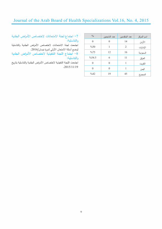
9
Journal of the Arab Board of Health Specializations Vol.16, No. 4, 2015
7
Journal of the Arab Board of Health Specializations Vol.16, No 3, 2015
%عدد الناجحين عدد المتقدميناسم المركز
1400األردن
50%21اإلمارات
75%1612السعودية
54.5%116العراق
100الكويت
100اليمن
42%4519المجموع
7- اجتماع لجنة االمتحانات الختصاص األمراض الجلدية والتناسلية:
والتناسلية الجلدية األمراض الختصاص االمتحانات لجنة اجتمعت لوضع أسئلة االمتحان األولي لدورة نيسان/2016.
الجلدية األمراض التنفيذية الختصاص اللجنة اجتماع -8والتناسلية:
اجتمعت اللجنة التنفيذية الختصاص األمراض الجلدية والتناسلية بتاريخ .2015/11/19

10
Journal of the Arab Board of Health Specializations Vol.16, No. 4, 2015
8
Journal of the Arab Board of Health Specializations Vol.16, No 3, 2015
خريجو المجلس العربي لالختصاصات الصحيةخالل الفترة من 2015/10/1 لغاية 2015/12/31
اختصاص الوالدة وأمراض النساء
اسم الطبيب مركز التدريبجمانة الشوربجي م. التوليد- سوريا
ماهر الباري م. حمد الطبية- قطرألونا الرواشدة م. األردن- األردنلميس محمد الحسن إعفاء- السودان
فادية التركماني م. التوليد- سوريا فتوى أحمد الظاهري م. الوحدة التعليمي- اليمنفاطمة عمر يسلم م.الوحدة التعليمي- اليمن مريم حامد شهاب م. الوحدة التعليمي-اليمن
سميرة علي التعزي م. الثورة العام- اليمنبلقيس عبد الحليم العريقي م. الثورة العام-اليمنرقية منصور شرف األهدل م. الثورة العام-اليمنجميلة حمود محمود م. السبعين- اليمنرحاب أحمد فضل الغوري م. السبعين- اليمننوال علي البدجي م. السبعين- اليمن
هدى أمين رشيد م. بغداد التعليمي- العراقشيماء عباس م. الكاظمي التعليمي-العراقعليا محمد علي م. الموصل التعليمي-العراقوزيرة محمد عثمان م. أربيل التعليمي- العراق
إيناس عبد الكريم السودانفتحية فرج محمد المرغني م. الجمهورية- ليبيا
أميمة عبد العزيز أحمد مصرمحمد عاطف لطفي األسطي إعفاء- مصر
هيثم شكري يوسف سويلم م. أحمد ماهر- مصرشيماء السويدي م. دبي- اإلمارات
فاطمة الزهراء عواد م. حمد الطبية- الدوحةنهاد يوسف األخرس إعفاء- سوريا
عبد اهلل عبد الشفيع ابراهيم م. طرابلس الطبي- ليبياحليمة مفتاح الزروق التير م. الجالء- ليبيا
آمال رمضان الصيد م. الجالء- ليبيادانة تركماني م. الزهراوي- سوريا
زينب الصادق م. طرابلس الطبي- ليبياسمية العكاري م. طرابلس الطبي- ليبيا
اختصاص طب األطفال
اسم الطبيب مركز التدريبعلياء صالح القحازي هـ. م. الثورة العام- اليمن
ياسر محمد أحمد أحمد ج. الخرطوم- الخرطومفواز سعيد الزهراني م. فهد للحرس- الرياض
وئام سليمان الميمان م. م. فيصل التخصصي- الرياضمحمد يحيى النعمي م. م. فهد المركزي- جازانمنى عبد الوهاب الصالح م. م. فهد الجامعي- الخبرفاطمة أحمد جبره م. م.عبد العزيز- السعوديةعيسى عايض الرشيدي م. م. عبد العزيز-السعوديةمعاذ حسم الحسن األحمد م. القوات المسلحة- تبوكهناء حسين أسعد زمزمي م. الوالدة واألطفال- جدة
شفقت بشير محمد م. الملك فهد- الباحةطلعت يحيى الجزار م. الوالدة واألطفال- اإلحساء
حليمة علي محمد الغامدي م. الوالدة واألطفال- جدةخلود محمد خميس م. الوالدة واألطفال- جدةلبنى عبد العزيز بيت المال ج. الملك سعود- السعودية
عبد العزيز الحسين ج. الخرطوم- السودان نايف محمد حمد القويعي م. قوى األمن- السعودية
دياال زاهر محمد زايد م. األردن- األردن رامي جمال أبو حجه م. التخصصي- األردن
نيروز محمد الجابري م. الجالء- ليبياعبد اهلل أحمد خليل إعفاء- مصر
مصطفى محمد الزفتاوي م. م. نصر للتأمين- مصررقية أحمد جمال دملخي م. اإلسالمي- األردن
11
Journal of the Arab Board of Health Specializations Vol.16, No. 4, 2015
9
Journal of the Arab Board of Health Specializations Vol.16, No 3, 2015
شريف محمد سليمان م. منشية البكري- مصرابتسام عثمان الخازمي م. الجالء- ليبيا
أيمن صبحي أحمد سعاده م. اإلسالمي- األردنعبير محمد موسى شتا م. المنصورة للتأمين- مصر
ابتسام أحمد سالم بعيو م. الجالء- ليبيامناسك كامل حسن م. حمد الطبية- الدوحة
زينب عبد الحي العوضي م. لطيفة- اإلماراتإيهاب حسين القادري م. الوحدة التعليمي- اليمن
فبين محي الدين محي الدين م. دبي- اإلماراتجيشا سانكار م. حمد الطبية- الدوحةسمر يعقوب عوض مقبول م. حمد الطبية- الدوحةفيحاء أحمد عبد القادر م. حمد الطبية- الدوحة
منال علم الدين عدي م. دبي- اإلماراتليلى صبحي أحمد بكر م. حمد الطبية- الدوحة
سحر يحيى نصار م. لطيفة- اإلماراتنبراس محمد أحمد القحطاني م. المفرق- اإلماراتعائشة محمد عبيد العنتلي م. المفرق- اإلماراتإيمان غالب علي الجابري م. لطيفة- اإلماراتعائشة شعيب فاثاني م. لطيفة- اإلمارات
تامر حمدي شوشة م. السلمانية الطبي- البحرين هناء عبد السالم مسعود م. حمد الطبية- الدوحةباسل ممدوح هبرة م. حمد الطبية- الدوحة
ليلى سالم عمر الهاشمي م. السلطاني- سلطنة عمانتيسير محمد سعيد الكود إعفاء- سوريا
سارة عثمان موسى م. حمد الطبية- الدوحةمحمد ماهر محمد الطيارة م. األطفال- سوريا محمد أمين النجار م. األطفال- سوريا ديما عز الدين الفرج م. األطفال- سوريا أيمن محمد تيسير زوزو م. األطفال- سوريا ديمة أسد الجرمقاني م. األطفال- سوريا بالل محمد سليمان علي م. األطفال- سوريا رندة فضل حمدي أيوبي م. األطفال- سوريا
سيرين عصام منيمنة م. المقاصد اإلسالمية- لبنانأسماء أحمد الظافر هـ. م. الثورة العام- اليمن
عبد اهلل العمر المشرف م. األطفال- سوريا
هشام محمد فياض م. األطفال- سوريا رنا إحسام الخانجي إعفاء- سوريا
محمد عبد الناصر عبيد م. األطفال- سوريامحمد محمد حديفة إعفاء- سوريا
نجاة عبد القادر هالل ج. الخرطوم- السودان منال عدنان المعد م. األطفال- سورياأنفال عدنان جعفر توفيق م. الوالدة- السودانمحمد بالل الخوام م. األطفال- سوريا
بسام ابراهيم أبو عامر م. البصرة التعليمي- العراقعلياء علي عبد اهلل م. الزهراء التعليمي-العراقأحمد ابراهيم عبد الواحد م. البصرة التعليمي- العراقسجى سحن الظاهر م. بصرة التعليمي- العراق
أحمد خليل الكرخي د. مدينة الطب- العراقغادة عبد الوهاب الخطاب م. الموصل التعليمي- العراقسماهر حاكم الوائلي م. كربالء التعليمي- العراق
رعد جاسم نوري صالح د. مدينة الطب- العراقسروان سفر اسماعيل م. الموصل التعليمي- العراق
بشير عبد اهلل عجيل م. بصرة التعليمي- العراقعالء هاشم العبد اهلل م. بصرة التعليمي- العراق
راغب بهنام زاكي م. الموصل التعليمي- العراق
اختصاص األمراض الجلدية والتناسلية
اسم الطبيب مركز التدريبمصطفى حازم توفيق م. الصدر التعليمي- العراقخنساء عبد األمير الياسري م. الكندي التعليمي- العراقسارة محمد علي شاكر م. الكندي التعليمي- العراق
اشجان ابراهيم علي الحجي م. م. خالد الجامعي-السعوديةهند محمد آل عطيف م. عسير المركزي- أبها
شوكت أبو بكر قادر م. بغداد التعليمي- العراقأزهر جبار الصبيحاوي م. الكندي التعليمي- العراق
سارة عبد الرحمن المطلقة م. عسير المركزي- أبهافجر منصور المنصور م. الحرس الوطني- الرياض
أزهار محمد عاللي م. م. عبد العزيز- السعودية

12
Journal of the Arab Board of Health Specializations Vol.16, No. 4, 2015
10
Journal of the Arab Board of Health Specializations Vol.16, No 3, 2015
شيخة نايف هني الرشيدي م. سلطان الطبية- السعوديةإيمان عبد اهلل آل عامر م. عسير المركزي-السعودية هنادي سعيد الساطي م. م. عبد العزيز- السعودية وليد عبد اهلل الصالحي م. الحرس الوطني- السعودية سلطان عقيل جعفري م. عسير المركزي- السعودية
طه حسين حبيب اهلل م. الحرس الوطني- جدةالعلياء هاشم اليونس م. الصدر التعليمي- العراقرغدة سعيد المعشري م. خليفة بن زايد- اإلمارات
سمر فوزي سابق م. حراء- السعودية
اختصاص جراحة الفم والوجه والفكين
اسم الطبيب مركز التدريبأشرف صديق عبد الغني إعفاء- السودانمشاعل محمود محجوب إعفاء- السودان
إيمان محمد طه جمعة إعفاء- مصرقيصر خليل عريبي م. الجراحات التخصصيعماد خضير عباس م. الجراحات التخصصيسيف نبيل جواد م. الجراحات التخصصيباسم علي قاسم م. الجراحات التخصصيعمار لؤي عبد القادر م. الجراحات التخصصيحيدر حميد عبد الكريم م. الجراحات التخصصيمنتظر محسن حسن م. الجراحات التخصصيخالد برهان خالد م. الجراحات التخصصي
حمزة صالح الشاوش م. المخ واألعصاب-ليبياابتسام عبد الفتاح جحي م. دمشق- سورياعمار بركات م. دمشق- سوريا
نسرين بهاء الدين يوسف م. الجامعة- دمشقهيثم محمد عياد م. ناصر- مصرمحمد عبد العزيز غانم م. ناصر- مصرفهمي شريف فهمي غالي م. ناصر- مصرأشرف أحمد عبد الفتاح م. ناصر- مصر
اختصاص التخدير والعناية المركزة
اسم الطبيب مركز التدريب
أحمد فتحي مصطفى الزيات م. الخرطوم- الخرطومأحمد هاشم حمودي م. أربيل التعليمي- العراق
محمد صادق النعسان إعفاء- العراقسامان حمه حسن م. أربيل- العراقزينب سمير عزيز الكروي م. الطب- العراق
عروية مزهر السوداني م. البصرة التعليمي- العراقعمر خالد جليل م. الطب- العراقحيدر عباس القيسي م. الطب- العراقنادية تحسين عيدان البياتي م. بغداد- العراقوحيد اسماعيل سنيار م. أربيل- العراقدابان جالل أيمن بياتي م. الطب- العراق
عبد اهلل طيب معروف م. أربيل التعليمي- العراقبسمة حقي اسماعيل الخياط م. الطب- العراقهوزان محمد حسين المرعب م. الطب- العراقاحمد موفق يونس الدركزلي م. الطب- العراق
جان جاك اسو م. الجامعة األميركية-لبنانغسان برهان دحروج م. الجامعة األميركية-لبنان
محمد أنور محمد حمزة م. طرابلس الطبي- ليبياحسام محمد القالي م. طرابلس المركزي- ليبيا
اختصاص علم األمراض التشريحي
اسم الطبيب مركز التدريبروال فتحي محمد األعرج م. الجامعة األردنية- األردن
والء فلمبان م. القوات المسلحة- السعودية مها تحسين فارس عكاوي م. الجامعة األردنية- األردن
نورا مصطفى كريم القيسي م. الكاظمية- العراقإيمان محمد حسين اليعبري م. القصر العيني- مصر
بشرى سعيد الحتروشي ج. السطان قابوس-سلطنة عمانمثال عصام علوان البيات م. م. فهد الجامعي- الخبر
يزن عبد اهلل العثمان م. الحسين للسرطان- األردنرضية عبد اهلل العجمي ج.السطان قابوس-سلطنة عمانزكية ابراهيم العجمي ج.السلطان قابوس-سلطنة عمانزهرة غصن هالل العبري ج.السلطان قابوس-سلطنة عمان
ماري لمع يوسف الياس م. األسد الجامعي- سوريا
13
Journal of the Arab Board of Health Specializations Vol.16, No. 4, 2015
11
Journal of the Arab Board of Health Specializations Vol.16, No 3, 2015
جيسيكا عون م. الجامعة األميركية- لبنانعبد اهلل باويزر م. ج. م. عبد العزيز- السعودية
زينة ضياء حامدي م. الجامعة األردنية- األردن عزة أحمد محمد زلفو المعمل القومي السوداني- السودان
نهى حلمي غانم معهد األورام- مصر
اختصاص الطب النفسي
اسم الطبيب مركز التدريب
نسيم عبد المحي الحجيلي م. م. عبد العزيز- السعوديةخضره أمين محمد أمين م. اشليخ خليفة- اإلمارات
معتز علي السيد علي إعفاء- مصررامي مجدي لبيب حنا م. المعمورة - مصر
إيمان محمد المدني البخاري م. الرازي- ليبيارانية أحمد عبدون م. الشيخ خليفة- اإلمارات
حنان رمضان الشريمي م. الرازي- ليبياعايض بن حمدان الغامدي م.ج. م. عبد العزيز- السعودية
يحيى بن خالد الكلباني م. ابن سينا- سلطنة عمانابراهيم بن الفريح م. العسكري- السعودية
صالح بن خالد الدهش م. م. فيصل- السعودية أحمد فالح زابن المطيري م. األمل- السعودية
حمده أحمد خليفة النشمي م. الطب النفسي- البحرين عزة محمد خدوم الهنائي ج. السلطان قابوس-سلطنة عمان
أسماء بن هويدي عبد النبي إعفاء- تونسعبد الواحد سعيد خان م. حمد الطبية- الدوحةالنا نمر عرب م. حمد الطبية- الدوحةياسمين حمدي عبد الرحيم م. حمد الطبية- الدوحةسهى محمود محمد محمود م. حمد الطبية- الدوحة
علي أحمد حسن التجلي إعفاء- الكويت حسن علي حسن أحمد م. السلمانية الطبي- البحرين
هاجر حسن اسحق شعبان م. الطب النفسي- البحرين أحمد باسل ابراهيم القيسي م. الرشاد التعليمي- العراق
إخالص محمد علي علي م. ابن سينا- العراقأماني ابراهيم فالح طعمة م. العباسية- مصر
وليد عبد المنعم وصفي م. م. النيل- مصرإيمان موسى الهوتي م. الطب النفسي- اإلمارات
هبة حسن المبشر الطيب م. بعشر- السودانأحمد سمير حسين الخرس م. م. فهد- السعودية
علي عسكر أحمد السيد إعفاء- مصر ماجد بن علي الحربي م. م. فهد- السعودية
خالد علي حسن عبد اهلل م. العباسية- مصرعائشة مانع سعد المانع م. م. فهد- السعودية رياض بن محمد العمري م. م. فهد- السعودية الشيماء عبد الرحمن مدخلي م. م. فهد- السعودية
علي حسين كاظم الشباني م. الديوانية- العراقثامر فهد عبد الكريم الشدوخي م. م. فهد- السعودية
اختصاص األمراض الباطنة
اسم الطبيب مركز التدريب
هبة عثمان خضر عثمان إعفاء- السودان عمر عبد الرحمن الحمادي م. الشيخ خليفة- اإلمارات
نورة سيف راشد النعيمي م. العين وتوام- العين نادية عبيد راشد مطروشي م. المفرق- أبو ظبينورة عبد الواحد صالح علي م. المفرق- أبو ظبي
خلود محمد أحمد بخاري م. راشد- دبيرندة الناير عبد الكريم حامد م. زايد العسكري- أبو ظبي
محمد منيب عزات عباس م. اإلسالمي- عمان عمر عبد الرحمن العتر م. األردن- عمان محمد عبد الرحمن مغربي م. األردن- عمان محمد وليد عبد الغفار نيروخ م. األردن- عمان بشار محمد مصطفى األعمر م. البشير- عمان محمد حسن سعيد الطراونة م. البشير- عمان هاني محمد عداد شطناوي م. البشير- عمان
مالك محمد اسحق مسودة م. التخصصي- عمان عبد الرحمن محمد مصطفى م. الجامعة األردنية- عمان محمد جمال عبد القادر المر م. الجامعة األردنية- عمان أحمد ماهر صالح الغزالي م. الجامعة األردنية-عمان

14
Journal of the Arab Board of Health Specializations Vol.16, No. 4, 2015
12
Journal of the Arab Board of Health Specializations Vol.16, No 3, 2015
عدنان غسان عدنان العتيبي م. الجامعة األردنية- عمانمايكل سالم ميخائيل أبو حديد م. الجامعة األردنية- عمان
روان المعتز باهلل اآلغا م. السلمانية- المنامةساجدة عبد الكريم سبت م. السلمانية- المنامةصفاء حسين نوروز غلوم م. السلمانية- المنامةيزن عبد النبي مسيب محمد م. السلمانية- المنامة
أحمد حمد خلف جواد القيسي م. ابن سينا- الموصل ساكار صمد شريف م. ابن سينا- الموصل مؤيد خلف ابراهبم خلف م. ابن سينا- الموصل
محمد عبد الغفور محمد م. البصرة التعليمي-البصرة حسين حاتم عمران الفتالوي م. الصدر التعليمي-العراق
علي عباس كريم زبيدي م. الكاظمية- بغدادغيث حامد حميد الجنابي م. الكاظمية- بغدادمصطفى صبحي حسين م. اليرموك- بغدادمصطفى كنعان خورشيد العبادي م. اليرموك- بغدادهديل أحمد كافي ابراهيم م. اليرموك- بغداد
بان سلطان حاجم سلطان م. بغداد- بغدادحسنين طعمة مهدي طعمة م. بغداد- بغدادقيس عبد اهلل عادي الخضر م. بغداد- بغدادكمال محمد مغير محمد الطيار م. بغداد- بغدادمازن باسل مزهر الكرخي م. بغداد- بغدادهدى عبد العباس محمد م. بغداد- بغداد
إلهام أحمد محمد القداري م. الثورة- صنعاءمروان عبد الرحمن عبد اهلل م. الجمهورية- عدن
مناف عزيز شعبان إعفاء- سورية ضاهر محمد العرب م. المواساة- سورية عامر توفيق زين الدين م. المواساة- سورية إيمان علي أحمد الرابطي م. طرابلس- طرابلسلبنى يوسف سالم الغنيمي م. طرابلس- طرابلسمجدي عثمان عبد السالم الخازمي م. طرابلس- طرابلس
أسامة سالم عبد الرحمن جاد اهلل م. ج. العرب- بنغازيأكرم رمضان فرج البركي م. ج. العرب- بنغازيمحمد علي ابراهيم حميدة م. ج. العرب- بنغازي
أحمد سالم راشد المقبالي م. السلطاني- سلطنة عمان
علي خميس سيف الريسي م. السلطاني- سلطنة عماننور الدين حمدان سهل م. معهد ناصر- مصر
اختصاص الجراحة العامة
اسم الطبيب مركز التدريبمجتبي صالح خضير م. الطب- بغدادمؤيد خميس مصطفى م. الطب- بغداد
مروان عزمي أبو فارة م. المقاصد الخيرية- القدسإيهاب السيد كحكة إعفاء- مصرباسم محمد محمود إعفاء- سوريا
أنس نعيم داوود م. البشير الحكومي- عمان جميلة أحمد حسين الجرادي م. الكويت الجامعي- صنعاءذي يزن محمد المريش م. الكويت الجامعي- صنعاءفؤاد حسن بن قديم م. الجمهورية التعليمي- عدن
سرمد عدنان محمد م. النجف- العراقحسنين عبد العباس كاظم م. النجف- العراقرزاق وهاب حميد م. النجف- العراق
عدنان عبد العظيم كاظم م. البصرة التدريبي- العراق ثائر جاسم جاسب م. البصرة التدريبي- العراقمحمد شهيد طعمة م. البصرة التدريبي- العراق
يونس ابراهيم عبد اهلل م. السلمانية الطبي- البحرين رفل رياض بدري م. الجامعة األردنية- عمان
محمد ظافر عالم م. طرابلس المركزي- طرابلسنور الدين عبد علي محمد م. السلمانية الطبي- المنامة
عمر سعيد ناصيف م. التخصصي- عمانعمر ذيب الزين م. المواساة الجامعي- دمشق
أحمد محمد عوض إعفاءمحمد حسن فتحي حسين إعفاء- مصر
خالد سعيد علي الشهراني م. خالد للحرس- جدةعماد خيري الشريف م. خالد للحرس- جدةشيرين سليم محمد شخاشيرو م. خالد للحرس- جدة
علي محمد فتحي ذياب م. البشير الحكومي- عماننزار محمد نمر عمرو إعفاء- األردن
بكر نزار أبو حية م. األردن- عمان