Embed Size (px)
Citation preview

Journ
al of
Plant P
atholo
gy
1
CHARACTERISATION OF NEOFUSICOCCUM SPECIES CAUSING MANGO DIEBACK IN ITALY
A.M. Ismail1, G. Cirvilleri2, L. Lombard3, P.W. Crous3,4,5,
J.Z. Groenewald3 and G. Polizzi2
1Plant Pathology Research Institute, Agriculture Research Center, 12619 Giza, Egypt 2Dipartimento di Gestione dei Sistemi Agroalimentari e Ambientali, Sezione di Patologia Vegetale, 95123
Catania, Italy 3CBS-KNAW Fungal Biodiversity Centre, Uppsalalaan 8, 3584 CT, Utrecht, The Netherlands
4Wageningen University and Research Centre (WUR), Laboratory of Phytopathology, Droevendaalsesteeg 1, 6708 PB Wageningen, The Netherlands
5Microbiology, Department of Biology, Utrecht University, Padualaan 8, 3584 CH Utrecht, The Netherlands
Running title: Mango dieback in Italy Corresponding author: G. Polizzi Fax: +39.095.7147283 E-mail: [email protected]
Journ
al of
Plant P
atholo
gy
2
SUMMARY
Species of Botryosphaeriaceae are important fungal pathogens of mango worldwide. A
survey of 11 mango orchards located in the provinces of Catania, Messina, Palermo and Ragusa
(Sicily, southern Italy), resulted in the isolation of a large number (76) of Neofusicoccum isolates
associated with decline and dieback symptoms. Isolates were identified based on morphology and
DNA sequence data analyses of the internal transcribed spacer region of the nrDNA and partial
translation of the elongation factor 1-alpha gene regions. Two species of Neofusicoccum were
identified, which included N. parvum and N. australe, the former of which was the dominant
species. The high incidence in local orchards and the pathogenicity results indicate that N.
parvum and N. australe are important pathogens of mango in Sicily where they may
significantly limit mango production.
Key words: Botryosphaeriaceae, dieback, ITS, EF-1α, Mangifera indica, Neofusicoccum,
INTRODUCTION
According to the ancient accounts of travelers and written historical records, the
cultivation mango (Mangifera indica; Anacardiaceae), a species native to India, began at least
4000 years ago (De Candolle, 1884). Mango trees are able to adapt to various environmental
conditions that are normally not conducive to growth of other fruit trees (Wolstenholme and
Whiley, 1995). The mango cv. Kensington Pride was first introduced into Sicily (southern Italy)
during 1980-1990 by growers in the Catania province. Thereafter, its cultivation expanded to the
other provinces of Sicily (Messina, Ragusa, Palermo) and to the nighbouring Calabria region. In
these regions, cvs Kensington Pride, R2E2, Maya, Kent, Irwin, Keitt and Tommy Atkins are the
most commonly grown cultivars (Anonymous, 2010).
During all stages of their life cycle, mango trees can be attacked by over 140 different
plant pathogens inciting diverse diseases (Prakash, 2004; Haggag, 2010), some of which have
become a limiting factor for mango production (Ploetz, 2004; Prakash, 2004; Javier-Alva et al.,
2009; de Olivera Costa et al., 2010). Tip dieback or decline, which is a complex disease, is

Journ
al of
Plant P
atholo
gy
3
considered a serious problem in various mango-producing countries (Ramos et al., 1991;
Johnson, 1992; Jacobs, 2002; Khanzada et al., 2004a, 2004b). The etiology of this disease
remained unclear for several years due to the different causal agents associated with it (Ploetz et
al., 2003).
Smith and Scudder (1951) found a Diplodia sp. associated with dieback of mango but did
not confirm its pathogenicity. Ramos et al. (1991) isolated Neofusicoccum ribis and a Diplodia
sp. from mango trees showing tip dieback in Florida (USA). Botryosphaeria dothidea, diplodia-
and fusicoccum-like asexual morphs, were reported as causal agents of fruit rot and decline of
mango (Ploetz, 2004). In Florida, Lasiodiplodia theobromae (as D. theobromae) and Fusicoccum
aesculi were found responsible for symptoms associated with decline on cvs Keit and Tommy
Atkins (Ploetz et al., 1996). The latter fungal species have also been reported from Brazil as
associated with mango dieback and stem-end rot (de Oliveira Costa et al., 2010). In western
Australia, Neoscytalidium dimidiatum and Ne. novaehollandiae have recently been reported as
causal agents of canker and dieback (Ray et al., 2010) whereas, in a subsequent survey, Sakalidis
et al. (2011) found that Pseudofusicoccum adansoniae, P ardesiacum, P. kimberleyense and
Lasiodiplodia pseudotheobromae were associated with canker and dieback. Other fungal species
in the family Botryosphaeriaceae, including L. theobromae, B. dothidea, Neofusicoccum parvum
and N. mangiferae have also been reported to cause stem-end rot (SER) of mango (Slippers et al.,
2005; Johnson, 2008; de Oliveira Costa et al., 2010). Recent phylogenetic studies revealed the
association of new members of Botryosphaeriaceae e.g., L. hormozganensis and L. iraniensis
with mango diseases in Iran and in Australia (Abdollahzadeh et al., 2010, Sakalidis et al., 2011)
and L. egyptiacae with mango dieback in Egypt (Ismail et al., 2012b).
Recent surveys conducted in mango orchards in Italy led to the discovery of several
diseases (Ismail et al., 2012a). During these surveys, plants with decline and tip dieback
symptoms were frequently observed, from which a large number of Botryosphaeriaceae isolates
were recovered that, as reported in this paper, were identified and assayed for pathogenicity
assessment.
MATERIALS AND METHODS

Journ
al of
Plant P
atholo
gy
4
Isolations. Isolations were made from 60 symptomatic plant samples showing dieback
symptoms on young twigs and branches, dark brown lesions on mature fruits, and necrosis and
brown discolouration under cambium tissues, resembling Botryosphaeriaceae infection (Fig. 1).
The collected plant materials were surface disinfected by sequential washing in 70% ethanol for
30 sec, a bleach solution (5% sodium hypochlorite) for 1 min followed by rinsing with sterile
water, and dried with a sterile filter paper. Small pieces, between the healthy and infected tissues
were excised and plated onto potato dextrose agar (PDA) amended with streptomycin sulfate (0.1
g/l). Plates were incubated at 25±2°C in the dark for 2–4 days. The putative isolates resembling
the colony morphology of botryosphaeriaceous taxa growing out from the tissues were sub-
cultured onto fresh PDA and incubated at 25±2°C for 5 days, and pure cultures were obtained by
excising and transferring a hyphal tip to fresh PDA plates.
DNA isolation and amplification. Genomic DNA was extracted from 10-day-old
cultures using the UltraCleanTM Microbial DNA Isolation Kit (MO-BIO Laboratories, Inc,
Carlsbad, USA) following the manufacturer’s protocol. The internal transcribed spacer region
(ITS) of the nrDNA including the 3’ end of 18S small subunit rRNA gene region, the first
internal transcribed spacer (ITS1), the complete 5.8S rRNA gene, the second internal transcribed
spacer (ITS2) and 5’ end of the 28S large subunit rRNA gene region were amplified using primer
pair V9G (de Hoog and Gerrits van den Ende, 1998) and ITS4 (White et al., 1990). A part of the
translation elongation factor 1-alpha (EF-1α) gene was amplified using primers EF1-728F
(Carbone and Kohn, 1999) and EF2 (O’Donnell et al., 1998). PCR conditions included final
concentrations of 0.5 U/µL of Taq polymerase (BIOLINE, San Diego, USA), 1× reaction buffer
(BIOLINE), 2–2.5 mM MgCl2 (BIOLINE), 0.4–0.6 mM of each dNTP and 0.12–0.2 µM of each
primer made up to a final volume of 12.5 µl with sterile deionized water. PCR conditions
included the following steps: an initial step of denaturation at 95°C for 5 min, followed by 40
cycles of 95°C for 30 sec, 52°C for 30 sec and 72°C for 1 min, with a final elongation step at
72°C for 7 min.
DNA phylogeny. Amplicons of the ITS region were sequenced in both directions using
ITS4 and the internal forward primer ITS5 (White et al., 1990). The PCR products of the EF–1α

Journ
al of
Plant P
atholo
gy
5
gene region were sequenced in both directions using the same primer pairs as for amplification.
Sequencing reactions were performed using BigDye Terminator v. 3.1 Cycle Sequencing Kit
(Perkin-Elmer Applied Bio Systems, Foster City, CA, USA) as recommended by the
manufacturer and run on an ABI PRISMTM 3100 DNA automated sequencer (Perkin-Elmer
Applied BioSystems, Foster City, CA, USA).
The obtained sequences were compared with those previously identified in GenBank
using the Basic Local Alignment Search Tool (BLAST) (Table 1). Sequences were aligned
together with those retrieved from GenBank using MAFFT v. 6.0 (Katoh and Toh, 2010) and
manually adjusted and corrected where necessary. The nucleotide substitution models were
determined individually for each gene region using MrModelTest v. 2.2 (Nylander, 2004). The
best fit model, HKY+I+G with dirichlet base frequencies, was selected for both ITS and EF–1α
sequence datasets. The 70% reciprocal NJ (Neighbour-Joining) bootstrap analysis was performed
for each gene region to determine congruency (Mason-Gamer and Kellogg, 1996; Gueidan et al.,
2007).
Bayesian analyses were performed using MrBayes v. 3.1.1 (Ronquist and Huelsenbeck,
2003). For the Bayesian analyses, a Markov Chain Monte Carlo (MCMC) (Larget and Simon,
1999) method was performed to confirm the topology of the tree, by running four chains
simultaneously starting from a random tree topology and ending at 1,000,000 generations with
trees saved every 100th generation. The burn-in value was graphically estimated from the
likelihood scores. The first 1000 trees were discarded from the analysis and the final consensus
tree constructed from the remaining trees. Trees were rooted to Phyllosticta capitalensis (CBS
115051) and P. citricarpa (CBS 102374) as out-group taxa.
The phylogenetic relationship among taxa was determined using PAUP* (Phylogenetic
Analysis Using Parsimony) v. 4.0b10 (Swofford, 2003). Maximum parsimony (MP) was
performed using the heuristic research option with random stepwise addition in 1,000 replicates,
tree bisection and reconnection (TBR) as branch swapping algorithms, and random taxon
addition sequences for the construction of maximum parsimony trees. Branches of zero length
were collapsed and all multiple equally parsimonious trees were saved. MAXTREES was set to
10,000. In the analysis all characters were unordered and had equal weight; gaps were treated as
missing data. Parameters measured for parsimony included tree length (TL), consistency index

Journ
al of
Plant P
atholo
gy
6
(CI), rescaled consistency index (RC) and retention index (RI). Bootstrap support values were
evaluated using 1,000 bootstrap replicates (Hillis and Bull, 1993). Sequences generated in this
study were deposited in GenBank (Table 1).
Morphological characterisation. To induce sporulation, a 6 mm diameter plug of
mycelium from each isolate was placed on 2% water agar medium (WA: 20 g agar/l) together
with autoclaved pine needles (Smith et al., 1996). The plates were incubated at 25±2°C under
near-ultraviolet (UV) light for 2–3 weeks. Dimensions of 50 conidia from each of 37 randomly
selected isolates were determined at 1.000× magnification mounted in 85% lactic acid. Vertical
sections were made through pycnidia with a Leica CM1100 microtome and mounted in lactic
acid. The 95% confidence intervals were determined for the conidia with extremes given in
parentheses. Cardinal growth temperatures were determined for each isolate on PDA at 10–35°C
in 5°C intervals in the dark. Colony colours were determined after 7 days on PDA at 25°C in the
dark using the colour charts of Rayner (1970) for comparison.
Pathogenicity tests. Three- to four-month-old mango cv. “Kensington Pride” seedlings
ranging in length from 40–60 cm, were used to determine the pathogenicity of nine isolates
representing two Neofusicoccum spp. (Table 1). The plants were maintained in a growth chamber
under artificial light (10/14 h light-and-dark cycles) at 25±2ºC and 70–80% relative humidity
(RH). Four plants for each isolate and the controls were used and arranged in a completely
randomised block design. Data were subjected to the analysis of variance one-way ANOVA and
the mean values of the lesions were compared using the Least Significant Difference (LSD) test
(P< 0.05) (Stat Soft, Inc. 2004). Isolates were cultured on PDA for 7 days at 25ºC in the dark.
Inoculations were performed after the outer tissues were disinfected with 70% ethanol, washed
with sterile distilled water and left to dry. Using a cork-borer, a 5 mm incision was made into the
epidermis, between two nodes and below the apex of the stem. A 5 mm diameter mycelial PDA
plug was removed from the edge of the actively growing test culture and placed in the wounds,
with the mycelium facing the cambium. The inoculated wounds were wrapped with Parafilm®,
(Laboratory Film, Chicago, IL, USA) to prevent desiccation and contamination. Control plants
were inoculated with a sterile PDA plug. Six weeks after inoculation the bark lesion lengths as

Journ
al of
Plant P
atholo
gy
7
well as the length of cambium discolouration were measured and data were log-transformed prior
to analysis. Re-isolation of the tested isolates was done from the margins of the necrotic lesions
on PDA to confirm Koch’s Postulates.
RESULTS
Isolations. In total, 76 isolates of Botryosphaeriaceae were recovered from four sites
located in southern-Italy (Sicily). Of these 41 isolates originated from Catania, 27 isolates from
Messina, six isolates from Palermo and two isolates from Ragusa. The majority of isolates were
recovered from twigs, branches, leaves, fruit and wood (26, 19, 18, 9 and 4, respectively). Of
these isolates, 37 were randomly selected and included in the phylogenetic analyses and
morphological studies. Isolates obtained in this study were deposited in the culture collection of
the Dipartimento di Gestione dei Sistemi Agroalimentari e Ambientali, Sez. Patologia Vegetale,
University of Catania, Italy. Representative isolates were also deposited in the culture collection
of the CBS-KNAW Fungal Biodiversity Centre, Utrecht, The Netherlands (Table 1).
Phylogeny. Amplicons of approximately 550 bp were obtained for ITS using primers
ITS4 and ITS5 and approximately 500 bp were obtained for EF–1α using the EF1-728F and EF2
primers. The combined dataset of ITS and EF–1α consisted of 72 taxa, which contained the
subset of 37 strains obtained during the survey. A further 35 sequences, including the out-group
species, were retrieved from GenBank. The combined dataset contained 909 characters after the
uneven ends were truncated. Of these characters, 536 were constant, 95 were uninformative and
278 were parsimony informative. A heuristic search revealed the most parsimonious tree (Fig. 2;
tree length = 670 steps, CI = 0.743, RI = 0.917 and RC = 0.682). No conflict between the two
gene partitions was detected by 70% reciprocal NJ bootstrap analysis (results not shown). The
obtained trees differed only in the arrangement of isolates within the terminal clades while their
overall topology was the same and were topologically identical to the 50% majority-rule
consensus tree illustrated in Fig. 2. Isolates obtained in this study were accommodated in two
distinct clades; of which the first clade contained the majority of strains, which grouped with
Neofusicoccum parvum strain CMW 9081 (culture ex-type) supported by a bootstrap value (BS)

Journ
al of
Plant P
atholo
gy
8
of 79%. Some strains formed two sub-clades where the first sub-clade contained isolates NF-17,
NF-38, NF-37 and NF-67 supported only by a Bayesian posterior probability (BPP) value of
0.95. The second sub-clade contained two isolates (NF-62 and NF-82) supported by a BS/BPP of
63/0.77. Four isolates (NF-70, NF-73, NF-76 and NF-77) clustered in a sub-clade in N. australe,
supported by a low BS of 64 and highly supported by a BPP of 1.0. The remaining two isolates
(NF-2 and NF-22) clustered together in a sub-clade (BS/BPP=58/1.0) with N. australe
(CMW6837, culture ex-type) (BS/BPP = 72/1.0).
Morphological characterisation. Isolates obtained in this study were separated into two
groups based on the phylogenetic inference as well as their conidia and culture morphology. In
the first group, conidiomata (Fig. 3a) were formed on pine needles within 10–15 days (Fig. 3a).
Pycnidia were solitary, mostly aggregated, globose to subglobose, the outer layers composed of
5–10 dark brown thick-walled textura angularis cell layers (Fig. 3b). Conidiogenous cells (Fig.
3c,d) were holoblastic, hyaline, cylindrical, 4.5–19.3 µm long, 1.5–2.7 µm wide. Conidia were
hyaline, (14.3–) 15.4–17.6 (–19.3) × (5.0–) 5.4–6.2 (–6.6), mean of 50 conidia ± SD = 16.5 ± 1.1
µm long, 5.8 ± 0.4 µm wide, L/W ratio = 2.8 (Fig. 3e,f). Colonies were initially white, becoming
glaucous grey to greenish grey on the upper surface, greenish grey in reverse (Fig. 3g,h). In the
second group, conidiomata were formed on pine needles within 10–20 days (Fig. 4a). Pycnidia
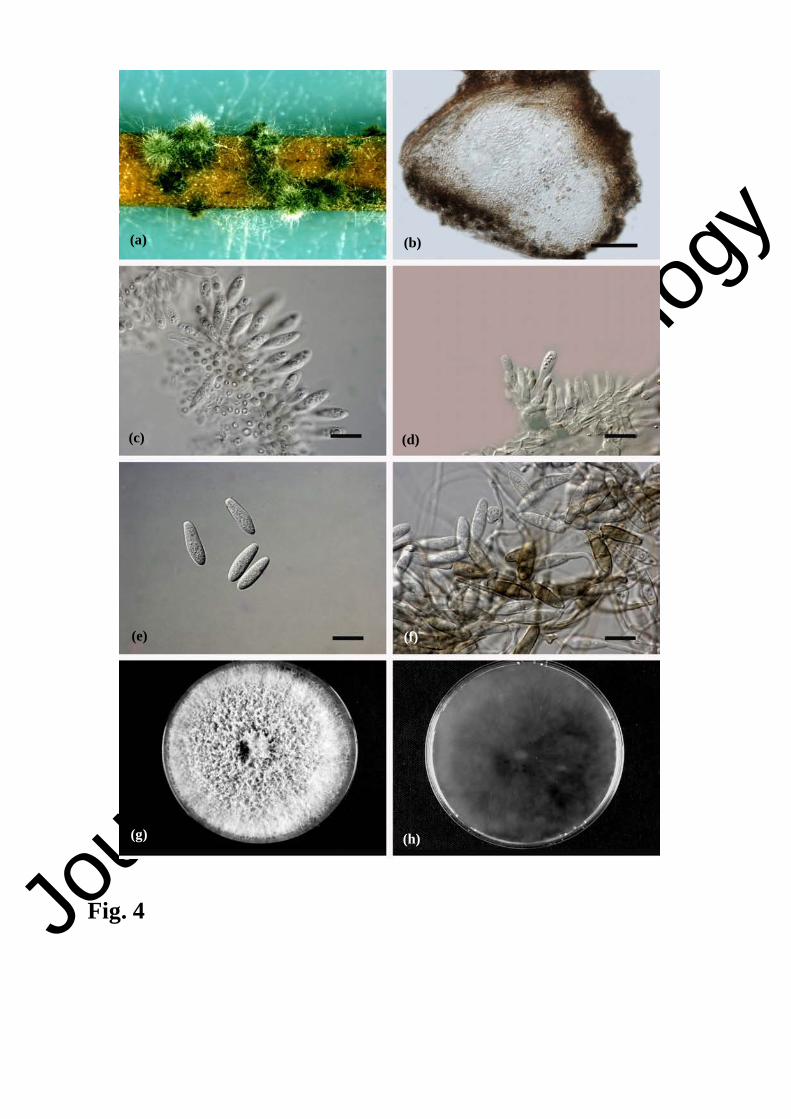
were solitary, subglobose to ellipsoidal, the outer layers composed of 4–6 dark brown, thick-
walled textura angularis cell layers (Fig. 4b). Conidiogenous cells (Fig. 4c,d) were holoblastic,
hyaline, cylindrical to subcylindrical and phialidic, 8.7–16.2 µm long, 1.8–3.7 µm wide. Conidia
were hyaline, (17.3–) 19.7–22.9 (–24.5) × (4.5–) 5.6–6.2 (–6.8), mean of 50 conidia ± SD = 21.3
± 1.6 µm long, 5.9 ± 0.3 µm wide, L/W ratio = 3.6 (Fig. 4e,f). Colonies were initially white,
becoming glaucous grey to greenish grey on the upper surface, and dark slate blue in reverse
(Fig. 4g,h).
The cardinal temperatures requirements for the growth of all isolates were: minimum
10°C, maximum 35°C, and optimum 25°C.
Pathogenicity tests. Six weeks after inoculation all seedlings showed bark lesions and
cambium discolouration. There was no significant variation observed in bark and cambium
Journ
al of
Plant P
atholo
gy
9
lesions produced among isolates of the same species. In general, N. parvum and N. australe
isolates were equally virulent and produced similar bark and cambium lesions, longer than that of
the controls (av. = 25.84 mm) (Fig. 5). However, the longest bark and cambium lesions were
developed by isolates NF-69 and NF-5 (av. 45.6 mm and 45 mm; av. 51.2 mm, 49.7 mm)
respectively. Although the remaining isolates developed smaller lesions, they also proved to be
pathogenic. Koch’s postulates were confirmed and the tested isolates were successfully recovered
from the inoculated tissues.
DISCUSSION
The present study represents the first attempt to identify botryosphaeriacous fungal
pathogens associated with M. indica in Italy. The first group consisted of isolates exhibited
culture and conidial morphology similar to those of N. parvum (Slippers et al., 2004, 2005). The
second group contained six isolates revealing culture and conidial characteristics similar to those
of N. australe (Slippers et al., 2004). Combined DNA sequence data and morphological features
confirmed the identity of the two groups of isolates as N. parvum and N. australe. These species
have been reported as plant pathogens under different climatic conditions and in very different
hosts (Slippers et al., 2004, 2005; van Niekerk et al., 2004; Damm et al., 2007; Begoude et al.,
2009; Sakalidis et al., 2011). N. parvum has been reported from mango in Australia (Johnson,
1992), South Africa (Jacobs, 2002), Peru (Javier-Alva et al., 2009) and Brazil (de Oliveira Costa
et al., 2010). In the present study, it was the most frequently isolated species from branches,
twigs, leaves, internal wood and fruits exhibiting various symptoms associated with dieback. This
fungus was recovered from almost all monitored areas (Catania, Messina and Palermo), but its
relative prevalence differed in each. N. parvum was identified in this study based on morphology
and phylogenetic inference. The differences among strains might be attributed to genetic
variation influenced by environmental conditions in different geographical areas where theses
strains were isolated. The conidial morphology did not differ greatly and resembled those
reported in previous studies (Slippers et al., 2004, 2005; de Oliveira Costa et al., 2010).
However, no septate conidia were observed as previously reported by Slippers et al. (2005) and
Oliveira Costa et al. (2010). N. australe was firstly described by Slippers et al. (2004) along with

Journ
al of
Plant P
atholo
gy
10
its sexual morph as occurring on several native Australian plant hosts, which include Banksia and
Eucalyptus, and also from a Protea sp. in South Africa, and on Pistachio in Italy. The fungus has
been reported to cause disease on other hosts such as Prunus spp. in South Africa (Damm et al.,
2007), olive in Italy (Lazzizera et al., 2008), grapevine in South Africa (van Niekerk et al., 2004),
Australia (Taylor et al., 2005), and more recently in Chile (Besoain et al., 2013). N. australe and
N. parvum were also reported as canker-causing agents on blueberry (Vaccinium spp.) in Chile
(Espinoza et al., 2009). N. australe was the second most dominant species isolated from twigs
and branches of mango showing typical dieback symptoms and found only in three sites, Ragusa,
Palermo and Catania.
The origin of the Neofusicoccum spp. obtained during this survey is unknown. Some of
the cultivated mango varieties in Sicily have been imported either as seeds or as plantlets from
Australia (Anonymous, 2010). This might suggest that N. parvum and N. australe have been
introduced into Italy with the importation of exotic mango plant materials (seeds or plantlets)
from Australia. Alternatively, most of the mango orchards were neglected and in close proximity
to various fruit trees which could have served as source of inoculum or as alternative hosts to
these fungi. Therefore, the epidemiology of these fungi needs to be studied further to understand
the ecology and the movement of these pathogens, in order to establish integrated control
strategies.
To our knowledge, this is the first report of these species causing dieback disease on
mango in Italy. It was not surprising to isolate these species from mango orchards, since they
have previously been reported on olive in southern Italy (Lazzizera et al., 2008). Dieback disease
by Botryosphaeriaceae could significantly limit future mango production in Sicily.
ACKNOWLEDGEMENTS
The work was partially funded by the CBS-KNAW Fungal Biodiversity Centre (CBS),
Utrecht, the Netherlands, University of Catania, Italy, and the Plant Pathology Research Institute,
Giza, Egypt. The Authors thank T. Yasseen for helpful contribution to collection of plant
samples.

Journ
al of
Plant P
atholo
gy
11
REFERENCES
Anonymous, 2010. La coltivazione del Mango in Sicilia. Seminario sullo Stato dell’Arte e Linee Guida dell’Impianto, Messina, Italy: 1-49.
Abdollahzadeh J., Javadi A., Mohammadi G.E., Zare R., Phillips A.J.L., 2010. Phylogeny and morphology of four new species of Lasiodiplodia from Iran. Persoonia 25: 1-10.
Begoude B.A.D., Slippers B., Wingfield M.J., Roux J., 2009. Botryosphaeriaceae associated with Terminalia catappa in Cameron, South Africa and Madagascar. Mycological Progress 9: 101-123.
Besoain X., Torres C., Díaz G.A., Latorre B.A., 2013. First report of Neofusicoccum australe associated with Botryosphaeria canker of grapevine in Chile. Plant Disease 97: 143.
Carbone I., Kohn L.M., 1999. A method for designing primer sets for speciation studies in filamentous ascomycetes. Mycologia 91: 553-556.
Damm U., Crous P.W., Fourie P.H., 2007. Botrysphaeriaceae as potential pathogens of Prunus species in South Africa, with descriptions of Diplodia Africana and Lasiodiplodia plurivora sp. nov. Mycologia 99: 664-680.
De Candolle A., 1884. Origin of Cultivated Plants. Hanfer, Kengan Paul, Trench, London, UK.
de Hoog G.S., Gerrits van den Ende A.H.G., 1998. Molecular diagnostics of clinical strains of filamentous basidiomycetes. Mycoses 41: 183-189.
de Oliveira Costa V.S., Michereff S.J., Martins R.B., Gava C.A.T., Mizubuti ESG, Câmara M.P.S., 2010. Species of Botryosphaeriaceae associated on mango in Brazil. European Journal of Plant Pathology 127: 509-519.
Espinoza J.G., Briceño E.X., Chávez E.R., Úrbez-Torres J.R., Latorre A., 2009. Neofusicoccum spp. associated with stem canker and dieback of blueberry in Chile. Plant Disease 93: 1187-1194.
Gueidan C., Roux C., Lutzoni F., 2007. Using multigene phylogeny analysis to assess generic delineation and character evolution in Verrucariaceae (Verrucariales, Ascomycota). Mycological Research 111: 1145-1168.

Journ
al of
Plant P
atholo
gy
12
Haggag W.M., 2010. Mango diseases in Egypt. Agriculture and Biology Journal of North America 1: 285-289.
Hillis D.M., Bull J.J., 1993. An empirical test of bootstrapping as a method for assessing confidence in phylogenetic analysis. Systematic Biology 42: 182-192.
Ismail A.M., Cirvilleri G., Polizzi G., 2012a. Characterisation and pathogenicity of Pestalotiopsis uvicola and Pestalotiopsis clavispora causing grey leaf spot of mango (Mangifera indica L.) in Italy. European Journal of Plant Pathology 135: 619-625.
Ismail A.M., Cirvilleri G., Polizzi G., Crous P.W., Groenewald J.Z., Lombard L., 2012b. Lasiodiplodia species associated with dieback disease of mango (Mangifera indica) in Egypt. Australasian Plant Pathology 41: 649-660.
Jacobs R., 2002. Characterisation of Botryosphaeria species from mango in South Africa. M.Sc. Thesis. University of Pretoria, Pretoria. South Africa.
Javier-Alva J., Gramaje D., Alvarez L.A., Armengol J., 2009. First report of Neofusicoccum parvum associated with dieback disease of mango trees in Peru. Plant Disease 93: 426.
Johnson G.I., 1992. Biology and control of stem end rot pathogens of mango. PhD Thesis. University of Queensland, Queensland, Australia.
Johnson G.I., 2008. Status of mango postharvest disease managment R&D: options and solutions for the Australian mango industry. Horticulture Australia Final Report for Project MG08017: 1-130.
Katoh K., Toh H., 2010. Parallelization of the MAFFT multiple sequence alignment program. Bioinformatics 26: 1899-1900.
Khanzada M.A., Lodhi A.M., Shahzad S., 2004a. Mango dieback and gummosis in Sindh, Pakistan caused by Lasiodiplodia theobromae. Online. Plant Health Progress http://www.plantmanagementnetwork.org/pub/php/diagnosticguide/2004/mango/.
Khanzada M.A., Lodhi A.M., Shahzad S., 2004b. Pathogenicity of Lasiodiplodia theobromae and Fusarium solani on mango. Pakistan Journal of Botany 36: 181-189.
Larget B., Simon D., 1999. Markov chain Monte Carlo algorithms for the Bayesian analysis of phylogenetic trees. Molecular Biology and Evolution 16: 750-759.

Journ
al of
Plant P
atholo
gy
13
Lazzizera C., Frisullo S., Alves A., Phillips A.J.L., 2008. Morphology, phylogeny and pathogenicity of Botryosphaeria and Neofusicoccum species associated with drupe rot of olives in southern Italy. Plant Pathology 57: 948-956.
Mason-Gamer R.J., Kellogg E.A., 1996. Testing for phylogenetic conflict among molecular data sets in the tribe Triticeae (Gramineae). Systematic Biology 45: 524-545.
Nylander J.A.A., 2004. MrModeltest v2. Program distributed by the author. Evolutionary Biology Centre, Uppsala University, Sweden.
O’Donnell K., Kistler H.C., Cigelnik E., Ploetz R.C., 1998. Multiple evolutionary origins of the fungus causing Panama disease of banana: concordant evidence from nuclear and mitochondrial gene genealogies. Proceedings National Academy of Science USA 95: 2044-2049.
Ploetz R.C., Benscher D., Vázquez A., Colls A., Nagel J., Schaffer B., 1996. A re-examination of mango decline in Florida. Plant Disease 80: 664-668.
Ploetz R.C., 2003. Diseases of mango. In: Ploetz R.C. (ed.). Diseases of Tropical Fruit Crops, pp. 327-363. APS Press, St. Paul, MN, USA.
Ploetz R.C., 2004. The major diseases of mango: strategies and potential for sustainable management. Acta Horticulturae 645: 137-150.
Prakash O., 2004. Diseases and disorders of mango and their management. In: Naqvi S.A.M.H. (ed.). Diseases of Fruits and Vegetables, pp. 511-619. Kluwer Academic Publishers, Dordrecht, The Netherlands.
Ramos L.J., Lara S.P., McMillan R.T., Narayanan K.R., 1991. Tip die back of mango (Mangifera indica) caused by Botryosphaeria ribis. Plant Disease 75: 315-318.
Ray J.D., Burgess T.I., Lanoiselet V.M., 2010. First record of Neoscytalidium dimidiatum and N. noveahollandiae on Mangifera indica and N. dimidiatum on Ficus carica in Australia. Australasian Plant Disease Notes 5: 48-50.
Rayner R.W., 1970. A Mycological Colour Chart. CMI and British Mycological Society, Kew, UK.
Ronquist F., Huelsenbeck J.P., 2003. MRBAYES 3: Bayesian phylogenetic inference under mixed models. Bioinformatics 19: 1572-1574.
Journ
al of
Plant P
atholo
gy
14
Sakalidis M.L., Ray J.D., Lanoiselet V., Hardy G.E.S., Burgess T.I., 2011. Pathogenic Botryosphaeriacea associated with Mangifera indica in the Kimberley Region of Western Australia. European Journal of Plant Pathology 130: 379-391.
Slippers B., Fourie G., Crous P.W., Denman S., Coutinho T.A., Wingfield B.D., Wingfield M.J., 2004. Multiple gene sequences delimit Botryosphaeria australis sp. nov. from B. lutea. Mycologia 96: 1030-1041.
Slippers B., Johnson G.I., Crous P.W., Coutinho T.A., Wingfield B., Wingfield M.J., 2005. Phylogenetic and morphological re-evolution of the Botryosphaeria species causing diseases of Mangifera indica. Mycologia 97: 99-110.
Smith H., Wingfield M.J., Crous, P.W., Coutinho, T.A., 1996. Sphaeropsis sapinea and Botryosphaeria dothidea endophytic in Pinus spp. and Eucalyptus spp. in South Africa. South African Journal of Botany 62: 86-88.
Smith P.F., Scudder G.K., 1951. Some studies of mineral deficiency symptoms in mango. Proceeding of the Florida State Horticulture Society 64: 243-248.
Swofford D.L., 2003. PAUP*. Phylogenetic analysis using parsimony (and other methods). Version 4. Sinaur Associates, Sunderland, MASS., USA.
Taylor A., Hardy G.E.St.J., Wood P., Burgess T., 2005. Identification and pathogenicity of Botryosphaeria species associated with grapevine decline in Western Australia. Australasian Plant Pathology 34: 187-195.
van Niekerk J.M., Crous P.W., Groenwald J.Z., Fourie P.H., Halleen F., 2004. DNA phylogeny, morphology and pathogenicity of Botryosphaeria species on grapevines. Mycologia 96: 781-798.
White T.J., Bruns T., Lee S., Taylor J., 1990. Amplification and direct sequencing of fungal ribosomal RNA genes for phylogenetics. In: Innis M.A., Gelfand D.H., Sninsky J.J., White T.J. (eds). PCR Protocols. A Guide to Methods and Applications, pp. 315-322. Academic Press, San Diego, CA, USA.
Wolstenholme B.N., Whiley A.W., 1995. Ecophysiology of the mango trees as a basis for pre-harvest management. South Africa Mango Growers’ Association Yearbook 15: 10-17.
Received February 25, 2013 Accepted April 8, 2013

Journ
al of
Plant P
atholo
gy
15
CAPTION TO FIGURES
Fig. 1. a, b. Mango dieback symptoms on the young twigs and branches starting from the tip
extending downwards; c. dark brown lesions on mature fruit lead to soft rot of the internal pulp
and sometimes water drops release from the necrotic area; d. with severe infection most of the
apical parts die and dry, leading to death of the whole tree; e, f. cross sections through trunks of
infected trees showing necrosis and brown vascular discolouration of the cambium tissues.
Fig. 2. The most parsimonious tree obtained from a heuristic research with 1,000 random
addition sequences of the combined sequences of the ITS and EF-1α sequence alignments of the
Neofusicoccum isolates recovered during the survey and other closely related species. Scale bar
shows 10 changes. Bootstrap support values and Bayesian posterior probability values are
indicated at the nodes. Isolates in bold were obtained during the survey.
Fig. 3. Neofusicoccum parvum NF-5 = CBS 130995. a.Pycnidia formed on pine needles on WA;
b. Longitudinal section through a pycnidium; c, d. Hyaline conidiogenous cells; e, f. Hyaline
conidia with granular contents. Colony morphology: g, front side; h, reverse side. - Scale bars: b
= 20 µm; c, d, e, f = 10 µm.
Fig. 4. Neofusicoccum australe NF-76 = CBS 130997. a. Pycnidia formed on pine needles on
WA; b. longitudinal section through a pycnidium; c, d. Hyaline conidiogenous cells; e. Hyaline
conidia with granular contents; f. Pale brown 1-3 septate conidia before germination. Colony
morphology: g. front side; h. reverse side. Scale bars: b = 20 µm; c, d, e, f = 10 µm.
Fig. 5. Pathogenicity test of Neofusicoccum species on mango seedlings cv. Kensington Pride.
Neofusicoccum parvum is represented by NF-69, NF-5, NF-4, NF-85, NF-20 and NF-50; N.
australe is represented by NF-70, NF-76 and NF-22. Columns represent the mean value of bark
and cambium lesions ± standard deviation (bars), which are significantly different p<0.05
according to LSD test.

Journ
al of
Plant P
atholo
gy
16
Table 1. List of isolates used in this study. Isolate numbers marked in bold arethose selected for pathogenicity tests. GenBank accession
Nos. in italics were generated in this study.
GenBank accession No. Species Identity Culture No. 1 Location Host Collector
ITS EF-1� Botryosphaeria dothidea CMW9075 New Zealand Populus sp. G.J. Samuels AY236950 AY236899 CMW8000 Switzerland Prunus sp. B. Slippers AY236949 AY236898 Neofusicoccum australe CMW6837T Australia Acacia sp. B. Slippers AY339262 AY339270 CMW 37395 Unknown Eucalyptus grandis Unknown JQ744577 JQ744598 NF-2 Sicily, Italy Mangifera indica A.M.Ismail JN814464 JN814507
NF-22= CBS 130996
Sicily, Italy M. indica A.M.Ismail JN814465 JN814508
NF-70 Sicily, Italy M. indica A.M.Ismail JN814466 JN814509
NF-76= CBS 130997
Sicily, Italy M. indica A.M.Ismail JN814467 JN814510
NF-73 Sicily, Italy M. indica A.M.Ismail JN814468 JN814511 NF-77 Sicily, Italy M. indica A.M.Ismail JN814469 JN814512 N. cordaticola CMW13992T South Africa Syzygium cordatum D. Pavlic EU821898 EU821868 CMW14056 South Africa S. cordatum D. Pavlic EU821903 EU821873 N. eucalypticola CMW6539 Australia E. grandis Unknown AY615141 AY615133 CMW6543 Australia Eucalyptus sp. Unknown AY615140 AY615132 N. kwambonambiense CMW14023T South Africa S. cordatum D. Pavlic EU821900 EU821870 CMW14140 South Africa S. cordatum D. Pavlic EU821919 EU821889 N. luteum CBS110299T Portugal Vitis vinifera A.J.L Phillips AY259091 AY573217 CBS110497 Portugal V. vinifera A.J.L Phillips EU673311 EU673277 N. mangiferae CMW7024T Australia M. indica G.I. Johnson AY615186 AY615173 CMW7797 Australia M. indica G.I. Johnson AY615188 AY615175 N. parvum CMW27135 China Eucalyptus sp. M.J. Wingfield HQ332205 HQ332221 CMW27110 China Eucalyptus sp. M.J. Wingfield HQ332202 HQ332218 CMW9081 T New Zealand Populus nigra Unknown AY236943 AY236888 NF-1 Sicily, Italy M. indica A.M.Ismail JN814429 JN814472
NF-5= CBS 130995
Sicily, Italy M. indica A.M.Ismail JN814430 JN814473
NF-13 Sicily, Italy M. indica A.M.Ismail JN814431 JN814474 NF-24 Sicily,I taly M. indica A.M.Ismail JN814432 JN814475

Journ
al of
Plant P
atholo
gy
17
NF-36 Sicily, Italy M. indica A.M.Ismail JN814433 JN814476 NF-38 Sicily, Italy M. indica A.M.Ismail JN814434 JN814477 NF-85 Sicily, Italy M. indica A.M.Ismail JN814435 JN814478 NF-33 Sicily, Italy M. indica A.M.Ismail JN814436 JN814479 NF-34 Sicily, Italy M. indica A.M.Ismail JN814437 JN814480 NF-40 Sicily, Italy M. indica A.M.Ismail JN814438 JN814481 NF-60 Sicily, Italy M. indica A.M.Ismail JN814439 JN814482 NF-44 Sicily, Italy M. indica A.M.Ismail JN814440 JN814483 NF-66 Sicily, Italy M. indica A.M.Ismail JN814441 JN814484 NF-37 Sicily, Italy M. indica A.M.Ismail N814442 JN814485 NF-84 Sicily, Italy M. indica A.M.Ismail JN814443 JN814486 NF-58 Sicily, Italy M. indica A.M.Ismail JN814444 JN814487 NF-52 Sicily, Italy M. indica A.M.Ismail JN814445 JN814488 NF-51 Sicily, Italy M. indica A.M.Ismail JN814446 JN814489 NF-67 Sicily, Italy M. indica A.M.Ismail JN814447 JN814490 NF-6 Sicily, Italy M. indica A.M.Ismail JN814448 JN814491 NF-28 Sicily, Italy M. indica A.M.Ismail JN814449 JN814492 NF-62 Sicily, Italy M. indica A.M.Ismail JN814450 JN814493 NF-4 Sicily, Italy M. indica A.M.Ismail JN814451 JN814494 NF-69 Sicily, Italy M. indica A.M.Ismail JN814452 JN814495 NF-17 Sicily, Italy M. indica A.M.Ismail JN814453 JN814496 NF-10 Sicily, Italy M. indica A.M.Ismail JN814454 JN814497 NF-50 Sicily, Italy M. indica A.M.Ismail JN814455 JN814498 NF-82 Sicily, Italy M. indica A.M.Ismail JN814456 JN814499 NF-32 Sicily, Italy M. indica A.M.Ismail JN814458 JN814501 NF-31 Sicily, Italy M. indica A.M.Ismail JN814459 JN814502 NF-20 Sicily, Italy M. indica A.M.Ismail JN814460 JN814503 N. ribis CMW7772T USA Ribes sp. B. Slippers AY236935 AY236877 CMW7054 USA Ribes sp. N.E. Stevens AF241177 AY236879 CMW7773 USA Ribes sp. B. Slippers/G. Hdler AY236936 AY236907 N. umdonicola CMW14058T South Africa S. cordatum D. Pavlic EU821904 EU821874 CMW14060 South Africa S. cordatum D. Pavlic EU821905 EU821875 N. vitifusiforme STE-U5050 South Africa V. vinifera J.M van Niekerk AY343382 AY343344 STE-U5252T South Africa V. vinifera J.M van Niekerk AY343383 AY343343 STE-U5820 South Africa Prunus salicina U. Damm EF445347 EF445389 Neoscytalidium dimidiatum CBS 499.66 Mali M. indica Unknown FM211432 EU144063

Journ
al of
Plant P
atholo
gy
18
CBS 204.33 Egypt Prunus sp. Unknown FM211429 EU144064 Ne. novaehollandiae CBS 122072 Australia Adansonia gibbosa Unknown EF585535 EF585581 CBS 122610 Australia Acacia synchronicia Unknown EF585536 EF585578 Phyllosticta capitalensis CBS 115051 Brazil Spondias mombin K.F. Rodriques FJ538325 FJ538383 P. citricarpa CBS 102374 Brazil Citrus aurantium Unknown FJ538313 FJ538371 Pseudofusicoccum adansoniae CBS 122053 Australia A. synchronicia Unknown EF585525 EF585569 CBS 122054 Australia Eucalyptus sp. Unknown EF585532 EF585570 Ps. kimberleyense CBS 122060 Australia A. gibbosa Unknown EU144058 E U144073 CBS 122061 Australia Ficus opposita Unknown EU144059 EU144074
1CMW = culture collection of the Forestry and Agricultural Biotechnology Institute, University of Pretoria, Pretoria, South Africa; CBS = CBS-KNAW Fungal Biodiversity Centre, Utrecht, The Netherlands; NF = culture collection of Dipartimento di Gestione dei Sistemi Agroalimentari e Ambientali, Catania, Italy; STE-U= Culture collection of the Department of Plant Pathology, University of Stellenbosch, South Africa. TEx-type cultures.

Journ
al of
Plant P
atholo
gy
.
(a) (b) (c)
(d) (e)
(f)
Fig 1 1

Journ
al of
Plant P
atholo
gy
Fig. 2.

Journ
al of
Plant P
atholo
gy
Fig. 3.
(a) (b)
(c) (d)
(e) (f)
(g) (h)
Journ
al of
Plant P
atholo
gy
Fig. 4
(a) (b)
(c) (d)
(e) (f)
(g) (h)

Journ
al of
Plant P
atholo
gy
Fig. 5
0
10
20
30
40
50
60
70
80 Bark lesion length Cambium lesion length
Neofusicoccum isolates