Embed Size (px)
Citation preview

Official Publication of the American Academy of Dental Sleep Medicine | www.jdsm.org
Journal of Dental Sleep Medicine
Volume 1, Number 1April 10, 2014Pages 1–76
ISSN 2333-9756
In This Issue
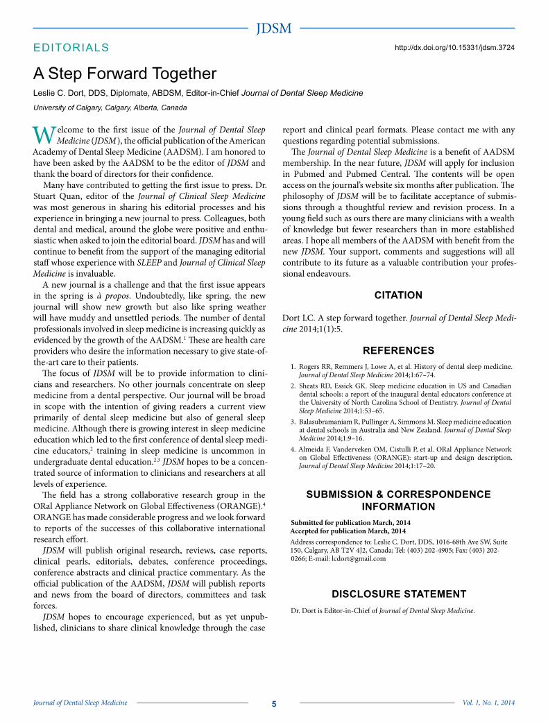
A Step Forward TogetherDort
New Journal Continues the Growth and Progress of Dental Sleep MedicineDemko
Sleep Medicine Education at Dental Schools in Australia and New ZealandBalasubramaniam, Pullinger, Simmons
ORal Appliance Network on Global Effectiveness (ORANGE): Start-Up and Design DescriptionAlmeida, Vanderveken, Cistulli, Fleury, Gagnadoux, Hoekema, Huynh, Hwang, Kuna, Kushida, Lavigne, Lowe, Marklund, Masse, Quinnell, Tsuda, Tsuiki


Editor-in-ChiefLeslie Dort, DDS
Deputy EditorOlivier Vanderveken, MD, PhD
Associate Editors Fernanda Almeida, DDS, PhDGilles Lavigne, DMD, PhDRose Sheats, DMD
Executive Director Jerome A. Barrett
Managing Editor Andrew Miller
Editorial BoardGhizlane Aarab, DDS, PhDPeter Cistulli, MD, PhDGreg Essick, DDS, PhDBernard Fleury, MDNelly Huynh, PhDSam Kuna, MDChris Lettieri, MDAlan Lowe, DMD, PhDMarie Marklund, DDS, PhDAntonio Romero-Garcia, DDS, PhDSatoru Tsuiki, DDS, PhD
Journal of Dental Sleep Medicine (JDSM) (Online 2333-9756; Website: www.jdsm.org) is published online quarterly on the 10th of January, April, July and October by the American Academy of Dental Sleep Medicine, 2510 North Frontage Road, Darien, IL 60561-1511, phone (630) 737-9705 and fax (630) 737-9790.
ANNUAL SUBSCRIPTION RATES: Subscription rates for Volume 1, 2014: Individual Online (US and International): $40.00; Institutional Online (US and International): $70.00. Prorated subscriptions are not available. Subscriptions begin with the January issue of the current
year. Renewals should be secured as early in the year as possible to avoid uninterrupted service. Questions about subscriptions (including payments, billing procedures, or policy matters) should be directed to the AADSM office at (630) 737-9705.
ADVERTISING: Digital advertising is available on www.jdsm.org. Please contact the National Sales Account Executive at [email protected] for complete information.
PERMISSION TO REPRODUCE: Written permission to reproduce, in print or electronically, whole articles or any parts of works, figures or tables
published in JDSM must be obtained prior to publication. Permission for republication must be arranged through the Copyright Clearance Center, Inc., 222 Rosewood Drive, Danvers, MA 01923, phone (978) 750-8400 or fax (978) 646-8600 or URL http://www.copyright.com. There are royalty fees associated with such permissions.
REPRINTS: For author reprints contact the AADSM office. For commercial reprint orders contact Cenveo Publisher Services, 4810 Williamsburg Road, #2, Hurlock, MD 21643 or [email protected].
DISCLAIMER: The statements and opinions contained in editorials and articles in this journal are solely those of the authors thereof and not of the American Academy of Dental Sleep Medicine, or of its officers, regents, members or employees. The Editor-in-Chief, the American Academy of Dental Sleep Medicine and its officers, regents, members and employees disclaim all responsibility for any injury to persons or property resulting from any ideas or products referred to in articles contained in this journal.
© 2014 American Academy of Dental Sleep Medicine
Journal of Dental Sleep MedicineVolume 1, Number 1 | April 10, 2014 | Pages 1–76
Official Publication of the American Academy of Dental Sleep Medicine


Table of Contents Vol. 1, No. 1
Instructions to authors are available online at www.jdsm.org
EDITORIALS
5A Step Forward TogetherLeslie C. Dort
7New Journal Continues the Growth and Progress of Dental Sleep MedicineB. Gail Demko
ORIGINAL ARTICLES
9Sleep Medicine Education at Dental Schools in Australia and New ZealandRamesh Balasubramaniam, Andrew Pullinger, Michael Simmons
17ORal Appliance Network on Global Effectiveness (ORANGE): Start-Up and Design DescriptionFernanda R. Almeida, Olivier M. Vanderveken, Peter A. Cistulli, Bernard Fleury, Frederic Gagnadoux, Aarnoud Hoekema, Nelly T. Huynh, Dennis Hwang, Samuel T. Kuna, Clete A. Kushida, Gilles Lavigne, Alan A. Lowe, Marie E. Marklund, Jean-Francois Masse, Timothy G. Quinnell, Hiroko Tsuda, Satoru Tsuiki
CASE REPORTS
21Effects of the Association of nCPAP and Tongue Positioner Device in OSAS Treatment: A Case ReportDomenico Ciavarella, Roberto Sabato, Battista Giovanni, Lorenzo Lo Muzio, Giuseppina Campisi, Cassano Michele, Lucio Lo Russo, Maria Pia Foschino Barbaro
25Oral Appliance Treatment in a Patient with Down SyndromeB. Gail Demko
REVIEW ARTICLES
27The Link between Sleep Bruxism, Sleep Disordered Breathing and Temporomandibular Disorders: An Evidence-based ReviewRamesh Balasubramaniam, Gary D. Klasser, Peter A. Cistulli, Gilles J. Lavigne
SPECIAL ARTICLES
39Definition of an Effective Oral Appliance for the Treatment of Obstructive Sleep Apnea and Snoring: A Report of the American Academy of Dental Sleep MedicineConsensus Conference Participants: Steven C. Scherr, Leslie C. Dort, Fernanda R. Almeida, Kathleen M. Bennett, Norman T. Blumenstock, B. Gail Demko, Gregory K. Essick, Sheri G. Katz, Paul M. McLornan, Katherine S. Phillips, Ronald S. Prehn, Robert R. Rogers, Thomas G. Schell, Rose D. Sheats, Flavia P. Sreshta
51Definition of an Effective Oral Appliance for the Treatment of Obstructive Sleep Apnea and SnoringConsensus Conference Participants: Steven C. Scherr, Leslie C. Dort, Fernanda R. Almeida, Kathleen M. Bennett, Norman T. Blumenstock, B. Gail Demko, Gregory K. Essick, Sheri G. Katz, Paul M. McLornan, Katherine S. Phillips, Ronald S. Prehn, Robert R. Rogers, Thomas G. Schell, Rose D. Sheats, Flavia P. Sreshta
53Sleep Medicine Education in US and Canadian Dental Schools: A Report of the Inaugural Dental Educators Conference at the University of North Carolina School of DentistryRose D. Sheats, Gregory K. Essick
67History of Dental Sleep MedicineRobert R. Rogers, John Remmers, Alan A. Lowe, Peter A. Cistulli, Jeff Prinsell, Donald Pantino
DENTAL SLEEP MEDICINE PEARLS
75Intervention for Oral Appliance Therapy Patients Presenting with Traumatic Anterior OcclusionSheri G. Katz

Journal of Dental Sleep Medicine Vol. 1, No. 1, 20145
JDSM
A Step Forward TogetherLeslie C. Dort, DDS, Diplomate, ABDSM, Editor-in-Chief Journal of Dental Sleep Medicine
University of Calgary, Calgary, Alberta, Canada
EDITORIALS
Welcome to the first issue of the Journal of Dental Sleep Medicine (JDSM), the official publication of the American
Academy of Dental Sleep Medicine (AADSM). I am honored to have been asked by the AADSM to be the editor of JDSM and thank the board of directors for their confidence.
Many have contributed to getting the first issue to press. Dr. Stuart Quan, editor of the Journal of Clinical Sleep Medicine was most generous in sharing his editorial processes and his experience in bringing a new journal to press. Colleagues, both dental and medical, around the globe were positive and enthu-siastic when asked to join the editorial board. JDSM has and will continue to benefit from the support of the managing editorial staff whose experience with SLEEP and Journal of Clinical Sleep Medicine is invaluable.
A new journal is a challenge and that the first issue appears in the spring is à propos. Undoubtedly, like spring, the new journal will show new growth but also like spring weather will have muddy and unsettled periods. The number of dental professionals involved in sleep medicine is increasing quickly as evidenced by the growth of the AADSM.1 These are health care providers who desire the information necessary to give state-of-the-art care to their patients.
The focus of JDSM will be to provide information to clini-cians and researchers. No other journals concentrate on sleep medicine from a dental perspective. Our journal will be broad in scope with the intention of giving readers a current view primarily of dental sleep medicine but also of general sleep medicine. Although there is growing interest in sleep medicine education which led to the first conference of dental sleep medi-cine educators,2 training in sleep medicine is uncommon in undergraduate dental education.2,3 JDSM hopes to be a concen-trated source of information to clinicians and researchers at all levels of experience.
The field has a strong collaborative research group in the ORal Appliance Network on Global Effectiveness (ORANGE).4 ORANGE has made considerable progress and we look forward to reports of the successes of this collaborative international research effort.
JDSM will publish original research, reviews, case reports, clinical pearls, editorials, debates, conference proceedings, conference abstracts and clinical practice commentary. As the official publication of the AADSM, JDSM will publish reports and news from the board of directors, committees and task forces.
JDSM hopes to encourage experienced, but as yet unpub-lished, clinicians to share clinical knowledge through the case
report and clinical pearl formats. Please contact me with any questions regarding potential submissions.
The Journal of Dental Sleep Medicine is a benefit of AADSM membership. In the near future, JDSM will apply for inclusion in Pubmed and Pubmed Central. The contents will be open access on the journal’s website six months after publication. The philosophy of JDSM will be to facilitate acceptance of submis-sions through a thoughtful review and revision process. In a young field such as ours there are many clinicians with a wealth of knowledge but fewer researchers than in more established areas. I hope all members of the AADSM with benefit from the new JDSM. Your support, comments and suggestions will all contribute to its future as a valuable contribution your profes-sional endeavours.
CITATION
Dort LC. A step forward together. Journal of Dental Sleep Medi-cine 2014;1(1):5.
REFERENCES1. Rogers RR, Remmers J, Lowe A, et al. History of dental sleep medicine.
Journal of Dental Sleep Medicine 2014;1:67–74.2. Sheats RD, Essick GK. Sleep medicine education in US and Canadian
dental schools: a report of the inaugural dental educators conference at the University of North Carolina School of Dentistry. Journal of Dental Sleep Medicine 2014;1:53–65.
3. Balasubramaniam R, Pullinger A, Simmons M. Sleep medicine education at dental schools in Australia and New Zealand. Journal of Dental Sleep Medicine 2014;1:9–16.
4. Almeida F, Vanderveken OM, Cistulli P, et al. ORal Appliance Network on Global Effectiveness (ORANGE): start-up and design description. Journal of Dental Sleep Medicine 2014;1:17–20.
SUBMISSION & CORRESPONDENCE INFORMATION
Submitted for publication March, 2014Accepted for publication March, 2014Address correspondence to: Leslie C. Dort, DDS, 1016-68th Ave SW, Suite 150, Calgary, AB T2V 4J2, Canada; Tel: (403) 202-4905; Fax: (403) 202-0266; E-mail: [email protected]
DISCLOSURE STATEMENTDr. Dort is Editor-in-Chief of Journal of Dental Sleep Medicine.
http://dx.doi.org/10.15331/jdsm.3724

Journal of Dental Sleep Medicine Vol. 1, No. 1, 20147
JDSM
New Journal Continues the Growth and Progress of Dental Sleep MedicineB. Gail Demko, DMD, D. ABDSM
Sleep Apnea Dentists of New England, Weston, MA; President, American Academy of Dental Sleep Medicine
EDITORIALS
“Without continual growth and progress, such words as improvement, achievement, and success have no meaning.” —Benjamin Franklin
The publication of this inaugural issue of the Journal of Dental Sleep Medicine by the American Academy of Dental
Sleep Medicine (AADSM) is a landmark achievement signi-fying that oral appliance therapy truly has come of age. This peer-reviewed, scientific and clinical journal is a product of the continual growth and progress of dentistry’s role in treating sleep-disordered breathing through collaboration with our physician colleagues.
This milestone is remarkable when you consider that it has been only about 30 years since the first reports describing the use of oral devices for the treatment of obstructive sleep apnea (OSA) began to appear in the medical literature.1,2 Practice parameters published by the American Academy of Sleep Medicine in 2006 validated oral appliance therapy as an appropriate treatment alternative to positive airway pressure (PAP) therapy for OSA,3 and today there is a wealth of scientific evidence supporting the effectiveness of mandibular advancement devices in reducing the severity of OSA and improving health outcomes.4
The rapid expansion of the scientific evidence base for oral appliance therapy has been mirrored by the growth of the AADSM as the only not-for-profit professional society that is dedicated exclusively to the practice of dental sleep medi-cine. Established by a small group of dentists in 1991, the AADSM now has more than 3,000 members as the organiza-tion approaches its silver anniversary. The AADSM always has attracted some of the best and brightest minds in dentistry, and our membership continues to grow as an increasing number of dentists recognize the important role they can play in reducing the burden of snoring and obstructive sleep apnea.
In addition to growing quantitatively, the AADSM has spurred qualitative progress in the practice of dental sleep medicine. Although dental sleep medicine began in indepen-dent commercial venues with a heavy emphasis on product sales, the AADSM was built on a cornerstone of evidence-based dentistry long before that term was used in any dental training program in the country. As a result the field has witnessed the emergence of university-based programs that are providing comprehensive education and training to teach dentists about oral appliance therapy at both the pre-doctoral and postdoctoral level. Evidence of the field’s scientific maturation can be seen in the recent establishment of the Oral Appliance Network for Global Effectiveness (ORANGE), an international collaboration supported by the AADSM that is mobilizing leading researchers to evaluate the long-term effectiveness and health outcomes of oral appliance therapy for OSA.
The growing knowledge base in dental sleep medicine has yielded significant improvements in the practice of dental sleep medicine. Gone are the days when some dentists promulgated the idea that oral appliance therapy is a simple treatment and that anyone can make an oral device. Now relying on skilled laboratories to craft custom-fabricated appliances using digital or physical impressions and models, today’s dental sleep medi-cine practitioners are much more than just oral device providers. We are clinicians with expertise in screening patients to iden-tify those who are most at risk for OSA, conducting thorough evaluations of patients’ oral anatomy, selecting the most appro-priate device for each patient, fitting and calibrating the device, assessing potential side effects and complications, monitoring adherence, and ensuring long-term effectiveness.
Although there has been an explosion of dental sleep medi-cine literature in recent years, much of this research has been published in medical journals that are inaccessible to the typical dental sleep medicine clinician. The Journal of Dental Sleep Medicine will span this gap by providing AADSM members with immediate and complimentary access to new research, case reports and commentaries. We are in the midst of an exciting stage in the progression of dental sleep medicine, bearing witness to a proliferation of novel advances and developments in oral appliance therapy. In the last year alone, we have seen evidence that mandibular advancement devices reduce the risk of fatal cardiovascular events in patients with severe OSA, treatment success can be predicted using a remotely controlled mandibular protrusion device, and objective measurement of compliance can be achieved using an embedded microsensor thermometer with on-chip integrated readout electronics.5-7 I am certain that we will see more progress in the years ahead, and AADSM members will stay abreast of these new findings and trends with the journal at their fingertips.
The publication of this journal also is significant because it will heighten recognition of dental sleep medicine among our physician colleagues, who regularly consult sleep medicine journals such as SLEEP and the Journal of Clinical Sleep Medi-cine. The AADSM has worked hard to forge strong ties with the AASM, understanding that teamwork between physicians and dentists promotes optimal care for the patients who suffer from sleep disordered breathing. Millions of people in the U.S. have undiagnosed and untreated OSA, and dentists are well positioned to address this crisis by screening the large number of patients who enter our offices every day, directing at-risk patients into the proper medical channels for a comprehen-sive sleep evaluation and diagnosis. Sleep medicine physicians also are gaining a better understanding of how oral appliance therapy is the most advantageous treatment for many patients with mild-to-moderate OSA, especially those who have a lower
http://dx.doi.org/10.15331/jdsm.3726

Journal of Dental Sleep Medicine Vol. 1, No. 1, 20148
Editorial—Demko
body mass index (BMI) or fail to comply with PAP therapy. Everyone benefits when physicians and dentists collaborate to achieve the common goal of providing the highest quality of care for OSA patients.
This journal’s publication is no small feat. It has taken nearly two years of planning, development, and coordination by Editor-in-Chief Leslie C. Dort, DDS, and the capable staff in the national office, who have worked diligently to bring the journal from a dream to a reality. Under the expert hand of Dr. Dort, and with the assistance of Deputy Editor Olivier Vanderveken, MD, PhD, talented associate editors and a dedicated editorial board, this journal will flourish.
The publication of the Journal of Dental Sleep Medicine by the AADSM is an important achievement that will promote continued growth and progress in the field of dental sleep medicine.
CITATIONDemko BG. New journal continues the growth and progress of dental sleep medicine. Journal of Dental Sleep Medicine 2014;1(1):7–8.
REFERENCES1. Cartwright RD, Samelson CF. The effects of a nonsurgical treatment
for obstructive sleep apnea. The tongue-retaining device. JAMA 1982;248:705-9.
2. Meier-Ewert K, Schäfer H, Kloss W. Treatment of sleep apnea by mandibular protracting device. [Abstract] Proceedings of the Seventh European Sleep Congress. Munich, 1984:217.
3. Kushida CA, Morgenthaler TI, Littner MR, et al. Practice parameters for the treatment of snoring and obstructive sleep apnea with oral appliances: An update for 2005. Sleep 2006;29:240-3.
4. Sutherland K, Vanderveken OM, Tsuda H, et al; on behalf of the ORANGE-Registry. Oral appliance treatment for obstructive sleep apnea: an update. J Clin Sleep Med 2014;10:215-27.
5. Anandam A, Patil M, Akinnusi M, Jaoude P, El-Solh AA. Cardiovascular mortality in obstructive sleep apnoea treated with continuous positive airway pressure or oral appliance: an observational study. Respirology 2013;18:1184-90.
6. Remmers J, Charkhandeh S, Grosse J, et al. Remotely controlled mandibular protrusion during sleep predicts therapeutic success with oral appliances in patients with obstructive sleep apnea. Sleep 2013;36:1517-25.
7. Vanderveken OM, Dieltjens M, Wouters K, De Backer WA, Van de Heyning PH, Braem MJ. Objective measurement of compliance during oral appliance therapy for sleep-disordered breathing. Thorax 2013;68:91-6.
SUBMISSION & CORRESPONDENCE INFORMATION
Submitted for publication February, 2014Accepted for publication February, 2014Address correspondence to: B. Gail Demko, DMD, 140 Merriam St., Weston, MA 02493; Tel: (617) 964-4028; Fax: (617) 595-4591; E-mail: [email protected]
DISCLOSURE STATEMENTDr. Demko has indicated no financial conflicts of interest.
Journal of Dental Sleep Medicine Vol. 1, No. 1, 20149
JDSM
Sleep Medicine Education at Dental Schools in Australia and New ZealandRamesh Balasubramaniam, BDSc, MS1; Andrew Pullinger, DDS, MSc2; Michael Simmons, DMD, FAGD3
1School of Dentistry, University of Western Australia, Perth, Western Australia, Australia; 2University of California, Los Angeles, School of Dentistry, CA; 3University of California, Los Angeles, School of Dentistry, Clinical Assistant Professor, Ostrow School of Dentistry, Los Angeles, CA
ORIGINAL ARTICLES
Background: Traditionally, the curriculum in Australian and New Zealand dental schools has largely ignored the need for future dentists to receive foundational education in the field of sleep medicine. The absence of official education accreditation standards means this increasing part of dental practice continues as a continuing education activity without proper accountability by organized dentistry. This manuscript evaluates the current status of education in sleep disorders to predoctoral dental students.Methods: All 10 dental schools in Australia and New Zealand were surveyed for information regarding their sleep medicine curriculum during the 2011 academic year. The head of each dental school or relevant course coordinator responded to a questionnaire.Results: One dental school did not respond, and 3 dental schools were unable to complete the survey, as they had not graduated a class. Therefore 6 of the potential 7 dental schools (85.7%) completed the survey. The average total predoctoral sleep medicine teaching time was 4.5 hours (SD 2.51; range 2 to 8 h). Five of the 6 dental schools spent most of their sleep medicine curriculum time teaching in the fifth year of 5-year programs (mean of 2.5 h; SD 2.88). Education time spent in sleep medicine was 55% didactic. All responding dental schools reviewed obstructive sleep apnea, 83% reviewed sleep bruxism, and 67% reviewed primary snoring.Conclusions: Although a definite beginning, current sleep medicine education at Australian and New Zealand dental schools still seems to be at an exposure level, and likely inadequate for competency in screening for sleep related breathing disorders as the primary requirement. It also seems to be minimal foundation for participating as a future dentist member of the sleep medicine team, which with further post graduation training may include providing oral appliance therapy for sleep disordered breathing when appropriate. This appears to be a similar outcome to the level of education in sleep medicine provided in the United States dental school predoctoral programs to date.Keywords: sleep medicine education, sleep disordered breathing, competency, dental sleep medicineCitation: Balasubramaniam R, Pullinger A, Simmons M. Sleep medicine education at dental schools in Australia and New Zealand. Journal of Dental Sleep Medicine 2014;1(1):9–16.
The field of dental sleep medicine (DSM) has become an area of importance for comprehensive dental education, needed
for contemporary dental practice.1 Traditionally, the curric-ulum in Australian and New Zealand dental schools has largely ignored the need for dentists to be educated in the field of sleep medicine, except for sleep bruxism. The education and training has been largely left to continuing education with little founda-tional oversight by organized dentistry or dental school accredi-tation standards within the context of the sleep medicine team.
The introduction and validation of oral appliances for the treatment of snoring and obstructive sleep apnea (OSA) has through years of research, resulted in dentists becoming involved in the treatment of patients with sleep disordered breathing (SDB).2,3 SDB is a group of common problems involving difficulties in breathing during sleep that may range from socially embarrassing snoring to severe and life-threat-ening OSA. These are caused primarily by negative pressures in the collapsible upper airway, which compromise the anatomic and physiologic capacity to maintain an adequate airway lumen during the various stages of sleep.4 SDB has been recognized to be a major risk factor for morbidity and mortality.5,6 There are cardiovascular, metabolic, and cognitive issues with undiag-nosed and untreated OSA. Specifically, OSA has been linked to systemic hypertension, myocardial infarction, stroke, congestive heart failure, atrial fibrillation, carotid artery atherosclerosis, glucose intolerance, diabetes, depression, and excessive daytime
sleepiness.7-9 Presence of any of these histories in the dental patient should precipitate questions about sleep disorders, even if the dentist is not involved in active treatments. Dentists can potentially treat OSA through growth and development orth-odontic intervention in children,10 oral appliance therapy,11 or orthognathic surgery.12 Based upon the significant percentage of the population with OSA, ranging from 10% to 26% in Austra-lian surveys,13 it is incumbent upon all health care providers to screen for these problems. Since dentists routinely examine the oral cavity, they also have a clear view of the oropharynx and are uniquely positioned to screen for potential anatomic risk features in patients with subjective or partner reports of SDB, unrestful sleep, or in patients reporting potentially associated medical problems. Therefore dentists could readily have an impact on the health status of our society if trained to a level of competency in sleep medicine.14 Screening as part of a wellness practice philosophy can evolve into active treatment by dentists subsequent to further training additional to a foundational dental school curriculum.
The American Academy of Sleep Medicine (AASM) prac-tice parameters recommend that the “oral appliance should be fitted by qualified dental personnel who are trained and experi-enced in the overall care of oral health, the temporomandibular joint, dental occlusion, and associated oral structures. Dental management of patients with oral appliances should be overseen by practitioners who have undertaken serious training in sleep
http://dx.doi.org/10.15331/jdsm.3728
Journal of Dental Sleep Medicine Vol. 1, No. 1, 201410
Sleep Medicine Education in Australia and New Zealand—Balasubramaniam et al.
medicine and/or sleep related breathing disorders with focused emphasis on the proper protocol for diagnosis, treatment, and follow-up”.11 The Australian Dental Association (AuDA) has a policy statement on the “use of dental appliances to treat sleep disorders”.15 The AuDA has endorsed the guidelines of the AASM practice parameters for the use of oral appliances in the treatment of snoring and OSA.
The 2009 Australian Schedule of Dental Services and Glos-sary, which is the official AuDA document governing the item number for dentists to use when providing and invoicing oral appliance therapy to treat OSA,16 also makes reference to the AASM practice parameters.11 Similarly, the board of the Austral-asian Sleep Association (ASA) in July 2010 endorsed guidelines for the “use of dental appliance therapy for the treatment of SDB to ensure a minimum standard of clinical practice.”17 Again, this document has recommended the practice protocol of the AASM.11 In New Zealand, no formal guidelines exist on the use of oral appliances for snoring and OSA. Nevertheless, the New Zealand Dental Council (NZDC) consider the provision of “corrective dental appliances” to fall within the scope of dental practice.18 It is apparent that the Academies and Associations in
Australia and New Zealand agree that dentists should be trained in the field of DSM if they choose to be involved in treating patients with SDB. However, all trainees should first be compe-tent in conducting a routine sleep disorder history screening in a new patient as part of routine dental care.
Given the potential societal advantages of training the next generation of dentists in the field of DSM at Australian and New Zealand dental schools, the authors carried out an investi-gation of current predoctoral dental curriculum in both coun-tries. The purpose was to establish the number of dental schools in Australia and New Zealand that include at least some DSM training at the predoctoral level and the current status of this education. Insight is anticipated for evolving structured and calibrated dental school sleep medicine education that will eventually become part of dental school accreditation standards and the foundation for standards of care in dental practice.
METHODS
All 10 dental schools in Australia and New Zealand were sent a survey to gather information regarding their sleep medicine curriculum for the 2011 academic year. The battery of ques-tions was based upon a study conducted on American dental schools.1 There were 8 categories in the questionnaire, which included: (1) hours spent teaching sleep medicine, (2) teaching methods, (3) department(s) involved in teaching, (4) topics discussed, (5) diagnosis reviewed, (6) all therapies discussed, (7) aspects of oral appliance therapy discussed, and (8) discus-sion of contemporary topics. (Appendix)
The questionnaires were mailed or e-mailed to the heads of all the dental schools in Australia and New Zealand who were instructed to forward it to the relevant course coordinator if necessary. The results were tabulated in Excel and analyzed.
RESULTS
Nine of 10 dental schools responded to the survey. Three of the recently established dental schools were unable to complete the questionnaire, as their curriculum was underdeveloped and they had not yet graduated a class. The other 6 dental schools completed the survey, resulting in 85.7% response rate for schools that had graduated a class.
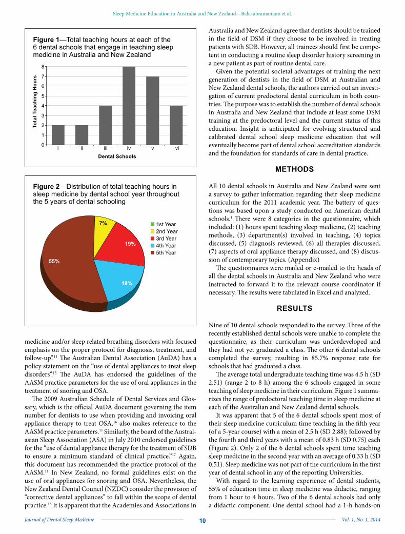
The average total undergraduate teaching time was 4.5 h (SD 2.51) (range 2 to 8 h) among the 6 schools engaged in some teaching of sleep medicine in their curriculum. Figure 1 summa-rizes the range of predoctoral teaching time in sleep medicine at each of the Australian and New Zealand dental schools.
It was apparent that 5 of the 6 dental schools spent most of their sleep medicine curriculum time teaching in the fifth year (of a 5-year course) with a mean of 2.5 h (SD 2.88); followed by the fourth and third years with a mean of 0.83 h (SD 0.75) each (Figure 2). Only 2 of the 6 dental schools spent time teaching sleep medicine in the second year with an average of 0.33 h (SD 0.51). Sleep medicine was not part of the curriculum in the first year of dental school in any of the reporting Universities.
With regard to the learning experience of dental students, 55% of education time in sleep medicine was didactic, ranging from 1 hour to 4 hours. Two of the 6 dental schools had only a didactic component. One dental school had a 1-h hands-on
Figure 1—Total teaching hours at each of the 6 dental schools that engage in teaching sleep medicine in Australia and New Zealand
0
1
2
3
4
5
6
7
8
i ii iii iv v viDental Schools
Tota
l Tea
chin
g H
ours
Figure 2—Distribution of total teaching hours in sleep medicine by dental school year throughout the 5 years of dental schooling
1st Year2nd Year3rd Year4th Year5th Year
55%
19%
19%
7%

Journal of Dental Sleep Medicine Vol. 1, No. 1, 201411
Sleep Medicine Education in Australia and New Zealand—Balasubramaniam et al.
(pre-clinical) laboratory component (4% of education time). Four of the 6 dental schools had a clinical component in their sleep medicine curriculum, which accounted for 41% of educa-tion time spent ranging from 0 to 5 hours. Detailed analysis revealed 2 of the 6 dental schools had a case-by-case and in-clinic discussion with 1 and 4 hours spent, respectively. Two of the dental schools had required rotation or clinical observa-tion with 1 and 5 hours spent, respectively.
Two of the 6 dental schools teaching sleep medicine had multiple dental departments contributing to the undergraduate dental curriculum. In one dental school, the oral medicine and orofacial pain departments were co-involved in teaching sleep medicine. In the other dental school, the teaching was divided between the oral medicine, oral and maxillofacial surgery, and prosthodontic departments. The oral medicine specialty was the most commonly involved department teaching the sleep medicine curriculum (4 of the 6 dental schools). Of interest, all teaching was undertaken at the undergraduate level. None of the dental schools reported involvement in teaching sleep medi-cine at the postgraduate or dental specialty program level. There is no information in the survey about any curriculum coordina-tion between departments.
With regard to sleep medicine topics discussed, all 6 responding dental schools discussed SDB, and 67% of dental schools discussed sleep bruxism (SB). The other topics discussed are summarized in Figure 3. All 6 responding dental schools reviewed OSA as a diagnosis. A high percentage of dental schools reviewed the diagnosis of SB and primary snoring (83% and 67%, respectively). Fifty percent of dental schools reviewed the diagnosis of restless legs syndrome and upper airway resistance syndrome. Periodic limb movement disorder and insomnia were discussed by 33% and 17% of dental schools, respectively.
Of note, the diagnosis of sleep phase shifts and REM behavior disorder were not discussed at all.
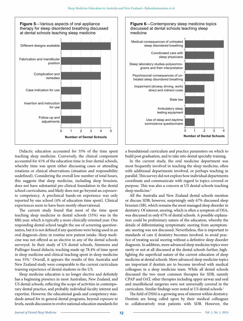
Dental school curriculum often involved discussion regarding various therapies for SDB (Figure 4). All dental schools involved in teaching sleep medicine covered the therapeutic interventions of continuous positive airway pressure (CPAP) and oral appli-ance therapy (OAT) in treating SDB. Four of the 6 responding dental schools discussed upper airway surgical therapies. Simi-larly, 4 responding dental schools discussed oral and maxillo-facial surgical therapies (orthognathic surgery) for SDB. Most dental schools discussed various aspects of oral appliance therapy for SDB (Figure 5).
Figure 6 summarizes the percentage of the responding dental schools involved in discussion of contemporary topics in the field of sleep medicine. The medical consequences of untreated SDB were discussed by 67% of the responding dental schools teaching sleep medicine. Only 50% of the responding dental schools taught topics related to coordinated care with sleep physicians, diagnostic need and interpretation of sleep studies, and the psychological consequences of untreated SDB. This questions how much DSM is being taught within the concept of the sleep medicine team. Only 17% of the responding dental schools taught the use of screening questionnaires for use in catching occult or undiagnosed sleep disorders.
DISCUSSION
Six of the seven responding dental schools engaging in some teaching of sleep medicine in their curriculum (85.7%) averaged 4.5 total hours, ranging from 2 to 8 hours. This is comparable with the average time of 3.92 hours spent teaching this topic at United States (US) dental schools that taught sleep medicine. However, it should be noted that 24.5% of US dental schools responding reported they did not cover the topic of sleep medicine at all.1
Figure 3—Topics covered in the sleep medicine in the undergraduate dental curriculum.
0 1 2 3 4 5 6
Other
Hypersomnias ofcentral origin
Insomnias
Sleep relatedmovement disorders
Circadian rhythmsleep disorders
Parasomnias
Sleep bruxism
Sleep relatedbreathing disorders
Number of Dental Schools
Figure 4—Sleep disordered breathing therapies discussed at dental schools in Australia and New Zealand teaching sleep medicine
0 1 2 3 4 5 6
CPAP
Oral appliance therapy
Oral and MaxillofacialSurgery
ENT surgical therapies
Orthodonticapproaches
Number of Dental Schools
Journal of Dental Sleep Medicine Vol. 1, No. 1, 201412
Sleep Medicine Education in Australia and New Zealand—Balasubramaniam et al.
Didactic education accounted for 55% of the time spent teaching sleep medicine. Conversely, the clinical component accounted for 41% of the education time in four dental schools, whereby time was spent either discussing cases or attending rotations or clinical observations (situation and responsibility undefined). Considering the overall low number of total hours, this suggests that sleep medicine, including sleep bruxism, does not have substantial pre-clinical foundation in the dental school curriculums, and likely does not go beyond an exposure-to competency. A preclinical hands-on experience was only reported by one school (4% of education time spent). Clinical experiences seem to have been mostly observational.
The current study found that most of the time spent teaching sleep medicine in dental schools (55%) was in the fifth year, which is typically a more clinically oriented year. One responding dental school taught the use of screening question-naires, but it is not defined if any questions were being used in an oral diagnosis clinic in routine new patient intake. Sleep medi-cine was not offered as an elective in any of the dental schools surveyed. In their study of US dental schools, Simmons and Pullinger found didactic teaching made up 78.4% of time spent in sleep medicine and clinical teaching spent in sleep medicine was 35%.1 Overall, it appears the results of this Australia and New Zealand study were comparable to the current curriculum training experience of dental students in the US.
Sleep medicine education is no longer elective and definitely has a beginning presence in most Australian, New Zealand, and US dental schools, reflecting the scope of activities in contempo-rary dental practice, and probably individual faculty interest and expertise. However, the educational goals and competency stan-dards aimed for in general dental programs, beyond exposure-to levels, needs discussion to evolve national education standards for
a foundational curriculum and practice parameters on which to build post graduation, and to take into dental specialty training.
In the current study, the oral medicine department was most frequently involved in teaching the sleep medicine, often with additional departments involved, or perhaps teaching in parallel. This survey did not explore how individual departments coordinate and communicate with regard to topics covered or purpose. This was also a concern at US dental schools teaching sleep medicine.1
All the Australia and New Zealand dental schools mention or discuss SDB; however, surprisingly only 67% discussed sleep bruxism (SB), which remains the most managed sleep disorder in dentistry. Of interest, snoring, which is often a symptom of OSA, was discussed in only 67% of dental schools. A possible explana-tion could be preliminary nature of the education, whereby the details of differentiating symptomatic snoring from asymptom-atic snoring was not discussed. Nevertheless, this is important to standards of care if dentistry becomes involved, to avoid prac-tice of treating social snoring without a definitive sleep disorder diagnosis. In addition, more advanced sleep medicine topics were rarely or not at all discussed at the dental schools further, high-lighting the superficial nature of the current education of sleep medicine at dental schools. More advanced sleep medicine topics are important if dentists are to become involved with medical colleagues in a sleep medicine team. While all dental schools discussed the two most common therapies for SDB, namely CPAP and OAT, other therapies including upper airway and oral and maxillofacial surgeries were not universally covered in the curriculum. Similar findings were noted at US dental schools.1
The field of DSM is a growing area of interest within dentistry. Dentists are being called upon by their medical colleagues to collaboratively treat patients with SDB. However, the
Figure 5—Various aspects of oral appliance therapy for sleep disordered breathing discussed at dental schools teaching sleep medicine
0 1 2 3 4 5
Different designs available
Fabrication and mandibularposition
Complication andremedies
Case indication for use
Insertion and instructionfor use
Follow-up andadjustments
Number of Dental Schools
Figure 6—Contemporary sleep medicine topics discussed at dental schools teaching sleep medicine
0 1 2 3 4
Use of sleep and daytimesomnolence questionnaire
Ambulatory sleeptesting equipment
State law
Impairment (drowsy driving, work)direct and indirect costs
Psychosocial consequences of un-treated sleep disordered breathing
Sleep laboratory studies-polysomno-grams and their interpretation
Coordinated care withsleep physicians
Medical consequences of untreatedsleep disordered breathing
Number of Dental Schools

Journal of Dental Sleep Medicine Vol. 1, No. 1, 201413
Sleep Medicine Education in Australia and New Zealand—Balasubramaniam et al.
question remains whether dentists have the foundational educa-tion required to competently treat patients with SDB. To date, there has been no formal study assessing educational prepared-ness of dentists in the field of sleep medicine in Australia and New Zealand. Bian in 2004 surveyed dentists in Indiana, USA, and found an overall deficiency in education regarding OSA and OAT.19 Undergraduate and postgraduate training were only reported by 16% and 30% of responders, respectively. Thirty-two percent reportedly were self-taught. Of concern, 58% of responders were unable to identify common signs and symptoms of OSA, and 40% stated knowing little or nothing about OSA.
Mindell et al. studied sleep education in medical school curriculum across countries.20 Findings revealed only 6 of the 19 medical schools in Australia responded to the survey. It was noted that 369 minutes was spent teaching sleep medicine in Australian medical schools, which was higher than the average of 146 minutes spent in other medical schools sampled from various parts of the world. Regardless, this highlights the limited time spent teaching sleep medicine at medical schools in Australia, which is problematic if the dentist is expecting their patient’s physician to be knowledgeable These findings were consistent with other studies, underscoring deficiencies in sleep medicine education in medical schools in the United States.21,22 Of interest, training in nursing schools in sleep medicine in the US is also not established, but it has been recommended that 40 hours educa-tion be required, which raises the requirement bar considerably.23
The Australian Dental Council (ADC) is an independent body for dental education and training in Australia. It is an external accreditation authority for the Dental Board of Australia. It sets the standard required of newly qualified dentists to be consid-ered “competent” to be able to care for the Australian public. Of concern, the ADC does not require competency in the field of DSM. In fact, this area of dentistry is not mentioned at all in the document on “Professional attributes and competencies of the newly qualified dentist”.24 Similarly, the NZDC and the New Zealand Dental Association do not address competency in the field of DSM.18 It is therefore not surprising that very little time is spent teaching sleep medicine at Australian and New Zealand dental schools. The administrators of dental schools could consider allocating more time teaching subjects such as DSM as part of foundational competencies. The current teaching of sleep medicine at dental schools might currently serve as a good introduction to the field but appears insufficient to safely treat patients with potentially associated serious medical conditions as required by the ASA and ADA guidelines.
The authors believe it is the responsibility of dental schools to provide foundational competencies in the field of DSM as required in all other aspects of dentistry. Universities should be the leaders in providing standards of care to reflect contem-porary practice, and to protect patients. Competencies in DSM have been recommended by Simmons and Pullinger.1 All new graduates should be moderately competent and confident in new patient intake sleep history triage through portal questions. All new graduates should be able to conduct a more compre-hensive sleep history in symptomatic patients and in patients with medical histories and comorbidities, which have associa-tions with SDB. Comprehensive history can be augmented by self-scoring published sleep questionnaires, combined with clinical examination for oropharyngeal anatomic risk factors.
Understanding the need and mechanism for referral for in lab polysomnography or out of sleep center test and motivating the patient to act requires an understanding and discussion of the potential medical consequences of untreated SDB. The dental graduate must be able to identify, refer, and document patients suspected of having SDB to their physician to request a sleep study and sleep diagnosis. Competency can only be achieved in these history and clinical screening requirements if they are routinely included in the Oral Diagnosis intake clinic process in dental schools for all patients, and the standard patient data-base. This is considered the first important step and is a require-ment for all of dentistry and not limited to a DSM expert clinic. The next level involves more training and potential treatment based on understanding of the outcomes of the medical sleep test and report. If suggested by the sleep physician, the dental graduate must be able to assess for suitability for an oral appli-ance for patients with SDB, provided they have received further clinical training post-graduation to “qualified” status or make the referral to such qualified dentist. “Qualified” dentists would provide follow-up care and testing as recommended by the AASM practice parameters.11 Similar, recommendations have been made for training in the US1; however, as of this date, the American Dental Education Association and the Council on Dental Accreditation have yet to implement foundational standards for DSM training in the US. Meanwhile, implementa-tion and the scope of a school’s DSM curriculum depend on the insight of each dental dean and the faculty, supported also by the input of recognized sleep medicine academies.
CONCLUSION
Dentists are uniquely positioned to help screen and co-treat SDB in a multidisciplinary approach, as part of a patients’ sleep medi-cine team. It is no longer a question of whether dentists can help identify undiagnosed and untreated SDB conditions, but rather how best to implement and participate in the field of sleep medi-cine. Currently there is an awareness level of education in most Australian and New Zealand dental schools; however, this falls short in foundational, screening, and treatment competencies. By working towards incorporating DSM education into dental school curriculum standards, dentists can improve quality of life, reduce the medical costs, protect patients, and create a greater awareness of the medical and social importance of good sleep.
REFERENCES1. Simmons MS, Pullinger A. Education in sleep disorders in US dental
schools DDS programs. Sleep Breath 2012;16:383-92.2. Barnes M, McEvoy RD, Banks S, et al. Efficacy of positive airway pressure
and oral appliance in mild to moderate obstructive sleep apnea. Am J Respir Crit Care Med 2004;170:656-64.
3. Gotsopoulos H, Chen C, Qian J, Cistulli PA. Oral appliance therapy improves symptoms in obstructive sleep apnea: a randomized, controlled trial. Am J Respir Crit Care Med 2002;166:743-8.
4. Sullivan CE, Issa FG. Obstructive sleep apnea. Clin Chest Med 1985;6:633-50.
5. Marshall NS, Wong KK, Liu PY, Cullen SR, Knuiman MW, Grunstein RR. Sleep apnea as an independent risk factor for all-cause mortality: the Busselton Health Study. Sleep 2008;31:1079-85.
6. Young T, Finn L, Peppard PE, et al. Sleep disordered breathing and mortality: eighteen-year follow-up of the Wisconsin sleep cohort. Sleep 2008;31:1071-8.

Journal of Dental Sleep Medicine Vol. 1, No. 1, 201414
Sleep Medicine Education in Australia and New Zealand—Balasubramaniam et al.
7. Marshall NS, Wong KK, Phillips CL, Liu PY, Knuiman MW, Grunstein RR. Is sleep apnea an independent risk factor for prevalent and incident diabetes in the Busselton Health Study? J Clin Sleep Med 2009;5:15-20.
8. Pack AI. Advances in sleep-disordered breathing. Am J Respir Crit Care Med 2006;173:7-15.
9. Lee SA, Amis TC, Byth K, et al. Heavy snoring as a cause of carotid artery atherosclerosis. Sleep 2008;31:1207-13.
10. Villa MP, Rizzoli A, Miano S, Malagola C. Efficacy of rapid maxillary expansion in children with obstructive sleep apnea syndrome: 36 months of follow-up. Sleep Breath 2011;15:179-84.
11. Kushida CA, Morgenthaler TI, Littner MR, et al. Practice parameters for the treatment of snoring and obstructive sleep apnea with oral appliances: an update for 2005. Sleep 2006;29:240-3.
12. Pirklbauer K, Russmueller G, Stiebellehner L, et al. Maxillomandibular advancement for treatment of obstructive sleep apnea syndrome: a systematic review. J Oral Maxillofac Surg 2011;69:e165-76.
13. Bearpark H, Elliott L, Grunstein R, et al. Snoring and sleep apnea. A population study in Australian men. Am J Respir Crit Care Med 1995;151:1459-65.
14. Ivanoff CS, Hottel TL, Pancratz F. Is there a place for teaching obstructive sleep apnea and snoring in the predoctoral dental curriculum? J Dent Educ 2012;76:1639-45.
15. Australian Dental Association Inc., Policy Statement 6.7: Use of dental appliances to treat sleep disorders. http://www.ada.org.au/about/policies.aspx. Accessed March 12, 2012.
16. The Australian Schedule of Dental Services and Glossary. 984: Bimaxillary oral appliance for diagnosed snoring and obstructive snoring and sleep apnoea. St Leonards: Australian Dental Association Inc., 2009.
17. Australasian Sleep Association: Dental appliance therapy for the treatment of sleep disordered breathing. http://www.sleep.org.au/about/dental-orofacial. Accessed March 12, 2012.
18. New Zealand Dental Council: Health Practitioners Competence Assurance Act 2003, 2012:7.
19. Bian H. Knowledge, opinions, and clinical experience of general practice dentists toward obstructive sleep apnea and oral appliances. Sleep Breath 2004;8:85-90.
20. Mindell JA, Bartle A, Wahab NA, et al. Sleep education in medical school curriculum: a glimpse across countries. Sleep Med 2011;12:928-31.
21. Rosen RC, Rosekind M, Rosevear C, Cole WE, Dement WC. Physician education in sleep and sleep disorders: a national survey of U.S. medical schools. Sleep 1993;16:249-54.
22. Orr WC, Stahl ML, Dement WC, Reddington D. Physician education in sleep disorders. J Med Educ 1980;55:367-9.
23. Lee KA, Landis C, Chasens ER, et al. Sleep and chronobiology: recommendations for nursing education. Nurs Outlook 2004;52:126-33.
24. Australian Dental Council: Professional attributes and competencies of the newly qualified dentist. June 2010. http://http://www.adc.org.au/documents/Attributes%20&%20Competencies_Dentist%20v1.0%20Final%2010-06-11.pdf. Accessed March 12, 2012.
ACKNOWLEDGEMENTSThe authors acknowledge Professor Marc Tennant and Associate Professor Estie Kruger of the Centre for Rural and Remote Oral Health for their as-sistance with this research.
SUBMISSION & CORRESPONDENCE INFORMATION
Submitted for publication May, 2013Submitted in final revised form January, 2014Accepted for publication January, 2014Address correspondence to: Ramesh Balasubramaniam, Perth Oral Medicine & Dental Sleep Centre, St John of God Hospital, Subiaco Clinic, Suite 311, 25 McCourt Street, Subiaco, WA 6008, Australia; Tel: +618 9382 2325; Fax: +618 9382 2328; Email: [email protected]
DISCLOSURE STATEMENTThis was not an industry supported study. The protocol of this research project was approved by the Human Research Ethics Committee at the University of Western Australia. The authors have indicated no financial conflicts of interest. All authors have contributed significantly and are in agreement with the content of the manuscript.

Journal of Dental Sleep Medicine Vol. 1, No. 1, 201415
Sleep Medicine Education in Australia and New Zealand—Balasubramaniam et al.
APPENDIX
Study on Sleep Disordered Breathing
By Dr. Ramesh Balasubramaniam
Dear Dr. ________ ________________,
The topic of Sleep Disordered Breathing ranging from primary snoring to severe sleep apnoea is currently becoming an area of high interest in many dental practices. However trickle down into dental school curricula seems to be only at its very early stages of development. As a result, main stream Dentistry seems little involved to date in establishment of standards of care in general practice, beyond its traditional interest in sleep bruxism. Meanwhile, Medicine voices increasing concern about the consequences of sleep pathology and dysfunction, and Dentistry wants to maintain a role in and contribution to a multidisci-plinary team in Sleep Medicine.
This brief study seeks to survey how much training our dental students currently receive in the area of Dental Sleep Medicine (DSM) and the field of Sleep. We sincerely request that you answer this brief 8 question questionnaire or send it on to the respec-tive department(s) for completion. Many thanks in advance for your consideration and prompt response.
For the 2011 Academic year please answer the following questions for your Bachelor of Dental Science program. Please circle all that apply or write in correct answers.
1. How many class or clinic hours are spent on teaching topics of Sleep Medicine in each program year. a. 1st Year 0 1 2 3 4 5 6 7 8 > 8b. 2nd Year 0 1 2 3 4 5 6 7 8 > 8c. 3rd Year 0 1 2 3 4 5 6 7 8 > 8d. 4th Year 0 1 2 3 4 5 6 7 8 > 8e. 5th Year 0 1 2 3 4 5 6 7 8 > 8
2. Is this experience (hours):
Didactic? Preclinicallaboratory?
ClinicalOne on one/
case by case in General Clinic
Required rotation or special clinic
observation
Required rotation or special clinic
active involvementElective
a. 1st Yearb. 2nd Yearc. 3rd Yeard. 4th Yeare. 5th Year
3. Which departments in your School teach about sleep disordered breathing: (please circle all that apply)a. Undergraduate Oral Medicine d. Undergraduate Orofacial Painb. Undergraduate Oral Surgery e. Undergraduate Prosthodontics/Restorativec. Undergraduate Orthodontics f. Other Undergraduate: _____________g. Post-graduate program:______________
4. Which topics, as classified by the American Academy of Sleep Medicine*, are discussed: (please circle all that apply) a. Insomnia* e. Parasomnias*b. Sleep related breathing disorders* f. Sleep related movement disorders*c. Hypersomnias of central origin* g. Other sleep disorders*d. Circadian rhythm sleep disorders* h. Sleep bruxism
Appendix continues on the following page
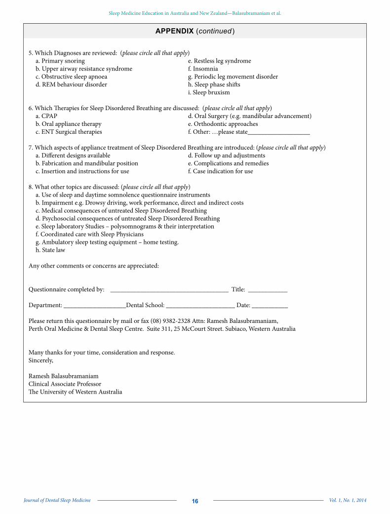
Journal of Dental Sleep Medicine Vol. 1, No. 1, 201416
Sleep Medicine Education in Australia and New Zealand—Balasubramaniam et al.
APPENDIX (continued )
5. Which Diagnoses are reviewed: (please circle all that apply)a. Primary snoring e. Restless leg syndromeb. Upper airway resistance syndrome f. Insomniac. Obstructive sleep apnoea g. Periodic leg movement disorderd. REM behaviour disorder h. Sleep phase shifts i. Sleep bruxism
6. Which Therapies for Sleep Disordered Breathing are discussed: (please circle all that apply)a. CPAP d. Oral Surgery (e.g. mandibular advancement)b. Oral appliance therapy e. Orthodontic approaches c. ENT Surgical therapies f. Other: …please state___________________
7. Which aspects of appliance treatment of Sleep Disordered Breathing are introduced: (please circle all that apply)a. Different designs available d. Follow up and adjustmentsb. Fabrication and mandibular position e. Complications and remediesc. Insertion and instructions for use f. Case indication for use
8. What other topics are discussed: (please circle all that apply)a. Use of sleep and daytime somnolence questionnaire instrumentsb. Impairment e.g. Drowsy driving, work performance, direct and indirect costsc. Medical consequences of untreated Sleep Disordered Breathingd. Psychosocial consequences of untreated Sleep Disordered Breathinge. Sleep laboratory Studies – polysomnograms & their interpretationf. Coordinated care with Sleep Physiciansg. Ambulatory sleep testing equipment – home testing.h. State law
Any other comments or concerns are appreciated:
Questionnaire completed by: ____________________________________ Title: ____________
Department: ___________________Dental School: _____________________ Date: ___________
Please return this questionnaire by mail or fax (08) 9382-2328 Attn: Ramesh Balasubramaniam, Perth Oral Medicine & Dental Sleep Centre. Suite 311, 25 McCourt Street. Subiaco, Western Australia
Many thanks for your time, consideration and response.Sincerely,
Ramesh BalasubramaniamClinical Associate Professor The University of Western Australia

Journal of Dental Sleep Medicine Vol. 1, No. 1, 201417
JDSM
ORal Appliance Network on Global Effectiveness (ORANGE): Start-Up and Design DescriptionFernanda R. Almeida, DDS, PhD1; Olivier M. Vanderveken, MD, PhD2; Peter A. Cistulli, MD, PhD3; Bernard Fleury, MD4; Frederic Gagnadoux, MD, PhD5; Aarnoud Hoekema, MD, DMD, PhD6; Nelly T. Huynh, PhD7; Dennis Hwang, MD8; Samuel T. Kuna, MD9; Clete A. Kushida, MD, PhD, RST, RPSGT10; Gilles Lavigne, DMD, PhD7; Alan A. Lowe, DMD, PhD1; Marie Marklund, DDS, PhD11; Jean-Francois Masse, DMD12; Timothy G. Quinnell, MD, FRACP, FRCP13; Hiroko Tsuda, DDS, PhD14; Satoru Tsuiki, DDS, PhD15
1University of British Columbia, Vancouver, BC, Canada; 2Faculty of Medicine and Health Sciences and Antwerp University Hospital, Edegem, University of Antwerp, Antwerp, Belgium; 3Department of Respiratory and Sleep Medicine, Royal North Shore Hospital and University of Sydney, Sydney, NSW Australia; 4Hôpital Saint Antoine, Hôpitaux Universitaires Est Parisien, Paris, France; 5Angers University Hospital, Angers, France; 6University Medical Center Groningen, University of Groningen, Groningen, The Netherlands; 7Université de Montréal, Montreal, Canada; 8Kaiser Permanente Fontana Medical Center, Fontana, CA; 9Philadelphia Veterans Affairs Medical Center and The University of Pennsylvania, Philadelphia, PA; 10Stanford Sleep Medicine Center, Redwood City, CA; 11Umea University, Umea, Sweden; 12Laval University, Quebec, Canada; 13Papworth Hospital, Cambridge, UK; 14Kyushu University, Fukuoka, Japan; 15Japan Somnology Center, Tokyo, Japan
ORIGINAL ARTICLES
Summary: Oral appliance (OA) therapy is the main non-surgical alternative to CPAP treatment in patients with obstructive sleep apnea (OSA). There are clear benefits from OA when compared to placebo, but a larger variability compared to CPAP has been documented for the reduction of OSA. These results are based on less than 30 randomized controlled trials. In addition, an important variability regarding study design and methodology has been observed in these studies published over the past 15 years. Therefore, a need for more knowledge in larger studies with standardized data collection is required to better understand the role and effectiveness of OA in patients with OSA.Study Objectives: Fifteen academic researchers from nine countries have founded a network focused on OA long-term outcomes. The primary aim of this network called ORANGE (ORal Appliance Network on Global Effectiveness) is to evaluate the long-term effectiveness of OA therapy in OSA patients and assess long-term health outcomes of OA therapy related to cardiovascular disease. Exploratory aims include: assessment of objective adherence and tolerance; incidence of cardiovascular events and related cardiovascular and cerebrovascular mortality; exploration of health care costs associated with this type of therapy across different countries; assessment of the cost-effectiveness of treatment; evaluation of side effects; examination of the impact of OA on quality of life; comparison of differences between OA types and titration methods; evaluation of the incidence of OA contraindications.Methods: In March 2012, researchers attended the first strategic meeting, funded by the American Academy of Dental Sleep Medicine (AADSM) in Chicago. During the meeting, objectives and feasibility of the cohort were discussed. Subcommittees were created to decide on data collection priorities and standardization, which were divided into anthropometrics, medical history, sleep test data, questionnaires, dental variables, side effects, adherence, and titration factors. Consecutive patients who consent to participate will be included, and the data will be entered in web-based software called REDCap (Research Electronic Data Capture). The finalized data to be included in the cohort were discussed and determined in June 2012 in Boston. The network met again in April 2013 in Paris to finalize patient data entry needs, charts, and ethics board requirements.Conclusion: ORANGE is a multinational observational cohort study, creating a unique opportunity to explore effectiveness and cardiovascular outcomes of OA therapy in OSA patients. Keywords: oral appliance, sleep apnea, ORANGE, cohort study, networkCitation: Almeida FR, Vanderveken OM, Cistulli PA, Fleury B, Gagnadoux F, Hoekema A, Huynh NT, Hwang D, Kuna ST, Kushida CA, Lavigne G, Lowe AA, Marklund M, Masse JF, Quinnell TG, Tsuda H, Tsuiki S. ORal Appliance Network on Global Effectiveness (ORANGE): start-up and design description. Journal of Dental Sleep Medicine 2014;1(1):17–20.
B reathing problems during sleep have an internation-ally reported prevalence of from 3% to 27% for primary
snoring, 4% to 20% for sleep disordered breathing, and 1% to 10% for obstructive sleep apnea (OSA).1 In the adult popula-tion, despite increasing knowledge and facilities, the majority of patients remain undiagnosed. The epidemic of obesity is a major contributor to the rise in the incidence of OSA in all developing countries. OSA prevalence is even higher in subpopulations with cardiovascular and metabolic comorbidities such as stroke, arterial hypertension, heart insufficiency, diabetes mellitus, or metabolic syndrome.2
One of the two main treatments for adult OSA is provided by dentists. The knowledge in dental sleep medicine is not fully disseminated. There is a lack of programs in the field to educate dentists and few centers worldwide have developed research in
this field. As a consequence, there are many dentists unaware of or not trained to provide oral appliance (OA) therapy, and as a consequence, a small number of studies have been conducted in the dental sleep medicine and oral appliance field.3
Oral appliances provide a simple, reversible, quiet, and cost-effective therapy for selected patients with OSA.4 The American Academy of Sleep Medicine (AASM) reviewed the available literature in 2006 and recommended that OAs may be used as first line therapy in adult patients with primary snoring, mild and moderate OSA and in patients with severe OSA who are intolerant of or refuse treatment with nasal CPAP.5,6 However, oral appliance therapy for OSA remains underutilized.
There are a variety of adjectives or synonyms for oral appli-ances—intraoral, dental, mandibular, device, splint, or pros-thesis. OAs can be divided into two major types: (1) those that
http://dx.doi.org/10.15331/jdsm.3730

Journal of Dental Sleep Medicine Vol. 1, No. 1, 201418
ORal Appliance Network on Global Effectiveness—Almeida et al.
reposition the mandible and the attached tongue, the mandib-ular advancement splints (MAS) or mandibular advancement devices (MAD); and (2) those that hold the tongue forward, the tongue retaining devices (TRD). OAs decrease OSA severity because of an increase in upper airway patency, the provision of a stable anterior position of the mandible and advancement of the tongue and its attached structures.7-9 OA therapy for OSA is a long-term commitment, so the appliance must be comfortable for the patient.10,11 MAS, like CPAP, require titration to achieve optimum efficacy.12,13 Previous studies have demonstrated also that if the titration is based on symptomatic improvement only, about 30% of patients who could ideally be treatment responders are missed.14,15
There are clear OA treatment effects when compared to placebo.16 While oral appliances have lesser efficacy in controlling OSA compared to CPAP, many studies suggest similar outcomes of these treatments in relation to improve-ments in blood pressure, endothelial function, sleepiness and quality of life.17-22 This discrepancy is generally hypoth-esized to be related to the greater acceptance and adherence to OA.17,23 Also, these studies are grounded on fewer than 30 randomized controlled trials published over the past 15 years, with a large variability regarding study design, methodology, type of appliance, and patient selection (mostly in mild-to-moderate OSA). There is only one study4 to our knowledge on cost-effectiveness, which is based on assumptions and not on prospective data analysis.
OSA is a chronic disease where long-term observational studies have suggested a beneficial impact of CPAP on cardio-vascular outcomes by demonstrating a reduced incidence of cardiovascular (CV) events in patients successfully treated compared to untreated or poorly adherent patients. Buchner and collaborators24 have also confirmed the decrease in CV morbidity and mortality in successfully treated mild-to-moderate OSA, and interestingly they have not distinguished CPAP from OA. Despite the study being focused on CPAP, there were 20 patients among the 209 treated patients who actually used OA and not CPAP. There are several other studies compiling evidence linking OSA and CV disease and that treatment of OSA may reduce these risks. However, as shown in other areas of medicine (hormone replacement), tests of whether treatment of OSA reduces cardiovascular morbidity and mortality require long-term, large-scale trials focused on “hard” cardiovascular outcomes. El-Sohl and collaborators25 recently found an equivalent reduction in fatal cardiovascular events under CPAP and OA compared to untreated severe OSA patients, but their sample size was relatively small and the study design retrospective analysis.
It is clear that the effectiveness of a treatment, especially for chronic diseases, is determined by a combination of efficacy and adherence. A major limitation of studies comparing OA and CPAP has been the lack of objective adherence monitoring for OA therapy. It was not until recently that Vanderveken and colleagues showed a reliable and now commercially avail-able monitor.21 With the advances in technology, the ability to measure objective adherence to OA is possible.
We believe this is the right time to start a large prospective cohort study focusing on OA effectiveness (being the product of the treatment’s efficacy and its adherence) and long-term CV
outcomes. A secondary and important initiative of the project will be to share the protocol used in this trial with interested institutions to further standardize, stimulate, and enhance new protocols in a wider number of research and clinical centers.
COHORT PLANNING
The ORANGE (Oral Appliance Network for Global Effec-tiveness) cohort started with the willingness of the American Academy of Dental Sleep Medicine (AADSM) to support a first strategic meeting to assess the viability of such an endeavor. In March 2012, the network’s first meeting comprised 15 academic related centers from 9 countries across the globe. The partner-ship presented a variety of specialists, involving physicians from University of Sydney (Australia), Stanford University (USA), University of Pennsylvania (USA), Kaiser Permanente (USA), Cambridge University (UK), Paris Hospital (France), Angers University Hospital (France), and University of Antwerp (Belgium), and dentists from Japan Somnology Center (Japan), Kyushu University (Japan), University of British Columbia (Canada), University of Montreal (Canada), Laval University (Canada), University of Groningen (Netherlands), and Umea University (Sweden). These centers have been involved with research in the field of OSA and also on OA therapy for many years. They have the necessary expertise to design and conduct the proposed trial. At this point, the only institution inter-ested in helping fund this initiative is the AADSM. Different subgroups are searching and submitting grants in their own regions (Canada, Europe, and Japan) to fund the parts of the trials conducted at their centers.
As decided during the first meeting, the primary aim is to evaluate the long-term effectiveness of OA therapy in OSA patients and the impact of OA therapy on CV morbidity and mortality. Secondary/exploratory aims include objective adher-ence and tolerance, cost-effectiveness of treatment, side effects, the impact of OA on quality of life and mood indices, health care costs of this type of therapy in different countries, indi-cations for combination of OA and CPAP, and comparison of different OA types and titration methods.
STUDY DESIGN
Once the group was developed and the main objectives were agreed upon, subcommittees were created to decide on data collection priorities and standardization, which were divided into anthropometrics, medical history, sleep test data, ques-tionnaires, dental variables, side effects, adherence, and titra-tion. It was decided that 1,000 consecutive patients who consent to participate will be included and the data will be entered in web-based software called REDCap (Research Electronic Data Capture). To overcome the challenges of a multicenter/multina-tional trial, we have taken various steps to minimize the differ-ences between centers. We have decided not to change each institution’s main clinical protocol, such as polysomnography versus portable monitoring, titration modality or OA design, but record those variables to enable us to use the data for future data analysis. Therefore all types of oral appliances will be accepted. However, since most centers tend to work mostly with custom-made, titratable appliances, they may represent the majority.

Journal of Dental Sleep Medicine Vol. 1, No. 1, 201419
ORal Appliance Network on Global Effectiveness—Almeida et al.
The effectiveness of the treatment will be measured as a combination of efficacy and adherence. Efficacy will be quanti-fied by the changes concerning the severity of sleep disordered breathing in terms of AHI and/or ODI during treatment26 as compared to baseline, improvement of symptoms, and improved health outcomes27 (e.g., FOSQ, SF-36) and adherence will be encouraged to be measured by a recently developed adherence monitoring system.21 Predictors of treatment outcome will be analyzed in relation to background data and living habits.28 In a parallel assessment, the cost-effectiveness of treatment will be analyzed, including variables such as the cost of treatment in each country as well as the generation of quality of life adjusted years based on the calculations from the changes before and after treatment on quality of life questionnaire, Short-Form 36.
The long term follow-up period will include a systematic assessment of the patients in years one, three, and five. For years two and four, a systematic phone interview will be utilized. A standard questionnaire will be used to follow noncom-pliant patients, and they will be followed within the same time intervals.
TIMELINES DURING THE START-UP PHASE
As summarized in Figure 1, timelines were collectively gener-ated by members of the network. During the second stra-tegic meeting held in April 2013 funded by the AADSM, data collection and forms were further discussed. All data collec-tion points have been reviewed and will be transferred into the REDCap database. In early 2014, participating centers will start the ethics approval process and all centers will start entering mock patients into the database. Once the centers have entered a few cases, the network will meet and determine the absolutely necessary versus desirable but not essential data fields.
Once ethical approval is granted, the centers will start collecting prospective data. By the end of 2014, the network will revisit the data points/forms to evaluate the burden of the study to the patient and clinician, the accuracy and completeness of questions and potential areas of missing data. Changes required will be implemented into REDCap with the respective final forms. It is expected that by the beginning of 2015 the first 100 patients will be entered in the database for long-term follow-up.
A quality assurance protocol will be implemented to assure completeness and accuracy of data. The network will be open to include a larger number of interested research centers. The protocol, questions, and structure of the network will be presented to the AADSM, and a possible clinical database may be then developed. A standardized protocol may facilitate the integration of dental and medical charts, develop a patient-centered approach, improve medical and dental communica-tion, and ensure long-term follow-up of patients.
Funding will constitute a barrier for the sustainability of the cohort. All centers have current funds to start collecting data; once pilot data are generated, this will facilitate funding oppor-tunities to enlarge the cohort and continue data collection.
In conclusion, the proposed cohort plans to generate data to fulfill the needs and identify the elements for integrated care that are central to providing patient-centered medicine, longi-tudinal evaluation of patients, and accessible, comprehensive, and coordinated treatment. As a multicenter group spread
through four continents, the elements of care will be sensitive to cultural differences, able to provide ongoing care for patients with chronic conditions with optimal coordination of care with the patient’s physician/dentist team. It is anticipated that by the year 2016, initial data analysis will take place.
REFERENCES1. Punjabi NM. The epidemiology of adult obstructive sleep apnea. Proc Am
Thorac Soc 2008;5:136-43.2. Young T, Peppard PE, Gottlieb DJ. Epidemiology of obstructive sleep
apnea: a population health perspective. Am J Respir Crit Care Med 2002;165:1217-39.
3. Huynh N, Emami E, Helman J, Chervin R. Interactions between sleep disorders and oral diseases. Oral Dis 2013 Jun 25. [Epub ahead of print].
4. Sadatsafavi M, Marra CA, Ayas NT, Stradling J, Fleetham J. Cost-effectiveness of oral appliances in the treatment of obstructive sleep apnoea-hypopnoea. Sleep Breath 2009;13:241-52.
5. Kushida CA, Morgenthaler TI, Littner MR, et al. Practice parameters for the treatment of snoring and obstructive sleep apnea with oral appliances: an update for 2005. Sleep 2006;29:240-3.
6. Ferguson KA, Cartwright R, Rogers R, Schmidt-Nowara W. Oral appliances for snoring and obstructive sleep apnea: a review. Sleep 2006;29:244-62.
7. Tsuiki S, Lowe AA, Almeida FR, Fleetham JA. Effects of an anteriorly titrated mandibular position on awake airway and obstructive sleep apnea severity. Am J Orthod Dentofacial Orthop 2004;125:548-55.
8. Tsuda H, Lowe AA, Chen H, Fleetham JA, Ayas NT, Almeida FR. The relationship between mouth opening and sleep stage-related sleep disordered breathing. J Clin Sleep Med 2011;7:181-6.
Figure 1—Timelines and future phase schedule.

Journal of Dental Sleep Medicine Vol. 1, No. 1, 201420
ORal Appliance Network on Global Effectiveness—Almeida et al.
9. Borel JC, Gakwaya S, Masse JF, Melo-Silva CA, Series F. Impact of CPAP interface and mandibular advancement device on upper airway mechanical properties assessed with phrenic nerve stimulation in sleep apnea patients. Respir Physiol Neurobiol 2012;183:170-6.
10. Doff MH, Hoekema A, Stegenga B. [Treatment of the obstructive sleep apnea syndrome. Side effects of a mandibular advancement device]. Ned Tijdschr Tandheelkd 2009;116:75-80.
11. Marklund M, Stenlund H, Franklin KA. Mandibular advancement devices in 630 men and women with obstructive sleep apnea and snoring: tolerability and predictors of treatment success. Chest 2004;125:1270-8.
12. Kuna ST, Giarraputo PC, Stanton DC, Levin LM, Frantz D. Evaluation of an oral mandibular advancement titration appliance. Oral Surg Oral Med Oral Pathol Oral Radiol Endod 2006;101:593-603.
13. Gauthier L, Laberge L, Beaudry M, Laforte M, Rompre PH, Lavigne GJ. Efficacy of two mandibular advancement appliances in the management of snoring and mild-moderate sleep apnea: a cross-over randomized study. Sleep Med 2009;10:329-36.
14. Almeida FR, Parker JA, Hodges JS, Lowe AA, Ferguson KA. Effect of a titration polysomnogram on treatment success with a mandibular repositioning appliance. J Clin Sleep Med 2009;5:198-204.
15. Fleury B, Rakotonanahary D, Petelle B, et al. Mandibular advancement titration for obstructive sleep apnea: optimization of the procedure by combining clinical and oximetric parameters. Chest 2004;125:1761-7.
16. Mehta A, Qian J, Petocz P, Darendeliler MA, Cistulli PA. A randomized, controlled study of a mandibular advancement splint for obstructive sleep apnea. Am J Respir Crit Care Med 2001;163:1457-61.
17. Phillips CL, Grunstein RR, Darendeliler MA, et al. Health outcomes of continuous positive airway pressure versus oral appliance treatment for obstructive sleep apnea: a randomized controlled trial. Am J Respir Crit Care Med 2013;187:879-87.
18. Otsuka R, Ribeiro de Almeida F, Lowe AA, Linden W, Ryan F. The effect of oral appliance therapy on blood pressure in patients with obstructive sleep apnea. Sleep Breath 2006;10:29-36.
19. Itzhaki S, Dorchin H, Clark G, Lavie L, Lavie P, Pillar G. The effects of 1-year treatment with a herbst mandibular advancement splint on obstructive sleep apnea, oxidative stress, and endothelial function. Chest 2007;131:740-9.
20. Gindre L, Gagnadoux F, Meslier N, Gustin JM, Racineux JL. Mandibular advancement for obstructive sleep apnea: dose effect on apnea, long-term use and tolerance. Respiration 2008;76:386-92.
21. Vanderveken OM, Braem MJ, Dieltjens M, De Backer WA, Van de Heyning PH. Objective measurement of the therapeutic effectiveness of continuous positive airway pressure versus oral appliance therapy for the treatment of obstructive sleep apnea. Am J Respir Crit Care Med 2013;188:1162.
22. Trzepizur W, Gagnadoux F, Abraham P, et al. Microvascular endothelial function in obstructive sleep apnea: Impact of continuous positive airway pressure and mandibular advancement. Sleep Med 2009;10:746-52.
23. Barnes M, McEvoy RD, Banks S, et al. Efficacy of positive airway pressure and oral appliance in mild to moderate obstructive sleep apnea. Am J Respir Crit Care Med 2004;170:656-64.
24. Buchner NJ, Sanner BM, Borgel J, Rump LC. Continuous positive airway pressure treatment of mild to moderate obstructive sleep apnea reduces cardiovascular risk. Am J Respir Crit Care Med 2007;176:1274-80.
25. Anandam A, Patil M, Akinnusi M, Jaoude P, El-Solh AA. Cardiovascular mortality in obstructive sleep apnoea treated with continuous positive airway pressure or oral appliance: an observational study. Respirology 2013;18:1184-90.
26. Masdeu MJ, Ayappa I, Hwang D, Mooney AM, Rapoport DM. Impact of clinical assessment on use of data from unattended limited monitoring as opposed to full-in lab PSG in sleep disordered breathing. J Clin Sleep Med 2010;6:51-8.
27. Smith IE, Quinnell TG. Obstructive sleep apnoea: relevance to non-sleep clinicians. Clin Med 2011;11:286-9.
28. Iftikhar IH, Kline CE, Youngstedt SD. Effects of exercise training on sleep apnea: a meta-analysis. Lung 2013 Sep 29. [Epub ahead of print].
SUBMISSION & CORRESPONDENCE INFORMATION
Submitted for publication December, 2013Accepted for publication December, 2013Address correspondence to: Fernanda R. Almeida, DDS, PhD, 2199 Wesbrook Mall, Vancouver, BC V6T 1Z3 Canada; Tel: (604) 822-3623; Fax: (604) 822-3562; E-mail: [email protected]
DISCLOSURE STATEMENTThis study development was supported by the American Academy of Dental Sleep Medicine. This is a start-up paper, and there has yet not been any data collection in any site. Dr. Vanderveken is co-promotor of the SomnoMed Grant in Dental Sleep Medicine at Antwerp University Hospi-tal and a co-investigator for a study supported by Inspire Medical Systems, Inc., but received no financial support. Dr. Cistulli is a chief investigator on sponsored clinical trials in obstructive sleep apnea for ResMed Inc and Exploramed Inc. His department receives equipment support for oral appliance research from SomnoMed Ltd, and he has a pecuniary interest in the company from previous involvement in product development. He is a medical advisor to Exploramed Inc (a US medical device incubator) and Zephyr Sleep Technologies. He has received speaker fees / travel support from ResMed Inc Fisher & Paykel Healthcare. Dr. Fleury is a consultant for BlueSom. Dr. Hoekema conducted a multicenter study on the (long-term) cardiovascular outcomes of oral appliance versus CPAP therapy at the University Medical Center Groningen. The study is sponsored by both CPAP (air liquide) and oral appliance Industry (Goedegebuure). The dental lab Goedegebuure holds shares in Somnomed. Both air liquide and Goedegebuure sponsored the multicenter study. Dr. Hwang is on the advisory board of SomnoMed. Dr. Kushida is principal investigator in research supported by Aerial BioPharma, Pacific Medico Co., Resmed, Apnex Medical, Impax Laboratories, and Cephalon; has served as a consul-tant for Apnex Medical, Seven Dreamers laboratories, Noven Pharmaceu-ticals, UCB, Philips-Respironics, and Zephyr; and receives royalties from Philips-Respironics. Dr. Quinnell has received travel, registration and subsistence support for the European Sleep Research Society Conference from UCB Pharma Ltd in September 2012. He is Chief Investigator for the Trial of Mandibular Advancement Devices in Obstructive Sleep Apnoea (TOMADO), which has been funded by the National Institute for Health Research Health Technology Assessment (NIHR HTA - UK; project number 08/110/03; results will be published in full in the Journal of Health Technology Assessment. The other authors have indicated no financial conflicts of interest.

Journal of Dental Sleep Medicine Vol. 1, No. 1, 201421
JDSM
Effects of the Association of nCPAP and Tongue Positioner Device in OSAS Treatment: A Case ReportDomenico Ciavarella, DDS1; Roberto Sabato, MD2; Giovanni Battista, DDS1; Lorenzo Lo Muzio, MD, DDS1; Giuseppina Campisi, DDS3; Michele Cassano, MD1; Lucio Lo Russo, DDS, PhD1; Maria Pia Foschino Barbaro, MD2
1Department of Clinical and Experimental Medicine, Faculty of Medicine, School of Dentistry, University of Foggia, Foggia, Italy; 2Department of Medical and Surgical Sciences, Faculty of Medicine, University of Foggia, Foggia, Italy; 3Department of Surgical and Oncological Disciplines, V. Margiotta Oral Medicine Unit, Palermo University Hospital, Palermo, Italy
CASE REPORTS
Study Objectives: Obstructive sleep apnea syndrome (OSAS) is a common disorder in middle-aged people associated with increased cardiovascular and cerebrovascular morbidity and mortality, excessive daytime somnolence, and impaired daytime cognitive function. Its management includes removal of risk factors (if feasible), nasal continuous positive airway pressure (nCPAP), surgical treatment (usually, reserved for cases in whom nCPAP failed), and, in non-severe cases, the application of oral appliances (mandible-advancement devices [MAD] or tongue positioner devices [TPD]). The beneficial effect of the association of TPD with nCPAP was investigated.Design: TPD associated with nCPAP was compared to other approaches: i.e., MAD, TPD alone, nCPAP alone.Patients: A 55-year-old man with moderate OSAS and a retrusive position of the mandible and the tongue.Interventions: series of polysomnographies and cephalometric evaluation.Measurements and Results: baseline parameters were: AHI (apnea-hypopnea index) 24.1 events/h, with 127 episodes of apnea (mean apnea period: 21.7 s), 90 episodes of hypopnea (mean hypopnea period: 37.2 s) and oxygen saturation (SpO2%) between 84% and 94%. The best improvements were obtained with nCPAP associated with TPD: AHI 2.3 events/h, 7 episodes of apnea (mean apnea period: 13 s), and SpO2% between 91% and 97%.Conclusions: the association between TPD, which helps in opening the upper airway space, and nCPAP may significantly improve nighttime respiratory function and sleep efficiency using lower nCPAP pressure.Keywords: obstructive sleep apnea syndrome, oral appliances, CPAP, cephalometry, tongue positionCitation: Ciavarella D, Sabato R, Battista G, Lo Muzio L, Campisi G, Cassano M, Lo Russo L, Foschino Barbaro MP. Effects of the association of nCPAP and tongue positioner device in OSAS treatment: a case report. Journal of Dental Sleep Medicine 2014;1(1):21–23.
Obstructive sleep apnea syndrome (OSAS) is a common disorder in middle-aged people (30-60 years), affecting
4% of men and 2% of women. OSAS may be associated with increased cardiovascular and cerebrovascular morbidity and mortality,1 excessive daytime somnolence, and impaired daytime cognitive function, which may be recognized as a cofactor in the etiology of road traffic accidents.2
OSAS management includes removal of risk factors, if feasible, nasal continuous positive airway pressure (nCPAP), surgical treatment (usually, reserved for cases in whom nCPAP failed)3 and, in non-severe cases, the application of oral appli-ances (mandible-advancement devices [MAD] or tongue posi-tioner devices [TPD])4.
In the present paper, the beneficial effect of the association of TPD with nCPAP in a moderate case of OSAS with a retrusive position of the mandible and the tongue.
REPORT OF CASE
A 55-year-old man with no sleep-related breathing disorders (SRBD) risk factors (e.g., obesity, increased neck circumference, anatomical abnormalities of the face5) experiencing daytime sleepiness, snoring and waking up at night was evaluated in the sleep laboratory of Respiratory Diseases center of the University of Foggia. The patient was continuously monitored in one night using a portable device (Embletta, Flaga, Reykjavik, Iceland).6 Recordings included airflow (by placing a nasal cannula at the
nose and at the mouth, and by oro-nasal thermistor); snoring (by a microphone placed at the neck); ECG; sleep position; thoracic-abdominal movements (detected through 2 piezo-electric belts); overnight oxygen saturation (by finger pulse-oximetry); electroencephalographic, electro-oculographic, and chin electromyographic recordings (by means of surface elec-trodes according to the international 10-20 electrode placement system for sleep).
Results from the baseline polysomnography (PSG) analysis are shown in Table 1. Briefly, an AHI of 24.1 events/h was calcu-lated, with 127 episodes of apnea (mean apnea period: 21.7 s), 90 episodes of hypopnea (mean hypopnea period: 37.2 s) and oxygen saturation (SpO2%) between 84% and 94%. The diag-nosis of moderate OSAS was made and the patient referred to the School of Dentistry of the University of Foggia for evalua-tion of intra- and extra-oral conditions associated with SRBD. Cephalometric evaluation, made on a lateral-head x-ray film, revealed a severe maxillary and mandibular retrusive position with a large tongue.
A MAD (Figure 1A-C) consisting of 2 acrylic splints capable to reposition the mandible 4 mm forward (measured with a George gauge from the tip of central inferior incisors with the mandible in resting position) was fabricated and the patient instructed to wear it for one month. Then, a second lateral head film and a PSG were performed (Table 1); since many apnea events were still present, another oral appliance, i.e., TPD (AveoTSD) (Figure 1D-E) was used and results re-evaluated
http://dx.doi.org/10.15331/jdsm.3732
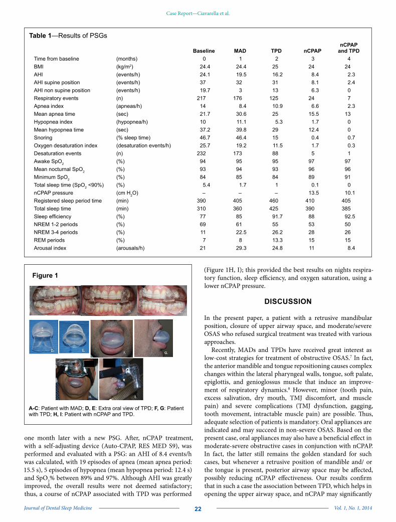
Journal of Dental Sleep Medicine Vol. 1, No. 1, 201422
Case Report—Ciavarella et al.
one month later with a new PSG. After, nCPAP treatment, with a self-adjusting device (Auto-CPAP, RES MED S9), was performed and evaluated with a PSG: an AHI of 8.4 events/h was calculated, with 19 episodes of apnea (mean apnea period: 15.5 s), 5 episodes of hypopnea (mean hypopnea period: 12.4 s) and SpO2% between 89% and 97%. Although AHI was greatly improved, the overall results were not deemed satisfactory; thus, a course of nCPAP associated with TPD was performed
(Figure 1H, I); this provided the best results on nights respira-tory function, sleep efficiency, and oxygen saturation, using a lower nCPAP pressure.
DISCUSSION
In the present paper, a patient with a retrusive mandibular position, closure of upper airway space, and moderate/severe OSAS who refused surgical treatment was treated with various approaches.
Recently, MADs and TPDs have received great interest as low-cost strategies for treatment of obstructive OSAS.7 In fact, the anterior mandible and tongue repositioning causes complex changes within the lateral pharyngeal walls, tongue, soft palate, epiglottis, and genioglossus muscle that induce an improve-ment of respiratory dynamics.8 However, minor (tooth pain, excess salivation, dry mouth, TMJ discomfort, and muscle pain) and severe complications (TMJ dysfunction, gagging, tooth movement, intractable muscle pain) are possible. Thus, adequate selection of patients is mandatory. Oral appliances are indicated and may succeed in non-severe OSAS. Based on the present case, oral appliances may also have a beneficial effect in moderate-severe obstructive cases in conjunction with nCPAP. In fact, the latter still remains the golden standard for such cases, but whenever a retrusive position of mandible and/ or the tongue is present, posterior airway space may be affected, possibly reducing nCPAP effectiveness. Our results confirm that in such a case the association between TPD, which helps in opening the upper airway space, and nCPAP may significantly
Figure 1
A-C: Patient with MAD; D, E: Extra oral view of TPD; F, G: Patient with TPD; H, I: Patient with nCPAP and TPD.
Table 1—Results of PSGs
Baseline MAD TPD nCPAPnCPAP
and TPDTime from baseline (months) 0 1 2 3 4BMI (kg/m2) 24.4 24.4 25 24 24AHI (events/h) 24.1 19.5 16.2 8.4 2.3AHI supine position (events/h) 37 32 31 8.1 2.4AHI non supine position (events/h) 19.7 3 13 6.3 0Respiratory events (n) 217 176 125 24 7Apnea index (apneas/h) 14 8.4 10.9 6.6 2.3Mean apnea time (sec) 21.7 30.6 25 15.5 13Hypopnea index (hypopnea/h) 10 11.1 5.3 1.7 0Mean hypopnea time (sec) 37.2 39.8 29 12.4 0Snoring (% sleep time) 46.7 46.4 15 0.4 0.7Oxygen desaturation index (desaturation events/h) 25.7 19.2 11.5 1.7 0.3Desaturation events (n) 232 173 88 5 1Awake SpO2 (%) 94 95 95 97 97Mean nocturnal SpO2 (%) 93 94 93 96 96Minimum SpO2 (%) 84 85 84 89 91Total sleep time (SpO2 <90%) (%) 5.4 1.7 1 0.1 0nCPAP pressure (cm H2O) – – – 13.5 10.1Registered sleep period time (min) 390 405 460 410 405Total sleep time (min) 310 360 425 390 385Sleep efficiency (%) 77 85 91.7 88 92.5NREM 1-2 periods (%) 69 61 55 53 50NREM 3-4 periods (%) 11 22.5 26.2 28 26REM periods (%) 7 8 13.3 15 15Arousal index (arousals/h) 21 29.3 24.8 11 8.4

Journal of Dental Sleep Medicine Vol. 1, No. 1, 201423
Case Report—Ciavarella et al.
improve nighttime respiratory function and sleep efficiency using lower nCPAP pressure.
REFERENCES1. Koskenvuo M, Kaprio J, Heikkilä K, Sarna S, Telakivi T, Partinen M.
Snoring is a risk factor for ischemic heart disease and stroke in men. Br Med J (Clin Res Ed) 1987;294:16-9.
2. Sassani A, Findley LJ, Kryger M, Goldlust E, George C, Davidson TM. Reducing motor-vehicle collisions, costs, and fatalities by treating obstructive sleep apnea syndrome. Sleep 2004;27:453-8.
3. Brigance JS, Miyamoto RC, Schilt P, et al. Surgical management of obstructive sleep apnea in infants and young toddlers. Otolaryngol Head Neck Surg 2009;140:912-6.
4. Ciavarella D, Sabatucci A, Mastrovincenzo M, et al. Obstructive sleep apnea treatment with a new oral appliance: Somnodent. Open Othorinolaryngol J 2009;3:34-8.
5. Bliwise DL, Feldman DE, Bliwise NG, et al. Risk factors for sleep disordered breathing in heterogeneous geriatric populations. J Am Geriatr Soc 1987;35:132-41.
6. Dingli K, Coleman EL, Vennelle M, et al. Evaluation of a portable device for diagnosing the sleep apnoea/hypopnoea syndrome. Eur Respir J 2003;21:253-9.
7. Eckhart J. Comparison of oral devices for snoring. CDA J 1998;26:611-23.8. Moore EK. Oral appliance treatment for obstructive sleep apnea. Oper
Tech Otolaryngogl 2007;18:52-6.
SUBMISSION & CORRESPONDENCE INFORMATION
Submitted for publication September, 2013Submitted in final revised form January, 2014Accepted for publication January, 2014Address correspondence to: Dr. Domenico Ciavarella, Via Rovelli n.48, 71100 Foggia-Italy; Tel: +39 347 6131225; Fax: +39 0881 588086; E-mail: [email protected]
DISCLOSURE STATEMENTThis was not an industry supported study. The authors have indicated no financial conflicts of interest.


Journal of Dental Sleep Medicine Vol. 1, No. 1, 201425
JDSM
molar area. At placement, his tongue naturally moved forward into the resulting interincisal space (Figure 1).
At follow-up, his stepfather reported that the patient sleeps much better with the OA in place and wakens more refreshed. His attitude is more positive and he is less prone to sullen behav-ior and decreased communication. Both parents were extremely happy with therapeutic outcomes. This patient has not yet been back to see his sleep physician for follow-up polysomnography.
Treatment of patients with disabilities can be very reward-ing. One must remember that device choice must be dictated by physical limitations both of oral anatomy and eye-hand co-ordination. There are number of devices presently on the mar-ket which can be easily used for this segment of our population who can be well served with an oral device.
CITATION
Demko BG. Oral appliance treatment in a patient with down syndrome. Journal of Dental Sleep Medicine 2014;1(1):25–26.
REFERENCES1. Arens R, Marcus CL. Pathophysiology of upper airway obstruction: a
developmental perspective. Sleep 2004;27:997-1019.2. Churchill SS, Kieckhefer GM, Landis CA, Ward TM. Sleep measurement
and monitoring in children with Down syndrome: a review of the literature, 1960-2010. Sleep Med Rev 2012;16:477-88.
3. de Miguel-Diez J, Villa-Asensi JR, Alvarez-Sala JL. Prevalence of sleep-disordered breathing in children with Down syndrome: polygraphic findings in 108 children. Sleep 2003;26:1006-9.
4. Yoshida K. Elastic retracted oral appliance to treat sleep apnea in mentally impaired patients and patients with neuromuscular disabilities. J Prosthet Dent 1999;81:196-201.
Oral Appliance Treatment in a Patient with Down SyndromeB. Gail Demko, DMD, D. ABDSM
Sleep Apnea Dentists of New England, Weston, MA; President, American Academy of Dental Sleep Medicine
CASE REPORTS
Treatment of patients with physical and mental disabilities is one of the more difficult undertakings in dental sleep
medicine. Most patients have minimal physical difficulty plac-ing, removing, and adjusting their oral appliance (OA). They understand the instructions that are given and comprehend the possibilities of side effects. Patients with Down syndrome, mild dementia, and those with physical limitations such as cerebral palsy and status post cerebral-vascular accident may find OA therapy extremely challenging. Many of these patients have al-ready failed positive airway pressure (PAP) and are sent to a sleep dentist for “salvage therapy.” While presenting challenges to the dental sleep medicine clinician, many of them can be-come happy and compliant patients.
Over 50% of persons with Down syndrome suffer from ob-structive sleep apnea secondary to mid-face deficiency and macroglossia.1-3
A high-functioning 26-year-old male with Down syndrome presented with a history of loud snoring and falling asleep when-ever he was a passenger in the car. He had undergone evaluation by an otolaryngologist who found no nasal abnormality and felt that soft tissue surgery was not indicated. The patient had also been evaluated by a maxillofacial surgeon, but the parents had decided not to proceed with surgical treatment. The patient had been on PAP using a number of different interfaces but always developed aerophagia and the pain resulted in his removing the CPAP apparatus within one hour.
The patient lives at home with his mother and stepfather. He works during the day in a protected workshop and tends to stay active by taking karate lessons. His normal bedtime is ~10:00 PM, and his sleep latency is anywhere from 10–60 min. His preferred sleep position is supine. He has a minimally restored 28-tooth dentition with an overjet of 0 mm and an overbite of 0 mm; his range of motion is +6 mm–0 mm.
The overall goal of treatment was to provide increased interin-cisal distance and mild advancement to allow the macroglossic tongue to move forward out of the upper airway.4 Particular concerns with this patient were his ability to place and remove the oral device without assistance, his limited range of mandib-ular motion, and a negative reaction to previous therapy.
Because the patient was relatively averse to the tight-fitting straps with PAP, it was decided not to use any device that re-quired heating under hot water prior to placement or was lami-nated for a very tight retentive fit. A hard acrylic device that could be made relatively loose, easy to place, and allow relatively free mandibular movement was chosen as most likely to be tol-erated by the patient. A hard acrylic Herbst device with no elas-tics was fabricated. The bite registration was made using a Pro gauge with a 10-mm interincisal bite fork in the hopes of creat-ing increased tongue space. Additional tongue space was devel-oped by keeping the interocclusal acrylic out of contact in the second molar area; the only contact was in the bicuspid—first
Figure 1
http://dx.doi.org/10.15331/jdsm.3734

Journal of Dental Sleep Medicine Vol. 1, No. 1, 201426
Case Report—Demko
SUBMISSION & CORRESPONDENCE INFORMATION
Submitted for publication January, 2014Accepted for publication January, 2014Address correspondence to: B. Gail Demko, DMD, 140 Merriam St., Weston, MA 02493; Tel: (617) 964-4028; Fax: (617) 595-4591; E-mail: [email protected]
DISCLOSURE STATEMENTThis was not an industry supported study. The author has indicated no financial conflicts of interest.
Journal of Dental Sleep Medicine Vol. 1, No. 1, 201427
JDSM
The Link between Sleep Bruxism, Sleep Disordered Breathing and Temporomandibular Disorders: An Evidence-based ReviewRamesh Balasubramaniam, BDSc, MS1; Gary D. Klasser, DMD2; Peter A. Cistulli, MD, PhD3; Gilles J. Lavigne, DDS, PhD4
1School of Dentistry, University of Western Australia, Perth, Australia; 2School of Dentistry, Louisiana State University, New Orleans, LA; 3Centre for Sleep Health and Research, Department of Respiratory and Sleep Medicine, Royal North Shore Hospital, and University of Sydney, NSW, Australia; 4Department of Oral Health, Faculty of Dental Medicine, Universite de Montreal, Surgery and Trauma Department, Hopital du Sacre Coeur de Montreal, Montreal, Canada
REVIEW ARTICLES
The potential link between sleep bruxism (SB), sleep disordered breathing (SDB), and temporomandibular disorders (TMD) is an area of intense interest in the dental sleep medicine field. There is a growing trend among some clinicians that treatment of SDB and hence SB may alleviate TMD related signs and symptoms. However the evidence base underpinning such a move in clinical practice is questionable. To date, we do not fully understand the relationship among these three entities and whether causality actually exists. This review examines the relationship(s) between SB, SDB, and TMD with the aim of defining whether these clinical disorders are concomitant, and the nature and direction of any causal relationships, with the ultimate goal of providing advice to clinicians about what to do when they identify this clinical cluster in a given patient. This review will provide a pathway for future research endeavors, which ultimately will be of great assistance in moving the field forward in an evidence-based manner.Keywords: Sleep bruxism, sleep disordered breathing, upper airway resistance syndrome, obstructive sleep apnea, temporomandibular disorders, comorbidity. Citation: Balasubramaniam R, Klasser GD, Cistulli PA, Lavigne GJ. The link between sleep bruxism, sleep disordered breathing and temporomandibular disorders: an evidence-based review. Journal of Dental Sleep Medicine 2014;1(1):27–37.
There has been recent interest in the relationship between sleep bruxism (SB) and sleep disordered breathing (SDB).
Some clinicians have advocated assessment for possible SDB in patients with temporomandibular disorders (TMD), with an underlying assumption that patients with TMD “caused” by SB may in fact have underlying SDB. In these patients, it is recommended that treatment of SDB which is assumed to be the “cause” of SB, will resolve TMD related signs and symptoms. This review will examine the relationship between sleep bruxism (SB), sleep disordered breathing (SDB), and temporomandib-ular disorders (TMD), with the aim of defining whether these clinical disorders are concomitant, the nature and direction of any causal relationships, with the ultimate goal of providing advice to clinicians about what to do when they identify this clinical cluster in a given patient.
OVERVIEW OF SLEEP BRUXISM, SLEEP DISORDERED BREATHING, AND
TEMPOROMANDIBULAR DISORDERS
Sleep bruxism (SB) is defined as a stereotyped movement disorder characterized by rhythmic masticatory muscle activity (RMMA) associated with tooth grinding (TG) and occasional tooth clenching.1 The definition of bruxism was recently reviewed by an international group of experts,2 and this updated definition has been adopted for the International Classification of Sleep Disorders-3, to be published online in 2013. This consensus work: (1) recognized that bruxism has two distinct circadian manifestations: sleep (indicated as sleep bruxism) or wakefulness (indicated as awake bruxism) and (2) reinforced that SB is a repetitive jaw-muscle activity characterized by clenching or grinding of the teeth and/or by
bracing or thrusting of the mandible. The prevalence of self-reports of tooth grinding to assess SB is about 8%.3 The gold standard for diagnosis of SB is a polysomnogram (PSG), and a one-night study is considered adequate for the diagnosis of moderate to high frequency SB. In some cases of low RMMA frequency, a second night study may be necessary to confirm the findings of the first night.4 A recent study involving 1,042 subjects who completed a questionnaire and underwent a PSG found that, based on the questionnaire alone, the prevalence was 12.5%. With the use of PSG alone regardless of the subjects report, the prevalence was 7.4%. The prevalence of SB when the questionnaire was combined with polysomnographic recording was 5.5%.5
Interest in SB has gained more attention due to a shift in the modern day employment environment and its putative recent association to sleep disorders such as insomnia and SDB.5 The word night or nocturnal as it relates to bruxism should no longer be used, since many workers sleep during the daytime due to flexible work schedules. Therefore, SB can occur anytime during a 24-h cycle. SB can be divided into two distinct catego-ries. They are: (1) primary or idiopathic SB, which is without an identifiable cause or any associated sociopsychological or medical problem; and (2) secondary SB, which is related to sociopsychological and/or medical conditions (e.g., movement or sleep disorder, neurologic or psychiatric condition, drug/chemical related). Practitioners must be aware that SB may occur concomitantly with many other sleep disorders such as insomnia, sleep epilepsy, REM behavior disorder (RBD), and SDB.
The etiology of SB is most likely multifactorial—the result of a combination of environmental, biological, and psycho-social influences; however, a causal association between these
http://dx.doi.org/10.15331/jdsm.3736

Journal of Dental Sleep Medicine Vol. 1, No. 1, 201428
Link between Sleep Bruxism, Sleep Disordered Breathing and Temporomandibular Disorders—Balasubramaniam et al.
factors and SB has yet to be established. The various etiolog-ical factors reported in the literature over the last half-century range from nicotine, ethanol, recreational drugs, and caffeine intake to peripheral mechanisms such as occlusal discrepan-cies, to a variety of central factors such as stress and psycho-social influences, alterations in catecholamine levels and other neurochemicals, and cardiac-autonomic interactions related to airway patency and salivary flow. In addition, there appears to be some minor evidence of genetic and familial predisposition for developing chronic SB.6-12 Historically, convincing argu-ments existed for a direct relationship between SB and occlusal factors, as early studies seemed to indicate that occlusal correc-tions diminished or stopped SB activity.13-15 However, later studies challenged occlusal disharmony as a principal contrib-uting factor; several also reported that SB activity was not reduced by occlusal therapy,16-20 thereby creating doubt as to the practice of this philosophy in relation to SB. Emotional stress was another etiological factor, that at one time, was considered to be highly responsible for SB. Early studies that monitored levels of SB looked for an association with stressful events, and some found that sleep masticatory muscle activity and peri-odic pain were increased during stressful periods.21-23 However, recent evidence has found this association to be true only in a small percentage of the population, thus minimizing the importance of this factor.5,7,8,24-26 There is, however, some degree of consensus that many SB individuals have an anxious person-ality (not an anxiety disorder) and are focused on successful performance.8,27-31
Sleep disordered breathing (SDB) may have several presen-tations. Two commonly identified conditions are obstructive sleep apnea and upper airway resistance syndrome (UARS).32-34 Classically, the prevalence of OSA has been reported to be 2% in women and 4% in men in the general population in the US. Among 50- to 60-year olds, 4% of women and 9.1% of men have OSA.35 However, a recent study has revealed that the preva-lence of SDB has been increasing in tandem with the obesity epidemic. In the US, the prevalence rate for moderate-to-severe OSA is 10% and 3% among 30- to 49-year-old men and women, respectively, and 17% and 9% among 50- to 70-year-old men and women, respectively.36
Classically, UARS is diagnosed by observation of episodes of increased respiratory effort associated with abnormal breathing pattern that does not meet the criteria for hypopnea (dura-tion > 10 seconds and drop in nasal flow amplitude of > 30%). Hence the arousals occur without evidence of OSA and are associated with excessive daytime sleepiness. Also, the lowest oxygen saturation at termination of the abnormal breathing event is > 92%, associated with well-defined cyclical alternating patterns (CAP) related to repetitive oscillations in autonomic-cardiac and EEG activities towards a rise in arousal pres-sure.32,37 The AASM Task Force38 has provided a proxy to the clinical definition UARS, the respiratory effort related arousal.39 In UARS the “S” is for syndrome and mandatory for sleepiness, which is not taken into account in RERA.39 RERA is clinically associated with increased inspiratory efforts reflected by more negative esophageal pressure, terminating with alpha wave or alpha and beta wave burst-related EEG arousals.40,41 OSA involves both obstructive apnea (cessation of airflow ≥ 90% compared to baseline with a duration ≥ 10 sec associated
with continued or increased inspiratory effort throughout the entire period of absent airflow) and hypopnea (a reduction in airflow ≥ 30% compared to baseline with a duration ≥ 10 sec, and with ≥ 3% oxygen desaturation compared to baseline, or an arousal).42
Patients with SDB typically have a number of physiologic traits that may explain the disorder. Typically, a small pharyn-geal airway combined with reduced pharyngeal dilator muscle activation during sleep may result in obstruction. The muscle responsiveness during sleep and arousal threshold in response to respiratory stimulation determines whether the individual develops SDB. Also the stability of ventilatory control deter-mines if the individual develops SDB. That is, if the respiratory drive is unstable, the pharyngeal dilator muscle response will also be unstable and hence promote upper airway collapse.43,44 SDB is considered a heterogeneous disorder in which anatomy is an important factor. However, in 56% of OSA subjects, non-anatomical factors such as genioglossus muscle responsiveness, arousal threshold, and respiratory control stability and loop gain contribute to the condition.45
TMD as defined by the American Academy of Orofa-cial Pain encompass a group of musculoskeletal and neuro-muscular conditions that involve the TMJs, the masticatory muscles, and all associated tissues.46 Based on a systematic review of studies that utilized the Research Diagnostic Criteria for TMD (RDC/TMD), the prevalence of TMD in the general population is 13% for muscle pain, 16% for disk derangement, and 9% for joint pain.47 These conditions primarily occur in the young and middle-aged adults and are twice as prevalent in females in the general population.48,49 Patients with TMD typically complain of masticatory muscle and TMJ pain, limited mandibular movements, and TMJ sounds associated with masticatory functions such as chewing.46 Although the etiology of TMD remains ambiguous, microtrauma is hypoth-esized to be a cause. It is reported that sustained and repeti-tious adverse loading of the masticatory system that occurs with SB can cause TMD.50-53 Based on the underlying assump-tion of some clinicians that SB is related to untreated SDB, it is posited that perhaps treatment of SDB will resolve signs and symptoms of TMD.
DOES THE LITERATURE SUPPORT AN ASSOCIATION BETWEEN SLEEP BRUXISM
AND SLEEP DISORDERED BREATHING?
A relationship between SB and SDB has been previously suggested54; however, it is yet to be demonstrated whether both entities are coincidental, causally related, linked to some arousal reactivity, or under some physiological state, which involves the triggering of one or the other.
Currently, there is no evidence to support the association or causality of SB and OSA. However, there do appear to be clinical commonalties between SB and OSA. SB is a complex process that may have a role in maintaining normal physiology, or is perhaps a component of pathophysiology not yet fully eluci-dated. However, there appears to be an association between SB and sleep position.55 Furthermore, an association among OSA, sleep position, and parafunctional activities (clenching) has been expressed in some patients.56 A link between OSA

Journal of Dental Sleep Medicine Vol. 1, No. 1, 201429
Link between Sleep Bruxism, Sleep Disordered Breathing and Temporomandibular Disorders—Balasubramaniam et al.
and those individuals manifesting SB is a possibility, as these entities share the common finding of an alteration in muscle activity/tone.57-59 Observation during hypercapnia (rise in CO2 level) has revealed that the genioglossus and masseter muscles increase in activity by ventilatory stimuli.58 Activation of the masseter muscle is thought to stabilize the mandible; hence enabling the genioglossus to dilate the upper airway more efficiently.57,59,60
There are a number of questions that arise due to the commonalities existing between OSA and SB. Are these commonalities related to upper airway obstruction, whereby SB may be a normal physiologic reactive-protective mecha-nism? If so, could SB be utilized as a reliable and valid surrogate clinical marker for OSA? A detailed exploration of these ques-tions utilizing an evidence-based narrative review approach will address these issues.
SB and SDB are of particular interest to practitioners who are involved in treating patients with either or both of these condi-tions. The coexistence, perhaps coincidental in terms of epide-miology (intersecting prevalence of SB decreasing and SDB increasing with age) and physiological mechanism triggering one or the other, has been reported in a number of pediatric and adult questionnaire- and PSG-based studies. For the purpose of this discussion, the pediatric population is considered subjects under the age of 17 years.
PEDIATRIC POPULATION
An investigation of the prevalence of sleep disorders based on a questionnaire telephone survey among 6- to 12-year-old healthy school children was carried out. The prevalence of sleep signs and symptoms, reported by parents, included descriptors related to habitual snoring (10.9%), cessation of breathing (1.5%), and TG (20.5%). Of interest, significant risk factors identified for habitual snoring comprised witnessed apnea, mouth breathing, snoring among first-degree relatives, morning headache, being male, allergic rhinitis, and witnessed sleep TG. Similarly, significant risk factors for witnessed sleep apnea comprised snoring, allergic rhinitis, morning tiredness, and daytime sleepiness. Among snorers, the odds ratios (OR) for witnessed sleep teeth grinding and morning headache were 1.56 and 1.53, respectively.61
A multiple-choice questionnaire study involving 1,605 chil-dren in Turkey (819 boys and 786 girls) aged 7-13 years was administered.62 Based on a 72.5% response rate, the prevalence of occasional snoring and habitual snoring was 38.9% and 3.5%, respectively. Of interest, habitual snorers had more daytime signs and symptoms (such as mouth breathing, sleepiness, headache, and hyperactivity) and nighttime signs and symp-toms (such as witnessed apnea, obstructed breathing, profuse sweating, restless sleep, enuresis, and TG). Also of significance, allergies, daytime mouth breathing, “shaking the child for apnea,” restless sleep, and hyperactivity were independent risk factors for habitual snoring. The prevalence of TG was 28.7% and 34.1% and significantly associated with occasional snoring and habitual snoring respectively.62 The validity of the ques-tionnaire used in this study was tested on 38 patients prior to the study; however, it was not reported on. It is important to note that the use of questionnaires to capture epidemiological
information regarding sleep apnea has limitations and requires improvement.63
In another study involving 119 snorers between ages 3 and 16, SB was evaluated via PSG. Seventy snorers (59%) were diagnosed with SB. The apnea index, apnea-hypopnea index (AHI), and REM sleep AHI were significantly greater among SB subjects than non-bruxers. The author suggested an association between SB and pediatric OSA.64
A PSG study involving 38 consecutive children with prob-able obstructive sleep apnea-hypopnea syndrome (OSAHS) was carried out. Thirty-five children (92.1%) diagnosed with OSAHS had excessive sleepiness (29.4%) and SB (34.3%) based on a questionnaire. All children (n = 8) with severe OSAHS were diagnosed with bruxism.65
ADULT POPULATION
Three groups of 25 patients with SDB were assessed for signs and symptoms consistent with functional somatic syndromes. All patients were clinically evaluated and had a PSG. The study revealed over 50% of patients with UARS (AHI < 10/h) reported SB; however, this finding was not clinically significant.66 It is important to note that the diagnosis of SB was based on observa-tion of tooth grinding by the bed partner or dentist observation of teeth wear, which are both unreliable methods of assessing SB.5
A telephone survey of 13,057 subjects over the age of 15 used the International Classification of Sleep Disorders minimal set criteria to assess the epidemiology of SB. This study revealed that snoring, choking/blocked breathing, breathing pauses, and OSA were significantly associated with the diagnosis SB and TG (not meeting criteria for SB). It also noted that subjects with OSA (OR = 1.8), loud snorers (OR = 1.4), and those with moderate daytime sleepiness (OR = 1.3) were at a higher probability but lower risk (i.e., low OR) of reporting SB.8 Obvious limitations of this study include use of telephone survey to assess SB and TG, as 20% of subjects did not have a bed partner and many were edentulous. Also subjects did not undergo a dental examination and PSG, which is the gold standard for assessing SB.5
In a full PSG study of 21 adult patients suspected of having SDB, 6 of 11 mild OSA patients (54%) and 4 of 10 moderate OSA patients (40%) were diagnosed with SB. Masseter muscle contraction episodes at the termination of an apneic event was noted in 3.5% in mild and 14.4% in moderate OSA patients.67 Critically, the diagnosis of SB was based on the combination of a questionnaire, clinical observation, and EMG measurement of masticatory muscles activity. Without the use of PSG and due to the small sample size of subjects for the diagnosis of SB, it is difficult to draw inference from this study.
In a study involving 24 sleep bruxers (15 females, age 23-67 years), sleep recordings revealed that only 4 subjects had a significant number of apnea-hypopnea events. A possible expla-nation for the lower prevalence may be related to the large age range and the predominantly female study population.68
A cross-sectional Japanese study of 1,930 residents (age 18-89 years) via a self-administered questionnaire and clinical examination, self-report SB was noted in 8% of individuals. It was higher among individuals between ages 30 and 49 years than those older than 60 years. SB was associated with snoring (OR: 2.58, p = 0.001).69 Limitations of this study include the

Journal of Dental Sleep Medicine Vol. 1, No. 1, 201430
Link between Sleep Bruxism, Sleep Disordered Breathing and Temporomandibular Disorders—Balasubramaniam et al.
subjective assessment of SB and inclusion of subjects missing some or all their teeth, and it did not take into account teeth contacts from use of prosthesis.
Based on the aforementioned studies, there appears to be a coexistence of SB and SDB in both pediatric and adult popula-tions. However, in a recent publication involving 1,042 subjects, SB was assessed based upon questionnaire and polysomno-graphic recordings, there was no significant association between SB and AHI and mean oxygen saturation.5 Limitations of this study include the absence of dental examination, not utilizing audio-video recording to exclude non-RMMA related facial movements, and the influence of the first-night effect. Certainly, prospective epidemiological studies utilizing PSG over several nights in large populations are warranted to further investigate the relationship between SB and SDB.
COMMON FEATURES OF SLEEP BRUXISM AND SLEEP DISORDERED BREATHING
Patients suspected of SB may present to the practitioner with signs and symptoms of tooth wear or damage, fractured dental restorations, dental implant failure, tooth hypersensitivity, tongue indentation, masticatory muscle pain, TMJ pain, and tension-type headache (Table 1). As stated previously, there is uncertainty as to when SB is considered a normal physiologic behavior found in the healthy adult population or considered to be a pathophysiologic disorder highlighted by the above-mentioned signs and symptoms.6 However, there appear to be common features in adult patients with SB and SDB related to sleep positions, oropharyngeal muscle activity, sleep arousal, and gastroesophageal reflux.
INFLUENCE OF SLEEP POSITION
Both OSA and SB patients spend 50% to 58% of their sleeping time in the supine position.70,71 In several studies
investigating the relationship between OSA and SB, there was either no statistical significance or only a slight trend toward an increased frequency of SB episodes among OSA patients in the supine position as compared to other sleep positions (side or mixed).54,56,72,73 However, these studies did not assess the relationship between SB and UARS. Miyawaki et al.55 reported that among SB adult patients, 74% of RMMA and swallowing events occurred in the supine position compared to 23% in the lateral decubitus position, suggesting that sleep position may be a factor in the frequency of oromotor events. The authors suggested that future studies might investigate the potential of postural modification in management of SB.
Based on limited studies, it appears that both SB and SDB can occur in the supine position. SB episodes do not exclusively occur in the supine position; however, studies on the effect of postural therapy on SB have not been carried out. In the case of SDB, sleep position appears to be related to the influence of gravity on the oropharyngeal tissues promoting upper airway obstruction.
OROPHARYNGEAL MUSCLE ACTIVITY
Masticatory and tongue muscle activity during sleep is thought to play an important role during SDB. In OSA the influence of gravity on the mandible, especially in the supine position, combined with masticatory and tongue muscle hypotonia results in a posterior shift of the mandible and tongue creating oropharyngeal narrowing and increased upper airway resis-tance. Yoshida assessed 14 adult apnea subjects and 6 snorers with PSG recordings, which included EMG of the genioglossus, masseter, and inferior lateral pterygoid muscles before, during, and after apneic events.57 The EMG amplitude was lower during an obstructive apnea event than before the event. The ampli-tudes were significantly higher after the apnea event. It was also found that EMG amplitude did not decrease during central apnea events. This study provided evidence that hypotonia
Table 1—Possible (but not all proven*) clinical consequences associated with sleep bruxism
DentalTemporomandibular Disorders (myogenous and arthrogenous) Other
Severe occlusal and incisal wear (chipping), tooth fracture and attrition
Masticatory muscle hypertrophy* (probably secondary to clenching, awake bruxism, habit of tic)
Lateral border of the tongue indentations/scalloping*
Tooth mobility* Masticatory muscle discomfort due to fatigue (may be with or without pain)
Reduction in salivary flow and/or xerostomia*
Hypersensitivity of teeth to air and cold or hot foods and beverages
Pericranial muscle tenderness or pain (considered a morning headache in absence of sleep disorder breathing or neurological condition)
Lip, cheek, or tongue biting
“Cracked tooth syndrome” and the frequent breakage of dental restorations
Stiff, tight mandible with reduced movement and/or difficulty with mastication of food upon awakening
Glossodynia due to parafunctional habits* (probably secondary to clenching, awake bruxism, habit or tic)
Exacerbation of periodontal disease (controversial issue)*
Temporomandibular joint discomfort or pain Excessive concern or anxiety about tooth wear
Failure of dental implants due to excessive forces – –
* These conditions are commonly associated with SB by clinicians, based upon clinical experience, but with little evidence of cause and effect relationships.122

Journal of Dental Sleep Medicine Vol. 1, No. 1, 201431
Link between Sleep Bruxism, Sleep Disordered Breathing and Temporomandibular Disorders—Balasubramaniam et al.
of masticatory and tongue muscles during sleep resulted in obstructive apnea events. Other studies support the suggestion that genioglossus, submental, and masseter muscle activation in OSA patients may occur in an attempt to stabilize the mandible and prevent collapse of the upper airway.59,60,74
Jaw opening at the end of inspiratory effort through submental muscle activation and tracheal tug has been shown to open the airway to allow mouth breathing; this is in contrast to jaw opening at the end of expiratory effort, which is known to narrow the airway.60 Similarly, increased distance between the tongue and posterior wall (from 11 to 17.5 mm) in subjects with neck extension in a jaw-closed position has been reported.75 These studies suggest that the combination of muscle tone and elasticity contribute to maintain the patency of the upper airway.
Based on the above studies, it appears the oropharyngeal and masticatory muscles activate and maintain tone during apneic events to maintain the patency of the upper airway. Although speculative, masticatory muscle activation during these apneic events combined with inadvertent tooth contacts may be diag-nosed as SB.
SLEEP AROUSAL
A common sign of SDB is arousal from sleep after an episode of upper airway obstruction. Similarly, SB is reported to occur within a microarousal, whereby there is a cluster of arousals during an abrupt shift of 3 to 10-15 seconds in EEG activity along with increased heart rate and muscle tone. Of interest, in otherwise healthy young SB patients, there is an increase in sympathetic activity 8 minutes preceding the SB-related elec-tromyographic (EMG) event; more specifically, RMMA. Also, as described below, 2 big breaths were observed in the physi-ological sequence preceding RMMA with a small but significant increase in blood pressure.76-79
In a prospective age- and sex-matched controlled pilot study, SB-RMMA events in children were reported to occur with arousals in 66% of cases. Also, 40% of SB patients had signifi-cant attention and behavior problems. Of interest, light snoring was associated with only 9% of SB episodes.39 In adults, a large sample size PSG study revealed no difference in OSA between SB and non-SB subjects; however, the study found that 52% of RMMA events were associated with sleep arousal.5
An association between SB and rise in respiration within arousal has been reported in a study assessing changes in respi-ration of 20 young healthy SB subjects without apnea-hypopnea manifestations. The results revealed increased respiratory ampli-tude of 8% to 23%, four seconds before RMMA. The respiratory amplitude was 60% to 82% one second before RMMA, which is at the onset of suprahyoid muscle activity, in the window before RMMA/TG being activated. Significantly, during RMMA, respi-ratory amplitude is maximal (108% to 206%). A rapid return of respiratory amplitude to baseline is noted after RMMA. When arousal was associated with RMMA, the amplitude was 11 times higher than when arousal occurred without RMMA.79
In a recent study, 19 subjects (age 53.1 ± 13.7) with OSA without SB were analyzed to assess masseter muscle contractions during sleep in association with arousals and apnea-hypopnea events. Respiratory events with arousals were significantly associated with longer event duration and/or larger transient
oxygen desaturation. Masseter muscle contraction increased significantly with increased duration of arousal. Of interest, the anterior tibialis muscle had a similar response pattern. The authors suggested that the muscle activity (i.e., both masseter and anterior tibialis) after respiratory events is a nonspecific motor phenomenon influenced by duration of arousals and not the presence of respiratory events.80
Arousals associated with SB and arousals associated with upper airway obstruction are well documented in the literature. It appears that in some cases these arousals are the same entity when SB is associated with upper airway obstruction, and other cases it appears to be related to a nonspecific motor phenom-enon. The question remains as to the purpose of these arousals, given their association with sleep fragmentation.
GASTROESOPHAGEAL REFLUX
Similarly, gastroesophageal reflux symptoms and events are more prevalent among subjects with OSA.81-83 Current evidence suggests an association between gastroesophageal reflux and SB. It has been noted that RMMA episodes occur when esophageal pH is reduced.84,85 Interestingly, the preva-lence of TMD among patients diagnosed with gastroesopha-geal reflux disease was 37%.86 As a proof of concept (i.e., SB stimulating salivary secretion), Ohmure et al. infused 5 mL of 0.1N HCl or saline into the esophagus of 12 healthy adult males without SB.87 It was noted that the frequency of EMG bursts, jaw muscle activation (presumptive to be RMMA) episodes, grinding noise, and RMMA/microarousal ratio was significantly higher during the 20-min period after acid infu-sion. Subjects who complained of heartburn did not reveal jaw muscle activation episodes 20 minutes after acid infu-sion, suggesting that jaw muscle activation and SB stimulate salivary secretion, thus enabling neutralization of esophageal acid.88-90 The acidic reflux content is thought to be cleared by a protective response involving arousal and swallowing to prevent mucosal injury and aspiration.91,92
ASSOCIATION BETWEEN TEMPOROMANDIBULAR DISORDERS AND
SLEEP DISORDERED BREATHING
It is likely that patients with low-frequency SB may present with painful TMD. Given that there is a relationship between SB and SDB, TMD may be diagnosed in patients with SDB. Petit et al.93 evaluated 100 adult patients diagnosed with OSA and found 2% of patients had significant signs and symptoms of TMD. In another study, Cunali et al.94 evaluated 87 adult patients with mild to moderate OSA and found 52% had TMD based on the RCD/TMD.
The OPPERA cohort and case-control studies found that signs and symptoms of OSA were associated with occurrence of TMD. In the cohort study, high probability of OSA was asso-ciated with increased incidence of first-onset TMD (adjusted hazard ratio = 1.73). Similarly, in the case-control study, high probability of OSA was associated with increase of chronic TMD (adjusted OR = 3.63).95 Further studies utilizing large case-control designs are warranted to explore the relationship between TMD and SDB.
Journal of Dental Sleep Medicine Vol. 1, No. 1, 201432
Link between Sleep Bruxism, Sleep Disordered Breathing and Temporomandibular Disorders—Balasubramaniam et al.
In a large case-control study involving a web-based registry of 1,511 adult TMD patients, approximately 10% of TMD patients had OSA, although this was not significantly different from controls.96 In another study utilizing the Douglass disor-ders questionnaire, 6% of 100 adult TMD patients had OSA compared to 4% of 100 age- and sex-matched controls, which was not clinically different.97 Of note, excessive daytime sleepi-ness was more frequent among masticatory myofascial pain patients.98
In a 3-month prospective study, 51 adult TMD patients were assessed by PSG, Beck Depression Index, and Brief Pain Inventory. The study revealed a trajectory increase in daily pain rating predicted by the presence of SDB. The authors postulated that treatment of SDB may minimize pain and suffering.99 Similarly, in a study assessing sleep disorders and their association with pain sensitivity, 53 masticatory myofas-cial pain adult patients were assessed: 43% had two or more sleep disorders such as insomnia and OSA; 28% of these patients were diagnosed with OSA. The author concluded that TMD patients complaining of sleep disturbances should have a sleep study.100
A commonly cited etiology for masticatory myofascial pain is SB. In a study involving 2-night PSG monitoring, 124 female patients with masticatory myofascial pain and 46 female controls, did not find a statistical significant difference in SB rates among patients (9.7%) and controls (10.9%). Of interest, self-reported rates for SB was 55.3% among mastica-tory myofascial pain cases compared to 15.2% among controls, hence bringing into question the validity of self-report of SB in clinical practice.101 However, further analysis of this study revealed that TMD patients have increased daytime dysfunc-tion, increased sleep stage N1 percentage, increased RERA, and increased respiratory arousal index compared to controls. The authors concluded that TMD is associated with sleep distur-bances and mild sleep breathing dysfunction.102 As discussed previously, a recent study did not report a significant associa-tion between SB and AHI and mean oxygen saturation.5
A similar study compared jaw-muscle EMG activity utilizing portable single-channel device for 4 nights in patients with craniofacial pain, pain-free conditions, tension-type headache, and healthy controls. There was no significant difference in EMG activity among the 4 groups. Of interest, pain related conditions had significantly greater night-to-night variability. It is important not to confuse the presence of jaw muscle EMG activity as diagnostic of SB-RMMA events as discussed previously.103
In a recently accepted paper for publication in the Journal of Sleep Research, PSG recordings of 10 patients with both OSA and SB were analyzed. Reportedly, significantly more SB-RMMA events followed the apnea-hypopnea events (i.e., occurred after the respiratory event [55%]), suggesting that SB-RMMA events occurring close to apnea-hypopnea events are a secondary form of sleep bruxism.104
Although speculative, underlying sleep disturbances and SDB may be associated with SB and to a lesser degree to TMD. On balance of the available literature, this relationship is equivocal. Also, oral parafunctional activity (SB) may not be the etiology for TMD as traditionally assumed. Further research is required to better elucidate these relationships.
MANAGEMENT OF SB AND ITS EFFECTS ON SLEEP DISORDERED BREATHING
The effect of SB treatment and its influence on SDB has been noted in a few studies. The notion that SB serves as a reactive-protective mechanism against upper airway collapse has been posited in the past.8,31,79,105,106 It is common dental practice to utilize oral appliance therapy when treating SB; however, its effect on SDB remains an enigma.
Twenty-eight moderate-to-severe SB adult subjects were treated with a thermoplastic mandibular advancement appliance (MAA) at 75% advancement and assessed by Bite Strip (EMG electrodes) for 30 days. Owing to limitation of the recording method (low specificity for RMMA from other orofacial motor activities), significant improvement of masseter muscle activity was observed and responses based on the Sleep Assessment Ques-tionnaire were noted (p < 0.05). Also there was significant reduc-tion of TMJ sounds and masseter muscle and temporalis muscle tenderness.107 The reliability of the use of Bite Strip for assessing SB over recorded masseter muscle activity is questionable, as its use has not been fully validated by independent investigators.
In a short term PSG, randomized, crossover, controlled but experimental study (not designed to asses efficacy), 13 adult SB subjects were fitted with a temporary thermoplastic custom fit MAA in 3 different positions: with freedom of move-ment, slightly advanced (< 40%), or more advanced (> 75%). The maxillary occlusal splint (MxOS-no advancement of the mandible) was used as the control device. The results revealed a 42% reduction in SB episodes per hour with the MxOS. The MAA in the freedom, < 40%, and > 75% advancement positions revealed 44%, 77%, and 83% reduction in SB episodes per hour, respectively. The authors hypothesized that the mechanism at which MAA reduces SB may be related to the dimension and configuration of the device, presence of pain, reduced freedom of movement, or upper airway patency.108
A subsequent experimental study from the same group was carried out in adult subjects randomized in a crossover, short-term study among 3 groups—25% protrusion of the MAA, 75% protrusion of the MAA, or mandibular occlusal splint (MdOS). The aim was to investigate the effect of a custom fitted adjust-able MAA on SB. Mean SB episodes per hour significantly were reduced by 39% and 47% with the MAA at 25% and 75% protrusion, respectively (p < 0.04), during a one-night PSG. The MdOS reduced the mean SB episodes by 34%; however, this was not statistically significant (p < 0.07). The authors concluded that short-term use of a reinforced MAA resulted in reduction of SB motor activity. Thus MAA may be a treatment option for patients with concomitant SB and SDB.109
In another short-term PSG study, 16 adolescents with SB, snoring, and headache underwent MAA therapy. PSGs were carried out at baseline and 3 positions (without connectors between upper and lower portions, maximum intercuspation, and 50% mandibular advancement) for 1 week in random order. Short-term use of a MAA reduced SB, snoring, and headache.110 Further investigation is necessary to assess the long-term effec-tiveness and safety of the MAA in adolescents with SB, SDB, and headache.
A pilot study involving 10 adult patients with snoring and OSA was carried out to assess the effects of MxOS on sleep.
Journal of Dental Sleep Medicine Vol. 1, No. 1, 201433
Link between Sleep Bruxism, Sleep Disordered Breathing and Temporomandibular Disorders—Balasubramaniam et al.
There was no statistical difference in median AHI62 between baseline and MxOS use at night. Of interest, 4 patients had aggravation of OSA with the use of the MxOS. Five of 10 of subjects had a 50% increase in AHI. Overall, the respiratory disturbance index (RDI) demonstrated a 30% increase when the MxOS was used at night. The split-night study showed a 40% increase in sleeping time with snoring. The authors suggested that patients who are candidates for oral appliances should be questioned about snoring and OSA prior to recommending oral appliance therapy.111
In a randomized, crossover, controlled study, 10 adult patients with snoring and OSA underwent 3 PSG recordings with their MxOS and 3 PSG recordings without their MxOS. There was a small increase in the mean AHI with MxOS, which was significantly higher than without MxOS. However, there was no difference in the Epworth Sleepiness Scale score. The authors concluded that MxOS was associated with OSA aggra-vation; however, the small size of the study limits its clinical relevance. It was suggested that the mechanism for increase AHI with MxOS could be related to reduced tongue space and rotation and anterior translation of the condyles, which conse-quently reduces the upper airway size.112 Further studies with larger sample sizes are warranted. Also the effect of the MdOS on snoring and OSA needs to be investigated.
It is routine for practitioners to increase the vertical dimen-sion for certain dental procedures. For example, oral appliances are used in dental practices often to treat patients with SB, TMD, and as an interim for dental rehabilitation. These procedures modify the space between maxillary and mandibular occlusal surfaces. Eighteen mild to severe adult OSA (AHI 5 to 45) patients completed a study assessing the effect of increasing the vertical dimension by 6 mm (interincisal) with MAA without protrusion and its effects on AHI. Increase of the AHI was noted in 9 patients, with 2 patients reaching statistical significance. The authors concluded that increasing the vertical dimension without mandibular protrusion might aggravate OSA in some patients. Therefore practitioners should screen patients for OSA prior to fabricating an oral appliance, which increase the vertical dimension without mandibular protrusion.113
Based on the above-mentioned studies, it appears that MAA is more efficacious than a non-protruding conventional oral appliance in reducing SB. However, the mechanism for this reduction may not be related only to improvement in airway patency. Also, the short-term studies assessing the use of MxOS in SB suggest that this type of oral appliance may increase AHI in subjects with OSA, although the increase in AHI may not be clinically significant. Caution is advised when drawing conclu-sions from the above studies, as oral appliances may indepen-dently improve both SB and SDB; hence a causal relationship cannot be assumed. The effects of other non-dental treatments for OSA such as CPAP therapy and its effects on SB would provide better insight on whether such a relationship exists.
MANAGEMENT OF TEMPOROMANDIBULAR DISORDERS AND ITS EFFECTS ON SLEEP
DISORDERED BREATHING
Twelve adult subjects with complaints of morning headache but without SBD and SB history were studied. Subjects underwent
4 PSG studies; a habituation study, a baseline study, neutral position MAA study, and 50% advancement MAA study. Both the neutral MAA and 50% MAA were associated with ≥ 70% morning headache reduction and ≥ 42% orofacial pain reduc-tion (p < 0.01). Also RMMA was significantly reduced with both the neutral MAA and 50% MAA (p < 0.05). Two subjects without previous SDB complaints had AHI > 5. Both subjects had beneficial reduction of AHI with 50% MAA. One subject with an initial AHI of 5.3 had reduction of the AHI to 2 with the use of both the neutral MAA and 50% MAA. Perhaps the neutral MAA was sufficient to prevent the retruded jaw posi-tion that typically occurs during sleep; however, this remains to be proven. The authors concluded that the reductions in orofacial pain and morning headache may partially be linked to reduction in RMMA; however, this cannot be assumed as the only explanation for a causal link.114 Certainly, further studies on orofacial pain and headache patients with mild SDB are warranted, as a recent study reported an increase in the pres-ence of RERA among female TMD subjects.102
NON-DENTAL THERAPIES FOR SLEEP DISORDERED BREATHING AND ITS EFFECTS
ON SLEEP BRUXISM
Continuous positive airway pressure (CPAP) is the gold stan-dard treatment for snoring and OSA.115 In a published case report, a 47-year-old male with severe SB and severe OSA (RDI = 47.6) was observed before and after CPAP therapy. Sixty-seven SB-related motor activity events (RMMA with TG sounds) were induced by apnea/hypopnea events, and only 6 occurred spontaneously. With CPAP, the RDI decreased to 4.1 and SB was undetectable. It was suggested that in a patient with severe SB and OSA, where oral appliance therapy is contraindicated, that CPAP may be an effective therapy in managing both conditions.116 Of interest, in patients with SB and SDB, perhaps the use of CPAP may be adequate, and hence there may be no need for the use of an occlusal splint to protect teeth from wear. Obviously, large sample size random-ized controlled trials are needed to further investigate such a suggestion.
In an open label study, the records of 571 adult CPAP treated patients were reviewed. One hundred thirty nine patients (24%) were identified as sleep bruxers, based on a history of tooth grinding and examination of tooth wear.117 Of note, two recent publications raised concerns about tooth grinding diag-nosis by history and clinical examination alone in absence of sleep recording, whereby the history of tooth grinding and the clinical estimations were inaccurate.5,118 That is, not all subjects that report tooth grinding based on history alone, were found to have SB during a PSG. The interest in this open study is the use of esophageal pressure to monitor UARS, which would be otherwise missed, given that SB is postulated to reduce airway obstruction. Of 95 SB patients who presented for follow-up, 69 used CPAP (56 nightly). SB identified by history and examina-tion alone was improved in 35 patients (50%). In the absence of RMMA index to diagnose SB, strong conclusions cannot yet be drawn. Also SB-RMMA and tooth grinding sounds are known to have night-to-night variability, a factor to consider during data interpretation.119

Journal of Dental Sleep Medicine Vol. 1, No. 1, 201434
Link between Sleep Bruxism, Sleep Disordered Breathing and Temporomandibular Disorders—Balasubramaniam et al.
In a retrospective study by the same researchers of the above-reported study, the records of 729 CPAP treated adult patients were reviewed. The authors identified 102 OSA/UARS patients by PSG and esophageal pressure monitoring and SB patients via the history and examination alone. Also CPAP compliance was assessed based on patient self-report. A multivariate analysis revealed statistically significant relationships between CPAP use and improvement in SB (p = 0.0054). Of interest, 25 patients had UARS, of whom 14 used CPAP nightly. Of these patients, 12 patients (86%) had improvement in SB. Also, the authors believe that esophageal pressure monitoring is essential to assess UARS, as SB may mask upper airway obstruction. Other-wise, RERA will not be identified and the threshold for the RDI may not be reached to identify underlying UARS or OSA. This study observed that using CPAP to treat OSA, i.e., maintenance of airway patency, also resulted in decreased SB.106 Significant limitations in this study are related to the assessment of SB being based solely upon history and examination, and RMMA not directly measured but rather assumed to have occurred. Also the temporal association between RMMA and decrease in AHI needs to be closely analyzed for individual subjects before firm conclusions can be drawn about such stimulating findings.
Another unexplored avenue is the benefit and efficiency of upper airway surgery to manage snoring and OSA in cases with SB. The incidence of SB was assessed in a study before and after adenotonsillectomy in children with SDB. Sixty-nine children with tonsillar hyperplasia and SDB were provided a question-naire and examination to determine the presence of SDB and SB before and after surgery.120 All 69 children were noted to have OSA, and 45.6% presented with SB. At 3-month follow-up, OSA was resolved in all patients, and only 11.8% continued to
have SB. The authors concluded that there is a “positive corre-lation” between SDB and SB and noted that SB was associated with airway obstruction in children. A major limitation of this study is that SDB and SB were assessed by unvalidated question-naire and examination. Also, it is unclear if observation of tooth grinding sounds and tooth wear were utilized as the method for assessing SB. Similar concerns are raised on the diagnosis of SDB, as PSG was not performed.120
In another study, 140 children (aged 4-12 years) were evalu-ated for obstructive symptoms secondary to adenotonsillar hypertrophy pre- and post-adenotonsillectomy. The preva-lence of SB based on a questionnaire completed by observant parents was 25.7% and 7.1% pre- and post-surgery, respectively. The authors concluded that SB-tooth grinding improved with adenotonsillectomy.121 It is obvious that blind and randomized assessments are needed before firm conclusions can be drawn from these two studies. Moreover, studies regarding the bene-fits and efficacy of mandibular advancement (orthognathic) surgery and palatal expansion surgery in teenagers and adults with SDB and SB are scarce. The effect of treatment on SB, SDB, and TMD is summarized in Table 2.
CONCLUSION
It appears that SB and SDB often coexist (Table 3). Both enti-ties appear to share common risk factors, with intersecting prevalences across the life span, and clinical features that influ-ence their clinical presentation. Hence this may challenge the clinical decision-making for diagnosis, comorbidities, and management of SB and SDB. The clinician has to be cautious in assuming causality just because treatment of SDB improves SB-TG in some patients. Individual differences in the era of personalized medicine prevent us from rapidly concluding on cause-and-effect relationships to be generalized to the whole population.
Vulnerability or predisposition to SDB and SB, respectively, needs to be identified, as indirect evidence is now emerging that perhaps SB may serve as a “reactive or protective mechanism” against upper airway obstruction. It appears that when patients with SB and/or painful TMD complain about insomnia, snoring and/or cessation of breathing during sleep, sleepiness of uniden-tified causes, or uncontrolled blood pressure, it is prudent to screen for the presence of SDB. Such is done in collaboration with sleep medicine specialists using either sleep laboratory or home recording systems with electromyography analysis of masseter/temporal muscle activity.
In patients with confirmed SB and concomitant SDB, after nasal examination to exclude obstruction, either a MMA or CPAP may be prescribed. The same is also suggested for TMD patients with SDB. Dentists need to be aware that current stan-dard maxillary oral appliances (occlusal splints) to protect teeth from attrition may not be appropriate treatment in the presence of SDB. That is, in some cases, occlusal splints may aggravate the underlying SDB. Also, in some cases, MAA may initiate or aggravate preexisting painful TMD in patients with SDB. Further prospective studies looking at the relationship between SB and SDB, and painful TMD and SDB are warranted before it may be translated into clinical guidelines and standards of practice.
Table 2—Effect of treatment on sleep bruxism, sleep disordered breathing, and temporomandibular disorders*
Treatment Comment
Effect of mandibular advancement appliance (MAA) on SB107-109
Reduction of SB event in the short term.
Effect of MAA on snoring, SB and headache110
Reduction of snoring, SB, and headache in the short term.
Effect of maxillary occlusal splint (MxOS) on snoring and OSA111,112
Aggravation of AHI and snoring with MxOS use in the short term.
Effect of raising the interincisal vertical dimension of a MAA without mandibular protrusion on OSA113
Aggravation of AHI with raising the interincisal vertical dimension of a MAA without protrusion in the short term.
Effect of neutral MAA and 50% MAA on patients with morning headache without SB and SDB114
Reduction in morning headache and orofacial pain with both neutral MAA and 50% MAA.
Effect of CPAP on SB and OSA106,116,117
CPAP may reduce SB and OSA.
Effect of adenotonsillectomy in children with snoring, OSA and SB120
Resolution of snoring and OSA and reduction of SB.
* Most studies are short-term and limited by sample size and results need replication.

Journal of Dental Sleep Medicine Vol. 1, No. 1, 201435
Link between Sleep Bruxism, Sleep Disordered Breathing and Temporomandibular Disorders—Balasubramaniam et al.
REFERENCES1. American Academy of Sleep Medicine. International classification of
sleep disorders. 2nd ed. Diagnostic and coding manual. Westchester, IL: American Academy of Sleep Medicine, 2005.
2. Lobbezoo F, Ahlberg J, Glaros AG, et al. Bruxism defined and graded: an international consensus. J Oral Rehabil 2013;40:2-4.
3. Lavigne GJ, Montplaisir JY. Restless legs syndrome and sleep bruxism: prevalence and association among Canadians. Sleep 1994;17:739-43.
4. Hasegawa Y, Lavigne G, Rompre P, Kato T, Urade M, Huynh N. Is there a first night effect on sleep bruxism? a sleep laboratory study. J Clin Sleep Med 2013;9:1139-45.
5. Maluly M, Andersen ML, Dal-Fabbro C, et al. Polysomnographic study of the prevalence of sleep bruxism in a population sample. J Dent Res 2013;92:97S-103S.
6. Lavigne GJ, Khoury S, Abe S, Yamaguchi T, Raphael K. Bruxism physiology and pathology: an overview for clinicians. J Oral Rehabil 2008;35:476-94.
7. Manfredini D, Lobbezoo F. Role of psychosocial factors in the etiology of bruxism. J Orofac Pain 2009;23:153-66.
8. Ohayon MM, Li KK, Guilleminault C. Risk factors for sleep bruxism in the general population. Chest 2001;119:53-61.
9. Ahlberg J, Savolainen A, Rantala M, Lindholm H, Kononen M. Reported bruxism and biopsychosocial symptoms: a longitudinal study. Community Dent Oral Epidemiol 2004;32:307-11.
10. Lavigne GL, Lobbezoo F, Rompre PH, Nielsen TA, Montplaisir J. Cigarette smoking as a risk factor or an exacerbating factor for restless legs syndrome and sleep bruxism. Sleep 1997;20:290-3.
11. Hojo A, Haketa T, Baba K, Igarashi Y. Association between the amount of alcohol intake and masseter muscle activity levels recorded during sleep in healthy young women. Int J Prosthodont 2007;20:251-5.
12. Abe Y, Suganuma T, Ishii M, et al. Association of genetic, psychological and behavioral factors with sleep bruxism in a Japanese population. J Sleep Res 2012;21:289-96.
13. Ramfjord SP. Bruxism, a clinical and electromyographic study. J Am Dent Assoc 1961;62:21-44.
14. Ash MM, Ramfjord SP. Occlusion. 4th ed. Philadelphia: W.B. Saunders, 1995.
15. Guichet NE. Occlusion: a teaching manual. Anaheim: The Denar Corporation, 1977.
16. Rugh JD, Barghi N, Drago CJ. Experimental occlusal discrepancies and nocturnal bruxism. J Prosthet Dent 1984;51:548-53.
17. Kardachi BJ, Bailey JO, Ash MM. A comparison of biofeedback and occlusal adjustment on bruxism. J Periodontol 1978;49:367-72.
18. Tsukiyama Y, Baba K, Clark GT. An evidence-based assessment of occlusal adjustment as a treatment for temporomandibular disorders. J Prosthet Dent 2001;86:57-66.
19. Clark GT, Tsukiyama Y, Baba K, Watanabe T. Sixty-eight years of experimental occlusal interference studies: what have we learned? J Prosthet Dent 1999;82:704-13.
20. Manfredini D, Visscher CM, Guarda-Nardini L, Lobbezoo F. Occlusal factors are not related to self-reported bruxism. J Orofac Pain 2012;26:163-7.
21. Rugh JD, Solberg WK. Electromyographic studies of bruxist behavior before and during treatment. J Calif Dent Assoc 1975;3:56-9.
22. Solberg WK, Clark GT, Rugh JD. Nocturnal electromyographic evaluation of bruxism patients undergoing short term splint therapy. J Oral Rehabil 1975;2:215-23.
23. Clark GT, Beemsterboer PL, Solberg WK, Rugh JD. Nocturnal electromyographic evaluation of myofascial pain dysfunction in patients undergoing occlusal splint therapy. J Am Dent Assoc 1979;99:607-11.
24. Pierce CJ, Chrisman K, Bennett ME, Close JM. Stress, anticipatory stress, and psychologic measures related to sleep bruxism. J Orofac Pain 1995;9:51-6.
25. Dao TT, Lavigne GJ, Charbonneau A, Feine JS, Lund JP. The efficacy of oral splints in the treatment of myofascial pain of the jaw muscles: a controlled clinical trial. Pain 1994;56:85-94.
26. Watanabe T, Ichikawa K, Clark GT. Bruxism levels and daily behaviors: 3 weeks of measurement and correlation. J Orofac Pain 2003;17:65-73.
27. Lavigne GJ, Manzini C, Kato T. Sleep bruxism. In: Kryger MH, Roth T, Dement WC, eds. Principles and practice of sleep medicine. Philadelphia: Elsevier, 2005.
28. Biondi M, Picardi A. Temporomandibular joint pain-dysfunction syndrome and bruxism: etiopathogenesis and treatment from a psychosomatic integrative viewpoint. Psychother Psychosom 1993;59:84-98.
29. Kampe T, Tagdae T, Bader G, Edman G, Karlsson S. Reported symptoms and clinical findings in a group of subjects with longstanding bruxing behaviour. J Oral Rehabil 1997;24:581-7.
30. Pingitore G, Chrobak V, Petrie J. The social and psychologic factors of bruxism. J Prosthet Dent 1991;65:443-6.
31. Bader GG, Kampe T, Tagdae T, Karlsson S, Blomqvist M. Descriptive physiological data on a sleep bruxism population. Sleep 1997;20:982-90.
32. Terzano MG, Parrino L, Sherieri A, et al. Atlas, rules, and recording techniques for the scoring of cyclic alternating pattern (CAP) in human sleep. Sleep Med 2001;2:537-53.
Table 3—Common clinical features between sleep bruxism and sleep disordered breathingClinical Features Comment
Both SB and SDB more common in supine sleep position
Studies required to assess influence of postural modification on SB (e.g., sleep positioned benefit not tested yet in a control design for SB)
Oropharyngeal and masticatory muscles activation and tonicity occurs during SB and SDB
Studies required to confirm if masticatory muscle activation during apneic events results in SB plus if it is more the tonic (e.g., clenching type) that is observed or the phasic RMMA
Sleep arousal Typically in SDB, arousal is observed after airway obstruction; in contrast, SB occurs within a sleep arousal
Gastroesophageal reflux Conceptually, acid reflux that occurs with SDB, results in a protective response (arousal and swallowing) to prevent mucosal injury and aspiration—indirect clinical evidence and one experimental study
Temporomandibular disorders Perhaps SB is associated with underlying SDB and consequently the etiology of TMD. Alternatively, SDB may be risk factor for TMD—vulnerability to be proven
Headache Both tension-type headache and migraine are associated with SB and SDB—based on population survey
Successful treatment with mandibular advancement appliance (MAA)
Further studies investigating the use of MAA in SB is warranted—experimental short term evidence at this stage and large studies required for replication
Successful treatment with CPAP Further studies required—only one case report
Successful treatment with upper airway surgery Missing evidence for link between SDB and SB

Journal of Dental Sleep Medicine Vol. 1, No. 1, 201436
Link between Sleep Bruxism, Sleep Disordered Breathing and Temporomandibular Disorders—Balasubramaniam et al.
33. Guilleminault C, Stoohs R, Clerk A, Simmons J, Labanowski M. From obstructive sleep apnea syndrome to upper airway resistance syndrome: consistency of daytime sleepiness. Sleep 1992;15:S13-6.
34. Guilleminault C, Stoohs R, Clerk A, Cetel M, Maistros P. A cause of excessive daytime sleepiness. The upper airway resistance syndrome. Chest 1993;104:781-7.
35. Young T, Palta M, Dempsey J, Skatrud J, Weber S, Badr S. The occurrence of sleep-disordered breathing among middle-aged adults. N Engl J Med 1993;328:1230-5.
36. Peppard PE, Young T, Barnet JH, Palta M, Hagen EW, Hla KM. Increased prevalence of sleep-disordered breathing in adults. Am J Epidemiol 2013 Apr 14 [Epub ahead of print].
37. Bao G, Guilleminault C. Upper airway resistance syndrome--one decade later. Curr Opin Pulm Med 2004;10:461-7.
38. Sleep-related breathing disorders in adults: recommendations for syndrome definition and measurement techniques in clinical research. The Report of an American Academy of Sleep Medicine Task Force. Sleep 1999;22:667-89.
39. Herrera M, Valencia I, Grant M, Metroka D, Chialastri A, Kothare SV. Bruxism in children: effect on sleep architecture and daytime cognitive performance and behavior. Sleep 2006;29:1143-8.
40. Guilleminault C, Stoohs R, Kim YD, Chervin R, Black J, Clerk A. Upper airway sleep-disordered breathing in women. Ann Intern Med 1995;122:493-501.
41. Black JE, Guilleminault C, Colrain IM, Carrillo O. Upper airway resistance syndrome. Central electroencephalographic power and changes in breathing effort. Am J Respir Crit Care Med 2000;162:406-11.
42. Berry RB, Brooks R, Gamaldo CE, Harding SM, Marcus CL, Vaughn BV. The AASM manual for the scoring of sleep and associated events: rules, terminology and technical specifications. Version 2.0.1. www.aasmnet.org. Darien, IL: American Academy of Sleep Medicine, 2013.
43. White DP. Pathogenesis of obstructive and central sleep apnea. Am J Respir Crit Care Med 2005;172:1363-70.
44. Eckert DJ, Malhotra A. Pathophysiology of adult obstructive sleep apnea. Proc Am Thorac Soc 2008;5:144-53.
45. Eckert DJ, White DP, Jordan AS, Malhotra A, Wellman A. Defining phenotypic causes of obstructive sleep apnea: identification of novel therapeutic targets. Am J Respir Crit Care Med 2013;188:996-1004.
46. Leeuw Rd, Klasser GD, American Academy of Orofacial Pain. Orofacial pain: guidelines for assessment, diagnosis, and management. 5th ed. Quintessence Pub Co, 2013.
47. Manfredini D, Guarda-Nardini L, Winocur E, Piccotti F, Ahlberg J, Lobbezoo F. Research diagnostic criteria for temporomandibular disorders: a systematic review of axis I epidemiologic findings. Oral Surg Oral Med Oral Pathol Oral Radiol Endod 2011;112:453-62.
48. LeResche L. Epidemiology of temporomandibular disorders: implica-tions for the investigation of etiologic factors. Crit Rev Oral Biol Med 1997;8:291-305.
49. Kohler AA. On temporomandibular disorders. Time trends, associated factors, treatment need and treatment outcome. Swed Dent J Suppl 2012:8 p preceding 11-119.
50. Carlsson GE, Egermark I, Magnusson T. Predictors of signs and symptoms of temporomandibular disorders: a 20-year follow-up study from childhood to adulthood. Acta Odontol Scand 2002;60:180-5.
51. Camparis CM, Siqueira JT. Sleep bruxism: clinical aspects and characteristics in patients with and without chronic orofacial pain. Oral Surg Oral Med Oral Pathol Oral Radiol Endod 2006;101:188-93.
52. Ahlberg K, Ahlberg J, Kononen M, Alakuijala A, Partinen M, Savolainen A. Perceived orofacial pain and its associations with reported bruxism and insomnia symptoms in media personnel with or without irregular shift work. Acta Odontol Scand 2005;63:213-7.
53. Johansson A, Unell L, Carlsson GE, Soderfeldt B, Halling A. Risk factors associated with symptoms of temporomandibular disorders in a population of 50- and 60-year-old subjects. J Oral Rehabil 2006;33:473-81.
54. Okeson JP, Phillips BA, Berry DT, Cook YR, Cabelka JF. Nocturnal bruxing events in subjects with sleep-disordered breathing and control subjects. J Craniomandib Disord 1991;5:258-64.
55. Miyawaki S, Lavigne GJ, Pierre M, Guitard F, Montplaisir JY, Kato T. Association between sleep bruxism, swallowing-related laryngeal movement, and sleep positions. Sleep 2003;26:461-5.
56. Phillips BA, Okeson J, Paesani D, Gilmore R. Effect of sleep position on sleep apnea and parafunctional activity. Chest 1986;90:424-9.
57. Yoshida K. A polysomnographic study on masticatory and tongue muscle activity during obstructive and central sleep apnea. J Oral Rehabil 1998;25:603-9.
58. Hollowell DE, Bhandary PR, Funsten AW, Suratt PM. Respiratory-related recruitment of the masseter: response to hypercapnia and loading. J Appl Physiol 1991;70:2508-13.
59. Hollowell DE, Suratt PM. Activation of masseter muscles with inspiratory resistance loading. J Appl Physiol 1989;67:270-5.
60. Hollowell DE, Suratt PM. Mandible position and activation of submental and masseter muscles during sleep. J Appl Physiol 1991;71:2267-73.
61. Ng DK, Kwok KL, Cheung JM, et al. Prevalence of sleep problems in Hong Kong primary school children: a community-based telephone survey. Chest 2005;128:1315-23.
62. Sahin U, Ozturk O, Ozturk M, Songur N, Bircan A, Akkaya A. Habitual snoring in primary school children: prevalence and association with sleep-related disorders and school performance. Med Princ Pract 2009;18:458-65.
63. Fedson AC, Pack AI, Gislason T. Frequently used sleep questionnaires in epidemiological and genetic research for obstructive sleep apnea: a review. Sleep Med Rev 2012;16:529-37.
64. Sheldon SH. Obstructive sleep apnea and bruxism in children. In: Bailey DR, ed. Sleep Med Clin 2010;5:163-88.
65. Gregorio PB, Athanazio RA, Bitencourt AG, Neves FB, Terse R, Hora F. [Symptoms of obstructive sleep apnea-hypopnea syndrome in children]. J Bras Pneumol 2008;34:356-61.
66. Gold AR, Dipalo F, Gold MS, O’Hearn D. The symptoms and signs of upper airway resistance syndrome: a link to the functional somatic syndromes. Chest 2003;123:87-95.
67. Sjoholm TT, Lowe AA, Miyamoto K, Fleetham JA, Ryan CF. Sleep bruxism in patients with sleep-disordered breathing. Arch Oral Biol 2000;45:889-96.
68. Bader G, Lavigne G. Sleep bruxism; an overview of an oromandibular sleep movement disorder. Sleep Med Rev 2000;4:27-43.
69. Kato T, Velly AM, Nakane T, Masuda Y, Maki S. Age is associated with self-reported sleep bruxism, independently of tooth loss. Sleep Breath 2012;16:1159-65.
70. Yoshida K. Influence of sleep posture on response to oral appliance therapy for sleep apnea syndrome. Sleep 2001;24:538-44.
71. Metersky ML, Castriotta RJ. The effect of polysomnography on sleep position: possible implications on the diagnosis of positional obstructive sleep apnea. Respiration 1996;63:283-7.
72. Okeson JP, Phillips BA, Berry DT, Baldwin RM. Nocturnal bruxing events: a report of normative data and cardiovascular response. J Oral Rehabil 1994;21:623-30.
73. Okeson JP, Phillips BA, Berry DT, Cook Y, Paesani D, Galante J. Nocturnal bruxing events in healthy geriatric subjects. J Oral Rehabil 1990;17:411-8.
74. Remmers JE, deGroot WJ, Sauerland EK, Anch AM. Pathogenesis of upper airway occlusion during sleep. J Appl Physiol 1978;44:931-8.
75. Morikawa S, Safar P, Decarlo J. Influence of the headjaw position upon upper airway patency. Anesthesiology 1961;22:265-70.
76. Huynh N, Kato T, Rompre PH, et al. Sleep bruxism is associated to micro-arousals and an increase in cardiac sympathetic activity. J Sleep Res 2006;15:339-46.
77. Kato T, Rompre P, Montplaisir JY, Sessle BJ, Lavigne GJ. Sleep bruxism: an oromotor activity secondary to micro-arousal. J Dent Res 2001;80:1940-4.
78. Nashed A, Lanfranchi P, Rompre P, et al. Sleep bruxism is associated with a rise in arterial blood pressure. Sleep 2012;35:529-36.
79. Khoury S, Rouleau GA, Rompre PH, Mayer P, Montplaisir JY, Lavigne GJ. A significant increase in breathing amplitude precedes sleep bruxism. Chest 2008;134:332-7.
80. Hida A, Kitamura S, Ohsawa Y, et al. In vitro circadian period is associated with circadian/sleep preference. Sci Rep 2013;3:2074.
81. Shepherd K, Hillman D, Holloway R, Eastwood P. Mechanisms of nocturnal gastroesophageal reflux events in obstructive sleep apnea. Sleep Breath 2011;15:561-70.
82. Green BT, Broughton WA, O’Connor JB. Marked improvement in nocturnal gastroesophageal reflux in a large cohort of patients with obstructive sleep apnea treated with continuous positive airway pressure. Arch Intern Med 2003;163:41-5.

Journal of Dental Sleep Medicine Vol. 1, No. 1, 201437
Link between Sleep Bruxism, Sleep Disordered Breathing and Temporomandibular Disorders—Balasubramaniam et al.
83. Ing AJ, Ngu MC, Breslin AB. Obstructive sleep apnea and gastroesophageal reflux. Am J Med 2000;108 Suppl 4a:120S-125S.
84. Miyawaki S, Tanimoto Y, Araki Y, Katayama A, Fujii A, Takano-Yamamoto T. Association between nocturnal bruxism and gastroesophageal reflux. Sleep 2003;26:888-92.
85. Miyawaki S, Tanimoto Y, Araki Y, Katayama A, Imai M, Takano-Yamamoto T. Relationships among nocturnal jaw muscle activities, decreased esophageal pH, and sleep positions. Am J Orthod Dentofacial Orthop 2004;126:615-9.
86. Gharaibeh TM, Jadallah K, Jadayel FA. Prevalence of temporomandibular disorders in patients with gastroesophageal reflux disease: a case-controlled study. J Oral Maxillofac Surg 2010;68:1560-4.
87. Ohmure H, Oikawa K, Kanematsu K, et al. Influence of experimental esophageal acidification on sleep bruxism: a randomized trial. J Dent Res 2011;90:665-71.
88. Helm JF, Dodds WJ, Hogan WJ, Soergel KH, Egide MS, Wood CM. Acid neutralizing capacity of human saliva. Gastroenterology 1982;83:69-74.
89. Allen ML, Orr WC, Woodruff DM, Duke JC, Robinson MG. The effects of swallowing frequency and transdermal scopolamine on esophageal acid clearance. Am J Gastroenterol 1985;80:669-72.
90. Mandel ID. The functions of saliva. J Dent Res 1987;66 Spec No:623-7.91. Orr WC, Robinson MG, Johnson LF. The effect of esophageal acid
volume on arousals from sleep and acid clearance. Chest 1991;99:351-4.92. Orr WC, Elsenbruch S, Harnish MJ, Johnson LF. Proximal migration of
esophageal acid perfusions during waking and sleep. Am J Gastroenterol 2000;95:37-42.
93. Petit FX, Pepin JL, Bettega G, Sadek H, Raphael B, Levy P. Mandibular advancement devices: rate of contraindications in 100 consecutive obstructive sleep apnea patients. Am J Respir Crit Care Med 2002;166:274-8.
94. Cunali PA, Almeida FR, Santos CD, et al. Prevalence of temporomandibular disorders in obstructive sleep apnea patients referred for oral appliance therapy. J Orofac Pain 2009;23:339-44.
95. Sanders AE, Essick GK, Fillingim R, et al. Sleep apnea symptoms and risk of temporomandibular disorder: OPPERA Cohort. J Dent Res 2013;92:S70-7.
96. Hoffmann RG, Kotchen JM, Kotchen TA, Temporomandibular disorders and associated clinical comorbidities. Clin J Pain 2011;27:268-74.
97. Douglass AB, Bornstein R, Nino-Murcia G, et al. The Sleep Disorders Questionnaire. I: Creation and multivariate structure of SDQ. Sleep 1994;17:160-7.
98. Collesano V, Segu M, Masseroli C, Manni R. Temporomandibular disorders and sleep disorders: which relationship? Minerva Stomatol 2004;53:661-8.
99. McInrue E, Hoehn J, Klick B, et al. Sleep disordered breathing is associated with a trajectory of increase pain in temporomandibular joint disorder (TMD): A three month prospective study. Sleep 2009;32(abstract suppl):A399.
100. Smith MT, Wickwire EM, Grace EG, et al. Sleep disorders and their association with laboratory pain sensitivity in temporomandibular joint disorder. Sleep 2009;32:779-90.
101. Vendrame M, Kaleyias J, Valencia I, Legido A, Kothare SV. Polysomnographic findings in children with headaches. Pediatr Neurol 2008;39:6-11.
102. Raphael KG, Sirois DA, Janal MN, et al. Temporomandibular pain disorders are associated with sleep disturbance and sleep disordered breathing in women: A controlled study. Sleep 2013;36(abstract suppl):A289.
103. Yachida W, Castrillon EE, Baad-Hansen L, et al. Craniofacial pain and jaw-muscle activity during sleep. J Dent Res 2012;91:562-7.
104. Saito M, Yamaguchi T, Mikami S, et al. Temporal association between sleep apnea-hypopnea and sleep bruxism events. J Sleep Res 4 Nov 2013. [Epub ahead of print].
105. Lavigne GJ, Kato T, Kolta A, Sessle BJ. Neurobiological mechanisms involved in sleep bruxism. Crit Rev Oral Biol Med 2003;14:30-46.
106. Simmons JH, Prehn R. Airway Protection: the missing link between nocturnal bruxism and obstructive sleep apnea. Sleep 2009;32(abstract suppl):A218.
107. Saueressig AC, Mainieri VC, Grossi PK, et al. Analysis of the influence of a mandibular advancement device on sleep and sleep bruxism scores by means of the BiteStrip and the Sleep Assessment Questionnaire. Int J Prosthodont 2010;23:204-13.
108. Landry ML, Rompre PH, Manzini C, Guitard F, de Grandmont P, Lavigne GJ. Reduction of sleep bruxism using a mandibular advancement device: an experimental controlled study. Int J Prosthodont 2006;19:549-56.
109. Landry-Schonbeck A, de Grandmont P, Rompre PH, Lavigne GJ. Effect of an adjustable mandibular advancement appliance on sleep bruxism: a crossover sleep laboratory study. Int J Prosthodont 2009;22:251-9.
110. Carra MC, Huynh NT, El-Khatib H, Remise C, Lavigne GJ. Sleep bruxism, snoring, and headaches in adolescents: short-term effects of a mandibular advancement appliance. Sleep Med 2013;14:656-61.
111. Gagnon Y, Mayer P, Morisson F, Rompre PH, Lavigne GJ. Aggravation of respiratory disturbances by the use of an occlusal splint in apneic patients: a pilot study. Int J Prosthodont 2004;17:447-53.
112. Nikolopoulou M, Ahlberg J, Visscher CM, Hamburger HL, Naeije M, Lobbezoo F. Effects of occlusal stabilization splints on obstructive sleep apnea: a randomized controlled trial. J Orofac Pain 2013;27:199-205.
113. Nikolopoulou M, Naeije M, Aarab G, Hamburger HL, Visscher CM, Lobbezoo F. The effect of raising the bite without mandibular protrusion on obstructive sleep apnoea. J Oral Rehabil 2011;38:643-7.
114. Franco L, Rompre PH, de Grandmont P, Abe S, Lavigne GJ. A mandibular advancement appliance reduces pain and rhythmic masticatory muscle activity in patients with morning headache. J Orofac Pain 2011;25:240-9.
115. Loube DI, Gay PC, Strohl KP, Pack AI, White DP, Collop NA. Indications for positive airway pressure treatment of adult obstructive sleep apnea patients: a consensus statement. Chest 1999;115:863-6.
116. Oksenberg A, Arons E. Sleep bruxism related to obstructive sleep apnea: the effect of continuous positive airway pressure. Sleep Med 2002;3:513-5.
117. Simmons JH, Prehn R. Nocturnal bruxism as a protective mechanism against obstructive breathing during sleep. Sleep 2008;31(abstract suppl):A199.
118. Raphael KG, Sirois DA, Janal MN, Wigren PE, Dubrovsky B, Nemelivsky LV, et al. Sleep bruxism and myofascial temporomandibular disorders: A laboratory-based polysomnographic investigation. J Am Dent Assoc 2012;143:1223-31.
119. Lavigne GJ, Guitard F, Rompre PH, Montplaisir JY. Variability in sleep bruxism activity over time. J Sleep Res 2001;10:237-44.
120. DiFrancesco RC, Junqueira PA, Trezza PM, de Faria ME, Frizzarini R, Zerati FE. Improvement of bruxism after T & A surgery. Int J Pediatr Otorhinolaryngol 2004;68:441-5.
121. Eftekharian A, Raad N, Gholami-Ghasri N. Bruxism and adenotonsillectomy. Int J Pediatr Otorhinolaryngol 2008;72:509-11.
122. Lobbezoo F, Lavigne GJ. Do bruxism and temporomandibular disorders have a cause-and-effect relationship? J Orofac Pain 1997;11:15-23.
SUBMISSION & CORRESPONDENCE INFORMATION
Submitted for publication September, 2013Submitted in final revised form December, 2013Accepted for publication January, 2014Address correspondence to: Ramesh Balasubramaniam, Perth Oral Medicine & Dental Sleep Centre, St John of God Hospital, Subiaco Clinic, Suite 311, 25 McCourt Street, Subiaco, WA 6008, Australia; Tel: +618 9382 2325; Fax: +618 9382 2328; Email: [email protected]
DISCLOSURE STATEMENTThis was not an industry supported study. The protocol of this research project was approved by the Human Research Ethics Committee at the University of Western Australia, Perth, Australia. All authors have contrib-uted significantly and are in agreement with the content of the manuscript. Dr. Cistulli is a chief investigator on sponsored clinical trials in obstructive sleep apnea for ResMed Inc and Exploramed Inc. His department receives equipment support for oral appliance research from SomnoMed Ltd, and he has a pecuniary interest in the company from previous involvement in product development. He is a medical advisor to Exploramed Inc (a US medical device incubator) and Zephyr Sleep Technologies. He has received speaker fees / travel support from ResMed Inc Fisher & Paykel Healthcare. The other authors have indicated no financial conflicts of interest.

Journal of Dental Sleep Medicine Vol. 1, No. 1, 201439
JDSM
Definition of an Effective Oral Appliance for the Treatment of Obstructive Sleep Apnea and Snoring: A Report of the American Academy of Dental Sleep MedicineConsensus Conference Participants: Steven C. Scherr, DDS, Moderator1*; Leslie C. Dort, DDS, Moderator2*; Fernanda R. Almeida, DDS, PhD3; Kathleen M. Bennett, DDS4; Norman T. Blumenstock, DDS5; B. Gail Demko, DMD6; Gregory K. Essick, DDS, PhD7*; Sheri G. Katz, DDS8; Paul M. McLornan, DDS9; Katherine S. Phillips, DDS10*; Ronald S. Prehn, DDS11*; Robert R. Rogers, DMD12; Thomas G. Schell, DMD13; Rose D. Sheats, DMD14*; Flavia P. Sreshta, DDS15
*Participated in writing the manuscript. 1Pikesville, MD; 2Calgary, Canada; 3Vancouver, Canada; 4Cincinnati, OH; 5Monroe Township, NJ; 6Weston, MA; 7Chapel Hill, NC; 8Decatur, GA; 9San Antonio, TX; 10Oakbrook, IL; 11The Woodlands, TX; 12Wexford, PA; 13Lebanon, NH, 14Chapel Hill, NC; 15Cleveland, OH
SPECIAL ARTICLES
In response to the demands of an emerging profession to set standards of care, the Board of Directors of the American Academy of Dental Sleep Medicine (AADSM) brought together leaders in the profession to develop the definition of an effective oral appliance for the treatment of sleep disordered breathing based on current research and clinical experience. On February 15-17, 2013, a consensus conference was held in Tampa, Florida. Fifteen leaders in the profession used the modified RAND/UCLA Appropriateness Method to craft an empiric definition of an effective oral appliance with emphasis on purpose, physical features and function. A definition was developed and in March 2013 was accepted by the Board of Directors of the AADSM.The purpose of this report is twofold. First, it presents a systematic review of all available level one and two literature (based on Oxford Centre methodology) to validate the accepted definition of an effective oral appliance. Second, this report details the processes employed and clarifies inclusion and exclusion rationale.Future research, improved methods, and innovations in biomaterials will continue to advance the profession of dental sleep medicine. This definition provides a foundation and framework to guide both future investigations and current treatment of individuals with sleep disordered breathing.Citation: Scherr SC, Dort LC, Almeida FR, Bennett KM, Blumenstock NT, Demko BG, Essick GK, Katz SG, McLornan PM, Phillips KS, Prehn RS, Rogers RR, Schell TG, Sheats RD, Sreshta FP. Definition of an effective oral appliance for the treatment of obstructive sleep apnea and snoring: a report of the American Academy of Dental Sleep Medicine. Journal of Dental Sleep Medicine 2014;1(1):39–50.
1.0 INTRODUCTION
Sleep disordered breathing constitutes a spectrum of repetitive upper airway narrowing episodes during sleep characterized by snoring, elevated upper airway resistance, and/or obstructive sleep apnea (OSA).1 Recurring airflow restriction often results in blood oxyhemoglobin desaturation, arousals from sleep, and sleep fragmentation.2,3 Common symptomatic manifestations include hypersomnolence4, insomnia, neurocognitive deficits,5,6 bed partner disturbance, mood disorders,7,8 nocturia,9 and fatigue. Diminished reaction time and increased susceptibility to motor vehicle crashes have also been reported.10,11 OSA is an independent risk factor for the development of hypertension, coronary artery disease, epithelial dysfunction leading to isch-emia,12 cardiac arrhythmias,13 stroke, insulin resistance,14,15 and all-cause mortality.16-22 Addressing the anatomic and physio-logic deficiencies related to sleep disordered breathing improves outcomes and quality of life.23-25
Oral appliances, designed for the treatment of sleep disor-dered breathing, are commonly used today.26 Oral appliances are silent, portable, noninvasive, and well tolerated.27,28 Most side effects tend to be transient, though permanent dental conse-quences may occur.29-33 There has been a proliferation of various designs since the first commercially available oral appliances were introduced in the 1980s.34 To date, the United States Food and Drug Administration (FDA) has cleared numerous oral
appliances, nearly all with 510k clearance, which acknowledges that the design has substantial equivalence to another appliance that had already been cleared.35 The vast majority of oral appli-ances today trace FDA clearance to a single orthodontic predi-cate device that was marketed prior to the 1976 passage of an amendment that required medical device registration with the FDA.36 FDA clearance is primarily directed at patient safety and requires minimal evidence of effectiveness.
Pierre Robin was the first to document the use of a mandibular advancement oral appliance for the treatment of nocturnal airway obstruction in 1923. However, oral appliances were apparently forgotten until 1982, when Cartwright and Samelson reported the use of a novel tongue retainer.38 Within a few years, several authors rediscovered mandibular advancement oral appliances.39 Research on these devices has increased exponentially since that time.40 Efficacy depends on a number of factors including severity of the sleep disordered breathing, materials, method of fabrica-tion, adjustability, and the degree of protrusion.41 Much creativity and ingenuity have gone into developing various oral appliance design features. This has contributed to confusion regarding which features are fundamental to treatment success. The absence of accepted standards of appliance design encumbers interpreta-tion and comparison of research findings. Third-party payers are challenged by the lack of an empirical definition of an effective oral appliance as well. In order to address these deficiencies, a consensus conference was held to develop an evidence-based
http://dx.doi.org/10.15331/jdsm.3738

Journal of Dental Sleep Medicine Vol. 1, No. 1, 201440
Definition of an Effective Oral Appliance for the Treatment of OSA and Snoring—Consensus Conference Participants
definition of an effective oral appliance for the treatment of sleep disordered breathing and to establish a standardized benchmark for both research and clinical practice.
The definition presented herein is intended to represent the best practices of oral appliance purpose, physical features, and function. Using a standardized definition for an effective oral appliance will foster consistency in clinical practice and research, allowing for results to be more easily compared. It is expected that the empirical definition of an effective oral appliance will be reevaluated and revised as new methods and evidence become available. This definition is not intended to supersede clinical judgment as individual patient needs may dictate alternative methods. It does serve as a clinically relevant standard for oral appliance selection by dental sleep medicine practitioners, and as a guidepost for developers of the next generation of oral appliances.
2.0 BACKGROUND
The American Academy of Sleep Medicine (AASM) first published practice parameters for the use of oral appliances in the treatment of OSA and snoring in 1995.42 These practice parameters were updated in 200543 and are currently being revised by a joint AADSM and AASM task force. “Oral appli-ances are indicated for use in patients with mild to moderate obstructive sleep apnea who prefer them to continuous posi-tive airway pressure (CPAP) therapy, or who do not respond to, are not appropriate candidates for, or who fail treatment attempts with CPAP.” “Oral appliances should be fitted by quali-fied dental personnel who are trained and experienced in the overall care of oral health, the temporomandibular joint, dental occlusion and associated oral structures. Dental management of patients with oral appliances should be overseen by practi-tioners who have undertaken serious training in sleep medicine and/or sleep related breathing disorders with focused emphasis on the proper protocol for diagnosis, treatment, and follow up (Option).”44 This AASM clinical practice guideline does not describe or define an oral appliance and acknowledges that research is needed to clarify oral appliance design characteristics.
The Centers for Medicare and Medicaid Services (CMS) formally acknowledged oral appliance therapy when it published a local coverage determination (LCD) for oral appliances for obstructive sleep apnea that became effective January 3, 2011.45 The LCD outlined a mechanical definition of an oral appliance based on a predicate appliance received by medical directors in 1999. In July 2012, CMS revised its definition of an oral appliance in the LCD46 to be so narrow as to initially disallow all oral appli-ance designs except one. Though a few additional designs have since been accepted by the Medicare Pricing, Data Analysis and Coding (PDAC) contractor, our conversations with Medicare representatives have made it clear that their working definition of an oral appliance is based more on serendipity and statutory regulation, rather than science and clinical relevance.
The AADSM invited a spectrum of clinical and research dental sleep medicine experts from across North America to participate in a consensus conference to develop a formal defini-tion of an effective oral appliance. Fifteen members accepted and attended a consensus conference held on February 15-17, 2013 in Tampa, Florida. All participants of the consensus conference
and contributors to the accompanying paper completed thor-ough conflict-of-interest statements and were found to have no conflicts of interest with regard to this subject.
3.0 METHODS
The consensus conference followed a modified RAND/UCLA Appropriateness Method.47 Approximately one month before the conference, participants were provided the latest litera-ture on oral appliance therapy, product information on oral appliances currently in use in clinical practice, and Medicare’s current billing and coverage criteria. Each participant was asked to review the materials provided and submit individual items that could be included in the final definition. These items, also referred to as elements, were to address the following about an oral appliance: the purpose (the intent of an oral appliance), physical features (what physical attributes are vital for an oral appliance to be effective), and function (how the oral appliance should work in order to fulfill its purpose). These individual elements were all compiled and each participant was asked to vote on a scale from 1-9 (1 being highly inappropriate and 9 being highly appropriate) whether it was appropriate to include each element in the final definition. Participants were asked to rate elements based on a combination of the evidence they reviewed and their best clinical judgment, considering the average patient and disregarding cost in order to focus on effec-tiveness. This Round 1 voting occurred via email before the conference. Scores were then grouped by median to determine the consensus of the group, and the level of agreement was determined based on the distribution of voting across the scale. This information comprised the starting point for the face-to-face discussions at the consensus conference.
Two members from the AADSM Board of Directors and staff members facilitated the conference and led participants through the consensus process. Over the course of three days, participants discussed the results from Round 1 voting in three sessions for elements that addressed the purpose, physical features, and function of an effective oral appliance. Additional rounds of voting were required to arrive at consensus and to develop a final list of elements that participants agreed were appropriate to be included in the final definition. The definition was drafted using this final list of elements which was unani-mously accepted by vote of the consensus conference partici-pants and later approved by the AADSM Board of Directors.
The purpose, key physical features and function of an effec-tive oral appliance included in the definition were supported by evidence collected through a comprehensive review of current peer-reviewed scientific literature on oral appliances. The litera-ture search was performed using a combination of medical subject heading (MeSH) terms and keywords in MEDLINE. The MeSH terms used were sleep apnea syndromes, snoring, orthodontic appliances, and mandibular advancement/instru-mentation. The disorder keywords used were sleep apnea, OSA, sleep-related breathing disorder(s), SRBD, sleep-disor-dered breathing, SDB, and snoring. The treatment keywords used were oral, intraoral, dental, orthodontic(s), mandibular, tongue retaining, tongue stabilizing, occlusal, or titratable paired with appliance(s), splint(s), or device(s). Search results were retrieved for literature published from the beginning of

Journal of Dental Sleep Medicine Vol. 1, No. 1, 201441
Definition of an Effective Oral Appliance for the Treatment of OSA and Snoring—Consensus Conference Participants
indexing by MEDLINE through March 8, 2013. The limits of the search were: humans, English and French, no editorials, letters, biographies, newspaper articles, comments, or case reports. The literature search strategy resulted in a total of 772 articles.
The Writing Group, comprised of six participants of the consensus conference, reviewed the abstracts of all the available literature to identify articles that would support the different elements included in the final definition. Each reviewer made note of which section(s) of the definition (purpose, physical features, or function) that the article supported.
The assessment of evidence in the accepted articles was performed using the Oxford Centre for Evidence-based Medi-cine Levels of Evidence table (See Table 1). All accepted studies were assigned a level of evidence by a member of the Writing Group. Any article where the level was in question was reviewed by an additional group member for a final decision. The final evidence used to support the definition of an effective oral appli-ance was limited to Level 1 and 2 studies, where possible. The final number of articles accepted as evidence, from both the liter-ature search and pearling, to support the final definition was 113.
This definition describes an effective oral appliance which should meet the needs of most patients in most situations. This definition should not, however, be considered inclusive of all proper methods of care or exclusive of other methods of care reasonably directed toward obtaining the same results. The ultimate judgment regarding the specific care of an individual patient must be made by the clinician, in light of the individual circumstances presented by the patient, available diagnostic tools, accessible treatment options, and resources.
4.0 DEFINITION
Following the conference, the drafted definition was presented to and approved by the AADSM Board of Directors in March 2013. The final approved definition is as follows:
The purpose of an oral appliance is to treat obstructive sleep apnea (OSA), primary snoring, and associated symptoms. Oral appliances are intended to decrease the frequency and/or duration of apneas, hypopneas, respira-tory effort related arousals (RERAs) and/or snoring events. Oral appliances have been demonstrated to improve nocturnal oxygenation as well as the adverse health and social consequences of OSA and snoring. Oral appliances are indicated for patients with mild to moderate OSA and primary snoring. Oral appliances are accepted therapy for patients with severe OSA who do not respond to or are unable or unwilling to tolerate positive airway pressure (PAP) therapies. Although oral appliances are typically
used as a stand-alone therapy, they can serve as an adjunct to PAP therapy and/or other treatment modalities for the management of OSA.
For this definition oral appliances refer to mandibular advancement devices because they are the most effective and widely used in clinical practice. Accordingly the func-tion of an oral appliance is to protrude and help stabilize the mandible in order to maintain a patent upper airway during sleep.
An oral appliance is custom fabricated using digital or physical impressions and models of an individual patient’s oral structures. As such, it is not a primarily prefabricated item that is trimmed, bent, relined or otherwise modified. It is made of biocompatible materials and engages both the maxillary and mandibular arches. The oral appliance has a mechanism that allows the mandible to be advanced in increments of 1 mm or less with a protrusive adjust-ment range of at least 5 mm. In addition, reversal of the advancement must be possible. The protrusive setting must be verifiable. The appliance is suitable for placement and removal by the patient or caregiver. It maintains a stable retentive relationship to the teeth, implants or eden-tulous ridge and retains the prescribed setting during use. An oral appliance maintains its structural integrity over a minimum of 3 years.
This definition includes the key design features of effec-tive oral appliances, is evidence based or, in the absence of evidence, is agreed upon using a modified RAND Appro-priateness Method process. Its intent is not to replace clin-ical judgment but instead represents a compilation of the best currently available appliance design features.
5.0 DISCUSSION OF EVIDENCE SUPPORTING THE DEFINITION
5.1 Purpose of an Effective Oral ApplianceThe purpose of an oral appliance is to treat obstructive sleep apnea, primary snoring, and associated symptoms. Historically, the most frequently measured outcomes of therapeutic effi-cacy and effectiveness of OSA treatment have been the apnea-hypopnea index (AHI) to measure severity of OSA and the Epworth Sleepiness Scale (ESS) to assess daytime somnolence. However, as oral appliance research has matured, measured outcomes have broadened to include effect on cardiovascular function, neurocognitive behavior, and quality of life.
Table 1—Oxford Centre for Evidence-Based Medicine 2011
Treatment Benefit Levels of Evidence48
Question Level 1 Level 2 Level 3 Level 4 Level 5Does this intervention help? (Treatment Benefits)
Systematic review of randomized trials or
n-of-1 trials
Randomized trial or observational study with dramatic effect
Nonrandomized controlled cohort/follow-up study
Case series, case-controlled studies, or historically controlled
studies
Mechanism-based reasoning
Journal of Dental Sleep Medicine Vol. 1, No. 1, 201442
Definition of an Effective Oral Appliance for the Treatment of OSA and Snoring—Consensus Conference Participants
5.1.1 Improvements in respiratory variables and daytime somnolenceThe efficacy and effectiveness of oral appliance therapy have been confirmed by several high quality studies, including randomized controlled trials, systematic reviews, and meta-analyses.3,40,43,49-61 These studies have validated, via overnight polysomnography (PSG), the utility of mandibular advance-ment oral appliances in decreasing the frequency and/or dura-tion of apneas, hypopneas, RERAs and/or snoring events, as well as improving nocturnal oxygenation. The ESS score, as a measure of daytime sleepiness, has been shown to normalize or improve by 2-4 points.55
In one of the earliest reports of a well-conducted study comparing pre- and post-appliance polysomnographic record-ings, Yoshida demonstrated that post-treatment AHI was signif-icantly reduced more than 50% of pre-treatment values.62 PSG parameters were not normalized, but these findings demon-strated that oral appliance therapy was capable of significantly improving sleep disordered breathing.
Subsequently, Marklund reported that in 72% of patients with mild to moderate obstructive sleep apnea, AHI was reduced to < 10 and that in the severe group, AHI was significantly reduced from a mean of 53 to a mean of 14.63 Other reports that followed corroborated these early findings and demonstrated longitudinal stability of the improvement in sleep parameters with oral appliances.64-70
In perhaps the largest study to date, Holley and colleagues described results of their retrospective study on a sample of 497 OSA patients with all levels of disease severity who were treated with oral appliance therapy.71 Oral appliance therapy reduced the mean AHI from 30.0 to 8.4, and the ESS improved signifi-cantly. A comparison of PSG parameters between oral appliance therapy and CPAP therapy was available for 397 subjects. Oral appliance therapy demonstrated equivalent efficacy relative to CPAP in the mild subjects (p = 0.15) where treatment was successful in reducing AHI < 5 in 76% of the CPAP and 62% of the oral appliance group. In the moderate and severe groups, CPAP was more effective than oral appliances in reducing AHI < 5 (71% vs. 51% in the moderate group and 63% vs. 40% in the severe group). However when the magnitude of reduc-tion in AHI was compared between treatments, the decrease in AHI was significant for only the severe group where CPAP decreased AHI by an additional 5.9 events/hour (p < 0.001). The amount of reduction in AHI by both treatments in the mild and moderate groups differed by less than 2 events/hour and was not statistically significant.
Oral appliances are also intended to manage snoring, and studies that examined respiratory variables often also included snoring in their outcome assessments.72 Some investigations, however, have specifically focused on snoring outcomes and have demonstrated the success of oral appliance therapy in improving this outcome.73-77 In an effort to begin comparing respiratory outcomes of different oral appliance designs, Gauthier evaluated two oral appliances in a cross-over study to judge if they differed in effect on respiratory variables including snoring. Both appliances were equally therapeutic in improving snoring and mild to moderate OSA.73 Stouder examined the effect of an oral appliance on several snoring parameters including frequency of snoring, average and peak loudness,
and anatomic site of snoring (palatal flutter or tongue base snoring).77 Snoring frequency, maximum and average loud-ness, and percent of palatal snoring all significantly decreased with the oral appliance. A decrease in tongue base snoring, although hypothesized, was not observed. On the other hand, the improvement in palatal snoring had not been anticipated, and the investigators speculated that mandibular advancement may have affected all levels of the pharyngeal airway, including the level of the soft palate.
5.1.2 Effect on cardiovascular functionIn addition to improvements in respiratory variables and daytime sleepiness, other health sequellae related to sleep disor-dered breathing that improved with oral appliance therapy included hypertension24,78-83 and cardiovascular function.84,85
The effect of oral appliance therapy on hypertension has been summarized in Iftikhar’s systematic review and meta-analysis of seven observational and randomized controlled trial studies.80 A reduction of approximately 2 mm in systolic, diastolic, and mean arterial pressure was reported among the pooled 399 participants that met the inclusion criteria for these studies. In another study, evaluation of the impact of oral appliance therapy on blood pressure revealed a significant improvement in night time diastolic blood pressure compared to CPAP.78 Lam studied the effect of oral appliance therapy on blood pressure and found significant improvement in systolic blood pressure that was maintained at one-year follow-up.81 Gotsopoulos demon-strated a reduction in 24-hour mean diastolic blood pressure in patients with AHI > 10 and concluded that these findings mirrored those found with CPAP.79 Otsuka reported a signifi-cant reduction in mean arterial blood pressure and diastolic blood pressure during monitoring over a 20-hour period, and reported significant reductions in systolic, diastolic, and mean arterial blood pressure during sleep.82
Phillips found no differences between CPAP and oral appli-ance therapy in 24-hour ambulatory blood pressure profiles. In a subgroup of initially hypertensive subjects, both treatments had equally salutary effects of improving 24-hour blood pressure between 2-4 mm Hg.24 In addition, in this same study, a reduc-tion in arterial stiffness of 1-2% from baseline was noted with no between-treatment differences.24 Likewise in a study comparing the impact of active and sham oral appliances on blood pressure, Andrén demonstrated at 3 months a reduction of 4.4 mm Hg in 24-hour mean systolic blood pressure with the active appliance in a subgroup of subjects with ambulatory daytime mean systolic BP > 135/85 mm Hg and AHI > 15 at baseline.86
In an effort to gain clarity on the effect of oral appliance therapy on markers for risk of development of cardiovascular disease, Itzhaki compared measurements of endothelial func-tion and oxidative stress in 12 patients with a mean AHI of 29.5.85 At 3 months and 1 year follow-up, subsequent to the start of oral appliance therapy, these markers improved to normal or near-normal scores, suggesting that oral appliance therapy was effective in reducing the risk of cardiovascular disease. Hoekema assessed cardiovascular function in untreated moderate to severe OSA patients without cardiovascular disease before and after randomization to either oral appliance therapy or CPAP.84 Using a marker of cardiac impairment from venous blood samples, both treatment groups demonstrated improvement.

Journal of Dental Sleep Medicine Vol. 1, No. 1, 201443
Definition of an Effective Oral Appliance for the Treatment of OSA and Snoring—Consensus Conference Participants
However, the change was statistically significantly greater in the group treated with oral appliance therapy, suggesting greater improvement in cardiac function relative to patients treated with CPAP. In a controlled cohort study, Anandam examined the impact of oral appliance therapy and CPAP on cardio-vascular mortality.87 The incidence of cardiovascular death in severe sleep apnea subjects treated with oral appliance therapy or CPAP was compared to those who refused treatment and healthy controls. The healthy controls had the lowest cardio-vascular mortality rate (0.28 per 100 person years) and those who declined treatment had the highest death rate (2.1 per 100 person years). Although the residual AHI for oral appliance treated subjects was significantly higher than for CPAP treated subjects, there was no difference in cardiovascular death rate (0.61, 0.56 per 100 person years) between the two treatment groups. Both oral appliance therapy and CPAP may be equally effective in reducing the risk of fatal cardiovascular events in patients with severe obstructive sleep apnea.87
5.1.3 Impact on quality of life and neurocognitive behaviorOverall quality of life24,88-94 and neurobehavioral outcomes24,88,89,93,95 have been shown to improve with oral appliance therapy. Walker-Engstrom examined three quality of life dimensions (vitality, contentment, and sleep quality) in subjects who were randomized to either uvulopalatopharyngo-plasty or oral appliance therapy.94 One year after intervention, both treatment groups demonstrated significant improvements in all three quality of life dimensions. Levendowski included several quality of life instruments in a small study of patients who underwent oral appliance therapy after failing CPAP treat-ment.89 Statistically significant reductions in sleepiness (76% of subjects) and depression (73% of subjects) were documented as well as improvement in a disease-specific quality of life index (60% of subjects). Saletu designed a study to examine not only respiratory variables but also additional outcomes of oral appli-ance therapy in a group of patients with all disease levels.93 Active and sham oral appliances were used to compare effects on morning mood, subjective impression of sleep quality, and cognitive and psychophysiological performance. All respiratory variables improved in the active oral appliance group compared to the sham appliance group. In addition, subjects demon-strated significant benefit in sleep quality, morning cognitive performance, fine motor activity, and reaction time.
Hoekema analyzed the effect of CPAP and oral appliances on simulated driving performance in a group of subjects ranging in OSA severity.95 Pre-treatment driving performance was similar between the two groups. After 2 to 3 months of treatment with CPAP or oral appliance therapy, subjects significantly improved equally in their performance on the driving test independent of which treatment was used.
While CPAP has been demonstrated to be more efficacious than oral appliance therapy when AHI was used as the primary outcome measure, when other outcome measures were exam-ined, oral appliance therapy has been demonstrated to be equiv-alent to CPAP.24,49,54,78,89 In studies of mild to moderate OSA, oral appliances have been no less effective than CPAP for improving PSG parameters, daytime somnolence, quality of life measures, and neurobehavioral function.51,53,54,71,78,88,96,97
In a landmark crossover study of 126 moderate to severe OSA patients, Phillips demonstrated that while CPAP was more effi-cacious than oral appliance therapy in reducing AHI, no differ-ence was detected when evaluating other health outcomes.24 Outcomes assessed included subjective sleepiness, driving simu-lator performance, and quality of life. Neurobehavioral outcomes improved similarly in ESS and quality of life with both treat-ments. However, oral appliance therapy outperformed CPAP on the Short Form (36) in four of eight domains and the overall mental component. Simulated driving performance improved equally in both treatments. At the time of the study, no objective measure of oral appliance adherence was available, but therapy adherence as reported by the subjects was superior to CPAP and may help explain the similar effectiveness of both treatments in neurobehavioral and quality of life outcomes.
5.1.4 Potential to enhance CPAP adherenceFinally, oral appliances may offer ways to improve CPAP compliance or effectiveness. Using phrenic nerve stimulation to assess upper airway dynamic properties, Borel established that concurrent use of nasal CPAP and oral appliances reduced velopharyngeal resistance to a greater extent than nasal CPAP alone.98 When oral appliances were used with nasal CPAP, maximal flow rate was significantly improved.
The simultaneous use of oral appliance therapy with CPAP is a relatively new concept in dental sleep medicine. While preliminary studies are promising, more research is warranted to validate improved effectiveness.
5.2 Physical Features of an Effective Oral ApplianceOral appliances employ various mechanisms of action to provide a more patent upper airway in order to alleviate signs and symptoms of OSA. Types of appliances include tongue retention devices, non-adjustable and adjustable oral appliances and single arch tongue depressing devices.
5.2.1 Comparison of oral appliance typesAlthough there is evidence that tongue retention devices can be efficacious and may be the only appliance choice for completely edentulous individuals who do not or cannot have dental implants,50,99 they are usually not as effective as oral appliances due to poor compliance.100 Patients reported that tongue reten-tion devices were not as comfortable as oral appliances100 and determining compliance has been problematic with tongue retention devices. Even if objective compliance measures can ascertain length of time in the mouth, the tongue retention device is only effective if the tongue is maintained in the bulb.101 Presently, there is no commercially available technology to confirm that the tongue is maintained in the bulb during the entire sleep period.
One of the first case series reports of tongue retention device use38 studied 20 subjects with severe OSA, and compared pre-treatment and post-treatment data for the tongue retention device to published pre-treatment and post-treatment data for uvulopalatopharyngoplasty (UPPP) and tracheostomy.102,103 Fourteen of the twenty subjects were studied before and after training with the device. Ten of the twenty subjects were studied before use of the device, after training with the device, and for 2 nights after 4-6 months of use. The authors then compared the

Journal of Dental Sleep Medicine Vol. 1, No. 1, 201444
Definition of an Effective Oral Appliance for the Treatment of OSA and Snoring—Consensus Conference Participants
duration of apneas and the number of apneas per hour of sleep that occurred prior to the use of a tongue retention device, UPPP and tracheostomy to a night with treatment, and found that the post-therapeutic results were similar among all three treat-ments. In a later randomized study Cartwright found tongue retaining devices provided additional treatment effect when added to positional therapy for some patients.99 Dort compared the outcomes of a tongue retention device to an identical device that was designed not to allow suction to develop on the tongue. In 32 mild OSA patients, only the device capable of developing suction produced significant reductions in the respiratory disturbance index (RDI) and snoring index (snores/hour).104 Deane compared the outcomes of a tongue retention device to a non-adjustable mandibular advancement splint. Although both devices showed a similar reduction in AHI, complete response (AHI < 5/hour) was achieved in 68% of subjects with the mandibular advancement splint and in only 45% of subjects with the tongue retention device. A clinically relevant finding was that compliance was poor with the tongue retention device and 91% of subjects preferred the mandibular advancement splint.100
Non-adjustable appliances that engage both maxillary and mandibular arches have demonstrated efficacy, but the inability to gradually adjust these appliances to increase protrusion without sectioning the appliance make it a less desirable and practical option for many patients.41,50,51,105 The results of studies of non-adjustable appliances are confounded by research meth-odology that effectively mimics adjustable appliances. In some studies, adjustable appliances were used to find the most effective mandibular position prior to fabrication of the non-adjustable appliances.106,107 In other studies, multiple non-adjustable appli-ances were fabricated if the initial non-adjustable appliance was uncomfortable108-112 or to obtain an increased treatment effect.113 Rose reported that a non-adjustable appliance was more effec-tive than an adjustable appliance; however, in this study the adjustable appliance was treated as if it were non-adjustable. Both appliances were set at 75% of maximal protrusion and no further titration was undertaken.114 Clinically, non-adjustable appliances are fabricated at a fixed protrusion that remains unchanged for the duration of treatment whereas the protrusive position of adjustable appliances is usually changed to increase treatment effect and patient comfort. Studies that reflect this clinical pathway revealed that the adjustable appliances resulted in greater improvements in the evaluated parameters.58
The only randomized trial that included a soft palate lifter found it had no significant effect on reducing snoring.115 We were unable to identify sufficient peer-reviewed level 1 or level 2 scientific evidence to support the use of single arch appliances for the treatment of OSA and conclude that an effective oral appliance must engage both maxillary and mandibular arches.
Due to the lack of high quality evidence to support the use of tongue retention devices, non-adjustable appliances, and single arch appliances, the definition developed by the consensus conference focused solely on custom-fabricated adjustable oral appliances, as these are the most commonly used in clinical prac-tice and have been shown to have the greatest efficacy.41,50,51,58
5.2.2 Custom versus non-custom oral appliancesNumerous studies have revealed that custom-fabricated oral appliances showed greater efficacy and patient acceptance than
non-custom (pre-fabricated) oral appliances.60,116,117 Pre-fabri-cated oral appliances tend to be bulky and ill-fitting, resulting in difficulties retaining the device on the oral structures.118 This diminishes the ability of the appliance to maintain a stable mandibular protrusive position during sleep and may increase patient discomfort.60 A direct comparison study of a pre-fabri-cated thermoplastic oral appliance and custom-fabricated oral appliance by Vanderveken in 2008 evaluated whether the non-custom appliance could be a more cost-effective option for the treatment of sleep disordered breathing. The pre-fabricated oral appliance failed to reduce the AHI and had limited success in reducing snoring. This may have been a result of the pre-fabri-cated appliance not being retained adequately by the teeth or allowing sufficient mandibular protrusion. Additionally, the pre-fabricated appliance had decreased patient acceptance due to discomfort associated with the lack of retention during sleep.117 A custom oral appliance was associated with increased patient comfort, had greater range of protrusive movement, and was more effective.
5.2.3 Protrusion mechanismResearch indicates that the ability to advance the mandible is a key design feature, and that there is a dose dependent impact on AHI reduction and reduction in snoring.41,51,58,60,118,119 Protrusive range of movement varies greatly in individual subjects. The mean protrusive capability has been reported as approximately 11 mm with a range of 6-14 mm.120 The position at which an oral appliance is most effective also varies.51,119-121 Although the most effective protrusion was found to be as little as 25% of maximum in 5% of subjects,122 most studies reported that subjects required protrusion over 50% of maximum.61,88,120,122
The mandibular protrusion mechanism should allow for advancement in increments of 1 mm or less over a minimum of 5 mm.41,60 Smaller increments of advancement can allow for evaluation of subjective parameters, while minimizing potential temporomandibular discomfort.60,114,120,123 Current data support a starting protrusion position of at least 50% of the patient’s protrusive range, but there is no consensus as to whether this is measured from a starting position of maximal intercuspa-tion or the maximally retruded position of the mandible.60,122 A minimum of 5 mm of advancement range in the mecha-nism allows for greater mandibular protrusion to be attempted without requiring modification of the protrusion mechanism.60
This definition makes no attempt to describe the exact nature of a suitable protrusion mechanism because effective design elements are continuously evolving.41,73,124-126 However, the mechanism should be stable, maintain the therapeutic level of protrusion/provider established position, and the patient or caregiver should be able to verify that the position is maintained to ensure optimal treatment effect. The mechanism should be reversible to allow for changes in the patient’s condition or to manage side effects.120,123 No recommendations regarding vertical adjustment were included in the definition. Vertical adjustability has been a controversial subject in the design of oral appliances. Despite anecdotal reports of patients benefiting from an increased vertical dimension, several studies suggested that increased vertical dimension, measured as inter-incisal distance, resulted in decreased patient acceptance and had no consistent impact on efficacy.41,106,127

Journal of Dental Sleep Medicine Vol. 1, No. 1, 201445
Definition of an Effective Oral Appliance for the Treatment of OSA and Snoring—Consensus Conference Participants
5.2.4 DurabilityIn order to secure FDA clearance, oral appliances must be demonstrated to be safe for intra-oral patient use. Thus the oral appliance must be fabricated using biocompatible materials. Although allowance for extreme circumstances must be made, the consensus of the conference participants was that oral appli-ances should be expected to maintain structural integrity for a minimum of 3 years of use.
5.3 Function of an Effective Oral ApplianceOSA is characterized by repetitive cycles of upper airway collapse with obstruction or limitation of airflow, followed by physiological arousal to restore airway patency in order to sustain life. The most effective oral appliances are mandibular advancement devices that stabilize the lower jaw in a forward and upward position. These devices function by reducing airway compliance and maintaining a patent upper airway during sleep.
Much research has been conducted over the past two decades to determine structural, anatomical and physiological factors that predict which patients with OSA will respond most favor-ably to oral appliance therapy. Many of these studies were conducted on upright and/or awake patients. Although the prognostic usefulness of these studies in patient selection for therapy has remained questionable, this evidence provides the major source of data bearing on the mechanisms by which mandibular advancement maintains airway patency.
In a systematic review published in 2004, Hoekema described three different plausible mechanisms underlying the efficacy of oral appliances in improving sleep respiration. First, mandib-ular advancement moves the suprahyoid and genioglossus muscles anteriorally, enlarging the airway, thus lessening the likelihood of its collapse. Second, downward movement of the mandible accompanies advancement, thereby exerting tension across the soft palate via the palatoglossal and palatopharyn-geal arches, thereby preserving the velopharyngeal airway space. Third, the oral appliance maintains a forward position of the mandible and hyoid bone during sleep, preventing back-ward rotation of the jaw and retrolapse of the tongue into the airway.57 Hoffstein in a systematic review of the oral appliance literature concluded more generally that mandibular advance-ment produces anatomical changes that “result in the altera-tions of the intrinsic relationships between different muscle groups controlling the upper airway caliber.”34 He noted that these changes could prevent obstruction, worsen obstruction or have no effect on an individual patient basis. Hoffstein also concluded that abnormal anatomy, as well as abnormal physi-ology, is required for sleep disordered breathing.34
Although many studies have provided some insight into the anatomical or neuromuscular response to an oral appliance during sleep or wakefulness, it is generally accepted that the manner in which an oral appliance functions is incompletely understood and that the mechanisms underlying improved patency during sleep is unlikely the same for all patients with OSA.128-131 For this reason the definition of an oral appliance did not attempt to ascribe any specific mechanism underlying the improvement in airway patency that could be assumed for the patients whose sleep respiration is normalized by therapy. However, review of the literature made it clear that mandib-ular advancement is the critical feature of an oral appliance
supporting its function in maintaining airway patency and that patency varies with the degree of advancement.
5.3.1 Efficacy of oral appliance therapy is dependent on mandibular advancement.Four systematic reviews described controlled trials in which the efficacy of oral appliances that advance the mandible was compared to that of control devices, which did not advance the mandible.34,40,50,57 Compared to the control devices, the AHI was more effectively lowered by the mandibular advancement appli-ances.61,132-134 Thus, the presence of an occlusal device between the teeth that variably separated the jaws due to its thickness, but did not advance the mandible, was ineffective in main-taining airway patency during sleep in patients with sleep disor-dered breathing.
5.3.2 Efficacy of mandibular advancement is dose dependentThree systematic reviews additionally concluded that efficacy of an oral appliance in normalizing sleep respiration increased with the degree of protrusion.34,40,57 These reviews described studies that demonstrated decreases in the AHI or oxygen desatura-tions with progressive mandibular advancement that was most commonly performed during the clinical titration of the oral appliance to achieve resolution of symptoms.63,113,114,123,135-138
5.3.3 Studies that support maintained airway patency during sleepA large number of studies have evaluated anatomical and neuromuscular parameters relevant to airway patency. The strongest studies regarding mechanisms of improved airway patency were those that obtained data from subjects while asleep in a supine position to mimic the natural situation in which OSA occurs most commonly. Many of these studies mimic the natural sleep situation by using controlled anes-thesia modalities to achieve nocturnal airway collapsibility. The weakest studies were those that obtained patency-relevant data from subjects while awake in an upright position. The patency of the airway in the upright position during wakeful-ness was not characteristic of that during natural sleep and the difference was likely greater for patients with OSA than for non-apneic individuals.139-141
5.3.4 Physiological evidence in support of maintained airway patency during sleepA decrease in the frequency and/or duration of apneas, hypop-neas, RERAs and/or snoring events, and improved nocturnal oxygenation can only occur if the airway is made more patent. Arguably, the single best measure of airway patency is the highest intraluminal air pressure below which the airway collapses.142 The more negative this pressure, the less likely the airway will collapse in response to the negative pressures gener-ated during inspiration.
In a landmark but small study, Kato demonstrated that mandibular advancement with appliances producing 2, 4 and 6 mm of jaw advancement progressively lowered the critical closing pressures from supra- to sub-atmospheric pressures in subjects (n = 4) who responded to the appliances with a signifi-cant reduction in nocturnal oxygen desaturations, but not in

Journal of Dental Sleep Medicine Vol. 1, No. 1, 201446
Definition of an Effective Oral Appliance for the Treatment of OSA and Snoring—Consensus Conference Participants
subjects who continued to exhibit significant oxygen desatura-tion (n = 2).138 The closing pressure represents the net outcome of all structural, anatomical and physiological factors at the time of its measurement, affecting the integrity of the upper airway. Measurements of the closing pressures were made during drug induced sleep and neuromuscular block in order to charac-terize the behavior of the completely passive pharynx, void of any reflex response to maintain airway patency. Similar results were obtained using less invasive methods by other teams of investigators. For example, by experimentally occluding nasal airflow during natural sleep, Ng estimated the closing pressure at the point at which nasal air pressure ceased to decrease with continued respiratory effort. The oral appliance not only lowered (by 10-20%) the upper airway closing pressures, but those patients who demonstrated the greatest improvements in airway patency exhibited the greatest improvements in the AHI with use of the oral appliance.143,144 These two small studies suggested that oral appliances do not maintain airway patency by altering the location at which airway occlusion is likely to occur.
5.3.5 Direct anatomical evidence in support of maintained airway patency during sleepConvincing evidence of improved airway patency has also been obtained by studies employing sleep endoscopy. This procedure has enabled direct observation of a patent airway upon mandib-ular advancement in sleeping patients who respond favorably to oral appliance therapy. Sleep was typically induced by the intravenous administration of pharmacological agents such as midazolam and/or propofol, that have shown similar effects on the airway as natural non-rapid eye movement (NREM) sleep.145 A flexible thin endoscope (laryngoscope) was inserted through a nostril enabling direct videoscopic imaging of the airway. Vroegop assessed improvements in airway patency of 200 patients with pre-made interocclusal records that captured the jaw position of the individual patient’s maximal comfort-able protrusion, a position similar to that anticipated for an oral appliance.146 The level, degree and configuration of airway collapse were compared with and without the simulation bite. The response of the airway to the simulation bite was found to be a significant predictor of a favorable response to oral appli-ance therapy (i.e., reduction of the AHI ≥ 50% compared to baseline).
Other groups have similarly observed improvement in airway patency upon mandibular advancement during sleep endoscopy. Johal and Battagel observed improved airway patency and a reduction of snoring upon advancement of the mandible 4 or 5 mm to mimic the effect of an oral appliance as well as upon place-ment of an oral appliance on the patients’ teeth.147-149 Consistent with these findings the oral appliance reduced the mean AHI, ESS scale scores, and partner-reported snoring scores.
5.3.6 Radiographic evidence in support of maintained airway patency during sleepMost radiographic studies have involved analyses of two-dimensional, sagittal images of the skull (lateral cephalographic images) taken at a single time point in upright, awake subjects. However, in a few studies sleep was induced pharmacologically to assess the impact of mandibular advancement on the airway over time in all three dimensions. For example, using ultrafast
computed tomography, Choi evaluated the cross-sectional areas of the airway with and without mandibular advancement (67% maximum protrusion).150 At each site, mandibular advancement was found to significantly restore in most subjects some of the loss in cross-sectional area observed upon induction of sleep, effectively maintaining patency of the upper airway. However, for a small number of subjects, no change was observed or there was a reduction in cross-sectional area at one or more levels.
Using a different three dimensional imaging technique, videofluoroscopy, Lee obtained similar results from 76 patients before and after receipt of a customized oral appliance.151,152 Images were obtained during wakefulness, sleep during normal oxygen saturation, and sleep during oxygen desaturation. The oral appliance was found to increase the retropalatal and retro-lingual spaces and decrease the length of the soft palate. Upon comparing the changes in patients who were deemed treatment success, widening of the retropalatal space was the primary discriminating observation. This demonstrated that not all airway changes suggestive of improved patency translated to better sleep respiration.
Numerous radiographic studies were conducted on awake patients to elucidate tissue changes upon mandibular advance-ment that correlate with measures of improved sleep respiration. A limitation in all of these papers is that the pharyngeal muscles actively contract to assist airway patency during wakefulness, particularly in patients with sleep disordered breathing.141 Thus, airway dimensions and physiological responses differ during wakefulness and sleep. Many of the studies employing lateral cephalographic images have concluded that the velopharyn-geal area is enlarged to a greater extent than the retroglossal or hypoglossal areas by an oral appliance or mandibular advance-ment. However, contemporary computed tomography (CT) and magnetic resonance imaging (MRI) of awake patients revealed both increases and decreases in the retroglossal and hypo-glossal areas. Moreover, the largest changes in the airway upon insertion of an oral appliance were often observed in the trans-verse dimension, limiting the interpretation of cephalographic images of the sagittal plane. These disparate findings suggested that the results of imaging studies are, in part, dependent on the methods employed. Thus, radiographic changes (lateral cephalographic, fluoroscopic, computerized tomographic, and magnetic resonant imaging) observed upon jaw advancement in awake patients have not provided to date a reliable means to predict treatment outcomes.
5.3.7 Electrophysiological evidence of maintained airway patency during sleepElectromyographic recordings of patients with OSA have been compared with and without oral appliances during poly-somnography. Yoshida found that activity in the genioglossus, lateral pterygoid, and masseter muscles was higher in sleeping patients when wearing an oral appliance.153 A similar finding was reported by Kurtulmus for the submental and masseter muscles.154 In both studies, the oral appliance significantly reduced the AHI of the patients. These studies suggested that an oral appliance may alter neuromuscular activity in muscles associated with tongue and mandibular protraction and eleva-tion and offer additional support that oral appliances improve airway patency during sleep.

Journal of Dental Sleep Medicine Vol. 1, No. 1, 201447
Definition of an Effective Oral Appliance for the Treatment of OSA and Snoring—Consensus Conference Participants
6.0 CONCLUSION
The science of dental sleep medicine has greatly expanded in recent years. After an exhaustive review of the literature, only level 1 and level 2 studies were referenced in this paper to support conference recommendations regarding the purpose, physical features, and function of an effective oral appliance. It is important to acknowledge that the definition developed does not represent endorsement of any one practice protocol, nor is it a comprehensive description of all available oral appli-ance designs. Rather, this definition delineates the best practices related to the essential elements of an effective oral appliance for the treatment of sleep disordered breathing.
Current literature provides robust evidence that custom, adjustable dual-arch mandibular advancement oral appli-ances are highly efficacious for the treatment of snoring and mild-moderate OSA. Though less efficacious than CPAP for improving AHI in moderate-severe OSA, several recent studies found that oral appliances and CPAP were equally effective at improving daytime somnolence, hypertension, neurocognitive function, quality of life indices, and cardiovascular mortality. Though little objective adherence data is available, numerous crossover studies have demonstrated oral appliance self-reported adherence to be superior to CPAP. There is much work yet to be done. Further comparative study is needed to establish the impact of various available appliance designs on therapeutic success, patient compliance and potential side effects. Only a few publications to date have explored the simultaneous use of oral appliances and CPAP. Preliminary results suggest combina-tion therapy may hold promise for those patients who are insuf-ficiently responsive to a monotherapy.
The function of an oral appliance is to protrude and help stabilize the mandible in order to maintain a patent upper airway during sleep. However, identification of the specific predominant mechanical and physiologic modes of action remains elusive. Most investigations of oral appliance function were limited by small sample size, as well as dependence on primarily upright and/or awake subjects which may not be an accurate reflection of the real world sleep state. Further research into oral appliance physiology in the sleeping, supine sleep apnea patient may help formulate strategies for identifying the best candidates for oral appliance therapy and facilitate devel-opment of the next generation of oral appliance design.
It is hoped that the publication of an empiric definition of an oral appliance will improve standardization for future research and produce more comparable results. It is also intended to serve as a useful guide to clinicians when evaluating and selecting oral appliances. We expect the definition will evolve as new information becomes available.
7.0 ACKNOWLEDGMENTSThe consensus conference participants are indebted to the proficient guidance and vital support provided by Sherene Thomas, PhD, Director of Science and Research, Lauren Loeding, MS, Science and Research Project Manager, Randi Prince, AADSM Coordinator, and Jerome Barrett, Execu-tive Director. We would like to acknowledge the foresight of the American Academy of Dental Sleep Medicine for recognizing an unfilled need and for willingly providing the financial and logistical support required. The manuscript authors would particularly like to thank each of the consen-sus conference participants who leant their valuable time and thoughtful
expertise to bring focus to the key elements of the definition of an effective oral appliance.
8.0 REFERENCES1. American Academy of Sleep Medicine. International Classification of
Sleep Disorders, 2nd ed.: Diagnostic and coding manual: American Academy of Sleep Medicine, 2005.
2. American Academy of Sleep Medicine. Sleep-related breathing disorders in adults: recommendations for syndrome definition and measurement techniques in clinical research. The Report of an American Academy of Sleep Medicine Task Force. Sleep 1999;22:667-89.
3. Epstein LJ, Kristo D, Strollo PJ, Jr., et al. Clinical guideline for the evaluation, management and long-term care of obstructive sleep apnea in adults. J Clin Sleep Med 2009;5:263-76.
4. Nieto FJ, Young TB, Lind BK, et al. Association of sleep-disordered breathing, sleep apnea, and hypertension in a large community-based study. Sleep Heart Health Study. JAMA 2000;283:1829-36.
5. Kim HC, Young T, Matthews CG, Weber SM, Woodward AR, Palta M. Sleep-disordered breathing and neuropsychological deficits. A population-based study. Am J Respir Critical Care Med 1997;156:1813-9.
6. Yaffe K, Laffan AM, Harrison SL, et al. Sleep-disordered breathing, hypoxia, and risk of mild cognitive impairment and dementia in older women. JAMA 2011;306:613-9.
7. Harris M, Glozier N, Ratnavadivel R, Grunstein RR. Obstructive sleep apnea and depression. Sleep Med Rev 2009;13:437-44.
8. Ong JC, Gress JL, San Pedro-Salcedo MG, Manber R. Frequency and predictors of obstructive sleep apnea among individuals with major depressive disorder and insomnia. J Psychosom Res 2009;67:135-41.
9. Parthasarathy S, Fitzgerald M, Goodwin JL, Unruh M, Guerra S, Quan SF. Nocturia, sleep-disordered breathing, and cardiovascular morbidity in a community-based cohort. PloS One 2012;7:e30969.
10. George CF. Reduction in motor vehicle collisions following treatment of sleep apnoea with nasal CPAP. Thorax 2001;56:508-12.
11. Tregear S, Reston J, Schoelles K, Phillips B. Obstructive sleep apnea and risk of motor vehicle crash: systematic review and meta-analysis. J Clin Sleep Med 2009;5:573-81.
12. Kohler M, Stradling JR. Mechanisms of vascular damage in obstructive sleep apnea. Nature reviews. Cardiology 2010;7:677-85.
13. Mehra R, Benjamin EJ, Shahar E, et al. Association of nocturnal arrhythmias with sleep-disordered breathing: The Sleep Heart Health Study. Am J Respir Critical Care Med 2006;173:910-6.
14. Goodson BL, Wung SF, Archbold KH. Obstructive sleep apnea hypopnea syndrome and metabolic syndrome: a synergistic cardiovascular risk factor. J Am Acad Nurse Pract 2012;24:695-703.
15. Ip MS, Lam B, Ng MM, Lam WK, Tsang KW, Lam KS. Obstructive sleep apnea is independently associated with insulin resistance. Am J Respir Critical Care Med 2002;165:670-6.
16. Balk EM, Moorthy D, Obadan NO, et al. Comparative Effectiveness Review No. 32. (Prepared by Tufts Evidence-based Practice Center under Contract No. 290-2007-10055-1). AHRQ Publication No. 11-EHC052-EF. Rockville (MD), 2011.
17. Marshall NS, Wong KK, Liu PY, Cullen SR, Knuiman MW, Grunstein RR. Sleep apnea as an independent risk factor for all-cause mortality: the Busselton Health Study. Sleep 2008;31:1079-85.
18. Punjabi NM, Caffo BS, Goodwin JL, et al. Sleep-disordered breathing and mortality: a prospective cohort study. PLoS Med 2009;6:e1000132.
19. Redline S, Yenokyan G, Gottlieb DJ, et al. Obstructive sleep apnea-hypopnea and incident stroke: the sleep heart health study. Am J Respir Critical Care Med 2010;182:269-77.
20. Shahar E, Whitney CW, Redline S, et al. Sleep-disordered breathing and cardiovascular disease: cross-sectional results of the Sleep Heart Health Study. Am J Respir Critical Care Med 2001;163:19-25.
21. Shamsuzzaman AS, Gersh BJ, Somers VK. Obstructive sleep apnea: implications for cardiac and vascular disease. JAMA 2003;290:1906-14.
22. Young T, Finn L, Peppard PE, et al. Sleep disordered breathing and mortality: eighteen-year follow-up of the Wisconsin sleep cohort. Sleep 2008;31:1071-8.
23. Li W, Xiao L, Hu J. The comparison of CPAP and oral appliances in treatment of patients with OSA: a systematic review and meta-analysis. Respir Care 2013;58:1184-95.

Journal of Dental Sleep Medicine Vol. 1, No. 1, 201448
Definition of an Effective Oral Appliance for the Treatment of OSA and Snoring—Consensus Conference Participants
24. Phillips CL, Grunstein RR, Darendeliler MA, et al. Health outcomes of continuous positive airway pressure versus oral appliance treatment for obstructive sleep apnea: a randomized controlled trial. Am J Respir Critical Care Med 2013;187:879-87.
25. Tan YK, L’Estrange PR, Luo YM, et al. Mandibular advancement splints and continuous positive airway pressure in patients with obstructive sleep apnoea: a randomized cross-over trial. Eur J Orthod 2002;24:239-49.
26. Sutherland K, Cistulli P. Mandibular advancement splints for the treatment of sleep apnea syndrome. Swiss Med Wkly 2011;141:w13276.
27. Randerath WJ, Verbraecken J, Andreas S, et al. Non-CPAP therapies in obstructive sleep apnoea. Eur Respir J 2011;37:1000-28.
28. Vanderveken OM, Dieltjens M, Wouters K, De Backer WA, Van de Heyning PH, Braem MJ. Objective measurement of compliance during oral appliance therapy for sleep-disordered breathing. Thorax 2013;68:91-6.
29. Chen H, Lowe AA, de Almeida FR, Fleetham JA, Wang B. Three-dimensional computer-assisted study model analysis of long-term oral-appliance wear. Part 2. Side effects of oral appliances in obstructive sleep apnea patients. Am J Orthod Dentofacial Orthop 2008;134:408-17.
30. de Almeida FR, Lowe AA, Tsuiki S, et al. Long-term compliance and side effects of oral appliances used for the treatment of snoring and obstructive sleep apnea syndrome. J Clin Sleep Med 2005;1:143-52.
31. Doff MH, Finnema KJ, Hoekema A, Wijkstra PJ, de Bont LG, Stegenga B. Long-term oral appliance therapy in obstructive sleep apnea syndrome: a controlled study on dental side effects. Clin Oral Investig 2013;17:475-82.
32. Doff MH, Veldhuis SK, Hoekema A, et al. Long-term oral appliance therapy in obstructive sleep apnea syndrome: a controlled study on temporomandibular side effects. Clin Oral Investig 2012;16:689-97.
33. Martinez-Gomis J, Willaert E, Nogues L, Pascual M, Somoza M, Monasterio C. Five years of sleep apnea treatment with a mandibular advancement device. Side effects and technical complications. Angle Orthod 2010;80:30-6.
34. Hoffstein V. Review of oral appliances for treatment of sleep-disordered breathing. Sleep Breath 2007;11:1-22.
35. FDA. FDA Class II Special Controls Guidance Document: Intraoral Devices for Snoring and/or Obstructive Sleep Apnea; Guidance for Industry and FDA. 2002 [cited September 9, 2013]; Available from: http://www.fda.gov/MedicalDevices/DeviceRegulationandGuidance/GuidanceDocuments/ucm072728.htm
36. Dental devices; classification for intraoral devices for snoring and/or obstructive sleep apnea. Final rule. Federal Register 2002;67:68510-2.
37. Bulletin de L’Academie de Medecine. In: 2 Janvier 1923. p. 37-41.38. Cartwright RD, Samelson CF. The effects of a nonsurgical treatment
for obstructive sleep apnea. The tongue-retaining device. JAMA 1982;248:705-9.
39. Meyer-Ewert K, Schafer H, Kloss W. Treatment of sleep apnea by mandibular protecting device. In: 7th European Congress of Sleep Research; 1984, 1984. p. A217.
40. Ferguson KA, Cartwright R, Rogers R, Schmidt-Nowara W. Oral appliances for snoring and obstructive sleep apnea: a review. Sleep 2006;29:244-62.
41. Ahrens A, McGrath C, Hagg U. A systematic review of the efficacy of oral appliance design in the management of obstructive sleep apnoea. Eur J Orthod 2011;33:318-24.
42. American Sleep Disorders Association. Practice parameters for the treatment of snoring and obstructive sleep apnea with oral appliances. American Sleep Disorders Association. Sleep 1995;18:511-3.
43. Kushida CA, Morgenthaler TI, Littner MR, et al. Practice parameters for the treatment of snoring and obstructive sleep apnea with oral appliances: an update for 2005. Sleep 2006;29:240-3.
44. AASM/AADSM. Policy Statement on the Diagnosis and Treatment of Obstructive Sleep Apnea. 2012 [cited September 9, 2013]; Available from: http://www.aasmnet.org/resources/pdf/AADSMJointOSApolicy.pdf
45. CMS. Oral Appliances for Obstructive Sleep Apnea (L28603). 2011 [cited September 9, 2013]; Available from: http://www.medicarenhic.com/dme/medical_review/mr_lcds/mr_lcd_archived/L28603_2011-01-01_PA_2011-01.pdf
46. CMS. Medicare LCD L28603 Revision OAOSA1. 2012 [cited September 9, 2013]; Available from: http://www.medicarenhic.com/dme/medical_review/mr_lcds/mr_lcd_current/L28603_2012-07-01_PA_2012-07.pdf
47. Fitch K, Bernstein SJ, Aguilar MD, et al. The RAND/UCLA Appropriateness Method User’s Manual. Santa Monica, CA: RAND, 2001.
48. OCEBM Levels of Evidence Working Group. The Oxford Levels of Evidence 2. [cited September 9, 2013]; Available from: http://www.cebm.net/index.aspx?o=5653
49. Oral appliances for obstructive sleep apnea: an evidence-based analysis. Ontario health technology assessment series 2009;9:1-51.
50. Oral appliances for treatment of snoring and obstructive sleep apnea: a review of clinical effectiveness. CADTH technology overviews 2010;1:e0107.
51. Aarab G, Lobbezoo F, Hamburger HL, Naeije M. Oral appliance therapy versus nasal continuous positive airway pressure in obstructive sleep apnea: a randomized, placebo-controlled trial. Respiration 2011;81:411-9.
52. Bennett LS, Davies RJ, Stradling JR. Oral appliances for the management of snoring and obstructive sleep apnoea. Thorax 1998;53 Suppl 2:S58-64.
53. Ferguson KA, Ono T, Lowe AA, Keenan SP, Fleetham JA. A randomized crossover study of an oral appliance vs nasal-continuous positive airway pressure in the treatment of mild-moderate obstructive sleep apnea. Chest 1996;109:1269-75.
54. Giles TL, Lasserson TJ, Smith BJ, White J, Wright J, Cates CJ. Continuous positive airways pressure for obstructive sleep apnoea in adults. Cochrane Database Syst Rev 2006:CD001106.
55. Hensley M, Ray C. Sleep apnoea. Clinical Evid 2009;2009.56. Hoekema A. Efficacy and comorbidity of oral appliances in the treatment
of obstructive sleep apnea-hypopnea: a systematic review and preliminary results of a randomized trial. Sleep Breath 2006;10:102-3.
57. Hoekema A, Stegenga B, De Bont LG. Efficacy and co-morbidity of oral appliances in the treatment of obstructive sleep apnea-hypopnea: a systematic review. Crit Rev Oral Biol Med 2004;15:137-55.
58. Lettieri CJ, Paolino N, Eliasson AH, Shah AA, Holley AB. Comparison of adjustable and fixed oral appliances for the treatment of obstructive sleep apnea. J Clin Sleep Med 2011;7:439-45.
59. Lim J, Lasserson TJ, Fleetham J, Wright J. Oral appliances for obstructive sleep apnoea. The Cochrane Database Syst Rev 2006:CD004435.
60. Marklund M, Verbraecken J, Randerath W. Non-CPAP therapies in obstructive sleep apnoea: mandibular advancement device therapy. Eur Respir J 2012;39:1241-7.
61. Mehta A, Qian J, Petocz P, Darendeliler MA, Cistulli PA. A randomized, controlled study of a mandibular advancement splint for obstructive sleep apnea. Am J Respir Critical Care Med 2001;163:1457-61.
62. Yoshida K. Prosthetic therapy for sleep apnea syndrome. J Prosthet Dent 1994;72:296-302.
63. Marklund M, Franklin KA, Sahlin C, Lundgren R. The effect of a mandibular advancement device on apneas and sleep in patients with obstructive sleep apnea. Chest 1998;113:707-13.
64. Aarab G, Lobbezoo F, Heymans MW, Hamburger HL, Naeije M. Long-term follow-up of a randomized controlled trial of oral appliance therapy in obstructive sleep apnea. Respiration 2011;82:162-8.
65. Ferguson KA, Ono T, Lowe AA, al-Majed S, Love LL, Fleetham JA. A short-term controlled trial of an adjustable oral appliance for the treatment of mild to moderate obstructive sleep apnoea. Thorax 1997;52:362-8.
66. Marklund M, Franklin KA. Long-term effects of mandibular repositioning appliances on symptoms of sleep apnoea. J Sleep Res 2007;16:414-20.
67. Marklund M, Franklin KA, Persson M. Orthodontic side-effects of mandibular advancement devices during treatment of snoring and sleep apnoea. Eur J Orthod 2001;23:135-44.
68. Randerath WJ, Heise M, Hinz R, Ruehle KH. An individually adjustable oral appliance vs continuous positive airway pressure in mild-to-moderate obstructive sleep apnea syndrome. Chest 2002;122:569-75.
69. Tegelberg A, Wilhelmsson B, Walker-Engstrom ML, et al. Effects and adverse events of a dental appliance for treatment of obstructive sleep apnoea. Swed Dent J 1999;23:117-26.
70. Walker-Engstrom ML, Tegelberg A, Wilhelmsson B, Ringqvist I. 4-year follow-up of treatment with dental appliance or uvulopalatopharyngo-plasty in patients with obstructive sleep apnea: a randomized study. Chest 2002;121:739-46.
71. Holley AB, Lettieri CJ, Shah AA. Efficacy of an adjustable oral appliance and comparison with continuous positive airway pressure for the treatment of obstructive sleep apnea syndrome. Chest 2011;140:1511-6.
72. O’Sullivan RA, Hillman DR, Mateljan R, Pantin C, Finucane KE. Mandibular advancement splint: an appliance to treat snoring and obstructive sleep apnea. Am J Respir Critical Care Med 1995;151:194-8.

Journal of Dental Sleep Medicine Vol. 1, No. 1, 201449
Definition of an Effective Oral Appliance for the Treatment of OSA and Snoring—Consensus Conference Participants
73. Gauthier L, Laberge L, Beaudry M, Laforte M, Rompre PH, Lavigne GJ. Efficacy of two mandibular advancement appliances in the management of snoring and mild-moderate sleep apnea: a cross-over randomized study. Sleep Med 2009;10:329-36.
74. Johnston CD, Gleadhill IC, Cinnamond MJ, Peden WM. Oral appliances for the management of severe snoring: a randomized controlled trial. Eur J Orthod 2001;23:127-34.
75. Maguire J, Steele JG, Gibson GJ, Wilson JA, Steen N, McCracken GI. Randomised cross-over study of oral appliances for snoring. Clin Otolaryngol 2010;35:204-9.
76. Robertson S, Murray M, Young D, Pilley R, Dempster J. A randomized crossover trial of conservative snoring treatments: mandibular repositioning splint and nasal CPAP. Otolaryngol Head Neck Surg 2008;138:283-8.
77. Stouder S, Jones L, Brietzke S, Mair EA. Does an oral appliance reduce palatal flutter and tongue base snoring? Otolaryngol Head Neck Surg 2007;136:827-31.
78. Barnes M, McEvoy RD, Banks S, et al. Efficacy of positive airway pressure and oral appliance in mild to moderate obstructive sleep apnea. Am J Respir Critical Care Med 2004;170:656-64.
79. Gotsopoulos H, Kelly JJ, Cistulli PA. Oral appliance therapy reduces blood pressure in obstructive sleep apnea: a randomized, controlled trial. Sleep 2004;27:934-41.
80. Iftikhar IH, Hays ER, Iverson MA, Magalang UJ, Maas AK. Effect of oral appliances on blood pressure in obstructive sleep apnea: a systematic review and meta-analysis. J Clin Sleep Med 2013;9:165-74.
81. Lam B, Sam K, Lam JC, Lai AY, Lam CL, Ip MS. The efficacy of oral appliances in the treatment of severe obstructive sleep apnea. Sleep Breath 2011;15:195-201.
82. Otsuka R, Ribeiro de Almeida F, Lowe AA, Linden W, Ryan F. The effect of oral appliance therapy on blood pressure in patients with obstructive sleep apnea. Sleep Breath 2006;10:29-36.
83. Yoshida K. Effect on blood pressure of oral appliance therapy for sleep apnea syndrome. Int J Prosthodont 2006;19:61-6.
84. Hoekema A, Voors AA, Wijkstra PJ, et al. Effects of oral appliances and CPAP on the left ventricle and natriuretic peptides. Int J Cardiol 2008;128:232-9.
85. Itzhaki S, Dorchin H, Clark G, Lavie L, Lavie P, Pillar G. The effects of 1-year treatment with a herbst mandibular advancement splint on obstructive sleep apnea, oxidative stress, and endothelial function. Chest 2007;131:740-9.
86. Andren A, Hedberg P, Walker-Engstrom ML, Wahlen P, Tegelberg A. Effects of treatment with oral appliance on 24-h blood pressure in patients with obstructive sleep apnea and hypertension: a randomized clinical trial. Sleep Breath 2013;17:705-12.
87. Anandam A, Patil M, Akinnusi M, Jaoude P, El Solh AA. Cardiovascular mortality in obstructive sleep apnea treated with continuous positive airway pressure or oral appliance: an observational study. Respirology 2013;18:1184-90.
88. Hoekema A, Stegenga B, Wijkstra PJ, van der Hoeven JH, Meinesz AF, de Bont LG. Obstructive sleep apnea therapy. J Dent Res 2008;87:882-7.
89. Levendowski DJ, Morgan TD, Patrickus JE, et al. In-home evaluation of efficacy and titration of a mandibular advancement device for obstructive sleep apnea. Sleep Breath 2007;11:139-47.
90. Machado MA, Prado LB, Carvalho LB, et al. Quality of life of patients with obstructive sleep apnea syndrome treated with an intraoral mandibular repositioner. Arq Neuropsiquiatr 2004;62:222-5.
91. Naismith SL, Winter VR, Hickie IB, Cistulli PA. Effect of oral appliance therapy on neurobehavioral functioning in obstructive sleep apnea: a randomized controlled trial. J Clin Sleep Med 2005;1:374-80.
92. Rose EC, Barthlen GM, Staats R, Jonas IE. Therapeutic efficacy of an oral appliance in the treatment of obstructive sleep apnea: a 2-year follow-up. Am J Orthod Dentofacial Orthop 2002;121:273-9.
93. Saletu A, Anderer P, Parapatics S, Matthai C, Matejka M, Saletu B. Effects of a mandibular repositioning appliance on sleep structure, morning behavior and clinical symptomatology in patients with snoring and sleep-disordered breathing. Neuropsychobiology 2007;55:184-93.
94. Walker-Engstrom ML, Wilhelmsson B, Tegelberg A, Dimenas E, Ringqvist I. Quality of life assessment of treatment with dental appliance or UPPP in patients with mild to moderate obstructive sleep apnoea. A prospective randomized 1-year follow-up study. J Sleep Res 2000;9:303-8.
95. Hoekema A, Stegenga B, Bakker M, et al. Simulated driving in obstructive sleep apnoea-hypopnoea; effects of oral appliances and continuous positive airway pressure. Sleep Breath 2007;11:129-38.
96. Clark GT, Blumenfeld I, Yoffe N, Peled E, Lavie P. A crossover study comparing the efficacy of continuous positive airway pressure with anterior mandibular positioning devices on patients with obstructive sleep apnea. Chest 1996;109:1477-83.
97. Engleman HM, McDonald JP, Graham D, et al. Randomized crossover trial of two treatments for sleep apnea/hypopnea syndrome: continuous positive airway pressure and mandibular repositioning splint. Am J Respir Critical Care Med 2002;166:855-9.
98. Borel JC, Gakwaya S, Masse JF, Melo-Silva CA, Series F. Impact of CPAP interface and mandibular advancement device on upper airway mechanical properties assessed with phrenic nerve stimulation in sleep apnea patients. Respir Physiol Neurobiol 2012;183:170-6.
99. Cartwright R, Ristanovic R, Diaz F, Caldarelli D, Alder G. A comparative study of treatments for positional sleep apnea. Sleep 1991;14:546-52.
100. Deane SA, Cistulli PA, Ng AT, Zeng B, Petocz P, Darendeliler MA. Comparison of mandibular advancement splint and tongue stabilizing device in obstructive sleep apnea: a randomized controlled trial. Sleep 2009;32:648-53.
101. Ono T, Lowe AA, Ferguson KA, Fleetham JA. A tongue retaining device and sleep-state genioglossus muscle activity in patients with obstructive sleep apnea. Angle Orthod 1996;66:273-80.
102. Fujita S, Conway W, Zorick F, Roth T. Surgical correction of anatomic azbnormalities in obstructive sleep apnea syndrome: uvulopalatopharyngoplasty. Otolaryngol Head Neck Surg 1981;89:923-34.
103. Weitzman ED, Kahn E, Pollak CP. Quantitative analysis of sleep and sleep apnea before and after tracheostomy in patients with the hypersomnia-sleep apnea syndrome. Sleep 1980;3:407-23.
104. Dort L, Brant R. A randomized, controlled, crossover study of a noncustomized tongue retaining device for sleep disordered breathing. Sleep Breath 2008;12:369-73.
105. Blanco J, Zamarron C, Abeleira Pazos MT, Lamela C, Suarez Quintanilla D. Prospective evaluation of an oral appliance in the treatment of obstructive sleep apnea syndrome. Sleep Breath 2005;9:20-5.
106. Pitsis AJ, Darendeliler MA, Gotsopoulos H, Petocz P, Cistulli PA. Effect of vertical dimension on efficacy of oral appliance therapy in obstructive sleep apnea. Am J Respir Critical Care Med 2002;166:860-4.
107. Zhou J, Liu YH. A randomised titrated crossover study comparing two oral appliances in the treatment for mild to moderate obstructive sleep apnoea/hypopnoea syndrome. J Oral Rehabil 2012;39:914-22.
108. Bloch KE, Iseli A, Zhang JN, et al. A randomized, controlled crossover trial of two oral appliances for sleep apnea treatment. Am J Respir Critical Care Med 2000;162:246-51.
109. Lam B, Sam K, Mok WY, et al. Randomised study of three non-surgical treatments in mild to moderate obstructive sleep apnoea. Thorax 2007;62:354-9.
110. Marklund M, Legrell PE. An orthodontic oral appliance. Angle Orthod 2010;80:1116-21.
111. Marklund M, Stenlund H, Franklin KA. Mandibular advancement devices in 630 men and women with obstructive sleep apnea and snoring: tolerability and predictors of treatment success. Chest 2004;125:1270-8.
112. Petri N, Svanholt P, Solow B, Wildschiodtz G, Winkel P. Mandibular advancement appliance for obstructive sleep apnoea: results of a randomised placebo controlled trial using parallel group design. J Sleep Res 2008;17:221-9.
113. Walker-Engstrom ML, Ringqvist I, Vestling O, Wilhelmsson B, Tegelberg A. A prospective randomized study comparing two different degrees of mandibular advancement with a dental appliance in treatment of severe obstructive sleep apnea. Sleep Breath 2003;7:119-30.
114. Rose E, Staats R, Virchow C, Jonas IE. A comparative study of two mandibular advancement appliances for the treatment of obstructive sleep apnoea. Eur J Orthod 2002;24:191-8.
115. Marklund M, Franklin KA. Dental appliances in the treatment of snoring. A comparison between an activator, a soft-palate lifter, and a mouth-shield. Swed Dent J 1996;20:183-8.
116. Ahrens A, McGrath C, Hagg U. Subjective efficacy of oral appliance design features in the management of obstructive sleep apnea: a systematic review. Am J Orthod Dentofacial Orthop 2010;138:559-76.
Journal of Dental Sleep Medicine Vol. 1, No. 1, 201450
Definition of an Effective Oral Appliance for the Treatment of OSA and Snoring—Consensus Conference Participants
117. Vanderveken OM, Devolder A, Marklund M, et al. Comparison of a custom-made and a thermoplastic oral appliance for the treatment of mild sleep apnea. Am J Respir Critical Care Med 2008;178:197-202.
118. Cooke ME, Battagel JM. A thermoplastic mandibular advancement device for the management of non-apnoeic snoring: a randomized controlled trial. Eur J Orthod 2006;28:327-38.
119. Gagnadoux F, Fleury B, Vielle B, et al. Titrated mandibular advancement versus positive airway pressure for sleep apnoea. Eur Respir J 2009;34:914-20.
120. Campbell AJ, Reynolds G, Trengrove H, Neill AM. Mandibular advancement splint titration in obstructive sleep apnoea. Sleep Breath 2009;13:157-62.
121. Tegelberg A, Walker-Engstrom ML, Vestling O, Wilhelmsson B. Two different degrees of mandibular advancement with a dental appliance in treatment of patients with mild to moderate obstructive sleep apnea. Acta Odontol Scand 2003;61:356-62.
122. Aarab G, Lobbezoo F, Hamburger HL, Naeije M. Effects of an oral appliance with different mandibular protrusion positions at a constant vertical dimension on obstructive sleep apnea. Clin Oral Investig 2010;14:339-45.
123. Fleury B, Rakotonanahary D, Petelle B, et al. Mandibular advancement titration for obstructive sleep apnea: optimization of the procedure by combining clinical and oximetric parameters. Chest 2004;125:1761-7.
124. Gauthier L, Laberge L, Beaudry M, Laforte M, Rompre PH, Lavigne GJ. Mandibular advancement appliances remain effective in lowering respiratory disturbance index for 2.5-4.5 years. Sleep Med 2011;12:844-9.
125. Ghazal A, Sorichter S, Jonas I, Rose EC. A randomized prospective long-term study of two oral appliances for sleep apnoea treatment. J Sleep Res 2009;18:321-8.
126. Lawton HM, Battagel JM, Kotecha B. A comparison of the Twin Block and Herbst mandibular advancement splints in the treatment of patients with obstructive sleep apnoea: a prospective study. Eur J Orthod 2005;27:82-90.
127. Nikolopoulou M, Naeije M, Aarab G, Hamburger HL, Visscher CM, Lobbezoo F. The effect of raising the bite without mandibular protrusion on obstructive sleep apnoea. J Oral Rehabil 2011;38:643-7.
128. Gale DJ, Sawyer RH, Woodcock A, Stone P, Thompson R, O’Brien K. Do oral appliances enlarge the airway in patients with obstructive sleep apnoea? A prospective computerized tomographic study. Eur J Orthod 2000;22:159-68.
129. L’Estrange PR, Battagel JM, Harkness B, Spratley MH, Nolan PJ, Jorgensen GI. A method of studying adaptive changes of the oropharynx to variation in mandibular position in patients with obstructive sleep apnoea. J Oral Rehabil 1996;23:699-711.
130. Sam K, Lam B, Ooi CG, Cooke M, Ip MS. Effect of a non-adjustable oral appliance on upper airway morphology in obstructive sleep apnoea. Respir Med 2006;100:897-902.
131. Woodson BT, Feroah T, Connolly LA, Toohill RJ. A method to evaluate upper airway mechanics following intervention in snorers. Am J Otolaryngol 1997;18:306-14.
132. Gotsopoulos H, Chen C, Qian J, Cistulli PA. Oral appliance therapy improves symptoms in obstructive sleep apnea: a randomized, controlled trial. Am J Respir Critical Care Med 2002;166:743-8.
133. Hans MG, Nelson S, Luks VG, Lorkovich P, Baek SJ. Comparison of two dental devices for treatment of obstructive sleep apnea syndrome (OSAS). Am J Orthod Dentofacial Orthop 1997;111:562-70.
134. Johnston CD, Gleadhill IC, Cinnamond MJ, Gabbey J, Burden DJ. Mandibular advancement appliances and obstructive sleep apnoea: a randomized clinical trial. Eur J Orthod 2002;24:251-62.
135. de Almeida FR, Bittencourt LR, de Almeida CI, Tsuiki S, Lowe AA, Tufik S. Effects of mandibular posture on obstructive sleep apnea severity and the temporomandibular joint in patients fitted with an oral appliance. Sleep 2002;25:507-13.
136. Esaki K, Kanegae H, Uchida T, Mizuma H, Sakamoto T, Kameyama T. Treatment of sleep apnea with a new separated type of dental appliance (mandibular advancing positioner). Kurume Med J 1997;44:315-9.
137. Gindre L, Gagnadoux F, Meslier N, Gustin JM, Racineux JL. Mandibular advancement for obstructive sleep apnea: dose effect on apnea, long-term use and tolerance. Respiration 2008;76:386-92.
138. Kato J, Isono S, Tanaka A, et al. Dose-dependent effects of mandibular advancement on pharyngeal mechanics and nocturnal oxygenation in patients with sleep-disordered breathing. Chest 2000;117:1065-72.
139. Fransson AM, Svenson BA, Isacsson G. The effect of posture and a mandibular protruding device on pharyngeal dimensions: a cephalometric study. Sleep Breath 2002;6:55-68.
140. Johal A, Sheriteh Z, Battagel J, Marshall C. The use of videofluoroscopy in the assessment of the pharyngeal airway in obstructive sleep apnoea. Eur J Orthod 2011;33:212-9.
141. Mezzanotte WS, Tangel DJ, White DP. Waking genioglossal electromyo-gram in sleep apnea patients versus normal controls (a neuromuscular compensatory mechanism). J Clinical Invest 1992;89:1571-9.
142. Dempsey JA, Veasey SC, Morgan BJ, O’Donnell CP. Pathophysiology of sleep apnea. Physiol Rev 2010;90:47-112.
143. Ng AT, Gotsopoulos H, Qian J, Cistulli PA. Effect of oral appliance therapy on upper airway collapsibility in obstructive sleep apnea. Am J Respir Critical Care Med 2003;168:238-41.
144. Ng AT, Qian J, Cistulli PA. Oropharyngeal collapse predicts treatment response with oral appliance therapy in obstructive sleep apnea. Sleep 2006;29:666-71.
145. Morrison DL, Launois SH, Isono S, Feroah TR, Whitelaw WA, Remmers JE. Pharyngeal narrowing and closing pressures in patients with obstructive sleep apnea. Am Rev Respir Dis 1993;148:606-11.
146. Vroegop AV, Vanderveken OM, Van de Heyning PH, Braem MJ. Effects of vertical opening on pharyngeal dimensions in patients with obstructive sleep apnoea. Sleep Med 2012;13:314-6.
147. Battagel JM, Johal A, Kotecha BT. Sleep nasendoscopy as a predictor of treatment success in snorers using mandibular advancement splints. J Laryngol Otol 2005;119:106-12.
148. Johal A, Battagel JM, Kotecha BT. Sleep nasendoscopy: a diagnostic tool for predicting treatment success with mandibular advancement splints in obstructive sleep apnoea. Eur J Orthod 2005;27:607-14.
149. Johal A, Hector MP, Battagel JM, Kotecha BT. Impact of sleep nasendoscopy on the outcome of mandibular advancement splint therapy in subjects with sleep-related breathing disorders. J Laryngol Otol 2007;121:668-75.
150. Choi JK, Hur YK, Lee JM, Clark GT. Effects of mandibular advancement on upper airway dimension and collapsibility in patients with obstructive sleep apnea using dynamic upper airway imaging during sleep. Oral Surg Oral Med Oral Pathol Oral Radiol Endod 2010;109:712-9.
151. Lee CH, Kim JW, Lee HJ, et al. Determinants of treatment outcome after use of the mandibular advancement device in patients with obstructive sleep apnea. Arch Otolaryngol Head Neck Surg 2010;136:677-81.
152. Lee CH, Kim JW, Lee HJ, et al. An investigation of upper airway changes associated with mandibular advancement device using sleep videofluoroscopy in patients with obstructive sleep apnea. Arch Otolaryngol Head Neck Surg 2009;135:910-4.
153. Yoshida K. Effect of a prosthetic appliance for treatment of sleep apnea syndrome on masticatory and tongue muscle activity. J Prosthet Dent 1998;79:537-44.
154. Kurtulmus H, Cotert S, Bilgen C, On AY, Boyacioglu H. The effect of a mandibular advancement splint on electromyographic activity of the submental and masseter muscles in patients with obstructive sleep apnea. Int J Prosthodont 2009;22:586-93.
SUBMISSION & CORRESPONDENCE INFORMATION
Submitted for publication October, 2013Accepted for publication October, 2013Address correspondence to: American Academy of Dental Sleep Medicine, 2510 N. Frontage Road, Darien, IL 60561; Tel: (630) 737-9705.
DISCLOSURE STATEMENTThis was not an industry supported study. The authors have indicated no financial conflicts of interest.
Journal of Dental Sleep Medicine Vol. 1, No. 1, 201451
JDSM
Definition of an Effective Oral Appliance for the Treatment of Obstructive Sleep Apnea and SnoringConsensus Conference Participants: Steven C. Scherr, DDS, Moderator1; Leslie C. Dort, DDS, Moderator2; Fernanda R. Almeida, DDS, PhD3; Kathleen M. Bennett, DDS4; Norman T. Blumenstock, DDS5; B. Gail Demko, DMD6; Gregory K. Essick, DDS, PhD7; Sheri G. Katz, DDS8; Paul M. McLornan, DDS9; Katherine S. Phillips, DDS10; Ronald S. Prehn, DDS11; Robert R. Rogers, DMD12; Thomas G. Schell, DMD13; Rose D. Sheats, DMD14; Flavia P. Sreshta, DDS15
1Pikesville, MD; 2Calgary, Canada; 3Vancouver, Canada; 4Cincinnati, OH; 5Monroe Township, NJ; 6Weston, MA; 7Chapel Hill, NC; 8Decatur, GA; 9San Antonio, TX; 10Oakbrook, IL; 11The Woodlands, TX; 12Wexford, PA; 13Lebanon, NH, 14Chapel Hill, NC; 15Cleveland, OH
SPECIAL ARTICLES
Oral appliances are an accepted and common treatment for sleep-related breathing disorders. Until now, the field has lacked an empiric definition of an effective oral appliance. A consensus conference was held on February 15-17, 2013 in Tampa, FL attended by 15 experts in the field of dental sleep medicine and dental sleep research. The purpose of the conference was to discuss available evidence and reach a consensus on a definition using a modified RAND Appropriateness Method process. See Definition of an Effective Oral Appliance for the Treatment of Obstructive Sleep Apnea and Snoring: A Report of the American Academy of Dental Sleep Medicine on page 39 in this issue of the Journal of Dental Sleep Medicine for details of the conference proceedings and available evidence supporting the final definition.Citation: Scherr SC, Dort LC, Almeida FR, Bennett KM, Blumenstock NT, Demko BG, Essick GK, Katz SG, McLornan PM, Phillips KS, Prehn RS, Rogers RR, Schell TG, Sheats RD, Sreshta FP. Definition of an effective oral appliance for the treatment of obstructive sleep apnea and snoring. Journal of Dental Sleep Medicine 2014;1(1):51.
FINAL DEFINITION
The purpose of an oral appliance is to treat OSA, primary snoring, and associated symptoms. Oral appliances are intended to decrease the frequency and/or duration of apneas, hypop-neas, respiratory effort related arousals (RERAs) and/or snoring events. Oral appliances have been demonstrated to improve nocturnal oxygenation as well as the adverse health and social consequences of OSA and snoring. Oral appliances are indi-cated for patients with mild to moderate OSA and primary snoring. Oral appliances are accepted therapy for patients with severe OSA who do not respond to or are unable or unwilling to tolerate PAP therapies. Although oral appliances are typically used as a stand-alone therapy, they can serve as an adjunct to PAP therapy and/or other treatment modalities for the manage-ment of OSA.
For this definition oral appliances refer to mandibular advancement devices because they are the most effective and widely used in clinical practice. Accordingly the function of an oral appliance is to protrude and help stabilize the mandible in order to maintain a patent upper airway during sleep.
An oral appliance is custom fabricated using digital or phys-ical impressions and models of an individual patient’s oral structures. As such, it is not a primarily prefabricated item that is trimmed, bent, relined or otherwise modified. It is made of biocompatible materials and engages both the maxillary and mandibular arches. The oral appliance has a mechanism that allows the mandible to be advanced in increments of 1 mm or less with a protrusive adjustment range of at least 5 mm. In addition, reversal of the advancement must be possible. The protrusive setting must be verifiable. The appliance is suit-able for placement and removal by the patient or caregiver. It maintains a stable retentive relationship to the teeth, implants or edentulous ridge and retains the prescribed setting during use. An oral appliance maintains its structural integrity over a minimum of 3 years.
This definition includes the key design features of effective oral appliances, is evidence based or, in the absence of evidence, is agreed upon using a modified RAND Appropriateness Method process. Its intent is not to replace clinical judgment but instead represents a compilation of the best currently avail-able appliance design features.
http://dx.doi.org/10.15331/jdsm.3740


Journal of Dental Sleep Medicine Vol. 1, No. 1, 201453
JDSM
Sleep Medicine Education in US and Canadian Dental Schools: A Report of the Inaugural Dental Educators Conference at the University of North Carolina School of DentistryRose D. Sheats, DMD, MPH1; Gregory K. Essick, DDS, PhD2
1Department of Orthodontics, and 2 Department of Prosthodontics and Regional Center for Neurosensory Disorders, University of North Carolina School of Dentistry, Chapel Hill, NC
SPECIAL ARTICLES
EXECUTIVE SUMMARY
The University of North Carolina School of Dentistry hosted the first ever national dental educators conference to gain an under-standing of the current status of sleep medicine education in US and Canadian dental schools. “Sleep Disordered Breathing in Dental School Education: Past, Present, and Future” took place in Chapel Hill, NC, on August 24-25, 2013, and was partially supported by the American Academy of Dental Sleep Medicine, the Dean of the University of North Carolina School of Dentistry, and the Dental Foundation of North Carolina. Commercial vendors with an interest in sleep medicine provided support via educational grants or exhibitor fees.
The objectives of the conference were to:1. Describe the need to provide education in sleep disor-
dered breathing to predoctoral and advanced dental education students, dental hygiene students, and other health care providers;
2. Compare and contrast models currently used in US or Canadian dental schools that provide education in sleep disordered breathing;
3. Provide recommendations for educational content of sleep disordered breathing curricular initiatives;
4. Discuss strategies for implementation of clinical training in provision of oral appliances for patients with sleep disordered breathing;
5. Develop lists of available teaching materials to assist with curriculum development in dental sleep medicine.
CONFERENCE STRUCTURE
While conference promotion was targeted to dental school administrators and faculty of all 57 public and private US and 10 Canadian dental schools, it was nonetheless open to private dental practitioners and other health care providers with an interest in the education of dental students and dental hygien-ists in sleep medicine. A total of 70 attendees participated, with over half representing 21 dental schools in the US, 3 in Canada, and 1 in India. Other attendees included 4 physicians as well as dentists in private practice, including one from Spain. Five dental school deans attended, including one dean each from Canada and India. Some dental schools sent more than one representative.
The format of the conference consisted of 1.5 days of invited speakers who presented information on sleep medicine
education in 1 of 3 half-day sessions each: predoctoral educa-tion, advanced dental education programs, and integration of oral health care providers and medicine. Speakers were mainly from academia and included 2 dental school deans and one physician. At the conclusion of each session, open floor discus-sions were held to solicit comments from conference partici-pants. Open floor discussions were recorded in their entirety but are presented in a summary format below.
KEY FINDINGS AND RECOMMENDATIONS
It was widely recognized that dental schools currently are not meeting the educational needs of their students with respect to education in sleep medicine. Although most dental schools provide a few hours of instruction in sleep medicine to pred-octoral and/or postdoctoral students, only a few dental schools at present have an established curriculum. A few schools have, or are, developing programs that aim to teach predoctoral students the pathophysiology and epidemiology of sleep disor-ders; how to screen, advise and refer patients at risk for SDB to sleep physicians for evaluation; and how to refer patients who are candidates for oral appliance therapy to dental clini-cians with advanced training in sleep medicine. Some schools additionally offer clinical training in oral appliance therapy to select advanced education in general dentistry students, partic-ularly those training in orofacial pain programs. There was no report of predoctoral students currently being trained to a level of competency in oral appliance therapy for sleep disordered breathing. Residency training in oral and maxillofacial surgery includes soft and hard tissue procedures to improve airway patency.
Considerable discussion centered around both the feasi-bility and desirability of providing sufficient didactic and clinical experience in the predoctoral curriculum to enable a dental graduate to participate collaboratively with their medical colleagues in providing oral appliance therapy. There was no consensus about the extent of sleep medicine education that should be provided in predoctoral dental education curri-cula, especially with respect to clinical experiences and level of competency. Some participants expressed the opinion that oral appliance therapy should be limited to individuals with advanced training in orofacial pain.
Only a few schools appear to have dedicated resources for sleep medicine education. Presenters from these schools described laudable established or developing programs that included continuing education for dentists in practice as well as
http://dx.doi.org/10.15331/jdsm.3742

Journal of Dental Sleep Medicine Vol. 1, No. 1, 201454
Inaugural Dental Educators Conference in Dental Sleep Medicine—Sheats and Essick
training for pre- and postdoctoral students. However, in other schools represented, time constraints within the predoctoral curriculum, lack of administrative support, and insufficient faculty expertise were identified as the most significant chal-lenges to the implementation of education programs in sleep medicine. It was argued that individual faculty in these schools must initiate efforts to establish programs in sleep medicine, but these were unlikely to succeed without the full endorse-ment and financial and administrative support from the dean.
Strong support was voiced for planning follow-up conferences to focus on unresolved issues. Some participants recommended inclusion of even more stakeholders, such as representatives of the American Dental Education Association, the Commission on Dental Accreditation, and the Joint Commission on National Dental Examinations in decisions regarding sleep medicine education in our dental schools. Although the University of North Carolina was urged to plan the next conference, UNC organizers encouraged other dental schools to share leadership in this regard as well.
Table 1 lists the speakers who presented at this conference. Table 2 tabulates the dental schools that were represented and the attending faculty who had full- or part-time appoint-ments. Table 3 lists remaining attendees and their expertise to provide insight into the breadth of interest in this important conference.
SUMMARY OF SESSION PRESENTATIONS
Saturday Aug 24, 2013 – Morning Session: Dental Sleep Medicine in Predoctoral Programs
Historical Perspective and Current Status of Dental Sleep Medicine Education in US Dental SchoolsMichael Simmons, DMD, Lecturer, University of California at Los Angeles; Clinical Assistant Professor, Ostrow School of Dentistry; Director, Simmons Dental Sleep Medicine, Tarzana, CA
A broad overview of the need for, and evolution of, sleep medi-cine care in the United States, including the emerging role of dental professionals was provided. To summarize the current status of dental sleep education in US dental schools, four ques-tions were posed and evidence was offered to answer them:
1. How has dental sleep medicine (DSM) evolved in the past? (see Endnote 1)
2. Where is DSM at present both in numbers of experts and education provided in the DDS programs as compared to our medical peers’ training?
3. Who are the DSM experts and educators at this time?4. Why should dentistry care about sleep medicine?
Additional unanswered questions for conference consideration were also proposed, including:
1. Who should teach DSM?2. What should be taught?3. Who should it be taught to?4. How should teaching be tested, validated, and
credentialed?5. What is to be the legacy of DSM teaching?
Subsequent to a 1988 Congressional investigation into the impact of sleep disorders in the US, both medicine and dentistry have grappled for 25 years with the challenges issued to health care professionals. The report ensuing from that landmark investigation exhorted health care professionals to increase the number of sleep medicine experts; to validate experts via training, testing, credentialing, and accreditation of their facili-ties; and to increase predoctoral university educational hours in topics of sleep.
Currently both medicine and dentistry include approxi-mately 3 hours of sleep medicine education in predoctoral university programs. Dentistry is increasing these educational hours more rapidly than medicine and focuses on sleep disor-dered breathing and the sleep related movement disorder of sleep bruxism. The growth in membership rosters in dental sleep medicine groups has been catching up with their medical counterparts, such that over the past 15 years, the ratios of dental to medical sleep academy members have evolved from 1:10 to the current ratio of 1:3.
Several groups that have potential to impact the teaching of DSM in dental schools include the American Dental Education Association (ADEA), the Commission on Dental Accreditation (CODA), General Practice Residencies, Advanced Education
Table 1—Conference speakers and affiliationsSpeaker Affiliation Location
Dr Leopoldo Correa
Tufts University School of Dental Medicine
Boston, MA
Ms Kristin Dillow University of North Carolina School of Dentistry
Chapel Hill, NC
Dr Greg Essick University of North Carolina School of Dentistry
Chapel Hill, NC
Dr Brent Golden University of North Carolina School of Dentistry
Chapel Hill, NC
Dean Timothy Hottel
University of Tennessee College of Dentistry
Memphis, TN
Dr Chris Ivanoff University of Tennessee College of Dentistry
Memphis, TN
Dr Robert Merrill University of CA at Los Angeles School of Dentistry
Los Angeles, CA
Ms Brittany Minichbauer
University of North Carolina School of Dentistry
Chapel Hill, NC
Dr John Rugh University of TX Health Science Center at San Antonio
San Antonio, TX
Dr Rose Sheats University of North Carolina School of Dentistry
Chapel Hill, NC
Dr John Stockstill East Carolina University School of Dental Medicine
Greenville, NC
Dr Clete Kushida Stanford University Medical Center
Redwood City, CA
Dr Katherine Phillips
Midwest Dental Sleep Center
Shorewood, IL
Dr Michael Simmons
Simmons Dental Sleep Medicine
Tarzana, CA

Journal of Dental Sleep Medicine Vol. 1, No. 1, 201455
Inaugural Dental Educators Conference in Dental Sleep Medicine—Sheats and Essick
Programs in General Dentistry, university pre- and postdoc-toral programs, non-profit and for-profit academies including industry sponsored courses, and even massive open online courses (MOOCs). ADEA’s Commission on Change and Inno-vation appears to be an appropriate access point for encouraging or supporting the incorporation of DSM into dental school curricula, as such an agenda is consistent with this Commis-sion’s goals and objectives.
Clarifications of specific terms in evolving DSM docu-ments were discussed. Because of the different ramifications of
various terms, careful consideration must be given to the use of such terms as “accreditation” compared to “credentialing,”
“protocols” compared to “guidelines” or “recommendations,” “novice” versus “advanced beginner,” “competent,” “proficient” or “expert” practitioner, and finally “policy statements” versus
“white papers.”Proposed reasons that the field of dental sleep medicine
should be actively engaged in the educational process included the ongoing and still unaddressed findings of the 1988 Congres-sional investigation, an obligation as health care professionals,
Table 2—Academic centers representedDental SchoolsDr Aurelio Alonso Case Western Reserve University Cleveland, OHDean Reena Ranjit-Kumar D J College of Dental Sciences & Research Modinagar, IndiaDr John Stockstill East Carolina University Greenville, NCDr Andrew Kious Georgia Regents University Augusta, GADr Terri Matthews Howard University Washington, DCDr Jean-Francois Masse Laval University Quebec, OntarioDr Allen Firestone Ohio State University Columbus, OHDr Jeffrey Goldberg State University of New York at Buffalo Buffalo, NYDr Leopoldo Correa Tufts University Boston, MADr Fernanda Almeida University of British Columbia Vancouver, BCDr Robert Merrill University of CA at Los Angeles Los Angeles, CADr James Delapp University of Colorado Aurora, CODr Francisco Plaza University of Detroit Mercy Detroit, MIDr Ales Obrez University of Illinois at Chicago Chicago, ILDr Reed Parker University of Iowa Iowa City, IADr Kurt Hassell University of Louisville Louisville, KYDr Larry Cohen University of Maryland Baltimore, MDDr Dennis Stiles University of Maryland Baltimore, MDDean Gilles Lavigne University of Montreal Montreal, QuebecMs Kristin Dillow University of North Carolina Chapel Hill, NCDr Greg Essick University of North Carolina Chapel Hill, NCDr Brent Golden University of North Carolina Chapel Hill, NCMs Brittany Minichbauer University of North Carolina Chapel Hill, NCDr Ralph Leonard University of North Carolina Chapel Hill, NCDr Estephan Moana University of North Carolina Chapel Hill, NCDr Emile Rossouw University of North Carolina Chapel Hill, NCDr Rose Sheats University of North Carolina Chapel Hill, NCDean Jane Weintraub University of North Carolina Chapel Hill, NCDr Robert Wright University of North Carolina Chapel Hill, NCDr Richard Bauer University of Pittsburgh Pittsburgh, PADr Michael Simmons University of Southern CA Los Angeles, CADean Timothy Hottel University of Tennessee Memphis, TNDr Chris Ivanoff University of Tennessee Memphis, TNDr Daniel Tylka, Sr University of Tennessee Memphis, TNDr John Rugh University of TX Health Science Center at San Antonio San Antonio, TX Dr Ronald Verrett University of TX Health Science Center at San Antonio San Antonio, TXDr Reed Boyd Virginia Commonwealth University Richmond, VADr Harmeet Chiang Virginia Commonwealth University Richmond, VADr James Schroeder Virginia Commonwealth University Richmond, VADean David Felton West Virginia University Morgantown, WV
Medical CentersHarold Kolodney, Jr, DMD University of Mississippi Medical Center Jackson, MSClete Kushida, MD Stanford University Medical Center Redwood City, CACharles Willson, MD East Carolina University School of Medicine Greenville, NC

Journal of Dental Sleep Medicine Vol. 1, No. 1, 201456
Inaugural Dental Educators Conference in Dental Sleep Medicine—Sheats and Essick
and especially because currently dentists have some of the best solutions to the ongoing challenges. A model was suggested, based on Alan Pack’s 2011 editorial on a similar topic (Sleep Medicine: Strategies for change. Journal of Clinical Sleep Medi-cine: Vol. 7, No. 6), that described the 8 steps required to trans-form the current model of limited dental school education devoted to DSM. The 8 steps progressed from establishing a sense of urgency → forming a powerful guiding coalition → creating a vision → communicating the vision → empowering others to act on the vision → planning and creating short-term wins → consolidating improvements and producing more change → institutionalizing new approaches.
Conference participants were exhorted to focus on “asking the right questions” in order to ensure the future of dental sleep medicine.
University of Tennessee College of Dentistry Vision and Model for Dental Sleep Medicine EducationChris S. Ivanoff, DDS, Associate Professor, and Timothy L. Hottel, DDS, MS, MBA, Dean, University of Tennessee College of Dentistry, Memphis, TN
A review of protocols and practices at the University of Tennessee College of Dentistry revealed that dental patients were not being queried about sleep disorders, screened for
sleep disordered breathing, or treated with oral appliances that improve sleep respiration. This recognition led to a progres-sive plan of dental sleep medicine education that includes: the institution of a school-wide sleep disorders screening program, changes in the predoctoral curriculum during all 4 years of predoctoral education, establishment of research protocols and continuing education for practicing dentists, recruitment of new faculty trained in dental sleep medicine, and remodeling of space for dedication to the treatment of patients with sleep disordered breathing.
The mission to increase student competency in dental sleep medicine begins during Phase I of the predoctoral program. By providing basic knowledge and skills during the early basic science, occlusion, and TMD/Sleep Medicine courses, students are enabled by traditional didactics (6 h) to screen for OSA more effectively when taking patients’ medical history. Lessons include basic concepts, terminology, the pathophysiology of sleep medicine as it relates to dentistry, and understanding of screening tools such as the Epworth Sleepiness Scale.
Phase II strategizes to prepare students to respond more effectively after OSA patients have been screened. Building on basic knowledge acquired during Phase I, the aim of Phase II is to prepare students with better multidisciplinary communi-cation skills to effectively conduct a professional consultation with medical reviewers and polysomnography technicians for appropriate referral. Proposed additions to the curriculum that are expected to be implemented soon include (1) one 3-h shad-owing rotation in a sleep clinic; (2) one 3-h lab in which students pair up to take protrusive records and fabricate a sleep appli-ance on each other; and (3) one 3-h problem-based learning session led by a multidisciplinary group to review sample cases or summary reports by a medical reviewer. The expectation is that these curriculum enhancements will reinforce all the prin-ciples learned in the TMD/Sleep Medicine Course.
Phase III aims to engage students after referral, by shadowing credentialed faculty in the Craniofacial Pain/Sleep Disordered Breathing Clinic, adding depth to student understanding of the clinical features and diagnosis of OSA as well as current dental and medical therapies.
Phase IV presses further by extending continued education to practicing dentists through symposia that bring together dental and medical sleep experts, e.g., “The Meeting of the Minds” Symposium held on June 7-8, 2013. By providing opportuni-ties to discuss topics ranging from screening to complex patient cases, Phase IV aims to build bridges between dentists and their medical colleagues.
An underlying premise of the University of Tennessee model is that basic acquisition of the language of polysomnography will enable students to screen for OSA better and to participate as functioning members of a multidisciplinary team to diagnose and treatment plan OSA patients. Increased research oppor-tunities are also anticipated to provide another dimension to actively engage students in dental sleep medicine. It is impor-tant to note that the proposed strategies will allow students to become involved in the treatment of OSA in a manner that does not violate AASM and AADSM standards of care.
Dean Hottel challenged dental school leadership, especially Deans, to recognize that they must make the commitment to sleep medicine education in the dental school environment if
Table 3—Other conference attendees
AttendeeHealth Profession City
Dr Dennis Bailey Dentist Greenwood Village, CO
Dr Steve Bender Dentist Plano, TXDr Jeffery Combs Dentist Butner, NCDr Martin Denbar Dentist Austin, TXDr Lacey Gane, II Dentist Lumberton, NCDr Grant Hensley Dentist Franklin, TNDr James Hogg Dentist Oak Brook, ILDr Andy Keaton Dentist Pikeville, KYDr Jill Keaton Dentist Pikeville, KYDr Steven Lanham Dentist Columbia, SCDr Timothy Martin Dentist Kingsport, TNDr John Merrill Dentist Charlotte, NCDr James Metz Dentist Columbus, OHDr Kent Moore Dentist Charlotte, NCDr Katherine Phillips Dentist Shorewood, ILDr Jose Rodriguez Dentist Marshfield, WIDr Tony Romero-Garcia, Sr Dentist Valencia, SpainDr David Schwartz Dentist Skokie, ILDr Keith Thornton Dentist Dallas, TXDr Dinah Vice Dentist Durham, NCDr Justin Vos Dentist Butner, NCDr Bruce White Dentist Gilbert, AZMs Dorina Thaci Dental
HygienistRenfrew, Ontario
Dr Sam Fleishman Physician Fayetteville, NC Dr Vincent McCarthy Physician Knoxville, TNMs Annie Gonzales Other Durham, NCMs Natalie Robinson Other Durham, NC

Journal of Dental Sleep Medicine Vol. 1, No. 1, 201457
Inaugural Dental Educators Conference in Dental Sleep Medicine—Sheats and Essick
the field is to flourish and remain within control of the dental community. Such leadership however obligates them to dedi-cate faculty resources and clinical space to enable appropriate didactic and clinical education for students. His boldness has led to hiring 2 full-time dental faculty members dedicated to this vision who have expertise in sleep medicine and board certification in TMD/orofacial pain. He is also renovating 1,300 sq ft of clinical space to tailor to the practice of dental sleep medicine. When a new 40,000 sq ft clinical addition to the dental school is completed, more space will be devoted to dental sleep medicine. The College of Dentistry is anticipating devel-oping a postgraduate program in orofacial pain in the future.
The College is reaching out to its medical sleep colleagues and establishing interactions with the medical sleep fellow-ship program, developing research protocols, and providing continuing education opportunities for dentists. These actions have clearly reinforced the need for a strong presence in dental sleep medicine education in dental schools, and have set a stan-dard of commitment for other schools to follow.
Physicians realize that oral appliance therapy is a viable therapeutic option for patients diagnosed with sleep disordered breathing. The Dean reported being warned that if dentists do not provide this treatment modality, physicians will. It is incum-bent on dental school educators to ensure that their graduates are provided the foundation in sleep medicine to become competent to meet the growing demand for oral appliance therapy.
Dental Sleep Medicine at the University of North Carolina School of DentistryRose D. Sheats, DMD, MPH, Affiliate Associate Professor, University of North Carolina School of Dentistry, Chapel Hill, NC
Interest in sleep medicine education, research and patient care at the University of North Carolina School of Dentistry has grown greatly over the past 5 years, providing in part the motivation for this conference. Strategies to respond to the increasing number of inquiries and requests by students, faculty, patients and practicing dentists in NC have been proposed and are under administrative review. Present activities depend largely on innovative use of existing resources allocated to other programs at the school.
Currently, sleep medicine teaching encompasses a total of about 16 hours, more than half of which is provided in advanced dental education programs in orthodontics, pediatric dentistry, and oral and maxillofacial surgery. Occasional lectures are provided to students in advanced education in general dentistry and the prosthodontics residency programs. Despite providing educational hours in sleep medicine greater than the average of all US dental schools, the teaching at UNC is fragmented, over-lapping, and not coordinated among the 3 faculty members from different departments who provide this education. Predoctoral dental and dental hygiene students receive 3 and 2 hours, respec-tively, of didactic education in separate offerings. No clinical or laboratory experience is provided in any pre- or postdoc-toral program. An increasing number of graduate students are requesting research opportunities in sleep medicine in fulfill-ment of their thesis requirements. These requests are being met. Patients from students and residents who seek oral appliances,
or are referred for oral appliance therapy by their physicians, are transferred for treatment in the faculty practice. Continuing education lectures in sleep medicine to dentists in practice in North Carolina are provided by the same faculty.
Dental students and directors of advanced education programs desire an opportunity for clinical experience in providing oral appliance therapy for their patients diagnosed with sleep disordered breathing. However, the lack of clinical faculty charged with didactic and clinical education responsi-bilities in dental sleep medicine, the absence of administrative expertise/support for medical insurance billing, administrative policy that prevents resident training in the dental faculty sleep medicine practice, and insufficient clinical resources including dedicated space for the practice of dental sleep medicine consti-tute current barriers to expanding this aspect of dental educa-tion in the UNC curriculum. Emerging programs in sleep medicine at other dental schools likely face very similar barriers, and solutions are sorely needed.
Recommendations to prepare graduates of the UNC School of Dentistry with a foundation in sleep medicine and clinical expe-rience in provision of oral appliance therapy (OAT) include:
1. Establishment of an administrative home for dental sleep medicine that will facilitate:a. Coordination of didactic teaching to increase the
amount of information provided, to minimize redundancies, and to more equitably distribute the burden on the limited number of faculty experts currently available at the School;
b. Creation of a didactic and clinical curriculum in sleep medicine for fellows in the orofacial pain program that would be available on an elective basis to residents in other advanced dental educa-tion programs
c. Development of a predoctoral, one-hour weekly didactic elective as a prerequisite to obtaining clinical experience in OAT;
2. Dedication of clinical space and equipment to eval-uate and treat patients referred for OAT by medical colleagues;
3. Administrative support of faculty experts to supervise predoctoral and advanced education student clinics devoted to OAT;
4. Development and implementation of a financial model based in part on medical insurance reimbursement that covers the cost of care delivery and enables the educa-tional program to grow; and
5. Encouragement of faculty and students at all levels to explore research opportunities in sleep medicine at the School of Dentistry or adjacent health professional schools.
Surgical Aspects of Sleep Medicine in Dental School CurriculumBrent Golden, DDS, MD, Clinical Assistant Professor, Univer-sity of North Carolina School of Dentistry, Chapel Hill, NC
Surgical treatment of sleep apnea is a key component of board certification for American oral and maxillofacial surgeons, and

Journal of Dental Sleep Medicine Vol. 1, No. 1, 201458
Inaugural Dental Educators Conference in Dental Sleep Medicine—Sheats and Essick
is therefore a core component of resident education throughout their training. On the other hand, predoctoral students are educated by surgery faculty only to a level of exposure with very little expanded discussion of surgical therapy.
Oral and maxillofacial surgeons are highly experienced in the diagnosis and surgical management of skeletal and soft tissue disproportion in the face and neck. These principles and surgical techniques have been demonstrated to be effective in modifying anatomic abnormalities that contribute to narrowing or obstruction of the airway at multiple levels.
Maxillomandibular advancement (MMA) surgery can address anatomic abnormalities in all the anatomic regions of the head and neck; only tracheostomy is more comprehensive. MMA surgery can have a direct or indirect effect on the nasal valve, nasal septum, nasal turbinates, palate, tongue, tonsillar pillar region, hyoid bone, and pharynx.
Maxillomandibular advancement as a primary or secondary treatment is a successful, safe, single-stage surgical interven-tion for clinically significant OSA with a therapeutic efficacy comparable to nasal CPAP. Oral and maxillofacial surgeons are uniquely qualified to provide this safe and effective option and should be a part of any comprehensive, multidisciplinary sleep disorder team.
Dental education should provide predoctoral dental students exposure to surgical aspects of treatment for sleep disordered breathing, such that they understand the indica-tions for referral.
What I Wish Dental School Had Taught Me about Dental Sleep MedicineKatherine S. Phillips, DDS, Dental Director, Midwest Dental Sleep Center, Chicago, IL
On the heels of graduating from dental school in 2008, this speaker was invited to join a dental practice which limited its practice to the provision of oral appliance therapy for sleep disordered breathing. She was ignorant of the fundamentals of sleep medicine and noted with regret that her dental educa-tion did not prepare her with an understanding of the medical significance of this condition and the role that dentists can play in its management.
In recognition of the paradigm shift in dentistry away from cottage industry to integration with medicine, sleep medicine in dentistry is well positioned to exploit this growing trend. Parallels can be drawn to the role of dentists in screening and referring for oral cancer, hypertension, diabetes, the effect of medications on the stomatognathic system, and a host of other medical conditions. With a prevalence as great as asthma and type 2 diabetes, dentists are in a unique position not only to screen for patients with sleep disordered breathing but also to provide first- and second-line therapy in collaboration with their medical colleagues whose role is to diagnose the condition and provide or refer for appropriate treatment.
Dental disciplines which overlap fundamentals of sleep medicine in dentistry include but are not limited to Physiology, Pathology, Temporomandibular Joint Disorders, Pharmacology, Oral Medicine/Oral Diagnosis, Physical Assessment, Pediatric Dentistry, Orthodontics, and Human Growth and Development.
Graduates of dental school predoctoral programs would be well served if they were educated in the following areas of sleep medicine:
1. medical history and screening for risk for sleep disor-dered breathing (SDB);
2. extra- and intra-oral clinical findings strongly associ-ated with risk for SDB, including jaw relationships, soft tissue and dental findings, and temporomandibular joint findings;
3. education in the specific examination of pediatric, TMD, and pain patients who may be at risk for SDB;
4. the fundamentals of oral appliance therapy and its side effects and management;
5. appropriate appliance selection, differences in design features, and rationale for selecting design features; and
6. appliance delivery, adjustment, and troubleshooting issues with oral appliances.
Given the important known consequences of untreated sleep disordered breathing and the effective treatment that dentists can offer as alternatives to continuous positive airway pressure, it is almost irresponsible to graduate dental students who are not prepared to collaborate with their medical colleagues in the management of this condition.
Open Floor Discussion: How Much Dental Sleep Medicine Should Be Taught To Predoctoral Dental Students?
Discussion was initiated with the exhortation that predoctoral dental students be educated minimally in the medical signifi-cance of untreated sleep disordered breathing, the benefits of oral appliance therapy (OAT), and the certainty that occlusal and skeletal changes will occur from treatment. In some instances, dentists are our own worst enemy when, in their lack of an overall understanding of this medical condition, they advise that OAT be discontinued due to unfavorable changes in the occlusion.
Recurrent themes of the Predoctoral Education Open Floor Discussion included the following:
1. At minimum predoctoral dental students should be educated in the screening for and understanding of consequences of untreated sleep disordered breathing in their patients.
2. Rather than creating separate courses in sleep medicine, predoctoral dental students should be introduced to the condition across several dental disciplines, including but not limited to anatomy, physiology, pathology, oral maxillofacial surgery, oral medicine/oral pathology, and temporomandibular disorders. Even modest curricular modifications, however, require that individual course directors understand the need to educate students in sleep disordered breathing and coordinate efforts to make these changes across multiple courses.
3. Dental educators must alter their thinking about “treat-ment” versus “management”. Many classic dental condi-tions such as caries can be “treated,” however chronic medical conditions such as sleep disordered breathing are not “treated” but rather “managed.”

Journal of Dental Sleep Medicine Vol. 1, No. 1, 201459
Inaugural Dental Educators Conference in Dental Sleep Medicine—Sheats and Essick
4. Appropriately educated dentists in sleep medicine have the skills to make a significant impact on the interdisci-plinary management with their physician colleagues of affected patients.
5. The American Dental Education Association (ADEA), as the “Voice” of dental education, should be included in the discussion of curricular modifications or trans-formations. Such involvement encompasses establish-ment of an ADEA Special Interest Group in sleep disordered breathing and creation of awareness of this ubiquitous condition at the dean’s level by the ADEA Council of Deans.
6. The Joint Commission on National Dental Examina-tions is creating a new examination to replace Parts I and II. The Integrated National Board Dental Exami-nation will seek to assess dental graduates’ ability to integrate the basic, behavioral, and clinical sciences to judge entry level competency in dentistry. As such, it will behoove dental school educators to provide a more integrated approach to teaching fundamental principles of dentistry. Sleep disordered breathing offers an ideal model for across-the-board blending of classic dental courses and clinical relevance.
7. Sleep disordered breathing is a chronic and costly medical condition with significant public health rami-fications. It affects 1 in 5 Americans with burdensome social, economic and health impacts. As such, it creates an extraordinary opportunity to integrate dentistry and oral health into a primary health care model that is increasingly focusing on the integration of all health care professional education. Team-based and collabora-tive approaches to health care delivery with emphasis on fiscal responsibility and outcomes assessments are becoming the cornerstone of the future of health care.
8. Dental school based sleep medicine research should be strongly encouraged to:a. demonstrate the engagement of dentists in the
field of sleep medicine beyond the delivery of care;b. provide greater awareness to the dental and
medical community of the role and responsibility of dentists to more actively participate in the management of patients with sleep disordered breathing; and
c. emphasize to students in all dental programs the importance of sleep medicine to the overall health of patients.
Saturday Aug 24, 2013 – Afternoon: Integrating Oral Health Providers and Medicine
Sleep Bruxism and Sleep Disordered Breathing: What and Why We Should Teach to Undergraduate Dental Students?Gilles Lavigne, DMD, FRCD (Oral medicine), PhD, hc (U Zurich) Professor and Dean, Faculty of Dental Medicine, Universite de Montreal Canada Research Chair in pain, sleep and trauma
At the Faculty of Dental Medicine of the University of Montreal, sleep and pain dental medicine have been integrated into the
education of undergraduate dental students in every year. Relevant material is included in basic science (physiology and neuroscience, psychology), oral medicine, occlusion splints, and orthodontics courses. In the final year, a 15-h series of lectures provides a foundation in sleep medicine by covering the following topics:
1. Review of sleep physiology, the neuroscience of pain and behavioral components related to both
2. Definitions, classification, and epidemiology of sleep disorders including: sleep disordered breathing, sleep bruxism, gastroesophageal reflux disease and xero-stomia during sleep, insomnia, periodic limb move-ments, REM behavior disorders (RBD), sleep and pain (TMD, neuropathic pain)
3. Risk factors and psychosocial causes4. Patient interview and clinical examination5. Differential diagnosis (before and after tests)6. Tools and tests (validity, sensitivity, specificity):
including imaging studies and polysomnography (home or in-lab; full or limited number of channels)
7. Who is conducting the sleep studies: technician under medical supervision?
8. Interpretation and limitation of sleep studies9. If insomnia, sleep disordered breathing, or sleep move-
ment disorders are suspected, need for appropriate referral to medical sleep clinics, ENT, neurology and/or psychology
10. Pathophysiology overview11. “Management” of sleep disordered breathing with clari-
fication that the term “treatment” is a misnomer since it suggests “curing” the condition. Students are educated in the following sequential approach to management:a. Counseling (sleep habits, sleep schedule, sleep
position, time of food intake, time of exercise)b. Cognitive behavioral treatment (CBT) and
physical/psychotherapy referralsc. Prescription (start with simple over-the-counter
medication to more powerful medications/RISK assessments and medical partnership)
d. Overview of various oral appliances, sleep aids (sleep positioning device/back cushion, sleep posi-tion alerting devices, etc.)
e. Surgical and orthodontics treatment options12. Follow-up protocol: 1 to 6 months with emphasis that
medical collaboration is mandatory if diagnosis of obstructive sleep apnea was made
Education in sleep medicine is also provided in the advanced education programs in orthodontics and, to a lesser extent, in pediatric dentistry. Each program develops its own curriculum, but residents may have up to 90 hours of seminars during their 3-year curriculum that include topics in orofacial pain, TMD, bruxism, sleep disorders and oral appliances, and research presentations. They gain clinical experience in oral appliance therapy in their respective graduate clinics under the tutelage of faculty with expertise in orofacial pain and sleep medicine. Residents may treat from 2 to 6 cases in their residency.
In summary, the dental school curriculum of one Cana-dian dental school provides a foundation in sleep medicine

Journal of Dental Sleep Medicine Vol. 1, No. 1, 201460
Inaugural Dental Educators Conference in Dental Sleep Medicine—Sheats and Essick
education that enables its graduates to participate in screening, recognition of risk factors, and collaboration with a sleep team to diagnose and manage their patients with obstructive sleep apnea.
Teaching Dental Sleep Medicine in an Evidence Based Practice and Research Methods CourseJohn D. Rugh, PhD, Professor, University of Texas Health Sciences Center Dental School at San Antonio, TX
Dental sleep medicine was introduced into the dental pre doctoral, residency, and faculty development programs at San Antonio in the context of an Evidence-Based Practice (EBP) and Clinical Research Elective Course. The inspiration for the course came from a grant funded by PHS/HRSA D86HP24480.
Forty-eight participants applied newly learned evidence-based practice, research, and critical thinking skills to the epide-miology, etiology, diagnosis, and management of obstructive sleep apnea. This one-semester, 32-h course employed team-based learning strategies. Dental students (n = 12), dental resi-dents (n = 13), dental faculty (n = 13), dental hygiene faculty (n = 3), and dental practitioners (n = 7) were assigned to 7 teams. During the semester these 7 teams engaged in multiple team problem-solving exercises and projects including: (1) the design of a National Practice-Based Research Network research protocol on extraction orthodontics and OSA; (2) an OSA screening program for dental continuing education courses; and (3) the writing and publication of 36 critically appraised topics (CATs) related to OSA in the UTHSCSA CATs Online Library (https://cats.uthscsa.edu/). These CATs have now been indexed on the British Trip Database (http://www.tripdatabase.com/). Course activities included students recording their sleep O2 values with wrist oximeters. Thirteen faculty from dentistry, medicine, basic sciences, and the library provided instruction and mentoring of the team projects.
Course evaluations and learning outcome measures were extremely positive. Participants were particularly pleased to be able to apply their newly learned EBP and research skills to the exciting new clinical area of sleep apnea. The team-based learning methods were very popular with the students and faculty but were found to be labor intensive for the faculty in comparison to the traditional lecture format. However, given the course’s popularity and very positive evaluations, the course is scheduled again next year using the same focus on OSA and team-based learning methodology.
What Role Can Dental Hygienists Play?Kristin Dillow, BSDH; Brittany Minichbauer, BSDH, Master’s Candidates in Dental Hygiene Education, University of North Carolina School of Dentistry, Chapel Hill, NC
Dental hygienists are on the front line regarding prevention and counseling. They perform patient oral cancer screenings, tobacco cessation guidance, nutritional counseling and extra-oral examinations. While dental hygienists focus on oral health, they also assess and advise on the patient’s overall health.
Acutely aware of the detrimental effects of periodontitis, dental hygienists focus most of their daily clinical activities on preventing and treating this disease. Current research, albeit
limited to only a few studies since 2009, suggests an association between periodontitis and obstructive sleep apnea (OSA). With this recognition, treating periodontal disease is not limited to the oral cavity alone as it is important to consider also the possible presence of sleep related breathing disorders affecting the patient’s overall health.
Ideally, education in sleep medicine should be offered in the dental hygiene curriculum; however, it is unknown to what extent sleep medicine is being taught in these programs. To address this question, the presenter (B.M.) conducted a survey to assess sleep medicine education in dental hygiene programs nationwide (over 300 programs). Preliminary results from the survey indicate a lack of formal sleep medicine education in most dental hygiene curriculums. Sleep bruxism was the most commonly reported topic covered. However, dental hygiene faculty expressed an interest in learning more about sleep medi-cine, which is encouraging for future integration of sleep medi-cine into the curriculum.
It was argued that dental hygienists in clinical practice can play an important role in evaluating patients periodically for risk factors associated with sleep related breathing disorders such as OSA. However, there are presently no widely accepted algorithms for screening patients in the dental office, nor is it known how patients who screen at high risk will use this infor-mation. The presenter (K.D.) described a well-planned clinical study in which she is assessing how patients in a community-based dental practice in Raleigh, NC, respond to OSA screening based on questionnaire and pulse oximetry, and whether those patients who screen at high risk for OSA follow up with their physician as recommended in the study.
The types of research described represent a first step in recog-nizing the dental hygienist’s role in a health care team dedicated to the identification and management of patients with sleep related breathing disorders.
Care Under One Roof ModelGregory K. Essick, DDS, PhD, Professor, University of North Carolina School of Dentistry, Chapel Hill, NC
Integrating oral appliance therapy into the delivery of care for sleep related breathing disorders has been a challenge for dental and medical professionals alike. Because of separate offices with inadequate channels of between-office communication, different record keeping systems, different financial models, and different modes of care delivery, treatment is often frag-mented with the possibility of the patient remaining untreated for long periods of time after diagnosis. A multidisciplinary care delivery model that integrates dental sleep medicine and sleep medicine under the same roof with educational and research components was proposed. In addition to providing a venue for the clinical cross-training of both dental and medical students and residents, the model offers distinct advantages to improved patient care, continuity of treatment, and the central coordina-tion of clinical and insurance-related benefits.
The care-under-one-roof concept entails the co-treatment of patients with sleep disordered breathing in the same facility, by which sleep physicians and team are in face-to-face-contact with dental sleep faculty and team (assistants, residents, and dental and dental hygiene students) during diagnostic, treatment, and

Journal of Dental Sleep Medicine Vol. 1, No. 1, 201461
Inaugural Dental Educators Conference in Dental Sleep Medicine—Sheats and Essick
follow-up procedures. Working under the same roof guaran-tees continuity of care and facilitates protocols involving both medical and dental personnel that are presently difficult to implement from separate offices, e.g., adjustment of an oral appliance during a PSG sleep study or implementation of CPAP combined with oral appliance therapy.
The academic care-under-one-roof model is envisioned as a partnership between a university-based American Academy of Sleep Medicine (AASM)-accredited sleep disorders center and the dental school associated with the university. The center would be accredited by the AASM and would be super-vised by a board certified sleep specialist, who would adhere to AASM practice parameters in the diagnosis and manage-ment of sleep disorders. The dental sleep medicine (DSM) program would be directed by a DSM trained dental faculty member, who would adhere to AASM practice parameters, American Academy of Dental Sleep Medicine (AADSM) protocols and evidence-based practices, all of which the trainees would experience and learn through their participa-tion. The importance of periodic follow-up, compliance, side effects monitoring, and longitudinal outcomes assessment would be emphasized.
Educational opportunities associated with the care-under-one-roof model are envisioned to include:
1. Weekly multidisciplinary conferences, a forum for cross-training of medical and dental personnel.
2. (Hands-on) Rotations for dental residents, enabling practice of skills taught by dental and medical faculty.
3. Grand rounds presentations by sleep physician fellows and dental residents, demonstrating their mastery of clinical care and supporting literature.
4. Development of an education framework, for eventual extension into the predoctoral DDS and MD curricula of the schools and allied health professional curricula.
5. Development of CE opportunities for physicians and dentists in private practice, sleep technologists, respira-tory therapists, and other allied professionals.
A Role for Oral Appliance Therapy and Practice Parameters UpdateClete Kushida, MD, PhD, Professor, Stanford University Medical Center Medical Director, Stanford Sleep Medicine Center, Director, Stanford Center for Human Sleep Research
The predisposing factors and anatomic determinants of upper airway collapse in obstructive sleep apnea (OSA) patients were discussed. In particular, the contributions of obesity, neck girth, and craniofacial dysmorphism to the pathogenesis of OSA were described, including hereditary factors and candidate genes for both OSA and craniofacial dysmorphism. The contributions of bony and soft tissue structures to an abnormal upper airway were highlighted, as well as changes to the airway that occur with oral appliances.
Recent studies on oral appliances were summarized, such as airway imaging studies before and after use of oral appliances and patient preferences, expectations, and factors impacting OSA treatment choice. The current AASM oral appliance (OA) practice parameters were reviewed and questions such
as oral appliances as second-line therapy to CPAP and upper airway surgery, OA indications for only mild-to-moderate OSA patients, and OA outcomes management were explored beyond the current OA practice parameters.
OSA mortality and cardiovascular risk were described, including studies that indicated changes in blood pressure with CPAP, upper airway surgery, and OAs. Other changes with OA treatment, such as sleepiness and cognition were discussed.
The Agency for Healthcare Research and Quality (AHRQ)-supported Comparative Outcomes Management with Electronic Data Technology (COMET) Study was described, particularly with respect to its comparative effectiveness trial comparing cardio-vascular risk in patients with OSA randomized to CPAP or OAs. The Patient-Centered Outcomes Research Institute (PCORI)-supported Sustainable Methods, Algorithms, and Research Tools for Delivering Optimal Care Study (SMART DOCS) was also discussed, with relevance to its use of new technology, such as OA titration and adherence monitoring. Lastly, future directions and needs for research on oral appliances were considered.
Open Floor Discussion: How Can We Integrate Sleep Medicine/Professionals into Dental Sleep Medicine Training Programs?
While the original focus of the afternoon sessions was to examine opportunities or models to integrate dental and medical educa-tion in the management of sleep disordered breathing, the open floor discussion tended to be narrower in its breadth. The following points were made:
1. The “Care Under One Roof ” model still poses signifi-cant challenges because of the barriers to accessing or sharing a clinical record, whether dental or medical.
2. Much support was expressed for the utilization of dental hygienists to participate in or assume the role of iden-tifying patients at risk for sleep disordered breathing in private dental practices. Distinction was made between
“identification” of at risk patients versus “screening” for at-risk patients as the latter implies use of a screening instrument (e.g., questionnaire, pulse oximeter).
3. The pilot program at the University of Texas Health Sciences Center at San Antonio described by Dr. Rugh demonstrated potential demand for education in sleep medicine when an elective course that was offered at 7 AM on Monday mornings for an entire semester reached its registration limit before all demand was met. Although the course was billed as a research methodology course, it was advertised as using the model of sleep disordered breathing to teach research principles.
4. One strategy to address the shortage of faculty with expertise in sleep disordered breathing included sharing of successful educational models from other institutions. Possibilities suggested were online learning modules or exchange of curricular content.
5. Patient-centered care will mandate that more effec-tive interactions be developed among medical sleep specialists, primary care physicians, and dentists in the realm of diagnosing and managing patients with sleep disordered breathing.
Journal of Dental Sleep Medicine Vol. 1, No. 1, 201462
Inaugural Dental Educators Conference in Dental Sleep Medicine—Sheats and Essick
Sunday Aug 25, 2013 – Dental Sleep Medicine in Advanced Dental Education Programs
Tufts School of Dental Medicine Model of Dental Sleep Medicine EducationLeopoldo P. Correa, BDS, MS, Associate Professor and Head of Dental Sleep Medicine Division, Department of Oral and Maxillofacial Pathology, Oral Medicine and Craniofacial Pain, Tufts University School of Dental Medicine, Boston, MA
Under the direction of Dr. Correa, a diplomate of the Amer-ican Board of Dental Sleep Medicine, Tufts University School of Dental Medicine provides a comprehensive education program in dental sleep medicine that includes the following components:
1. Predoctoral education of dental students2. Postdoctoral education of students in advanced educa-
tion programs3. Education of dental hygiene students4. Mini-residency for dentists (Continuing Education)5. International training of dental students at their home
schools6. Sleep fellowship collaboration with Tufts Medical
Center7. Dental sleep medicine fellowship
The predoctoral education of dental students extends over the first 3 years of their curriculum (5 h of lecture + one half-day clinical rotation). In Year 01, dental students learn about the history of sleep medicine, the physiology of sleep, the causes and consequences of sleepiness and recommendations for improving sleep quality (“Basics of Sleep Medicine” lecture, 1.5 h). In Year 02, an overview is provided of different types of sleep disorders; their pathophysiology, consequences, and manage-ment options; and of screening for sleep disordered breathing, which includes the oropharyngeal examination and identifica-tion of common symptoms (“Sleep Disorders,” 1.5 h). In Year 03, dental students are introduced to the dental sleep medicine patient examination, and fitting of dental devices used in the therapy of obstructive sleep apnea. They are also familiarized with assessment of the patient for potential complications such as temporomandibular disorders (TMD) and bite changes (“Oral Appliance Therapy for the Management of Obstructive Sleep Apnea,” 2.0 h). In groups of 8, the third-year students are additionally required to participate one half-day in the Dental Sleep Clinic. During this rotation, the students are required to demonstrate an understanding of the pathogenesis and management of patients with obstructive sleep apnea who are being considered for a dental device. This includes performing an appropriate clinical history intake, examination, assessment, and treatment planning. During these clinical sessions the attending faculty supervises all trainee activities, and teaching occurs in a patient-centered manner.
Postdoctoral education is offered as an elective to the advanced dental education students. The elective course consists of 10 lecture sessions of 1.5 h each with rotations of a half-day in each of the Dental Sleep Clinic, the sleep laboratory, and the Department of Pulmonary, Critical Care and Sleep Medicine
at Tufts Medical Center. Lecture sessions address the following topics: Session 1: Basics of Sleep Medicine Session 2: Psychiatric Disorders and Sleep Disturbances Session 3: Medical Conditions Affecting Sleep Session 4: Sleep Disorders Part I Session 5: Sleep Disorders Part II Session 6: Screening and Diagnosis of Sleep Disordered
Breathing Session 7: Assessment of Sleep Patients Session 8: Sleep Bruxism Session 9: Temporomandibular Disorder (TMD) and
Sleep Disordered Breathing (SDB) Session 10: Review of the Program and Final Written
Examination
Advanced dental education residents are expected to participate in patient assessments; sleep study interpretations; the selection, fitting, and adjustment of dental devices for treatment of sleep disordered breathing; use of sleep monitors during the titration of oral appliances; and to demonstrate an understanding of the potential limitations and complications of dental devices.
Education of students in the School of Dental Hygiene consists of one 2-h lecture on dental sleep medicine.
Tufts University School of Dental Medicine provides many continuing educational opportunities for dentists in private prac-tice, the most extensive of which is the Mini-Residency Program. This program consists of 3 modules of on-site lecture and hands-on instruction over a period of 6 months. Each module provides 3 days of instruction, for a total of 9 days of training. The partici-pants complete assignments between the modules.
As part of the dental sleep medicine teaching mission of Tufts University School of Dental Medicine, Dr. Correa frequently travels to Mexico to teach. Students in Monterrey follow a similar curriculum to what is offered to dental students at Tufts University.
Finally, 2 fellowship opportunities exist within the Dental Sleep Clinic at Tufts University School of Dental Medicine. One program enables physicians to rotate in the clinic and learn about dental sleep medicine (1 day per week for 3 months). The second program is a one-year full-time fellowship in Dental Sleep Medicine for dentists. This program focuses on the inter-actions of dental sleep medicine, sleep medicine, temporoman-dibular disorders, and the role of the dentist in the screening and treatment of sleep disorders. The clinical training includes performing a full dental, sleep, and TMD history including medical systems; examination of the head, face, and neck for assessing the anatomical and physiological structures related to airway; review and understanding of polysomnograms; and formulating and implementing an interdisciplinary treatment plan in conjunction with other medical providers.
The core of the teaching program at Tufts University School of Dental Medicine is the Dental Sleep Clinic, located in the Craniofacial Pain Center where approximately 30-35 patients are seen every week under Dr. Correa’s supervision. This clinic provides the opportunity to educate predoctoral dental and dental hygiene students, advanced dental education students, dentists and physicians in a collaborative and well-integrated environment.
Journal of Dental Sleep Medicine Vol. 1, No. 1, 201463
Inaugural Dental Educators Conference in Dental Sleep Medicine—Sheats and Essick
American Academy of Oral Facial Pain Perspective: UCLA ProgramRobert L. Merrill, DDS, MS, Clinical Professor and Director, Graduate Orofacial Pain Program, University of California at Los Angeles School of Dentistry
The UCLA School of Dentistry Graduate Orofacial Pain Resi-dency Program was the first orofacial pain program in the country to be accredited by the Commission on Dental Accred-itation (CODA). This occurred in February and August of 2011. The Orofacial Pain (OFP) residency program was established in 1990 and now provides the home for training in sleep medicine in the dental school. The pain program first began receiving sleep referrals from UCLA Neurology and Pulmonary Medi-cine in 2004, and training of the residents in sleep medicine was expanded in 2006 under the direction of Dr. Dennis Bailey and Dr. Merrill. In 2008, a Sleep Mini-Residency program was developed to provide “in-depth and comprehensive training” in sleep medicine for dentists in practice (“Advanced Clinical Training” dentists). At the completion of the mini-residency, the dentist is prepared to competently treat patients with sleep related breathing disorders (SRBD).
The mini-residency consists of 5 Friday/Saturday sessions of on-site training over a period of 5 months. Physician colleagues from UCLA Neurology and Pulmonary Medicine provide lectures during the mini-residency, as well as referrals of patients to be treated in the OFP Clinic. OFP residents and Advanced Clinical Training (ACT) dentists attend all sessions of the Sleep Mini-Resi-dency for the didactic aspect of their pain program (60 h). For the clinical phase of their training, the OFP residents and ACT dentists see SRBD patients in the OFP clinic under the guidance of the clinic attendings, including Drs. Bailey and Merrill.
Thirty percent (30%) of the patients treated in the OFP Clinic are sleep patients. The graduate training program is char-acterized by close collaboration with medical colleagues and is integrated into the medical center. The residents generate consultation letters and SOAP notes directly in the medical center’s EPIC medical record, as well as order and retrieve sleep studies, laboratory tests and imaging through the electronic medical record system. The sleep physicians support the use of home sleep testing devices for titrating the oral appliances when administered by the residents or ACT students prior to a follow-up PSG in the hospital’s sleep laboratory.
Dr. Merrill believes that education in oral appliance therapy as a component of sleep medicine education in dental schools should be (1) undertaken only by advanced general dentistry education programs in orofacial pain and (2) limited to advanced education residents and dentists trained to at least a minimum level of competency in orofacial pain and sleep medi-cine. He noted the commonly observed relationship between sleep and pain disorders and the responsibility of the dentist to refer patients with dual complaints of orofacial pain and poor sleep to a sleep center/laboratory for further evaluation for a sleep problem. Moreover, patients with SRBD who are referred to a dentist for management with an oral appliance should be expertly evaluated for orofacial pain prior to initiating treat-ment for the SRBD. The treating dentist must be capable of managing the temporomandibular pain that may result from progressive mandibular advancement.
At the present time sleep medicine is unlikely to be recog-nized by CODA as a specialty within general dentistry. However, CODA has set standards for training in sleep medicine within the standards for orofacial pain mandating education in sleep medicine as well as clinical experience in oral appliance therapy for SRBD. OFP programs are the logical place for sleep medi-cine because of the strong overlap between pain disorders and sleep disorders. Students of these programs are trained to perform complex head and neck neurological examinations, oropharyngeal/nasal airway examinations, and stomatognathic and musculoskeletal examinations, including a comprehensive evaluation of TMJ dysfunction.
Dr. Merrill argued that skills required for the treatment of sleep patients are not practical expectations for predoctoral dental students. He also noted that “CODA no longer requires TMD to be taught in dental schools…How can DDS students proceed to treat a sleep patient with an MAD that can compli-cate an existing TMD/OFP condition?” Additionally, treating SRBD patients with oral appliances requires close follow-up to manage problems that can occur with an appliance that advances the jaw. Undergraduate programs generally are not good at managing follow-up once the dental student has received credit for a procedure.
Predoctoral education in sleep medicine should follow the model used to expose predoctoral dental students to the OFP Clinic for care of complex TMD/orofacial pain disorders. Dental students whose patients are referred to them for oral appliance therapy should, in turn, refer the patient to the OFP/oral medicine clinic for care and would benefit from observa-tion of the management of and possibly limited participation in the patient’s care. Many aspects of patient care with oral appli-ance therapy do not fit well into a predoctoral dental program, e.g., home sleep test monitoring, appliance titration/adjustment over a 3-4 month period, and periodic follow-up visits for as long as the patient is being managed with OAT.
Dr. Merrill’s recommendation is that predoctoral programs include lectures on sleep medicine to broaden the awareness of the student regarding health issues of their patients. They should be educated to identify patients at risk for potential sleep disorders, know how to make an appropriate referral to a sleep clinic for further evaluation and care, and be able to shadow the OFP resident providing care for the student’s patient.
A Brief Summary of Temporomandibular Disorders/Orofacial Pain and Dental Sleep Medicine: An Historical Perspective of Predoctoral and Postdoctoral Level Curriculum Development for Related DisciplinesJohn W. Stockstill, DDS, MS, Clinical Associate Professor, East Carolina University School of Dental Medicine, Greenville, NC
Predoctoral and postdoctoral TMD/Orofacial Pain curricula in US and Canadian dental schools have evolved over the past 25 years yet remain a “work in progress.” Educational confer-ences for the development of the curriculum were held in 1990, 1992, and 2000, and as a result of these consensus-focused meetings, educational guidelines for teaching TMD/Orofacial Pain were proposed. Collaborative alliances between the Amer-ican Academy of Orofacial Pain, the Association of Univer-sity Temporomandibular Disorder/Orofacial Pain Programs

Journal of Dental Sleep Medicine Vol. 1, No. 1, 201464
Inaugural Dental Educators Conference in Dental Sleep Medicine—Sheats and Essick
(AUTOPP), academicians and clinicians were created, and numerous publications highlighted the effort of these groups to establish dental educational standards for TMD/Orofacial Pain teaching and clinical utilization.
In 2007, 2011, and 2012, the results of educationally based TMD/Orofacial Pain surveys were published, with these surveys highlighting the need for further calibration and renewed focus upon evidence-based curricula at the predoctoral and postdoctoral levels in this discipline. In conjunction with this recent renewal for curriculum updating came a simultaneous and similar need for the development of dental sleep medicine curriculum in US and Canadian schools. It is within the context and content of the previous TMD/Orofacial Pain conferences that a model or framework for developing a dental sleep medi-cine (DSM) curriculum was proposed.
In keeping with an overriding theme of “consensus building,” the proposed manner in which this DSM curriculum may be developed closely mimics the previous TMD conference models yet more efficiently supports the inclusion of adminis-trative, educational, and clinical groups in an effort to maintain open channels of communication during this developmental process. This model for inclusion offers an equitable partner-ship to all interested groups in such a way as to ensure coopera-tion and collaboration in this new and equally evolving effort, the development of the curriculum in dental sleep medicine. In conclusion, it is recommended that “instead of reinventing the wheel, simply improve upon it”; that is, use the TMD confer-ences as a model for current and future DSM curriculum development.
Educational MaterialsRose D. Sheats, DMD, MPH, Affiliate Associate Professor, University of North Carolina School of Dentistry, Chapel Hill, NC
Due to a desire to provide adequate time for the open floor discussion which was scheduled to follow, this presentation was canceled; however, course participants were advised that written materials for this presentation were included in the course handouts. The intent of the presentation was to assist faculty who sought guidance in developing a foundational curriculum in dental sleep medicine. The materials included a summary of the UNC School of Dentistry dental sleep medi-cine seminars and lectures with lecture objectives and reading lists, a list of recommended readings, and other resources for additional educational materials in the form of educational CDs, textbooks, journals, and professional organizations.
Open Floor Discussion: What Distinguishes Education in Dental Sleep Medicine in Advanced Education Programs from Education in Predoctoral Dental Programs?
Recurring themes of this discussion follow:1. Agreement that predoctoral dental students should
minimally be educated to a level of competencya. in the identification of patients at risk for sleep
disordered breathing (SDB)b. in the ability to make appropriate referrals for
management of SDB patients
c. in the ability to explain the consequences of untreated SDB.
2. Unless a process is developed in each dental school to ensure appropriate follow-up of patients treated with oral appliance therapy (OAT) consistent with practice guidelines, predoctoral dental students should not be providing oral appliance treatment.
3. Restriction of OAT to graduates of advanced education programs, especially only orofacial pain graduates, will not solve the challenge of insufficient practitioners to meet health demand. To alleviate the burden, it may be appropriate for predoctoral students to be educated to competency in treating the “simple” cases of sleep disor-dered breathing. Clear guidelines must be developed to clarify which SDB patients are classified as “simple.”
4. Insufficient faculty expertise in both predoctoral and advanced education programs limit the ability to adequately educate students in predoctoral or graduate programs. Recommendations to develop strategies to share resources among dental schools included:a. emulating the East Carolina University School
of Dentistry educational model of electronic access to experts by remote practices by which ECU dental students obtain much of their clinical education at a distance from the source
b. purchasing/selling/sharing sleep modules devel-oped by other programs with established educa-tional programs in dental sleep medicine in dental education
c. implementing the UCLA model of an extended mini-residency program in sleep medicine at other schools.
Conference Summary: Detailed Recommendations and Follow-upDrs. Rose Sheats/Greg Essick
The broad issues that emerged from this conference were that sleep disordered breathing, a major global public health burden that affects one in five Americans, provides an opportunity for dentists to make a significant impact on the overall health of the nation and the world. Dental schools are woefully unpre-pared to educate their graduates to offer appropriate treatment options to manage this condition in support of their medical colleagues. This void in education has permitted more nimble splinter groups and commercial entities to create educational niches which may not serve the best interests of patients.
Physicians express concern about identifying dentists compe-tently trained in OAT. To ensure that oral health care providers are educationally qualified at a minimum to identify sleep disor-dered breathing, dental school leaders are urged to recognize the urgency of providing a foundation in sleep medicine for all dental graduates. Such education may entail a paradigm shift in dental education that will need to build on an interdependence with medical education and physician educators.
Specific recommendations of conference attendees included:1. Publication of proceedings of this inaugural dental
educators conference

Journal of Dental Sleep Medicine Vol. 1, No. 1, 201465
Inaugural Dental Educators Conference in Dental Sleep Medicine—Sheats and Essick
2. Identification of a core group of educators interested in taking the next steps
3. Development of an action plan with deadlines to address the educational challenges described. Plan may include:a. Establishment of a “Dental School Curriculum
Committee” to propose educational guidelines for pre- and postdoctoral dental school programs
b. Creation of an American Dental Education Asso-ciation Special Interest Group in sleep medicine
4. Creation of liaisons with other stakeholders such as the Commission on Dental Accreditation, the American Dental Education Association, the American Academy of Orofacial Pain, the American Academy of Dental Sleep Medicine, and the American Academy of Sleep Medicine.
CONFERENCE EVALUATIONS
At the conclusion of the conference, participants completed program evaluations to score program content, topic organiza-tion, and usefulness and relevance of the information. These three areas were judged favorably with scores of 4.55, 4.52, and 4.4, respectively (maximum score: 5.0). The response rate of 41% was partially influenced by the fact that many attendees had departed by the conclusion of the conference.
Attendees observed that course objectives may have been too ambitious, noting that little was accomplished in the develop-ment of a strategy to implement teaching of dental sleep medi-cine in dental schools (score: 3.97) or in the creation of teaching materials to assist faculty who seek to initiate sleep education in their respective programs (score: 3.73). The other course objectives scored very favorably with the objective of describing the need to provide education in sleep disordered breathing in dental schools ranking highest (score: 4.40).
Recommendations for future meetings included:1. establishment of work groups to formulate specific
recommendations for educational goals and objectives2. development of awareness campaigns among the public
and health care providers to foster an understanding of oral appliance therapy
3. strategies to improve interdisciplinary education among all health care providers
4. creation of a formal position statement on the impor-tance of sleep medicine education in dental school curricula.
Participants were firm in their belief that follow up conferences were mandatory for a successful crusade to integrate sleep medicine education in dental school curricula. This inaugural
conference laid a foundation that should guide the agenda and expectations for future meetings.
ENDNOTE
1. Although not the intent of the conference, the use of the popular term “dental” sleep medicine, rather than, for example
“sleep medicine in dental education or dentistry,” was discussed with the argument that it is inaccurate, confusing, and possibly minimizes the medical implications of sleep disorders or trivial-izes the dentist’s role in managing it. Sleep disorders comprise medical conditions, but the term “dental” may suggest that some other “sleep” conditions are affecting the dentition. Practitio-ners and leaders in the field were urged to consider abandoning the term “dental sleep medicine” in favor of “sleep medicine” and to recognize that dentists contribute to an interdisciplinary team of health care providers that seek to optimize the care for each patient.
CITATION
Sheats RD, Essick GK. Sleep medicine education in US and Canadian dental schools: a report of the inaugural dental educa-tors conference at the University of North Carolina School of Dentistry. Journal of Dental Sleep Medicine 2014;1(1):53–65.
ACKNOWLEDGMENTSThe authors gratefully acknowledge partial support of this conference from the American Academy of Dental Sleep Medicine, Dean Jane Weintraub of the University of North Carolina School of Dentistry, the Dental Founda-tion of North Carolina, and Airway Management, Inc., Carrolton, TX.
SUBMISSION & CORRESPONDENCE INFORMATION
Submitted for publication February, 2014Accepted for publication February, 2014Address correspondence to: Rose D. Sheats, DMD, MPH, University of North Carolina School of Dentistry, CB 7450, Chapel Hill, NC 27599; E-mail: [email protected]
DISCLOSURE STATEMENTThis conference was supported in part by educational grants from the American Academy of Dental Sleep Medicine, the Dean’s Office of the University of North Carolina School of Dentistry, the Dental Foundation of North Carolina, and Airway Management, Inc., Carrolton, TX. Both authors are members of the American Academy of Dental Sleep Medicine which provided partial financial support of this conference. Both authors contributed significantly to its preparation. Dr. Essick receives from SleepI-mage and Airway Management, Inc. in-kind support for teaching and research activities at the University of North Carolina School of Dentistry. Dr. Sheats has indicated no financial conflicts of interest.


Journal of Dental Sleep Medicine Vol. 1, No. 1, 201467
JDSM
History of Dental Sleep MedicineRobert R. Rogers, DMD, DABDSM1; John Remmers, MD2; Alan A. Lowe, DMD, PhD, ABDSM3; Peter A. Cistulli, MD, PhD4; Jeff Prinsell, DMD, MD, DABDSM5; Donald Pantino, DDS, ABDSM6
1Pittsburgh Dental Sleep Medicine, Wexford, PA; 2Sedona, AZ; 3Department of Oral Health Sciences, University of British Columbia, Vancouver, BC, Canada; 4Department of Respiratory Medicine, Royal North Shore Hospital, Saint Leonard’s, NSW, Australia; 5Marietta, GA; 6Islip, NY
SPECIAL ARTICLES
As this inaugural issue of The Journal of Dental Sleep Medicine debuts, it is my pleasure to recount the historical highlights
of the development of Dental Sleep Medicine over the past 25 years. The youthfulness of this nascent field allows us the luxury of calling on some of the original pioneers for their direct recol-lection and comment in this regard. I have chosen to let each submission stand essentially on its own to reflect the individual author’s unique, personal take on the subject. As such, there may be some redundancies in the content and a difference in writing style. I am hoping this adds to the depth and texture of the presentation.
The basic underpinnings of Dental Sleep Medicine are the critical roles sleep and breathing play in the maintenance of overall health and well-being and indeed, life itself. Pres-ently, it is well known that sleep deprivation and sleep disor-ders are highly prevalent and are intimately related to adverse social, health, and occupational problems. Snoring is no longer thought to be benign, and obstructive sleep apnea is viewed in epidemic proportion worldwide. Fortunately, continuous posi-tive airway pressure (CPAP) has been found to prevent upper airway collapse, normalize nocturnal sleep and breathing, alleviate daytime hypersomnolence, and decrease associated medical comorbidity. However, despite the benefits positive airway pressure intervention can provide, adherence remains a significant issue and effective treatment options are needed.
The impact of tongue and mandibular positioning on upper airway patency has been well known for over 100 years and remains the functional basis of the “jaw thrust” during CPR maneuvers. In the early 1900s, surgeons occasionally treated micrognathic infants by suturing the tongue to the lower lip in a forward position attempting to open and stabilize the upper airway during sleep. By 1930, helmets and chin straps were utilized by physicians for mandibular repositioning in an effort to accomplish the same goal. The first described use of an intra-oral mandibular repositioning device is generally attrib-uted to Pierre Robin, a French pediatrician, in 1923.1 Since then, surgical advancement of the maxilla and mandible has been reported and, in 1982, Charles Samuelson, a Chicago psychia-trist, designed a tongue retaining device that was shown to be effective.2
Substantial progress has been documented in the expanding literature. A milestone review appeared in the 1995 literature summarizing the efficacy of oral appliance therapy (OAT) for the first time and suggested clinical practice parameters.3,4 In 2005, these two documents were revised to reflect the newer data in this growing field,5,6 and the scientific literature is replete today with investigations supporting OAT. Most recently, the Amer-ican Academy of Sleep Medicine has, for the first time, published guidelines for the evaluation, management, and long-term care
of obstructive sleep apnea in adults that cites OAT as an effective option for management of sleep disordered breathing.7
Today, Dental Sleep Medicine represents a synergistic blend of medicine and dentistry as dentists bring their unique skills associated with the stomatognathic system to bear on the prob-lems that physicians face attempting to create and maintain a patent airway during sleep.
—Robert R. Rogers
DENTISTS, DENTISTRY, AND SLEEP APNEA: AN UNINHIBITED HISTORY AND PERSONAL
PERSPECTIVE
For a physician and non-historian to write on the historical role of dentists in sleep science might be presumptuous or folly depending on your viewpoint. I will attempt to mitigate concerns by restricting my comments to sleep apnea.
Dentistry was pivotal in the earliest elucidation of sleep apnea. In 1932 the well-known French dental surgeon, Pierre Robin, described a breathing impairment during sleep caused by pharyngeal obstruction in children with micrognathia and glossoptosis.1 This was the first clear demonstration that oral-pharyngeal anatomic abnormalities can induce a narrowing of the pharyngeal airway that obstructs breathing during sleep. In this seminal contribution, Robin laid the groundwork for understanding the role of pharyngeal obstruction in the patho-genesis of sleep apnea. Arguably, the entire field of sleep disor-dered breathing was founded by a dentist who discovered that anatomic abnormalities of the pharynx lead to obstruction of breathing during sleep.
Thirty-eight years later, another dental scientist, Eberhart Sauerland, provided a complimentary insight to Robin’s when he discovered the respiratory action of pharyngeal muscles. While not formally educated as a dentist, “Ebo” was a dental academician at the UCLA Dental School. In 1970, he reported that during inspiration, humans displayed a burst of EMG activity in the genioglossus muscle, thereby dilating and stiff-ening the pharynx when subatmospheric intraluminal pressure acts to narrow it.8 In 1976 with Ron Harper, he extended this finding by showing that the rhythmic respiratory bursting of the genioglossus is exhibited by normal humans during sleep.9 Thus, the contributions of two dental scientists, Robin and Sauerland, laid the foundation for our understanding of that pharyngeal structure and function engage in a complex neural-anatomical interaction that maintains pharyngeal airway during wakeful-ness and sleep.
In the early 1970s, Sauerland and I serendipitously moved to the same institution, the University of Texas Medical Branch,
http://dx.doi.org/10.15331/jdsm.3744

Journal of Dental Sleep Medicine Vol. 1, No. 1, 201468
History of Dental Sleep Medicine—Rogers et al.
met, and collaborated. Two kinds of creation myths are told: one where life arises from the mud and another where it falls from the sky. Both sources of creativity contributed to our research collaboration. Wallowing in the mud, I was trying to compre-hend why some people stop breathing when they sleep. Sadly, I found that the culprit was the pharynx, an area of total igno-rance for me. Ebo, a preeminent expert on pharynx, appeared from the sky, illuminating the relevant anatomy and the respira-tory actions of the pharyngeal muscles. Our collaboration was magical; we arrived at the first comprehensive understanding of sleep apnea, showing that it originates from chemoreceptor-driven, cyclic variation in the activity of the genioglossus, which in turn, causes repetitive opening and closure of the pharyngeal airway.10
The need for convenient methods of treating sleep apnea has stimulated advances in dental sleep science. The rather barbaric features of standard medical therapy, continuous positive airway pressure (CPAP), spurred dentists to invent dental appli-ances that might relieve pharyngeal obstruction during sleep by protruding the mandible. The specifics of this development are related elsewhere in this issue. The point to be emphasized here, however, is that dental scientists developed a novel therapy and carried out appropriate validation research. Noteworthy in this regard is the pioneering study of Clark,11 providing clear evidence that mandibular protruding dental appliances can be efficacious in eliminating OSA, and, hence, may constitute an alternative CPAP therapy. Thus, the problematic aspects of the medical treatment of OSA set the stage for advances in dental sleep science.
The relative efficaciousness of oral appliances therapies has been evaluated by clinical trials involving the collaboration of dental and medical investigators. Three will be mentioned here. Lowe collaborated with medical colleagues in a crossover comparison of nasal CPAP and oral appliance therapy. The results revealed that oral appliance therapy, while preferred by patients, had lower efficaciousness rate than CPAP.5 A randomized clinical trial with long-term follow-up, involving the Swedish dentists Tegelberg and Ringqvist, compared oral appliance therapy to pharyngeal surgery and showed that oral appliance therapy has superior effectiveness to surgery in the long term.12 Hoekema carried out a large randomized trial comparing CPAP with oral appliance therapy in treating OSA.13 This study showed that the two therapies are comparably effec-tive in treating OSA when adherence and symptomatic improve-ment are taken into account.
That sleep apnea conveys significant cardiovascular risk implies that any successful therapy for this disease must be highly effective. This has particular significance for oral appli-ance therapy, as it is efficacious in only half of all apneics.14 In other words, for oral appliances to become a front-line thera-peutic option for sleep apnea, methods for selecting favorable candidates for this therapy must be developed. Such predic-tive methods are being actively explored, and dentists have participated in this search. Marklund and Tsuiki have described promising clinical features that may be helpful in this regard.15,16 Dort and Charkhandeh have shown that a remotely controlled mandibular positioner used in a polysomnographic setting provides an accurate selection method.17,18 While the search is far from over, there is ample reason to be optimistic about the
advent of methods that will allow selection of patients for oral appliance therapy.
This brief historical review highlights the resonance, collabo-ration, and mutual reinforcement that have occurred between the two fields—sleep medicine and dental sleep medicine. Current and anticipated technological advances suggest that we are now embarking on a new phase of this relationship, one where the sleep dentist and physician will collaborate efficiently in widespread use of oral appliances to manage OSA. In other words, we may be entering a new era of sleep medicine, one where dental sleep medicine plays a central role: A perfect time to launch a new journal.
—John E. Remmers
EVOLUTION OF ORAL APPLIANCES
Oral appliances (OAs) have evolved in the treatment of sleep disordered breathing as an increasingly popular alternative to more established therapies, including continuous positive airway pressure. Although the early literature consisted of case reports and a number of small case series, a significant number of randomized controlled studies have been reported in the last decade, which clearly document the efficacy of both preformed and custom-made appliances. The concept of moving the tongue and jaw forward to correct a compromised airway has been effectively used for many decades in anesthesiology and orthodontics and is now being applied to OSA.
Each of the more than 100 currently available OAs has a primary effect on either the tongue or the tongue and mandible together. Several appliances move the mandible anteriorly. The tongue is affected by all the appliances either by direct forward movement of the muscle itself or by changes secondary to an altered mandibular position. In 1923, Pierre Robin described glossoptosis (tongue obstruction) due to mandibular hypot-rophy and used a monobloc functional appliance to move the mandible forward. Since then, many variations in mandibular repositioning appliances have been used to affect growth, change airway size, and alter the dentition. A maxillary appliance with an attached esophageal tube has been used to open and advance the mandible. Meier-Ewert was the first to describe a rigid mandibular repositioning appliance to move the mandible forward that was effective in reducing OSA. Numerous reports based on single case studies also documented the use of similar appliances.
A significant number of “boil and bite” appliances have been developed over the years. They are very easy to fit and adjust directly on the patient and appear to be well tolerated, but retention issues may appear in the long term. Failure rates with thermoplastic appliances appear to be higher than with custom-made appliances. Based on their low success rates, it has also been suggested that they not be used as screening tools for custom-made appliances. The overall amount of mandib-ular protrusion is not controlled and is not reproducible in the absence of study models and an accurate bite registration.
Preformed and custom-made appliances have also been designed to hold the tongue forward during sleep. One custom-made appliance has an anterior bulb which, by means of nega-tive pressure, holds the tongue forward during sleep. For those patients with blocked nasal passages, a modified appliance with

Journal of Dental Sleep Medicine Vol. 1, No. 1, 201469
History of Dental Sleep Medicine—Rogers et al.
lateral airway tubes is also available. Tongue appliances have been studied in various body positions and in conjunction with other forms of therapy.
Over time, the concept of an adjustable appliance to allow titration of the mandibular position over time was developed. Dentists realized early on that determining the correct jaw posi-tion to fully open the airway was the most difficult step to use OAs successfully. Considerable variations in the initial comfort-able range of the anteroposterior movement of the mandible and differences in the speed and the amount of forward jaw position that any given patient could tolerate were found. Single jaw posi-tion appliances often needed to be remade if the initial jaw posi-tion proved to be inadequate. Gradual titration forward of the mandible without the necessity of making a new appliance each time became the objective, and numerous adjustable mandib-ular repositioning appliances were invented and marketed. It was also observed that a subgroup of patients, particularly those who suffered from sleep bruxism, often experienced a consider-able amount of jaw discomfort in the morning after wearing a rigid hard acrylic single jaw position OA. A need to develop OAs that could allow for lateral jaw movement as well as some degree of vertical jaw opening was addressed.
At the same time, retention issues were identified as a major concern, since if the mandible drops out of the appliance, effec-tiveness is significantly reduced. In addition, major advances in dental materials significantly improved the flexibility and strength of thermosensitive acrylic resin materials. Appliances made of temperature-sensitive material that the patient could heat in hot water before insertion that would cool and harden somewhat intraorally were found to have better retention than traditionally designed cold cure acrylic appliances. Techniques for the determination of the correct jaw position that can be obtained during sleep studies have been developed. The combi-nation of adjustability, lateral and vertical jaw movement, increased retention, and better defined titration protocols all significantly improved the effectiveness of OAs, and a myriad of appliances have been designed and marketed.
As OAs continue to evolve over time, several questions require further study. How can one easily identify the obstruc-tion site in a cost-effective way so as to utilize the most effective OA? Which patients are ideally suited for an OA? Which appli-ance will be most effective in any one patient? What is the long-term compliance with these appliances? What are the long-term periodontal implications? Are there any long-term deleterious effects?
Unfortunately, aggressive marketing of specific appliances together with professional disagreements as to the snoring/ OSA caseload distribution have slowed down the acceptance of this treatment modality. The efficacy of OAs for the effective treatment of snoring and/or OSA is no longer in question. Only their correct management and supervision require clarification. However, the issue of patient compliance with OAs does need to be evaluated further. Recent compliance monitor develop-ments suggest that a randomized clinical trial with an accurate monitor embedded directly into the OA can now be under-taken. As the field exists today, if the initial assessment is coor-dinated by the attending physician and good communication is established with the dentist involved, a significant number of subjects with snoring and/or OSA can be effectively treated
with a variety of currently available OAs. The evolution of these appliances over time has been rapid and successful in providing effective management of OSA.
—Alan A. Lowe
INDICATIONS/EFFICACY OF OAT
Pierre Robin is generally credited with the earliest clinical work on OAT, when in 1902 he described a “monoblock” device for the treatment of glossoptosis.19 He subsequently used an oral appliance to reposition the mandible.20 A further 50 years elapsed before the earliest reports of the use of oral appliances for the treatment of snoring and obstructive sleep apnea (OSA), involving a tongue retaining device2 or a mandibular advance-ment device.21 The evidence base supporting the role of OAT in the management of OSA has gradually evolved over the last 30 years, reaching the point today where OAT is increasingly recognized as a viable and effective treatment option for many patients.
The last decade in particular has witnessed substantial advances in the field through rigorous research evaluating the efficacy and effectiveness of OA therapy, and particularly mandibular advancement devices. The earliest randomized controlled trials of OAT were published in 1996 by Clark et al.22 and Ferguson et al.14 In 2001, Mehta at al. published the first placebo-controlled study in the field, and advocated for a strin-gent definition of treatment outcome that is on par with CPAP treatment.23 This spawned a series of important studies, and the strengths of these newer studies include rigorous clinical trial designs, further comparisons to placebo treatment (e.g., inac-tive oral devices or tablet placebo), adoption of more stringent definitions of treatment outcome, direct comparisons to the gold standard therapy (i.e., CPAP), and an increasing focus on health outcomes.
The early focus of research was on polysomnographic and symptomatic outcomes, including snoring, daytime sleepi-ness and other major symptoms, and patient preference. More recently there has been an important and substantial shift to more relevant health outcomes, including neuropsychological and cardiovascular endpoints. There is now strong evidence from randomized controlled trials that OAT is effective in the prevention or significant reduction in the number of obstruc-tive respiratory events and arousals and improvement in arterial oxygen saturation across the full spectrum of OSA severity.24
Ultimately the goal of any OSA treatment is to prevent the occurrence of complete or partial upper airway collapse during sleep. OAT has been associated with a reduction of the apnea-hypopnea index (AHI) to normal levels (< 5/h) in 36% to 50% of patients, and up to 70% of patients achieve an AHI < 10/h.24 Using the most liberal definition of treatment outcome, namely a > 50% reduction in AHI, an average of 65% of patients achieve this outcome.24 With regard to oxygen saturation parameters, the improvements noted with OAT is of smaller magnitude than the changes in AHI, and rarely to normal levels. Some studies have reported improved sleep architecture. Similarly, reductions in arousal index have been reported. It seems likely that differences in OAT designs have a major influence on the extent to which these parameters are improved, but research on this is lacking.

Journal of Dental Sleep Medicine Vol. 1, No. 1, 201470
History of Dental Sleep Medicine—Rogers et al.
In terms of downstream health outcomes, OAT appears to lead to improvement in subjective daytime sleepiness, as measured by the Epworth Sleepiness Scale. A limited number of studies using objective tests of sleepiness and simulated driving performance report improvements equivalent to those with CPAP. General and disease-specific quality of life are improved with OAT. Reports on the effect of OAT on neurophysiological function are limited but show an improved performance in some neurocognitive assessments after oral device treatment. As OSA is associated with increased cardiovascular morbidity and mortality, modifying this risk is an important goal of treatment. Modest reductions in blood pressure (2 to 4 mm Hg) following treatment with OAT have been reported in both uncontrolled and randomized placebo-controlled trials.25 Currently long-term studies into the effect of OAT on cardiovascular endpoints are lacking. However, there is some evidence that OAT may improve intermediate endpoints such as oxidative stress, endo-thelial function, and arterial stiffness.26
Improvements in long-term adverse health consequences are of course dependent on sustained efficacy of the treatment over time. However, less is currently known about the effectiveness of OAT therapy long term. Studies reevaluating patients 1 to 5 years after treatment initiation indicate that there is a reason-ably high rate of sustained control, even in cases of severe OSA.27 Reductions in efficacy can be attributed to issues such as weight gain or device wear and tear and highlight the need for long-term dental and medical follow-up to sustain effectiveness.
A meta-analysis of studies comparing OAT to CPAP found OAT less efficacious in improving polysomnographic measures of OSA (AHI, oxygen saturation). CPAP reduces the AHI, improves sleep efficiency, and attenuates oxyhemoglobin desaturation to a greater degree than OAT.28 However reports of similar improvements in health outcomes (e.g., blood pres-sure, sleepiness) suggest that there may not be such a discrep-ancy in clinical practice. Specifically, suboptimal compliance to CPAP reduces its clinical effectiveness, and recent research suggests that the health outcomes of CPAP and OAT are equivalent, at least in the short term.29 Among patients who responded to both therapies, there is strong preference for oral appliances.
The earliest clinical practice parameters on the use of OAT in OSA were produced by the American Academy of Sleep Medicine in 1995.4 The subsequent growth in robust evidence was incorporated in an updated comprehensive review of OA therapy published in 2006, along with updated practice param-eters for their use.5,6 As of 2006, clinical practice parameters of the American Academy of Sleep Medicine (AASM) state that oral devices are indicated as a first-line therapy for patients with mild-to-moderate OSA who prefer an oral appliance over CPAP or who fail treatment attempts or are inappropriate candidates for CPAP.6 OAT can effectively treat OSA across a range of disease severity; however, it is currently recommended that patients with severe OSA initially try CPAP (due to its superior efficacy) before considering an oral device. Similarly, CPAP therapy is preferred in severely symptomatic patients who require urgent treatment (such as in cases of sleepy drivers) and those with medical comorbidities, as CPAP may be immediately effective, whereas oral device therapy requires an extended acclimatiza-tion period until optimal therapeutic benefit is achieved. Given
the continuing growth in the evidence base, there is now a need to further update practice parameters to reflect an expanded role of OAT in the management of OSA.
Future comparative effectiveness studies are needed to assess long-term outcomes of OAT in comparison to CPAP. Such work will be enhanced by the ability to objectively monitor OAT use and emerging methods to reliably predict treatment outcome, using phenotypic approaches or single-night titration methods. Research is also needed to assess the influence of appliance design features on efficacy and compliance, as well as methods to optimize the titration process.
—Peter A. Cistulli
SURGICAL APPROACHES
OSA surgery is generally indicated when conservative treat-ments such as CPAP, oral appliances, weight reduction, posi-tional, and other behavioral therapies are unsuccessful or intolerable. It can be safe and therapeutic if performed compe-tently and on the correctly identified specific anatomic sites or levels that contribute to upper airway obstruction, which vary between patients. There are many surgical procedures30 and approaches that can be classified anatomically as upper airway bypass (e.g., tracheostomy), intrapharyngeal versus extrapha-ryngeal, and unilevel versus multilevel. For cases of multiple levels or diffusely complex sites of disproportionate upper airway anatomy, it is often difficult to decide when and how to prioritize and combine surgical procedures in one or more stages.
One approach is to perform certain procedures in a stepwise fashion according to a methodical protocol,31 usually beginning with intrapharyngeal surgery on the soft palate (e.g., uvulopala-topharyngoplasty [UPPP]) and other anatomic structures such as the tonsils/adenoids or segmental soft tissues such as the tongue base that compromise or impinge upon the velo-orohy-popharyngeal airway. However, this may result in unnecessary multiple operations that may be painful, dysfunctional, expen-sive, subtherapeutic, and, ultimately a deterrent for patients to pursue additional surgery.
Extrapharyngeal surgery such as maxillomandibular advancement (MMA) pulls forward the anterior pharyngeal soft tissues suspended from the maxilla, mandible, and hyoid to enlarge and stabilize the entire velo-orohypopharyngeal airway with minimal risks of airway embarrassment resulting from edema in the immediate postoperative period or recurrent OSA due to cicatricial scarring and stenosis, because the osteotomies are outside the pharyngeal airway lumen.32 Although highly therapeutic, telegnathic MMA, which can be a secondary or primary operation with or without adjunctive procedures such as an anterior inferior mandibular osteotomy, removal of ante-rior mandibular lingual tori, septoplasty with turbinate reduc-tion, sinus curettage, and cervicofacial lipectomy,33 is a long and technically difficult surgery with risks of neurosensory deficits, malocclusion, and unavoidable changes in facial appearance that can be unesthetic.
The plethora of surgical procedures and approaches can be confusing and intimidating, the selection of which should not be industry-driven or specialty-specific, but rather based on the individual patient’s sites of disproportionate upper airway

Journal of Dental Sleep Medicine Vol. 1, No. 1, 201471
History of Dental Sleep Medicine—Rogers et al.
anatomy and stated preference following comprehensive informed consent that includes a discussion of the risks versus benefits and alternatives to treatment.34 Preoperative assess-ment involves integration of clinical and radiographic exami-nation including multiple airway imaging modalities, as well as a thorough understanding of the patient’s OSA severity and other medical comorbidities that may require multidisciplinary management perioperatively.
Patients should be monitored closely, particularly for airway management with continuous pulse oximetry, in the immediate postoperative period. Repeat diagnostic PSG, usually obtained several months postoperatively, is necessary to document thera-peutic efficacy. Based on percent reduction in AHI, multilevel approaches with MMA and adjunctive extrapharyngeal proce-dures are generally more therapeutic than multilevel combina-tions of intrapharyngeal procedures.33,34 However, long-term success of OSA surgery should be measured by a yet undefined comprehensive algorithm of standardized multiple weighted outcome parameters that includes PSG measurements, health, performance, and quality-of-life variables.
—Jeffrey R. Prinsell
MULTIDISCIPLINARY APPROACH TO SLEEP DISORDERED BREATHING: THE ROLE OF
THE DENTIST
Sleep impacts nearly every aspect of health, development, and well -being. The most common sleep disorder is sleep disor-dered breathing (SDB). The number of health concerns linked to SDB continues to increase. Most health care providers and safety and business leaders directly or indirectly deal with the consequences of SDB on a daily basis, presenting both a chal-lenge and an opportunity.
Twenty-five years ago, dentists would not have expected to develop clinical practices as exist today concentrated on caring for those with SDB. Because the dentist-patient relationship is long-term with regular follow-up, dentists are well positioned to participate in the recognition, prevention, and treatment of SDB.
Many dental practitioners can participate in the field of SDB. General practitioners, maxillofacial surgeons, and orthodon-tists can work together in a multidisciplinary approach to the prevention and treatment of SDB.
Dentists trained in dental sleep medicine can offer an evidence-based alternative therapy to those who have been diagnosed with SDB and are unable, unwilling, or simply unsuccessful with other therapeutic interventions. Most impor-tantly every dentist through routine examination and history can help screen, refer, and care for these patients. These are but a few of the skills and abilities that allow us to work with and be an integral part of the SDB team. Once we understand the medical, cognitive, psychological, social, behavioral and subse-quent economic consequences of SDB, the need for a multidis-ciplinary approach to SDB becomes clear.
Understanding the complex nature of SDB gives us the opportunity to seek out, educate, and work with other members of the healthcare and business communities to participate in a multidisciplinary approach to patient care. This relationship is very different depending geography, demographics, education,
philosophy, and other factors. What we all have in common is caring for and treating the patient.
—Don Pantino
EVOLUTION OF THE AMERICAN ACADEMY OF DENTAL SLEEP MEDICINE (AADSM)
As sleep medicine and the therapeutic modalities to treat sleep disordered breathing evolved, it became apparent that an option to continuous positive airway pressure was sorely needed. Although oral appliances were first utilized in the early 1900s to stabilize the upper airway during sleep, the resurgence in their use did not occur until the 1980s. At this point in time, research supporting their use was scant and the number of practitioners utilizing oral appliances for this purpose was miniscule.
Quite serendipitously in 1990, this author received a phone call from a dentist-friend (Arthur Strauss, DDS) inquiring as to whether or not I was aware of oral appliances being used to treat snoring and obstructive sleep apnea. I admitted that I was not and struggled to remember exactly what obstructive sleep apnea actually was since my academic dental school days were fading quickly into my past. As such, we decided to attend a continuing education course in San Antonio entitled, “Training in the Principles of the Snore Guard Dental Orthosis,” deliv-ered by a dentist, Thomas E. Meade, DDS, who had done some seminal oral appliance research with his medical colleague, Wolfgang Schmidt-Nowara, MD. That weekend changed my life, the lives of many of our patients, and the course of dental sleep medicine. (see Box 1).
Upon returning from San Antonio, we convened a very informal telephone “study club,” comprising a handful of us who had placed a modest number of boil and bite Snore Guard appliances for the purposes of discussing our experiences. As memory serves, this small group included me, Arthur Strauss, DDS; Peter George, DDS; Alan Lowe, DMD, PhD; Michael Alvarez, DDS; Gary Johnson, DDS; Jeffery Hall, DDS; and Don Rosenbloom, DDS. A year into this, the bold step of creating a formal society was taken, so named the Sleep Disorder Dental Society (SDDS). These Founding Members placed a small ad in the Journal of the American Dental Association soliciting members. To our great surprise, a number of dentists responded and created a core group of Charter Members. In 1992, the first annual meeting was held in Phoenix, Arizona, with attendance of approximately 25 of our founding and charter members.
Renamed The Academy of Dental Sleep Medicine in 2000 and later the American Academy of Dental Sleep Medicine in 2006, the Academy today lists approximately 3,000 total members (Figure 1), with more than 230 being located outside the United States in 27 foreign countries. Instrumental in this impressive growth from the beginning, was Mary Beth Rogers, the Academy’s first Executive Director. She operated the Academy during its first decade from the den in our house and nurtured the fledgling organization during the delicate, formative years building it into a viable, credible organization. As the scope and magnitude increased, Jerry Barrett was asked to bring his special talent and sophisticated resources to bear upon the evolution of the society and bring it to the level it is today. Thanks to these two committed individuals, there are currently over 3,000 members with nearly 200 board-certified

Journal of Dental Sleep Medicine Vol. 1, No. 1, 201472
History of Dental Sleep Medicine—Rogers et al.
Diplomates and 5 International Certificants. The Academy is the leading professional organization promoting the special interest area of dental sleep medicine and advocates for excel-lence in clinical care, providing educational opportunities to its members and other health professionals.
Specifically, the purpose of the organization is to:• establish, update and maintain standards for the
treatment of sleep disordered breathing by dentists utilizing oral appliances and/or surgical techniques;
• establish an exam process for certification in dental sleep medicine (oral appliance track and surgical track);
• provide a forum for the exchange of information on dental sleep medicine;
• promote the role of a dentist in the treatment mix of sleep disordered breathing; and
• represent the discipline in relation to professional health organizations, federal/local regulatory bodies, and federal/private health insurers.
In 1998, through the tireless efforts of Harold Smith, DDS, the American Board of Dental Sleep Medicine (ABDSM) was established and created the first certification program in dental sleep medicine. This provided a means for dentists in the field of dental sleep medicine to be recognized as possessing the knowledge and skills necessary to interface with physi-cians and competently manage appropriate sleep disordered breathing patients. The ABDSM is an entity independent from the AADSM with input from a professional testing organization regarding test questions and analysis of results.
In 2012, Steven Scherr, DDS, guided a committee that created an AADSM Office Accreditation program which supports standards and requirements for billing Medicare and other insurers while identifying accredited dental offices as a highest level provider. The Office Accreditation program focuses on designated quality standards such as administration, financial management, human resource management, consumer services, performance management, product safety, information manage-ment, and supplier product-specific service requirements.
Practitioner education stands as an essential part of the Academy mission. Numerous courses have been developed and are continually offered including Essentials of Dental Sleep
Box 1—AADSM Timeline1990 Eight dentists form a telephone study club to discuss utilizing
oral appliances1991 The 8 founding members and 26 charter members start the
Sleep Disorders Dental Society (SDDS)1992 SDDS becomes incorporated and bylaws are created1992 Inaugural Annual Meeting of the SDDS takes place in
Phoenix, AZ drawing 25 attendees1995 American Academy of Sleep Medicine publishes first ever
Review Paper and Practice Parameters regarding oral appliance therapy
1996 American Dental Association approves SDDS educational program for continuing education credit
1997 Springer publishes the first volume of Sleep and Breathing, the official peer-reviewed, scientific journal of the SDDS
1998 SDDS founds the Certification Program, which will accredit approximately 200 dentists between 1998 and 2013
1999 SDDS honors first recipients of the Pierre Robin Academic Award and Distinguished Service Award at the 8th annual meeting
2000 SDDS becomes the Academy of Dental Sleep Medicine (ADSM)
2000 ADSM publishes the first issue of Dialogue, the official publication of the organization
2002 ADSM comes under the management of the American Academy of Sleep Medicine
2002 Sleep and Breathing becomes an accepted, peer-reviewed journal in the Index Medicus
2002 Academy’s national office moves from Wexford, PA to Westchester, IL
2002 ADSM membership surpasses 300 dental professionals2003 The 12th Annual Meeting runs in conjunction with the annual
meeting of the Associated Professional Sleep Societies in Chicago, IL
2003 ADSM sends its first monthly e-news update2003 ADSM creates the Mentor-Protégé Program2004 American Board of Dental Sleep Medicine (ABDSM) replaces
the ADSM’s Certification Program for the administration of board certification in dental sleep medicine
2004 ADSM honors first recipients of the Honorary Member Award and Best Abstract in Dental Sleep Medicine Awards at the 13th annual meeting in Philadelphia, PA
2005 ADSM offers its first Introduction to Dental Sleep Medicine Course in Miami FL, drawing more than 50 attendees
2006 American Academy of Sleep Medicine publishes a revised Review Paper and Practice Parameters for the Treatment of Snoring and Obstructive Sleep Apnea with Oral Appliances in the journal SLEEP
2006 ADSM becomes the American Academy of Dental Sleep Medicine (AADSM)
2007 Diplomats of the ABDSM conduct the first AADSM Study Club via teleconference
2007 AADSM Board of Directors develops a 3-year Strategic Plan2009 AADSM offers its first Advanced Study Club Program and
Advanced Course in Oral Appliance Therapy to meet the needs of experienced clinicians
2009 The 18th annual meeting in Seattle, WA attracts more than 700 attendees
2012 AADSM Office Accreditation Program offered2013 AADSM offers Board Review Course2013 AADSM membership approaches 3,000 members, including
approximately 200 board-certified Diplomates of the ABDSM2014 Inaugural issue of the AADSM peer-reviewed Journal of
Dental Sleep Medicine debuts
Figure 1—Membership growth 2001–2012

Journal of Dental Sleep Medicine Vol. 1, No. 1, 201473
History of Dental Sleep Medicine—Rogers et al.
Medicine, Advanced Dental Sleep Medicine, Board Review Course, and Practical Demonstration to mention a few.
By the 12th annual meeting in 2003, attendance was at nearly 500. Attendance approached 800 people at the 20th anniversary meeting in 2011. Meet the Professor Sessions, introduced during the 17th annual meeting in 2008, steadily expanded from three the first year offered to the nine sessions currently available at the annual meeting. In Boston, 2012, the meeting expanded to a 2-track program for the first time, and the Academy saw record-breaking attendance with over 1,100 attendees at this 21st annual meeting (Figure 2).
—Robert R. Rogers
FUTURE OF DENTAL SLEEP MEDICINE
This is been an overview highlighting the history of dental sleep medicine and the American Academy of Dental Sleep Medicine. As we look to the future, we must not lose sight of the innumer-able contributions of the many researchers, educators, inventors, and practitioners whose collective efforts have given increasing credence to the role of the dentist in the management of sleep disordered breathing.
The field of dental sleep medicine will undoubtedly continue to grow through advancing research and technology, allowing the role of the dentist to continually morph and expand. As a result, the relationship between the dentist and physician will become more finely tuned, to the benefit of all concerned. And as our younger colleagues enter the ranks of the dental profes-sion, it will become critical that dental school curricula include meaningful courses in dental sleep medicine. The seeds have already germinated, as several leading universities already offer their students guidance in this area.
Objective research supporting dental sleep medicine together with physician acceptance and increasing patient demand have influenced insurance carriers to offer benefits for oral appliance therapy and surgical procedures to manage sleep disordered breathing. Their focus on access to care, efficacy of treatment, and conservation of resources will challenge and guide us as our
clinical protocols evolve. Likewise, the commercial interests of manufacturers may play some role as we blend their contribu-tion into responsible clinical care.
Especially provocative is the evolution of the oral appli-ance itself and how it may affect creation and maintenance of a patent airway during sleep. How nice it would be to be able to identify good candidates for oral appliance therapy and to accu-rately pinpoint upper airway sites of obstruction. Our best and brightest thinkers and tinkerers are tasked with inventing and testing different methods to influence oral/ pharyngeal anatomy to prevent airway collapse. And as oral appliances and surgical procedures reach many millions of people, a better understanding of side effects and adherence loom as critical topics of study and investigation.
All told, the past 25 years have seen the creation and evolu-tion of a unique, new branch of dentistry that now serves to offer physicians an effective option to manage a life-threatening malady. Indeed, millions of people worldwide have benefited from dental sleep medicine and will continue to do so in the years to come.
—Robert R. Rogers
APPENDIX
Past Presidents of the American Academy of Dental Sleep Medicine
• Robert R. Rogers, DMD—1991–1992• Arthur M. Strauss, DDS—1992–1993• R. Michael Alvarez, DDS—1993–1994• Jeffrey R. Prinsell, DMD, MD—1994–1995• Robert R. Rogers, DMD—1995–1996• Lawrence I. Barsh, DMD—1996–1997• Kenneth Hilsen, DDS—1997–1998• Dennis R Bailey, DDS—1998–1999• Robert R. Rogers, DMD—1999–2000• Donald A. Pantino, DDS—2000–2002• Harold A. Smith, DDS—2002–2004• Kent E. Moore, DDS, MD—2004–2006• Jeffrey R. Prinsell, DMD, MD—2006–2008• Jeffrey P. Pancer, DDS—2008–2010• Sheri Katz, DDS—2010–2012• B. Gail Demko, DMD—2012–2014
CITATION
Rogers RR, Remmers J, Lowe AA, Cistulli PA, Prinsell J, Pantino D, Rogers MB. History of dental sleep medicine. Journal of Dental Sleep Medicine 2014;1(1):67–74.
REFERENCES1. Robin P. Relapse of the base of tongue considered as a new cause of naso-
pharyngeal respiratory problems. Bulletin of the Academy of Medicine 1923.
2. Cartwright RD, Samelson CF. The effects of a nonsurgical treatment for obstructive sleep apnea. The tongue-retaining device. JAMA 1982;248:705-9.
3. Schmidt-Nowara W, Lowe A, Wiegand L, Cartwright R, Perez-Guerra F, Menn S. Oral appliances for the treatment of snoring and obstructive sleep apnea: a review. Sleep 1995;18:501-10.
Figure 2—Meeting attendance 2004–2013

Journal of Dental Sleep Medicine Vol. 1, No. 1, 201474
History of Dental Sleep Medicine—Rogers et al.
4. Practice parameters for the treatment of snoring and obstructive sleep apnea with oral appliances. American Sleep Disorders Association. Sleep 1995;18:511-3.
5. Ferguson KA, Cartwright R, Rogers R, Schmidt-Nowara W. Oral appliances for snoring and obstructive sleep apnea: a review. Sleep 2006;29:244-62.
6. Kushida CA, Morgenthaler TI, Littner MR, et al. Practice parameters for the treatment of snoring and obstructive sleep apnea with oral appliances: an update for 2005. Sleep 2006;29:240-3.
7. Epstein LJ, Kristo D, Strollo PJ Jr., et al. Clinical guideline for the evaluation, management and long-term care of obstructive sleep apnea in adults. J Clin Sleep Med 2009;5:263-76.
8. Sauerland EK, Mitchell SP. Electromyographic activity of the human Genioglossus muscle in response to respiration and to positional changes of the head. Bull Los Angeles Neurol Soc 1970;35:69-73.
9. Sauerland EK, Harper RM. The human tongue during sleep: electromyographic activity of the genioglossus muscle. Exp Neurol 1976;51:160-70.
10. Remmers JE, deGroot WJ, Sauerland EK, Anch AM. Pathogenesis of upper airway occlusion during sleep. J Appl Physiol Respir Environ Exerc Physiol 1978;44:931-8.
11. Clark GT, Arand D, Chung E, Tong D. Effect of anterior mandibular positioning on obstructive sleep apnea. Am Rev Respir Dis 1993;147:62Q.
12. Walker-Engstrom ML, Tegelberg A, Wilhelmsson B, Ringqvist I. 4-year follow-up of treatment with dental appliance or uvulopalatopharyngo-plasty in patients with obstructive sleep apnea: a randomized study. Chest 2002;121:739-46.
13. Hoekema A, Stegenga B, Wijkstra PJ, van der Hoeven JH, Meinesz AF, de Bont LG. Obstructive sleep apnea therapy. J Dent Res 2008;87:882-7.
14. Ferguson KA, Ono T, Lowe AA, Keenan SP, Fleetham JA. A randomized crossover study of an oral appliance vs nasal-continuous positive airway pressure in the treatment of mild-moderate obstructive sleep apnea. Chest 1996;109:1269-75.
15. Marklund M, Stenlund H, Franklin KA. Mandibular advancement devices in 630 men and women with obstructive sleep apnea and snoring: tolerability and predictors of treatment success. Chest 2004;125:1270-8.
16. Tsuiki S, Kobayashi M, Namba K, et al. Optimal positive airway pressure predicts oral appliance response to sleep apnoea. Eur Respir J 2010;35:1098-105.
17. Dort LC, Hadjuk E, Remmers JE. Mandibular advancement and obstructive sleep apnoea: a method for determining effective mandibular protrusion. Eur Respir J 2006;27:1003-9.
18. Remmers J, Charkhandeh S, Grosse J, et al. Remotely controlled mandibular protrusion during sleep predicts therapeutic success with oral appliances in patients with obstructive sleep apnea. Sleep 2013;36:1517-25, 25A.
19. Robin P. Demonstration pratique sur la construction et la mise enbouche d’un Nouvelle appareil de redressement. Revue de Stomatologie 1902;9:561-90.
20. Robin P. Glossoptosis due to atresia and hypotrophy of the mandible. Am J Dis Child 1934;48:541-47.
21. Meier-Ewert K, Brosig B. Treatment of sleep apnea by prosthetic mandibular advancement. In: Peter H, Podszus T, von Wichert P, eds. Sleep related disorders and internal diseases. Springer, 1987:341-45.
22. Clark GT, Blumenfeld I, Yoffe N, Peled E, Lavie P. A crossover study comparing the efficacy of continuous positive airway pressure with anterior mandibular positioning devices on patients with obstructive sleep apnea. Chest 1996;109:1477-83.
23. Mehta A, Qian J, Petocz P, Darendeliler MA, Cistulli PA. A randomized, controlled study of a mandibular advancement splint for obstructive sleep apnea. Am J Respir Crit Care Med 2001;163:1457-61.
24. Cistulli PA, Ferguson KA, Lowe A. Oral appliances for sleep-disordered breathing. In: Kryger MH, Roth T, Dement WC, eds. Principles and practice of sleep medicine. Elsevier Saunders, 2011:1261-77.
25. Gotsopoulos H, Kelly JJ, Cistulli PA. Oral appliance therapy reduces blood pressure in obstructive sleep apnea: a randomized, controlled trial. Sleep 2004;27:934-41.
26. Itzhaki S, Dorchin H, Clark G, Lavie L, Lavie P, Pillar G. The effects of 1-year treatment with a herbst mandibular advancement splint on obstructive sleep apnea, oxidative stress, and endothelial function. Chest 2007;131:740-9.
27. Marklund M, Sahlin C, Stenlund H, Persson M, Franklin KA. Mandibular advancement device in patients with obstructive sleep apnea: long-term effects on apnea and sleep. Chest 2001;120:162-9.
28. Lim J, Lasserson TJ, Fleetham J, Wright J. Oral appliances for obstructive sleep apnoea. Cochrane Database Syst Rev 2004:CD004435.
29. Phillips CL, Grunstein RR, Darendeliler MA, et al. Health outcomes of continuous positive airway pressure versus oral appliance treatment for obstructive sleep apnea: a randomized controlled trial. Am J Respir Crit Care Med 2013;187:879-87.
30. Aurora RN, Casey KR, Kristo D, et al. Practice parameters for the surgical modifications of the upper airway for obstructive sleep apnea in adults. Sleep 2010;33:1408-13.
31. Riley RW, Powell NB, Guilleminault C. Obstructive sleep apnea syndrome: a review of 306 consecutively treated surgical patients. Otolaryngol Head Neck Surg 1993;108:117-25.
32. Prinsell JR. Maxillomandibular advancement surgery in a site-specific treatment approach for obstructive sleep apnea in 50 consecutive patients. Chest 1999;116:1519-29.
33. Prinsell JR. Primary and secondary telegnathic maxillomandibular advancement, with or without adjunctive procedures, for obstructive sleep apnea in adults: a literature review and treatment recommendations. J Oral Maxillofac Surg 2012;70:1659-77.
34. Caples SM, Rowley JA, Prinsell JR, et al. Surgical modifications of the upper airway for obstructive sleep apnea in adults: a systematic review and meta-analysis. Sleep 2010;33:1396-407.
SUBMISSION & CORRESPONDENCE INFORMATION
Submitted for publication February, 2014Accepted for publication February, 2014Address correspondence to: Robert R Rogers, DMD, Pittsburgh Dental Sleep Medicine, PC, 11676 Perry Highway, Building III, Suite 3201, Wexford, PA 15090, Tel: (724) 935-6670; Fax: (724) 935-6758; E-mail: [email protected]
DISCLOSURE STATEMENTThis was not an industry supported study. Dr. Cistulli is a chief investiga-tor on sponsored clinical trials in obstructive sleep apnea for ResMed Inc and Exploramed Inc. His department receives equipment support for oral appliance research from SomnoMed Ltd, and he has a pecuniary interest in the company from previous involvement in product development. He is a medical advisor to Exploramed Inc (a US medical device incubator) and Zephyr Sleep Technologies. He has received speaker fees / travel support from ResMed Inc Fisher & Paykel Healthcare. The other authors have indicated no financial conflicts of interest.

Journal of Dental Sleep Medicine Vol. 1, No. 1, 201475
JDSM
Intervention for Oral Appliance Therapy Patients Presenting with Traumatic Anterior OcclusionSheri G. Katz, DDS
Private practice, Atlanta, GA
DENTAL SLEEP MEDICINE PEARLS
Occlusal changes are some of the most troublesome side-effects associated with long term use of oral appliances
(OA) in the treatment of snoring and obstructive sleep apnea (OSA). These changes often present as a decrease in both overjet and overbite. The orthodontic forces of the OA cause the upper incisors to tip backward and the lower incisors to move forward1. It is thought that up to 35% of patients experience overjet reduc-tions of more than 1 mm. When the occlusal changes are asso-ciated with an accompanying posterior open bite or associated with traumatic anterior occlusion, the patient may be forced to discontinue OA therapy.
Are there clinical features one must consider when evaluating a new patient? Are there interventions that may be employed to either avoid these orthodontic changes or to correct the changes once they have occurred?
While evaluating a new patient, consider their presenting occlusion. “Those with smaller overjets and overbites may be more likely to experience unfavorable occlusal changes.”1,2 Follow up patients, newly presenting with traumatic anterior occlusion and a decrease in posterior occlusion may benefit from the following intervention (Figure 1):
1. On the OA: relieve the interior facial surface of the associated maxillary anterior teeth.
2. Add a small drop of acrylic in the middle of the lingual surface, on the splint, of these same teeth.
3. You may need to shape the added acrylic and/or further remove acrylic from the facial surface, to allow the patient to fully close into the OA.
The patient should experience relief within 1-2 nights.
CITATION
Katz SG. Intervention for oral appliance therapy patients presenting with traumatic anterior occlusion. Journal of Dental Sleep Medicine 2014;1(1):75.
REFERENCES1. Marklund M, Legrell PE. An orthodontic oral appliance. Angle Orthod
2010;80:1116-21.2. Almeida FR, Lowe AA, Otsuka R, Fastlicht S, Farbood M, Tsuiki S.
Long-term sequellae of oral appliance therapy in obstructive sleep apnea patients: Part 2. Study-model analysis. Am J Orthod Dentofacial Orthop 2006;129:205-13.
SUBMISSION & CORRESPONDENCE INFORMATION
Submitted for publication February, 2014Submitted in final revised form February, 2014Accepted for publication March, 2014Address correspondence to: Sheri Katz, DDS, 2172 Lavista Road, Atlanta, GA 30329; Tel: (404) 321-2722; Fax: (404) 343-1845; E-mail: [email protected]
DISCLOSURE STATEMENTThis was not an industry supported study. Dr. Katz has indicated no finan-cial conflicts of interest.
Figure 1—Oral appliance with acrylic dots on lingual surface.
http://dx.doi.org/10.15331/jdsm.3746