Embed Size (px)
Citation preview
A
Ca
b
a
ARAA
KMFCVV
1
ocDaisopm
wfivTpTt
1d
Journal of Chromatography B, 880 (2012) 82– 89
Contents lists available at SciVerse ScienceDirect
Journal of Chromatography B
jo ur n al hom ep age: www.elsev ier .com/ locate /chromb
monolith purification process for virus-like particles from yeast homogenate
laire S. Burdena, Jing Jina, Ales Podgornikb, Daniel G. Bracewell a,∗
The Advanced Centre for Biochemical Engineering, Department of Biochemical Engineering, University College London, Torrington Place, London WC1E 7JE, UKBIA Separations, d.o.o., Teslova 30, 1000 Ljubljana, Slovenia
r t i c l e i n f o
rticle history:eceived 3 August 2011ccepted 22 October 2011vailable online 18 November 2011
eywords:onoliths
oulingonfocalaccinesirus-like particle
a b s t r a c t
Monoliths are an alternative stationary phase format to conventional particle based media for largebiomolecules. Conventional resins suffer from limited capacities and flow rates when used for viruses,virus-like particles (VLP) and other nanoplex materials. The monolith structure provides a more open porestructure to improve accessibility for these materials and better mass transport from convective flow andreduced pressure drops. To examine the performance of this format for bioprocessing we selected thechallenging capture of a VLP from clarified yeast homogenate. Using a recombinant Saccharomyces cere-visiae host it was found hydrophobic interaction based separation using a hydroxyl derivatised monolithhad the best performance. The monolith was then compared to a known beaded resin method, wherethe dynamic binding capacity was shown to be three-fold superior for the monolith with equivalent 90%recovery of the VLP. To understand the impact of the crude feed material confocal microscopy was usedto visualise lipid contaminants, deriving from the homogenised yeast. It was seen that the lipid formed a
layer on top of the column, even after regeneration of the column with isopropanol, resulting in increas-ing pressure drops with the number of operational cycles. Removal of the lipid pre-column significantlyreduces the amount and rate of this fouling process. Using Amberlite/XAD-4 beads around 70% of thelipid was removed, with a loss of VLP around 20%. Applying a reduced lipid feed versus an untreated feedfurther increased the dynamic binding capacity of the monolith from 0.11 mg/mL column to 0.25 mg/mL column.. Introduction
Chromatography is a commonly used method in the purificationf biologics, with the market dominated by conventional parti-le based resins. But with large nanoplexes, such as viruses andNA, these resins show low capacities and long processing timess titres are typically relatively low. To improve the limited capac-ty of large particles, companies produced resins with smaller beadizes, however the consequence is a higher resistance to the flowf the mobile phase [1]. The 1990s saw the development of solidhases designed for nanoplexes based on membranes [2–4] andonoliths for viruses and plasmid DNA [5–9].Research on the first monoliths appeared around 1967 but
as abandoned before it was revived in the late 1980s. Therst commercial columns from BIA separations called CIM (Con-ective Interaction Media) disks were on the market in 1998.he stationary phase of monoliths is formed from a continuous
orous material and can be in the shape of disks or columns.he characteristics of monoliths are a high porosity allowing massransport by convection, low pressure drops and a good capacity for∗ Corresponding author.E-mail address: [email protected] (D.G. Bracewell).
570-0232/$ – see front matter © 2011 Elsevier B.V. All rights reserved.oi:10.1016/j.jchromb.2011.10.044
© 2011 Elsevier B.V. All rights reserved.
nanoplexes [1,10,11]. Particle based absorbents require the trans-port of molecules though their pores via diffusion, this can be slowor even prevent large molecules accessing the large internal surfacearea. The open pore structure in monoliths uses flow to improve themass transfer to the sites of adsorption [12].
Hence monoliths exhibit flow-independent performance andcan run over a range of flow rates, which would be unachievablewith a conventional resin column, indicating that the adsorptionis not mass transfer limited. The advantages of this can be seen inreduced processing times, although the flow rate becomes limitedby the high-pressures formed at very high rates [10,13,14]. Scaleup of the monolith based columns is complex as the manufactur-ing process is highly exothermic, resulting in the possibility of aninhomogeneous structure [11]. Therefore monoliths are changedfrom axial flow in the disks to radial flow in the larger columns [1].Columns of up to 8 L have been produced this way for a commercialplasmid DNA process [8].
Virus-like particles (VLP) are a proven alternative to viral vac-cines, their success is in part due to the fact that they contain nogenetic material but mimic the overall structure of virus particles
[15]. This still causes the immune response needed to induce pro-tection in the patient but with fewer health risks over the classicattenuated, or inactivated viral vaccines [16]. Currently two typesof VLP vaccines are available on the market against the hepatitis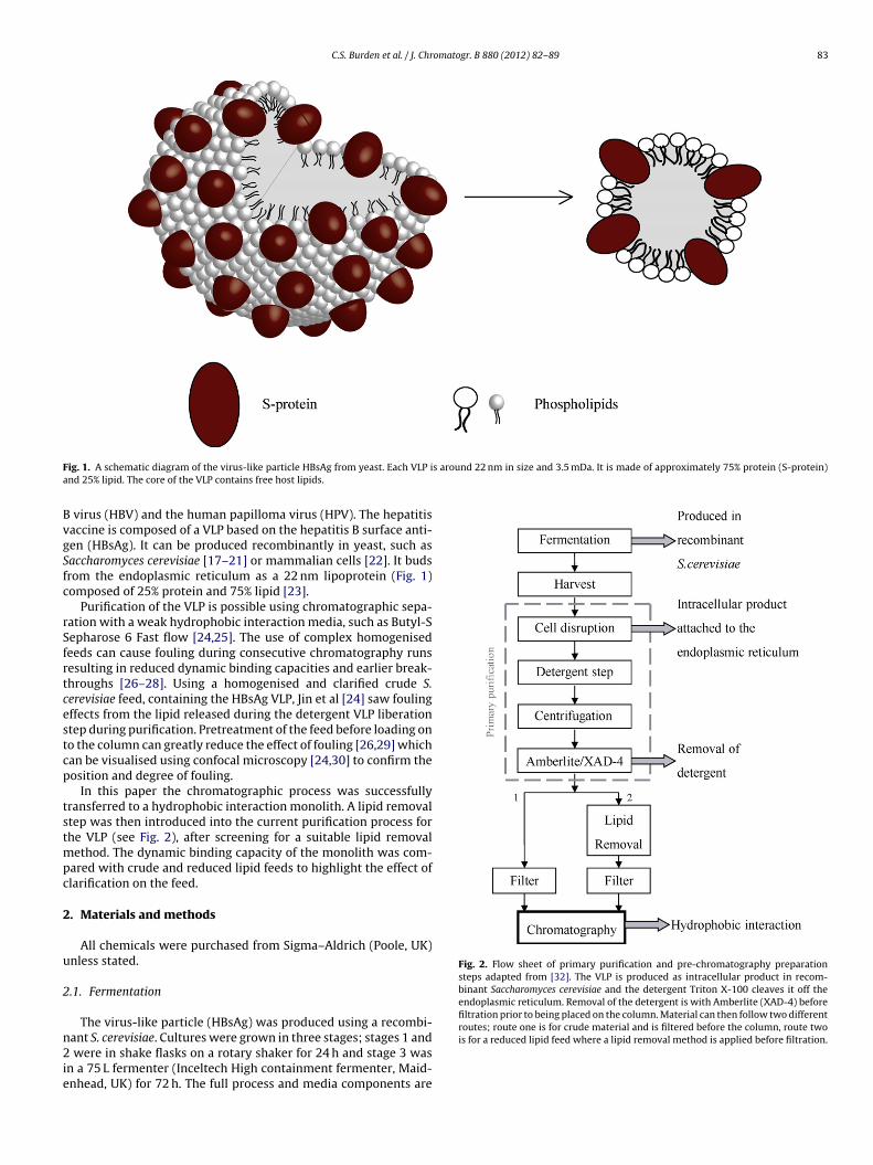
C.S. Burden et al. / J. Chromatogr. B 880 (2012) 82– 89 83
F s around 22 nm in size and 3.5 mDa. It is made of approximately 75% protein (S-protein)a
BvgSfc
rSfrtcestcp
tstmpc
2
u
2
n2ie
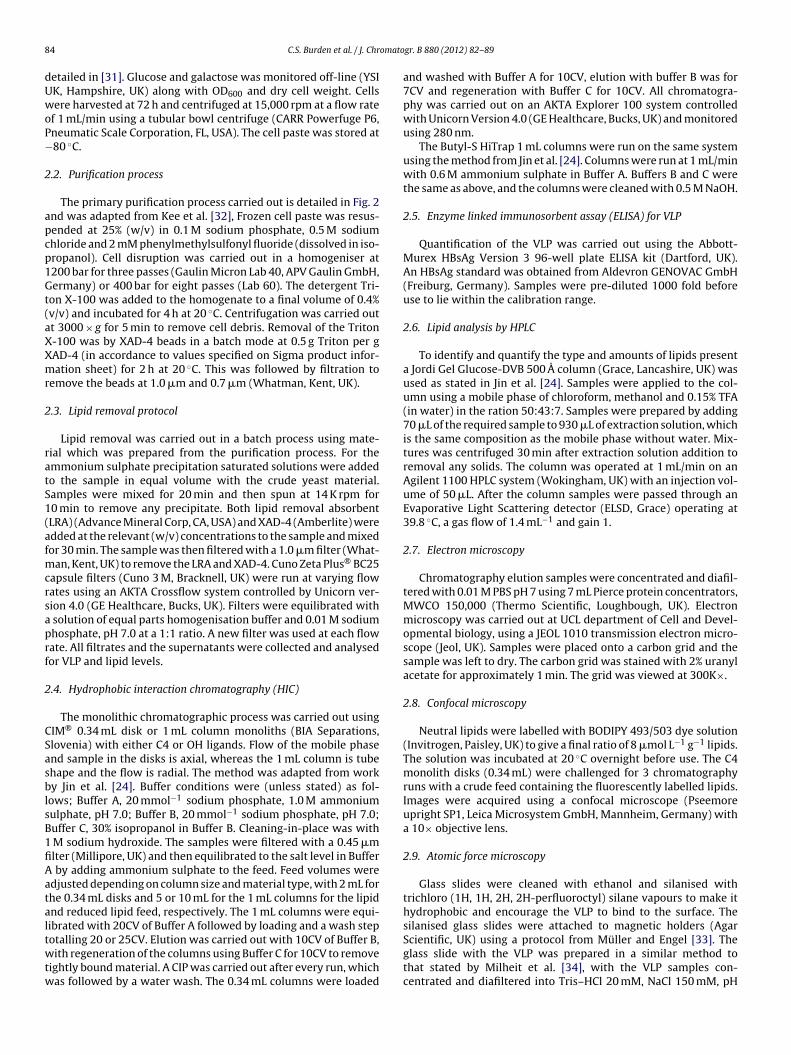
Fig. 2. Flow sheet of primary purification and pre-chromatography preparationsteps adapted from [32]. The VLP is produced as intracellular product in recom-binant Saccharomyces cerevisiae and the detergent Triton X-100 cleaves it off theendoplasmic reticulum. Removal of the detergent is with Amberlite (XAD-4) beforefiltration prior to being placed on the column. Material can then follow two differentroutes; route one is for crude material and is filtered before the column, route two
ig. 1. A schematic diagram of the virus-like particle HBsAg from yeast. Each VLP ind 25% lipid. The core of the VLP contains free host lipids.
virus (HBV) and the human papilloma virus (HPV). The hepatitisaccine is composed of a VLP based on the hepatitis B surface anti-en (HBsAg). It can be produced recombinantly in yeast, such asaccharomyces cerevisiae [17–21] or mammalian cells [22]. It budsrom the endoplasmic reticulum as a 22 nm lipoprotein (Fig. 1)omposed of 25% protein and 75% lipid [23].
Purification of the VLP is possible using chromatographic sepa-ation with a weak hydrophobic interaction media, such as Butyl-Sepharose 6 Fast flow [24,25]. The use of complex homogenisedeeds can cause fouling during consecutive chromatography runsesulting in reduced dynamic binding capacities and earlier break-hroughs [26–28]. Using a homogenised and clarified crude S.erevisiae feed, containing the HBsAg VLP, Jin et al [24] saw foulingffects from the lipid released during the detergent VLP liberationtep during purification. Pretreatment of the feed before loading ono the column can greatly reduce the effect of fouling [26,29] whichan be visualised using confocal microscopy [24,30] to confirm theosition and degree of fouling.
In this paper the chromatographic process was successfullyransferred to a hydrophobic interaction monolith. A lipid removaltep was then introduced into the current purification process forhe VLP (see Fig. 2), after screening for a suitable lipid removal
ethod. The dynamic binding capacity of the monolith was com-ared with crude and reduced lipid feeds to highlight the effect oflarification on the feed.
. Materials and methods
All chemicals were purchased from Sigma–Aldrich (Poole, UK)nless stated.
.1. Fermentation
The virus-like particle (HBsAg) was produced using a recombi-
ant S. cerevisiae. Cultures were grown in three stages; stages 1 andwere in shake flasks on a rotary shaker for 24 h and stage 3 wasn a 75 L fermenter (Inceltech High containment fermenter, Maid-nhead, UK) for 72 h. The full process and media components are
is for a reduced lipid feed where a lipid removal method is applied before filtration.
8 omato
dUwoP−
2
apcp1Gt(aXXmr
2
ratS1(afmcrsaprf
2
CSasblsB1fiAataltwtw
4 C.S. Burden et al. / J. Chr
etailed in [31]. Glucose and galactose was monitored off-line (YSIK, Hampshire, UK) along with OD600 and dry cell weight. Cellsere harvested at 72 h and centrifuged at 15,000 rpm at a flow rate
f 1 mL/min using a tubular bowl centrifuge (CARR Powerfuge P6,neumatic Scale Corporation, FL, USA). The cell paste was stored at80 ◦C.
.2. Purification process
The primary purification process carried out is detailed in Fig. 2nd was adapted from Kee et al. [32], Frozen cell paste was resus-ended at 25% (w/v) in 0.1 M sodium phosphate, 0.5 M sodiumhloride and 2 mM phenylmethylsulfonyl fluoride (dissolved in iso-ropanol). Cell disruption was carried out in a homogeniser at200 bar for three passes (Gaulin Micron Lab 40, APV Gaulin GmbH,ermany) or 400 bar for eight passes (Lab 60). The detergent Tri-
on X-100 was added to the homogenate to a final volume of 0.4%v/v) and incubated for 4 h at 20 ◦C. Centrifugation was carried outt 3000 × g for 5 min to remove cell debris. Removal of the Triton-100 was by XAD-4 beads in a batch mode at 0.5 g Triton per gAD-4 (in accordance to values specified on Sigma product infor-ation sheet) for 2 h at 20 ◦C. This was followed by filtration to
emove the beads at 1.0 �m and 0.7 �m (Whatman, Kent, UK).
.3. Lipid removal protocol
Lipid removal was carried out in a batch process using mate-ial which was prepared from the purification process. For themmonium sulphate precipitation saturated solutions were addedo the sample in equal volume with the crude yeast material.amples were mixed for 20 min and then spun at 14 K rpm for0 min to remove any precipitate. Both lipid removal absorbentLRA) (Advance Mineral Corp, CA, USA) and XAD-4 (Amberlite) weredded at the relevant (w/v) concentrations to the sample and mixedor 30 min. The sample was then filtered with a 1.0 �m filter (What-
an, Kent, UK) to remove the LRA and XAD-4. Cuno Zeta Plus® BC25apsule filters (Cuno 3 M, Bracknell, UK) were run at varying flowates using an AKTA Crossflow system controlled by Unicorn ver-ion 4.0 (GE Healthcare, Bucks, UK). Filters were equilibrated with
solution of equal parts homogenisation buffer and 0.01 M sodiumhosphate, pH 7.0 at a 1:1 ratio. A new filter was used at each flowate. All filtrates and the supernatants were collected and analysedor VLP and lipid levels.
.4. Hydrophobic interaction chromatography (HIC)
The monolithic chromatographic process was carried out usingIM® 0.34 mL disk or 1 mL column monoliths (BIA Separations,lovenia) with either C4 or OH ligands. Flow of the mobile phasend sample in the disks is axial, whereas the 1 mL column is tubehape and the flow is radial. The method was adapted from worky Jin et al. [24]. Buffer conditions were (unless stated) as fol-
ows; Buffer A, 20 mmol−1 sodium phosphate, 1.0 M ammoniumulphate, pH 7.0; Buffer B, 20 mmol−1 sodium phosphate, pH 7.0;uffer C, 30% isopropanol in Buffer B. Cleaning-in-place was with
M sodium hydroxide. The samples were filtered with a 0.45 �mlter (Millipore, UK) and then equilibrated to the salt level in Buffer
by adding ammonium sulphate to the feed. Feed volumes weredjusted depending on column size and material type, with 2 mL forhe 0.34 mL disks and 5 or 10 mL for the 1 mL columns for the lipidnd reduced lipid feed, respectively. The 1 mL columns were equi-ibrated with 20CV of Buffer A followed by loading and a wash step
otalling 20 or 25CV. Elution was carried out with 10CV of Buffer B,ith regeneration of the columns using Buffer C for 10CV to removeightly bound material. A CIP was carried out after every run, whichas followed by a water wash. The 0.34 mL columns were loaded
gr. B 880 (2012) 82– 89
and washed with Buffer A for 10CV, elution with buffer B was for7CV and regeneration with Buffer C for 10CV. All chromatogra-phy was carried out on an AKTA Explorer 100 system controlledwith Unicorn Version 4.0 (GE Healthcare, Bucks, UK) and monitoredusing 280 nm.
The Butyl-S HiTrap 1 mL columns were run on the same systemusing the method from Jin et al. [24]. Columns were run at 1 mL/minwith 0.6 M ammonium sulphate in Buffer A. Buffers B and C werethe same as above, and the columns were cleaned with 0.5 M NaOH.
2.5. Enzyme linked immunosorbent assay (ELISA) for VLP
Quantification of the VLP was carried out using the Abbott-Murex HBsAg Version 3 96-well plate ELISA kit (Dartford, UK).An HBsAg standard was obtained from Aldevron GENOVAC GmbH(Freiburg, Germany). Samples were pre-diluted 1000 fold beforeuse to lie within the calibration range.
2.6. Lipid analysis by HPLC
To identify and quantify the type and amounts of lipids presenta Jordi Gel Glucose-DVB 500 A column (Grace, Lancashire, UK) wasused as stated in Jin et al. [24]. Samples were applied to the col-umn using a mobile phase of chloroform, methanol and 0.15% TFA(in water) in the ration 50:43:7. Samples were prepared by adding70 �L of the required sample to 930 �L of extraction solution, whichis the same composition as the mobile phase without water. Mix-tures was centrifuged 30 min after extraction solution addition toremoval any solids. The column was operated at 1 mL/min on anAgilent 1100 HPLC system (Wokingham, UK) with an injection vol-ume of 50 �L. After the column samples were passed through anEvaporative Light Scattering detector (ELSD, Grace) operating at39.8 ◦C, a gas flow of 1.4 mL−1 and gain 1.
2.7. Electron microscopy
Chromatography elution samples were concentrated and diafil-tered with 0.01 M PBS pH 7 using 7 mL Pierce protein concentrators,MWCO 150,000 (Thermo Scientific, Loughbough, UK). Electronmicroscopy was carried out at UCL department of Cell and Devel-opmental biology, using a JEOL 1010 transmission electron micro-scope (Jeol, UK). Samples were placed onto a carbon grid and thesample was left to dry. The carbon grid was stained with 2% uranylacetate for approximately 1 min. The grid was viewed at 300K×.
2.8. Confocal microscopy
Neutral lipids were labelled with BODIPY 493/503 dye solution(Invitrogen, Paisley, UK) to give a final ratio of 8 �mol L−1 g−1 lipids.The solution was incubated at 20 ◦C overnight before use. The C4monolith disks (0.34 mL) were challenged for 3 chromatographyruns with a crude feed containing the fluorescently labelled lipids.Images were acquired using a confocal microscope (Pseemoreupright SP1, Leica Microsystem GmbH, Mannheim, Germany) witha 10× objective lens.
2.9. Atomic force microscopy
Glass slides were cleaned with ethanol and silanised withtrichloro (1H, 1H, 2H, 2H-perfluoroctyl) silane vapours to make ithydrophobic and encourage the VLP to bind to the surface. Thesilanised glass slides were attached to magnetic holders (Agar
Scientific, UK) using a protocol from Müller and Engel [33]. Theglass slide with the VLP was prepared in a similar method tothat stated by Milheit et al. [34], with the VLP samples con-centrated and diafiltered into Tris–HCl 20 mM, NaCl 150 mM, pH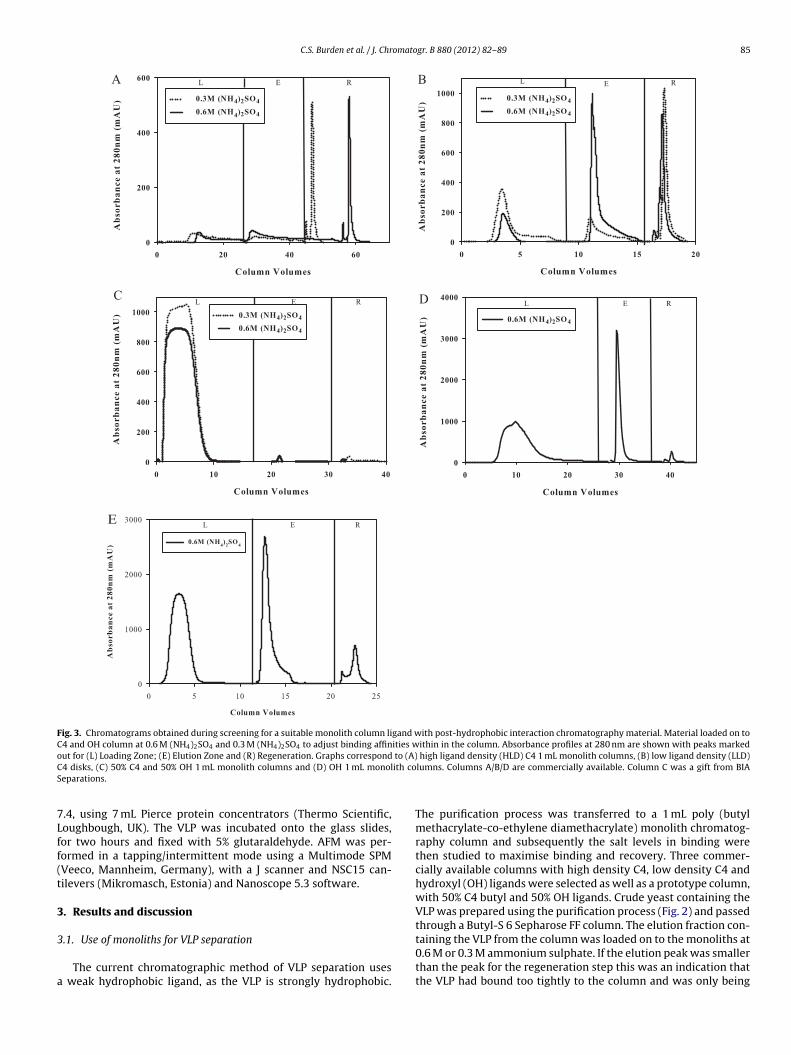
C.S. Burden et al. / J. Chromatogr. B 880 (2012) 82– 89 85
L E RA E RLB
C REL RELD
L E RE
Fig. 3. Chromatograms obtained during screening for a suitable monolith column ligand with post-hydrophobic interaction chromatography material. Material loaded on toC4 and OH column at 0.6 M (NH4)2SO4 and 0.3 M (NH4)2SO4 to adjust binding affinities within in the column. Absorbance profiles at 280 nm are shown with peaks markedo to (A)C ith coS
7Lff(t
3
3
a
ut for (L) Loading Zone; (E) Elution Zone and (R) Regeneration. Graphs correspond4 disks, (C) 50% C4 and 50% OH 1 mL monolith columns and (D) OH 1 mL monoleparations.
.4, using 7 mL Pierce protein concentrators (Thermo Scientific,oughbough, UK). The VLP was incubated onto the glass slides,or two hours and fixed with 5% glutaraldehyde. AFM was per-ormed in a tapping/intermittent mode using a Multimode SPMVeeco, Mannheim, Germany), with a J scanner and NSC15 can-ilevers (Mikromasch, Estonia) and Nanoscope 5.3 software.
. Results and discussion
.1. Use of monoliths for VLP separation
The current chromatographic method of VLP separation uses weak hydrophobic ligand, as the VLP is strongly hydrophobic.
high ligand density (HLD) C4 1 mL monolith columns, (B) low ligand density (LLD)lumns. Columns A/B/D are commercially available. Column C was a gift from BIA
The purification process was transferred to a 1 mL poly (butylmethacrylate-co-ethylene diamethacrylate) monolith chromatog-raphy column and subsequently the salt levels in binding werethen studied to maximise binding and recovery. Three commer-cially available columns with high density C4, low density C4 andhydroxyl (OH) ligands were selected as well as a prototype column,with 50% C4 butyl and 50% OH ligands. Crude yeast containing theVLP was prepared using the purification process (Fig. 2) and passedthrough a Butyl-S 6 Sepharose FF column. The elution fraction con-
taining the VLP from the column was loaded on to the monoliths at0.6 M or 0.3 M ammonium sulphate. If the elution peak was smallerthan the peak for the regeneration step this was an indication thatthe VLP had bound too tightly to the column and was only being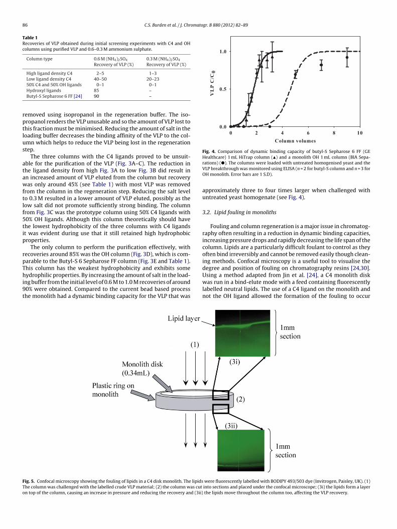
86 C.S. Burden et al. / J. Chromatogr. B 880 (2012) 82– 89
Table 1Recoveries of VLP obtained during initial screening experiments with C4 and OHcolumns using purified VLP and 0.6–0.3 M ammonium sulphate.
Column type 0.6 M (NH4)2SO4
Recovery of VLP (%)0.3 M (NH4)2SO4
Recovery of VLP (%)
High ligand density C4 2–5 1–3Low ligand density C4 40–50 20–2350% C4 and 50% OH ligands 0–1 0–1
rptlus
atawftlf5tip
rpThi9t
Fig. 4. Comparison of dynamic binding capacity of butyl-S Sepharose 6 FF (GEHealthcare) 1 mL HiTrap column (�) and a monolith OH 1 mL column (BIA Sepa-
FTo
Hydroxyl ligands 85 –Butyl-S Sepharose 6 FF [24] 90 –
emoved using isopropanol in the regeneration buffer. The iso-ropanol renders the VLP unusable and so the amount of VLP lost tohis fraction must be minimised. Reducing the amount of salt in theoading buffer decreases the binding affinity of the VLP to the col-mn which helps to reduce the VLP being lost in the regenerationtep.
The three columns with the C4 ligands proved to be unsuit-ble for the purification of the VLP (Fig. 3A–C). The reduction inhe ligand density from high Fig. 3A to low Fig. 3B did result inn increased amount of VLP eluted from the column but recoveryas only around 45% (see Table 1) with most VLP was removed
rom the column in the regeneration step. Reducing the salt levelo 0.3 M resulted in a lower amount of VLP eluted, possibly as theow salt did not promote sufficiently strong binding. The columnrom Fig. 3C was the prototype column using 50% C4 ligands with0% OH ligands. Although this column theoretically should havehe lowest hydrophobicity of the three columns with C4 ligandst was evident during use that it still retained high hydrophobicroperties.
The only column to perform the purification effectively, withecoveries around 85% was the OH column (Fig. 3D), which is com-arable to the Butyl-S 6 Sepharose FF column (Fig. 3E and Table 1).his column has the weakest hydrophobicity and exhibits some
ydrophilic properties. By increasing the amount of salt in the load-ng buffer from the initial level of 0.6 M to 1.0 M recoveries of around0% were obtained. Compared to the current bead based processhe monolith had a dynamic binding capacity for the VLP that was
ig. 5. Confocal microscopy showing the fouling of lipids in a C4 disk monolith. The lipidshe column was challenged with the labelled crude VLP material; (2) the column was cut
n top of the column, causing an increase in pressure and reducing the recovery and (3ii)
rations) (�). The columns were loaded with untreated homogenised yeast and theVLP breakthrough was monitored using ELISA (n = 2 for butyl-S column and n = 3 forOH monolith. Error bars are 1 S.D).
approximately three to four times larger when challenged withuntreated yeast homogenate (see Fig. 4).
3.2. Lipid fouling in monoliths
Fouling and column regeneration is a major issue in chromatog-raphy often resulting in a reduction in dynamic binding capacities,increasing pressure drops and rapidly decreasing the life span of thecolumn. Lipids are a particularly difficult foulant to control as theyoften bind irreversibly and cannot be removed easily though clean-ing methods. Confocal microscopy is a useful tool to visualise thedegree and position of fouling on chromatography resins [24,30].
Using a method adapted from Jin et al. [24], a C4 monolith diskwas run in a bind-elute mode with a feed containing fluorescentlylabelled neutral lipids. The use of a C4 ligand on the monolith andnot the OH ligand allowed the formation of the fouling to occurwere fluorescently labelled with BODIPY 493/503 dye (Invitrogen, Paisley, UK). (1)into sections and placed under the confocal microscope; (3i) the lipids form a layer
the lipids move throughout the column too, affecting the VLP recovery.
C.S. Burden et al. / J. Chromatogr. B 880 (2012) 82– 89 87
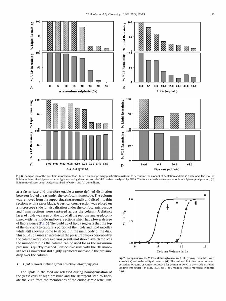
F cation material to determine the amount of depletion and the VLP retained. The level ofl d analysed by ELISA. The four methods were (a) ammonium sulphate precipitation; (b)l
abwsaalpoowTttpld
3
ta
Fig. 7. Comparison of the VLP breakthrough curves of 1 mL hydroxyl monoliths witha crude (�) and reduced lipid material (�). The reduced lipid feed was preparedby adding 0.3 g/mL of Amberlite/XAD-4 for 30 min at 20 ◦C to the crude material.
ig. 6. Comparison of the four lipid removal methods tested on post primary purifiipid was determined by evaporative light scattering detection and the VLP retaineipid removal absorbent (LRA); (c) Amberlite/XAD-4 and (d) Cuno filters.
t a faster rate and therefore enable a more defined distinctionetween fouled areas under the confocal microscope. The columnas removed from the supporting ring around it and sliced into thin
ections with a razor blade. A vertical cross-section was placed on microscope slide for visualisation under the confocal microscopend 1 mm sections were captured across the column. A distinctayer of lipids was seen on the top of all the sections analysed, com-ared with the middle and lower sections which had a lower degreef fluorescence (Fig. 5). The build up of lipids suggests that the topf the disk acts to capture a portion of the lipids and lipid micelleshile still allowing some to deposit in the main body of the disk.
his build up causes an increase in the pressure drop experienced byhe column over successive runs (results not shown) which reduceshe number of runs the column can be used for as the maximumressure is quickly reached. Consecutive runs with the OH mono-
ith sees a slower but still highly significant increase in the pressurerop over the column.
.3. Lipid removal methods from pre-chromatography feed
The lipids in the feed are released during homogenisation ofhe yeast cells at high pressure and the detergent step to liber-te the VLPs from the membranes of the endoplasmic reticulum,
Binding was under 1 M (NH4)2SO4 pH 7 at 3 mL/min. Points represent triplicateruns.
88 C.S. Burden et al. / J. Chromatogr. B 880 (2012) 82– 89
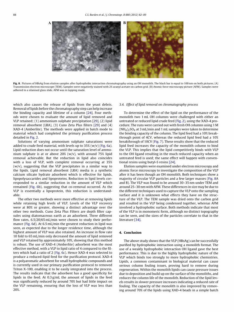
Fig. 8. Pictures of HBsAg from elution samples after hydrophobic interaction chromatography using an OH monolith. The black bar is equal to 100 nm on both pictures. (A)Transmission electron microscope (TEM). Samples were negatively stained with 2% uranyl acetate on carbon grid. (B) Atomic force microscopy picture (AFM). Samples werea
wRtoVrXmd
aLnrw(tclrrVa
wwosflmsh1aietpiiTTlwt2
dhered to a silanised glass slide. AFM was in tapping mode.
hich also causes the release of lipids from the yeast debris.emoval of lipids before the chromatography step can help increasehe binding capacity and lifetime of a column [24]. Four meth-ds were chosen to evaluate the amount of lipid removed andLP retained; (1) ammonium sulphate precipitation [29], (2) lipidemoval absorbent (LRA), (3) Cuno Zeta Plus filters [29] and (4)AD-4 (Amberlite). The methods were applied in batch mode toaterial which had completed the primary purification process
etailed in Fig. 2.Solutions of varying ammonium sulphate saturations were
dded to crude feed material, with levels up to 35% (w/v) (Fig. 6a).ipid reduction does not occur until the saturation level of ammo-ium sulphate is at or above 20% (w/v), with around 75% lipidemoval achievable. But the reduction in lipid also coincidesith a loss of VLP, with complete removal occurring at 35%
w/v), suggesting that the VLP precipitates in a similar way tohe lipids. Lipid removal absorbent (LRA) media is a syntheticalcium silicate hydrate adsorbent which is effective for lipids,ipopolysaccarides and lipoproteins. Reduction in lipid levels cor-esponded to a similar reduction in the amount of VLP whichemained (Fig. 6b), suggesting that co-removal occurred. As theLP is essentially a lipoprotein, this reduction is understand-ble.
The other two methods were more effective at removing lipidshile retaining high levels of VLP. Levels of the VLP recoveryere at 80% or greater, showing a distinct advantage over the
ther two methods. Cuno Zeta Plus Filters are depth filter cap-ules using diatomaceous earth as an adsorbent. Three differentow rates, 6.5/20/65 mL/min were chosen to study their perfor-ance (Fig. 6d). At 6.5 mL/min the greatest reduction in lipid was
een, as expected due to the longer residence time, although theighest amount of VLP was also retained. An increase in flow rate0 fold to 65 mL/min only decreased the amount of lipid removednd VLP retained by approximately 10%, showing that this methods robust. The use of XAD-4 (Amberlite) adsorbent was the mostffective method, with a VLP to lipid ratio of 4 compared to the fil-ers which had a ratio of 2 (Fig. 6c). Hence XAD-4 was selected toroduce a reduced-lipid feed for the purification protocol. XAD-4
s a polyaromatic adsorbent for small hydrophobic compounds ands currently used in our primary purification protocol to removedriton X-100, enabling it to be easily integrated into the process.he results indicate that the adsorbent has a good specificity for
ipids in the feed. At 0.3 g/mL the amount of lipids in the feedas significantly reduced by around 70% but had little impact onhe VLP remaining, ensuring that the loss of VLP was less than0%.
3.4. Effect of lipid removal on chromatography process
To determine the effect of the lipid on the performance of themonolith two 1 mL OH columns were challenged with either anuntreated or reduced lipid crude feed (Fig. 2), using the XAD-4 pro-cedure. The runs were carried out with fresh OH columns using 1 M(NH4)2SO4 at 3 mL/min and 1 mL samples were taken to determinethe binding capacity of the column. The lipid feed had a 10% break-through point of 4CV, whereas the reduced lipid feed had a 10%breakthrough of 10CV (Fig. 7). These results show that the reducedlipid feed increases the capacity of the monolith column to bindthe VLP. This implies that the lipid competitively binds with VLPto the OH ligand resulting in the much reduced capacity when anuntreated feed is used; the same effect will happen with conven-tional resins using butyl-S resins [24].
Elution samples were examined under electron microscopy andatomic force microscopy to investigate the composition of the VLPafter it has been though an OH monolith. Both techniques show amajority of circular VLP particles and a few larger masses (Fig. 8Aand B). The VLP was found to be around 30–35 nm under TEM andaround 25–30 nm with AFM. These differences in size may be due tothe different techniques used to capture the VLP onto the samplingsurface and it is unknown what effects they have on the struc-ture of the VLP. The TEM sample was dried onto the carbon gridand resulted in the VLP being condensed together, whereas AFMinvolved a hydrophobic surface. Both methods indicate that mostof the VLP is in monomeric form, although no distinct topographycan be seen, and the sizes of the particles correlate to that in theliterature [34].
4. Conclusion
The above study shows that the VLP (HBsAg) can be successfullypurified by hydrophobic interaction using a monolith format. Theuse of a weakly hydrophobic interaction OH ligand gave the bestperformance. This is due to the highly hydrophobic nature of theVLP which binds too strongly to more hydrophobic chemistries.Lipids, a common contaminant in biological material can causeserious column fouling issues, proving hard to remove duringregeneration. Within the monolith lipids can cause pressure issuesdue to deposition and build up on the surface of the monoliths, and
reduces the column life of the monolith. Reduction of the lipid lev-els results in slower pressure increases indicating a reduced rate offouling. The capacity of the monolith is also improved by remov-ing around 70% of the lipids using XAD-4 beads in a simple batch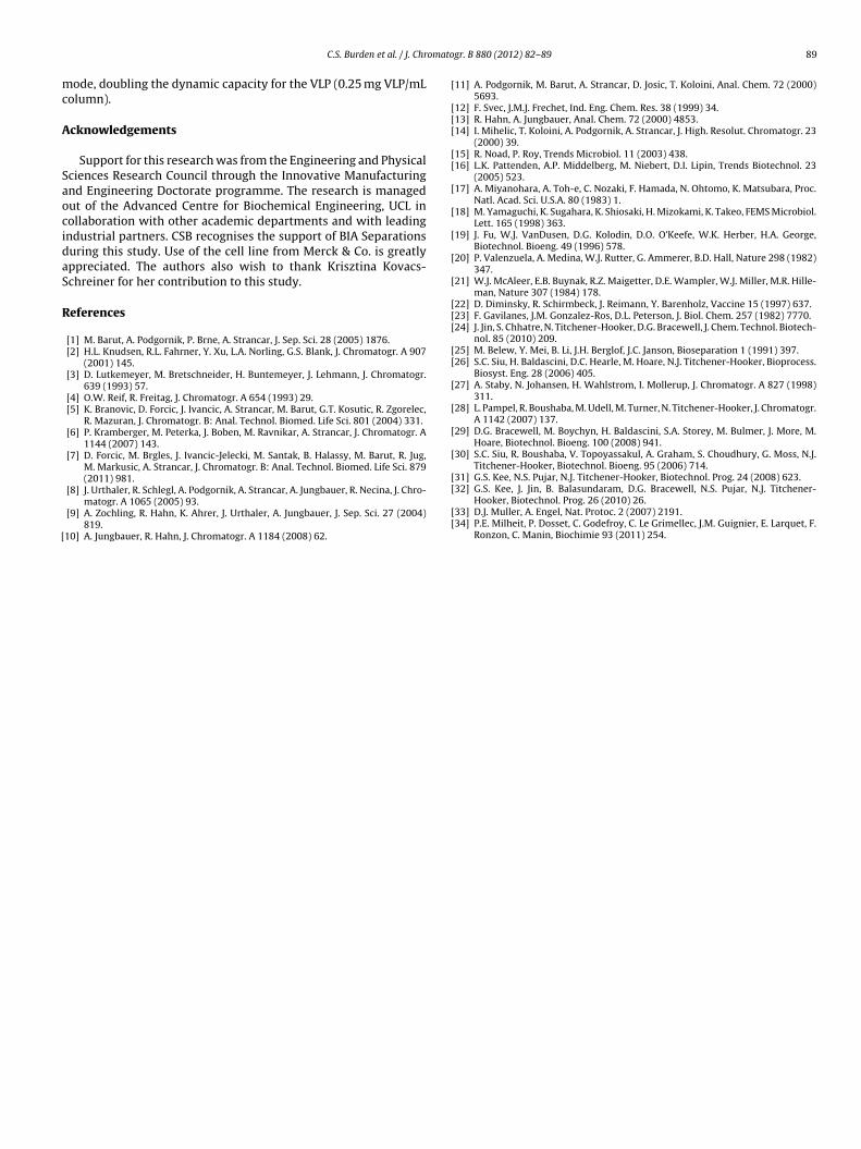
omato
mc
A
SaocidaS
R
[
[
[[[
[[
[
[
[
[
[
[[[
[[
[
[
[
[
[
C.S. Burden et al. / J. Chr
ode, doubling the dynamic capacity for the VLP (0.25 mg VLP/mLolumn).
cknowledgements
Support for this research was from the Engineering and Physicalciences Research Council through the Innovative Manufacturingnd Engineering Doctorate programme. The research is managedut of the Advanced Centre for Biochemical Engineering, UCL inollaboration with other academic departments and with leadingndustrial partners. CSB recognises the support of BIA Separationsuring this study. Use of the cell line from Merck & Co. is greatlyppreciated. The authors also wish to thank Krisztina Kovacs-chreiner for her contribution to this study.
eferences
[1] M. Barut, A. Podgornik, P. Brne, A. Strancar, J. Sep. Sci. 28 (2005) 1876.[2] H.L. Knudsen, R.L. Fahrner, Y. Xu, L.A. Norling, G.S. Blank, J. Chromatogr. A 907
(2001) 145.[3] D. Lutkemeyer, M. Bretschneider, H. Buntemeyer, J. Lehmann, J. Chromatogr.
639 (1993) 57.[4] O.W. Reif, R. Freitag, J. Chromatogr. A 654 (1993) 29.[5] K. Branovic, D. Forcic, J. Ivancic, A. Strancar, M. Barut, G.T. Kosutic, R. Zgorelec,
R. Mazuran, J. Chromatogr. B: Anal. Technol. Biomed. Life Sci. 801 (2004) 331.[6] P. Kramberger, M. Peterka, J. Boben, M. Ravnikar, A. Strancar, J. Chromatogr. A
1144 (2007) 143.[7] D. Forcic, M. Brgles, J. Ivancic-Jelecki, M. Santak, B. Halassy, M. Barut, R. Jug,
M. Markusic, A. Strancar, J. Chromatogr. B: Anal. Technol. Biomed. Life Sci. 879(2011) 981.
[8] J. Urthaler, R. Schlegl, A. Podgornik, A. Strancar, A. Jungbauer, R. Necina, J. Chro-matogr. A 1065 (2005) 93.
[9] A. Zochling, R. Hahn, K. Ahrer, J. Urthaler, A. Jungbauer, J. Sep. Sci. 27 (2004)819.
10] A. Jungbauer, R. Hahn, J. Chromatogr. A 1184 (2008) 62.
[
[[
gr. B 880 (2012) 82– 89 89
11] A. Podgornik, M. Barut, A. Strancar, D. Josic, T. Koloini, Anal. Chem. 72 (2000)5693.
12] F. Svec, J.M.J. Frechet, Ind. Eng. Chem. Res. 38 (1999) 34.13] R. Hahn, A. Jungbauer, Anal. Chem. 72 (2000) 4853.14] I. Mihelic, T. Koloini, A. Podgornik, A. Strancar, J. High. Resolut. Chromatogr. 23
(2000) 39.15] R. Noad, P. Roy, Trends Microbiol. 11 (2003) 438.16] L.K. Pattenden, A.P. Middelberg, M. Niebert, D.I. Lipin, Trends Biotechnol. 23
(2005) 523.17] A. Miyanohara, A. Toh-e, C. Nozaki, F. Hamada, N. Ohtomo, K. Matsubara, Proc.
Natl. Acad. Sci. U.S.A. 80 (1983) 1.18] M. Yamaguchi, K. Sugahara, K. Shiosaki, H. Mizokami, K. Takeo, FEMS Microbiol.
Lett. 165 (1998) 363.19] J. Fu, W.J. VanDusen, D.G. Kolodin, D.O. O’Keefe, W.K. Herber, H.A. George,
Biotechnol. Bioeng. 49 (1996) 578.20] P. Valenzuela, A. Medina, W.J. Rutter, G. Ammerer, B.D. Hall, Nature 298 (1982)
347.21] W.J. McAleer, E.B. Buynak, R.Z. Maigetter, D.E. Wampler, W.J. Miller, M.R. Hille-
man, Nature 307 (1984) 178.22] D. Diminsky, R. Schirmbeck, J. Reimann, Y. Barenholz, Vaccine 15 (1997) 637.23] F. Gavilanes, J.M. Gonzalez-Ros, D.L. Peterson, J. Biol. Chem. 257 (1982) 7770.24] J. Jin, S. Chhatre, N. Titchener-Hooker, D.G. Bracewell, J. Chem. Technol. Biotech-
nol. 85 (2010) 209.25] M. Belew, Y. Mei, B. Li, J.H. Berglof, J.C. Janson, Bioseparation 1 (1991) 397.26] S.C. Siu, H. Baldascini, D.C. Hearle, M. Hoare, N.J. Titchener-Hooker, Bioprocess.
Biosyst. Eng. 28 (2006) 405.27] A. Staby, N. Johansen, H. Wahlstrom, I. Mollerup, J. Chromatogr. A 827 (1998)
311.28] L. Pampel, R. Boushaba, M. Udell, M. Turner, N. Titchener-Hooker, J. Chromatogr.
A 1142 (2007) 137.29] D.G. Bracewell, M. Boychyn, H. Baldascini, S.A. Storey, M. Bulmer, J. More, M.
Hoare, Biotechnol. Bioeng. 100 (2008) 941.30] S.C. Siu, R. Boushaba, V. Topoyassakul, A. Graham, S. Choudhury, G. Moss, N.J.
Titchener-Hooker, Biotechnol. Bioeng. 95 (2006) 714.31] G.S. Kee, N.S. Pujar, N.J. Titchener-Hooker, Biotechnol. Prog. 24 (2008) 623.
32] G.S. Kee, J. Jin, B. Balasundaram, D.G. Bracewell, N.S. Pujar, N.J. Titchener-Hooker, Biotechnol. Prog. 26 (2010) 26.33] D.J. Muller, A. Engel, Nat. Protoc. 2 (2007) 2191.34] P.E. Milheit, P. Dosset, C. Godefroy, C. Le Grimellec, J.M. Guignier, E. Larquet, F.
Ronzon, C. Manin, Biochimie 93 (2011) 254.