Embed Size (px)
Citation preview

1
The Mon1-Ccz1 GEF activates the Rab7 GTPase Ypt7 via a longin
fold-Rab interface and association with PI-3-P-positive membranes
Margarita Cabrera1, Mirjana Nordmann1,*, Angela Perz1, David Schmedt2, Andreas
Gerondopoulos3, Francis Barr3, Jacob Piehler2, Siegfried Engelbrecht-Vandré1,
and Christian Ungermann1
1 University of Osnabrück Department of Biology/Chemistry Biochemistry section Barbarastrasse 13 49076 Osnabrück, Germany 2 University of Osnabrück Department of Biology/Chemistry Biophysics section 3 Department of Biochemistry University of Oxford South Parks Road OX1 3QU Oxford, United Kingdom *Present address: University of Osnabrück Department of Biology/Chemistry Section of Molecular Cell Biology 1 To whom correspondence should be addressed: Email: [email protected] Phone: +49-541-969-2752 (C.U.) Characters: (without spaces) : 40,299
© 2014. Published by The Company of Biologists Ltd.Jo
urna
l of C
ell S
cien
ceA
ccep
ted
man
uscr
ipt
JCS Advance Online Article. Posted on 10 January 2014

2
Abstract
For their function in fusion and signaling, Rab GTPases need to be converted into their active
GTP form. We previously identified the conserved Mon1-Ccz1 complex as the guanine
nucleotide exchange factor (GEF) of the yeast Rab7 GTPase Ypt7. To address the possible
GEF mechanism, we generated a homology model of the predicted longin domains of Mon1
and Ccz1 using as template the Rab-binding surface of the TRAPP complex. Based on this,
we identified mutations in both yeast Mon1 and Ccz1 that block Ypt7 activation, without
affecting heterodimer formation and intracellular localization of Mon1 and Ccz1 at
endosomes. Strikingly, the activity of the isolated Mon1-Ccz1 complex for Ypt7 is highly
stimulated on membranes, and is promoted by the same anionic phospholipids such as
phosphatidylinositol-3-phosphate (PI-3-P) that also support membrane-association of the GEF
complex. Our data imply that the GEF activity of the Mon1-Ccz1 complex towards
Rab7/Ypt7 requires the interface formed by their longin domains and profits strongly from its
association with the organelle surface.
Jour
nal o
f Cel
l Sci
ence
Acc
epte
d m
anus
crip
t

3
Introduction
Within the endomembrane system, vesicles transport cargo between organelles. These
organelles need to maintain their identity despite the fact that they continuously fuse with
incoming vesicles and loose membrane due to vesicles generation. Rab GTPases, tethering
factors and the SNARE machinery localize to specific organelles and cooperate to promote
fusion of membranes. Rabs are small GTP/GDP-binding proteins with C-terminal prenyl
anchors, which function as molecular switches. They bind in their GTP-form to tethering
factors to bring membranes into proximity before SNAREs present in each membrane form
four-helix bundles and thus drive bilayer mixing.
Due to the slow rate of dissociation of GDP, Rabs require guanine nucleotide
exchange factors (GEFs) for their conversion into the active GTP-bound form, which can
interact with effectors on membranes such as tethering factors or lipid kinases. In addition,
Rabs depend on a GTPase activating protein (GAP) to hydrolyze the GTP to GDP, and then
become substrates of the GDI chaperone, which can extract the Rab-GDP and keep it soluble
in the cytoplasm (Lachmann et al., 2011; Hutagalung et al., 2011; Itzen et al., 2011; Barr,
2013).
Among the Rab interactors, GEFs are most critical as they provide the activated Rab at
a specific location in the cell. In this context, structures of Rab-GEF complexes and the
subsequent analyses have been very informative. For the exocytic Sec4 Rab, the GEF Sec2
promotes nucleotide exchange via its α-helical domain (Dong et al., 2007; Sato et al., 2007).
Rab5-like proteins that operate in the endosomal pathway depend on Vps9-domain containing
GEFs (Delprato et al., 2004; Delprato et al., 2007). DENN domains were identified as a
signature domain for multiple GEFs (Yoshimura et al., 2010; Allaire et al., 2010; Marat et al.,
2011), and molecular details of their function were revealed for DENND1-BS and Rab35
(Wu et al., 2011).
Jour
nal o
f Cel
l Sci
ence
Acc
epte
d m
anus
crip
t

4
GEFs may also act as part of a multiprotein complex. The best-characterized example
is the heptameric TRAPPI, which interacts with the Rab1/Ypt1 protein via a surface
consisting of four subunits, Bet5, Trs23, and the two flanking copies of Bet3 (Kümmel el al,
2005; Kim et al., 2006; Cai et al., 2008). GEF dimers are required to activate Rab6/Ypt6
(Ric1-Rgp1; Pusapati et al., 2012; Siniossoglou et al., 2000) and Rab7/Ypt7 (Mon1-Ccz1;
Nordmann et al., 2010; Gerondopoulos et al., 2012), although structural information on these
complexes is lacking. In agreement with the function of the Mon1-Ccz1 dimer in Ypt7
activation, endosomal biogenesis is strongly impaired if either of the two proteins is defective
(Wang et al., 2002; Kinchen et al., 2010; Poteryaev et al., 2010; Nordmann et al., 2010;
Yousefian et al., 2013).
TRAPP subunits Bet5 and Trs23 interact both with each other and with Ypt1 via their
longin domains (Kim et al., 2006; Cai et al., 2008). These are arranged into a continuous ten-
strand β sheet supporting two parallel α helices, which provide a significant portion of the
interacting residues. Interestingly, the formation of the Mon1-Ccz1 heterodimer also requires
the predicted longin domains of both subunits (Nordmann et al., 2010). This suggests that the
architecture and mode of GEF action of Mon1 and Ccz1 on Ypt7 might resemble those of the
TRAPPI complex. Here, we present evidence that Mon1 and Ccz1, which are linked to
Rab7/Ypt7 both in mammals and yeast, cooperate via their longin-domain surface to activate
the Rab GTPase. This activity is strongly promoted upon membrane recruitment of the GEF,
suggesting that previously determined activities might have been significantly
underestimated.
Results
Mon1 and Ccz1 require a common interface for Ypt7 activation
Jour
nal o
f Cel
l Sci
ence
Acc
epte
d m
anus
crip
t

5
Secondary structure prediction using PredictProtein (http://predictprotein.org) and PsiPred
(http://bioinf.cs.ucl.ac.uk/psipred/) identified possible longin domains in Ccz1 (aa 1-156) and
Mon1 (aa 180-287), which are required for the assembly of the Mon1-Ccz1 heterodimer
(Nordmann et al., 2010). This suggests a similar structure/function relationship for Mon1-
Ccz1 and the TRAPPI GEF complex, since the two TRAPP subunits Trs23 and Bet5 also
contain longin domains that contact the Rab GTPase Ypt1 directly (Kim et al., 2006; Cai et
al., 2008). We therefore built a homology model for the predicted longin domains of Mon1-
Ccz1 using Trs23 and Bet5 as templates.
Longin domains consist of a central antiparallel β sheet (S1-S5) with one α helix
(H1) on one side and two α helices (H2, H3) on the opposing side. Trs23 and Bet5 interact
edge to edge via the central β-sheets of their longin domains and contact Ypt1 primarily via
residues residing in the helices H1 of both subunits (Figure 1A). Consequently, mutants in
each H1 helix result in loss of GEF activity towards Ypt1 (Cai et al., 2008) (Figure 1B). In
addition, Ypt1 is recognized by loop S1-S2 and strand S2 of Trs23 (Figure 1B) and some
residues provided by the two copies of Bet3 (Cai et al., 2008). We used the Genesilico
Metaserver (Kurowski et al., 2003) to assigned Ccz1 (aa 1-156) to Bet5 and Mon1 (aa 180-
287) to Trs23, and built a homology model for Ypt7/MC1 based on the Ypt1/TRAPP
complex (see methods for details). The resulting model showed only few clashes with
satisfactory stereochemistry (Figure 1C).
Based on this model and the previously characterized TRAPP mutants, we introduced
mutations in Mon1 and Ccz1 that should interfere with GEF activity (Figure 1D). Notably, for
TRAPPI and DENND1B-S, double and triple mutants had to be generated in order to abolish
GEF activity completely (Cai et al., 2008; Wu et al., 2011). We therefore used a similar
approach here, and first followed vacuole morphology of Mon1 and Ccz1 mutants. Deletion
of Mon1 or Ccz1 results in multiple small vacuoles (Figure 2A-C). This defect is rescued by
expression of Mon1 and Ccz1, either from a normal promoter or overproduced (Figure 2A,
Jour
nal o
f Cel
l Sci
ence
Acc
epte
d m
anus
crip
t

6
C). For Mon1, we identified mutations in helix H1 (G209W, T213K) and the neighboring S1-
S2 region (G191P, K192D) that resulted in vacuolar defects (Figure 2A). Defects in the single
G191P mutant were only observed if expression of Mon1 was lowered (Figure 2C). Ccz1
mutants displayed similar defects if mutated in the H1 region (G47W, G51M)(Figure 2B).
These defects were not due to mislocalization, as GFP-tagged Mon1 and Ccz1 mutants were
still found in dot-like structures, which likely represent endosomes, similar to the wild-type
proteins (Figure 2D, E). To test if any of the mutations interfered with the Rab interaction, we
relied on a yeast-three-hybrid assay, where both Mon1 and Ccz1 were co-expressed with
Ypt7. This assay clearly demonstrated interaction of Mon1 and Ccz1 with the GDP-locked
version of Ypt7 (T22N), whereas none of the mutants of Mon1 or Ccz1 did (Figure 2F). In
agreement with this, the exclusive vacuolar localization of Ypt7 was lost in both mutants as
observed for the GEF deletion strains (Figure 3A)(Cabrera and Ungermann, 2013). We next
asked if the observed defect was due to complex assembly. However, neither mutation
affected the formation of the dimer, and we could purify Mon1-Ccz1 complexes with similar
stoichiometries as observed for the wild-type complex using the established tandem-affinity
purification protocol (Figure 3B)(Nordmann et al., 2010). We then asked if the purified
mutant complexes are still functional and used the vacuole fusion assay to measure their GEF
activity (Figure 3C). In this assay, fusion of vacuoles from two different tester strains allows
the protease present in one vacuole population to process immature alkaline phosphatase,
which is found in the other vacuole population (Wickner, 2002). To test for GEF activity, we
preincubated vacuoles with a non-specific GAP to generate Ypt7-GDP and then used Mon1-
Ccz1 to reactivate Ypt7 (Nordmann et al., 2010). Whereas the wild-type Mon1-Ccz1 complex
nicely restored fusion, neither mutant was able to counteract GAP activity (Figure 3C). In
sum, mutations in either Mon1 or Ccz1, which had been introduced based on our homology
model, maintained the GEF complex and its localization, but abolished both Ypt7 interaction
and GEF activity.
Jour
nal o
f Cel
l Sci
ence
Acc
epte
d m
anus
crip
t

7
The Mon1-Ccz1 complex binds negatively charged membranes
Having established that Mon1 and Ccz1 cooperate in Rab7/Ypt7 activation, we asked how
activity of the complex might be regulated. Previous analyses of the C.elegans Mon1 protein
SAND-1 indicated that it binds phosphoinositide-3-phosphate (PI-3-P)(Poteryaev et al.,
2010), even though depletion of PI-3-P did not affect Mon1-Ccz1 localization in Drosophila
(Yousefian et al., 2013). We used single Mon1 and Ccz1 proteins isolated from yeast deleted
for the other binding partner, and tested their binding specificity to nitrocellulose strips coated
with different lipid species (Figure 4A). When blots were decorated against Mon1 or Ccz1,
we observed strong interactions with PI-3-P, PI-5-P and phosphatidylserine (PS).
Interestingly, the human Mon1a protein had the same preference for PI-3-P and PS, indicating
conserved membrane preferences of Mon1-Ccz1 across species. To confirm yeast Mon1-Ccz1
binding to membranes with negatively charged lipids, we generated liposomes with vacuolar
lipid composition with or without PI-3-P, incubated them with Mon1-Ccz1 complex and
separated the lipid-bound fraction from the unbound by flotation (Cabrera et al., 2010).
Surprisingly, Mon1-Ccz1 was mainly recovered in the liposome fraction (Figure 4B),
regardless of the presence of PI-3-P in the bilayer. This indicates that either Mon1-Ccz1 does
not show a lipid preference or that our methodology is not sensitive enough to reveal possible
differences. We thus employed reflectance interference (Rif), a label-free detection method,
which determines protein binding to solid-supported membranes (Figure 4C)(Gavutis et al.,
2005). Using this experimental strategy, we observed a more efficient binding of Mon1-Ccz1
to PS and PI-3-P containing membranes than to PC membranes (Figure 4D, see Table). This
indicates that Mon1-Ccz1 has an inherent affinity to membranes that is strengthened by
negatively charged phospholipids.
Membrane association enhances Mon1-Ccz1 GEF activity to membrane bound Ypt7
Jour
nal o
f Cel
l Sci
ence
Acc
epte
d m
anus
crip
t

8
Although GEFs operate on membranes to promote both Rab activation and localization, their
activities are usually determined in solution. We wondered if the activity of Mon1-Ccz1
might be underestimated in our previous measurements (Nordmann et al., 2010). To mimic at
least part of the Rab activation cascade, we immobilized Ypt7 via a C-terminal hexahistidine-
tag on vesicles containing DOGS-NTA lipids (1,2-dioleoyl-sn-glycero-3-[(N-(5-amino-1-
carboxypentyl)iminodiacetic acid)succinyl])(Sot et al., 2013), and then measured GEF
activity. Unlike in solution, where modest Mon1-Ccz1 activity was observed (Nordmann et
al., 2010), binding of Ypt7 to vesicles resulted in dramatically enhanced activity (Figure 5A).
We then reduced the concentration of Mon1-Ccz1, and estimated an approximately 1600-fold
increase in GEF activity (Figure 5B, I). Of note, the determined GEF activity of Mon1-Ccz1
without membrane-anchored Ypt7 is very similar to our previous approximation (Nordmann
et al., 2010). This increase in GEF activity was dependent on the membrane-localization of
Ypt7 as vesicles without DOGS-NTA were without effect (Figure 5C). As expected, Mon1
longin mutant (G209W, T213K) and Rab5-GEF Vps9 were inactive in the GEF assay (Figure
5D). We then asked if the increase in membrane binding as observed before (Figure 4D)
would correlate with GEF activity. We therefore generated DOGS-NTA containing vesicles
without or with PS and PI-3-P in different concentrations and determined GEF activity.
Whereas activity was clearly observed if Ypt7 was immobilized on PC-containing
membranes, it was further two-fold increased by additional negatively charged phospholipids
(Figure 5E, I). To discriminate whether PS and PI-3-P only contribute to the membrane
recruitment of the GEF complex, we used a His-tagged version of the complex that can
associate with membranes independently of these lipids. Interestingly, the presence of PS and
PI-3-P resulted in additional stimulation of the GEF activity, which may reflect an additional
role of these lipids in Mon1-Ccz1 activation (Figure 5F).
The HOPS complex binds activated Ypt7 on the late endosome and vacuole, and
might act in a positive feedback loop to promote Mon1-Ccz1 activity. Indeed, the Vps39
Jour
nal o
f Cel
l Sci
ence
Acc
epte
d m
anus
crip
t
9
subunit of the HOPS complex interacts with Mon1 (Wang et al., 2003; Nordmann et al.,
2010), and additional interactions of metazoan Mon1 with HOPS subunits were reported
(Poteryaev et al., 2010). We therefore localized GFP-tagged Mon1 or Ccz1 in cells
overexpressing the entire HOPS complex. Under these conditions, Mon1 and Ccz1
accumulated on vacuolar membranes, presumably due to the reported interactions of the GEF
complex with HOPS (Figure 5G). We then asked if the interaction also affects activity of the
GEF complex, and therefore added purified HOPS (approximately two-fold excess) to the
GEF assay (Figure 5H). Even though we observed a slight increase in activity, we consider
the effect rather minimal. In sum, GEF activity of Mon1-Ccz1 is strongly increased on
membranes, but is not affected by the HOPS complex.
Discussion
Within this study, we have addressed the molecular function of the Mon1-Ccz1 complex.
Mon1-Ccz1 activity requires residues within the predicted longin domains of both subunits,
which we could identify based on a homology model with the TRAPPI complex. Consistent
with this, Mon1 and Ccz1 longin mutants still form a heterodimer and localize like the wild-
type complex, but cannot promote nucleotide exchange in Ypt7. Using a modified GEF assay,
we further show that Mon1-Ccz1 is strongly active once it is recruited to the membranes,
suggesting that GEF activities might have been underestimated previously.
The presence of longin domains in Mon1-Ccz1 and TRAPPI and the similar
phenotype of the mutants suggest that both GEFs might share a related Rab-binding surface
and nucleotide exchange mechanism. Also, the recently resolved DENND1 GEF domain
contains a longin fold, although it binds Rab35 via a different surface compared to TRAPPI-
Ypt1 interaction (Wu et al., 2011). We are aware that within the TRAPPI complex, the two
Bet3 subunits contribute to the binding of Ypt1 and the C-terminal residues of Bet3A have a
critical role to promote GEF activity (Cai et al., 2008). We thus consider it likely that
Jour
nal o
f Cel
l Sci
ence
Acc
epte
d m
anus
crip
t

10
additional segments proximal to the longin domains of Mon1-Ccz1 will be required for full
activity. We also note that our model is limited by the structural differences between Ypt1 and
Ypt7 especially in their amino terminal regions. However, at this point, our working model
allows us to predict critical residues for GEF activity (Figures 1, 2).
In this context, we realized that roadblock domains that are found in subunits of the
EGO/ragulator complex have been predicted as GEFs for the Rag GTPases (BarPeled et al.,
2013). Even though the order of secondary structure elements differs between roadblock and
longin domains, their overall structure is quite similar (Kurzbauer et al., 2004; Qian et al.,
2005; Zhang et al., 2012). These domains likely have evolved as general interactors of small
GTPases (Levine et al., 2013), and a subfamily might act as dimeric GEFs. Importantly, the
dimeric BLOC-3 complex also harbors a longin domain in each of its two subunits Hps1 and
Hps4 and may function very similar to Mon1-Ccz1 (Gerondopoulos et al., 2012). It thus is
conceivable that these domains have evolved as general interactors of small GTPases.
Intriguingly, the dimeric Ric1-Rgp1 complex, which activates Ypt6 (Siniossoglou et al.,
2000), also depends on both subunits for activity, and might also use a shared surface to
interact with its Rab Ypt6/Rab6.
Even though we have shown GEF activity of Mon1-Ccz1 before in vitro, we did not
expect a 1600-fold increase in activity (kcat/KM) upon immobilization of Ypt7 on membranes.
This may be in part due to the membrane recruitment of both proteins. Although the assay
does not recapitulate the entire cycle, it revealed that Mon1-Ccz1 activity is far higher than
previously anticipated. GEFs like the Legionella DrrA protein have strong activity in vitro,
which is sufficient to counteract Rab extraction by GDI (Schöbel et al., 2009; Zhu et al.,
2010; Suh et al., 2010). It is thus likely that other GEFs use similar mechanisms and will
explain how GEFs that are mistargeted to the mitochondria can drive the recruitment of Rabs
to this organelle (Gerondopoulos et al., 2012; Blümer et al., 2013), even though some
targeting information may reside in additional proteins and the Rab itself (Cabrera and
Jour
nal o
f Cel
l Sci
ence
Acc
epte
d m
anus
crip
t

11
Ungermann, 2013). Our adjustment in the GEF assay now provides insight into the activity of
Mon1-Ccz1. We could demonstrate that acidic phospholipids such as PS and PI-3-P clearly
enhanced the inherent ability of the yeast and human complex to bind membranes in
agreement with earlier findings on the C.elegans Mon1 (Poteryaev et al., 2010). Interestingly,
in Drosophila the Mon1-Ccz1 complex localizes even in the absence of PI-3-P to membranes
(Yousefian et al., 2013). We consider it likely that acidic phospholipids also facilitate the
Rab-GEF interaction by optimally positioning the GEF complex relative to the Rab. We
believe that similar assays with other GEFs will also uncover stronger GEF activities. A more
extreme case was observed for Rasal, a RasGAP that displays activity only when it is
recruited with its Ras substrate to the same membrane (Sot et al., 2013). As GEFs function
only on membranes in vivo, we consider our approach more suitable to mimic the in vivo
situation on organelles.
One aspect that we could not yet clarify is the timing of Mon1-Ccz1 recruitment. It is
possible that the complex is always active on endosomal membranes or it may be activated
only once endosomes have matured, and thus contribute to the exchange of Rab5 for Rab7
(Rink et al., 2005; Poteryaev et al., 2010). With respect to yeast, it is puzzling that the
amounts of Ypt7 on endosomes are rather low, even though we observe Mon1-Ccz1 mostly
on endosomes (Nordmann et al., 2010). This suggests that the complex might be further
regulated in vivo. At this point, our data indicate that Mon1-Ccz1 acts primarily at endosomes
and its interaction with HOPS might create an additional GEF microdomain on vacuoles.
Within our experimental set-up, Mon1-Ccz1 activity was not affected significantly by HOPS,
and Ypt7 localization was not disturbed in the absence of Vps39, which binds to Mon1 in
vitro (Nordmann et al., 2010). We expect that the further dissection of the role of other
endosomal factors that may function in a Rab cascade will help us to reveal how Mon1-Ccz1
function is controlled in vivo.
Jour
nal o
f Cel
l Sci
ence
Acc
epte
d m
anus
crip
t

12
In summary, our data reveal that the Mon1-Ccz1 GEF complex activates Ypt7 via a
common interface, has highest activity on membranes and acts independently of the HOPS
complex.
Jour
nal o
f Cel
l Sci
ence
Acc
epte
d m
anus
crip
t
13
Materials and Methods
Yeast strains and molecular biology
Strains and plasmids used in this study are listed in Table S1 and S2 respectively. Deletions
and tagging of genes were done using homologous recombination of PCR fragments (Janke et
al., 2004; Puig et al., 1998). mCherry-tagged Ypt7 was expressed from plasmid pRS414-
TPIpr-mCherry-V5, which was generated from plasmid pRS415-TPIpr-mCherry-V5-ATG8 (a
gift from Fulvio Reggiori, University Medical Center Utrecht, The Netherlands). Mon1 and
Ccz1 mutants in were generated using the QuickChange Site-Directed Mutagenesis Kit from
Stratagene (La Jolla, CA, USA). The catalytic domain of Gyp1 (249-637) was purified from
BL21 strain containing plasmid pET22-Gyp1-46 (Nordmann et al., 2010). Protein expression
was induced by addition of 0.5 mM isopropyl-d-thiogalactoside overnight at 20°C.
Purification was performed using the nickel-nitrilotriacetic acid resin (QIAGEN, Hilden,
Germany) and elution with 300 mM imidazole.
Yeast three hybrid assay
YEP352-Mon1, pACT2-Ccz1 and pFBT9-Ypt7 plasmids containing wild-type and mutant
versions of Mon1, Ccz1 and Ypt7 were co-transformed into PJ69-4A strain and plated on
minimal media lacking leucine, tryptophan and uracil. Transformants were patched first on
plates lacking leucine, tryptophan and uracil and afterwards on plates lacking leucine,
tryptophan, uracil, histidine and adenine. Four clones were analyzed for each combination and
one is shown.
Modeling of Mon1 and Ccz1
The Genesilico metaserver (https://genesilico.pl/meta2) (Kuroski et al., 2003) was used to
identify remotely related proteins of known structure (by allowing for much more freedom in
Jour
nal o
f Cel
l Sci
ence
Acc
epte
d m
anus
crip
t
14
sequence alignments). Secondary structure predictions were carried out with either
PredictProtein or PsiPred (http://predictprotein.org, http://bioinf.cs.ucl.ac.uk/psipred/). Both
published structures of the TRAPP complex were identified (PDB IDs 3CUE 2J3T; protein
data bank codes from www.pdb.org). 3CUEc and 2J3Tc (chain c of yeast and mouse Bet5
respectively) were identified for the first 150 residues of Ccz1. The server also reports Bet5
structures for Mon1 fragment (180-287) but with lower score. The template for modeling
consisted of Ypt7-GDP (PDB ID 1KY3) (Constantinescu et al., 2002) and mammalian Bet5
and Trs23 (PDB ID 2J3T) and was obtained by 3D alignment with the respective chains of
Ypt1/TRAPP structure (PDB ID 3CUE). A PDZL domain, which is lacking in yeast Trs23
was removed from the template. The complex formed by Ypt7GDP and the longin domains of
Mon1 and Ccz1 was modeled using YASARA 13.4.21 (Krieger et al., 2002). The resulting
model resembled the Ypt1-Trs23-Bet5 structure and showed only few clashes in the
contacting residues. The model scored as optimal or satisfactory considering the correctness
of backbone (Ramachandran plot) and side-chain dihedrals, as well as packing interactions.
Note that the model does not include Ypt7 residues (38-40 and 67-76), which are missing in
the reported structure.
Microscopy
Yeast cells were grown to mid-log phase in YPD, YPG, or synthetic complete (SDC) medium
lacking selected amino acids or nucleotides, collected by centrifugation, washed once with
SDC or SGC medium supplemented with all amino acids, and immediately analyzed by
fluorescence microscopy. For FM4-64 staining of vacuoles, cells were incubated with 30 μM
FM4-64 for 30 min, washed twice with YPD medium, and incubated in the same medium
without dye for 1 h. Images were acquired with a Leica DM5500 B microscope equipped with
a SPOT Pursuit camera equipped with an internal filter wheel (D460sp, BP460-515 and
D580lp; Leica Microsystems GmbH), fluorescence filters (49002 ET-GFP (FITC/Cy2): Exc.
Jour
nal o
f Cel
l Sci
ence
Acc
epte
d m
anus
crip
t

15
ET470/40x, Em. ET525/50m; Wide Green: Exc. D535/50, Em. E590lp; 49008 ET-mCherry,
Texas Red: Exc. ET560/40x, Em. ET630/75m Chroma Technology Corp.), and Metamorph 7
software (Visitron Systems, Munich, Germany). Images were processed using Image J 1.42
(National Institute of Health) and Autoquant x v1.3.3 (Media Cybernetics, Inc).
Tandem Affinity Purification (TAP)
Tandem Affinity purification was performed as described (Nordmann et al., 2010; Ostrowicz
et al., 2010; Bröcker et al., 2012). Three liters of culture were grown at 30°C to OD600 ~ 4 and
cells were harvested by centrifugation. Cells were lysed in buffer containing 50 mM
HEPES/NaOH, pH 7.4, 300 mM NaCl, 1.5 mM MgCl2, 1x FY protease inhibitor mix (Serva,
Heidelberg, Germany), 0.5 mM PMSF and 1 mM DTT. Lysates were centrifuged for 90 min
at 100.000 g, and supernatants were incubated with IgG Sepharose beads for 2 hours at 4°C.
Beads were isolated by centrifugation at 800 g for 5 min, and washed with 15 ml lysis buffer
containing 0.5 mM DTT. Bound proteins were eluted by TEV cleavage, and analyzed on
SDS-PAGE. For Mon1-Ccz1, buffer exchange via NAP5 columns to the fusion reaction
buffer was performed. For HOPS, buffer exchange was omitted.
Vacuole fusion
Vacuoles were isolated from yeast strains BJ3505 (pep4∆) and DKY6281 (pho8∆) via ficoll
gradient centrifugation as described (Cabrera et al., 2008). Fusion reactions containing 3 µg
of each vacuole type were incubated in fusion reaction buffer (1 mM PIPES/KOH, pH 6.8, 20
mM sorbitol, 5 mM MgCl2, 125 mM KCl), 10 μM CoA, 0.01 μg of His-Sec18, 1 mM GTP,
with or without ATP-regenerating system (0.5 mM ATP, 40 mM creatine phosphate, 0.1
mg/ml creatine kinase) for 90 min at 27°C and developed for 5 min. Where indicated, Mon1-
Ccz1 or recombinant Gyp1-46 was added. Fusion values correspond to the average of 2-4
samples.
Jour
nal o
f Cel
l Sci
ence
Acc
epte
d m
anus
crip
t

16
Interaction of Mon1-Ccz1 with lipids
Single Mon1 and Ccz1 were purified from strains lacking the respective binding partner.
Proteins were incubated with the lipid strips (Invitrogen) that had been blocked with TBS-
Tween and 3% fatty acid free BSA for 1 h at room temperature and protein binding detected
by immunoblot. Purification of human Mon1a was done as described (Gerondopoulos et al.,
2012). Blots were blocked with TBS-Tween and 3% fatty acid free BSA as described by the
manufacturer (Invitrogen). Incubation with human Mon1-Ccz1 was done at 4°C overnight.
Vesicles isolation
Lipids were purchased from Avanti Polar Lipids, except ergosterol (Sigma) and
phosphatidylinositol-3-phosphate (PI-3-P) (Mobitech/Echelon). A vacuolar lipid mixture
(Zinser et al., 1991; Mima et al., 2008) containing (mol %) di-oleoyl-phosphatidylcholine
(DOPC; 47%), di-oleoyl-phosphatidyl-ethanolamine (DOPE; 18%), soy phosphatidyl-inositol
(PI; 18%), di-oleoyl-phosphatidyl-serine (DOPS; 4.4%), di-oleoyl-phosphatidic acid (DOPA;
2%), cardiolipin (1.6%), ergosterol (8%), diacylglycerol (DAG; 1%) and NBD-PE (1%) was
prepared by evaporation, and resuspended in HEPES-KOAc (HK) buffer (50 mM HEPES-
KOH, pH 7.2 and 120 mM KOAc). Where indicated, phosphatidylinositol-3-phosphate (PI-3-
P, 1%) was added. After five steps of thawing and freezing in liquid nitrogen, the liposome
suspension (2 mM) was extruded through polycarbonate filters of 400, 200, 100 nm pore size
using a hand extruder (Avanti). The liposome size was determined by dynamic light scattering
((DynaPro Titan, Wyatt Technology Europe GmbH, Dernbach, Germany). To obtain the
vesicles used in the GEF assays, the lipid mixture was prepared by evaporation of the desired
lipids, resuspended in HEPES-NaCl buffer containing 20 mM HEPES-NaOH, pH 7.4, 150
mM NaCl, and 1 mM MgCl2 to a final concentration of 3 mM, and subjected to nine steps of
thawing and freezing in liquid nitrogen.
Jour
nal o
f Cel
l Sci
ence
Acc
epte
d m
anus
crip
t

17
Liposome flotation assay
The assay was performed as described (Cabrera et al., 2010). In brief, purified Mon1-Ccz1
complex (200 nM) was incubated with liposomes (0.75 mM) in 150 µl HKM buffer (HK
buffer with 1 mM MgCl2) at room temperature for 5 min. 100 µl of a 75% sucrose solution in
HKM buffer was added to adjust the sucrose concentration to 30%. The suspension was
overlaid with two layers (200 µl 25% sucrose solution in HKM and 50 µl HKM buffer).
Gradients were centrifuged at 220,000 g for 1 h at 20°C. Proteins from the bottom (250 µl)
and top (50 µl) fractions were trichloracetic acid (TCA)-precipitated, analyzed by SDS-PAGE
and sypro-orange staining (Invitrogen). Protein binding was detected using VersaDoc imaging
system (Biorad GmbH, Munich).
Real-time membrane binding studies
Binding of Mon1Ccz1 to artificial membranes was monitored in real time by reflectance
interference (RIf) detection in a flow system as in principle described previously (Gavutis et
al., 2005). Briefly, RIf allows label-free detection of protein binding to the surface of a glass
substrate coated with a thin silica layer. All experiments were carried with HEPES-NaCl
buffer (20 mM Hepes, pH 7.5, 150 mM NaCl) as running buffer. Continuous membranes
were obtained by fusing small unilamellar lipid vesicles composed of POPC only, 90% POPC
and 10% POPS or 90% POPC and 10% PI-3-P, onto clean transducer slides. Prior to each
measurement the system and membrane were washed by successive injections of imidazole
(500 mM, 250 µl, 86 s) and EDTA (250 mM, 250 µl, 86 s). Subsequently, 100 nM of the
Mon1-Ccz1 complex was injected in a volume of 250 µl for 185 s, and its dissociation was
monitored for 150 s by rinsing with HBS. A final washing step with imidazole removed all
the bound protein from the membrane to recover it for the next measurement and served as
proof of an intact bilayer.
Jour
nal o
f Cel
l Sci
ence
Acc
epte
d m
anus
crip
t

18
GEF assay on vesicles
GEF assays were performed as described in (Nordmann et al., 2010). Briefly, 500 pmoles of
the Rab Ypt7-His were preloaded with MANT-GDP, and incubated with 160 μl vesicles for
10 min at 25°C prior to the incubation with different amounts of Mon1-Ccz1 complex.
MANT-fluorescence was recorded for the indicated time in a fluorimeter with a temperature
controlled cuvette and a stirring device (Jasco, Gross-Umstadt, Germany). Samples were
excited at 366 nm and fluorescence was detected at 443 nm. After 200 sec, GTP was added to
final concentrations of 0.1 mM to trigger the exchange reaction. The decrease of MANT-GDP
fluorescence is used as read-out of nucleotide release.
Acknowledgements
We would like to thank Daniel Kümmel for feedback on the manuscript, and all members of
the Ungermann lab for critical suggestions. This work was supported by the SFB 944 (project
P11), and by the Hans-Mühlenhoff foundation (to C.U.). J.P. is funded by the SFB 944
(project P9), and F.B received support by the Wellcome Trust (082467/Z/07/Z).
Jour
nal o
f Cel
l Sci
ence
Acc
epte
d m
anus
crip
t

19
References
Allaire, P. D., Marat, A. L., Dall'Armi, C., Di Paolo, G., Mcpherson, P. S. and Ritter, B.
(2010). The Connecdenn DENN domain: a GEF for Rab35 mediating cargo-specific
exit from early endosomes. Mol Cell 37, 370–382.
Bar-Peled, L., Chantranupong, L., Cherniack, A. D., Chen, W. W., Ottina, K. A.,
Grabiner, B. C., Spear, E. D., Carter, S. L., Meyerson, M. and Sabatini, D. M.
(2013). A Tumor Suppressor Complex with GAP Activity for the Rag GTPases That
Signal Amino Acid Sufficiency to mTORC1. Science 340, 1100–1106.
Barr, F. A. (2013). Rab GTPases and membrane identity: Causal or inconsequential? J
Cell Biol 202, 191–199.
Blumer, J., Rey, J., Dehmelt, L., Mazel, T., Wu, Y. W., Bastiaens, P., Goody, R. S. and
Itzen, A. (2013). RabGEFs are a major determinant for specific Rab membrane
targeting. J Cell Biol 200, 287–300.
Boriack-Sjodin, P. A., Margarit, S. M., Bar-Sagi, D. and Kuriyan, J. (1998). The structural basis of the activation of Ras by Sos. Nature 394, 337–343.
Bröcker, C., Kuhlee, A., Gatsogiannis, C., Kleine Balderhaar, H. J., Hönscher, C.,
Engelbrecht-Vandré, S., Ungermann, C. and Raunser, S. (2012). Molecular
architecture of the multisubunit homotypic fusion and vacuole protein sorting
(HOPS) tethering complex. Proceedings of the National Academy of Sciences 109,
1991–1996.
Cabrera, M. and Ungermann, C. (2008). Purification and in vitro analysis of yeast
vacuoles. Meth. Enzymol. 451, 177–196.
Cabrera, M., Langemeyer, L., Mari, M., Rethmeier, R., Orban, I., Perz, A., Bröcker, C.,
Griffith, J., Klose, D., Steinhoff, H.-J., et al. (2010). Comment on: Phosphorylation of
a membrane curvature-sensing motif switches function of the HOPS subunit Vps41
in membrane tethering. J Cell Biol 191, 845–859.
Cabrera, M. and Ungermann, C. (2013) Guanine nucleotide exchange factors (GEFs)
have a critical but not exclusive role in organelle localization of Rab GTPases. J Biol
Chem 288, 28704-28712.
Cai, Y., Chin, H. F., Lazarova, D., Menon, S., Fu, C., Cai, H., Sclafani, A., Rodgers, D. W.,
La Cruz, de, E. M., Ferro-Novick, S., et al. (2008). The structural basis for activation
of the Rab Ypt1p by the TRAPP membrane-tethering complexes. Cell 133, 1202–
1213.
Constantinescu, A., Rak, A., Alexandrov, K., Esters, H., Goody, R. and Scheidig, A.
(2002). Rab-Subfamily-Specific Regions of Ypt7p Are Structurally Different from
Other RabGTPases. Structure (Camb) 10, 569–79.
Delprato, A. and Lambright, D. (2007). Structural basis for Rab GTPase activation by
VPS9 domain exchange factors. Nat Struct Mol Biol 14, 406–412.
Delprato, A., Merithew, E. and Lambright, D. (2004). Structure, exchange
Jour
nal o
f Cel
l Sci
ence
Acc
epte
d m
anus
crip
t

20
determinants, and family-wide rab specificity of the tandem helical bundle and Vps9
domains of Rabex-5. Cell 118, 607–617.
Dong, G., Medkova, M., Novick, P. and Reinisch, K. M. (2007). A catalytic coiled coil:
structural insights into the activation of the Rab GTPase Sec4p by Sec2p. Mol Cell 25,
455–462.
Gavutis, M., Lata, S., Lamken, P., Müller, P. and Piehler, J. (2005). Lateral ligand-
receptor interactions on membranes probed by simultaneous fluorescence-
interference detection. Biophys J 88, 4289–4302.
Gerondopoulos, A., Langemeyer, L., Liang, J.-R., Linford, A. and Barr, F. A. (2012).
BLOC-3 Mutated in Hermansky-Pudlak Syndrome Is a Rab32/38 Guanine Nucleotide
Exchange Factor. Curr Biol 1–5.
Hutagalung, A. H. and Novick, P. J. (2011). Role of Rab GTPases in Membrane Traffic
and Cell Physiology. Physiological Reviews 91, 119–149.
Itzen, A. and Goody, R. S. (2011). GTPases involved in vesicular trafficking: structures
and mechanisms. Semin Cell Dev Biol 22, 48–56.
Janke, C., Magiera, M., Rathfelder, N., Taxis, C., Reber, S., Maekawa, H., Moreno-
Borchart, A., Doenges, G., Schwob, E., Schiebel, E., et al. (2004). A versatile
toolbox for PCR-based tagging of yeast genes: new fluorescent proteins, more
markers and promoter substitution cassettes. Yeast 21, 947–962.
Kim, Y., Raunser, S., Munger, C., Wagner, J., Song, Y., Cygler, M., Walz, T., Oh, B. and
Sacher, M. (2006). The architecture of the multisubunit TRAPP I complex suggests a
model for vesicle tethering. Cell 127, 817–830.
Kinchen, J. M. and Ravichandran, K. S. (2010). Identification of two evolutionarily
conserved genes regulating processing of engulfed apoptotic cells. Nature 1–6.
Krieger, E., Koraimann, G. and Vriend, G. (2002). Increasing the precision of
comparative models with YASARA NOVA--a self-parameterizing force field. Proteins
47, 393–402.
Kummel, D., Muller, J., Roske, Y., Henke, N. and Heinemann, U. (2006). Structure of
the Bet3-Tpc6B Core of TRAPP: Two Tpc6 Paralogs Form Trimeric Complexes with
Bet3 and Mum2. Journal of Molecular Biology.
Kurowski, M. A. and Bujnicki, J. M. (2003). GeneSilico protein structure prediction
meta-server. Nucleic Acids Res 31, 3305–3307.
Kurzbauer, R., Teis, D., de Araujo, M., Maurer-Stroh, S., Eisenhaber, F., Bourenkov,
G., Bartunik, H., Hekman, M., Rapp, U., Huber, L., et al. (2004). Crystal structure of
the p14/MP1 scaffolding complex: how a twin couple attaches mitogen-activated
protein kinase signaling to late endosomes. Proc Natl Acad Sci U S A 101, 10984–
10989.
Lachmann, J., Ungermann, C. and Engelbrecht-Vandré, S. (2011). Rab GTPases and
tethering in the yeast endocytic pathway. smallgtpases 2, 182–186.
Jour
nal o
f Cel
l Sci
ence
Acc
epte
d m
anus
crip
t

21
Marat, A. L., Dokainish, H. and Mcpherson, P. S. (2011). DENN domain proteins:
regulators of Rab GTPases. Journal of Biological Chemistry 286, 13791–13800.
Nordmann, M., Cabrera, M., Perz, A., Bröcker, C., Ostrowicz, C. W., Engelbrecht-
Vandré, S. and Ungermann, C. (2010). The Mon1-Ccz1 complex is the GEF of the
late endosomal Rab7 homolog Ypt7. Curr Biol 20, 1654–1659.
Ostrowicz, C. W., Bröcker, C., Ahnert, F., Nordmann, M., Lachmann, J., Peplowska,
K., Perz, A., Auffarth, K., Engelbrecht-Vandré, S. and Ungermann, C. (2010).
Defined subunit arrangement and rab interactions are required for functionality of
the HOPS tethering complex. Traffic 11, 1334–1346.
Poteryaev, D., Datta, S., Ackema, K., Zerial, M. and Spang, A. (2010). Identification of
the Switch in Early-to-Late Endosome Transition. Cell 141, 497–508.
Puig, O., Rutz, B., Luukkonen, B., Kandels-Lewis, S., Bragado-Nilsson, E. and
Seraphin, B. (1998). New constructs and strategies for efficient PCR-based gene
manipulations in yeast. Yeast 14, 1139–1146.
Pusapati, G. V., Luchetti, G. and Pfeffer, S. R. (2012). Ric1-Rgp1 complex is a guanine
nucleotide exchange factor for the late Golgi Rab6A GTPase and an effector of the
medial Golgi Rab33B GTPase. Journal of Biological Chemistry 287, 42129–42137.
Qian, C., Zhang, Q., Wang, X., Zeng, L., Farooq, A. and Zhou, M. (2005). Structure of the
adaptor protein p14 reveals a profilin-like fold with distinct function. Journal of
Molecular Biology 347, 309–321.
Rink, J., Ghigo, E., Kalaidzidis, Y. and Zerial, M. (2005). Rab conversion as a
mechanism of progression from early to late endosomes. Cell 122, 735–749.
Sato, Y., Fukai, S., Ishitani, R. and Nureki, O. (2007). Crystal structure of the Sec4p.Sec2p complex in the nucleotide exchanging intermediate state. Proc Natl Acad Sci USA 104, 8305–8310.
Schöbel, S., Oesterlin, L. K., Blankenfeldt, W., Goody, R. S. and Itzen, A. (2009). RabGDI displacement by DrrA from Legionella is a consequence of its guanine nucleotide exchange activity. Mol Cell 36, 1060–1072.
Siniossoglou, S., Peak-Chew, S. Y. and Pelham, H. R. (2000). Ric1p and Rgp1p form a
complex that catalyses nucleotide exchange on Ypt6p. EMBO J 19, 4885–4894.
Sot, B., Behrmann, E., Raunser, S. and Wittinghofer, A. (2013). Ras GTPase activating
(RasGAP) activity of the dual specificity GAP protein Rasal requires colocalization
and C2 domain binding to lipid membranes. Proceedings of the National Academy of
Sciences 110, 111–116.
Suh, H.-Y., Lee, D.-W., Lee, K.-H., Ku, B., Choi, S.-J., Woo, J.-S., Kim, Y.-G. and Oh, B.-H.
(2010). Structural insights into the dual nucleotide exchange and GDI displacement
activity of SidM/DrrA. EMBO J 29, 496–504.
Wang, C.-W., Stromhaug, P. E., Kauffman, E. J., Weisman, L. S. and Klionsky, D. J.
(2003). Yeast homotypic vacuole fusion requires the Ccz1-Mon1 complex during the
Jour
nal o
f Cel
l Sci
ence
Acc
epte
d m
anus
crip
t

22
tethering/docking stage. J Cell Biol 163, 973–985.
Wang, C.-W., Stromhaug, P. E., Shima, J. and Klionsky, D. J. (2002). The Ccz1-Mon1
protein complex is required for the late step of multiple vacuole delivery pathways. J
Biol Chem 277, 47917–47927.
Wickner, W. (2002). NEW EMBO MEMBER'S REVIEW: Yeast vacuoles and membrane
fusion pathways. EMBO J 21, 1241–7.
Wu, X., Bradley, M. J., Cai, Y., Kümmel, D., La Cruz, de, E. M., Barr, F. A. and Reinisch,
K. M. (2011). Insights regarding guanine nucleotide exchange from the structure of a
DENN-domain protein complexed with its Rab GTPase substrate. Proceedings of the
National Academy of Sciences.
Yoshimura, S.-I., Gerondopoulos, A., Linford, A., Rigden, D. J. and Barr, F. A. (2010).
Family-wide characterization of the DENN domain Rab GDP-GTP exchange factors. J
Cell Biol 191, 367–381.
Yousefian, J., Troost, T., Grawe, F., Sasamura, T., Fortini, M. and Klein, T. (2013).
Dmon1 controls recruitment of Rab7 to maturing endosomes in Drosophila. J Cell Sci
126, 1583–1594.
Zhang, T., Péli-Gulli, M.-P., Yang, H., de Virgilio, C. and Ding, J. (2012). Ego3 Functions
as a Homodimer to Mediate the Interaction between Gtr1-Gtr2 and Ego1 in the EGO
Complex to Activate TORC1. Structure 1–10.
Zhu, Y., Hu, L., Zhou, Y., Yao, Q., Liu, L. and Shao, F. (2010). Structural mechanism of
host Rab1 activation by the bifunctional Legionella type IV effector SidM/DrrA.
Proceedings of the National Academy of Sciences 107, 4699–4704.
Figure legends
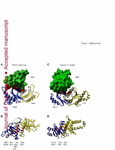
Figure 1. Model of Mon1-Ccz1 longin domains in complex with the Rab Ypt7
(A, B) Structure of yeast TRAPP subunits Trs23 (PDB ID 3CUEa) and Bet5 (PDB ID
3CUEc). Residues forming Ypt1-binding surface are indicated in red (Cai et al., 2008). In (B),
the Trs23-Bet5 segment is turned by 90°, while Ypt1 is not shown. Residues of the Ypt1-
binding surface are listed below the structure.
Jour
nal o
f Cel
l Sci
ence
Acc
epte
d m
anus
crip
t

23
(C, D) Modeling of predicted Mon1 and Ccz1 longin domains was based in the structure of
Trs23 and Bet5 (YASARA). Residues within Mon1 and Ccz1 longin domains, which could
participate in the interaction with Ypt7-GDP (Constantinescu et al., 2002) are shown in red.
Representation of the putative Rab-GEF interface is shown as in (B), and postulated
interacting residues are listed below the model.
Figure 2. Identification of mutants that affect Mon1-Ccz1 GEF activity.
(A, B, C) Analysis of vacuole morphology in Mon1 (A, C) and Ccz1 (B) mutants. Vacuoles
were stained with FM4-64 in cells overexpressing wild-type (wt) or Mon1/Ccz1 mutants.
Mutations were generated based on TRAPPI GEF mutants (Cai et al., 2008) in S1-S2 and H1
regions of Mon1 and Ccz1 longin domains. In (C), wild-type and mutant Mon1 was expressed
from the NOP1 promoter to reduce expression. Scale bar 5µm.
(D, E) Localizations of GFP-tagged Mon1 and Ccz1 mutants were analyzed by fluorescence
microscopy. Both mutants remained associated with endosomes as observed for wild-type
(wt) proteins. Scale bar 5µm.
(F) Analysis of Mon1-Ccz1 interaction with Ypt7 by yeast three-hybrid. Plasmids coding for
Ypt7, Mon1 and Ccz1 were cotransformed in PJ69-4A strain and protein interaction was
detected by cell growth in selective plates (SD-leu-trp-ura-his-ade).
Figure 3. Mon1-Ccz1 and TRAPPI GEF complexes may operate in a similar mode.
(A) Localization of mCherry-tagged Ypt7 is perturbed by mutations of Mon1-Ccz1 as
observed in GEF deletion strains. Ypt7 was detected by fluorescence microscopy. Size bar, 5
µm.
(B) Mon1-Ccz1 wild-type (wt) and mutant complexes were isolated using tandem-affinity
purification (see Methods), resolved by SDS-PAGE, and visualized by Coomassie-staining.
Jour
nal o
f Cel
l Sci
ence
Acc
epte
d m
anus
crip
t

24
(C) Mon1 and Ccz1 mutants cannot counteract GAP activity. Vacuoles from the two tester
strains were treated with the GAP Gyp1 and Mon1-Ccz1 complex where indicated. Vacuole
fusion was detected after 90 min at 26°C in a colorimetric enzyme assay
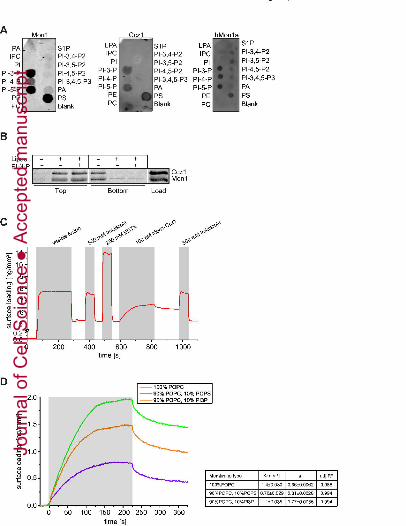
Figure 4. Mon1-Ccz1 displays preference for binding to PS and PI-3-P lipids.
(A) Mon1-Ccz1 binding to lipids. Purified yeast Mon1 and Ccz1 proteins, as well as human
Mon1a, which was purified as described (Gerondopoulos et al., 2012), were incubated with
lipids strips either for one hour at room temperature (yeast Mon1 and Ccz1) or at 4°C
overnight (for hMon1a) and binding was detected by immunoblot. Similar results were
obtained with hCcz1a (not shown).
(B) Mon1-Ccz1 associates with liposomes independently of the presence of PI-3-P. Binding
of purified yeast Mon1-Ccz1 complex to liposomes with or without PI-3-P was monitored by
flotation.
(C, D) Real-time and label-free detection of Mon1-Ccz1 binding to lipid membranes. (C)
Typical assay including fusion of small unilamellar lipid vesicles onto the transducer surface,
conditioning of the resulting membrane, binding of purified Mon1-Ccz1 complex and elution
with imidazole. The grey bars mark the injection periods. (D) Comparison of Mon1-Ccz1
binding to membranes with different lipid compositions as indicated in (C) (see methods for
details). Kd1 was calculated after normalization and fitting the dissociation phase to a double
exponential. A table with the fitting values is shown to the right.
Figure 5. Mon1-Ccz1 GEF activity is stimulated by PS and PI-3-P lipids.
500 pmoles of Ypt7-His loaded with MANT-GDP were used in all experiments. Lipid
concentration is indicated in mol% in brackets. Mon1-Ccz1 concentrations are indicated in
each figure legend.
Jour
nal o
f Cel
l Sci
ence
Acc
epte
d m
anus
crip
t

25
(A) GEF activity of Mon1-Ccz1 towards Ypt7 is increased in the presence of POPC
(52.5):DOGS-NTA(30):PS(10):PI-3-P(7.5) vesicles. 500 pmoles of Mon1-Ccz1 complex was
used.
(B) Titration of Mon1-Ccz1 complex in the GEF assay containing POPC (67.5):DOGS-NTA
(15):PS(10):PI-3-P(7.5) vesicles. Mon1-Ccz1 complex was used at the indicated
concentration.
(C) Vesicles stimulate Mon1-Ccz1 GEF activity only if they contain bound Ypt7. POPC
(67.5): DOGS-NTA(15):PS(10):PI-3-P(7.5) and POPC (82.5) PS(10):PI-3-P(7.5) vesicles
were used in the presence of 25 pmoles of Mon1-Ccz1 complex.
(D) Rab5-GEF Vps9 and Mon1 G209W, T213K-Ccz1 mutant complex are inactive towards
membrane-bound Ypt7. 25 pmoles or Vps9 or Mon1 G209W T213K-Ccz1 complex were
added. POPC (67.5):DOGS-NTA(15):PS(10):PI-3-P(7.5) vesicles were used in this assay.
(E) Complete GEF stimulation requires PS and PI-3-P lipids. GEF activity of Mon1-Ccz1 was
compared in vesicles of different composition, POPC(85):DOGS-NTA(15), POPC
(67.5):DOGS-NTA(15):PS(10):PI-3-P(7.5) and POPC(77):DOGS-NTA(15):PS(5):PI-3-P(3).
Mon1-Ccz1 concentration was 25 pmoles.
(F) PS and PI-3-P contribution to Mon1-Ccz1 recruitment and activation. Where indicated, 25
pmoles of Mon1-Ccz1 complex carrying a His-tag were added. POPC(85):DOGS-NTA(15)
and POPC (67.5):DOGS-NTA(15):PS(10):PI-3-P(7.5) vesicles were used in the GEF assay.
(G) Excess of HOPS relocalizes Mon1 and Ccz1 to the vacuole membranes. Distribution of
Mon1 and Ccz1 was analyzed in wild-type (wt) and cells overexpressing all HOPS subunits
by fluorescence microscopy.
(H) Mon1-Ccz1 GEF activity was slightly enhanced by the HOPS complex. GEF assay
included 500 pmoles of Ypt7-His loaded with MANT-GDP, 25 pmoles Mon1-Ccz1 complex,
HOPS complex at the indicated concentration and POPC(85):DOGS-NTA(15) vesicles.
Jour
nal o
f Cel
l Sci
ence
Acc
epte
d m
anus
crip
t

26
(I) Estimation of Mon1-Ccz1 GEF activity. kcat/Km was calculated after fitting the nucleotide
release curves to a double exponential curve.
Jour
nal o
f Cel
l Sci
ence
Acc
epte
d m
anus
crip
t
Jour
nal o
f Cel
l Sci
ence
Acc
epte
d m
anus
crip
t

Jour
nal o
f Cel
l Sci
ence
Acc
epte
d m
anus
crip
t

Jour
nal o
f Cel
l Sci
ence
Acc
epte
d m
anus
crip
t
Jour
nal o
f Cel
l Sci
ence
Acc
epte
d m
anus
crip
t

Jour
nal o
f Cel
l Sci
ence
Acc
epte
d m
anus
crip
t