Embed Size (px)
Citation preview

BioMed Central
Journal of Cardiovascular Magnetic Resonance
ss
Open AcceCase reportCardiovascular magnetic resonance of myocardial infarction after blunt chest trauma: a heartbreaking soccer-shotHannibal Baccouche*, Torsten Beck, Martin Maunz, Peter Fogarassy and Martin BeyerAddress: Medizinische Klinik II, Klinikum Kirchheim unter Teck, Kreiskliniken Esslingen, Germany
Email: Hannibal Baccouche* - [email protected]; Torsten Beck - [email protected]; Martin Maunz - [email protected]; Peter Fogarassy - [email protected]; Martin Beyer - [email protected]
* Corresponding author
AbstractCardiac injury occasionally occurs as a result of blunt chest trauma. Most cardiac complications inchest trauma are due to myocardial contusion rather than direct damage to the coronary arteries.However, traumatic coronary injury has been reported, and a variety of underlyingpathophysiological mechanisms have been proposed. We present a 26 year old patient presentingwith an acute coronary syndrome as a consequence of a soccer-shot impact to the chest. CMRshowed apical inferior infarction, as well as multiple small septal lesions which were presumed tohave resulted from embolization. The culprit lesion was a proximal 75% LAD stenosis with aprominent plaque-rupture and thrombus-formation, and the distal LAD was occluded bythromboembolic material.
BackgroundIn blunt chest trauma patients, it is important to considermyocardial injury, which is mostly the result of myocar-dial contusion. The consequences of contusion includeECG-changes, arrhythmia and necrotic damage of theheart muscle. Direct damage to the coronary arteries how-ever is a rare finding. Data published on the prevalence ofcardiac affection in chest trauma and its further sub-cate-gorization is sparse. Maenza and colleagues performed ameta-analysis on cardiac complications in blunt cardiactrauma including more than 4600 patients. The preva-lence of cardiac complications varied between 2.6% and4.5%. Myocardial infarction or complications assuminginfarction occurred only in a minority of the patients(between 5 and 7%) [1]. Christensen et al. identified 77published cases of acute myocardial infarction in bluntchest trauma. The LAD was the most commonly affected
vessel, followed by the RCA and RCX. Mechanisms of cor-onary-damage included dissection, plaque-rupture withthrombosis and/or thromboembolism, spasm, vessel rup-ture and epicardial hematoma with external compression.Patients suffering from a traumatic injury of a coronaryartery were considerably younger than the normal age ofpresentation of coronary disease (> 80% were less than 45years old). The most common trauma causing myocardialinfarction came from road traffic accidents, followed bysporting accidents including soccer [2].
The time interval from injury to coronary vessel occlusionshowed a highly variable course reaching from immediateonset to a delay of several weeks [3]. Diagnosticapproaches for cardiac involvement in chest traumapatients comprise cardiac biomarkers (creatine kinase andtroponin), ECG-testing and echocardiography. Computed
Published: 11 October 2009
Journal of Cardiovascular Magnetic Resonance 2009, 11:39 doi:10.1186/1532-429X-11-39
Received: 7 June 2009Accepted: 11 October 2009
This article is available from: http://www.jcmr-online.com/content/11/1/39
© 2009 Baccouche et al; licensee BioMed Central Ltd. This is an Open Access article distributed under the terms of the Creative Commons Attribution License (http://creativecommons.org/licenses/by/2.0), which permits unrestricted use, distribution, and reproduction in any medium, provided the original work is properly cited.
Page 1 of 4(page number not for citation purposes)

Journal of Cardiovascular Magnetic Resonance 2009, 11:39 http://www.jcmr-online.com/content/11/1/39
tomography has been used both for visualizing a dam-aged coronary vessel and detecting regional myocardialperfusion defects [4]. CMR has been successfully used inchest trauma [5,6], although its exact role in assessing car-diac involvement has not yet been clearly defined.
Case presentationA 26 year old male patient was referred to our hospitalwith an acute coronary syndrome. He had experiencedblunt chest trauma while playing soccer, taking a highvelocity shot on the chest. The trauma raised an intenseepisode of retrosternal chest pain. After a short pain-freeinterval more intense chest-pain recurred leading to emer-gency hospital admission, after which acute inferior ST-segment-elevation myocardial infarction was diagnosed.(Figure 1, full motion images can be viewed at http://www.kk-es.de/A_HEARTBEAKING_SOCCER-SHOT/Figure1/index.html).
With the exception of a family history of myocardial inf-arction, no other cardiovascular risk factors were present.Furthermore, there was no evidence indicating that a viralprodrome suggestive of myocarditis was present.
Cardiac catheterization revealed single vessel-coronary-artery-disease with a proximal 75% left anterior descend-ing artery (LAD) stenosis showing prominent plaque rup-ture with thrombus formation. There wasthromboembolic distal LAD occlusion of the prominentLAD vessel, which was also supplying the apical inferiorportion of the heart by wrapping around the LV apex,therefore leading to inferior infarction. (Figure 2, full
motion images can be viewed at http://www.kk-es.de/A_HEARTBEAKING_SOCCER-SHOT/Figure2/index.html).
Bare metal stenting of the proximal lesion was success-fully performed (no remaining stenosis). Due to the local-isation of distal thromboembolism, thrombus aspirationwas no option. Medical therapy with glycoprotein IIb/IIIa-inhibitor, clopidogrel, aspirin and heparin wasadministered and distal flow was able to be restored.Serial serum creatinekinase measurements revealed amaximum CK-rise to 1950 U/l.
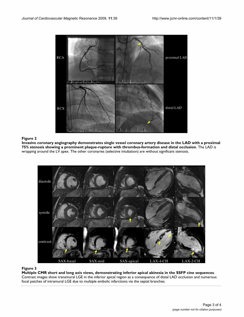
CMR was performed to assess myocardial viability (1.5 TAvanto, Siemens Medical Systems, Erlangen, Germany).Cine images were acquired using fast gradient echosteady-state free precession sequences (SSFP) demonstrat-ing inferior apical akinesia (Figure 3). Systolic left ven-tricular function was mildly impaired (LVEDV 166 ml,LVEF 58%). Ten minutes after injection of 0.2 mmol/kgGd-DTPA (Bayer Schering, Germany), CMR was per-formed using inversion-recovery-gradient-echo-tech-nique, adjusting inversion time to null normalmyocardium. Late gadolinium enhancement (LGE) waspresent in the inferior apical region of the heart withmainly transmural extent. Additional unexpected focalpatches of intramurally distributed LGE were also seen inthe interventricular septum, most likely correlating withmultiple small embolic infarctions via the septal branchesof the LAD. (Figure 3, full motion images can be viewed athttp://www.kk-es.de/A_HEARTBEAKING_SOCCER-SHOT/Figure3/index.html).
Twelve lead ECG demonstrating a classical picture of inferiorly located STEMI with ST-segment elevation in the inferior leads (II, III, aVF) and ST-segment depression in lead I and aVLFigure 1Twelve lead ECG demonstrating a classical picture of inferiorly located STEMI with ST-segment elevation in the inferior leads (II, III, aVF) and ST-segment depression in lead I and aVL.
I
II
III
aVR
aVL
aVF
V2
V1
V3
V4
V5
V6
Page 2 of 4(page number not for citation purposes)
Journal of Cardiovascular Magnetic Resonance 2009, 11:39 http://www.jcmr-online.com/content/11/1/39
Page 3 of 4(page number not for citation purposes)
Invasive coronary angiography demonstrates single vessel coronary artery disease in the LAD with a proximal 75% stenosis showing a prominent plaque-rupture with thrombus-formation and distal occlusionFigure 2Invasive coronary angiography demonstrates single vessel coronary artery disease in the LAD with a proximal 75% stenosis showing a prominent plaque-rupture with thrombus-formation and distal occlusion. The LAD is wrapping around the LV apex. The other coronaries (selective intubation) are without significant stenosis.
RCA
RCX distal LAD
proximal LAD
Multiple CMR short and long axis views, demonstrating inferior apical akinesia in the SSFP cine sequencesFigure 3Multiple CMR short and long axis views, demonstrating inferior apical akinesia in the SSFP cine sequences. Contrast images show transmural LGE in the inferior apical region as a consequence of distal LAD occlusion and numerous focal patches of intramural LGE due to multiple embolic infarctions via the septal branches.
diastole
systole
contrast
SAX-basal SAX-mid SAX-apical LAX-4-CH LAX-2-CH

Journal of Cardiovascular Magnetic Resonance 2009, 11:39 http://www.jcmr-online.com/content/11/1/39
Publish with BioMed Central and every scientist can read your work free of charge
"BioMed Central will be the most significant development for disseminating the results of biomedical research in our lifetime."
Sir Paul Nurse, Cancer Research UK
Your research papers will be:
available free of charge to the entire biomedical community
peer reviewed and published immediately upon acceptance
cited in PubMed and archived on PubMed Central
yours — you keep the copyright
Submit your manuscript here:http://www.biomedcentral.com/info/publishing_adv.asp
BioMedcentral
The further clinical course of the patient was uneventful.At 2 months of follow up, CMR showed normal global leftventricular function and the myocardial scarring hadclearly shrunk. The patient was not suffering from any car-diac symptoms and was able to enjoy sports again withoutlimitations.
ConclusionAcute myocardial infarction is a rare entity in chesttrauma. It may result from contusion or damage to thecoronary arteries. In the latter, plaque rupture, thrombusformation, coronary artery dissection, focal spasm andother rare mechanisms have to be considered. As clinicalmanagement changes if coronary perfusion is impaired, itis crucial to identify those patients in need for invasiveworkup including cardiac catheterization und angi-oplasty. Diagnostic standard approaches to cardiac injuryin chest trauma patients by ECG, lab-testing and echocar-diography can be supplemented by tomographic imagingas demonstrated by CMR.
ConsentWritten informed consent was obtained from the patientfor publication of this case report and any accompanyingimages. A copy of the written consent is available forreview by the Editor-in-Chief of this journal.
Competing interestsThe authors declare that they have no competing interests.
Authors' contributionsHB conceived and designed this case report, was involvedin data acquisition (performed the CMR scan) and inter-pretation and drafted both the text and figure file. TBdesigned this case report, was involved in data acquisition(performed cardiac-catheterization and PCI on thepatient), carried out literature review and drafted theimage file. MM was involved in data acquisition (cardiaccatheterization, literature) and clinical management ofthe patient, made contributions to drafting both text andimage files and the webpage and critically reviewed thedocuments. PF was involved in data acquisition, interpre-tation and patient management, contributed to draftingof text and images, designed the webpage and criticallyreviewed the documents. MB was involved in data acqui-sition and interpretation and contributed to drafting oftext, images and webpage and critically reviewed all docu-ments. MB supervised the clinical management of thepatient and finally this case report. All authors takeresponsibility for the entire content of this report andhave read and approved the submission of this manu-script.
References1. Maenza RL, Seaberg D, D'Amico F: A meta-analysis of blunt car-
diac trauma: ending myocardial confusion. Am J Emerg Med1996, 14:237-241.
2. Christensen MD, Nielsen PE, Sleight P: Prior blunt chest traumamay be a cause of single vessel coronary disease; hypothesisand review. Int J Cardiol 2006, 108:1-5.
3. Bjornstad JL, Pillgram-Larsen J, Tonnessen T: Coronary artery dis-section and acute myocardial infarction following blunt chesttrauma. World J Emerg Surg 2009, 4:14.
4. Oghlakian G, Maldjian P, Kaluski E, Saric M: Acute myocardial inf-arction due to left anterior descending coronary artery dis-section after blunt chest trauma. Emerg Radiol 2009 in press.
5. Mohrs OK, Magedanz A, Schlosser T: Extensive myocardial inf-arction and left ventricular thrombus after chest collisionduring a soccer match. Clin Cardiol 2006, 29:547-548.
6. Southam S, Jutila C, Ketai L: Contrast-enhanced cardiac MRI inblunt chest trauma: differentiating cardiac contusion fromacute peri-traumatic myocardial infarction. J Thorac Imaging2006, 21:176-178.
Page 4 of 4(page number not for citation purposes)