Embed Size (px)
Citation preview
BioMed Central
Journal of Cardiovascular Magnetic Resonance
ss
Open AcceResearchA dual propagation contours technique for semi-automated assessment of systolic and diastolic cardiac function by CMRWei Feng1, Hosakote Nagaraj2, Himanshu Gupta2, Steven G Lloyd2, Inmaculada Aban3, Gilbert J Perry2, David A Calhoun2, Louis J Dell'Italia2 and Thomas S Denney Jr*1Address: 1Electrical and Computer Engineering Department, Auburn University, Auburn, AL 36849, USA, 2Division of Cardiovascular Disease, University of Alabama at Birmingham, Birmingham, AL 35294, USA and 3Department of Biostatistics, University of Alabama at Birmingham, Birmingham, AL 35294, USA
Email: Wei Feng - [email protected]; Hosakote Nagaraj - [email protected]; Himanshu Gupta - [email protected]; Steven G Lloyd - [email protected]; Inmaculada Aban - [email protected]; Gilbert J Perry - [email protected]; David A Calhoun - [email protected]; Louis J Dell'Italia - Dell'[email protected]; Thomas S Denney* - [email protected]
* Corresponding author
AbstractBackground: Although cardiovascular magnetic resonance (CMR) is frequently performed to measure accurate LVvolumes and ejection fractions, LV volume-time curves (VTC) derived ejection and filling rates are not routinelycalculated due to lack of robust LV segmentation techniques. VTC derived peak filling rates can be used to accuratelyassess LV diastolic function, an important clinical parameter. We developed a novel geometry-independent dual-contourpropagation technique, making use of LV endocardial contours manually drawn at end systole and end diastole, tocompute VTC and measured LV ejection and filling rates in hypertensive patients and normal volunteers.
Methods: 39 normal volunteers and 49 hypertensive patients underwent CMR. LV contours were manually drawn onall time frames in 18 normal volunteers. The dual-contour propagation algorithm was used to propagate contoursthroughout the cardiac cycle. The results were compared to those obtained with single-contour propagation (usingeither end-diastolic or end-systolic contours) and commercially available software. We then used the dual-contourpropagation technique to measure peak ejection rate (PER) and peak early diastolic and late diastolic filling rates (ePFRand aPFR) in all normal volunteers and hypertensive patients.
Results: Compared to single-contour propagation methods and the commercial method, VTC by dual-contourpropagation showed significantly better agreement with manually-derived VTC. Ejection and filling rates by dual-contourpropagation agreed with manual (dual-contour – manual PER: -0.12 ± 0.08; ePFR: -0.07 ± 0.07; aPFR: 0.06 ± 0.03 EDV/s,all P = NS). However, the time for the manual method was ~4 hours per study versus ~7 minutes for dual-contourpropagation. LV systolic function measured by LVEF and PER did not differ between normal volunteers and hypertensivepatients. However, ePFR was lower in hypertensive patients vs. normal volunteers, while aPFR was higher, indicative ofaltered diastolic filling rates in hypertensive patients.
Conclusion: Dual-propagated contours can accurately measure both systolic and diastolic volumetric indices that canbe applied in a routine clinical CMR environment. With dual-contour propagation, the user interaction that is routinelyperformed to measure LVEF is leveraged to obtain additional clinically relevant parameters.
Published: 13 August 2009
Journal of Cardiovascular Magnetic Resonance 2009, 11:30 doi:10.1186/1532-429X-11-30
Received: 26 March 2009Accepted: 13 August 2009
This article is available from: http://www.jcmr-online.com/content/11/1/30
© 2009 Feng et al; licensee BioMed Central Ltd. This is an Open Access article distributed under the terms of the Creative Commons Attribution License (http://creativecommons.org/licenses/by/2.0), which permits unrestricted use, distribution, and reproduction in any medium, provided the original work is properly cited.
Page 1 of 13(page number not for citation purposes)

Journal of Cardiovascular Magnetic Resonance 2009, 11:30 http://www.jcmr-online.com/content/11/1/30
BackgroundTemporal changes in left ventricular (LV) volume over thecardiac cycle provides fundamental information regardingsystolic and diastolic function of the heart but is difficultto measure by standard clinical techniques. Cine cardio-vascular magnetic resonance (CMR) using serial short axisslices is well accepted as a gold standard for measuringgeometry-independent ventricular volumes [1,2]. Meas-urement of LV ED and ES volumes is based on drawingcontours at the ED and ES time points. If contours couldbe reliably identified in all acquired time frames, ventricu-lar volume-time curves (VTC) could be constructed, fromwhich important parameters of ventricular function suchas peak ejection rates (PER) and peak filling rates (PFR)[3,4] can be derived. These parameters may complementflow indices [5] in the assessment of diastolic function.
Fully automated contouring techniques have been aresearch topic for many years [6-15], and, more recently,techniques have been developed for propagating contoursdrawn at single time frame to the remaining time frames[16-22]. While the accuracy of these methods continues toimprove, contour review and editing by a trained expert isstill mandatory. A common problem encountered inmyocardial contour identification is the presence of pap-illary muscles; following the echocardiographic conven-tion, papillary muscles are often excluded from theendocardial contour. At ED in the short axis CMR image,papillary muscles are usually not a problem because theyare separated from the LV wall. During systole, however,the papillary muscles move close to the LV wall, and it canbe difficult to distinguish papillary muscles from the heartwall without carefully examining the images. For this rea-son, fully automatic contouring routines often have diffi-culty detecting papillary muscles, and may includepapillary muscle volume as part of the LV cavity volumein ED and as outside the LV cavity (i.e., in the myocardialmuscle volume) in ES. This potentially affects the derivedvolumes and masses.
Consequently, we propose a semi-automated methodwhich leverages the user interaction in drawing ED and EScontours by automatically propagating them to all othertime frames in a typical cardiac scan. This dual-contourpropagation technique has the potential to more accu-rately exclude papillary muscles from the LV wall than sin-gle-contour propagation techniques or fully automatedtechniques. The proposed dual-contour propagation tech-nique will not require additional work by the user,because at most institutions contours are already rou-tinely drawn at ED and ES to compute standard volumes,myocardial mass, and ejection fraction. The purpose ofthis study was to develop a novel semi-automated tech-nique using dual-contour propagation to measure ven-tricular volumes throughout the cardiac cycle, and
compare this method to manual and single-contour tech-niques in normal volunteers and hypertensive patients.
MethodsSubjectsThe study was approved by the appropriate institutionalreview boards and informed consent was obtained fromall the participants. 39 normal human volunteers (NLs)and 49 hypertensive (HTN) patients consecutivelyenrolled in a study of resistant hypertension (defined asrequiring 3 or more anti-hypertensive medications toachieve blood pressure < 140/90 mmHg) participated inthis study. All patients were in sinus rhythm at the time ofCMR.
Image AcquisitionCMR was performed on a 1.5-T scanner (CV/i, GE Health-care, Milwaukee, WI) optimized for cardiac application.ECG-gated, breath-hold steady state free precision tech-nique was used to obtain standard (2, 3 and 4 Chamber,Short Axis) views using the following parameters – slicethickness 8 mm with no gap between short-axis slices,field-of-view 44 × 44 cm, scan matrix 256 × 128, flip angle45°, typical TR/TE = 3.8/1.6 ms; typical acquired temporalresolution approximately 40 ms); data reconstructed to 20cardiac phases.
Image AnalysisIn all scans, LVED and LVES endocardial contours weremanually drawn on all short axis slices between the mitralannulus and apex [23] with exclusion of the papillarymuscles. These contours were then automatically propa-gated to all the other frames in the acquisition using thedual-contour propagation algorithm described below. Forvalidation, LV contours were manually drawn on all timeframes in 18 randomly-selected normal scans by a Level 3trained CMR specialist. These contours were used as a goldstandard for evaluating and validating the dual-contourpropagation algorithm.
Contour PropagationNon-rigid registration (NRR) [24] was used to propagatethe contours manually drawn at end-diastole and end-sys-tole to all other time frames in the acquisition. The NRRalgorithm computed a deformation field that warped asource image to fit a template image. The deformationfield was then used to propagate contours defined on thetemplate image to the source image. Details of this algo-rithm are provided in the Appendix. All algorithms wereimplemented in MATLAB (The Mathworks, Natick, MA).
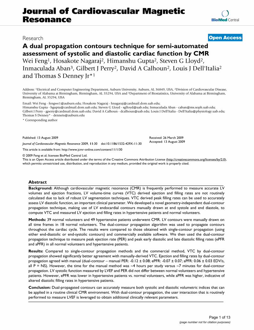
The dual-contour propagation scheme shown in Figure 1was used to propagate both ED and ES contours to allother time frames in the sequence. First, the NRR algo-rithm was used to propagate ED contours forward in time
Page 2 of 13(page number not for citation purposes)

Journal of Cardiovascular Magnetic Resonance 2009, 11:30 http://www.jcmr-online.com/content/11/1/30
through systole and backward in time through diastole(white arrows in Figure 1). Next, ES contours were propa-gated forward in time through diastole and backward intime through systole (gray arrows in Figure 1). The con-tours propagated from ED and ES were then combined, asdescribed in the Appendix, into a single set of endocardialand epicardial contours.
Volumetric AnalysisThe LV volume at each time frame was computed by sum-ming the volumes defined by the contours in each slice.The contour propagation procedure, however, propagatedcontours in all slices that were contoured at ED, and, nearthe base, the LV margin may have moved through theimage plane in systole. To address this problem, the NRRalgorithm was used in a long-axis slice to track a user-selected point near the mitral annulus through the image
sequence. The displacement of this point was used todetermine how much each short-axis slice should beincluded in the volume computation [4,25]. For example,if the mitral annulus displaced 12 mm between ED andthe current time frame and the slice thickness was 8 mm,the most basal slice would not be used in the volumecomputation and 50% of the second-most-basal slice vol-ume would be used in the total volume. This mitral annu-lus tracking procedure was used to compute volumes forall the contouring techniques discussed in this paper,including manual contouring and automatic contourpropagation algorithms.
Once the volumes were computed in each time frame, aVTC curve was constructed and differentiated with respectto time. End-diastole was defined as the maximum-vol-ume time frame, and end-systole was defined as the min-
Contour propagation strategyFigure 1Contour propagation strategy. Manually-drawn contours at ED (top-left) and ES (2nd row and 2nd column) were propa-gated to other time frames and combined.
Page 3 of 13(page number not for citation purposes)

Journal of Cardiovascular Magnetic Resonance 2009, 11:30 http://www.jcmr-online.com/content/11/1/30
imum-volume time frame. Early diastole and late diastolewere defined as the first and second halves respectively ofthe diastolic interval. The peak ejection rate (PER) wasdefined as the maximum negative time derivative duringthe systolic interval. The early diastolic and late diastolicpeak filling rates (ePFR and aPFR) were defined as themaximum derivative during the early and late diastole.
Comparison Between Single and Dual-Contour PropagationMost existing contour propagation techniques propagatecontours from either ED or ES time frames [8,16,20-22,26]. Volumes computed from dual-propagated con-tours, single-propagated contours from ED and ES usingthe NRR method described above, and single-propagatedcontours from ED and ES using CAAS MRV for Windows,version 3.2 (Pie Medical Imaging, Maastricht, the Nether-lands), software were compared to volumes computedfrom manual contours on nine randomly-selected normalstudies (identified by NV1-NV9). A VTC was computedfor each type of contours for each study. To compare VTCscomputed from different types of contours, differenceswere computed at each time point by subtracting the man-ual volume from the propagated volumes.
Inter-User VariabilityTo assess inter-user variability in volumes computed frompropagated contours, nine normal studies (NV10-NV18)were randomly selected. For each study, two sets of con-tours were manually drawn at ED and ES by differentusers. Each user was a Level 3 trained CMR specialist orequivalent. Each set of ED and ES contours was propa-gated using the dual contour technique, and VTCs andejection/filling rates were computed.
LV Mass EvaluationTo evaluate stability of dual-propagated contoursthroughout the cardiac cycle, LV mass measurements werecomputed in each time frame for studies NV10-NV18from dual-propagated contours and compared to thatfrom one set of manually-drawn contours.
Comparison of PER and PFR Values in Normals and HypertensivesThe dual-contour propagation algorithm was used topropagate contours to all time frames and compute VTCsand ejection/filling rates in all 39 normals and 49 hyper-tensives.
Statistical AnalysisComparisons of LV volumes computed from differentpropagation schemes and comparisons of LV masses com-puted from dual-propagated and manual contours wereperformed using mixed modeling via PROC MIXED (SASversion 9.1). To account for the repeated measures within
a subject, a compound symmetry correlation structure wasassumed. In the LV volume study, confidence intervals onthe differences based on the fitted mixed model were con-structed each at 99% level to achieve a joint confidencelevel of at least 95% for this set of confidence intervalsusing Bonferroni adjustment [27].
Comparisons of PER and PFR values computed fromdual-propagated and manually-drawn contours and com-parisons of contours propagated by two different userswere performed using two-tailed paired t-tests, correlationanalysis, and Bland-Altman analysis. PER and PFR valuesderived from dual-propagated contours in hypertensivepatients were compared to normals using unpaired t-tests.In all these statistical tests, a P-value less than 5% was con-sidered statistically significant.
ResultsComparison Between Single and Dual-Contour PropagationDifferences between propagated contours and manualcontours resulted in differences in VTCs. Figure 2 showsVTCs from a normal volunteer. ED-propagated contourswith NRR resulted in volume overestimation near ES, andES-propagated contours with NRR produced volumeunderestimation in early systole and late diastole. Dual-propagated contours showed excellent agreementthroughout the entire cardiac cycle. Both ED and ES prop-agated contours using CAAS MRV underestimated the vol-umes as compared to the manually drawn, gold standardvolumes, more than NRR propagated contours through-out the cardiac cycle; the CAAS MRV propagation methodalso changes the manually-drawn ED and ES contoursslightly, so the volume difference is not zero at ED or ESin these curves.
Table 1 shows confidence intervals of the volume differ-ences between each propagation method and manual.ED-propagated contours with NRR overestimate LV vol-ume, whereas ES-propagated contours with NRR underes-timate LV volume. In comparison, both ED-propagatedand ES-propagated contours with CAAS MRV underesti-mate LV volume by a larger margin. However, the dual-propagated volumes were not statistically different frommanually-contoured volumes.
The average computation time for dual-contour propaga-tion (not including manually contouring the ED and EScontours) was 7.3 minutes for a single study on a 2.6 GHzdual-core personal computer with 4 GB of RAM. Auto-mated contour propagation using CAAS MRV requiredless than 1 minute per study. Manual contouring of allslices and phases (typically 12 to 14 short axis slices × 20cardiac phases) required approximately 4 hours per study.
Page 4 of 13(page number not for citation purposes)

Journal of Cardiovascular Magnetic Resonance 2009, 11:30 http://www.jcmr-online.com/content/11/1/30
Validation of Functional ParametersFigure 3 shows the VTCs in normal volunteers derivedfrom the manually-drawn contours and dual-propagatedcontours. The manual and dual-propagated VTCs werequite close to each other in all nine studies – particularlyduring systole and early diastole. This similarity betweenmanual and propagated VTCs means that the contoursmanually drawn at ED and ES were consistently propa-gated to the other time frames in the cine sequence.
No statistically-significant differences were foundbetween PER, ePFR and aPFR rates computed from manu-ally-drawn contours and dual-propagated contours (Table2). The correlation coefficients between the PER, ePFR andaPFR values were 0.92, 0.95 and 0.96 respectively (all P <0.001). Figure 4 shows scatter and Bland-Altman plotscomparing the manual and dual-propagated measure-ments of ejection and filling rates.
Inter-User VariabilityNo significant difference was found between PER, ePFRand aPFR values computed from contours propagatedwith ED and ES contours drawn by two different users(User1 and User2). The differences (User2-User1)
between PER, ePFR and aPFR values were 0.07 ± 0.16EDV/s (P = 0.24), -0.03 ± 0.05 EDV/s (P = 0.11) and -0.01± 0.05 EDV/s (P = 0.50) respectively. The correlation coef-ficients for the PER, ePFR and aPFR values were 0.95 (P <0.0001), 0.99 (P < 0.0001) and 0.99 (P < 0.0001) respec-tively. Figure 5 shows scatter and Bland-Altman plotscomparing User1 and User2 measurements of PER, ePFRand aPFR.
LV Mass EvaluationNo significant difference was found between normalizedLV masses (LV mass/ED LV mass) computed from dual-propagated contours and manual contours. The normal-ized LV mass difference (propagated-manual) was -0.015± 0.0077 (P = 0.08). Figure 6 shows the mean normalizedLV mass throughout the cardiac cycle averaged over stud-ies NV10-NV18. The normalized LV mass computed fromdual-propagated contours was close to that from the man-ual contours and both remain stable throughout the car-diac cycle, indicating that the propagated contours were asstable as the manual contours.
LV-volume-versus-time curves for a normal human volunteerFigure 2LV-volume-versus-time curves for a normal human volunteer. They were computed from different sets of contours: manually-drawn contours in each time frame (red), contours propagated from the manual ED contours using NRR (green), contours propagated from the manual ES contours using NRR (cyan), contours propagated from both ED and ES contours using NRR (blue), contours propagated from manual ED contours using CAAS MRV (black), and contours propagated from manual ES contours using CAAS MRV (magenta).
10 20 30 40 50 60 70 80 90 100
30
40
50
60
70
80
90 NV4
LV V
olum
e (m
l)
% R−R Interval
ManualDualEDESCAAS MRV EDCAAS MRV ES
Page 5 of 13(page number not for citation purposes)

Journal of Cardiovascular Magnetic Resonance 2009, 11:30 http://www.jcmr-online.com/content/11/1/30
Page 6 of 13(page number not for citation purposes)
Table 1: Differences between LV volumes (expressed as fraction of EDV) computed from propagated contours and manually-drawn contours.
Volume Difference (EDV)
Mean ± SE 99% Confidence Interval P
Dual NRR – Manual -0.19 ± 0.56 -1.74 1.35 0.7316ED NRR – Manual 1.61 ± 0.56 0.08 3.14 0.0069ES NRR – Manual -3.50 ± 0.56 -5.03 -1.97 <0.0001ED CAAS – Manual -6.54 ± 0.55 -8.06 -5.02 <0.0001ES CAAS – Manual -11.05 ± 0.55 -12.57 -9.54 <0.0001
SE = standard error.
LV-volume-versus-time curves for nine normal human volunteersFigure 3LV-volume-versus-time curves for nine normal human volunteers. They were computed from two different sets of contours: manually-drawn contours in each time frame (red) and dual-propagated contours (blue).
20 40 60 80 10050
100
150NV10
LV V
olum
e (m
l)
20 40 60 80 100
60
80
100
120
NV11
20 40 60 80 10050
100
150
NV12
20 40 60 80 100
60
80
100 NV13
LV V
olum
e (m
l)
20 40 60 80 100
60
80
100
120
140NV14
20 40 60 80 100
60
80
100
120NV15
20 40 60 80 10050
100
150 NV16
LV V
olum
e (m
l)
% R−R Interval20 40 60 80 100
40
60
80
100
120 NV17
% R−R Interval20 40 60 80 100
40
60
80
100 NV18
% R−R Interval

Journal of Cardiovascular Magnetic Resonance 2009, 11:30 http://www.jcmr-online.com/content/11/1/30
Peak Ejection and Filling Rates in HTNFigure 7 shows typical VTCs for a normal volunteer and ahypertensive patient measured from dual-propagatedcontours. Peak ejection rates are similar in both curves,but the early diastolic filling rate is lower in the hyperten-sive patient than in the normal while the late filling rate ishigher.
Figure 8 shows the mean peak ejection and filling ratesmeasured from dual-propagated contours in all 39 nor-mals and 49 patients with hypertension. In hypertensives,PER was not different from normal (3.4 ± 0.1 vs. 3.2 ± 0.1EDV/sec, P = NS). Diastolic filling rates, however, werealtered compared to normals, demonstrating diastolicdysfunction in hypertension that is common in thispatient group: ePFR was lower than normal (2.6 ± 0.1 vs.3.2 ± 0.1 EDV/sec, P < 0.0001), but aPFR was higher thannormal (2.4 ± 0.1 vs. 1.6 ± 0.1 EDV/sec, P < 0.0001).
DiscussionIn this paper, we described a novel dual-contour propaga-tion technique for measuring volume-time curves (VTCs),validated it against manually drawn contours, and dem-onstrated its utility in a clinically-relevant patient popula-tion. This method requires nothing more than standardshort-axis and long-axis CMR acquisitions and routinelydrawn ED and ES contours. We show that the dual-prop-agated contours can be used to accurately measure peakejection and filling rates compared to the reference stand-ard of manually-drawn contours. The dual-contour prop-agation technique provides a fast, practical means ofmeasuring volume-based indices of systolic and diastolicventricular function from routine clinical CMR.
Several techniques have been proposed for propagatingcontours in CMR [16-22] and other modalities [28-33],but these techniques only propagate contours from a sin-gle time frame. While propagating contours from onlyone time frame requires less user interaction, we foundthat the resulting volumes are less accurate compared todual propagation. If contours are only defined at ED,propagated contours with NRR may not be able to sepa-
rate papillary muscles from the LV wall at ES resulting inthe volume differences at ES (Figure 2). Propagating onlyES contours with NRR may solve this problem, but vol-ume differences occur in late diastole as demonstrated inFigure 2.
van Guens, et al. [15] proposed an automated method fordrawing contours at ED and ES. The required user inputwas minimal – only four manually-drawn epicardial con-tours on two and four-chamber views at ED and ES – butvolumes were only validated at ED and ES. In addition,for registration purposes, this method requires that bothlong axis and multiple short-axis acquisitions be per-formed with reproducible breath-hold positions, whichcan sometimes be difficult to obtain under clinical condi-tions. The contour propagation method proposed in thispaper, however, does not have this limitation since con-tours are propagated in each slice independently.
Investigators have previously described use of volumetime indices for measuring systolic and diastolic function[34,35]. CMR allows measurement of ventricular volumesthroughout the cardiac cycle independent of geometricassumptions. The excellent spatial resolution and imagecontrast make it potentially the most accurate clinically-applicable non-invasive technique for assessment ofsystolic and diastolic function. To provide an illustrationof the utility of our propagation method in clinical assess-ment of patients with risk factors for heart failure, thedual-contour propagation technique was employed toassess the physiology of LV systolic and diastolic functionin 49 patients consecutively enrolled in a study of resistanthypertension. The images in this study contain the normalrange of image quality and presence of artifacts encoun-tered under routine clinical conditions. The concentricallyhypertrophied LV in the HTN patients had a normal LVejection fraction and LV peak ejection rate; however, earlypeak filling was decreased and late filling rate wasincreased, consistent with diastolic dysfunction.
The inter-user and intra-user variability in the propagatedcontours depend on the inter-user and intra-user variabil-ity of the semi-automatically-drawn contours at ED andES. This variability has been studied in [15,36,37]. In ourstudy, no significant difference was found between PER,ePFR and aPFR values computed from contours propa-gated from ED and ES contours drawn by two differentusers.
A limitation of contour propagation algorithms in generalis that any errors in the seed contours get propagated to allother time frames. Consequently, it is especially impor-tant to ensure accurate seed contours before propagation.Also in this paper, papillary muscle volume was consid-ered part of the LV blood volume. Since the papillary mus-
Table 2: Differences between peak ejection and filling rates computed from manually-drawn and dual-propagated contours.
Rate Difference (EDV/s)
Mean ± SE 95% Confidence Interval P
PER -0.12 ± 0.08 -0.29 0.06 0.16ePFR -0.07 ± 0.07 -0.23 0.08 0.31aPFR 0.06 ± 0.03 -0.02 0.13 0.11
Differences are dual-propagated minus manual.PER: peak ejection rate; ePFR: early diastolic filling rate; aPFR: late diastolic filling rate; SE: standard error.
Page 7 of 13(page number not for citation purposes)

Journal of Cardiovascular Magnetic Resonance 2009, 11:30 http://www.jcmr-online.com/content/11/1/30
Page 8 of 13(page number not for citation purposes)
Scatter and Bland-Altman plots of LV ejection and filling rates computed from manual contours (Manual) and dual-propagated contours (Prop)Figure 4Scatter and Bland-Altman plots of LV ejection and filling rates computed from manual contours (Manual) and dual-propagated contours (Prop). The plotted rates include peak ejection rate (PER) (a, b), early diastolic filling rate (ePFR) (c, d), and late diastolic filling rate (aPFR) (e, f) values in end-diastolic volumes (EDV)/sec. The dashed lines in Bland-Alt-man plots represent the mean and mean ± two standard deviations of the difference between Prop and Manual rates.
2 3 4
2
2.5
3
3.5
4
Manual PER
Prop
PER
2 3 4−0.6
−0.4
−0.2
0
0.2
0.4
(Manual+Prop)/2 PER
(Pro
p −
Man
ual)
PER
2 2.5 3 3.52
2.5
3
3.5
Manual ePFR
Prop
ePF
R
2 3 4−0.6
−0.4
−0.2
0
0.2
0.4
(Manual+Prop)/2 ePFR
(Pro
p −
Man
ual)
ePFR
0.5 1 1.5 2
1
1.5
2
Manual aPFR
Prop
aPF
R
0.5 1 1.5 2−0.2
0
0.2
0.4
0.6
(Manual+Prop)/2 aPFR
(Pro
p −
Man
ual)
aPFR
a b
c d
e f

Journal of Cardiovascular Magnetic Resonance 2009, 11:30 http://www.jcmr-online.com/content/11/1/30
Page 9 of 13(page number not for citation purposes)
Scatter and Bland-Altman plots of LV PER (a,b), ePFR (c,d), and aPFR (e,f) values in EDV/sec computed from contours manually drawn at ED and ES by two different users (User1 and User2) and propagatedFigure 5Scatter and Bland-Altman plots of LV PER (a,b), ePFR (c,d), and aPFR (e,f) values in EDV/sec computed from contours manually drawn at ED and ES by two different users (User1 and User2) and propagated.
2 2.5 3 3.5
2.5
3
3.5
User1 PER
Use
r2 P
ER
2 3 4−0.4
−0.2
0
0.2
0.4
0.6
(User1+User2)/2 PER
(Use
r2−
Use
r1) P
ER
2 2.5 3 3.5
2
2.5
3
3.5
User1 ePFR
Use
r2 e
PFR
2 3 4−0.15
−0.1
−0.05
0
0.05
0.1
(User1+User2)/2 ePFR
(Use
r2−
Use
r1) e
PFR
1 1.5 2
1
1.5
2
User1 aPFR
Use
r2 a
PFR
0.5 1 1.5 2−0.15
−0.1
−0.05
0
0.05
0.1
(User1+User2)/2 aPFR
(Use
r2−
Use
r1) a
PFR
a b
c d
e f

Journal of Cardiovascular Magnetic Resonance 2009, 11:30 http://www.jcmr-online.com/content/11/1/30
cle volume is relatively constant throughout the cardiaccycle, subtracting the papillary muscle volume wouldreduce the blood volume by the same amount in allphases and would not significantly affect filling or ejec-tion rates, which are the key parameters determined inthis work. Although not evaluated in the present study, webelieve that another potential advantage of the dual-con-
tour propagation approach is that defining contours attwo time points provides increased robustness to imagingartifacts.
In conclusion, the dual-contour propagation techniqueprovides a fast, accurate and practical means of measuringvolume-based indices of systolic and diastolic ventricularfunction from routine clinical CMR.
Competing interestsThe authors declare that they have no competing interests.
Authors' contributionsWF developed the dual-contour propagation algorithm,drew one set of contours for the inter-user variabilitystudy, and drafted the manuscript. HN performed manualcontouring of normal datasets including one set of con-tours for the inter-user variability analysis. HG partici-pated in the study design and helped draft the manuscript.SGL participated in the study design and helped draft themanuscript. IA participated in the study design and per-formed the statistical analysis. GJP helped draft the man-uscript. DAC participated in study coordination. LJDparticipated in study coordination and helped draft themanuscript. TSD conceived of the study, participated instudy design and coordination and helped draft the man-uscript. All authors read and approved the final manu-script.
AppendixNon-Rigid RegistrationFirst, a square region of interest (ROI) was defined thatenclosed epicardial contours in the template image. Sincethe NRR was applied sequentially starting from either EDor ES, an epicardial contour (either manually-drawn orpropagated) was always available in the template image.
Next, a two-dimensional displacement field was definedon the template ROI. The deformation field was parame-terized by a tensor product of quadratic, uniformly-spacedB-splines:
where pis a point in the template ROI, β(p) is a B-splinebasis function, μi is a control point, C is the number ofcontrol points and vi is the knot location associated withthe i-th control point. Eight control points were used ineach dimension. The B-spline order and number of con-trol points were determined empirically.
The B-spline control points, μ, were computed to mini-mize the sum of squared differences between the pixel
m p p v( ; )μμ μμ ββ= −( )=∑ i i
i
C
1
Mean LV mass (normalized to ED LV mass) at different phases of the cardiac cycle computed from manual contours and dual-propagated contours averaged over nine normal studiesFigure 6Mean LV mass (normalized to ED LV mass) at differ-ent phases of the cardiac cycle computed from man-ual contours and dual-propagated contours averaged over nine normal studies.
0 50 1000
0.85
1
% R−R Interval
Nor
mal
ized
LV
Mas
s
Manual
Dual
Volume versus time (VTC) plots computed from dual-propa-gated contours for a normal volunteer (solid) and a hyper-tensive patient (dashed)Figure 7Volume versus time (VTC) plots computed from dual-propagated contours for a normal volunteer (solid) and a hypertensive patient (dashed).
0 20 40 60 80 10030
40
50
60
70
80
90
100
110
%R-R Interval
LV V
olum
e (m
l)
Page 10 of 13(page number not for citation purposes)

Journal of Cardiovascular Magnetic Resonance 2009, 11:30 http://www.jcmr-online.com/content/11/1/30
intensities in the template image, It, and the source image,Is:
where Ω is the set of pixels in the template ROI.
The cost function in Eq. (1) was optimized using a multi-resolution strategy to speed up computation and avoidlocal minima in the cost function. First, the template ROIwas resampled from 64 × 64 to 16 × 16 pixels and thenumber of control points was reduced to four in each
dimension. The reduced resolution control points werethen computed using Levenberg optimization algorithmwith an analytical gradient and Hessian. This process wasthen repeated using template ROI resampled from 64 × 64to 32 × 32 pixels. The number of control points was keptat four in each dimension, and the result of the coarser res-olution optimization was used as the starting point for theoptimization. Next, the displacement field was interpo-lated to eight control points in each dimension, and Eq.(1) was optimized on the full-resolution 64 × 64 templateROI. The source image was resampled accordingly in eachmulti-resolution layer to match the resolution of the tem-plate ROI. The result was a spatially continuous mapping
J I It k s k k( ) ;μμ μμ= ( ) − + ( )( )⎡⎣ ⎤⎦∈Ω
∑ p p m ppk
2
(1)
Peak LV ejection rate, early diastolic filling rate, and late diastolic filling rate in EDV/s in normal volunteers (NL) and patients with primary hypertension (HTN)Figure 8Peak LV ejection rate, early diastolic filling rate, and late diastolic filling rate in EDV/s in normal volunteers (NL) and patients with primary hypertension (HTN). * P < 0.05 vs. normal.
NL HTN0
0.5
1
1.5
2
2.5
3
3.5
4Peak Systolic Ejection Rate
EDV/
s
NL HTN0
0.5
1
1.5
2
2.5
3
3.5Peak Early Diastolic Filling Rate
*
Peak Atrial Diastolic Filling Rate
NL HTN0
0.5
1
1.5
2
2.5
3
*
Table 3: Control points for generating ED-propagated contour weights.
% Systolic Interval ED – Propagated Contour Weight % Diastolic Interval ED-PropagatedContour Weight
0.00 1.00 0.00 0.0016.67 0.90 7.69 0.1033.33 0.75 15.38 0.2550.00 0.50 23.08 0.4066.67 0.25 30.77 0.5083.33 0.10 38.46 0.65100.00 0.00 46.15 0.75
53.85 0.8561.54 0.9069.23 0.9576.92 1.0084.62 1.0092.31 1.00100.00 1.00
ES-propagated contour weights are one minus the ED-propagated weights.
Page 11 of 13(page number not for citation purposes)

Journal of Cardiovascular Magnetic Resonance 2009, 11:30 http://www.jcmr-online.com/content/11/1/30
of points from the template ROI to the source image.Finally, each contour point in the template ROI wasmapped to the source image using the final displacementfield.
Combining Contours Propagated From ED and ESAs described above, the NRR was used to propagate con-tours from both ED and ES to all other time frames in asequence. These propagations resulted in two contours foreach time frame (except at ED and ES). The two contourswere combined into a single B-spline contour using aweighted-least-squares fit. The ED-propagated contourweight for a given time was computed using cubic-splineinterpolation from the empirically-determined controlpoints in Table 3. The end-systolic ES-propagated contourweight is one minus the ED contour weight. The ED-prop-agated and ES-propagated weights at a given frame arebased on their distances from the ED and ES frames. Forexample, as the distance of a frame from ED increases, itsED-propagated weights decreases and its ES-propagatedweights increases.
AcknowledgementsThis work was supported by NIH grant P50-HL077100.
References1. Bellenger NG, Davies LC, Francis JM, Coats AJ, Pennell DJ: Reduc-
tion in sample size for studies of remodeling in heart failureby the use of cardiovascular magnetic resonance. J CardiovascMagn Reson 2000, 2:271-278.
2. Schalla S, Nagel E, Lehmkuhl H, Klein C, Bornstedt A, SchnackenburgB, Schneider U, Fleck E: Comparison of magnetic resonancereal-time imaging of left ventricular function with conven-tional magnetic resonance imaging and echocardiography.Am J Cardiol 2001, 87:95-99.
3. Zeidan Z, Erbel R, Barkhausen J, Hunold P, Bartel T, Buck T: Analysisof global systolic and diastolic left ventricular performanceusing volume-time curves by real-time three-dimensionalechocardiography. Journal of the American Society of Echocardiogra-phy 2003, 16:29-37.
4. Maceira AM, Prasad SK, Khan M, Pennell DJ: Normalized left ven-tricular systolic and diastolic function by steady state freeprecession cardiovascular magnetic resonance. J CardiovascMagn Reson 2006, 8:417-426.
5. Rathi VK, Doyle M, Yamrozik J, Williams RB, Caruppannan K, TrumanC, Vido D, Biederman RW: Routine evaluation of left ventricu-lar diastolic function by cardiovascular magnetic resonance:a practical approach. J Cardiovasc Magn Reson 2008, 10:36.
6. Kaus MR, von Berg J, Weese J, Niessen W, Pekar V: Automatedsegmentation of the left ventricle in cardiac MRI. Med ImageAnal 2004, 8:245-254.
7. Kirschbaum SW, Baks T, Gronenschild EH, Aben JP, Weustink AC,Wielopolski PA, Krestin GP, de Feyter PJ, van Geuns RJ: Addition ofthe long-axis information to short-axis contours reducesinterstudy variability of left-ventricular analysis in cardiacmagnetic resonance studies. Invest Radiol 2008, 43:1-6.
8. Lorenzo-Valdes M, Sanchez-Ortiz GI, Elkington AG, Mohiaddin RH,Rueckert D: Segmentation of 4D cardiac MR images using aprobabilistic atlas and the EM algorithm. Med Image Anal 2004,8:255-265.
9. Lynch M, Ghita O, Whelan PF: Automatic segmentation of theleft ventricle cavity and myocardium in MRI data. Comput BiolMed 2006, 36:389-407.
10. Merrifield R, Keegan J, Firmin D, Yang GZ: Dual contrast True-FISP imaging for left ventricular segmentation. Magn ResonMed 2001, 46:939-945.
11. Mitchell SC, Bosch JG, Lelieveldt BP, van der Geest RJ, Reiber JH,Sonka M: 3-D active appearance models: segmentation of car-diac MR and ultrasound images. IEEE Trans Med Imaging 2002,21:1167-1178.
12. Pluempitiwiriyawej C, Moura JM, Fellow , Wu YJ, Ho C: STACS:new active contour scheme for cardiac MR image segmenta-tion. IEEE Trans Med Imaging 2005, 24:593-603.
13. Uzumcu M, van der Geest RJ, Sonka M, Lamb HJ, Reiber JH, LelieveldtBP: Multiview active appearance models for simultaneoussegmentation of cardiac 2- and 4-chamber long-axis mag-netic resonance images. Invest Radiol 2005, 40:195-203.
14. van Assen HC, Danilouchkine MG, Frangi AF, Ordas S, WestenbergJJ, Reiber JH, Lelieveldt BP: SPASM: a 3D-ASM for segmentationof sparse and arbitrarily oriented cardiac MRI data. MedImage Anal 2006, 10:286-303.
15. van Geuns RJ, Baks T, Gronenschild EH, Aben JP, Wielopolski PA,Cademartiri F, de Feyter PJ: Automatic quantitative left ven-tricular analysis of cine MR images by using three-dimen-sional information for contour detection. Radiology 2006,240:215-221.
16. Hautvast G, Lobregt S, Breeuwer M, Gerritsen F: Automatic con-tour propagation in cine cardiac magnetic resonanceimages. IEEE Trans Med Imaging 2006, 25:1472-1482.
17. Noble NM, Hill DL, Breeuwer M, Schnabel JA, Hawkes DJ, GerritsenFA, Razavi R: Myocardial delineation via registration in a polarcoordinate system. Acad Radiol 2003, 10:1349-1358.
18. Ranganath S: Contour extraction from cardiac MRI studiesusing snakes. IEEE Trans Med Imaging 1995, 14:328-338.
19. Uzumcu M, van der Geest RJ, Swingen C, Reiber JH, Lelieveldt BP:Time continuous tracking and segmentation of cardiovascu-lar magnetic resonance images using multidimensionaldynamic programming. Invest Radiol 2006, 41:52-62.
20. van der Geest RJ, Buller VG, Jansen E, Lamb HJ, Baur LH, van der WallEE, de Roos A, Reiber JH: Comparison between manual andsemiautomated analysis of left ventricular volume parame-ters from short-axis MR images. J Comput Assist Tomogr 1997,21:756-765.
21. van der Geest RJ, Lelieveldt BP, Angelie E, Danilouchkine M, SwingenC, Sonka M, Reiber JH: Evaluation of a new method for auto-mated detection of left ventricular boundaries in time seriesof magnetic resonance images using an Active AppearanceMotion Model. J Cardiovasc Magn Reson 2004, 6:609-617.
22. Wierzbicki M, Drangova M, Guiraudon G, Peters T: Validation ofdynamic heart models obtained using non-linear registrationfor virtual reality training, planning, and guidance of mini-mally invasive cardiac surgeries. Med Image Anal 2004,8:387-401.
23. Marcus JT, Gotte MJ, DeWaal LK, Stam MR, van der Geest RJ,Heethaar RM, Van Rossum AC: The influence of through-planemotion on left ventricular volumes measured by magneticresonance imaging: implications for image acquisition andanalysis. J Cardiovasc Magn Reson 1999, 1:1-6.
24. Kybic J, Unser M: Fast parametric elastic image registration.IEEE Transactions on Image Processing 2003, 12:1427-1442.
25. Heiberg E, Wigström L, Carlsson M, Bolger AF, Karlsson M: TimeResolved Three-dimensional Automated Segmentation ofthe Left Ventricle. Proceedings of IEEE Computers in Cardiology2005:599-602.
26. von Berg J, Lorenz C: A geometric model of the beating heart.Methods Inf Med 2007, 46:282-286.
27. Neter J, Kutner MH, Wasserman W, Nachtsheim CJ: Applied LinearStatistical Models 4th edition. McGraw-Hill/Irwin; 1996.
28. Chandrashekara R, Mohiaddin RH, Rueckert D: Analysis of 3-Dmyocardial motion in tagged MR images using nonrigidimage registration. IEEE Trans Med Imaging 2004, 23:1245-1250.
29. Huang J, Abendschein D, Davila-Roman VG, Amini AA: Spatio-tem-poral tracking of myocardial deformations with a 4-D B-spline model from tagged MRI. IEEE Trans Med Imaging 1999,18:957-972.
30. Lorenz C, von Berg J: A comprehensive shape model of theheart. Med Image Anal 2006, 10:657-670.
31. Luo G, Heng PA: LV shape and motion: B-spline-based deform-able model and sequential motion decomposition. IEEE TransInf Technol Biomed 2005, 9:430-446.
32. Spottiswoode BS, Zhong X, Hess AT, Kramer CM, Meintjes EM, May-osi BM, Epstein FH: Tracking myocardial motion from cine
Page 12 of 13(page number not for citation purposes)

Journal of Cardiovascular Magnetic Resonance 2009, 11:30 http://www.jcmr-online.com/content/11/1/30
Publish with BioMed Central and every scientist can read your work free of charge
"BioMed Central will be the most significant development for disseminating the results of biomedical research in our lifetime."
Sir Paul Nurse, Cancer Research UK
Your research papers will be:
available free of charge to the entire biomedical community
peer reviewed and published immediately upon acceptance
cited in PubMed and archived on PubMed Central
yours — you keep the copyright
Submit your manuscript here:http://www.biomedcentral.com/info/publishing_adv.asp
BioMedcentral
DENSE images using spatiotemporal phase unwrapping andtemporal fitting. IEEE Trans Med Imaging 2007, 26:15-30.
33. Stegmann MB, Olafsdottir H, Larsson HB: Unsupervised motion-compensation of multi-slice cardiac perfusion MRI. MedImage Anal 2005, 9:394-410.
34. Bonow RO, Bacharach SL, Green MV, Kent KM, Rosing DR, LipsonLC, Leon MB, Epstein SE: Impaired left ventricular diastolic fill-ing in patients with coronary artery disease: assessment withradionuclide angiography. Circulation 1981, 64:315-323.
35. Harizi RC, Bianco JA, Alpert JS: Diastolic function of the heart inclinical cardiology. Arch Intern Med 1988, 148:99-109.
36. Karamitsos TD, Hudsmith LE, Selvanayagam JB, Neubauer S, FrancisJM: Operator induced variability in left ventricular measure-ments with cardiovascular magnetic resonance is improvedafter training. J Cardiovasc Magn Reson 2007, 9:777-783.
37. Steen H, Nasir K, Flynn E, El-Shehaby I, Lai S, Katus HA, Bluemcke D,Lima JA: Is magnetic resonance imaging the 'reference stand-ard' for cardiac functional assessment? Factors influencingmeasurement of left ventricular mass and volumes. Clin ResCardiol 2007, 96:743-751.
Page 13 of 13(page number not for citation purposes)