Embed Size (px)
Citation preview

INTRODUCTION
Breast cancer, the most common malignancy in women,
is characterized by a distinct pattern of metastases involv-
ing regional lymph nodes, lung, bone, and liver. Even
though early stage breast cancers are not life threatening,
metastatic breast cancer is responsible for the majority
of cancer-related deaths. Metastasis is the result of sev-
eral sequential steps and represents a highly organized,
nonrandom, and organ-selective process.(1) Chemokines
and their respective receptors regulate chemotaxis and the
transendothelial migration of leukocytes during immune
and inflammatory reactions.(2) Although different kinds
of cancer cells express different chemokine receptors, the
most common expressed on human cancer cells is the
CXC chemokine receptor CXCR4, which has been found
in at least 23 different types of human cancer, includ-
ing those of epithelial, mesenchymal, and hematopoietic
origin.(3) Expression of CXCR4 has been demonstrated
in breast cancer, melanoma, some leukemias,(4) prostate
and ovarian cancers,(5) B-cell lymphomas,(6) primary
pancreatic cancer cell lines,(7) renal cell carcinoma(8) and
bladder cancer.(9) CXCR4 is activated by its only known
ligand, stromal cell-derived factor-1α(SDF-1α). Activa-
tion of CXCR4 induces signal transduction pathways in
Purpose: A CXCR4/stroma derived factor-1α(SDF-1α,CXCL12) interaction is involved in many metastatic cancermechanisms, including breast cancer. The primary objec-tives of this study were to investigate the correlation bet-ween CXCR4 and axillary lymph node metastasis and toclarify the interaction between CXCR4 in primary tumor cellsand SDF-1αin metastatic lymph nodes. An analysis of thecorrelation between CXCR4, SDF-1αand clinicopathologicfeatures was also performed. Methods: Representativeareas from 44 invasive ductal carcinomas were selected forconstruction of tissue microarrays using a 5 mm punch. Breastcancers (n=44), metastatic axillary lymph nodes (n=18) andnon-metastatic axillary lymph nodes (n=26) were immuno-histochemically stained for CXCR4, SDF-1α, estrogen recep-tor (ER), progesterone receptor (PR) and HER2. The para-meters of age, tumor size, nuclear grade, histologic grade,lymph node status and pathologic node (pN) stage pN0 to
pN3 were evaluated. Results: CXCR4 expression was neg-atively correlated with increased age (p=0.005) and positivelycorrelated with a large tumor size (p=0.043) and PR expres-sion (p=0.027). CXCR4 expression was not correlated withmetastatic lymph nodes (p=0.079) and SDF-1αexpressionin metastatic lymph nodes (p=0.062). However, CXCR4nuclear positivity is correlated with lymph node metastasis(p=0.044). SDF-1αwas not correlated with any clinicopathol-ogic feature in a statistically significant manner. Conclusion:An evaluation of young age, large tumor size and PR expres-sion helps predict lymph node metastasis and poor prognosis.Expression of CXCR4 nuclear positivity is correlated with apoor prognosis.
Key Words: Breast, Carcinoma, Chemokine CXCL12, CXCR4, Neoplasm meta-stasis
Journal ofBreast
CancerORIGINAL ARTICLE
Expression of CXCR4 and SDF-1ααin Primary Breast Cancers andMetastatic Lymph NodesJong-Ok Kim, Kwang-Sun Suh1, Dong-Ho Lee2, Hae-Joung Sul, Jung-Uee Lee, Kyu-Sang Song1
Department of Pathology, The Catholic University of Korea, Daejeon St. Mary’s Hospital; 1Department of Pathology, Chungnam National University;2Department of Surgery, The Catholic University of Korea, Daejeon St. Mary’s Hospital, Daejeon, Korea
Correspondence: Kyu-Sang Song
Department of Pathology, Chungnam National University College
of Medicine, 6 Munhwa 1-dong, Jung-gu, Daejeon 301-721, Korea
Tel: 042-580-8234, Fax: 042-581-5233
E-mail: [email protected]
Received: July 29, 2009 Accepted:November 17, 2009
This article was supported by the Clinical Research Institute of
the Catholic University Daejeon St. Mary’s Hospital.
249
J Breast Cancer 2009 December; 12(4): 249-56 DOI: 10.4048/jbc.2009.12.4.249
cancer cells that are important in the cancer metastasis
procedure with involvement in cytoskeletal rearrange-
ment, actin polymerization,(4) pseudopodia formation,
firm adhesion to endothelial cells, directional migration
and proliferation.(10)Dewan et al.(11) found that the SDF-
1 receptor CXCR4 was over-expressed in human breast
cancer tissues and was linked to the nodal spread of
breast cancer. Also, CXCR4-expressing breast cancer
cells aggressively metastasized in secondary organs. Sun
et al.(12) found that SDF-1αwas expressed in all lymph
cells in lymph nodes, including metastatic and non-
metastatic, suggesting that the expression of SDF-1α
in lymph nodes probably participates in CXCR4 signal-
directed migration of breast cancer cells into the lymph
system. SDF-1 was specifically expressed in malignant
cell types including breast cancers, pancreatic cancers,
glioblastomas and oral squamous cell carcinoma.(6,13)
Neutralization of SDF-1/CXCR4 interaction significantly
inhibits the metastasis of breast cancer cells to distant
organs.(11) Helbig et al.(14) found that studies using
mouse models of breast cancer and specimens from
human tumors had emphasized the importance of the
tumor microenvironment in controlling the SDF-1/CXCR4
signaling pathway.
The primary objectives of this study were to investigate
the correlation between CXCR4 and axillary lymph node
metastasis and to clarify the interaction of CXCR4 in pri-
mary tumor cells and SDF-1αin metastatic lymph nodes.
An analysis of the correlation between CXCR4, SDF-1α,
and clinicopathologic features was also performed.
METHODS
Patients and tissue specimens
Forty-four invasive ductal carcinomas, which were
surgically resected in the Department of Surgery and
diagnosed in the Department of Pathology at the Catholic
University Daejeon St. Mary’s Hospital from January
2000 through January 2009, were obtained. Ipsilateral
axillary lymph node dissection was performed in all cases.
Clinicopathologic parameters were evaluated, including
age at initial diagnosis, tumor size, nuclear grade, histo-
logic grade, lymph node metastasis and pathologic node
(pN) stage pN0 to pN3. The histologic grade was assessed
according to a modified Bloom-Richardson-Scarff grad-
ing system.
Construction of tissue microarrays (TMAs)
Hematoxylin-eosin stained slides were reviewed for
each case and areas of interest were marked on each slide.
The arrays were constructed using a 5 mm punch on a
Bee-Cher arrayer. Corresponding regions were circled on
the ‘‘donor’’paraffin block using a marker pen. Samples
were then arrayed on a ‘‘recipient’’blank block. A total
of 19 samples were available for 1 array.
Immunohistochemistry
Immunohistochemical staining for CXCR4 (Abcam,
Cambridge, UK), SDF-1α(Abcam, Cambridge, UK), estro-
gen receptor (ER) (Immunotech, Paris, France), proges-
terone receptor (PR) (Immunotech, Paris, France) and
HER2 (DAKO, Copenhagen, Denmark) were performed
on TMA sections. Briefly, 4 μm thick sections were cut
from paraffin blocks. The wax was removed from the
paraffin sections using xylen, followed by rehydration
through a series of graded alcohols, placement in 10 mL
of citrate buffer, and submitted for heat retrieval using
a vapor lock for 30 min. After heating, the slides were
allowed to cool to room temperature then briefly washed
with tris-buffered saline (DAKO, Copenhagen, Denmark).
Then the slides were immersed in a 3% H2O2 solution to
block endogenous peroxidase activity, and incubated
for 1 hr with primary antibodies for ER, PR and HER2.
Primary antibodies for CXCR4 and SDF-1αwere incu-
bated overnight at 4℃. After incubation, slides were
stained with peroxidase-labeled streptavidin-biotin com-
plex (Zymed, San Francisco, USA) and counterstained
with Mayer’s hematoxylin.
CXCR4 was considered to be positive when breast can-
cer cells showed the cytoplasm, the nucleus, and/or the
complete membrane as a brown color. The staining pattern
of tumors for CXCR4 was defined as cytoplasmic expres-
sion, nuclear expression or membranous expression using
the criteria in Table 1.(15,16)
250 Jong-Ok Kim, et al.

SDF-1αwas considered to be positive if more than
moderate cytoplasmic staining of malignant cells, but
not nuclei, was observed. If the sum of the proportion
score and the intensity score(17) was more than 3, ER
and PR were considered to be positive. Only cells with a
membranous staining score 3 were considered positive for
HER2.
We performed immunohistochemical staining for both
ER and PR three times because of a low percentage for
positivity.
Statistical analysis
The SPSS for Window 12 software package (SPSS Inc.,
Chicago, USA) was used for statistical analysis.
The tumor size, nuclear grade, histologic grade, and
pathology of lymph nodes (pN) of the clinicopathologic
features were analyzed using the chi-square test. Fisher’s
exact test was evaluated used for the remaining clinico-
pathologic features. The intergroup difference in CXCR4
and tumor size was evaluated by Student’s t-test. Pear-
son’s r correlation was also used to estimate correlation
coefficients between hormone receptors and histologic or
CXCR4 and SDF-1αExpression in Primary and Metastatic Breast Cancer 251
Table 1. Consideration of CXCR4 positivity
Intensity Pattern DefinitionPercentage
Moderate Cytoplasmic >50% Cytoplasmic expressionStrong Cytoplasmic >30% Cytoplasmic expressionWeak, moderate, strong Nuclear >80% Nuclear expressionWeak, moderate Nuclear and cytoplasmic >80% Nuclear experssionWeak, moderate, strong Membranous and cytoplasmic >10% of membrane staining Membranous expression
Figure 1. H&E and immunohistochemical staining in invasive ductal carcinoma. Small irregular shaped glands and nests infiltrate thestroma (H&E stain, ×200) (A). Immunohistochemical staining for CXCR4 is positive in cytoplasm and nucleus (B, ×400), cytoplasm(C, ×200), and membrane (D, ×400). SDF-1αis positive in the cytoplasm in primary breast carcinoma (E, ×200) and metastatic lymphnode (F, ×200).
A B
C
D E F
C
nuclear grade, between ER expression and HER2 expres-
sion, and between age and CXCR4 expression. p-values
of less than0.05 were considered statistically significant.
RESULTS
Interaction of CXCR4 and SDF-1ααin breast cancer,
lymph nodes, normal breast tissue, and clinico-
pathologic features
In total, 44 invasive ductal carcinomas were analyzed
(Figure 1A). Patient age ranged from 28-77 yr with a
mean age of 52 yr. CXCR4 expression in breast cancer
was negatively correlated with increased age (≥52 yr)
(p=0.005), and positively correlated with a larger tumor
size (p=0.043 analyzed using the Fisher’s exact test and
p=0.020 evaluated by Student’s t-test) and PR expression
(p=0.027). CXCR4 expression in breast cancer was not
correlated with metastatic lymph nodes (p=0.079) nor
SDF-1αexpression in metastatic lymph nodes (p=0.062)
(Tables 2, 3).
The staining patterns of CXCR4 in breast cancer varied
as follows: no staining (19 cases), nuclear expression (15
cases, Figure 1B), cytoplasmic expression (9 cases, Figure
1C) and membranous expression (1 case, Figure 1D).
CXCR4 nuclear expression in breast cancer appeared
more frequently in lymph node-positive tumors (55.6%,
252 Jong-Ok Kim, et al.
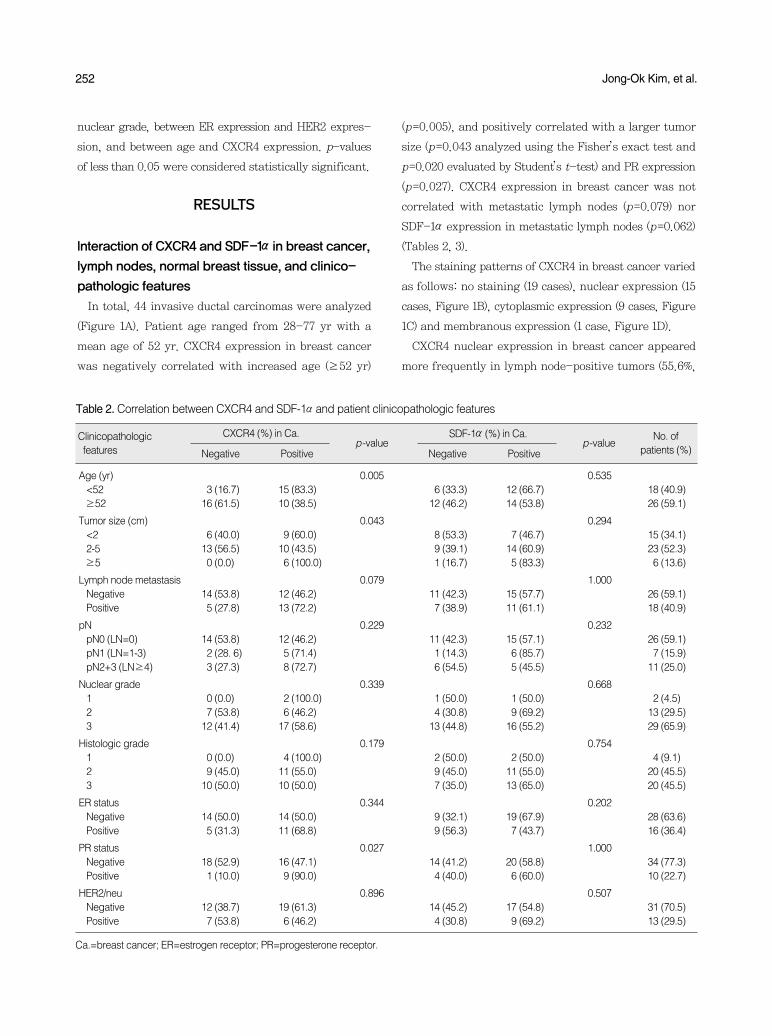
Table 2. Correlation between CXCR4 and SDF-1αand patient clinicopathologic features
Clinicopathologicfeatures
CXCR4 (%) in Ca.
Negative Positivep-value
SDF-1α(%) in Ca.
Negative Positivep-value
No. ofpatients (%)
Age (yr) 0.005 0.535<52 3 (16.7) 15 (83.3) 6 (33.3) 12 (66.7) 18 (40.9)≥52 16 (61.5) 10 (38.5) 12 (46.2) 14 (53.8) 26 (59.1)
Tumor size (cm) 0.043 0.294<2 6 (40.0) 9 (60.0) 8 (53.3) 7 (46.7) 15 (34.1)2-5 13 (56.5) 10 (43.5) 9 (39.1) 14 (60.9) 23 (52.3)≥5 0 (0.0) 6 (100.0) 1 (16.7) 5 (83.3) 6 (13.6)
Lymph node metastasis 0.079 1.000Negative 14 (53.8) 12 (46.2) 11 (42.3) 15 (57.7) 26 (59.1)Positive 5 (27.8) 13 (72.2) 7 (38.9) 11 (61.1) 18 (40.9)
pN 0.229 0.232pN0 (LN=0) 14 (53.8) 12 (46.2) 11 (42.3) 15 (57.1) 26 (59.1)pN1 (LN=1-3) 2 (28. 6) 5 (71.4) 1 (14.3) 6 (85.7) 7 (15.9)pN2+3 (LN≥4) 3 (27.3) 8 (72.7) 6 (54.5) 5 (45.5) 11 (25.0)
Nuclear grade 0.339 0.6681 0 (0.0) 2 (100.0) 1 (50.0) 1 (50.0) 2 (4.5)2 7 (53.8) 6 (46.2) 4 (30.8) 9 (69.2) 13 (29.5)3 12 (41.4) 17 (58.6) 13 (44.8) 16 (55.2) 29 (65.9)
Histologic grade 0.179 0.7541 0 (0.0) 4 (100.0) 2 (50.0) 2 (50.0) 4 (9.1)2 9 (45.0) 11 (55.0) 9 (45.0) 11 (55.0) 20 (45.5)3 10 (50.0) 10 (50.0) 7 (35.0) 13 (65.0) 20 (45.5)
ER status 0.344 0.202Negative 14 (50.0) 14 (50.0) 9 (32.1) 19 (67.9) 28 (63.6)Positive 5 (31.3) 11 (68.8) 9 (56.3) 7 (43.7) 16 (36.4)
PR status 0.027 1.000Negative 18 (52.9) 16 (47.1) 14 (41.2) 20 (58.8) 34 (77.3)Positive 1 (10.0) 9 (90.0) 4 (40.0) 6 (60.0) 10 (22.7)
HER2/neu 0.896 0.507Negative 12 (38.7) 19 (61.3) 14 (45.2) 17 (54.8) 31 (70.5)Positive 7 (53.8) 6 (46.2) 4 (30.8) 9 (69.2) 13 (29.5)
Ca.=breast cancer; ER=estrogen receptor; PR=progesterone receptor.
p=0.023) than in lymph node-negative tumors (19.2%).
CXCR4 cytoplasmic expression in breast cancer was not
correlated with lymph node metastasis (Table 4). CXCR4
expression was absent or minor in normal breast epithelia.
Breast cancer cells were stained by SDF-1αin primary
breast cancers (Figure 1E) and in the lymph nodes (Figure
1F). Lymphoid cells were also stained by SDF-1αin pri-
mary breast cancers and in the lymph nodes. SDF-1α
expression was negative in normal ductal and lobular
cells. SDF-1αin breast cancer was not correlated with
any clinicopathologic features (Table 2).
Correlation between hormone receptor (ER and
PR) expression, and CXCR4 and SDF-1αα
Coexpression of ER and PR was present in 10 breast
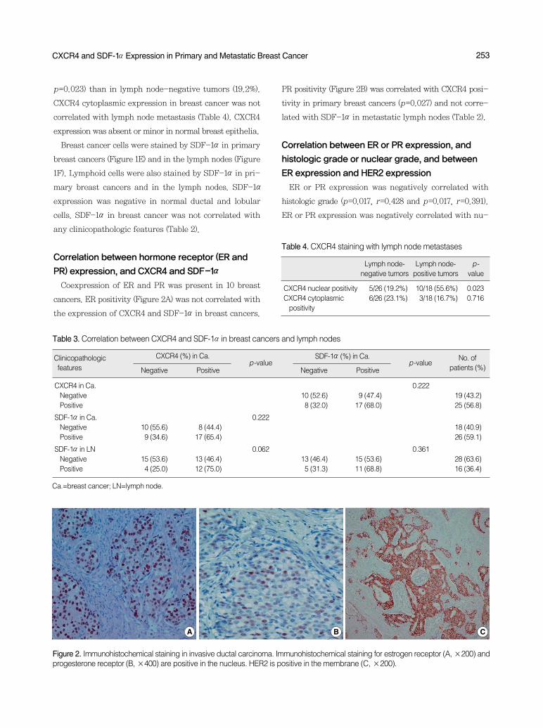
cancers. ER positivity (Figure 2A) was not correlated with
the expression of CXCR4 and SDF-1αin breast cancers.
PR positivity (Figure 2B) was correlated with CXCR4 posi-
tivity in primary breast cancers (p=0.027) and not corre-
lated with SDF-1αin metastatic lymph nodes (Table 2).
Correlation between ER or PR expression, and
histologic grade or nuclear grade, and between
ER expression and HER2 expression
ER or PR expression was negatively correlated with
histologic grade (p=0.017, r=0.428 and p=0.017, r=0.391).
ER or PR expression was negatively correlated with nu-
CXCR4 and SDF-1αExpression in Primary and Metastatic Breast Cancer 253
Table 3. Correlation between CXCR4 and SDF-1αin breast cancers and lymph nodes
Clinicopathologicfeatures
CXCR4 (%) in Ca.
Negative Positivep-value
SDF-1α(%) in Ca.
Negative Positivep-value
No. ofpatients (%)
CXCR4 in Ca. 0.222Negative 10 (52.6) 9 (47.4) 19 (43.2)Positive 8 (32.0) 17 (68.0) 25 (56.8)
SDF-1αin Ca. 0.222 Negative 10 (55.6) 8 (44.4) 18 (40.9)Positive 9 (34.6) 17 (65.4) 26 (59.1)
SDF-1αin LN 0.062 0.361 Negative 15 (53.6) 13 (46.4) 13 (46.4) 15 (53.6) 28 (63.6)Positive 4 (25.0) 12 (75.0) 5 (31.3) 11 (68.8) 16 (36.4)
Ca.=breast cancer; LN=lymph node.
Table 4. CXCR4 staining with lymph node metastases
Lymph node-negative tumors
Lymph node-positive tumors
p-value
CXCR4 nuclear positivity 5/26 (19.2%) 10/18 (55.6%) 0.023CXCR4 cytoplasmic 6/26 (23.1%) 3/18 (16.7%) 0.716
positivity
Figure 2. Immunohistochemical staining in invasive ductal carcinoma. Immunohistochemical staining for estrogen receptor (A, ×200) andprogesterone receptor (B, ×400) are positive in the nucleus. HER2 is positive in the membrane (C, ×200).
A B C

clear grade (p=0.029, r=0.397 and p=0.014, r=0.392).
ER expression trended toward a negative correlation with
HER2 expression (p=0.61, r=0.282).
Correlation between HER2, CXCR4 in breast
cancer and metastatic lymph nodes
HER2 positivity (Figure 2C) was not correlated with
CXCR4 expression in breast cancers and metastatic lymph
nodes (p=0.896 and p=0.258) (Table 2). Positivity of CXCR4
and/or HER2 in primary carcinoma was not significantly
correlated with metastatic lymph nodes (p=0.088).
DISCUSSION
The mechanism of metastasis to lymph nodes is not well
understood. Recent studies have shown that CXCR4,
CCR7, and c-MET are involved in directing tumor cells
to lymph nodes.(7) Activation of SDF-1α/CXCR4 increas-
es secretion of MMP2 and MMP9. These signaling events
result in changes in cellular motility and invasive and
adhesive properties in breast cancer cells. Collectively,
these events may lead tumor cells to metastasize to lymph
nodes. Experimental studies have suggested that several
tumor types metastasize to distant organs, such as the
lungs and bones, via the CXCR4-mediated signaling path-
way.(3)
Sun et al.(12) found that SDF-1αis involved in CXCR4-
stimulated directed metastasis of cancer cells. CXCR4
protein expression increases with tumor aggressive-
ness, and levels of SDF-1 are higher in metastatic lesions
than in primary tumor sites. Tumor invasion and metas-
tasis involve complex procedures in which the invasion
of tumor cells and passage through the basement mem-
brane, fibroblast monolayers and endothelial cells are
regarded as key processes before the tumor cells migrate
to target organs. Directed migration of tumor cells stim-
ulated by activation of CXCR4 is dependent on an SDF-
1αgradient.(9) It has been reported that 100-1,000 ng/
mL of SDF-1αis an optimal concentration to stimulate
the migration of monocytes, lymphocytes, endothelial
cells, and tumor cells. By using time-lapse monitoring
of the locomotion of individual cells, it was shown that
SDF-1αaccelerates the motility of several different kinds
of human tumor cells, including breast cancer, rhab-
domyosarcoma and small lung cancer cell lines.(10) The
mechanisms by which SDF-1αeffects tumor cell motil-
ity include changes/rearrangement of the cytoskeletal
proteins, augmentation of the number and thickness of
F-actin bundles, and calcium flux.(11) Based on a Matrigel
invasion model, SDF-1αis probably also involved in cancer
cell growth and survival, in addition to stimulating cancer
cell migration. Because cancer cells experience little growth
when deprived of a blood supply during metastasis to
distant sites, SDF-1αprobably helps in the survival and
spread of cancer cells by stimulating their proliferation.
CXCR4/SDF-1αparticipates in vivo and directs migra-
tion in vitro.
Tumor cell metastasis may be correlated with attraction
of SDF-1αto the metastatic organ. Our results showed
that CXCR4 expression in breast cancers tended toward
a positive correlation with SDF-1αexpression in lymph
nodes-positive tumors, even though the correlation was
not statistically significant (p=0.062).
A high level of cytoplasmic CXCR4 expression occurs
more commonly, but not significantly, in lymph node-posi-
tive tumors.(15,18) The results of Woo et al.(19) showed
that expression of CXCR4 in the cytoplasm is not associ-
ated with axillary lymphatic metastasis. CXCR4 is a mem-
brane-bound G-protein-coupled receptor that can be
internalized together with its ligand SDF-1 upon activation.
Therefore, cytoplasmic reactivity, especially when co-
expressed together with SDF-1, may reflect receptor inter-
nalization and, thus, a state of activation of CXCR4.(20)
Predominantly nuclear CXCR4 staining was more likely
to be statistically significant in lymph node-negative
tumors.(15,18) Su et al.(21) reported that a high level of
CXCR4 nuclear expression was not associated with lymph
node involvement. Spano et al.(22) pointed out that in
non-small cell lung carcinoma, CXCR4-positive nuclear
staining might be associated with survival. They opined
that the nuclear retention of CXCR4 decreases cell pro-
liferation and metastasis by inhibiting the interaction with
SDF-1α. Shim et al.(23) stated that SDF-1 induces both
down-regulation of membranous CXCR4 expression and
254 Jong-Ok Kim, et al.
degradation of the internalized CXCR4 receptors via the
lysosome pathway. These results suggest that a reduc-
tion in CXCR4 membrane staining in metastatic lymph
nodes is associated with progression of breast cancer
metastasis.
Our results showed that CXCR4 nuclear positivity is
correlated with lymph node metastasis and that CXCR4
expression trended toward a positive correlation with
metastatic lymph nodes (p=0.079). These results indicate
that different patterns of CXCR4 expression in cells pro-
bably lead to differences in the biological behavior of the
respective tumor cells. Evaluation of the expression pat-
terns will help predict lymph node metastasis and clinical
outcomes.
Woo et al.(19) found that neither cytoplasmic nor nuclear
CXCR4 expression shows a significant correlation with
other characteristics including age, tumor stage, node
stage, tumor-node-metastasis (TNM) stage, histological
grade, ER, PR, or HER2 status.
We found that the positivity of CXCR4 in breast can-
cer is negatively correlated with increased age (≥52 yr).
Assuming a normal age distribution in breast cancer
occurrance, the sample mean (52 yr) of observed ages is
considered to be an estimate of the population mean. The
p-value and the correlation coefficient (Pearson’s r) for
increased age (≥52 yr) and CXCR4 positivity were 0.005
and -0.445, respectively. These show an inverse relation-
ship between increased age (≥52 yr) and CXCR4 positivity.
High-intensity immunohistochemical staining for
CXCR4 was associated with large tumors in papillary
thyroid carcinomas (p=0.02)(24) and CXCR4 was also
expressed strongly in large tumors of non-small cell
lung cancers (>3 cm, p=0.042).(25)
Woo et al.(19) found that among 105 breast cancers,
26 cases (24.8%) were classified as T1, 63 cases (60%) as
T2 and 16 cases (15.2%) as T3. Nuclear and cytoplasmic
CXCR4 were not correlated with tumor size. However,
our results showed that a larger tumor size was corre-
lated with CXCR4 expression.
Salvucci et al.(26) found an inverse correlation between
cytoplasmic CXCR4 staining and ER positivity, and no
correlation between either cytoplasmic or nuclear CXCR4
staining and PR positivity. A negative estrogen and a
progesterone receptor status were significantly correlated
with a high level of nuclear CXCR4 expression and with
lymph node metastasis. Furthermore, there was a signif-
icant correlation between a high level of nuclear CXCR4
expression and lymph node metastasis and receptor nega-
tive estrogen and the progesterone status.(19) We found
that CXCR4 was positively correlated with PR expression
but wasn’t with ER positivity.
Recently, the Breast Pathology Study Group of the
Korean Society of Pathologist collected 1,198 breast car-
cinoma samples from seven university hospitals and per-
formed ER staining with the three antibodies, 1D5, 6F11
and SP1. The ER positivity rates were 68.6% for SP1,
59.5% for 1D5 and 54.8% for 6F11.(27) In our study, the
ER expression rate in breast cancers was 36.4% with the
1D5 estrogen receptor clone. Also, among the other 72
excised breast cancers without lymph node dissection,
the ER expression rate was 35 cases (48.6%). In our study,
negative ER or PR expression was correlated with a high
histologic grade or high nuclear grade. These ER expres-
sion rates may be attributable to high histologic grade
cancers, high nuclear grade cancers, different kinds of
antibodies used, differences between the products of man-
ufacturing companies, the duration of fixation, the buffer
solutions used, tumor heterogeneity, or a small number
of cases.
CONCLUSION
An evaluation of young age, large tumor size and PR
expression helps predict lymph node metastasis and poor
prognosis. Expression of CXCR4 nuclear positivity is cor-
related with a poor prognosis. A larger study is needed to
verify these results and further study is needed to deter-
mine the clinical significance of SDF-1α/CXCR4 expres-
sion.
REFERENCES
1. Nicolson GL. Paracrine and autocrine growth mechanisms in tumormetastasis to specific sites with particular emphasis on brain and lungmetastasis. Cancer Metastasis Rev 1993;12:325-43.
CXCR4 and SDF-1αExpression in Primary and Metastatic Breast Cancer 255
2. Mo_hle R, Schittenhelm M, Failenschmid C, Bauts F, Kratz-AlbersK, Serve H, et al. Functional response of leukaemic blasts to stromalcell-derived factor-1 correlates with preferential expression of thechemokine receptor CXCR4 in acute myelomonocytic andlymphoblastic leukaemia. Br J Haematol 2000;110:563-72.
3. Kang H, Watkins G, Douglas-Jones A, Mansel RE, Jiang WG. Theelevated level of CXCR4 is correlated with nodal metastasis of humanbreast cancer. Breast 2005;14:360-7.
4. Kijima T, Maulik G, Ma PC, Tibaldi EV, Turner RE, Rollins B, et al.Regulation of cellular proliferation, cytoskeletal function, and signaltransduction through CXCR4 and c-Kit in small cell lung cancer cells.Cancer Res 2002;62:6304-11.
5. Balkwill F. The significance of cancer cell expression of the chemo-kine receptor CXCR4. Semin Cancer Biol 2004;14:171-9.
6. Uchida D, Begum NM, Almofti A, Nakashiro K, Kawamata H, Ta-teishi Y, et al. Possible role of stromal-cell-derived factor-1/CXCR4signaling on lymph node metastasis of oral squamous cell carcinoma.Exp Cell Res 2003;290:289-302.
7. Fernandis AZ, Prasad A, Band H, Klosel R, Ganju RK. Regulationof CXCR4 mediated chemotaxis and chemoinvasion of breast cancercells. Oncogene 2004;23:157-67.
8. Pan J, Mestas J, Burdick MD, Phillips RJ, Thomas GV, Reckamp K,et al. Stromal derived factor-1 (SDF-1/CXCL12) and CXCR4 in renalcell carcinoma metastasis. Mol Cancer 2006;5:56.
9. Scotton CJ, Wilson JL, Milliken D, Stamp G, Balkwill FR. Epithelialcancer cell migration: a role for chemokine receptors? Cancer Res2001;61:4961-5.
10. Adams GB, Chabner KT, Foxall RB, Weibrecht KW, Rodrigues NP,Dombkowski D, et al. Heterologous cells cooperate to augment stemcell migration, homing, and engraftment. Blood 2003;101:45-51.
11. Dewan MZ, Ahmed S, Iwasaki Y, Ohba K, Toi M, Yamamoto N.Stromal cell-derived factor-1 and CXCR4 receptor interaction in tumorgrowth and metastasis of breast cancer. Biomed Pharmacother 2006;60:273-6.
12. Sun YX, Wang J, Shelburne CE, Lopatin DE, Chinnaiyan AM, RubinMA, et al. Expression of CXCR4 and CXCL12 (SDF-1) in humanprostate cancers (PCa) in vivo. J Cell Biochem 2003;89:462-73.
13. Geminder H, Sagi-Assif O, Goldberg L, Meshel T, Rechavi G, WitzIP, et al. A possible role for CXCR4 and its ligand, the CXC chemo-kine stromal cell-derived factor-1, in the development of bone marrowmetastases in neuroblastoma. J Immunol 2001;167:4747-57.
14. Helbig G, Christopherson KW 2nd, Bhat-Nakshatri P, Kumar S, Ki-shimoto H, Miller KD, et al. NF-kappaB promotes breast cancer cellmigration and metastasis by inducing the expression of the chemokinereceptor CXCR4. J Biol Chem 2003;278:21631-8.
15. Cabioglu N, Yazici MS, Arun B, Broglio KR, Hortobagyi GN, PriceJE, et al. CCR7 and CXCR4 as novel biomarkers predicting axillary
lymph node metastasis in T1 breast cancer. Clin Cancer Res 2005;11:5686-93.
16. Tsoli E, Tsantoulis PK, Papalambros A, Perunovic B, England D,Rawlands DA, et al. Simultaneous evaluation of maspin and CXCR4in patients with breast cancer. J Clin Pathol 2007;60:261-6.
17. Harvey JM, Clark GM, Osborne CK, Allred DC. Estrogen receptorstatus by immunohistochemistry is superior to the ligand-bindingassay for predicting response to adjuvant endocrine therapy in breastcancer. J Clin Oncol 1999;17:1474-81.
18. Cabioglu N, Sahin A, Doucet M, Yavuz E, Igci A, Yildirim OE, et al.Chemokine receptor CXCR4 expression in breast cancer as a poten-tial predictive marker of isolated tumor cells in bone marrow. ClinExp Metastasis 2005;22:39-46.
19. Woo SU, Bae JW, Kim CH, Lee JB, Koo BW. Significant correlationbetween nuclear CXCR4 expression and axillary lymph node metas-tasis in hormonal receptor negative breast cancer. Ann Surg Oncol2008;15:281-5.
20. Liu F, Lang R, Wei J, Fan Y, Cui L, Gu F, et al. Increased expressionof SDF-1/CXCR4 is associated with lymph node metastasis of inva-sive micropapillary carcinoma of the breast. Histopathology 2009;54:741-50.
21. Su YC, Wu MT, Huang CJ, Hou MF, Yang SF, Chai CY. Expressionof CXCR4 is associated with axillary lymph node status in patientswith early breast cancer. Breast 2006;15:533-9.
22. Spano JP, Andre F, Morat L, Sabatier L, Besse B, Combadiere C, etal. Chemokine receptor CXCR4 and early stage non-small cell lungcancer: pattern of expression and correlation with outcome. Ann Oncol2004;15:613-7.
23. Shim HS, Lau SK, Devi S, Yoon Y, Cho HT, Liang Z. Lower ex-pression of CXCR4 in lymph node metastases than in primary breastcancers: Potential regulation by ligand-dependent degradation andHIF-1a. Biochem Biophys Res Commun 2006;346:252-8.
24. Wagner PL, Moo TA, Arora N, Liu YF, Zarnegar R, Scognamiglio T,et al. The chemokine receptors CXCR4 and CCR7 are associatedwith tumor size and pathologic indicators of tumor aggressiveness inpapillary thyroid. Ann Surg Oncol 2008;15:2833-41.
25. Song JS, Jung JK, Park JC, Kim DK, Jang SJ. Association of CXCR4expression with metastasis and survival among patients with non-small cell lung cancer. Korean J Pathol 2008;42:358-64.
26. Salvucci O, Bouchard A, Baccarelli A, Deschenes J, Sauter G, Si-mon R, et al. The role of CXCR4 receptor expression in breast cancer:a large tissue microarray study. Breast Cancer Res Treat 2006;97:275-83.
27. Bae YK, Gong GY, Kang J, Lee AW, Cho EY, Lee JS, et al. Estrogenreceptor expression in Korean breast carcinoma and comparison ofthree anti-ER antibodies. 61th fall annual meeting of Korean Societyof Pathologists. 2009;43. abstract #4.
256 Jong-Ok Kim, et al.


![Intradermal microbubbles and contrast-enhanced ultrasound ......Axillary lymph node status is an important prognostic factor in patients with breast cancer [1]. Sentinel lymph node](https://img.pdfslide.us/doc/110x75/60cf0037673f955c2d5ad2b4/intradermal-microbubbles-and-contrast-enhanced-ultrasound-axillary-lymph.jpg)