Embed Size (px)
Citation preview

Cancer Res Treat. 2017 Aug 17 [Epub ahead of print]
pISSN 1598-2998, eISSN 2005-9256
https://doi.org/10.4143/crt.2017.210
│ http://www.e-crt.org │ 1Copyright ⓒ 2017 by the Korean Cancer AssociationThis is an Open-Access article distributed under the terms of the Creative Commons Attribution Non-Commercial License (http://creativecommons.org/licenses/by-nc/3.0/)
which permits unrestricted non-commercial use, distribution, and reproduction in any medium, provided the original work is properly cited.
Open Access
Feasibility of Charcoal Tattooing of Cytology-Proven Metastatic Axillary Lymph Node at Diagnosis and Sentinel Lymph Node Biopsyafter Neoadjuvant Chemotherapy in Breast Cancer Patients
Original Article
Purpose
Sentinel lymph node biopsy (SLNB) can be performed when node-positive disease is con-
verted to node-negative status after neoadjuvant chemotherapy (NCT). Tattooing nodes
might improve accuracy but supportive data are limited. This study aimed to investigate the
feasibility of charcoal tattooing metastatic axillary lymph node (ALN) at presentation followed
by SLNB after NCT in breast cancers.
Materials and Methods
Twenty patients with cytology-proven node metastases prospectively underwent charcoal tat-
tooing at diagnosis. SLNB using dual tracers and axillary surgery after NCT were then per-
formed. The detection rate of tattooed node and diagnostic performance of SLNB were
analyzed.
Results
All patients underwent charcoal tattooing without significant morbidity. Sentinel and tattooed
nodes could be detected during surgery after NCT. Nodal pathologic complete response
was achieved in 10 patients. Overall sensitivity, false-negative rate (FNR), negative predictive
value, and accuracy of hot/blue SLNB were 80.0%, 20.0%, 83.3%, and 90.0%, respectively.
Retrieving more nodes and favorable nodal response were associated with improved per-
formance. The best accuracy was observed when excised tattooed node was calculated
together (FNR, 0.0%). Cold/non-blue tattooed nodes of five patients were removed during
non-sentinel axillary surgery but clinicopathological parameters did not differ compared to
patients with hot/blue tattooed node detected during SLNB, suggesting the importance of
the tattooing procedure itself to improve performance.
Conclusion
Charcoal tattooing of cytology-confirmed metastatic ALN at presentation is technically fea-
sible and does not limit SLNB after NCT. The tattooing procedure without additional preop-
erative localization is advantageous for improving the diagnostic performance of SLNB in
this setting.
Key words
Breast neoplasms, Charcoal, Neoadjuvant therapy,
Sentinel lymph node biopsy, Tattoo
Seho Park, MD, PhD1,2
Ja Seung Koo, MD, PhD3
Gun Min Kim, MD4
Joohyuk Sohn, MD, PhD4
Seung Il Kim, MD, PhD1
Young Up Cho, MD, PhD1
Byeong-Woo Park, MD, PhD1
Vivian Youngjean Park, MD, PhD5
Jung Hyun Yoon, MD, PhD5
Hee Jung Moon, MD, PhD5
Min Jung Kim, MD, PhD5
Eun-Kyung Kim, MD, PhD5
+ + + + + + + + + + + + + + + + + + + + + + + + + + + + + + + + + + + + + + + + + + + + + + + + + + + + + + + + + + + ++ + + + + + + + + + + + + + + + + + + + + + + + + + + + + + + + + + + + + + + + + + + + + + + + + + + + + + + + + + + ++ + + + + + + + + + + + + + + + + + + + + + + + + + + + + + + + + + + + + + + ++ + + + + + + + + + + + + + + + + + + ++ + + + + + + + + + + + + + + + + + + + + + + + + + + + + + + + + + + + + + + ++ + + + + + + + + + + + + + + + + + + ++ + + + + + + + + + + + + + + + + + + + + + + + + + + + + + + + + + + + + + + ++ + + + + + + + + + + + + + + + + + + ++ + + + + + + + + + + + + + + + + + + +
Correspondence: Min Jung Kim, MD, PhD
Department of Radiology and Research Institute
of Radiological Science, Severance Hospital,
Yonsei University College of Medicine,
50-1 Yonsei-ro, Seodaemun-gu, Seoul 03722, Korea
Tel: 82-2-2228-7400
Fax: 82-2-393-3035
E-mail: [email protected]
Received May 2, 2017
Accepted August 7, 2017
*Seho Park and Ja Seung Koo contributed equally
to this work.
1Division of Breast Surgery, Department ofSurgery, Yonsei University College of Medicine, Seoul, 2Frontier Research Instituteof Convergence Sports Science, Yonsei University, Seoul, 3Department ofPathology, Yonsei University College of Medicine, Seoul, 4Division of Medical Oncology, Department of Internal Medicine,Severance Hospital, Yonsei University Collegeof Medicine, Seoul, 5Department of Radiologyand Research Institute of Radiological Science,Severance Hospital, Yonsei University Collegeof Medicine, Seoul, Korea

Cancer Res Treat. 2017 Aug 17 [Epub ahead of print]
2 CANCER RESEARCH AND TREATMENT
Introduction
Sentinel lymph node biopsy (SLNB) is a standard proce-
dure for managing patients with clinical node-negative
breast cancer and it lowers surgical morbidity and improves
quality of life compared to axillary lymph node dissection
(ALND) while providing similar survival rates [1,2]. How-
ever, the oncologic safety of SLNB remains controversial in
patients with cytology-proven node-positive disease at pres-
entation that is converted to clinical node-negative status
after receiving neoadjuvant chemotherapy (NCT). Nodal
metastasis can be eradicated by NCT in up to approximately
40% of patients with positive axillary lymph node (ALN)
metastasis at diagnosis [3,4], but the possible effects of NCT
are alterations or disruptions of lymphatic vessels or non-
uniform tumor regression of metastatic ALNs, which can
limit the usefulness of SLNB in these patients [5].
Several prospective clinical trials were undertaken to
determine the accuracy of SLNB after NCT in patients with
clinical or histological node-positive disease and the overall
false-negative rate (FNR) of SLNB was 14.2% in the SENTinel
NeoAdjuvant (SENTINA) study, 12.6% in the American Col-
lege of Surgeons Oncology Group (ACOSOG) Z1071 (Alli-
ance) trial, and 13.3% in the Sentinel Node Biopsy Following
Neoadjuvant Chemotherapy (SN FNAC) trial [6-8]. In cases
where dual tracers were used, > 2 sentinel lymph nodes
(SLNs) were retrieved, or immunohistochemistry was used;
however, FNRs < 10% were clinically acceptable for SLNB
[4]. Thus, the National Comprehensive Cancer Network
(NCCN) guidelines recommend SLNB or ALND if biopsy-
proven metastatic ALNs are converted to clinically node-
negative status after preoperative systemic therapy [9].
Importantly, the NCCN guidelines emphasize that marking
sampled ALNs with a clip or tattoo before NCT can improve
the diagnostic performance of SLNB [10,11].
A biopsy-proven metastatic ALN has to be marked if tar-
geted axillary dissection is to be performed after NCT and
this marking has to take place by an experienced radiologist
with specialized equipment such as a clip [12]. Preoperative
localization with a wire or radioactive seed is also necessary
and this additional invasive procedure might be uncomfort-
able for patients. Furthermore, clinicians must also check
possible clip migration and determine the radiation safety of
the radioactive seed [4]. In comparison, additional localiza-
tion is not required for the tattooing technique of positive
node although limited studies have been reported since its
introduction [11]. The diagnostic performance of the tattoo-
ing procedure incorporated into SLNB should be studied
with this background in mind. In addition, marked metasta-
tic ALN before NCT is not always detected as hot and/or
blue nodes at surgery. For clarifying the association between
tattooed ALN at diagnosis and dual traced nodes after NCT,
SLNB was considered as results of hot/blue ALNs retrieved
alone and modified SLNB was defined as findings of dual
traced or residual marked, suspiciously enlarged nodes in
this study.
The main aims of the present study were primarily to
investigate whether black tattoo injected in cytology-proven
metastatic ALN at presentation could be detected intraoper-
atively in patients treated with NCT and to examine the fea-
sibility of charcoal tattooing in breast cancers. The second
was to evaluate the diagnostic performance of SLNB with or
without marking the biopsy-proven metastatic ALN by cal-
culating the detection rate, sensitivity, FNR, negative predic-
tive value (NPV), and accuracy. The last was to determine
the concordance between charcoal-tattooed ALN and SLNs
detected by radioisotopes and blue dye. If a discordance was
found, we tried to identify which clinicopathological param-
eters were associated with the discordance.
Materials and Methods
1. Patient enrollment
Twenty patients with clinical T1-T3 breast cancer at diag-
nosis who received NCT between October 2015 and July 2016
were prospectively enrolled from Yonsei Cancer Center, Sev-
erance Hospital, Yonsei University College of Medicine,
Seoul, Korea. All patients had cytology-proven ALN metas-
tases prior to NCT. Patients with a past history of allergic
reaction to charcoal agents, inflammatory or T4 breast carci-
noma, or stage IV cancer at diagnosis were excluded. At pres-
entation, all patients with clinically enlarged ALNs or with
radiologically suspicious nodal findings of loss of fatty
hilum, cortical thickening > 3 mm, round shape, markedly
hypoechoic cortex, or increased peripheral blood flow
underwent ultrasound-guided fine needle aspiration cytol-
ogy of the most suspicious or largest ALN to confirm the
presence of metastasis.
2. Ultrasound-guided charcoal tattooing on cytology-con-
firmed ALN
Prior to NCT, the main breast tumor was marked with a
metallic clip and then the cytology-proven metastatic ALN
was tattooed with charcoal suspension (Charcotrace, Phebra,
Sydney, Australia) through ultrasound-guidance by a radi-
ologist (M.J.K.) who had more than 14 years of specialized
experience in breast imaging. To minimize obstacles to
pathologic examination, the charcoal was mainly injected

Seho Park, Metastatic ALN Tattooing and SLNB after NCT
Cancer Res Treat. 2017 Aug 17 [Epub ahead of print] 3
into the capsular area of the metastasis-confirmed ALN and
a needle-point injection track was made from the ALN to the
skin of the axilla. A median dose of 0.5 mL (range, 0.2 to 0.8
mL) was used for node tattooing.
3. Response to NCT and clinicopathological factors
After four cycles of anthracycline followed by taxane-con-
taining NCT, the responses of the primary tumor and nodes
were comprehensively evaluated using clinical and radiolog-
ical examinations, mainly with ultrasound or breast magnetic
resonance images. For cases with human epidermal growth
factor receptor 2 (HER2)‒positive disease, trastuzumab was
incorporated into the NCT regimens. According to the
Response Evaluation Criteria in Solid Tumors ver. 1.1, tumor
response to NCT was considered as complete or partial
response and non-response was defined with stable or pro-
gressive disease. The ALN response to NCT was defined
with the disappearance of metastatic ALNs or no suspicious
imaging findings in the axilla. Nodal non-response was con-
sidered when suspicious findings in the axilla remained after
completion of NCT.
Pathologic stages were based on the AJCC Cancer Staging
Manual, seventh edition [13]. The modified Scarff-Bloom-
Richardson grading system was used for histologic grading.
Tumors with ≥ 1% nuclear-stained cells by immunohisto-
chemistry using core needle biopsy specimens were consid-
ered positive for estrogen receptor (ER) and progesterone
receptor (PR) according to the American Society of Clinical
Oncology/College of American Pathologists (ASCO/CAP)
guidelines [14]. HER2 staining was scored as 0, 1+, 2+, or 3+
according to the ASCO/CAP guidelines [15]. In cases with
HER2 2+ results, silver in situ hybridization (SISH) was per-
formed using the INFORM HER2 Dual ISH DNA Probe
Cocktail Assay (Ventana Medical Systems, Tucson, AZ) with
an automated slide stainer according to the manufacturer’s
protocols. HER2 gene amplification was defined with a HER2
gene/chromosome 17 copy number ratio ≥ 2.0 or a HER2
gene/chromosome 17 copy number ratio < 2.0 with an aver-
age HER2 copy number ≥ 6.0 signals/cell according to the
ASCO/CAP guidelines [15]. An immunohistochemistry
score of 3+ or gene amplification by SISH was considered
HER2-positive. During the study period, Ki-67 staining was
mainly performed on post-treatment surgical specimens for
residual carcinomas in our institution and was not investi-
gated in the present study.
Based on ER, PR, HER2, and histologic grade findings,
molecular phenotypes were categorized into the following
four subgroups: luminal A (ER-positive and/or PR-positive,
HER2-negative and grade 1 or 2), luminal B (ER-positive
and/or PR-positive, HER2-negative and grade 3; or ER-pos-
itive and/or PR-positive, and HER2-positive), HER2-
enriched (ER-negative, PR-negative, and HER2-positive),
and triple-negative breast cancer (TNBC) (ER-negative,
PR-negative, and HER2-negative).
4. SLNB, axillary surgery, and pathologic assessment
SLNs were detected using a combination of radioisotopes
and blue dye techniques except for one detected with a
radioisotope alone. In brief, 0.5 mCi 99mTc-Phytate (Korea
Atomic Energy Research Institute, Daejeon, Korea) was
diluted in 0.5 mL saline and injected into the subcutaneous
layer of the areolar tissue in the direction of the main primary
tumor at the Department of Nuclear Medicine on the day of
surgery. In the operation room just before surgery was initi-
ated, 0.8% indigo carmine dye (Carmine, Korea United
Pharm Inc., Seoul, Korea) was injected into the periareolar
area followed by a massage of the injection site. SLNs were
excised and defined as hot and/or blue-colored nodes with
a handheld gamma probe (Neoprobe Gamma Detection Sys-
tem, Neoprobe Corp., Dublin, OH) at gross visual inspection
or with a radioactive count of ≥ 10% of the ex vivo count of
the hottest node. During SLNB, clinically palpable cold or
non-blue axillary nodal tissues were also excised and cate-
gorized as axillary sampling. Charcoal-tattooed ALN and
track were removed during axilla surgery and included in
the SLNB or axillary sampling procedures. SLNs and axillary
sampling tissues were then sent to frozen section analysis.
After SLNB and/or axillary sampling, the decision to per-
form further concomitant completion level I/II ALND was
made taking into account patient’ consent, initial nodal bur-
den, node responses during and after NCT, or frozen section
results of SLNB and/or axillary sampling.
All excised ALNs including SLNs were subjected to rou-
tine pathological examinations, which included hematoxylin
and eosin staining with or without immunohistochemistry
for cytokeratin in accordance with previously published rec-
ommendations [16]. ALNs were considered positive if
metastatic foci were > 0.2 mm and/or > 200 tumor cells
(≥ ypN1mi) according to the AJCC Cancer Staging Manual,
seventh edition [13]. Any nodes with isolated metastatic foci
of ≤ 0.2 mm were considered negative [ypN0(i+)], but were
not considered to have achieved a node pathologic complete
response (pCR). Regarding primary tumor responses to
NCT, the absence of in situ or invasive carcinomas (ypT0) or
residual in situ carcinoma alone without invasive disease
(ypTis) constituted a tumor pCR.
5. Statistical analysis
The detection of charcoal-tattooed node was investigated
and its association with the detection of SLNs was analyzed.
Detection failure of SLNs was defined as an inability to iden-

tify hot and/or blue nodes by lymphoscintigraphy or gamma
probe with visual inspection. Sensitivity was calculated by
dividing true positive (TP) findings by TP plus false-negative
(FN) findings. FNR was defined as the proportion of patients
with negative SLNs who subsequently had metastatic ALNs
among patients with ≥ 1 positive lymph node. NPV was cal-
culated by dividing true negative (TN) findings by TN plus
Cancer Res Treat. 2017 Aug 17 [Epub ahead of print]
4 CANCER RESEARCH AND TREATMENT
Parameter No. of patients (%)
Age (yr)
≤ 50 11 (55.0)
> 50 9 (45.0)
BMI (kg/m2)
≤ 25 15 (75.0)
> 25 5 (25.0)
Laterality
Left 13 (65.0)
Right 7 (35.0)
Location of the main lesion
Upper outer quadrant 7 (35.0)
Upper inner quadrant 4 (20.0)
Upper central area 2 (10.0)
Mediocentral area 2 (10.0)
Lower outer quadrant 2 (10.0)
Lower inner quadrant 1 (5.0)
Lower central area 1 (5.0)
Subareolar area 1 (5.0)
Histologic type
IDC-NOS 19 (95.0)
Mucinous carcinoma 1 (5.0)
Clinical tumor stage at diagnosis
cT1 6 (30.0)
cT2 11 (55.0)
cT3 3 (15.0)
Clinical node stage at diagnosis
cN1 12 (60.0)
cN2 5 (25.0)
cN3 3 (15.0)
Regimens of NCT
AC–wP±H 9 (45.0)
AC–T±H 11 (55.0)
Tumor response to NCT
Response 14 (70.0)
Non-response 6 (30.0)
Node response to NCT
Response 11 (55.0)
Non-response 9 (45.0)
Pathologic tumor stage
ypT0-is 6 (30.0)
ypT1-2 14 (70.0)
Pathologic node stage
ypN0 10 (50.0)
ypN1-2 10 (50.0)
Pathologic TNM stage
Stage 0 6 (30.0)
Stage 1 4 (20.0)
Stage 2 7 (35.0)
Stage 3 3 (15.0)
Table 1. Clinicipathological characteristics of the enrolled
patients
Table 1. Continued
BMI, body mass index; IDC-NOS, invasive ductal carci-
noma-not otherwise specified; NCT, neoadjuvant chemo-
therapy; AC, anthracycline followed by cyclophosphamide;
wP, weekly paclitaxel; H, trastuzumab; T, docetaxel; TNM,
tumor node metastasis; HER2, human epidermal growth
factor receptor 2; TNBC, triple-negative breast cancer;
SLNB, sentinel lymph node biopsy; ALND, axillary lymph
node dissection.
Parameter No. of patients (%)
Histologic grade
I 3 (15.0)
II 13 (65.0)
III 4 (20.0)
Lymphovascular invasion
Absent 15 (75.0)
Present 5 (25.0)
Perinodal extension
Absent 15 (75.0)
Present 5 (25.0)
Estrogen receptor
Negative 6 (30.0)
Positive 14 (70.0)
Progesterone receptor
Negative 8 (40.0)
Positive 12 (60.0)
HER2
Negative 15 (75.0)
Positive 5 (25.0)
Molecular phenotype
Luminal A 10 (50.0)
Luminal B 4 (20.0)
HER2-enriched 2 (10.0)
TNBC 4 (20.0)
Type of breast surgery
Breast-conserving surgery 11 (55.0)
Total mastectomy 9 (45.0)
Type of axillary surgery
SLNB alone 2 (10.0)
SLNB+axillary sampling 6 (30.0)
SLNB+ALND 12 (60.0)

FN findings. Accuracy was defined as the proportion of
patients with TP or TN among patients with successful
SLNB. The significances of differences in SLNB performance
of subgroups and in clinicopathological parameters between
groups were evaluated using the chi-square test or Fisher
exact test. The Mann-Whitney U test was used to compare
the median values of continuous numerical data. All statisti-
cal tests were two-sided, and p < 0.05 was considered statis-
tically significant. SPSS ver. 23.0 (IBM Corp., Armonk, NY)
and SAS ver. 9.4 (SAS Inc., Cary, NC) were used for analy-
sis.
6. Ethical statement
This study was approved by the Institutional Review
Board (IRB) of Severance Hospital, Yonsei University Health
System, Seoul, Korea (IRB No. 4-2015-0465) and registered to
the Clinical Research Information Service (CRIS, http://
cris.nih.go.kr/cris/index.jsp; No. KCT0002370), Korea. Writ-
ten informed consent was obtained from all patients prior to
study entry.
Results
1. Patient characteristics and ultrasound-guided charcoal
tattooing procedure
The mean age of 20 patients was 52.3±10.9 years (range, 34
to 70 years) at diagnosis. The mean size of the main primary
tumor at presentation was 3.1±1.8 cm (range, 1.2 to 8.4 cm).
Table 1 shows the clinicopathological characteristics and
treatment patterns of the enrolled patients. No significant
side effects or complications occurred during charcoal tat-
tooing of metastatic ALN. After finishing the tattooing pro-
cedure, 16 patients (80%) underwent NCT on the very day
that charcoal tattooing was performed and the time interval
between charcoal tattooing and NCT ranged from 0 to 5
days. The median time interval between charcoal tattooing
and surgery was 181 days (range, 165 to 197 days).
Final permanent pathology of breast and ALNs is pre-
sented in Table 1. Preoperative imaging studies of three
patients with initially suspicious non-axillary regional lymph
nodes showed no radiological evidence of non-axillary
regional metastasis after completion of NCT. Therefore,
when pathologic results of the breast and axilla were consid-
ered, both tumor and nodal pCR were determined in six
patients (30.0%). There was no statistical difference in the
nodal pCR rate according to the type of axillary surgery
(ALND 41.7% vs. axillary sampling 62.5%, p=0.650) (Table 1).
2. Axillary surgery and diagnostic performance of SLNB
One or more hot and/or blue SLNs were detected in all
patients (detection rate, 100%). The median number of SLNs
and total ALNs (SLNs+non-sentinel ALNs) retrieved was 3.0
(range, 1 to 12) and 10.0 (range, 4 to 18), respectively. The
median number of non-sentinel ALNs excised was 4.5
(range, 0 to 15). The number of pathologically retrieved
nodes in two patients who underwent SLNB alone was four
and 10, respectively. During axillary surgery, charcoal-tat-
tooed ALN and track were easily identified by visual inspec-
tion and excised in all patients (detection rate of tattooed
node, 100%). According to the type of axillary surgery, the
median number of SLNs was not different (n=3 in each
group, p=0.624), but the median number of non-sentinel
Seho Park, Metastatic ALN Tattooing and SLNB after NCT
Cancer Res Treat. 2017 Aug 17 [Epub ahead of print] 5
Final ALN status after NCT
Parameter Positive (n=10) Total
Non-sentinel ALN (+) Non-sentinel ALN (‒)Negative (n=10)
SLNB
Positive 3 5 - 8
Negative 2 - 10 12
Modified SLNBa)
Positive 4 6 - 10
Negative 0 - 10 10
Table 2. Results of SLNB and axillary surgery
SLNB, sentinel lymph node biopsy; ALN, axillary lymph node; NCT, neoadjuvant chemotherapy. a)Modified SLNB is the
final results of hot or blue-colored sentinel lymph nodes and initial cytology-proven, charcoal-tattooed lymph node excised
during axillary sampling procedure.

ALNs was significantly larger in the ALND group; therefore,
the median number of total ALNs removed was significantly
higher in the ALND group (ALND, 13 [range, 7 to 18] vs.
axillary sampling, 4.5 [range, 4 to 10]; p < 0.001).
Table 2 compares SLNB results of removing hot and/or
blue SLNs with the final ALN status. In addition to hot
and/or blue SLNs, charcoal-tattooed node was calculated
together as the modified SLNB. The diagnostic performance
of the modified SLNB showed the highest values as follows:
sensitivity, 100%; FNR, 0.0%; NPV, 100%; and accuracy,
100%.
Overall diagnostic performance of SLNB is presented in
Cancer Res Treat. 2017 Aug 17 [Epub ahead of print]
6 CANCER RESEARCH AND TREATMENT
Fig. 1. Gross and microscopic findings of a charcoal-tattooed lymph node in a patient presenting with a false-negative
sentinel lymph node biopsy using dual tracers. (A) Gross picture of a retrieved charcoal-tattooed axillary lymph node. (B)
Ex vivo radioisotope counts of the tattooed node showing minimal uptake (value, 35). (C) Microscopic photograph of residual
metastatic carcinomas and charcoal pigments in the tattooed axillary node (H&E staining, ×100).
A B C
Table 3. Diagnostic performance of SLNB in all and subgroups of patients
Parameter Sensitivity (%) FNR (%) NPV (%) Accuracy (%)
Overall (n=20) 80.0 20.0 83.3 90.0
95% CI 55.2-100.0 0.0-44.8 62.2-100.0 76.9-100.0
No. of retrieved SLNs
≤ 2 (n=2) 0.0 100.0 50.0 50.0
3 (n=11) 75.0 25.0 87.5 90.9
≥ 4 (n=7) 100.0 0.0 100.0 100.0
p-value 0.089 0.089 0.576 0.305
Type of axillary surgery
Axillary sampling (n=8) 66.7 33.4 83.3 87.5
ALND (n=12) 85.7 14.3 83.3 91.7
p-value > 0.999 > 0.999 > 0.999 > 0.999
Node response to NCT
Response (n=11) 100.0 0.0 100.0 100.0
Non-response (n=9) 71.4 28.6 50.0 77.8
p-value > 0.999 > 0.999 0.091 0.190
Molecular phenotype
Luminal (n=14) 100.0 0.0 100.0 100.0
Non-luminal (n=6) 33.3 66.7 60.0 66.7
p-value 0.067 0.067 0.152 0.079
SLNB, sentinel lymph node biopsy; FNR, false-negative rate; NPV, negative predictive value; CI, confidence interval; SLNs,
sentinel lymph nodes; ALND, axillary lymph node dissection; NCT, neoadjuvant chemotherapy.

Seho Park, Metastatic ALN Tattooing and SLNB after NCT
Cancer Res Treat. 2017 Aug 17 [Epub ahead of print] 7
Table 4. Clinicopathological characteristics between patients whose SLNs concordantly included a charcoal-tattooed lymph
node and those not
Parameter Concordant (n=15) Discordant (n=5) p-valuea)
Age (yr)
≤ 50 10 (66.7) 1 (20.0) 0.127
> 50 5 (33.3) 4 (80.0)
BMI (kg/m2)
≤ 25 12 (80.0) 3 (60.0) 0.560
> 25 3 (20.0) 2 (40.0)
Laterality
Left 10 (66.7) 3 (60.0) > 0.999
Right 5 (33.3) 2 (40.0)
Clinical tumor stage at diagnosis
cT1 3 (20.0) 3 (60.0) 0.131
cT2-3 12 (80.0) 2 (40.0)
Clinical node stage at diagnosis
cN1 10 (66.7) 2 (40.0) 0.347
cN2-3 5 (33.3) 3 (60.0)
Regimens of NCT
AC–wP±H 7 (46.7) 2 (40.0) > 0.999
AC–T±H 8 (53.3) 3 (60.0)
Tumor response to NCT
Response 10 (66.7) 4 (80.0) > 0.999
Non-response 5 (33.3) 1 (20.0)
Node response to NCT
Response 9 (60.0) 2 (40.0) 0.617
Non-response 6 (40.0) 3 (60.0)
Pathologic tumor stage
ypT0-is 4 (26.7) 2 (40.0) 0.613
ypT1-2 11 (73.3) 3 (60.0)
Pathologic node stage
ypN0 7 (46.7) 3 (60.0) > 0.999
ypN1-2 8 (53.3) 2 (40.0)
Pathologic TNM stage
Stage 0 4 (26.7) 2 (40.0) 0.613
Stage 1-3 11 (73.3) 3 (60.0)
Histologic grade
I/II 12 (80.0) 4 (80.0) > 0.999
III 3 (20.0) 1 (20.0)
Lymphovascular invasion
Absent 11 (73.3) 4 (80.0) > 0.999
Present 4 (26.7) 1 (20.0)
Perinodal extension
Absent 11 (73.3) 4 (80.0) > 0.999
Present 4 (26.7) 1 (20.0)
Estrogen receptor
Negative 3 (20.0) 3 (60.0) 0.131
Positive 12 (80.0) 2 (40.0)
Progesterone receptor
Negative 5 (33.3) 3 (60.0) 0.347
Positive 10 (66.7) 2 (40.0)

Table 3 including subgroups. When more SLNs were excised,
the sensitivity, NPV, and accuracy improved and FNR
decreased, although statistical significance was not demon-
strated. Similarly, wider axillary surgery or favorable nodal
response was associated with improved diagnostic perform-
ance of SLNB. Better performance was exhibited in patients
with luminal A or B subtypes than in those with non-luminal
(HER2-enriched or TNBC) subtypes with borderline signifi-
cance.
3. Association between sentinel and tattooed lymph nodes
Charcoal-tattooed ALN was among the hot and/or blue
SLNs in 15 patients (75.0%) but removed during cold and
non-blue axillary nodes sampling in five cases (25.0%).
Among 15 patients with concordance between the tattooed
node and SLNs, eight (53.3%) showed residual metastatic
disease in the charcoal-tattooed ALN. The other seven
patients (46.7%) were determined to have negative conver-
sion of the tattooed node and finally achieved nodal pCR.
Among five patients with discordance between the tattooed
ALN and SLNs, three (60.0%) showed negative sentinel and
tattooed nodes and finally achieved nodal pCR. Of the
remaining two patients, one 61-year-old patient showed that
three SLNs detected were negative, but the charcoal-tattooed
non-sentinel ALN showed residual metastasis (Fig. 1). In the
other 59-year-old patient, only one SLN was retrieved.
Frozen section and permanent pathology of the SLN con-
firmed the presence of a few atypical cells and isolated tumor
cells with a maximal diameter of 100 μm [ypN0(i+)], respec-
tively. Subsequently, ALND was performed and metastatic
carcinomas in four out of 12 ALNs were found including
micrometastasis (450 μm) in the tattooed ALN.
Regarding the intensity order of the uptaken radioisotope,
the tattooed node was the hottest in eight patients, the sec-
ond hottest in three, and the third hottest in four. When clin-
icopathological characteristics of patients with concordant
SLNs and tattooed node were compared to those of patients
with discordant findings, no statistically significant param-
eters were identified (Table 4).
Discussion
In patients with clinically or histopathologically node-pos-
itive disease at presentation, SLNB can decrease morbidity
while increasing quality of life by allowing ALND to be omit-
ted when the axilla is converted to clinically node-negative
status after NCT, similar benefits to those proven in initially
node-negative patients [17,18]. However, the diagnostic per-
formance and oncological safety of SLNB has been evolving
and long-term follow-up results are required to clearly define
the role of SLNB in these patients. Recently, the SENTINA,
ACOSOG Z1071, and SN FNAC trials showed SLNB being
Cancer Res Treat. 2017 Aug 17 [Epub ahead of print]
8 CANCER RESEARCH AND TREATMENT
Table 4. Continued
Parameter Concordant (n=15) Discordant (n=5) p-valuea)
HER2
Negative 11 (73.3) 4 (80.0) > 0.999
Positive 4 (26.7) 1 (20.0)
Molecular phenotype
Luminal A 8 (53.3) 2 (40.0) 0.319
Luminal B 4 (26.7) 0 (0.0)
HER2-enriched 1 (6.7) 1 (20.0)
TNBC 2 (13.3) 2 (40.0)
Type of breast surgery
Breast-conserving surgery 9 (60.0) 2 (40.0) 0.617
Total mastectomy 6 (40.0) 3 (60.0)
Type of axillary surgery
SLNB±axillary sampling 6 (40.0) 2 (40.0) > 0.999
SLNB+ALND 9 (60.0) 3 (60.0)
SLN, sentinel lymph node; BMI, body mass index; NCT, neoadjuvant chemotherapy; AC, anthracycline followed by cyclophos-
phamide; wP, weekly paclitaxel; H, trastuzumab; T, docetaxel; HER2, human epidermal growth factor receptor 2; TNBC,
triple-negative breast cancer; SLNB, sentinel lymph node biopsy; ALND, axillary lymph node dissection. a)p-value was calcu-
lated by the Fisher exact test.
performed in this setting under somewhat different protocols
and the results of these studies found the following technical
issues to be conclusively associated with SLNB accuracy:
marking of biopsied ALNs, use of dual tracers, numbers of
SLNs retrieved, and immunohistochemical evaluation [4,19].
The National Comprehensive Cancer Network guidelines
recommend either type of axillary surgery as a category 2B
and also state the technical considerations [9].
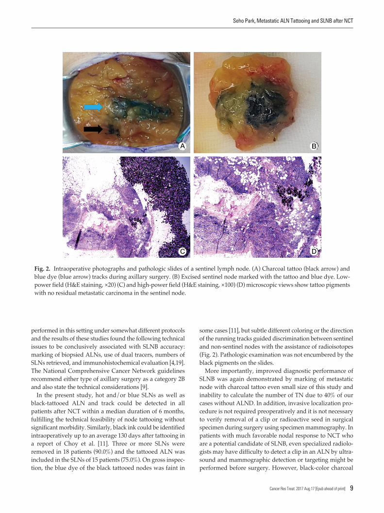
In the present study, hot and/or blue SLNs as well as
black-tattooed ALN and track could be detected in all
patients after NCT within a median duration of 6 months,
fulfilling the technical feasibility of node tattooing without
significant morbidity. Similarly, black ink could be identified
intraoperatively up to an average 130 days after tattooing in
a report of Choy et al. [11]. Three or more SLNs were
removed in 18 patients (90.0%) and the tattooed ALN was
included in the SLNs of 15 patients (75.0%). On gross inspec-
tion, the blue dye of the black tattooed nodes was faint in
some cases [11], but subtle different coloring or the direction
of the running tracks guided discrimination between sentinel
and non-sentinel nodes with the assistance of radioisotopes
(Fig. 2). Pathologic examination was not encumbered by the
black pigments on the slides.
More importantly, improved diagnostic performance of
SLNB was again demonstrated by marking of metastatic
node with charcoal tattoo even small size of this study and
inability to calculate the number of TN due to 40% of our
cases without ALND. In addition, invasive localization pro-
cedure is not required preoperatively and it is not necessary
to verify removal of a clip or radioactive seed in surgical
specimen during surgery using specimen mammography. In
patients with much favorable nodal response to NCT who
are a potential candidate of SLNB, even specialized radiolo-
gists may have difficulty to detect a clip in an ALN by ultra-
sound and mammographic detection or targeting might be
performed before surgery. However, black-color charcoal
Seho Park, Metastatic ALN Tattooing and SLNB after NCT
Cancer Res Treat. 2017 Aug 17 [Epub ahead of print] 9
Fig. 2. Intraoperative photographs and pathologic slides of a sentinel lymph node. (A) Charcoal tattoo (black arrow) and
blue dye (blue arrow) tracks during axillary surgery. (B) Excised sentinel node marked with the tattoo and blue dye. Low-
power field (H&E staining, ×20) (C) and high-power field (H&E staining, ×100) (D) microscopic views show tattoo pigments
with no residual metastatic carcinoma in the sentinel node.
A B
C D

was easily detected during surgery without any additional
mechanical device or equipment. Clinically negligible risk of
charcoal migration or absorption was presented in this study
and additional radiation hazard did not exist. However,
long-term benefits and complications of charcoal tattooing
should be further validated using large cohort studies.
According to predefined SLNB and pathological node
response, the FNR of this study with a small cohort was
worse than that of a prior meta-analyses [20,21]. However,
when the protocols of the SN FNAC study were followed,
the performance of SLNB was comparable when ypN0(i+)
was considered node-positive [8]. Although the clinical rele-
vance of very small residual nodal disease after NCT remains
unclear [22], the more number of SLNs retrieved or the wider
type of axillary surgery increased the accuracy of SLNB. Sim-
ilar to this study, Mamtani et al. [23] reported that at least
three SLNs including abnormally palpable nodes during sur-
gery could be removed in 86% of patients, but the SENTINA
and ACOSOG Z1071 studies reported that approximately
one-third to one-half of patients could have ≥ 3 SLNs
retrieved, respectively [6,7]. It is challenging to determine
whether ≥ 3 SLNs can be retrieved in the majority of patients
after NCT.
Persistently suspicious ALN metastasis after completion
of NCT is not an indication for SLNB in initially node-posi-
tive patients [9,18]. In the ACOSOG Z1071 trial, approxi-
mately 70% of patients presented normalized nodal features
on axillary ultrasound after NCT and these patients showed
acceptable SLNB performance with a FNR of 9.8% [24]. Cur-
rent cohorts with favorable nodal response to NCT seen on
preoperative work-ups also demonstrated better perform-
ance of SLNB with tattooing. Therefore, axillary restaging by
image modalities after NCT could inform patient selection
and further improvements of optimal techniques should be
investigated in this setting [19]. In addition, molecular phe-
notypes are well-known predictive factors of NCT response.
Although the small sample size in this study was not conclu-
sive, luminal subtypes showed better performance than non-
luminal subtypes in contrast to findings from prior studies
[3,25]. More research is needed to answer the remaining
questions.
Basically, SLNB should be used to excise hot and/or blue
nodes detected by dual tracers as well as suspicious enlarged
cold and non-blue ALNs intraoperatively in both adjuvant
and neoadjuvant settings [26,27]. In the present study, 11
patients underwent suspicious ALN sampling including five
tattooed node excisions and of them, three (27.3%) showed
additional two residual macronodal and one residual
micronodal metastasis from the sampled ALNs. According
to the practically recommended SLNB technique, the per-
formance of the modified SLNB in this study was the best.
Our results again emphasize the removal of suspiciously
cold or non-blue ALNs during SLNB. However, considering
the chance of non-palpable partial nodal response to NCT
and the need for balance between surgical morbidity and
diagnostic accuracy, charcoal marking at diagnosis followed
by excision of the biopsy-proven ALN after NCT, so called
targeted axillary dissection, can be one of the best ways to
improve the performance in concordance with suggestions
from the ACOSOG Z1071 study [10,28]. Furthermore, there
was no significant difference in clinicopathological parame-
ters between patients with tattooed ALN included in SLNB
and axillary sampling, suggesting the importance of the
marking technique itself to achieve successful targeted axil-
lary dissection.
Patterns of lymphatic drainage to SLNs in breast cancer are
known to be individual characteristic and are even detected
in internal mammary chains by lymphoscintigraphy [29].
Langer’s axillary arch, an anatomical variation, is found in
7% to 10% of cases during surgery or by preoperative diag-
Cancer Res Treat. 2017 Aug 17 [Epub ahead of print]
10 CANCER RESEARCH AND TREATMENT
Fig. 3. Images and tattooed sentinel node of a patient with an axillary arch. (A) The 18F-fluorodeoxyglucose (FDG) positron
emission tomography–computed tomography (PET-CT) scan shows increased FDG uptake by the metastatic axillary lymph
node in the level I left axilla (arrow). (B) The PET-CT scan demonstrates the left axillary arch that is also known as the axil-
lopectoral muscle (double arrow). (C) The hot and tattooed sentinel lymph node is retrieved.
A B C

Seho Park, Metastatic ALN Tattooing and SLNB after NCT
Cancer Res Treat. 2017 Aug 17 [Epub ahead of print] 11
nosis and can affect SLNB [30]. A 47-year-old woman who
underwent SLNB guided by only a radioisotope had a left
axillary arch noted in her initial diagnostic work-up, but her
tattooed node with the third intensity of modest uptake was
unexpectedly located in the deep portion of the axilla during
surgery, which might have been missed if done without cau-
tion (Fig. 3). Fortunately, guided by the tattoo track, the
biopsy-proven tattooed SLN could be easily removed and
technical mistakes were prevented. Although the clinical
impact of the axillary arch on SLNB in this setting is currently
uncertain, these rare circumstances can be happen to anyone
and should be kept in mind.
In conclusion, our initial experience with charcoal tattoo-
ing of cytology-confirmed metastatic ALN at presentation
showed technical feasibility without significant morbidity in
breast cancer patients treated with NCT. SLNB using dual
tracers after NCT is not limited by positive node tattooing
and has been demonstrated to improve accuracy. Further-
more, invasive preoperative localization is not additionally
required to detect the marked node. The tattooing procedure
can prevent technical errors during SLNB. A multicenter
study with a large study population is necessary to deter-
mine the clinical implications of the node-tattooing technique
including long-term side effects and its oncological safety in
this setting.
Conflicts of Interest
Conflict of interest relevant to this article was not reported.
Acknowledgments
This study was supported by a faculty research grant of Yonsei
University College of Medicine for 2015 (grant No. 6-2015-0161) and
by the Ministry of Education of the Republic of Korea and the
National Research Foundation of Korea (NRF-2015S1A5B8036349
and NRF-2017R1A2B4010407).
1. Lyman GH, Somerfield MR, Bosserman LD, Perkins CL,Weaver DL, Giuliano AE. Sentinel lymph node biopsy for patients with early-stage breast cancer: American Society ofClinical Oncology clinical practice guideline update. J ClinOncol. 2017;35:561-4.
2. Bromham N, Schmidt-Hansen M, Astin M, Hasler E, ReedMW. Axillary treatment for operable primary breast cancer.Cochrane Database Syst Rev. 2017;1:CD004561.
3. Park S, Park JM, Cho JH, Park HS, Kim SI, Park BW. Sentinellymph node biopsy after neoadjuvant chemotherapy in patients with cytologically proven node-positive breast cancerat diagnosis. Ann Surg Oncol. 2013;20:2858-65.
4. Pilewskie M, Morrow M. Axillary nodal management follow-ing neoadjuvant chemotherapy: a review. JAMA Oncol.2017;3:549-55.
5. Jatoi I, Benson JR, Toi M. De-escalation of axillary surgery inearly breast cancer. Lancet Oncol. 2016;17:e430-41.
6. Kuehn T, Bauerfeind I, Fehm T, Fleige B, Hausschild M, HelmsG, et al. Sentinel-lymph-node biopsy in patients with breastcancer before and after neoadjuvant chemotherapy (SEN-TINA): a prospective, multicentre cohort study. Lancet Oncol.2013;14:609-18.
7. Boughey JC, Suman VJ, Mittendorf EA, Ahrendt GM, WilkeLG, Taback B, et al. Sentinel lymph node surgery after neoad-juvant chemotherapy in patients with node-positive breastcancer: the ACOSOG Z1071 (Alliance) clinical trial. JAMA.2013;310:1455-61.
8. Boileau JF, Poirier B, Basik M, Holloway CM, Gaboury L,Sideris L, et al. Sentinel node biopsy after neoadjuvant
chemotherapy in biopsy-proven node-positive breast cancer:the SN FNAC study. J Clin Oncol. 2015;33:258-64.
9. Gradishar WJ, Anderson BO, Balassanian R, Blair SL, BursteinHJ, Cyr A, et al. Invasive breast cancer version 1.2016, NCCNclinical practice guidelines in oncology. J Natl Compr CancNetw. 2016;14:324-54.
10. Caudle AS, Yang WT, Krishnamurthy S, Mittendorf EA, BlackDM, Gilcrease MZ, et al. Improved axillary evaluation follow-ing neoadjuvant therapy for patients with node-positive breastcancer using selective evaluation of clipped nodes: implemen-tation of targeted axillary dissection. J Clin Oncol. 2016;34:1072-8.
11. Choy N, Lipson J, Porter C, Ozawa M, Kieryn A, Pal S, et al.Initial results with preoperative tattooing of biopsied axillarylymph nodes and correlation to sentinel lymph nodes in breastcancer patients. Ann Surg Oncol. 2015;22:377-82.
12. Shin K, Caudle AS, Kuerer HM, Santiago L, Candelaria RP,Dogan B, et al. Radiologic mapping for targeted axillary dis-section: needle biopsy to excision. AJR Am J Roentgenol.2016;207:1372-9.
13. Edge SB, Byrd DR, Compton CC, Fritz AG, Greene FL, TrottiA. AJCC cancer staging manual. 7th ed. New York: Springer;2010.
14. Hammond ME, Hayes DF, Dowsett M, Allred DC, HagertyKL, Badve S, et al. American Society of Clinical Oncology/Col-lege Of American Pathologists guideline recommendations forimmunohistochemical testing of estrogen and progesteronereceptors in breast cancer. J Clin Oncol. 2010;28:2784-95.
15. Wolff AC, Hammond ME, Hicks DG, Dowsett M, McShane
References

Cancer Res Treat. 2017 Aug 17 [Epub ahead of print]
12 CANCER RESEARCH AND TREATMENT
LM, Allison KH, et al. Recommendations for human epider-mal growth factor receptor 2 testing in breast cancer: AmericanSociety of Clinical Oncology/College of American Patholo-gists clinical practice guideline update. J Clin Oncol. 2013;31:3997-4013.
16. Weaver DL. Pathology evaluation of sentinel lymph nodes inbreast cancer: protocol recommendations and rationale. ModPathol. 2010;23 Suppl 2:S26-32.
17. Glechner A, Wockel A, Gartlehner G, Thaler K, StrobelbergerM, Griebler U, et al. Sentinel lymph node dissection only ver-sus complete axillary lymph node dissection in early invasivebreast cancer: a systematic review and meta-analysis. Eur JCancer. 2013;49:812-25.
18. Mastrangelo S, McMasters K, Ajkay N. Surgical managementof the axilla in breast cancer. Am Surg. 2016;82:475-86.
19. Rubio IT. Sentinel lymph node biopsy after neoadjuvant treat-ment in breast cancer: work in progress. Eur J Surg Oncol.2016;42:326-32.
20. van Nijnatten TJ, Schipper RJ, Lobbes MB, Nelemans PJ, Beets-Tan RG, Smidt ML. The diagnostic performance of sentinellymph node biopsy in pathologically confirmed node positivebreast cancer patients after neoadjuvant systemic therapy: asystematic review and meta-analysis. Eur J Surg Oncol.2015;41:1278-87.
21. El Hage Chehade H, Headon H, El Tokhy O, Heeney J, KasemA, Mokbel K. Is sentinel lymph node biopsy a viable alterna-tive to complete axillary dissection following neoadjuvantchemotherapy in women with node-positive breast cancer atdiagnosis? An updated meta-analysis involving 3,398 patients.Am J Surg. 2016;212:969-81.
22. King TA, Morrow M. Surgical issues in patients with breastcancer receiving neoadjuvant chemotherapy. Nat Rev ClinOncol. 2015;12:335-43.
23. Mamtani A, Barrio AV, King TA, Van Zee KJ, Plitas G,Pilewskie M, et al. How often does neoadjuvant chemotherapyavoid axillary dissection in patients with histologically con-
firmed nodal metastases? Results of a prospective study. AnnSurg Oncol. 2016;23:3467-74.
24. Boughey JC, Ballman KV, Hunt KK, McCall LM, MittendorfEA, Ahrendt GM, et al. Axillary ultrasound after neoadjuvantchemotherapy and its impact on sentinel lymph node surgery:results from the American College of Surgeons OncologyGroup Z1071 Trial (Alliance). J Clin Oncol. 2015;33:3386-93.
25. Enokido K, Watanabe C, Nakamura S, Ogiya A, Osako T,Akiyama F, et al. Sentinel lymph node biopsy after neoadju-vant chemotherapy in patients with an initial diagnosis of cytology-proven lymph node-positive breast cancer. ClinBreast Cancer. 2016;16:299-304.
26. Lyman GH, Giuliano AE, Somerfield MR, Benson AB 3rd, Bodurka DC, Burstein HJ, et al. American Society of ClinicalOncology guideline recommendations for sentinel lymphnode biopsy in early-stage breast cancer. J Clin Oncol. 2005;23:7703-20.
27. Amersi F, Giuliano AE. Management of the axilla. HematolOncol Clin North Am. 2013;27:687-702.
28. Boughey JC, Ballman KV, Le-Petross HT, McCall LM, Mitten-dorf EA, Ahrendt GM, et al. Identification and resection ofclipped node decreases the false-negative rate of sentinellymph node surgery in patients presenting with node-positivebreast cancer (T0-T4, N1-N2) who receive neoadjuvant che-motherapy: results From ACOSOG Z1071 (Alliance). AnnSurg. 2016;263:802-7.
29. Kawase K, Gayed IW, Hunt KK, Kuerer HM, Akins J, Yi M, etal. Use of lymphoscintigraphy defines lymphatic drainage pat-terns before sentinel lymph node biopsy for breast cancer. JAm Coll Surg. 2006;203:64-72.
30. Ando J, Kitamura T, Kuroki Y, Igarashi S. Preoperative diag-nosis of the axillary arch with multidetector row computed tomography and the axillary arch in association with anatom-ical problems of sentinel lymph node biopsy. Breast Cancer.2010;17:3-8.