Embed Size (px)
Citation preview

www. biophotonics-journal.orgJournal of
BIOPHOTONICS
REPRINT

REVIEW ARTICLE
The need for speed
Jeffrey L. Suhalim1, John C. Boik2, Bruce J. Tromberg1, and Eric O. Potma*; 1
1 Beckman Laser Institute, Department of Biomedical Engineering, University of California, Irvine2 Department of Chemistry, University of California, Irvine
Received 4 January 2012, accepted 14 January 2011Published online 17 February 2012
Key words: nonlinear microscopy, vibrational imaging, coherent Raman scattering, tissue imaging
1. Introduction
If the distribution of all the biomolecular constitu-ents that make up biological tissue could be reliablyvisualized into high-resolution, three-dimensionalmaps, then biomedical scientists would be able to in-vestigate tissue biology and pathology directly froma molecular perspective. Such a visualization tooldoes not currently exist, but given the enormous im-pact that it could have on the field of biomedicinedeveloping one is a laudable goal.
Optical techniques have been superior in gener-ating high-resolution maps of live tissues. At thesame time, the molecular contrast offered by opticalimaging is a far cry from the ideal molecular contrasttool mentioned above. It is clear that fluorescenceand refraction based techniques will never be ableto identify an arbitrary molecular compound in tis-sue, since only a handful of endogenous compounds
fluoresce, and refractive effects carry no significantmolecular specificity. On the other hand, vibrationalimaging techniques offer more promise. A wide vari-ety of molecular compounds can be probed, includ-ing important classes such as water, proteins, lipids,and carbohydrates. Although it ultimately falls shortof the idealized tool – the variation in bond types istoo limited to allow full discernment of all molecu-lar species – its capacity to identify many importantgroups, and its label-free, non-invasive characteris-tics, make it stand out over all other optical techni-ques as being the closest to the ideal.
The conventional vibrational imaging techniqueis Raman microscopy. Introduced in 1966, Ramanmicroscopy combines spectroscopic vibrational con-trast with a high spatial resolution. It has found manyapplications in biological imaging, material scienceand forensic research [1, 2]. The chemical contrast isderived from the Raman spectrum, a rich collection
# 2012 by WILEY-VCH Verlag GmbH & Co. KGaA, Weinheim
Journal of
BIOPHOTONICS
One of the key enabling features of coherent Ramanscattering (CRS) techniques is the dramatically improvedimaging speed over conventional vibrational imagingmethods. It is this enhanced imaging acquisition rate thathas guided the field of vibrational microscopy into theterritory of real-time imaging of live tissues. In this fea-ture article, we review several aspects of fast vibrationalimaging and discuss new applications made possible bythe improved CRS imaging capabilities. In addition, wereflect on the current limitations of CRS microscopy andlook ahead at several new developments towards real-time, hyperspectral vibrational imaging of biological tis-sues.
Mosaic of CARS images of cerebellum tissue section ofa domestic pig.
* Corresponding author: e-mail: [email protected]
J. Biophotonics 5, No. 5–6, 387–395 (2012) / DOI 10.1002/jbio.201200002
of signatures that can be traced to chemical bondvibrations [3, 4]. The amount of detail that can beextracted from the Raman spectrum is impressive.For instance, the chemical selectivity of Raman spec-troscopy has been used in cancer [5–7] and athero-sclerosis [8–10] research to discriminate healthy fromdiseased tissues, based on only small differences inchemical composition. Similarly, glucose in bloodplasma can be detected at physiologicically relevantconcentrations on the order of 10 mg/dl [11]. Theseexamples point to the general applicability of Ramanspectroscopic sensitivity for the detection of molecu-lar compounds in biological materials [12].
Practical limitations have so far prevented Ra-man microscopy from reaching its full potential. Onethese limitations – the most important – is speed.Conventional Raman microscopes, which are basedon the spontaneous Raman effect, cannot deliver theimaging speed required for routine biomedical imag-ing applications. The intrinsically weak Raman signalsseverely limit the achievable image acquisition rate.
The development of coherent Raman scattering(CRS) microscopy techniques over the last decadehas resulted in important steps toward resolving thespeed issue [13–16]. Although CRS exhibits severalunique attributes, its most important contribution tobiomedical imaging has been the dramatic reductionin acquisition time. In this feature article, we will dis-cuss several ways in which the improved speed ofCRS microscopy has transformed the biomedicalimaging field. In addition, we will touch on some ofthe challenges that lie ahead in moving towards therealization of a more generally applicable visualiza-tion technique.
2. Limitations of spontaneous Ramanscattering
The signal strength obtained by spontaneous Ramanmicroscopy, and thus the acquisition speed, is limitedby the small Raman cross sections of organic com-pounds and the inability to maximize laser intensitydue to concerns over sample damage. The Ramancross section, a material property, is a measure ofthe probability that a given molecular vibration willproduce a Raman-shifted photon. Cross sections areon the order of 10�29 cm2, which for a typical (pure)organic liquid roughly translates into one out of10 million incident photons producing one Ramanscattered photon, assuming incident light travelsthrough 1 cm of liquid.
For macroscopic Raman measurements, such lowscattering efficiencies do not pose a limitation per se.For instance, 10 mW of visible excitation light willgenerate roughly 109 Raman scattered photons per
second in a 1 cm cell of organic liquid. This is morethan enough to collect a high quality Raman spec-trum. For microscopy applications, however, the lowscattering efficiency does make a difference. For ex-ample, probing a volume of 1 cubic micrometer with10 mW of laser power, roughly 105 photons are scat-tered per second. Given that only a fraction of theseare actually detected, the number of photons regis-tered is well below 100 counts per millisecond. Usingspectrometers equipped with electron multiplyingCCD cameras, such counting rates force the shortestpixel dwell times to be in the ms range and up [17].These are the fastest Raman acquisition speeds cur-rently available; speed is not limited by detectioncapabilities as much as the finite photon flux. Thesemaximized rates can only be achieved for highlyconcentrated and condensed materials, such as pureliquids and solids. Longer acquisition times areneeded for diluted and heterogeneous samples, suchas biological tissues and cells. Imaging of live biolo-gical specimens requires pixel dwell times on the or-der of a microsecond. Under these conditions, theprocess of spontaneous Raman scattering cannot de-liver sufficient amounts of photons to enable suffi-ciently fast imaging. For real time imaging of biologi-cal materials, other approaches must be used thatscatter more photons per time unit.
One strategy to increase the photon count is toincrease the incident power. Raising the power isnot a general solution, however, as photodamage tothe sample can occur. Another strategy is surface-en-hanced Raman scattering (SERS), which avoids theneed for high power by concentrating the light in na-noscopic hotspots [18–20]. The high excitation den-sity in these hotspots generates detectable Ramansignals even at low copy numbers approaching thesingle molecule limit [21, 22]. This approach is unsui-table for general imaging purposes, however, due tothe localized nature of SERS and its reliance on exo-genous nanoparticles.
Yet another strategy to boost the Raman signal isto select only those molecules with strong electronictransitions, which in turn electronically enhance thevibrational Raman response [23]. While this reso-nant Raman approach is useful for the spectroscopicinvestigation of particular molecular chromophores,it is not generally applicable for the label-free visua-lization of biological materials.
Coherent Raman scattering avoids many of theabove mentioned limitations. CRS imaging techni-ques form a convenient solution for boosting theamount of Raman scattered photons. Stronger sig-nals are obtained because in CRS the molecules aredriven coherently, which makes them radiate in unison[24, 25]. The resulting signal is coherently amplifiedthrough constructive interference in a well-defined,phase-matched direction [26]. The directional signalenables efficient detection of the Raman response.
J. L. Suhalim et al.: The need for speed388
Journal of
BIOPHOTONICS
# 2012 by WILEY-VCH Verlag GmbH & Co. KGaA, Weinheim www.biophotonics-journal.org
The coherent amplification effect is particularlyeffective when the number of Raman scatterers infocus is high. For instance, when the microscopicfocal volume is filled with lipids, the number ofdetected photons in coherent anti-Stokes Ramanscattering (CARS), generated from the CH2 stretch-ing mode, can easily exceed 102 per microsecond at10 mW of illumination. With such high signal levels,real-time Raman imaging of biological tissues be-comes feasible.
CRS techniques, including CARS and stimulatedRaman scattering (SRS), have opened up Raman-based imaging to a broad range of biological andbiomedical applications. The stronger signal levels inCRS allow for image acquisition at video rate, corre-sponding to 30 frames per second and pixel dwelltimes as short as 100 ns. Applications that were pre-viously unthinkable to a Raman spectroscopist, suchas real-time imaging of live human tissues, have nowbecome a reality.
3. Fast imaging with CRS
The fast imaging capability of CRS microscopy en-ables completion of various tasks that were pre-viously beyond the scope of conventional Raman mi-croscopy. First, the faster imaging speed permitsimaging of large tissue segments at high resolution.Because tissue maps are composed of millions ofpoint measurements, fast imaging capabilities are es-sential to generate such maps within a realistic timeframe. Second, high-speed imaging has allowed imag-ing of live tissues. The rapid motion of live samplescan give rise to blurring of images, which is naturallycircumvented when the imaging acquisition rate suf-ficiently high. Third, fast imaging enables visualiza-tion of dynamic processes. Many biologically rele-vant processes occur over short enough time spansto preclude the use of slower spontaneous Ramantechnologies. Below we summarize some recent ad-vances in these areas.
3.1 Imaging of large tissue sections
An important application of high speed CRS imag-ing is the visualization of large tissue segments. Che-mical and structural details relevant to the biologyand pathology of tissues manifest themselves on awide range of length scales, from sub-cellular detailsto mesoscopic scale tissue morphology. While cover-ing surface areas of up to several cm2, chemical mapsof tissues need to retain sufficient resolution to cap-ture microscopic features as well. This broad range ofspatial scales requires a high density of data points.
To accomplish this with laser-scanning micro-scopy, large-scale tissue maps are typically generatedby recording adjacent individual images at a smallerfield of view, which form the tiles of a larger mosaic.Consequently, the total amount of individual pointmeasurements is large, and the compiled tissue mapscan easily contain more than several Gigapixels. Fastimaging capabilities are crucial to keep the acquisi-tion of such large maps within realistic time frames.Here, the difference between a microsecond and amillisecond pixel dwell time is vast, as the shorterdwell time would enable the recording of a 1 Giga-pixel map in approximately 17 minutes while thelonger pixel dwell time would require 12 days ofmeasurement time.
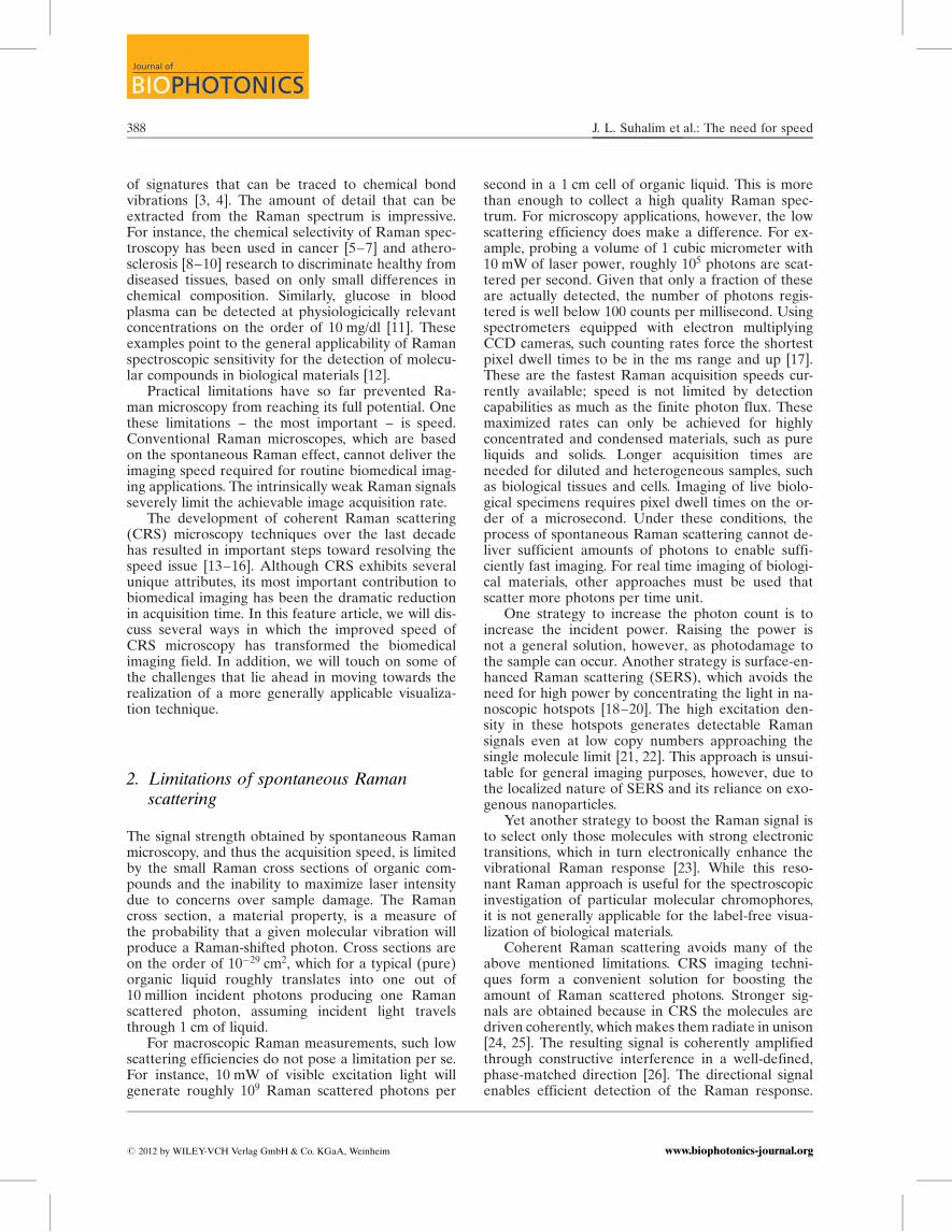
Centimeter scale tissue maps have been con-structed in several ex vivo CRS studies. Examplesinclude neural injuries [27] and myelination disor-ders in live spinal tissues [28–30], mapping of athe-rosclerotic plaques in aortas [31–33], demarcation ofbrain tumor boudaries [34], and establishing correla-tions between mammary tumor growth and lipid in-gestion [35]. In these studies, tissue maps are gener-ated that cover up to several centimeters in lateraland hundreds of microns in axial distance, while pre-serving the (sub-)micrometer resolution offered bythe high numerical aperture objective. An exampleis given in Figure 1, where a mosaic of CARS images
Figure 1 Mosaic of CARS images of cerebellum tissue sec-tion of a domestic pig. Raman shift was set at the2850 cm�1 CH2 symmetric stretching vibration to visualizelipids. Bright signals correspond to white matter (lipid-rich,myelined axons) and dimmer signals correspond to greymatter (lipid poor areas). Total area is 1.4 � 1.4 cm2. Re-printed in part with permission from Ref. [36].
J. Biophotonics 5, No. 5–6 (2012) 389
REVIEWREVIEWARTICLEARTICLE
# 2012 by WILEY-VCH Verlag GmbH & Co. KGaA, Weinheimwww.biophotonics-journal.org

provides chemical contrast between lipid-rich whitematter and lipid-poor grey matter in a cerebellum tis-sue section [36].
3.2 Imaging of live tissues
Raman imaging of live tissues is an application inwhich fast imaging capability is indispensable. Thereare several reasons why high image acquisition rates,from several frames per second up to video rate, areadvantageous for live tissue imaging applications.First, the region of interest in the tissue of animalsor human subjects is typically a small part of a muchlarger area. To find the target area, or to inspect fea-tures over a larger area, it is necessary to move thefield of view laterally and axially through the tissue.This microscopic browsing of a macroscopic tissuerequires real-time imaging to maximize the efficiencyof the microscopist and to minimize discomfort tothe subject.
Second, the intrinsic movement of the tissue, dueto pulsation of blood vessels, breathing, or positionaladjustments by the subject, poses a challenge to mi-croscopic imaging. Where motions in the tissue occurat a faster pace than the imaging frame rate, blurringof the images is inevitable. Tissue motions can mani-fest over several length scales, from microscopic tocomplete translation of the tissue sample, introdu-cing artifacts in the image that are difficult to elimi-nate retro-actively. Imaging at video-rate enables im-age acquisition in which the individual frames showminimal blurring due to pulse pressure, breathing, orother common movements. Under these conditions,lateral translational movements in the range of up toseveral tens of microns can be addressed with post-processing image correction methods, producing vir-tually motion free time-lapse recordings [37].
Third, fast imaging can mitigate photodamagecaused by excitation radiation. Shorter pixel dwelltimes minimize persistent light exposure in each illu-minated spot, which reduces the photodamage thatis caused by linear heat absorption. In general, fasterimaging enables quicker acquisitions and thus mini-mal light exposure per unit area examined.
High speed CRS microscopy has been success-fully used to collect chemical selective signals fromvarious tissues in rodent models in vivo. Video-rateCARS microscopy of live mice enabled visualizationof lipid rich structures in the skin, including sebac-eous glands and dermal adipocytes [38]. The fastimaging capability of CARS was also crucial in stud-ies focused on demyelination processes in the spinalcord of mice in vivo (Figure 2) [27–29, 39, 40], produc-ing important insights into the pathology of neuralinjuries and possible treatments for multiple sclerosis[41, 42]. In addition, fast CARS signal acquisition
has been key for the intravital identification of circu-lating tumor cells in blood vessels of mice [43].
Besides live tissue imaging in small animal mod-els, CRS imaging has been applied to the examina-tion of human skin in vivo. Using a microscope sys-tem equipped with a flexible arm that is optimizedfor clinical use, Konig et al demonstrated depth re-solved CARS imaging of lipid and water distributionsin human skin in vivo [44, 45]. Similarly, video-rateSRS imaging was shown to be capable of generatingprotein density maps of skin of human subjects [46].It was found that topically applied d-DMSO perco-lated predominantly through the skin along hairshafts. Work in this direction indicates the capabilitiesof CRS modalities for expanded clinical applications.
3.3 Visualizing dynamic processes
Prior to the development of CRS imaging, the longacquisition times characteristic of vibrational micro-scopy limited applications to the study of inanimatespecimens. The fast imaging capability introduced byCRS has permanently opened the door to the exam-ination of live cells and tissues. As part of this, faster
Figure 2 (online color at: www.biophotonics-journal.org)Fast imaging enables visualization of live tissues. Multimo-dal nonlinear imaging of spinal nerves in live mouse. Mye-lin (green) is visualized with CARS, and axonal body (red)is observed with two-photon fluorescence through Thy1-YFP labeling. Reflectance contrast (blue) is used for gen-eral guidance. Image size is 112.5 � 112.5 mm2. Image cour-tesy of E. Belanger, A. Daradich, B. Aube, D. Cote, Uni-versite Laval.
J. L. Suhalim et al.: The need for speed390
Journal of
BIOPHOTONICS
# 2012 by WILEY-VCH Verlag GmbH & Co. KGaA, Weinheim www.biophotonics-journal.org

imaging enables a time- and space-resolved view ofdynamic processes of biological relevance.
The sub-second CRS image acquisition rate issufficient to register a wide range of dynamic pro-cesses in live cells. For instance, CARS microscopyhas been used to determine the water diffusion andpermeability parameters in amoeba and neutrophilecells (Figure 3) [47, 48]. The combination of highspeed imaging and sensitivity to lipids has further-more propelled several CARS studies on the dy-namics of intracellular lipid droplets. Examples in-clude the characterization of lipid droplet traffickingin adrenal cortical tumor cells [49], lipid dropletgrowth and distribution in human hepatoma cells[50], lipid droplet remodeling due to lipolytic stimu-lation in fibroblast cells [51], and lipid droplet varia-bility in induced adipogenesis [52]. In addition to vi-sualizing mobile lipid droplets in individual cells,CRS microscopy has also been used to identify lipidreservoirs and study lipid metabolism in living nema-tode organisms [53–56]. Nematodes can be very mo-bile in the seconds to minutes range, even when re-straints to movement are employed.
Besides capturing stills of moving eukaryotic cellsand organisms, high speed CRS imaging has alsoproven useful for studying diffusion and chemicalkinetics in a variety of biological systems in vitro.Examples include the use of CARS to examine thereal-time dissolution of the drug theophylline anhy-drate from tablets [57], and the use of SRS to followthe diffusion of pharmaceutically relevant agentssuch as ketoprofen and propylene glycol throughskin specimens ex vivo [58], Also, multiplex CARShas been used successfully for following the kinetics
of lipid digestion by lipase enzymes (Figure 4) [59].The spectral acquisition rate was reduced to 20 msper pixel, which allowed complete spatial mappingof the digestion of 10 micrometer-sized lipid dropletson a timescale relevant to lipase kinetics. For biolo-gical samples of this nature, comparable image ac-quisition times are extremely challenging to achievewith spontaneous Raman microscopy.
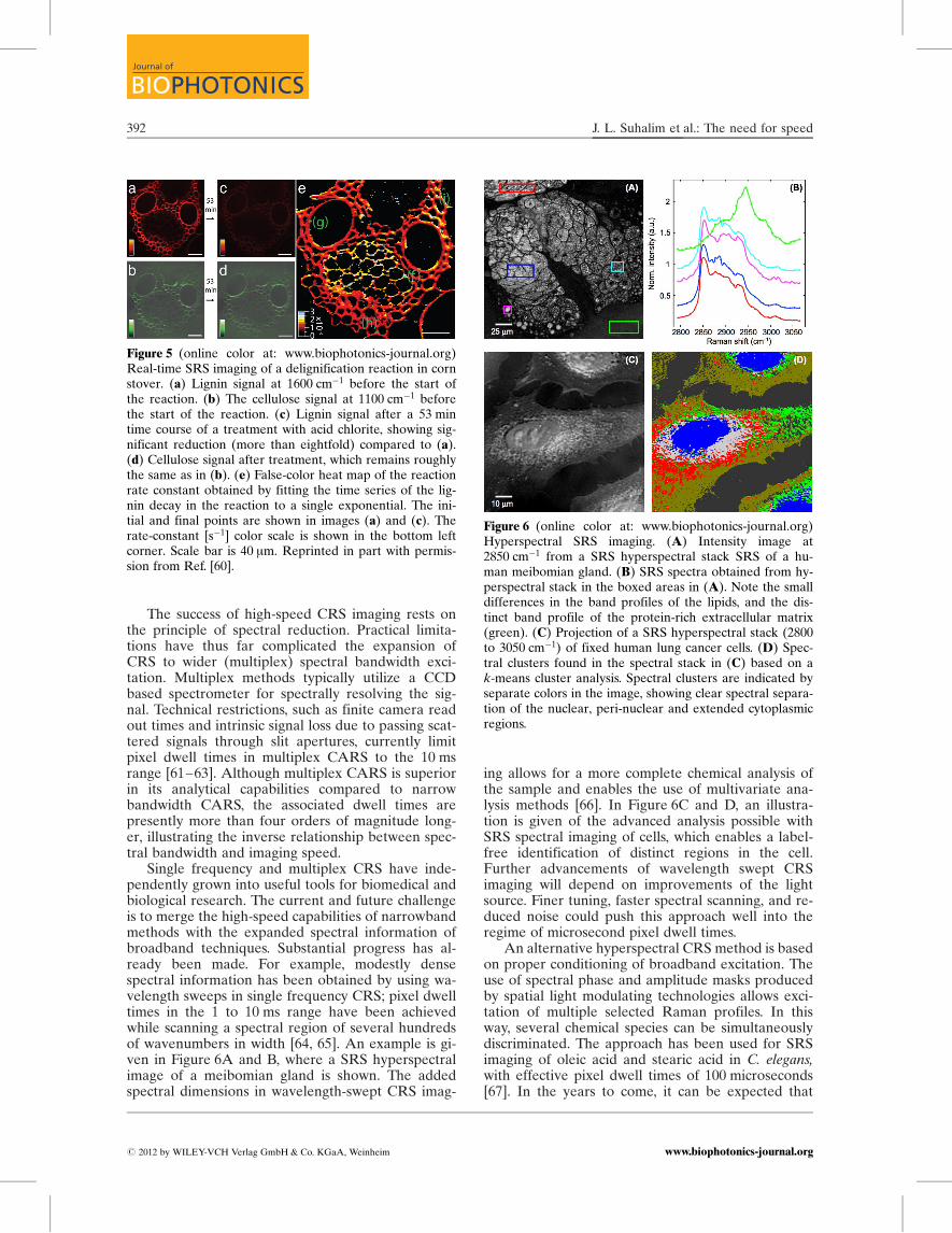
Another example of the capability of CRS to re-solve spatially variant chemical kinetics is demon-strated in studies on delignification and cellulosehydrolysis in plant materials. Saar et al recently gen-erated spatiotemporal maps of the chemically in-duced delignification process in corn stover speci-mens, allowing a spatial analysis of reaction dynamics(Figure 5) [60]. This work nicely demonstrates theunique analytical capabilities of fast chemical imag-ing, which enables the direct measurement of bondselective chemical conversions.
4. Limitations of high-speed CRS imaging
The fastest implementations of CRS imaging havethus far been based on narrow bandwidth (singlefrequency) excitation of selected Raman lines. Usingpicosecond laser beams, CRS imaging can routinelybe accomplished with sub-microsecond pixel dwelltimes, generating images with contrast based on anarrow segment (1–10 cm�1) of the Raman spectrum.Compared to spontaneous Raman microspectro-scopy, speed is gained at the cost of information loss;the narrow bandwidth provides limited spectral in-formation. For several applications, most notably thevisualization of lipids, the single frequency vibrationalcontrast is sufficient for quantitative identificationof the target compounds. Many other applications,however, would benefit from a higher informationdensity in the spectral dimension.
Figure 3 (online color at: www.biophotonics-journal.org)Dynamic recording of water effusion from D. discoideumcells using fast CARS imaging. Raman shift was set at the3220 cm�1 OH stretching vibration to visualize water. Effu-sion maps were obtained by rapidly flushing D2O throughthe cells. A spatio-temporal effusion map acquired from aselected area (top left, black box) of the cell is shown.Adapted from Ref. [47].
Figure 4 (online color at: www.biophotonics-journal.org)Time-resolved images of the digestion of a glyceryl triole-ate droplet by porcine pancreatic lipase. (a) Bright-fieldmicroscopy images; (b) false-color images obtained byCARS microspectroscopy of glyceryl trioleate (red) and li-polytic products (green). Scale bar ¼ 5 mm, pixel step size ¼1 mm, 41 � 41 pixels, measurement time per image ¼ 35 s.Enzyme concentration ¼ 1.5 mg mL�1. Reprinted with per-mission from Ref. [59]. Copyright (2010) American Chemi-cal Society.
J. Biophotonics 5, No. 5–6 (2012) 391
REVIEWREVIEWARTICLEARTICLE
# 2012 by WILEY-VCH Verlag GmbH & Co. KGaA, Weinheimwww.biophotonics-journal.org
The success of high-speed CRS imaging rests onthe principle of spectral reduction. Practical limita-tions have thus far complicated the expansion ofCRS to wider (multiplex) spectral bandwidth exci-tation. Multiplex methods typically utilize a CCDbased spectrometer for spectrally resolving the sig-nal. Technical restrictions, such as finite camera readout times and intrinsic signal loss due to passing scat-tered signals through slit apertures, currently limitpixel dwell times in multiplex CARS to the 10 msrange [61–63]. Although multiplex CARS is superiorin its analytical capabilities compared to narrowbandwidth CARS, the associated dwell times arepresently more than four orders of magnitude long-er, illustrating the inverse relationship between spec-tral bandwidth and imaging speed.
Single frequency and multiplex CRS have inde-pendently grown into useful tools for biomedical andbiological research. The current and future challengeis to merge the high-speed capabilities of narrowbandmethods with the expanded spectral information ofbroadband techniques. Substantial progress has al-ready been made. For example, modestly densespectral information has been obtained by using wa-velength sweeps in single frequency CRS; pixel dwelltimes in the 1 to 10 ms range have been achievedwhile scanning a spectral region of several hundredsof wavenumbers in width [64, 65]. An example is gi-ven in Figure 6A and B, where a SRS hyperspectralimage of a meibomian gland is shown. The addedspectral dimensions in wavelength-swept CRS imag-
ing allows for a more complete chemical analysis ofthe sample and enables the use of multivariate ana-lysis methods [66]. In Figure 6C and D, an illustra-tion is given of the advanced analysis possible withSRS spectral imaging of cells, which enables a label-free identification of distinct regions in the cell.Further advancements of wavelength swept CRSimaging will depend on improvements of the lightsource. Finer tuning, faster spectral scanning, and re-duced noise could push this approach well into theregime of microsecond pixel dwell times.
An alternative hyperspectral CRS method is basedon proper conditioning of broadband excitation. Theuse of spectral phase and amplitude masks producedby spatial light modulating technologies allows exci-tation of multiple selected Raman profiles. In thisway, several chemical species can be simultaneouslydiscriminated. The approach has been used for SRSimaging of oleic acid and stearic acid in C. elegans,with effective pixel dwell times of 100 microseconds[67]. In the years to come, it can be expected that
Figure 5 (online color at: www.biophotonics-journal.org)Real-time SRS imaging of a delignification reaction in cornstover. (a) Lignin signal at 1600 cm�1 before the start ofthe reaction. (b) The cellulose signal at 1100 cm�1 beforethe start of the reaction. (c) Lignin signal after a 53 mintime course of a treatment with acid chlorite, showing sig-nificant reduction (more than eightfold) compared to (a).(d) Cellulose signal after treatment, which remains roughlythe same as in (b). (e) False-color heat map of the reactionrate constant obtained by fitting the time series of the lig-nin decay in the reaction to a single exponential. The ini-tial and final points are shown in images (a) and (c). Therate-constant [s�1] color scale is shown in the bottom leftcorner. Scale bar is 40 mm. Reprinted in part with permis-sion from Ref. [60].
Figure 6 (online color at: www.biophotonics-journal.org)Hyperspectral SRS imaging. (A) Intensity image at2850 cm�1 from a SRS hyperspectral stack SRS of a hu-man meibomian gland. (B) SRS spectra obtained from hy-perspectral stack in the boxed areas in (A). Note the smalldifferences in the band profiles of the lipids, and the dis-tinct band profile of the protein-rich extracellular matrix(green). (C) Projection of a SRS hyperspectral stack (2800to 3050 cm�1) of fixed human lung cancer cells. (D) Spec-tral clusters found in the spectral stack in (C) based on ak-means cluster analysis. Spectral clusters are indicated byseparate colors in the image, showing clear spectral separa-tion of the nuclear, peri-nuclear and extended cytoplasmicregions.
J. L. Suhalim et al.: The need for speed392
Journal of
BIOPHOTONICS
# 2012 by WILEY-VCH Verlag GmbH & Co. KGaA, Weinheim www.biophotonics-journal.org

spectral conditioning methods will improve, both inspeed and bandwidth, ultimately combining the high-speed imaging capabilities of picosecond CRS withthe spectral breadth of Raman spectroscopy.
5. Conclusions
CRS methods have one key advantage over sponta-neous Raman microscopy: speed. The (sub-)micro-second pixel dwell times offered by narrowband CRSimaging methods have initiated a new era of chemi-cal imaging applications in biology and biomedicine.Single frequency CARS and SRS have already prov-en indispensable in the study of lipids and lipid me-tabolism in live tissues and cells; such investigationswould not have been possible with conventionalspontaneous Raman techniques. Because of its imag-ing speed and chemical contrast, CRS has pushedthe concept of label-free and noninvasive chemicalimaging closer to clinical biomedical applications.The challenge ahead is to marry the speed qualitiesof single frequency scanning with the superior spec-tral information of broadband Raman spectroscopy.Recent developments suggest several approaches bywhich this could occur and provide a glimpse towardthe ideal of clinically relevant, real-time chemical in-spection of live tissues.
Acknowledgements We acknowledge support from theNational Institutes of Health, Grant P41-RR-01192 (LaserMicrobeam and Medical Program, LAMMP), from theNational Science Foundation, Grant CHE-0847097. J.C.B.acknowledges financial support from the Life ExtensionFoundation.
References
[1] M. Delhaye and M. Migeon, C. R. Acad. Sc. Paris 262,1513 (1966).
[2] G. Turrell and J. Corset, Raman Microscopy. Devel-opments and Applications (Academic Press, San Die-go, 1996).
[3] C. V. Raman and K. S. Krishnan, Proc. Roy. Soc. Lon-don 122, 23 (1929).
Jeffrey L. Suhalim obtaineda B.S. in Bioengineeringfrom the University of Cali-fornia, Riverside, where healso received the Bioengi-neering Academic Excel-lence Award at the HonorsConvocation. He is currentlya graduate student in theBiomedical Engineeringprogram at the University ofCalifornia, Irvine. He is fo-cusing on the development
and applications of nonlinear optical imaging techni-ques in cardiovascular and eye research.
Dr. John C. Boik is Presidentof the nonprofit researchgroup New Earth BioMed.He received his Ph.D. Bio-medical Sciences from theUniversity of Texas HealthSciences Center in Houstonand conducted his Post-doctoral work at StanfordUniversity Statistics Depart-ment. He is currently a vis-iting scientist at the Univer-sity of California, Irvine.
Dr. Bruce J. Tromberg is theDirector of the BeckmanLaser Institute and MedicalClinic at the University ofCalifornia, Irvine (UCI) andprincipal investigator of theLaser Microbeam and Medi-cal Program, an NIH Na-tional Biomedical Technol-ogy Center. He is a Professorin the departments of Bio-medical Engineering andSurgery and co-leads the
Onco-imaging and Spectroscopy Program in UCI’sChao Family Comprehensive Cancer Center. His re-search interests are in biomedical optics, including dif-fuse optics, non-linear microscopy, metabolic imaging,and photodynamic therapy.
Dr. Eric O. Potma is an As-sociate Professor in the De-partment of Chemistry atthe University of California,Irvine (UCI). He holds anadjunct position in theBeckman Laser Instituteand Medical Clinic at UCI.His research group is activein developing nonlinear op-tical imaging techniques forthe purpose of interrogating
biological tissues and nanostructured materials.
J. Biophotonics 5, No. 5–6 (2012) 393
REVIEWREVIEWARTICLEARTICLE
# 2012 by WILEY-VCH Verlag GmbH & Co. KGaA, Weinheimwww.biophotonics-journal.org

[4] G. Placzek, Handbuch der Radiologie, edited byE. Marx (Academische-Verlag, Leipzig, 1934), p. 205.
[5] H. Yamazaki, S. Kaminaka, E. Kohda, M. Mukai, andH. Hamaguchi, Radiat. Med. 21, 1 (2003).
[6] G. Shetty, C. Kendall, N. Shepherd, N. Stone, andH. Barr, Br. J. Cancer 94, 1460 (2006).
[7] S. K. Teh, W. Zheng, K. Y. Ho, M. Teh, K. G. Yeoh,and Z. Huang, Brit. J. Cancer 98, 457 (2008).
[8] H. P. Buschman, J. T. Motz, G. Deinum, T. J. Romer,M. Fitzmaurice, J. R. Kramer, A. v. d. Laarse, A. V.Bruschke, and M. S. Feld, Cardiovas. Pathology 10, 59(2001).
[9] G. V. Nogueira, L. Silveira, A. A. Martin, R. A. Zan-garo, M. T. T. Pacheco, M. C. Chavantes, and C. A.Pasqualucci, J. Biomed. Opt. 10, 031117 (2005).
[10] J. T. Motz, M. Fitzmaurice, A. Miller, S. J. Gandhi,A. S. Haka, L. H. Galindo, R. R. Dasari, J. R. Kramer,and M. S. Feld, J. Biomed. Opt. 11, 021003 (2006).
[11] C. R. Kong, I. Barman, N. C. Dingari, J. W. Kang,L. Galindo, R. R. Dasari, and M. S. Feld, AIP Adv. 1,032175 (2011).
[12] P. R. Carey, Biochemical Applications of Raman andResonance Raman Spectroscopies (Academic Press,Toronto, 1982).
[13] J. X. Cheng, Appl. Spectrosc. 91, 197 (2007).[14] C. L. Evans and X. S. Xie, Annu. Rev. Anal. Chem. 1,
883 (2008).[15] W. Min, C. W. Freudiger, S. Lu, and X. S. Xie, Annu.
Rev. Phys. Chem. 62, 507 (2011).[16] A. Volkmer, J. Phys. D 38, R59 (2005).[17] O. Hollricher, Confocal Raman Microscopy, edited by
T. Dieing, O. Hollricher, and J. Toporski (Springer-Verlag, Berlin, 2011).
[18] D. L. Jeanmaire and R. P. V. Duyne, J. Electroanal.Chem. 84, 1 (1977).
[19] M. Moskovits, Rev. Mod. Phys. 57, 783 (1985).[20] A. Otto, I. Mrozek, H. Grabborn, and A. Akemann,
J. Phys. Condens. Matter 4, 1143 (1992).[21] S. Nie and S. R. Emory, Science 275, 1102 (1997).[22] J. A. Dieringer, R. B. Lettan, K. A. Scheidt, and R. P.
V. Duyne, J. Am. Chem. Soc. 129, 16249 (2007).[23] J. Behringer, Raman Spectroscopy, edited by H. A.
Szymanski (Plenum Press, New York, 1967), p. 168.[24] J. A. Armstrong, N. Bloembergen, J. Ducuing, and P. S.
Pershan, Phys. Rev. 127, 1918 (1962).[25] P. D. Maker and R. W. Terhune, Phys. Rev. 137, A801
(1965).[26] J. X. Cheng, A. Volkmer, L. D. Book, and X. S. Xie,
J. Phys. Chem. B 105, 1277 (2001).[27] F. Henry, D. Cote, M. A. Randolph, E. A. Z. Rust,
R. W. Redmond, I. E. Kochevar, C. P. Lin, and J. M.Winograd, Plastic and Reconstructive Surgery 123,123S (2009).
[28] T. B. Huff and J. X. Cheng, J. Microsc. 225, 175 (2007).[29] T. B. Huff, Y. Shi, Y. Yan, H. Wang, and J. X. Cheng,
IEEE J. Sel. Top. Quant. Electron. 14, 4 (2008).[30] H. Wang, Y. Fu, P. Zickmund, R. Shi, and J. X. Cheng,
Biophys. J. 89, 581 (2005).[31] H. W. Wang, I. M. Langohr, M. Sturek, and J. X. Cheng,
Artherioscler. Thromb. Vasc. Biol. 29, 1342 (2009).
[32] H. W. Wang, T. T. Le, and J. X. Cheng, Opt. Commun.281, 1813 (2008).
[33] R. S. Lim, A. Kratzer, N. P. Barry, S. Miyazaki-Anzai,M. Miyazaki, W. W. Mantulin, M. Levi, E. O. Potma,and B. J. Tromberg, J. Lipid Res. 51, 1729 (2010).
[34] C. L. Evans, X. Xu, S. Kesari, X. S. Xie, S. T. C. Wong,and G. S. Young, Opt. Express 15, 12076 (2007).
[35] T. T. Le, C. W. Rehrer, T. B. Huff, M. B. Nichols, I. G.Camarillo, and J. X. Cheng, Mol. Imaging 6, 205(2007).
[36] T. Meyer, N. Bergner, C. Bielecki, C. Krafft, D. Aki-mov, B. F. M. Romeike, R. Reichart, R. Kalff, B. Die-tzek, and J. Popp, J. Biomed. Opt. 16, 021113 (2011).
[37] I. Veilleux, J. A. Spencer, D. P. Bliss, D. Cote, andC. P. Lin, IEEE J. Sel. Top. Quant. Electron. 14, 10(2008).
[38] C. L. Evans, E. O. Potma, M. Puoris’haag, D. Cote,C. Lin, and X. S. Xie, Proc. Natl. Acad. Sci. USA 102,16807 (2005).
[39] Y. Fu, H. Wang, T. B. Huff, R. Shi, and J. X. Cheng,J. Neurosci. Res. 85, 2870 (2007).
[40] J. Imitola, S. Rasmussen, Y. Liu, T. Chitnis, S. J. Khoury,D. Cote, X. S. Xie, C. P. Lin, and R. L. Sidman, J.Biomed. Opt. 16, 021109 (2011).
[41] Y. Shi, S. Kim, R. B. Borgens, K. Park, R. Shi, andJ. X. Cheng, Nat. Nanotechnol. 5, 80 (2010).
[42] T. B. Huff, Y. Shi, W. Sun, W. Wu, R. Shi, and J. X.Cheng, PLoS One 6, e17176 (2011).
[43] T. T. Le, T. B. Huff, and J. X. Cheng, BMC Cancer 9,42 (2009).
[44] H. G. Breunig, R. Buckle, M. Kellner-Hofer, M. Wei-nigel, J. Lademann, W. Sterry, and K. Konig, Microsc.Res. Techn., DOI: 10.1002/jemt.21082 (2011).
[45] K. Konig, H. G. Breunig, R. Buckle, M. Kellner-Hofer,M. Weinigel, E. Buttner, W. Sterry, and J. Lademann,Laser Phys. Lett. 8, 465 (2011).
[46] B. G. Saar, C. W. Freudiger, J. Reichman, S. C. Michael,G. R. Holtom, and X. S. Xie, Science 330, 1368 (2010).
[47] E. O. Potma, W. P. d. Boeij, P. J. M. v. Haastert, andD. A. Wiersma, Proc. Natl. Acad. Sci. USA 98, 1577(2001).
[48] E. P. Reeves, H. Lu, H. L. Jacobs, C. G. M. Messina,S. Bolsover, G. Gabella, E. O. Potma, A. Warley,J. Roes, and A. W. Segal, Nature 416, 291 (2002).
[49] X. Nan, E. O. Potma, and X. S. Xie, Biophys. J. 91,728 (2006).
[50] R. K. Lyn, D. C. Kennedy, S. M. Segan, D. R. Blais,Y. Rouleau, A. F. Pegoraro, X. S. Xie, A. Stolow, andJ. P. Pezacki, Virology 394, 130 (2009).
[51] T. Yamaguchi, N. Omatsu, E. Morimoto, H. Nakashi-ma, K. Ueno, T. Tanaka, K. Satouchi, F. Hirose, andT. Osumi, J. Lipid Res. 48, 1078 (2007).
[52] T. T. Le and J. X. Cheng, PLoS One 4, e5189 (2009).[53] T. Hellerer, C. Axang, C. Brackmann, P. Hillertz,
M. Pilon, and A. Enejder, Proc. Natl. Acad. Sci. USA104, 14658 (2007).
[54] C. Morck, L. Olsen, C. Kurth, A. Persson, N. J. Storm,E. Svensson, J. O. Jansson, M. Hellqvist, A. Enejder,N. J. Faergeman, and M. Pilon, Proc. Natl. Acad. Sci.USA 106, 18285 (2009).
J. L. Suhalim et al.: The need for speed394
Journal of
BIOPHOTONICS
# 2012 by WILEY-VCH Verlag GmbH & Co. KGaA, Weinheim www.biophotonics-journal.org

[55] T. T. Le, H. M. Duren, M. N. Slipchenko, C. D. Hu,and J. X. Cheng, J. Lipid Res. 51, 672 (2009).
[56] M. C. Wang, W. Min, C. W. Freudiger, G. Ruvkun, andX. S. Xie, Nat. Meth. 8, 135 (2011).
[57] M. Windbergs, M. Jurna, H. L. Offerhaus, J. L. Herek,K. Kleinebudde, and C. J. Strachan, Anal. Chem. 81,2085 (209).
[58] B. G. Saar, L. R. Contreras-Rojas, X. S. Xie, and R. H.Guy, Mol. Pharm. 8, 969 (2011).
[59] J. P. R. Day, G. Rago, K. F. Domke, K. P. Velikov, andM. Bonn, J. Am. Chem. Soc. 132, 8433 (2010).
[60] B. G. Saar, Y. Zeng, C. W. Freudiger, Y. S. Liu, M. E.Himmel, X. S. Xie, and S. Y. Ding, Angew. Chem. Int.Ed. 49, 5476 (2010).
[61] S. H. Parekh, Y. J. Lee, K. A. Aamer, and M. T. Cice-rone, Biophys. J. 99, 2695 (2010).
[62] M. Okuno, H. Kano, P. Leproux, V. Couderc, J. P. R.Day, M. Bonn, and H. Hamaguchi, Angew. Chem. 49,6773 (2010).
[63] C. Pohling, T. Buckup, and M. Motzkus, J. Biomed.Opt. 16, 021105 (2011).
[64] S. Begin, B. Burgoyne, V. Mercier, A. Villeneuve,R. Vallee, and D. Cote, Biomed. Opt. Express 2, 1296(2011).
[65] R. S. Lim, J. L. Suhalim, S. Miyazaki-Anzai, M. Miya-zaki, M. Levi, E. O. Potma, and B. J. Tromberg, J. Li-pid Res. 52, 2177 (2011).
[66] M. Miljkovic, T. Chernenko, M. J. Romeo, B. Bird,C. Matthaus, and M. Diem, Analyst. 135, 2002 (2010).
[67] C. W. Freudiger, W. Min, G. R. Holtom, B. Xu, M. Dan-tus, and X. S. Xie, Nat. Photon. 5, 103 (2011).
Register now for the freeWILEY-VCH Newsletter!www.wiley-vch.de/home/pas
In ‘Laser Imaging and Manipulation in Cell Biology’, the editor has gathered a team of international experts to present the latest advances in the field of laser imaging and manipulation techniques. The result is broad coverage of the interactions with biological samples to perform novel optical manipulationoperations, both on the cellular and tissue levels.
Of interest to physicists working and researching laser tissue mechanisms, cell biologists investigating new imaging and manipulation operation on the cellular level, medical doctors working with new laser therapies and diagnostic tools, as well as engineers developing new technologies in the field of optics and lasers.
2010. XIV, 246 pages, 71 figures, 3 in color. Hardcover.€ 99.– /£ 85.– /US$ 135.–ISBN: 978-3-527-40929-7
WILEY-VCH • P.O. Box 10 11 61 • 69451 Weinheim, GermanyFax: +49 (0) 62 01 - 60 61 84e-mail: [email protected] • http://www.wiley-vch.de
Edited by FRANCESCO S. PAVONEEuropean Laboratory for Non Linear Spectroscopy, and Department of Physics, University of Florence
Laser Imaging and Manipulation in Cell Biology
+++ Suggested Reading +++ Suggested Reading +++ Suggested Reading +++
J. Biophotonics 5, No. 5–6 (2012) 395
REVIEWREVIEWARTICLEARTICLE
# 2012 by WILEY-VCH Verlag GmbH & Co. KGaA, Weinheimwww.biophotonics-journal.org