Embed Size (px)
Citation preview

lable at ScienceDirect
Journal of Archaeological Science xxx (2011) 1e14
Contents lists avai
Journal of Archaeological Science
journal homepage: http : / /www.elsevier .com/locate/ jas
Exceptional preservation of a prehistoric human brain from Heslington,Yorkshire, UK
Sonia O’Connor a,*, Esam Ali b, Salim Al-Sabah c, Danish Anwar c,d, Ed Bergströmd,e, Keri A. Brown f,Jo Buckberry a, Stephen Buckley g, Matthew Collins h, John Denton i, Konrad M. Dorling f, Adam Dowle c,d,Phil Duffey j, Howell G.M. Edwards b, Elsa Correia Faria f, Peter Gardner f, Andy Gledhill a, Karl Heaton d,e,Carl Heron a, Rob Janawaya, Brendan J. Keely e, David King j, Anthony Masinton g, Kirsty Penkman h,Axel Petzold k, Matthew D. Pickering e, Martin Rumsby l, Holger Schutkowski a, Kimberley A. Shackleton e,Jerry Thomas c,d, Jane Thomas-Oates d,e, Maria-Raimonda Usai g, Andrew S. Wilson a, Terry O’Connor g
aArchaeological Sciences, University of Bradford, Richmond Road, Bradford BD7 1DP, UKbChemical and Forensic Sciences, University of Bradford, Richmond Road, Bradford BD7 1DP, UKc Technology Facility, Department of Biology (15), University of York, PO Box 373, York YO10 5YW, UKdCentre of Excellence in Mass Spectrometry, University of York, Heslington, York YO10 5DD, UKeDepartment of Chemistry, University of York, Heslington, York YO10 5DD, UKfManchester Interdisciplinary Biocentre, University of Manchester, 131 Princess Street, Manchester M1 7DN, UKgDepartment of Archaeology, University of York, King’s Manor, York YO1 7EP, UKhBioArCh, Departments of Biology, Archaeology and Chemistry, University of York, York YO10 5DD, UKiDepartment of Laboratory Medicine, Stopford Building, University of Manchester, Oxford Road, Manchester M13 9PT, UKjYorkHospital, Wigginton Road, York YO31 8HE, UKkDepartment of Neuroimmunology, UCL Institute of Neurology, Queen Square, London WC1N 3BG, UKlDepartment of Biology, University of York, Heslington, York YO10 5YW, UK
a r t i c l e i n f o
Article history:Received 11 October 2010Received in revised form22 February 2011Accepted 24 February 2011
Keywords:Brain tissueWaterloggingBurial environmentAdipocerePutrefactionDecapitation
* Corresponding author. Tel.: þ44 1274 236498.E-mail address: [email protected] (S. O’Con
0305-4403/$ e see front matter � 2011 Elsevier Ltd.doi:10.1016/j.jas.2011.02.030
Please cite this article in press as: O’ConnorJournal of Archaeological Science (2011), do
a b s t r a c t
Archaeological work in advance of construction at a site on the edge of York, UK, yielded human remainsof prehistoric to Romano-British date. Amongst these was a mandible and cranium, the intra-cranialspace of which contained shrunken but macroscopically recognizable remains of a brain. Although thedistinctive surface morphology of the organ is preserved, little recognizable brain histology survives.Though rare, the survival of brain tissue in otherwise skeletalised human remains from wet burialenvironments is not unique. A survey of the literature shows that similar brain masses have beenpreviously reported in diverse circumstances. We argue for a greater awareness of these brain massesand for more attention to be paid to their detection and identification in order to improve the reportingrate and to allow a more comprehensive study of this rare archaeological survival.
� 2011 Elsevier Ltd. All rights reserved.
1. Introduction
In August 2008, a human skull containing the remains of a brainwas discovered in a waterlogged pit at Site A1, Heslington East, York,UK (Fig. 1). The excavation, directed by Mark Johnson of theYork Archaeological Trust (YAT), was undertaken for theUniversity ofYork ahead of construction of their new campus (Johnson, 2008;Dean, 2008). A multi-disciplinary team was brought together to
nor).
All rights reserved.
, S., et al., Exceptional preseri:10.1016/j.jas.2011.02.030
investigate the brain and the circumstances of its preservation. Thesurvival of brain tissue inhuman remainsmaybe expectedwhere thebiodeterioration of soft tissues has been inhibited, whether throughdeliberate mummification or particular conditions of the burialenvironment (Cockburn et al., 1998; Aufderheide, 2003). Familiarexamples include the desiccated sand burials and embalmedmummies of Ancient Egypt (David, 1997; Karlik et al., 2007; Lewinand Harwood Nash, 1977); the deeply frozen bodies of the Franklinexpedition (Beattie and Geiger, 1987; Notman et al., 1987), the 5000year-old Tyrolean Ice Man (Hess et al., 1998; Spindler, 1993) and Incamummies of the high Andes (Ceruti, 2004); the tanned bog bodiesfrom across Northern and Western Europe (Brothwell and Gill-
vation of a prehistoric human brain from Heslington, Yorkshire, UK,
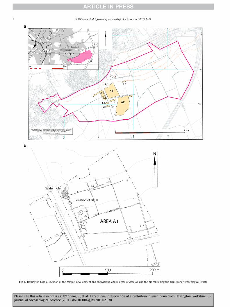
Fig. 1. Heslington East. a, Location of the campus development and excavations, and b, detail of Area A1 and the pit containing the skull (York Archaeological Trust).
S. O’Connor et al. / Journal of Archaeological Science xxx (2011) 1e142
Please cite this article in press as: O’Connor, S., et al., Exceptional preservation of a prehistoric human brain from Heslington, Yorkshire, UK,Journal of Archaeological Science (2011), doi:10.1016/j.jas.2011.02.030

Fig. 2. The skull as found (York Archaeological Trust).
S. O’Connor et al. / Journal of Archaeological Science xxx (2011) 1e14 3
Robinson, 2002; Spatz et al.,1958); bodies sealed in lead-coffins, suchas St Bees man (Tapp and O’Sullivan, 1982); and crypt burials such asthose at Spitalfields Church, London (Adams and Reeve, 1987). In allthese cases, where brain tissue persists, there is survival of otherinternal andexternal soft tissues, suchas lung, heart,muscle, skin andhair, unless theywere deliberately removed in antiquity. However, intheHeslingtoncase, brain survives in the absenceofother soft tissues.
Here we report the archaeological circumstances of thisremarkable find, the recording and sampling of the brain masses,macroscopic, radiographic and histological observations on themasses, and the results of a literature search to locate otherexamples of brain survival in skeletalised human remains, fromwhichwe discuss the conditions that appear to favour preservation.Subsequent papers will report in detail the biomolecular analysesthat are ongoing in order to characterise the surviving material inmore detail. The overall aim of the project is to understand thetaphonomic trajectory that led to exceptional preservation, andhence to determine whether particular aspects of the death andinhumation of this individual may have contributed to the survivalof what is ordinarily a most vulnerable soft tissue.
This paper has been compiled by Sonia O’Connor (PI) based onreports from and discussions with the numerous colleaguesengaged in the investigation of this specimen, and revised by herand Terry O’Connor. Specific contributions to the investigationsreported here were as follows:
Identification and recovery of the brain, critical review ofprevious cases of brain survival in excavated skeletons and thecharacteristics of adipocere e Sonia O’Connor (University ofBradford).3D Photography and laser scanning e Anthony Masinton(University of York).High-resolution laser scanninge PhilipDodds andAndyWarriner(Konica Minolta).CTandMRI scanninge PhilDuffeyandDavidKing (YorkHospital).Examination and recording of the skull e Jo Buckberry(University of Bradford), Terry O’Connor (University of York).Sediment thin-section e Raimonda Usai (University of York).Scanning Electron Microscopy e Andrew Wilson (University ofBradford).Bone thin-sectioneHolger Schutkowski (University of Bradford).Soft tissue thin-sectione JohnDenton (University ofManchester).DNA e Keri Brown (University of Manchester).Brain neurochemistryeAxel Petzold (University College London).Tissue and sediment chemistry e Salim Al-Sabah, Danish Anwar,Ed Bergström, Stephen Buckley, Matthew Collins, Adam Dowle,Karl Heaton, Brendan Keely, Matthew Pickering, Kirsty Penkman,Martin Rumsby, Kimberley Shackleton, Jerry Thomas, JaneThomas-Oates (University of York), Esam Ali, Howell Edwards,Andy Gledhill, Carl Heron (University of Bradford), Konrad Dorl-ing, Elsa Correia Faria, Peter Gardner (University of Manchester).
The project was funded by the University of York and EnglishHeritage.
2. Material
The Heslington East campus of the University of York lies to thesouth-east of the city (grid ref SE636506), and is constructed onformer agricultural land. The northern edge of the site is marked byan eastewest trending glacial moraine, from which the land fallson a gentle southward slope. The subsoil parent material isa heterogeneous mix of mostly glaciofluvial silts, sands and gravels.Survey and excavation work was undertaken in 2003e2008 inadvance of construction. A number of former water channels were
Please cite this article in press as: O’Connor, S., et al., Exceptional preserJournal of Archaeological Science (2011), doi:10.1016/j.jas.2011.02.030
identified, together with linear ditches of prehistoric (mostly IronAge) date, consistent with the drainage of water from springs andseepage along the moraine slope. One such spring had beenadapted into a series of well points, two of which had wickerlinings, sited at the junction of a number of ditches and apparentland boundaries, and including posts and wattle indicating it tohave been managed from the Bronze Age through to the middleIron Age. To the south of these features were a dozen or so pitswhose contents were not typical of occupation waste, hinting atsomemore ceremonial function that persisted from the Bronze Agethrough to the early Roman period. Many were marked by a singlestake, and their non-trivial contents included ‘burned’ cobbles oflocal stone. In addition the headless body of a red deer Cervuselaphus had been deposited in a palaeochannel, one of the earliestfeatures of the site, and an unworked red deer antler was found inan Iron Age ditch.
In one of the pits, a dark-stained human cranium with articu-lated mandible lay approximately face-down with no othercontents, in a matrix described as a moist, friable to plastic darkbrown organic rich, soft sandy clay with occasional beige sandflecks/spotting (context 2619) (Fig. 2). The skull and a small numberof animal bone fragments were recovered from 2619. After recoveryof the skull, it was noted to contain a resilient mass not consistentwith inwashed silt. Inspection of the endocranial cavity through theforamen magnum showed the presence of ‘a quantity of brightyellow material’ (R. Cubitt, pers. comm.). Subsequent endoscopy(Fig. 3), radiography of the cranium and examination of a sample ofthe mass, identified it as consistent with brain masses previouslyseen in skeletalised human remains from the Hull Magistrates’Court site in Kingston-upon-Hull, UK (O’Connor, 2002). Thecranium, with its articulated mandible and vertebrae C1 and C2,and a possibly unrelated single intermediate phalanx, was removedto cold storage pending further imaging and extraction of the brainmasses. DNA sequencing of samples from the brain gave a nearestmatch in haplogroup J1d, a recently defined haplogroup, first seeninmodern DNA sequences from just a few individuals from Tuscanyand the Near East. So far it has not been identified in Britain butfurther sampling in British populations may yet find this hap-logroup. Alternatively it may have existed in the past in Britain andbeen lost through genetic drift. A calibrated radiocarbon date of673e482BC was obtained from collagen extracted from themandible (OxA-20677: 2469 � 34 bp).
vation of a prehistoric human brain from Heslington, Yorkshire, UK,

Fig. 3. View of the brain through the foramen magnum using endoscopy (SoniaO’Connor).
S. O’Connor et al. / Journal of Archaeological Science xxx (2011) 1e144
3. Methods
3.1. Recording of the skull and brain
Following removal of only the most superficial adhering sedi-ment, the craniumwas imaged by (i) digital photography (including3D) and low-resolution laser scanning, to give a detailed recordof itsexternal condition prior to opening the cranium to access the brainmass, and (ii) by conventional medical CT and MRI, to record theposition and internal structure of the brain mass (Fig. 4). Adheringsediment from the eye sockets and the exterior of the craniumwassampled for geochemical analyses. The skull was then recorded andmeasured according to standard osteoarchaeology protocols(Buikstra and Ubelaker, 1994; Brickley and McKinley, 2004). A
Fig. 4. CT section of the cranium showing two of the larger fragments, which may bethe cerebral hemispheres separated by the sagittal cleft (David King).
Please cite this article in press as: O’Connor, S., et al., Exceptional preserJournal of Archaeological Science (2011), doi:10.1016/j.jas.2011.02.030
conventional autopsy saw-cutwasmade around the cranium a littleabove the temporal line, allowing most of the parietal bones andparts of the frontal and occipital bones to be lifted away in one piece.The brain masses could be seen surrounded by sediment in theoccipital portion of the endocranial space (Fig. 5). The brain masseswere removed, and then cleaned using a small, blunt, plastic spatulato lift away lumps of sediment and a soft, fine, paintbrush anda gentle stream of distilled water to clean the surfaces of eachmass.Sediment from the intra-cranial space was retained, and thecranium, mandible and cervical vertebrae were further washed andexamined. A full photographic record was made throughout,including low-power photomicroscopy and video. A 3D photo-graphic recordwasmade of the brainmasses immediately followingcleaning, and the brain masses and elements of the skull werescanned with a high-resolution 3D laser digitising system, theKonica Minolta Range 7. The bones were allowed to air-dry slowly,and were then re-examined for signs of trauma or other evidencepertinent to death and deposition. Light microscopy and SEM,carried out under low-vacuum conditions in an environmentalchamberwithout coating or otherwise preparing thematerial, wereemployed in the examination of detail.
3.2. Sampling
Sampling of the brain was restricted as much as possible to oneof the masses for the sake of consistency and to preserve theanatomically distinctive portions for future study. The pieceselected for sampling retained some external morphology and alsoareas of fractured surface, revealing the interior, but was largeenough to provide sufficient material for all the proposed analyticalprocedures. Separate samples were taken for a range of biomolec-ular analyses and to be fixed for histological study. The sample sizeswere appropriate to the minimum required for each analyticalprotocol (in the range of 20 mge2 g). The inwashed sediments andthe bone of the cranium were also sampled.
Fig. 5. The brain remains and sediment in situ in the opened cranium. Two of thelarger masses are indicated by the arrows (York Archaeological Trust).
vation of a prehistoric human brain from Heslington, Yorkshire, UK,

Fig. 6. Associated vertebrae C2. a, Peri-mortem damage and b, anterior aspect showingcut-marks (Jo Buckberry).
S. O’Connor et al. / Journal of Archaeological Science xxx (2011) 1e14 5
3.3. To characterise the internal and external deposits on thecranium
Sediments internal and external to the cranium were examinedby light microscopy and SEM, carried out as above. Samples werealso taken for biomolecular analyses. A coherent lump of sedimentfrom the interior of the cranium was sampled for micromorpho-logical analysis. The liquid phase was gradually replaced withacetone, then with a styrene-rich resin (Crystic 17449). After twomonths curing, 30 mm thin sections were cut from the block andexamined using polarized light microscopy and the criteria definedby Stoops (2003).
3.4. To investigate the state of preservation of the bone
To gain an overview of the state of preservation of the bone,a loose fragment, approximately 10 � 15 mm, was taken from closeto asterion on the left side of the cranium. This fragment wasembedded in epoxy resin, cut by microtome to 70 mm thickness,polished and mounted. The sections were viewed by transmittedlight at 200e400� magnification.
3.5. To explore the surviving morphology and histology of the brain
Low-power reflected light microscopy was undertaken to char-acterise surfacesof the surviving tissue. SEMwascarriedout as above.For histological analysis, tissue fixed in neutral buffered formalin (pH7.1) was dehydrated through alcohol, cleared in xylene, thenembedded in paraffin wax. The block was then microtomed to give5 mm thin-sections, which were mounted onto glass microscopeslides. The sections were stained with toluidine blue at pH 4.0, thenwithhaematoxylin andeosin. TEMwasundertakenbypost-fixing theformalin-fixed tissue in osmium tetroxide, embedding it in aralditeresin and staining the sectionswith lead citrate and uranium acetate.
3.6. To explore the chemistry of the brain
The range of biomolecular analyses in progress is directedtowards testing the brain masses for characteristic brain chemistry,and investigating the composition of the apparently stable materialto which the brain has undergone taphonomic alteration. Onlypreliminary results are presented here. Highly sensitive neuro-immunological techniques, together with proteomic analyses, havedemonstrated the presence of a range of brain-specific proteins,mainly of structural nature (Petzold, 2005). We are currentlyinvestigating how these proteins survived and what insight theymay give on the circumstances between death, the burial envi-ronment and preservation of the Heslington brain. An analysis ofDNA from the brain and from the skull has also been undertaken.Full results of all of these analyses will be reported in subsequentpapers.
4. Results
4.1. Examination of the skeletal remains
Anthropological examination showed the skull to be that ofa male, following criteria outlined by Buikstra and Ubelaker (1994)and Bass (1995). Based on Meindl and Lovejoy’s (1985) criteria forcranial suture closure, and on Brothwell’s (1972) method for molarattrition, an age-at-death estimate of 26e45 years was obtained,most likely in the younger half of this range. Cranial measurementsshow the cranium to be unexceptional in size and shape, with nosignificant evidence of disease. Examination of the two associatedvertebrae showed the arch of C2 to be fractured on either side of the
Please cite this article in press as: O’Connor, S., et al., Exceptional preserJournal of Archaeological Science (2011), doi:10.1016/j.jas.2011.02.030
centrum (Fig. 6). The fracture surfaces are consistent with peri-mortem fracture, and their location is consistent with a traumaticspondylolisthesis of C2. A cluster of c. 9 transversely-directed finecut-marks made by a thin-bladed instrument, such as a knife, arevisible on the anterior aspect of the centrum of C2. SEM examina-tion confirmed these to be peri-mortem (Fig. 7). Fine cracks on thecranial base and disruption of the right temporal suture are mostlypost-mortem, but three appear to be peri-mortem e two to theoccipital and one to the greaterwing of the sphenoid. The traumaticspondylolisthesis and the cut-marks are consistent with death byan abrupt trauma to the neck, followed by deliberate and carefuldismemberment of the head between C2 and C3.
4.2. Preservation of the bone
Although superficially well preserved, the skull has obviouslyundergone loss of organic matter, making it necessary to resampleto obtain sufficient collagen for radiocarbon dating. That said,microanatomical features are very well preserved: lamellar andosteon structures are readily recognized, and osteocyte lacunae can
vation of a prehistoric human brain from Heslington, Yorkshire, UK,

Fig. 7. SEM of the peri-mortem cut-marks on the anterior aspect of C2 (Andy Wilson).
S. O’Connor et al. / Journal of Archaeological Science xxx (2011) 1e146
be resolved throughout the sections (Fig. 8). Brown staining,possibly from humic compounds in the burial environment, isstrongest at the surface of the bone, fading towards the interior.Most significantly, there is no trace of microbial activity, bacterial orfungal, with none of the porosity or ‘tunnelling’ that is character-istic of putrefactive microorganisms (Jans et al., 2004). However, inplaces the bone displays linear cracks and erosive patterns to thereversal lines of osteons, structural damage that has been inter-preted as the result of intermittent periods of dry and moist-satu-rated burial environments (Piepenbrink and Schutkowski, 1987;Smith et al., 2002).
4.3. Deposits within the skull
Micromorphological examination showed an unsorted, predom-inantly quartzose, sediment, with low grain weathering, no texturalpedofeatures, and only a few traces of randomly-arranged planttissues. The results show a very low degree of pedogenesis acting on
Fig. 8. Thin-section of the bone of the skull indicating the overall very good preser-vation but also structural cracks in the compact bones and around Haversian systems(Holger Schutkowski).
Please cite this article in press as: O’Connor, S., et al., Exceptional preserJournal of Archaeological Science (2011), doi:10.1016/j.jas.2011.02.030
the sediment. The lack of pedofeatures indicates little movement ofwater within the sediment after its deposition within the skull, andlittle or no movement of decomposition products through voids orthrough the matrix.
4.4. Examination of the brain masses
CT of the cranium had revealed several fragments of brain looseinside and mixed with the denser sediment. The brain masses hadrecognisable sulci and gyri and many internal voids which aremainly post-mortem features (Fig. 4). CT could not differentiatebetween the brain cortex (grey matter) and underlying medulla(white matter). Subsequent MRI produced similarly useful imagesbut did not elucidate this point.
Within the cranium, therewerefivemajor brainmasses andmanymillimetre-scale fragments of brain tissue. Following superficialcleaning of the masses, gross anatomical features such as well-defined sulci and gyri were clearly identifiable. The tissue wasodourless, had a smooth surfacewith a resilient, tofu-like texture andwasmore pink/brown or tan in colour in daylight than had appearedwhen first viewed by electric light within the cranium, when it hadappeared yellow (Fig. 9). The scale of the surface convolutions, takenwith theoverall volumeof survivingmaterial, indicated that thebrainhad shrunken to perhaps 20% of the volume of a fresh brain (i.e. toabout 250e300 ml). One of the largest masses, approximately70 mm � 60 mm � 30 mm, also had an area of black membranousmaterial, perhaps a fragment of the meninges (Fig. 9b). Where themasses had fractured, they had a soft, granular texture and werelighter in colour than the exterior surfaces (Fig. 9c). The expecteddistribution of white and grey matter could not be discernedmacroscopically.
Imaging by 3D laser scanning of the major fragments wassuccessful, except for one fragment that, when turned over, dis-torted to the extent that the data sets could not be integrated. Theimages reconstructed from the data allow the fragments to beviewed from all angles and brought together in different combi-nations to help identify the portions of the brain that have survived,without the risks associated with handling the fragments them-selves. The photographs can also be digitally added to the surface ofthe 3D reconstructions (Fig. 10) and the scan data could be used toproduce replicas of the fragments using rapid phototyping tech-niques. Imaging by micro-CT was unsuccessful but it is hoped thatmore useful results may be obtained in the future if the resultsreported here, and subsequent analyses of composition, allowa more precise calibration of equipment to optimise the imaging.
4.5. Histological examination of the brain masses
Both toluidine blue and haematoxylineeosin staining of thebrain sections showed a homogeneous, amorphous substance thathad not retained any cellular or matrix structure. TEM also did notdetect any surviving cellular structure although these images didshow the presence of numerous morphologically-degraded struc-tures characteristic of the myelin sheath of nerve fibres (Fig. 11). Afew bacterial spores could be recognised on TEM, but no othertraces of putrefactive bacteria or fungi where evident. This obser-vation is more consistent with degradation by sterile autolysis thanwith putrefaction. SEM captured the spongy, granular nature of thefracture surface of the brain mass (Fig. 12) but added little to theunderstanding of the surviving histology.
4.6. Biomolecular analysis of the brain masses
It is only possible here to provide a summary of the initial resultsof the array of qualitative, quantitative and compositional
vation of a prehistoric human brain from Heslington, Yorkshire, UK,
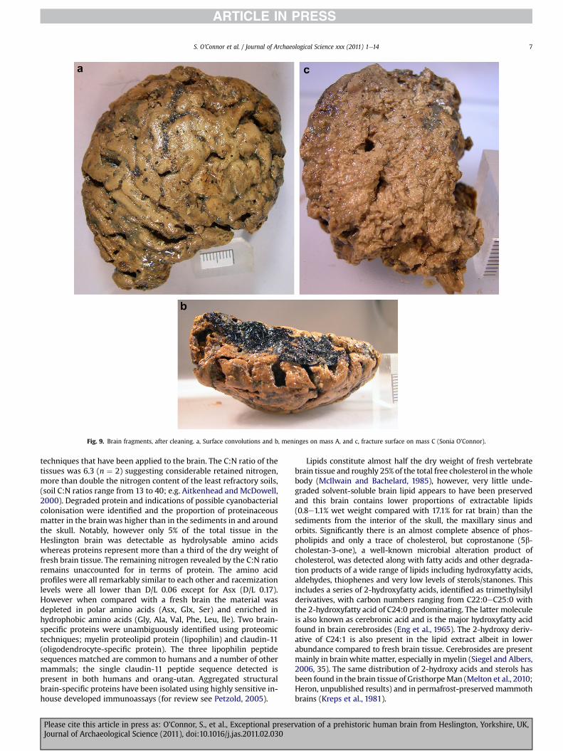
Fig. 9. Brain fragments, after cleaning. a, Surface convolutions and b, meninges on mass A, and c, fracture surface on mass C (Sonia O’Connor).
S. O’Connor et al. / Journal of Archaeological Science xxx (2011) 1e14 7
techniques that have been applied to the brain. The C:N ratio of thetissues was 6.3 (n ¼ 2) suggesting considerable retained nitrogen,more than double the nitrogen content of the least refractory soils,(soil C:N ratios range from 13 to 40; e.g. Aitkenhead and McDowell,2000). Degraded protein and indications of possible cyanobacterialcolonisation were identified and the proportion of proteinaceousmatter in the brainwas higher than in the sediments in and aroundthe skull. Notably, however only 5% of the total tissue in theHeslington brain was detectable as hydrolysable amino acidswhereas proteins represent more than a third of the dry weight offresh brain tissue. The remaining nitrogen revealed by the C:N ratioremains unaccounted for in terms of protein. The amino acidprofiles were all remarkably similar to each other and racemizationlevels were all lower than D/L 0.06 except for Asx (D/L 0.17).However when compared with a fresh brain the material wasdepleted in polar amino acids (Asx, Glx, Ser) and enriched inhydrophobic amino acids (Gly, Ala, Val, Phe, Leu, Ile). Two brain-specific proteins were unambiguously identified using proteomictechniques; myelin proteolipid protein (lipophilin) and claudin-11(oligodendrocyte-specific protein). The three lipophilin peptidesequences matched are common to humans and a number of othermammals; the single claudin-11 peptide sequence detected ispresent in both humans and orang-utan. Aggregated structuralbrain-specific proteins have been isolated using highly sensitive in-house developed immunoassays (for review see Petzold, 2005).
Please cite this article in press as: O’Connor, S., et al., Exceptional preserJournal of Archaeological Science (2011), doi:10.1016/j.jas.2011.02.030
Lipids constitute almost half the dry weight of fresh vertebratebrain tissue and roughly 25% of the total free cholesterol in thewholebody (McIlwain and Bachelard, 1985), however, very little unde-graded solvent-soluble brain lipid appears to have been preservedand this brain contains lower proportions of extractable lipids(0.8e1.1% wet weight compared with 17.1% for rat brain) than thesediments from the interior of the skull, the maxillary sinus andorbits. Significantly there is an almost complete absence of phos-pholipids and only a trace of cholesterol, but coprostanone (5b-cholestan-3-one), a well-known microbial alteration product ofcholesterol, was detected along with fatty acids and other degrada-tion products of a wide range of lipids including hydroxyfatty acids,aldehydes, thiophenes and very low levels of sterols/stanones. Thisincludes a series of 2-hydroxyfatty acids, identified as trimethylsilylderivatives, with carbon numbers ranging from C22:0eC25:0 withthe 2-hydroxyfatty acid of C24:0 predominating. The latter moleculeis also known as cerebronic acid and is the major hydroxyfatty acidfound in brain cerebrosides (Eng et al., 1965). The 2-hydroxy deriv-ative of C24:1 is also present in the lipid extract albeit in lowerabundance compared to fresh brain tissue. Cerebrosides are presentmainly in brainwhite matter, especially in myelin (Siegel and Albers,2006, 35). The same distribution of 2-hydroxy acids and sterols hasbeen found in the brain tissue of GristhorpeMan (Melton et al., 2010;Heron, unpublished results) and in permafrost-preservedmammothbrains (Kreps et al., 1981).
vation of a prehistoric human brain from Heslington, Yorkshire, UK,

Fig. 10. Brain fragment (mass A) reconstructed from 3D data. a, rendering of KonicaMinolta laser scan data and b, with photo textured rendering (Anthony Mastinson).
Fig. 11. TEM of myelin structure preserved in the brain tissue (John Denton).
Fig. 12. SEM of a fracture surface of the brain tissue (Andy Wilson).
S. O’Connor et al. / Journal of Archaeological Science xxx (2011) 1e148
Please cite this article in press as: O’Connor, S., et al., Exceptional preserJournal of Archaeological Science (2011), doi:10.1016/j.jas.2011.02.030
There are no biomarkers indicative of artificial preservationtechniques such as smoking or embalming in the Heslington brainmass. Fatty acids are not a major component of the lipid extractsand there is no evidence for the formation of adipocere, since noneof its characteristic products, such as steroid degradation products(Adachi et al., 1997), were detected. Instead, high-resolution massspectrometry and sequential thermal desorption and pyrolysisGCeMS, together with TLC, indicate that a high molecular weight,long chain, hydrocarbon material now forms a major hydrophobiccomponent of the brain mass. Research in progress aims to char-acterise this material.
5. Discussion
The aim of drawing together these results so far is to bring thisremarkable specimen to the attention of the research community.Two questions are central to any discussion. First, was thereanything particular about the circumstances of death and deposi-tion of this head that may have predisposed the degradation of thebrain to a more stable material that retains so much of the originalmorphology? Second, just how exceptional is this survival?
5.1. Death and deposition
It is rare to be able to postulate the cause of death for skeletonisedhuman remains of archaeological origin. In this case, we haveevidence for a traumatic spondylolisthesis of the neck and forsubsequent, almost surgical, dismemberment of the head. If theformer was not fatal, the latter certainly would have been. Knife cutson the axis indicate decapitation by means of a thin-bladed knife,inserted from the anterior aspect (i.e. from the front of the throat)and repeatedly pulled transversely across the neck. It is unlikely thatthis could have been undertaken on a conscious individual. Thissequence of events would have separated the head from the circu-latory system and thus from the internal organs from whichendogenous putrefaction would spread, and would also haveallowed blood to drain from the detached head. Open wounds areassociatedwith an increased rate of putrefaction but burialmay havefollowed quite rapidly, reducing infection by exogenous putrefactiveorganisms and slowing decomposition considerably (Fiedler and
vation of a prehistoric human brain from Heslington, Yorkshire, UK,

S. O’Connor et al. / Journal of Archaeological Science xxx (2011) 1e14 9
Graw, 2003). Below 21 �C the activity of most putrefactive bacteriawould have been inhibited, and in ground temperatures a fewdegrees above freezing it could have been a week before the firstsigns of putrefaction occurred (Mant, 1987). Certainly, the bonehistology is inconsistent with any putrefaction of skeletal tissues,and the same appears to be the case for the brain tissue itself.
The results are inconsistent with post-mortem curation. Thecharacteristic signs of early putrefaction are absent from the bonehistology and the chemistry of the surviving brain masses is incon-sistent with any deliberate intervention such as smoking. Further-more, it is unlikely that deliberate curation through smoking ordesiccation would have preserved brain tissue in the absence of anyother soft tissues. Indeed, desiccation is likely to have led to rapidcollapse of the brain tissues. There are no superficialmarkings on theskull indicative of excarnation, so it is improbable that a morecompletely-preserved head was deliberately stripped of survivingskin and muscle before interring the skull. Despite the place that‘trophy heads’ appear to have played in Iron Age societies andevidence for curationofhuman remains in theBronzeAge (Aldhouse-Green, 2002; Parker-Pearson et al., 2005), there is no evidence thatthis case is anything other than a decapitated head in which post-mortemputrefactionwas rapidly inhibited. That inhibition,we argue,was likely achieved through rapid burial into a fine-grained wetsediment. This is a distinctive and unusual sequence of events, andcould be taken as an explanation for the exceptional brain preserva-tion. However, similar preservation has been noted in other caseswheredecapitation clearly did notoccur (Beriault et al.,1981;Clausenet al., 1979; Dailey et al., 1972; Doran et al., 1986; Flinn, 1989;Haneveld, 1984; Oakley, 1960; O’Connor, 2002; Pääbo et al., 1988;Papageorgopoulou et al., 2010; Pilleri and Schwab, 1970; Royal andClark, 1960 and Tkocz et al., 1979). What, if anything, does theHeslington case have in commonwith these others surviving brains?
5.2. Yet another unique ancient brain?
Even a brief survey of the forensic literature confirms that braintissue is normally expected to decompose rapidly in the immediatepost-mortem period and survival of brain when other soft tissueshave decomposed is reported as exceptional, as in the case of thedried brain found in a skull by police in the bushveld near Rad-fontein, South Africa (Eklektos et al., 2006) or even ‘unique’, as inthe case of several examples recovered from a mass grave inBulgaria, buried 45e50 years previously in loose, stony soil(Radanov et al., 1992). However, here and in Appendix 1, wesummarise the results of a literature search that shows the survivalof brain in otherwise skeletalised remains from waterlogged sedi-ments to be unusual but by no means unique.
Perhaps the earliest published accounts of this type of preser-vation come from pre-Revolution France. Between December 1785and October 1787, the crowded and foetid cemetery of St Innocents’Church was cleared as a public health measure (Sourkes, 1992).Contemporary accounts report that brain matter was found in largenumbers of otherwise skeletalised bodies evenwhere the skull wasruptured by soil pressure (Thouret, 1790). A substance likened to‘white cheese’, and known as gras or grave wax, that had formedover some of the bodies preserving the shape of the flesh, andorgans such as the brain, heart and liver was subsequently named‘adipocere’ (Fourcroy, 1791). Significantly, Thouret noted that, evenat the earliest stages of decomposition, the brain always showedless deterioration than other soft tissues and was often the last tosurvive. The best-preserved examples came from those commongraves in which adipocere frequently covered the bones. Thesebrains had recognisable hemispheres and convolutions, fillingabout a quarter to a third of the cranial cavity, even after 20 or 30years burial. These brain masses were pulpy, soft and ‘fusible’
Please cite this article in press as: O’Connor, S., et al., Exceptional preserJournal of Archaeological Science (2011), doi:10.1016/j.jas.2011.02.030
between the fingers, although some were firmer, more solid andlooked more friable. Outwardly they had the colour of fresh brainbut internally the colour varied, some with a differentiationbetween the whitish medullary substance (white matter) anda covering of greyer cortex (grey matter). The brains were odour-less, the only smell being that of the adipocere surrounding thebones. Brain masses were also found in individual interments inwhich adipocere had not formed. In drier contexts such as charnelhouses, surviving brains were described as very small and black-ened on the surface but whiter within and very hard. Thouret(1791) also cites earlier reports of brains preserved in burials,several recorded in the 17th century including a corpse of 50 years’burial with a brain that was white, oily and odourless; several fromAvignon being moist, soft and apparently un-deteriorated; and thesurvival of soft and apparently un-deteriorated brains from a largenumber of executed men thrown into a well and only retrieved 80years later. Fourcroy was so intrigued by the unexpected preser-vation of brain tissue that he embarked on a detailed study ofhuman and other animal brains, laying the roots of the modernstudy of neurochemistry (Fourcroy, 1793).
Appendix 1 lists examples of brains in otherwise skeletonisedremains reported in the literature since 1960. The specimens fromFloridaarewell-known,andallderive fromarangeofwater-saturatedenvironments (Beriault et al., 1981; Clausen et al., 1979; Dailey et al.,1972; Doran et al., 1986; Pääbo et al., 1988; Royal and Clark, 1960).The Zihl Canal specimens are important as they were not deliberateinhumations, but the victims of a flood that in 1 BC collapseda wooden bridge and immediately buried all (Pilleri and Schwab,1970). Rapid burial was also inferred for the specimen from Scole,the context and body position of which led Turner-Walker (pers.comm.) to suggest furtive burial, aswith the victimof amurder.Manyof the cases listed in Appendix 1 are from cemeteries associatedwithmedieval monastic houses (Flinn, 1989; Haneveld, 1984; O’Connor,2002; Tkocz et al., 1979). This may be nothing more than a chanceassociation: medieval cemeteries constitute a high proportion of theburials excavated in northern Europe. However, an association withwet or waterlogged sediments is very clear.
The treatment and investigation afforded these brains havevaried considerably, making systematic comparison quite difficult.A number were immediately fixed in formalin, several have beensubjected to conventional histological section and staining, andonly a few have any chemical characterisation. CT examination ofthewell-preserved specimens fromKingston-upon-Hull (O’Connor,2002) confirmed internal structures consistent with brain, albeitdistorted and reduced in volume (Figs.13 and 14). Inmost cases, thespecimens were recognised as brain masses largely, it would seem,because they retained the characteristic surface convolutions,albeit in shrunken form, and were located in the intra-cranialcavity. In the cases of Droitwich (Oakley, 1960) and Quimper-Bre-tagne (Papageorgopoulou et al., 2010), the survival of the brain inthe form of adipocere has been proposed.We think this unlikely, forreasons discussed below.
In addition to the specimens listed in Appendix 1, publicitysurrounding the Heslington brain has brought to our attention otherrecent examples. Stephen Rowland, Oxford Archaeology North, hasprovided details of a 19th century example from a coffin burial inwaterlogged clay at a cemetery in Blackpool, Lancashire, UK, andJonny Geber, Margaret Gowen & Co Ltd., Heritage and ArchaeologyConsultants, informed us of several in various states of preservation(wet,moist anddry) fromaGreat Famine (1845e52)massgraveat thesite of the former unionworkhouse in Kilkenny City, Ireland. A newsreport that has not yet led to academic publication describes three1800 year-old brains fromskeletons excavated fromheavy,moist clayat the site of Aoya-Kamijichi, Tottori Prefecture, Japan (Science 27April 2001: 617. http://www.sciencemag.org/cgi/content/short/292/
vation of a prehistoric human brain from Heslington, Yorkshire, UK,
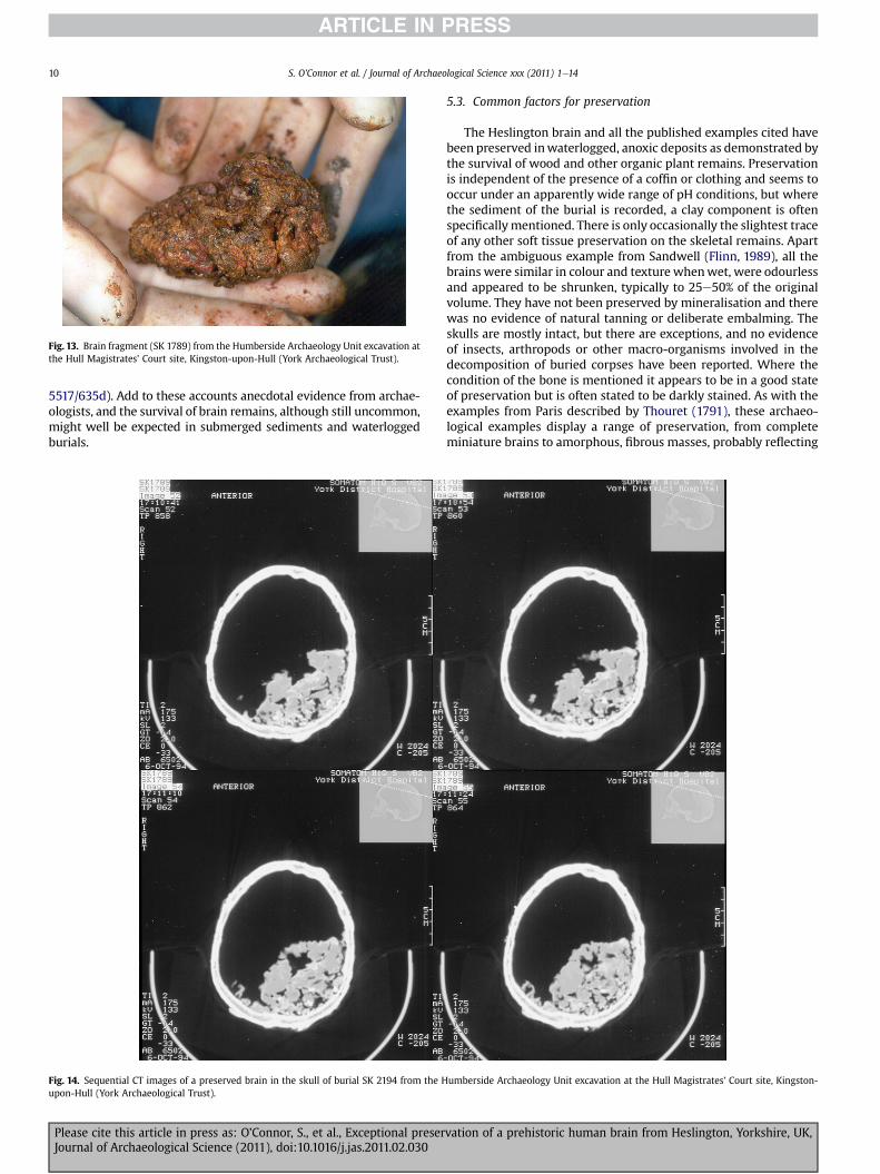
Fig. 13. Brain fragment (SK 1789) from the Humberside Archaeology Unit excavation atthe Hull Magistrates’ Court site, Kingston-upon-Hull (York Archaeological Trust).
S. O’Connor et al. / Journal of Archaeological Science xxx (2011) 1e1410
5517/635d). Add to these accounts anecdotal evidence from archae-ologists, and the survival of brain remains, although still uncommon,might well be expected in submerged sediments and waterloggedburials.
Fig. 14. Sequential CT images of a preserved brain in the skull of burial SK 2194 from the Hupon-Hull (York Archaeological Trust).
Please cite this article in press as: O’Connor, S., et al., Exceptional preserJournal of Archaeological Science (2011), doi:10.1016/j.jas.2011.02.030
5.3. Common factors for preservation
The Heslington brain and all the published examples cited havebeen preserved inwaterlogged, anoxic deposits as demonstrated bythe survival of wood and other organic plant remains. Preservationis independent of the presence of a coffin or clothing and seems tooccur under an apparently wide range of pH conditions, but wherethe sediment of the burial is recorded, a clay component is oftenspecificallymentioned. There is only occasionally the slightest traceof any other soft tissue preservation on the skeletal remains. Apartfrom the ambiguous example from Sandwell (Flinn, 1989), all thebrains were similar in colour and texture whenwet, were odourlessand appeared to be shrunken, typically to 25e50% of the originalvolume. They have not been preserved by mineralisation and therewas no evidence of natural tanning or deliberate embalming. Theskulls are mostly intact, but there are exceptions, and no evidenceof insects, arthropods or other macro-organisms involved in thedecomposition of buried corpses have been reported. Where thecondition of the bone is mentioned it appears to be in a good stateof preservation but is often stated to be darkly stained. As with theexamples from Paris described by Thouret (1791), these archaeo-logical examples display a range of preservation, from completeminiature brains to amorphous, fibrous masses, probably reflecting
umberside Archaeology Unit excavation at the Hull Magistrates’ Court site, Kingston-
vation of a prehistoric human brain from Heslington, Yorkshire, UK,

S. O’Connor et al. / Journal of Archaeological Science xxx (2011) 1e14 11
the state of putrefaction reached before this process was inhibitedby equilibration with the burial environment. Differences in theirhistology and chemistry relate as much to their different states ofpreservation as to differences in the techniques applied to theirinvestigation. Variation in the colours and textures of those thathad subsequently dried out also fit Thouret’s descriptions and againmay reflect the extent of putrefaction that had occurred beforepreservation. Finally those brains that have not been stored informalin, such as the Kingston-upon-Hull, Blackpool and Hesling-ton examples, have proved quite stable for more than a year whenkept hydrated in chilled storage, without physically deteriorating,developing an odour or apparently supporting fungal or bacterialgrowths. The persistent material that these brains have formedseems to be far more stable than fresh brain tissue: in that respect,it no more resembles fresh brain than casein plastic resembles themilk from which it was formed.
In the cemetery sites only a proportion of the burials producedrecognisable remains of brains. Obviously not all the graves willhave been suitably waterlogged and anoxic from the time ofinterment. However, observations of the distribution of othersurviving organic materials (including textiles, leather and wood)at Kingston-upon-Hull indicate that even continuously water-logged graves with intact skulls did not always produce braintissue. Clearly factors other than the immediate burial environmentmust also influence the taphonomic trajectory of the brains. In oneinstance, Zihl, all the intact skulls contained very well preservedbrain tissue, which in this unusual case can probably be directlyassociated with near-instantaneous death and burial (Pilleri andSchwab, 1970).
In attempting to understand the preservation of brains in ske-letalised human remains, it is not helpful to conflate them withstudies undertaken on brains from tanned bog bodies, frozen,desiccated, smoked or embalmed mummies and other instanceswhere there has been significant additional soft tissue preservation.Brains discovered in skeletons from mass graves have not alwaysbeen in typically wet or waterlogged sites (Brothwell and Gill-Robinson, 2002, 125; Radanov et al., 1992) but here the concentra-tion of decaying bodies themselves may be creating the wet, anoxicconditions that seem to predispose preservation of the brain. Theshrunken brain of a young adult female, from a 15th or 16th centuryburial in Yongin, Korea, investigated in 2005 (Kim et al., 2008), isverysimilar to theHeslingtonbrain.Remainsof themeninges (dura),blood vessels, grey andwhitematter,myelin structures and possiblebacterial spores were observed and the soft tissue proved positivefor both lipids and proteins. However the mechanism of preserva-tion may be very different to the other cases reported here as hairwas also preserved and therewas soft tissue in the orbits and skin onthe skull. Yongon is an example of a type of highly regulated burialpractice inwhich the corpsewas interred in a doublewooden coffin,incorporatinga layerof carbon,whichwas then thicklyencapsulatedin a limeesoilmixture. Korean tombsprepared in thiswayoften leadto complete mummificationwhere the sealing of the coffin remainsintact and frequently brains still survive evenwhere little other softtissue is preserved. Kim et al. (2008) suggest that the limeesoilmixturemay be involved in this process. From their account it is notclear howwet the burials are but thefibrous appearance of thewoodsuggests that it has degraded in a wet, alkaline environment.
Even brains recovered from skeletonised bodies in submergedmarine environments, such as the 8 strong crew of the Americancivil war submarine HL Hunley (Jamie Downs pers. comm.) mayhave become preserved in a subtly different way. These brains andthose from a few individuals who went down with the Tudor warship, the Mary Rose, were similar to the terrestrial examples incolour and texture. However the Mary Rose examples ‘liquifiedwithin a matter of minutes’ unless immediately submerged in
Please cite this article in press as: O’Connor, S., et al., Exceptional preserJournal of Archaeological Science (2011), doi:10.1016/j.jas.2011.02.030
alcohol (Allen and Elkerton, 2005). Althoughmany of the terrestrialexamples were relatively quickly fixed in formalin, none had star-ted to liquefy and several examples, including Heslington, havesurvived unfixed in chilled storage for extended periods. Thedissolution reported for the Mary Rose specimens suggests thatthese have not formed the same persistent material.
5.4. Suggested mechanisms for preservation
Despite a lack of chemical evidence or other soft tissues,Haneveld (1984) speculates that the preservation of the Dordrechtbrains is due to long-term submersion in a cold slightly acid tanningenvironment. Others have speculated that alkaline burial conditionsmay be involved (Doran et al., 1986; Tkocz et al., 1979). In the case ofWarmWater Springs, Royal andClark (1960) suggest that the groundwater or sediments might have antibiotic properties, as stalagmiteevidence indicates that the surviving brain mass was depositedbefore the site was inundated. However, as evidenced by the otherexamples cited here, the sediments probably only needed to havebeen wet and anoxic for preservation to have occurred in theintervening period. The most commonly held hypothesis is that thebrains have become adipocere (Oakley, 1960; Papageorgopoulouet al., 2010; Tkocz et al., 1979), making it necessary to discuss thiscompound and its formation in some detail.
Limited analysis of preserved brain from the Paris cemetery alsoled Thouret (1791) to link the waxy material of the brain with adi-pocere. Today the term is strictly defined as the persistent waxy,complex formed by the hydrolysis and hydrogenation of adiposetissues during decomposition. This is a bacterially-mediated processthat ismost frequently associatedwith immersion inwater or burialinwaterlogged ground thoughmoisture and bacteria intrinsic to thedecomposing tissue can be sufficient to produce adipocere. Itsformation inhibits further post-mortem changes and can makea corpse almost completely resistant to decay. Under favourableburial conditions considerable development of adipocere can occurwithin 30 days of interment (Fiedler and Graw, 2003). Observationsof post World War II exhumations showed a direct correlationbetween body fat deposits and adipocere distribution (Mant, 1987).Adipocere was more localised to the cheeks, buttocks and abdomenin men than inwomen, reflecting in general their different patternsof subcutaneous fat distribution, and was more or less absent fromthe skeletons of emaciated concentration camp victims. Burial ofa fully clothed body, directly into the soil, seemed to have producedthe most rapid formation of adipocere.
The major lipid constituents (90e98%: Albright and Stern, 1998)of adipose tissue are triacylglycerols (TAGs) and these are brokendown to form glycerol and mostly even-C number saturated fattyacids. As the formation of adipocere proceeds, the unsaturated fatsin the adipose tissue are depleted, the proportion of palmitic, stearicand myristic acids increases and hydroxystearic acid is formed.Although commonly soft, greasy and white, adipocere can bereddish brown when fresh, become grey in older deposits, anddevelop a dry and brittle texture (Garland and Janaway,1989). Thesedifferences in physical properties are due partly to cation exchangeduring different stages of formation of the adipocere, producingdifferent salts of fatty acids. In the early stages of decompositionthere is initially an abundance of sodium and then potassium saltsas tissue cell structures disintegrate, but later, salts from themineralcomponents of the soil predominate. Potassium salts produce thewax-like deposits and sodium salts form a harder crumbly material(Vane and Trick, 2005). In the later stages of decomposition a quitebrittle adipocere can be formed where calcium and magnesiumsalts are readily available (Forbes et al., 2005a).
Experimental work on adipocere formation by Forbes et al.(2005a,b,c) confirmed that the ideal factors for adipocere formation
vation of a prehistoric human brain from Heslington, Yorkshire, UK,

S. O’Connor et al. / Journal of Archaeological Science xxx (2011) 1e1412
are thepresenceofmoisture, bacteria, anaerobic burial conditions anda mildly alkaline pH. Adipocere formed readily between approxi-mately pH5e9 butwas greatly curtailed in soils doctored to be highlyacid (approx.pH2.4)orhighlyalkaline (approx. pH12.6).Other factorsinhibiting formation included low temperature, aerobic conditionsand sterilised soil. These results are consistent with the pivotal role ofsoil anaerobic bacteria in adipocere formation as their activity is cur-tailed in extremes of pH, low temperatures and aerobic conditions.Otherwise soil type had little effect on the rate of adipocere formationexcept that it was reduced in clayey soil (Forbes et al., 2005a).
Once formed, adipocere can be very resistant if the environmentdoes not change. Fiedler et al. (2009) report a burial in which adi-pocere was still found after 1600 years even though the water tablewas thought to have varied considerably. However it is not the finalproduct of decomposition and will itself decay over time. The half-life of adipocere in anaerobic conditions has been experimentallydetermined to be between 11 and 82 years: in air this is reduced 10-fold, to as little as 0.7 years (Fründ and Schoenen, 2009). Oxidationand decay by aerobic bacteria appear to be the main factors in thisincreased rate of deterioration (Fiedler and Graw, 2003).
As a mechanism for the preservation of brains from wet burialenvironments the formation of adipocere has several shortcomings.If the brains are preserved by adipocere, why is evidence of othersoft tissue preservation almost entirely absent? Those brainremains not immediately placed in formalin did not deterioratewith the speed expected of fresh brain or of adipocere, but provedremarkably stable in dark, wet and chilled conditions. In addition,concomitant with the conversion of adipose tissue to adipocere isan increase in volume, sufficient to hidewounds or perforations thesize of bullet holes (Mant, 1987). All of the brains in the literaturecited, despite remaining hydrated, had shrunken substantially.Thouret (1790) observed that even in the Parisian mass graves(anything from 12 to 1500 individuals) where bodies werecompletely enveloped in a solid mass of adipocere, the preservedbrains only filled a quarter to a third of the cranial cavity. Finally,although fresh brain tissue does contain ample lipids these aremostly of phospholipids and glycolipids. TAGs and free fatty acidsaccount for only a few percent of the total lipid content and areprobably associated with the blood and blood vessels, rather thanthe neural tissue (Suzuki, 1972). It is, therefore, highly unlikely thatwet-preserved brains become adipocere.
It is possible, however, that adipocere is part of themechanism ofpreservation. Itwas once thought that tissues such asmuscle becameadipocere (adiponeogenesis) but it has since been shown that theadipocere is formed from the liquified fatty acids that penetrate thedecomposing muscle tissue under the pressure of gases formedduring putrefaction (Fiedler and Graw, 2003). As a result, adipocerecan formwithin tissues andorganswith little intrinsic fat content andthe state of preservation of the tissue will depend on how decayed itwas at the time the adipocere formed. In this way, soft tissues, eyes,genital organs and internal organs, including the brain, can bepreserved by adipocere (Fiedler and Graw, 2003). In all of these casesofwet-preservedbrains, adipocerehasbeenpositively identifiedonlyin the Quimper-Bretagne brain (Papageorgopoulou et al., 2010). Asthe analysis was qualitative it could easily have been a minorconstituent but its presence may have helped inhibit normal deteri-orationwhilstmore persistentmaterial formed. It is possible that thered/brown deposit found on the surface of this specimen was adi-pocere but this was not analysed. In contrast, no adipocere wasdetected in the Heslington brain, probably because it was separatedfrom the body, which indicates that adipocerewithin the brain tissueis not essential to the formation of a persistent mass.
The analytical study of the Heslington brain so far shows that,although chemical breakdown of proteins and lipids has beeninitiated, significant quantities of protein-derived and lipid-derived
Please cite this article in press as: O’Connor, S., et al., Exceptional preserJournal of Archaeological Science (2011), doi:10.1016/j.jas.2011.02.030
material have been preserved. The preserved material retainsa higher proportion of hydrophobic components (amino acids inthe case of proteins and high molecular weight hydrocarbon-likematerial in the case of the lipids) than fresh brain matter. It is notpossible at the present time to determine if the hydrophobicityreflects the selective preservation of constituents that are notamenable to degradation or if it has acted in someway as a physico-chemical barrier preventing bacterial and fungal attack. Thechemical analytical techniques applied to the other brains citedwould not have been capable of detecting the high molecularweight material and its possible role in preservation is undergoingfurther investigation. These further studies include this brain andcomparative modern materials.
6. Conclusions
All the published examples of wet preservation cited involvedsome level of investigation of the brain remains to evaluatesurviving histology and the nature of the persistent material thathad formed. Mostly they were regarded in isolation with littleattention paid to the condition of the skeletal remains or detail oftheir burial environment. No attempt has beenmade to understandthem as a group, to look systematically for common factors thatmight have led to their preservation or to see them, in an archae-ological context, as evidence of the processes of death and burial.Based on the experience gained from the Hull Magistrates’ Courtexcavation, the Heslington Brain Project has taken a more holisticview and its findings challenge many of the assumptions made inthe past, such as the mechanism of preservation and their status asrare ‘curiosities’ rather than significant archaeological evidence.
Although the chemistry of the Heslington brain requires furtherinvestigation, it is clear that adipocere is not a pre-requisite in thesecases. The literatureyields a surprisingnumberof instances inwhichbrain is the only surviving soft tissue associated with otherwiseskeletalised remains. In only one of these does there appear to beevidence for the presence of adipocere (Papageorgopoulou et al.,2010). The simplest explanation that accounts for all of the caseswould seem to be that the circumstances of death and burial thatpredispose the formation of adipocere may also predispose theformation of a stable brain mass, but that neither process drives ornecessitates the other. Burial immediately following death can onlybe inferredwith certainty for theZihl assemblage, forwhichwehaveproposed that the exceptional histological preservation may bea consequence of that rapid burial. If so, and if other means ofdetermining the post-mortem, pre-burial interval can be developed,it may be possible to infer important details of the circumstances ofdeath and burial of more human remains based on the survival andstate of preservation of these remarkable ancient brains.
Acknowledgements
Thanks go to Richard Hall, Rachel Cubitt, Martin Stockwell,Bryan Antoni and Jane McComish (York Archaeological Trust) fortheir support throughout this project, and for information aboutthe circumstances of excavation. CT and MR imaging, the recordingof the skull and removal of the brain were undertaken at YorkHospital. We thank Gwen Hayley and her team at the CT Depart-ment and the staff of the hospital mortuary, particularly Peter Hilland Kevin Breheney, for their expertise and patience. We are verygrateful to Paul Bowman, Philip Dodds and Andy Warriner ofKonica Minolta Sensing Europe B.V., for generously providing theexpertise and equipment for the high-resolution laser scanning.Thanks also go to Richard Allen (University of York) for use oflaboratory space and facilities for the sampling of the brain, Ching-Hua Lu (University College London) for assisting with the brain-
vation of a prehistoric human brain from Heslington, Yorkshire, UK,

S. O’Connor et al. / Journal of Archaeological Science xxx (2011) 1e14 13
specific protein samples, Bill Christie and Anna Nicolaou (Univer-sity of Bradford) for their help in identifying the 2-hydroxyfattyacids and Michael Fagan (Hull-York Medical School) for testing thepracticality of micro-CT examination.
Appendix 1
Records of brain masses in skeletalised human remains since1960.
This catalogue concisely summarises the following information:location, date, number, context, description, investigative methods,further comments, and source.
Droitwich, U.K. Romano-British; 1 specimen female 44e55 yrsold; burial in wooden coffin at 3 m depth; small pieces of brain,shrunken, visible surface convolutions; unspecified chemicaltests indicated replacement by ‘wax’, and noted substantialproportion of clay; identified as adipocere by Prof H. Spatz, buthis reasoning is not given; (Oakley, 1960).Florida, Warm Mineral Springs, USA. 8000 � 200BC; 1 brainfrom 7 individuals; under ‘several feet of soft sediment’ con-taining leaves and wood in flooded, shallow limestone cave;pieces of soft, white brain mass with macroscopic structure;histological staining, ashing, unspecified chemical analysis;brain mass discoloured and shrank, possibly due to immersionin formalin; (Royal and Clark, 1960).Florida, Little Salt Spring, U.S.A. 6860 � 110BP; 1 brain from anunknown number of individuals excavated from cemeterythought to contain over 1000 inhumations; hard water, anaer-obic, buried in peat with well-preserved wooden objects andplant remains in a wet, ‘muck-filled’ hollow leading to a sink-hole; ‘substantial portion’ of brain, surface convolutions,‘cellular processes’, ‘macroscopically well-preserved neuraltissue’; aDNA; (Clausen et al., 1979; Pääbo et al., 1988).Florida, Windover, USA. Ca. 7500BP; 91 brains from 168 indi-viduals; burials in peaty sediments in swampy pond; shrunkento ¼ original size, tan-grey, soft, granular, fragile, some recog-nisable gross anatomy; CT, MRI, histological sections, TEM,aDNA; noted myelin traces; (Doran et al., 1986; Glen Doran pers.comm. 2008).Zihl Canal, Coraux, Switzerland. La Têne; 18 brains from 18individuals; under remains of wooden bridge in catastrophicflood debris at 6 m depth; greyish-white, ‘humid’, surfaceconvolutions, reduced in volume; histological section; axonsand possible fungal structures identified; (Pilleri and Schwab,1970).Svenborg, Funen, Denmark. Medieval; 56 brains from 74 indi-viduals; Franciscan cemetery, graves cut into alkaline claysfrequently flooded; preservation varied but not in relation topresence of coffin, dark grey, soft, ‘soapy’, shrunken to ½ size;fixed in formalin, histological sections, SEM, ‘qualitative’chemical analysis unspecified; traces of axons noted, cholesteroland phospholipids detected; (Tkocz et al., 1979).Dordrecht, Netherlands. 14th century; number unknown;Minorites monastic cemetery; ‘well preserved’ with visibleconvolutions; fixed in formalin, CT, MRI, histological sections,TEM; CT confirmed ventricles and post-mortem cavities, stain-ing confirmed neurons, cell fibrillary structures, vague contoursof vascular structures, lipids and myelin not detected, no fungidetected; (Haneveld, 1984).Sandwell, U.K. 14th century; 1 brain from 1 individual; gravewithin Benedictine Priory, waterlogged, neutral; soft, blackmaterial within cranium; fixed in formalin, histological section,electron probe microanalysis, no organic analysis undertaken;traces of shroud material survived; (Flinn, 1989).
Please cite this article in press as: O’Connor, S., et al., Exceptional preserJournal of Archaeological Science (2011), doi:10.1016/j.jas.2011.02.030
Kingston-upon-Hull, Magistrates’ Court, U.K. Medieval; 25brains from ca 250 individuals; Augustinian Friary cemeteryinside and outside buildings, waterlogged; grey-brown, soft,slightly resilient, shrunken, convolutions visible, one apparentlyliquefied then reformed as soft, granular endocast, becomingnodular and granular or black, glossy and resinous on drying instorage; CT, histological section, FT Raman spectroscopy;(O’Connor, 2002).Scole, Norfolk, U.K. Roman; 1 brain from 1 individual; burialbeneath wooden plank in alluvium, riverside, possible murdervictim; granular endocast; (O’Connor, 2002; G. Turner-Walkerpers. comm.).Aalborg, Denmark. Medieval; 10 brains from an unknownnumber of individuals; Franciscan friary cemetery; varyingstates of preservation; (O’Connor, 2002; J. Nielson in litt.).Salisbury, U.K. Medieval; 1 brain from an unknown number ofindividuals; Friary burial; identified as a fungal pseudomorph bya morbid anatomist, no analyses; (O’Connor, 2002; M. Corfieldpers. comm.).Norwich, U.K. 1 brain; context and date uncertain; (O’Connor,2002; B. Ayers pers comm.).Quimper-Bretagne, France. AD1250e1275; 1 brain from 1individual, 18-month-old child; skeletonised remains wrappedin leather within wooden coffin, burial in wet clay; greyish,compact, convoluted surface, traces of meninges, harder, darkermedial surface may be more degraded; fixed in formalin, CT,MRI, histological section, aDNA, GCeMS; Nissl bodies identified,diverse fatty acids detected, poor DNA preservation (formalin);(Papageorgopoulou et al., 2010).
References
Adachi, J., Ueno, Y., Miwa, A., Asano, M., Nishimura, A., Tatsuno, Y., 1997. Epi-coprostanol found in adipocere from five human autopsies. Lipids 32, 1155e1160.
Adams, M., Reeve, J., 1987. Excavations at christ church spitalfields 1984e6.Antiquity 61 (232), 247e256.
Aitkenhead, J., McDowell, W., 2000. Soil C:N ratio as a predictor of annual riverineDOC flux at local and global scales. Global Biogeochemical Cycle 14, 127e138.
Albright, A.L., Stern, J.S., 1998. Adipose tissue. In: Fahey, T.D. (Ed.), Encyclopedia ofSports Medicine and Science. Internet Society for Sport Science. http://www.sportsci.org/encyc/adipose/adipose.html last accessed 07.11.10.
Aldhouse-Green, M., 2002. Dying for the Gods: Human Sacrifice in Iron Age andRoman Europe. NPI Media Group, Stroud.
Allen, M.J., Elkerton, A., 2005. Human remains: other scientific analyses. In:Gardiner, J., Allen, M.J. (Eds.), Before the Mast: Life and Death Aboard the MaryRose. The Mary Rose Trust Ltd., Portsmouth, p. 563.
Aufderheide, A.C., 2003. The Scientific Study of Mummies. Cambridge UniversityPress, Cambridge.
Bass, W.M., 1995. Human Osteology: a Laboratory and Field Manual. MissouriArchaeological Society, Colombia.
Beattie, O., Geiger, J., 1987. Frozen in Time: The Fate of the Franklin Expedition.Bloomsbury, London.
Beriault, J., Carr, R., Stipp, J., Johnson, R., Meeder, J., 1981. The archaeologicalsalvage of the Bay West site, Collier County, Florida. Florida Anthropologist 34,39e58.
Brickley, M., McKinley, J.I. (Eds.), 2004. Guidelines to the Standards for RecordingHuman Remains. IFA Paper No. 7. BABAO/IFA.
Brothwell, D.R., 1972. Digging up Bones, second ed. British Museum (NaturalHistory), London.
Brothwell, D., Gill-Robinson, H., 2002. Taphonomic and forensic aspects of bogbodies. In: Haglund, W.D., Marcella, H.S. (Eds.), Advances in ForensicTaphonomy: Method, Theory, and Archaeological Perspectives. CRC Press, BocaRaton, pp. 119e133.
Buikstra, J., Ubelaker, D.H., 1994. Standards for Data Collection from Human SkeletalRemains. Arkansas Archeological Survey, Arkansas.
Ceruti, C., 2004. Human bodies as objects of dedication at Inca mountain shrines(northewest Argentina). World Archaeology 36 (1), 103e122.
Clausen, C.J., Cohen, A.D., Emitiani, C., Holman, J.A., Stipp, J.J., 1979. Little Salt Spring,Florida: a unique underwater site. Science 203, 609e614.
Cockburn, A., Cockburn, E., Reyman, T.A. (Eds.), 1998. Mummies, Disease andAncient Cultures, second ed. Cambridge University Press, Cambridge.
Dailey, R.C., Morrell, L.R., Cockrell, W.A., 1972. The St. Marks Military cemetery (8WA108). In: Bureau of Historic Sites and Properties Bulletin 2. Florida Departmentof State, Tallahassee, FL, pp. 1e24.
vation of a prehistoric human brain from Heslington, Yorkshire, UK,

S. O’Connor et al. / Journal of Archaeological Science xxx (2011) 1e1414
David, A.R., 1997. Disease in Egyptian mummies: the contribution of new technol-ogies. Lancet 349, 1760e1763.
Dean, G., 2008. A ditch in time. Further excavations at Heslington East, York.Yorkshire Archaeology Today 15, 11e14.
Doran, G.H., Dickel, D.N., Ballinger, W.E., Agee, O.F., Laipis, P.J., Hauswirth, W.W.,1986. Anatomical, cellular and molecular analysis of 8,000-yr-old human braintissue from the Windover archaeological site. Nature 323, 803e806.
Eklektos, N., Dayal, M.R., Manger, P.R., 2006. A forensic case study of a naturallymummified brain from the bushveld of South Africa. Journal of Forensic Science51 (3), 498e503.
Eng, L.F., Gerstl, B., Hayman, R.B., Lee, Y.L., Tietsort, R.W., Smith, J.K., 1965. The2-hydroxy fatty acids in white matter of infant and adult brains. Journal of LipidResearch 6, 135e139.
Fiedler, S., Buegger, F., Klaubert, B., Zipp, K., Dohrmann, R., Witteyer, M., Zarei, M.,Graw, M., 2009. Adipocere withstands 1600 years of fluctuating groundwaterlevels in soil. Journal of Archaeological Science 26, 1328e1333.
Fiedler, S., Graw, M., 2003. Decomposition of buried corpses, with special referenceto the formation of adipocere. Naturwissenschaften 90, 291e300.
Flinn, R.M., 1989. Ghosts in the ground e the staining of buried human remains.Humanbiologia Budapestinensis 19, 53e57.
Forbes, S.L., Dent, B.B., Stuart, B.H., 2005a. The effect of soil type on adipocereformation. Forensic Science International 154, 35e43.
Forbes, S.L., Stuart, B.H., Dent, B.B., 2005b. The effect of the burial environment onadipocere formation. Forensic Science International 154, 24e34.
Forbes, S.L., Stuart, B.H., Dent, B.B., 2005c. The effect of the method of burial onadipocere formation. Forensic Science International 154, 44e52.
Fourcroy, A.F., 1791. Deuxième mémoire sur les matières animales trouvées dans laCimetière des Innocens à Paris, pendant les fouilles qu’on a faites en 1786 &1787. Examen chimique de la matière grasse des cadavers contenus dans lesfosses communes. Annales de Chimie 8, 17e73.
Fourcroy, A.F., 1793. Examen chimique du cerveau de plusieurs animaux. Annales deChimie 16, 282e322.
Fründ, H.-C., Schoenen, D., 2009. Quantification of adipocere degradation with andwithout access to oxygen and to the living soil. Forensic Science International188, 18e22.
Garland, A.N., Janaway, R.C., 1989. The taphonomy of inhumation burials. In:Roberts, C.A., Lee, F., Bintliff, J. (Eds.), Burial Archaeology: Current Research,Methods and Developments. BAR British Series 211. British ArchaeologicalReports, Oxford, pp. 15e37.
Haneveld, G.T., 1984. 14th century naturally preserved brains from the Netherlands.In: Capecchi, V., Rabino Massa, E. (Eds.), Proceedings of the PalaeopathologyAssociation 5th European Meeting, Siena 1984. Siena University Press, Siena,pp. 157e162.
Hess, M.W., Klima, G., Pfaller, K., Künzel, K.H., Gaber, O., 1998. Histological investi-gations on the Tyrolean Ice Man. American Journal of Physical Anthropology106, 521e532.
Jans, M.M.E., Nielsen-Marsh, C.M., Smith, C.I., Collins, M.J., Kars, H., 2004. Charac-terisation of microbial attack on archaeological bone. Journal of ArchaeologicalScience 31, 87e95.
Johnson, M., 2008. East side story. Excavations at Heslington East, York. YorkshireArchaeology Today 14, 11e13.
Karlik, S.J., Bartha, R., Kennedy, K., Chhem, R., 2007. MRI and multinuclear MRspectroscopy of 3,200-year-old Egyptian mummy brain. American Journal ofRoentgenology 189 (2), W105eW110.
Kim, M.J., Oh, C.S., Lee, I.S., Lee, B.H., Choi, J.H., Lim, D.-S., Yi, Y.S., Han, W.-J.,Kim, Y.-S., Bok, G.D., Lee, S.D., Shin, D.H., 2008. Human mummified brain froma medieval tomb with lime-soil mixture barrier of the Joseon Dynasty, Korea.International Journal of Osteoarchaeology 18, 614e623.
Kreps, E.M., Avrova, N.F., Chebotarëva, M.A., Chirkovskaya, E.V., Levitina, M.V.,Pomazanskaya, L.F., 1981. Brain lipids in fossilized mammoths Mammuthusprimigenius. Comparative Biochemistry and Physiology Part B: Biochemistryand Molecular Biology 68 (1), 135e140.
Lewin, P.K., Harwood-Nash, D., 1977. X-ray computed tomography on an ancientEgyptian brain. IRCS Journal of Medical Science 5, 78.
Mant, A.K., 1987. Knowledge acquired from post-War exhumations. In:Boddington, A., Garland, A.N., Janaway, R.C. (Eds.), Death, Decay and Recon-struction. Approaches to Archaeology and Forensic Science. ManchesterUniversity Press, Manchester, pp. 65e78.
McIlwain, H., Bachelard, H.S., 1985. Biochemistry and the Central Nervous System.Churchill Livingstone, Edinburgh.
Please cite this article in press as: O’Connor, S., et al., Exceptional preserJournal of Archaeological Science (2011), doi:10.1016/j.jas.2011.02.030
Meindl, R.S., Lovejoy, C.O., 1985. Ectocranial suture closure: a revised method for thedetermination of skeletal age at death based on the lateral-anterior sutures.American Journal of Physical Anthropology 68, 57e66.
Melton, N., Montgomery, J., Knusel, C.J., Batt, C., Needham, S., Parker Pearson, M.,Sheridan, A., Heron, C., Horsley, T., Schmidt, A., Evans, A., Carter, E., Edwards, H.,Hargreaves, M., Janaway, R., Lynnerup, N., Northover, P., O’Connor, S., Ogden, A.,Taylor, T., Wastling, V., Wilson, A., 2010. Gristhorpe Man: an Early Bronze Agelog-coffin burial scientifically defined. Antiquity 84, 796e815.
Notman, D.N.H., Anderson, L., Beattie, O.B., Amy, R., 1987. Arctic paleoradiology:portable radiographic examination of two frozen sailors from the Franklinexpedition (1845e1848). American Journal of Roentgenology 149 (2), 347e350.
Oakley, K.P., 1960. Ancient preserved brains. Man 60, 90e91.O’Connor, S., 2002. Brain pseudomorphs: grey matter, grey sediments and grey
literature. In: Dobney, K., O’Connor, T. (Eds.), Bones and the Man: Studies inHonour of Don Brothwell. Oxbow Books, Oxford, pp. 41e50.
Pääbo, S., Gifford, J.A., Wilson, A.C., 1988. Mitochondrial DNA sequences from 7,000year old brain. Nucleic Acids Research 16 (20), 9775e9787.
Papageorgopoulou, C., Rentsch, K., Raghavan, M., Hofmann, M.I., Colacicco, G.,Gallien, V., Bianucci, R., Rühli, F., 2010. Preservation of cell structures ina medieval infant brain: a paleohistological, paleogenetic, radiological andphysico-chemical study. NeuroImage 50, 893e901.
Parker-Pearson, M., Chamberlain, A.T., Collins, M.J., Craig, O.E., Marshall, P.,Mulville, J., Smith, H., Chenery, C., Cook, G., Craig, G., Evans, J., Hiller, J.,Montgomery, J., Schwenninger, J.-L., Taylor, G., Wess, T., 2005. Evidence formummification in Bronze age Britain. Antiquity 79, 529e546.
Petzold, A., 2005. Neurofilament phosphoforms: surrogate markers for axonalinjury, degeneration and loss. (Review). Journal of Neurological Science 233,183e198.
Piepenbrink, H., Schutkowski, H., 1987. Decomposition of skeletal remains in desertdry soil. A roentgenological study. Human Evolution 2, 481e491.
Pilleri, G., Schwab, H., 1970. Morphological structures in 2100-year old celtic brains.Man, New Series 5 (4), 701e702.
Radanov, S., Stoev, S., Davidov, M., Nachev, S., Stanchev, N., Kirova, E., 1992. A uniquecase of naturally occurring mummification of human brain tissue. InternationalJournal of Legal Medicine 105, 173e175.
Royal, W., Clark, E., 1960. Natural preservation of human brain, Warm MineralSprings, Florida. American Antiquity 26 (2), 285e287.
Siegel, G.J., Albers, R.W., 2006. Basic Neurochemistry: Molecular, Cellular andMedical Aspects. Elsevier, London.
Smith, C.I., Nielsen-Marsh, C.M., Jans, M.M.E., Arthur, P., Nord, A.G., Collins, M.J.,2002. The strange case of Apigliano: early ‘fossilization’ of medieval bone insouthern Italy. Archaeometry 44, 405e415.
Sourkes, T.L., 1992. The origins of neurochemistry: the chemical study of the brainin France at the end of the eighteenth century. Journal of the History ofMedicine and Allied Sciences 47 (3), 322e339.
Spatz, H., Kenk, E., Diezel, P.B., 1958. Der Gehirnrest der Moorleiche von Windeby.Praehistorische Zeitschrift 36, 129e156.
Spindler, K., 1993. The Man in the Ice. Weidenfeld and Nicholson, London.Stoops, G., 2003. Guidelines for Analysis and Description of Soil and Regolith Thin
Sections. Soil Science Society of America, Madison.Suzuki, K., 1972. Chemistry and metabolism of brain lipids. In: Albers, R.W.,
Siegel, G.J., Katzman, R., Agranoff, B.W. (Eds.), Basic Neurochemistry, first ed.Little Brown and Co., Boston, pp. 207e227.
Tkocz, I., Bytzer, P., Bierring, F., 1979. Preserved brains in medieval skulls. AmericanJournal of Physical Anthropology 51, 197e202.
Tapp, E., O’Sullivan, D., 1982. St Bee’s Man: the autopsy. In: Haneveld, G.T.,Perizonius, W.R.K., Janssens, P.J. (Eds.), Proceedings of the PalaeopathologyAssociation 4th European Meeting, Middelberg-Antwerpen. PaleopathologyAssociation, Utrect, pp. 178e182.
Thouret, M., 1790. Rapport sur les exhumations du cimetière et de l’église des SaintsInnocents. In: Histoire de la Société Royal de Médecine avec les Mémoires deMédecine et de Physique Médicale. Théophile Barrois jr, Paris, pp. 238e271.
Thouret, M., 1791. Mémoire. Sur la nature de la substance du cerveau, & sur lapropriété qu’il paroît avoir de se conserver long-tems après toutes les autresparties, dans les Corps qui se décomposent au sein de la terre. Journal dePhysique 38, 329e340 (Translation in: Tower, D.B., 1994. Brain Chemistry andthe French Connection 1791e1841. Raven Press, New York, pp. 20e45).
Vane, C.H., Trick, J.K., 2005. Evidence of adipocere in a burial pit from the foot andmouth epidemic of 1967 using gas chromatography-mass spectrometry.Forensic Science International 154, 19e23.
vation of a prehistoric human brain from Heslington, Yorkshire, UK,