Embed Size (px)
Citation preview
Journal of American Science, 2011;7(10) http://www.americanscience.org
http://www.americanscience.org [email protected] 152
Rumen Constituents and Ciliates Generic & Species Composition in Water Buffaloes (Bubalus bubalis) in Egypt
Baraka, T. A.
Dept. Medicine and Infectious Diseases, Faculty of Veterinary Medicine, Cairo University, Egypt.
Abstract: Rumen liquor physical, biochemical and ciliates orders, families, species, genera, forma and their dimensions in 37 native buffaloes (Bubalus bubalis) belonging to private farms in Giza Governorate were performed. Obtained results revealed that mean value of pH was 6.70 ± 0.12, total protozoa count 15.85 ± 6.29 ×10⁴, generic composition of Entodinium, Diplodinium, Epidinium and Holotricha were 95.71 ± 2.62, 1.18 ± 0.11, 2.5 0± 0.21 and 0.61 ± 0.41% respectively. Ammonia concentration, volatile fatty acids, total protein, calcium, phosphorus, copper and zinc levels were 373.65 ± 9.189 mmol/L, 73.25 ± 7.57 mmol/L, 9.47 ±1.66 g/L, 1.57 ±0.50 mmol/L, 2.96 ±0.67 mmol/L, 6.57 ± 1.11 µmol/L and 23.12 ± 3.80 µmol/L respectively. In addition; 13 new species belonging to 6 genera were identified. They were Entodinium rectangulatum f. caudatum, Entodinium longinucleatum, Entodinium longinucleatum f. spinolobum, Entodinium ciclum, Diplodinium monocanthum, Eudiplodinium magii, Ostrachodinium gracile, Ostrachodinium nucleulobum, Ostrachodinium tiete, Ostrachodinium mammosum, Metadinium banksi, Metadinium esalqum, Epidinium caudatum and Epidinium bicaudatum. On conclusion; physical, biochemical and cellular constituents of water buffalo should be put in consideration during clinical examination and evaluation of rumen performance, and typing of buffalo rumen ciliates needs further investigation to be a guide during rumen juice transplantation. [Baraka, T. A. Rumen Constituents and Ciliates Generic & Species Composition in Water Buffaloes (Bubalus bubalis) in Egypt. Journal of American Science 2011; 7(10): 152-160]. (ISSN: 1545-1003). http://www.americanscience.org. Key words: Buffaloes, rumen constituents, ciliates. 1. Introduction
Water buffalo (Bubalus bubalis) population in the world is actually about 168 million head; 161 million can be found in Asia (95.83 percent); 3.717 million are in Africa, almost entirely in Egypt (2.24 percent); total number in Egypt reached about 3.717million in 2003, of which 42 percent were dairy cows, 6 percent buffalo bulls, 32 percent heifers less than two years old and 20 percent male calves less than two years old. While the annual growth rate for the buffalo population approached 3 percent over the last two decades, it still only accounts for 1 percent of the cattle population. Water buffalo is economically a very important farm animal for the farmers in Egypt as the aggregate share of buffalo milk, from all types of production systems is about 81 percent of total milk production in Egypt (Borghese, 2005). Examination of rumen fluid has a great importance in the diagnosis of microbial and biochemical alterations of reticulum and rumen and to determine the cause of diseases of the fore-stomachs (Barbosa, 2007).
Until now, no comprehensive recording of rumen ciliates genera and species in buffaloes in Egypt was established since the paper conducted by Selim et al. (1996) and no available data concerning the biochemical constituents of rumen.
The aim of this work was to investigate
physical, biochemical and cellular constituents of buffalo rumen liquor in Egypt; to be used as indicator for clinical and laboratory evaluation of rumen function and performance. 2. Materials and Methods:
This study was conducted on 37 native buffaloes (Bubalus Bubalis) belonging to private farms in Giza Governorate. Samples were collected using a rubber stomach tube passed through wooden mouth gag and connected to a suction pump. Examination was applied immediately for pH (using SMP1 pH-meter), color, odor and consistency; then divided and stored for determination of total protozoa count (TPC) according to the method described by Dehority (1984) and generic protozoa composition according to Dehority (1993). Ammonia concentration was determined in preserved samples according to the method described by Zapletal (1967); while volatile fatty acids concentrations were determined according to the method described by Cottyn and Boucque (1968). Biochemical constituents (total protein, calcium, phosphorus, copper and zinc) were analyzed in the supernatant of centrifuged strained liquor using Apel PD-303S spectrophotometer and the specific chemical kits purchased from Biogenomics-Egypt.
Ciliates were identified and their dimensions

Journal of American Science, 2011;7(10) http://www.americanscience.org
http://www.americanscience.org [email protected] 153
were measured in wet smears of strained, stained rumen liquor with methylene green formal saline (MGFS) under glass covers using 100×/1.25 oil immersion lens of research microscope (Boeco-Germany), micrometer eye piece (MOB-1-16×) and digital camera (Canon A650 IS 20.0 MP). Identification and description of rumen ciliates were performed depending on the morphology reported in atlases prepared by Ogimoto and Imai (1981) and Dehority (1993), with special reference to recent ciliates descriped by Bayram (2000), Bayram et al. (2001), Baraka and Dehority (2003), Mermer et al. (2003), Bayram and Karaoglu (2005), Bayram and Sezgen (2006) and Baraka (2010).
These examinations were applied at Laboratory of Rumenology, Department of Medicine and Infectious Diseases, Faculty of Veterinary Medicine, Cairo University. The obtained data were statistically analyzed using the SPSS Statistical Computer Software. Copyright (c) SPSS Inc., 2007 version 16.0. 3. Results and Discussion:
The examination of rumen juice of buffaloes showed that their color ranged between dark green to olive green color according to the given ration constituents and seasons; odor was aromatic, while the consistency were slimy. Rumen pH ranged between 6.60-6.80 with mean value of 6.70±0.12, in agreement with values recorded in by Paul and Srvastava (2002) and Franzolin et al. (2010), while Philip and Al-Badrani (2008) mentioned higher values.
Estimated total protozoa count (Table 1) was in agreement with that mentioned by Dehority (1979), Lopez et al. (2004) and Tsankova, et al. (2010). Higher values were recorded by Philip and Al-Badrani (2008), Rispoli et al. (2009), Franzolin and Alves (2010), Franzolin et al. (2010), while low values were mentioned by Imai et al., 1981(b), Shimizu et al., 1983 and Barbosa et al. (2010).
According to distribution of genera, species and forms of rumen ciliates in buffalo, our data were in the ranges recorded by Tsankova et al. (2010) and lower than that recorded by Imai et al. (1981b) and Selim et al. (1996) in Egypt; while was higher than that recorded by Imai et al. (1981a) an and Shimizu et al. (1983). Generic composition showed that Entodinium spp. was the major component and more than 80% in agreement with that mentioned by Dehority (2003); but, Barbosa et al. (2010) and Franzolin et al. (2010) mentioned lower percentages and higher for Diplodinium spp. Epidinium spp. percentage was in agreement with that recorded by Franzolin and Alves (2010).
Microscopic examination of stained strained rumen juice samples revealed the identification of nine genera, 25 species, and three forms (Fig. 1). In comparison with the last study carried out in Egypt by Selim et al., (1996), we found 13 species belonging to 6 genera were not recorded before, (marked with: NS). They are Entodinium rectangulatum f. caudatum, Entodinium longinucleatum, Entodinium longinucleatum f. spinolobum, Entodinium ciclum, Diplodinium monocanthum, Eudiplodinium magii, Ostrachodinium gracile, Ostrachodinium nucleulobum, Ostrachodinium tiete, Ostrachodinium mammosum, Metadinium banksi, Metadinium esalqum, Epidinium caudatum and Epidinium bicaudatum.
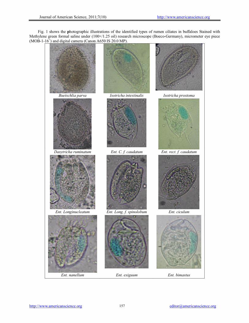
The identified genera and species can be ordered as following: Order: PROSTOMATIDA Family: BUETSCHLIIDAE Genus: BUETSCHLIA Buetschlia parva
Body is ovoid with attenuated anterior end and round posterior one. Whole body is uniformly ciliated except anterior long cilia around cytostome. Ectoplasm at anterior end is thick and macronucleus is near posterior end and globular in shape. There is an anterior concretion vacuole. Oval contractile vacuole is anterior to the nucleus and to one side. No cytopyge. The body dimensions (length × width) were 30-70 ×20-50 µm. Order: TRICHOSTOMATORIDA Family: ISOTRICHIDAE Genus: ISOTRICHA Isotricha prostoma
Body is oval and uniformly covered with cilia. Body is tapered at the level of cytostome which is sub-terminal. Macronucleus is kidney shape. Mouth is located at the end opposite the leading or anterior end. The body dimensions were 80-100 × 50-120 µm. Isotricha intestinalis
Body is oval and macronucleus is kidney shape. Cytostome is more sub-terminal at the level of macronucleus. Mouth is on one side of the cell equidistant between the posterior end and the middle. The body dimensions were 90-200 × 45-150 µm. Genus: DASYTRICHA Dasytricha ruminatum
Body is oval, covered with cilia in spiral longitudinal rows and is smaller than isotricha and commonly occurs in greater numbers in the rumen.
Journal of American Science, 2011;7(10) http://www.americanscience.org
http://www.americanscience.org [email protected] 154
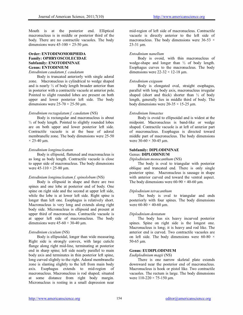
Mouth is at the posterior end. Elliptical macronucleus is in middle or posterior third of the body. There are no contractile vacuoles. The body dimensions were 45-100 × 25-50 µm. Order: ENTODINOMORPHIDA Family: OPHRYOSCOLECIDAE Subfamily: ENOTODININAE Genus: ENTODINIUM Entodinium caudatum f. caudatum
Body is truncated anteriorly with single adoral zone. Macronucleus is cylindrical to wedge shaped and is nearly ½ of body length broader anterior than in posterior with a contractile vacuole at anterior pole. Pointed to slight rounded lobes are present on both upper and lower posterior left side. The body dimensions were 25-70 × 25-50 µm. Entodinium rectagulatum f. caudatum (NS)
Body is rectangular and macronucleus is about ½ of body length. Pointed to slightly rounded lobes are on both upper and lower posterior left side. Contractile vacuole is at the base of adoral membranelle zone. The body dimensions were 25-50 × 25-40 µm. Entodinium longinucleatum
Body is ellipsoid, flattened and macronucleus is as long as body length. Contractile vacuole is close to upper side of macronucleus. The body dimensions were 45-110 × 25-80 µm. Entodinium longinucleatum f. spinolobum (NS)
Body is ellipsoid in shape and there are two spines and one lobe at posterior end of body. One spine on right side and the second at upper left side, while the lobe is at lower left side. Right spine is longer than left one. Esophagus is relatively short. Macronucleus is very long and extends along right body side. Micronucleus is ellipsoid and present at upper third of macronucleus. Contractile vacuole is at upper left side of macronucleus. The body dimensions were 45-60 × 30-40 µm. Entodinium ciculum (NS)
Body is ellipsoidal, longer than wide measuring. Right side is strongly convex, with large cuticle flange along right mid-line, terminating at posterior end in sharp spine; left side nearly parallel to main body axis and terminates in thin posterior left spine, long curved slightly to the right. Adoral membranelle zone is slanting slightly to the left from main body axis. Esophagus extends to mid-region of macronucleus. Macronucleus is rod shaped; situated at some distance from right body margin. Micronucleus is resting in a small depression near
mid-region of left side of macronucleus. Contractile vacuole is directly anterior to the left side of macronucleus. The body dimensions were 36-53 × 23-31 µm. Entodinium nanellum
Body is ovoid, with thin macronucleus of wedge-shape and longer than ½ of body length. Esophagus curves to the macronucleus. The body dimensions were 22-32 × 12-18 µm. Entodinium exiguum
Body is elongated oval, straight esophagus, parallel with long body axis, macronucleus irregular shaped (short and thick) shorter than ½ of body length, generally lies in middle third of body. The body dimensions were 20-35 × 15-25 µm. Entodinium bimastus
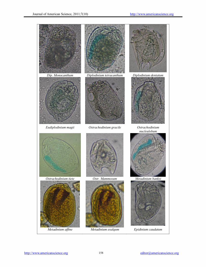
Body is ovoid to ellipsoidal and is widest at the midpoint. Macronucleus is band-like or wedge shaped. Contractile vacuole is at left of anterior part of macronucleus. Esophagus is directed toward middle part of macronucleus. The body dimensions were 30-60 × 30-45 µm. Subfamily: DIPLODININAE Genus: DIPLODINIUM Diplodinium monocanthum (NS)
The body is oval to triangular with posterior oblique and truncated end. There is only single posterior spine. Macronucleus is sausage in shape with anterior curved end toward the ventral aspect. The body dimensions were 60-90 × 40-60 µm. Diplodinium tetracanthum
The body is oval to triangular and ends posteriorly with four spines. The body dimensions were 60-80 × 40-60 µm. Diplodinium dentatum
The body has six heavy incurved posterior spines. Spine on right side is the longest one. Macronucleus is long; it is heavy and rod like. The anterior end is curved. Two contractile vacuoles are on left side. The body dimensions were 60-80 × 50-65 µm. Genus: EUDIPLODINIUM Eudiplodinium magii (NS)
There is one narrow skeletal plate extends downward near the posterior end of macronucleus. Macronucleus is hook or pistol like. Two contractile vacuoles. The rectum is large. The body dimensions were 110-220 × 75-150 µm.
Journal of American Science, 2011;7(10) http://www.americanscience.org
http://www.americanscience.org [email protected] 155
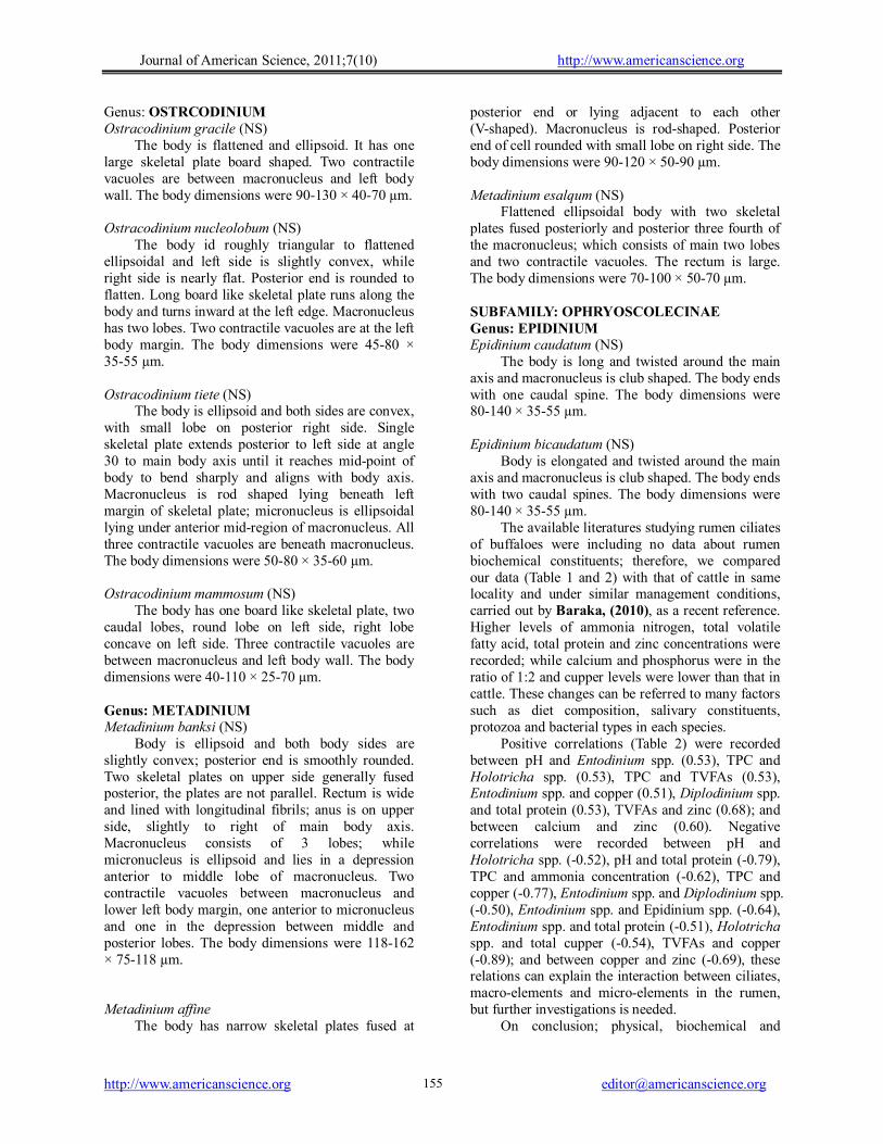
Genus: OSTRCODINIUM Ostracodinium gracile (NS)
The body is flattened and ellipsoid. It has one large skeletal plate board shaped. Two contractile vacuoles are between macronucleus and left body wall. The body dimensions were 90-130 × 40-70 µm. Ostracodinium nucleolobum (NS)
The body id roughly triangular to flattened ellipsoidal and left side is slightly convex, while right side is nearly flat. Posterior end is rounded to flatten. Long board like skeletal plate runs along the body and turns inward at the left edge. Macronucleus has two lobes. Two contractile vacuoles are at the left body margin. The body dimensions were 45-80 × 35-55 µm. Ostracodinium tiete (NS)
The body is ellipsoid and both sides are convex, with small lobe on posterior right side. Single skeletal plate extends posterior to left side at angle 30 to main body axis until it reaches mid-point of body to bend sharply and aligns with body axis. Macronucleus is rod shaped lying beneath left margin of skeletal plate; micronucleus is ellipsoidal lying under anterior mid-region of macronucleus. All three contractile vacuoles are beneath macronucleus. The body dimensions were 50-80 × 35-60 µm. Ostracodinium mammosum (NS)
The body has one board like skeletal plate, two caudal lobes, round lobe on left side, right lobe concave on left side. Three contractile vacuoles are between macronucleus and left body wall. The body dimensions were 40-110 × 25-70 µm. Genus: METADINIUM Metadinium banksi (NS)
Body is ellipsoid and both body sides are slightly convex; posterior end is smoothly rounded. Two skeletal plates on upper side generally fused posterior, the plates are not parallel. Rectum is wide and lined with longitudinal fibrils; anus is on upper side, slightly to right of main body axis. Macronucleus consists of 3 lobes; while micronucleus is ellipsoid and lies in a depression anterior to middle lobe of macronucleus. Two contractile vacuoles between macronucleus and lower left body margin, one anterior to micronucleus and one in the depression between middle and posterior lobes. The body dimensions were 118-162 × 75-118 µm. Metadinium affine
The body has narrow skeletal plates fused at
posterior end or lying adjacent to each other (V-shaped). Macronucleus is rod-shaped. Posterior end of cell rounded with small lobe on right side. The body dimensions were 90-120 × 50-90 µm. Metadinium esalqum (NS)
Flattened ellipsoidal body with two skeletal plates fused posteriorly and posterior three fourth of the macronucleus; which consists of main two lobes and two contractile vacuoles. The rectum is large. The body dimensions were 70-100 × 50-70 µm. SUBFAMILY: OPHRYOSCOLECINAE Genus: EPIDINIUM Epidinium caudatum (NS)
The body is long and twisted around the main axis and macronucleus is club shaped. The body ends with one caudal spine. The body dimensions were 80-140 × 35-55 µm. Epidinium bicaudatum (NS)
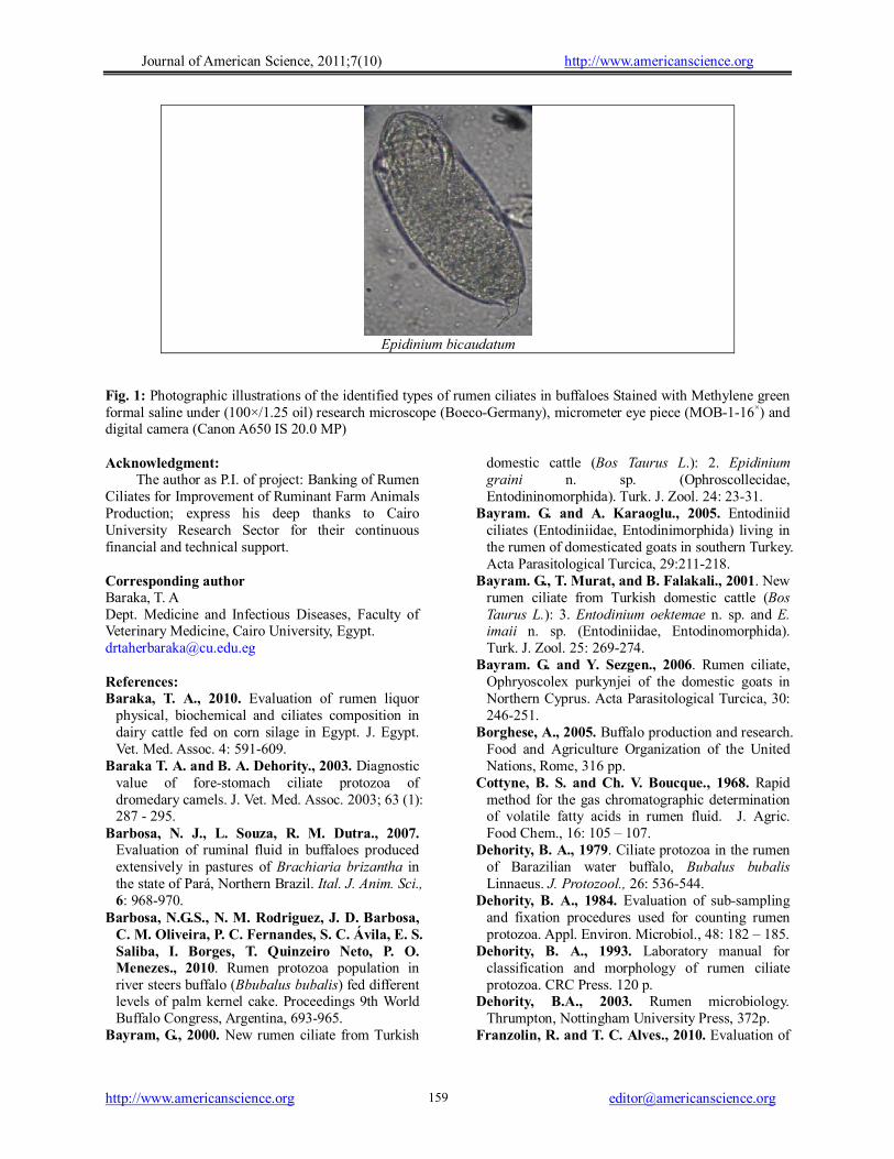
Body is elongated and twisted around the main axis and macronucleus is club shaped. The body ends with two caudal spines. The body dimensions were 80-140 × 35-55 µm.
The available literatures studying rumen ciliates of buffaloes were including no data about rumen biochemical constituents; therefore, we compared our data (Table 1 and 2) with that of cattle in same locality and under similar management conditions, carried out by Baraka, (2010), as a recent reference. Higher levels of ammonia nitrogen, total volatile fatty acid, total protein and zinc concentrations were recorded; while calcium and phosphorus were in the ratio of 1:2 and cupper levels were lower than that in cattle. These changes can be referred to many factors such as diet composition, salivary constituents, protozoa and bacterial types in each species.
Positive correlations (Table 2) were recorded between pH and Entodinium spp. (0.53), TPC and Holotricha spp. (0.53), TPC and TVFAs (0.53), Entodinium spp. and copper (0.51), Diplodinium spp. and total protein (0.53), TVFAs and zinc (0.68); and between calcium and zinc (0.60). Negative correlations were recorded between pH and Holotricha spp. (-0.52), pH and total protein (-0.79), TPC and ammonia concentration (-0.62), TPC and copper (-0.77), Entodinium spp. and Diplodinium spp. (-0.50), Entodinium spp. and Epidinium spp. (-0.64), Entodinium spp. and total protein (-0.51), Holotricha spp. and total cupper (-0.54), TVFAs and copper (-0.89); and between copper and zinc (-0.69), these relations can explain the interaction between ciliates, macro-elements and micro-elements in the rumen, but further investigations is needed.
On conclusion; physical, biochemical and
Journal of American Science, 2011;7(10) http://www.americanscience.org
http://www.americanscience.org [email protected] 156
cellular constituents of water buffalo should be put in consideration during clinical examination and evaluation of rumen performance, and typing of
buffalo rumen ciliates needs further investigation to be a guide during rumen juice transplantation.
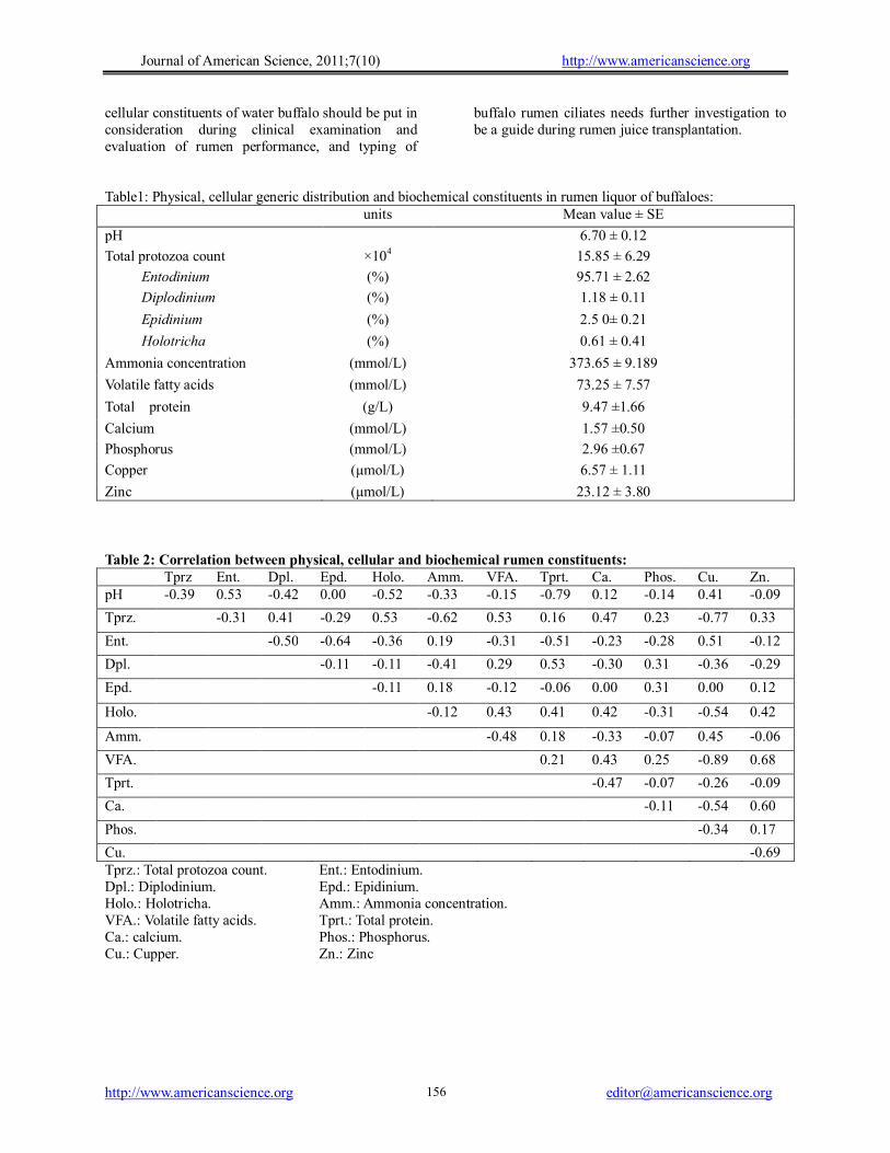
Table1: Physical, cellular generic distribution and biochemical constituents in rumen liquor of buffaloes: units Mean value ± SE
pH 6.70 ± 0.12
Total protozoa count ×104 15.85 ± 6.29
Entodinium (%) 95.71 ± 2.62
Diplodinium (%) 1.18 ± 0.11
Epidinium (%) 2.5 0± 0.21
Holotricha (%) 0.61 ± 0.41
Ammonia concentration (mmol/L) 373.65 ± 9.189
Volatile fatty acids (mmol/L) 73.25 ± 7.57
Total protein (g/L) 9.47 ±1.66
Calcium (mmol/L) 1.57 ±0.50
Phosphorus (mmol/L) 2.96 ±0.67
Copper (µmol/L) 6.57 ± 1.11
Zinc (µmol/L) 23.12 ± 3.80 Table 2: Correlation between physical, cellular and biochemical rumen constituents: Tprz Ent. Dpl. Epd. Holo. Amm. VFA. Tprt. Ca. Phos. Cu. Zn. pH -0.39 0.53 -0.42 0.00 -0.52 -0.33 -0.15 -0.79 0.12 -0.14 0.41 -0.09
Tprz. -0.31 0.41 -0.29 0.53 -0.62 0.53 0.16 0.47 0.23 -0.77 0.33
Ent. -0.50 -0.64 -0.36 0.19 -0.31 -0.51 -0.23 -0.28 0.51 -0.12
Dpl. -0.11 -0.11 -0.41 0.29 0.53 -0.30 0.31 -0.36 -0.29
Epd. -0.11 0.18 -0.12 -0.06 0.00 0.31 0.00 0.12
Holo. -0.12 0.43 0.41 0.42 -0.31 -0.54 0.42
Amm. -0.48 0.18 -0.33 -0.07 0.45 -0.06
VFA. 0.21 0.43 0.25 -0.89 0.68
Tprt. -0.47 -0.07 -0.26 -0.09
Ca. -0.11 -0.54 0.60
Phos. -0.34 0.17
Cu. -0.69 Tprz.: Total protozoa count. Ent.: Entodinium. Dpl.: Diplodinium. Epd.: Epidinium. Holo.: Holotricha. Amm.: Ammonia concentration. VFA.: Volatile fatty acids. Tprt.: Total protein. Ca.: calcium. Phos.: Phosphorus. Cu.: Cupper. Zn.: Zinc
Journal of American Science, 2011;7(10) http://www.americanscience.org
http://www.americanscience.org [email protected] 157
Fig. 1 shows the photographic illustrations of the identified types of rumen ciliates in buffaloes Stained with Methylene green formal saline under (100×/1.25 oil) research microscope (Boeco-Germany), micrometer eye piece (MOB-1-16×) and digital camera (Canon A650 IS 20.0 MP).
Buetschlia parva Isotricha intestinalis Isotricha prostoma
Dasytricha ruminatum Ent. C. f. caudatum Ent. rect. f. caudatum
Ent. Longinucleatum Ent. Long. f. spinolobum Ent. ciculum
Ent. nanellum Ent. exiguum Ent. bimastus
Journal of American Science, 2011;7(10) http://www.americanscience.org
http://www.americanscience.org [email protected] 158
Dip. Monocanthum Diplodinium tetracanthum Diplodinium dentatum
Eudiplodinium magii Ostrachodinium gracile Ostrachodinium nucleulobum
Ostrachodinium tiete Ostr. Mammosum Metadinium banksi
Metadinium affine Metadinium esalqum Epidinium caudatum
Journal of American Science, 2011;7(10) http://www.americanscience.org
http://www.americanscience.org [email protected] 159
Epidinium bicaudatum
Fig. 1: Photographic illustrations of the identified types of rumen ciliates in buffaloes Stained with Methylene green formal saline under (100×/1.25 oil) research microscope (Boeco-Germany), micrometer eye piece (MOB-1-16×) and digital camera (Canon A650 IS 20.0 MP) Acknowledgment:
The author as P.I. of project: Banking of Rumen Ciliates for Improvement of Ruminant Farm Animals Production; express his deep thanks to Cairo University Research Sector for their continuous financial and technical support. Corresponding author Baraka, T. A Dept. Medicine and Infectious Diseases, Faculty of Veterinary Medicine, Cairo University, Egypt. [email protected] References: Baraka, T. A., 2010. Evaluation of rumen liquor
physical, biochemical and ciliates composition in dairy cattle fed on corn silage in Egypt. J. Egypt. Vet. Med. Assoc. 4: 591-609.
Baraka T. A. and B. A. Dehority., 2003. Diagnostic value of fore-stomach ciliate protozoa of dromedary camels. J. Vet. Med. Assoc. 2003; 63 (1): 287 - 295.
Barbosa, N. J., L. Souza, R. M. Dutra., 2007. Evaluation of ruminal fluid in buffaloes produced extensively in pastures of Brachiaria brizantha in the state of Pará, Northern Brazil. Ital. J. Anim. Sci., 6: 968-970.
Barbosa, N.G.S., N. M. Rodriguez, J. D. Barbosa, C. M. Oliveira, P. C. Fernandes, S. C. Ávila, E. S. Saliba, I. Borges, T. Quinzeiro Neto, P. O. Menezes., 2010. Rumen protozoa population in river steers buffalo (Bbubalus bubalis) fed different levels of palm kernel cake. Proceedings 9th World Buffalo Congress, Argentina, 693-965.
Bayram, G., 2000. New rumen ciliate from Turkish
domestic cattle (Bos Taurus L.): 2. Epidinium graini n. sp. (Ophroscollecidae, Entodininomorphida). Turk. J. Zool. 24: 23-31.
Bayram. G. and A. Karaoglu., 2005. Entodiniid ciliates (Entodiniidae, Entodinimorphida) living in the rumen of domesticated goats in southern Turkey. Acta Parasitological Turcica, 29:211-218.
Bayram. G., T. Murat, and B. Falakali., 2001. New rumen ciliate from Turkish domestic cattle (Bos Taurus L.): 3. Entodinium oektemae n. sp. and E. imaii n. sp. (Entodiniidae, Entodinomorphida). Turk. J. Zool. 25: 269-274.
Bayram. G. and Y. Sezgen., 2006. Rumen ciliate, Ophryoscolex purkynjei of the domestic goats in Northern Cyprus. Acta Parasitological Turcica, 30: 246-251.
Borghese, A., 2005. Buffalo production and research. Food and Agriculture Organization of the United Nations, Rome, 316 pp.
Cottyne, B. S. and Ch. V. Boucque., 1968. Rapid method for the gas chromatographic determination of volatile fatty acids in rumen fluid. J. Agric. Food Chem., 16: 105 – 107.
Dehority, B. A., 1979. Ciliate protozoa in the rumen of Barazilian water buffalo, Bubalus bubalis Linnaeus. J. Protozool., 26: 536-544.
Dehority, B. A., 1984. Evaluation of sub-sampling and fixation procedures used for counting rumen protozoa. Appl. Environ. Microbiol., 48: 182 – 185.
Dehority, B. A., 1993. Laboratory manual for classification and morphology of rumen ciliate protozoa. CRC Press. 120 p.
Dehority, B.A., 2003. Rumen microbiology. Thrumpton, Nottingham University Press, 372p.
Franzolin, R. and T. C. Alves., 2010. Evaluation of
Journal of American Science, 2011;7(10) http://www.americanscience.org
http://www.americanscience.org [email protected] 160
diets with increasing corn grain levels on rumen protozoa population and liquid outflow rate in buffalo. Proceedings 9th World Buffalo Congress, Argentina, 663-665.
Franzolin, R., P. R. Fabrício and V. B. Weber., 2010. Effects of dietary energy and nitrogen supplements on rumen fermentation and protozoa population in buffalo and zebu cattle. R. Bras. Zootec., 3: 549-555.
Imai, S., C. H. Chang, K. Ogimoto, J. Fujita., 1981 (a). Rumen ciliate protozoa of the water buffalo (Bubalus bubalis) in Taiwan. Bull. Nippon Vet.Zootech. Coll. 30: 77-81
Imai, S., Ogimoto, K., Fujita, J. 1981 (b). Rumen ciliate protozoal fauna of the water buffalo Bubalus bubalis (Linnaeus) in Okinawa, Japan. Bull. Nippon Vet.Zootech. Coll. 30: 82-85.
Lopez, P. L., E. C. Ricohermoso, M. S. Banes., 2004. variation in microbial counts in the rumen of slaughtered carabaos. abstract of researches on the philippine water buffalo. Philippine Carabao Center, pp: 113-114
Mermer, A., S. Rastgldi,G. Ergen, and B. Gocmen., 2003. Occurance of rumen ciliates, Elytroplastron bubali in Turkis domestic goats. Acta Parasitologica Turcica. 27: 270-272.
Ogimoto, K. and S. Imai., 1981. Atlas of rumen microbiology. Japan Scientific Soc. Press, Tokyo.
Paul, N. B. and A. Srvastava., 2002. In vitro rumen fermentation as affected by different levels of pure
fatty acids. Indian Vet. J. 79: 20-22. Philip, K.A. and B. A. Al-Badrani., 2008.
Changes in ruminal contents of buffaloes suffering from digestive disorders. Iraqi Vet. Med. Sci. J. 2: 151-163.
Rispoli, T. B., I. L. Rodrijues, R. Neto, R. Kazama, O. P. Prado, L. M. Zeoula, P. B. Arsuri., (2009). Ruminal ciliate protozoa of cattle and buffalo fed on diet supplemented with monensin or extracts from propolis. Pesq. Agropec. Bas., Brasilia, 44: 92-97.
Selim, H .M., O. Yamato, O. Elkabbany, F. Koroloss, Y. Maede., 1996. Comparative study on rumen ciliates in buffalo, cattle and sheep in Egypt. J. Vet. Med. Sci. 58: 799-801.
Shimizu, M., M. Kinoshita, J. Fujita, S. Imai., 1983. Rumen ciliates protozoal fauna of the zibu cattle, (Bos indicus) and water buffalo (Bubalus bubalis) in Philippines. Bull. Nippon Vet. Zootech. 18: 83-88.
Tsankova, M., K. Sivokova, K. Dimov, M. Marinov., 2010. Effects of the enzyme preparation Xybeten-cel on some fermentation processes in the rumen of buffalo calves. Proceedings 9th World Buffalo Congress. Argentina, Buenos Aires., p. 658-662.
Zapletal, O., 1967. The toxicity of urea and possibility its influence at cattle. Ph. D. Thesis. Brno, (Czech Republic), 120 pp.
9/30/2011