Embed Size (px)
Citation preview
Journal Name
COMMUNICATION
This journal is © The Royal Society of Chemistry 20xx J. Name., 2013, 00, 1-3 | 1
Please do not adjust margins
Please do not adjust margins
a. King Abdullah University of Science and Technology (KAUST), Division of Physical Sciences and Engineering (PSE), Thuwal 23955-6900, Saudi Arabia.
b. King Abdullah University of Science and Technology (KAUST), SABIC Corporate Research and Innovation Center, Thuwal, 23955-6900, Saudi Arabia.
c. King Abdullah University of Science and Technology (KAUST), Imaging and Characterization Lab, Thuwal 23955-6900, Saudi Arabia.
d. York Nanocentre, University of York, Heslington, York YO10 5DD, U.K.
e. Mathematical Institute, University of Oxford, Oxford OX2 6GG, U.K.
Electronic Supplementary Information (ESI) available: See DOI: 10.1039/x0xx00000x
Received 00th January 20xx,
Accepted 00th January 20xx
DOI: 10.1039/x0xx00000x
www.rsc.org/
Trapping Shape-Controlled Nanoparticle Nucleation and Growth Stages via Continuous-Flow Chemistry
Alec P. LaGrow,*ad
Tabot M. D. Besong,a Noktan M. AlYami,
a Khabiboulakh Katsiev,
b Dalaver H.
Anjum,c Ahmed Abd Elkader,
a Pedro M. F. J. Costa,
a Victor M. Burlakov,
e Alain Goriely
e and Osman
M. Bakr*a
Continuous flow chemistry is used to trap the nucleation and
growth stages of platinum-nickel nano-octahedra with second
time resolution and high throughputs to probe their properties
ex-situ. The growth starts from poorly crystalline particles
(nucleation) at 5 seconds, to crystalline 1.5 nm particles bounded
by the {111}-facets at 7.5 seconds, followed by truncation and
further growth to octahedral nanoparticles at 20 seconds.
Nanoparticles have become ubiquitous in the literature for
their applications, in catalysis,1 magnetism,
2,3 biomedicine
2,3
and optics.1,2
Solution synthesis techniques for nanoparticles
have rapidly increased in sophistication, with a library of sizes
and shapes being generated.4 However, controlling the growth
of nanoparticles is still more of an art due to the lack of
techniques that can probe the initial stages of nanoparticle
growth with the necessary time resolution and sensitivity.4,5
For shape-controlled nanoparticles syntheses, it is typically
considered that initial nuclei grow into a seed crystal that
controls the growth into the final shaped nanoparticle.4
However, direct evidence for the identity of the initial nuclei
and how faceting develops have not been experimentally
shown. The insensitivity of in-situ techniques to the formation
of defects and crystal facets is a major barrier for obtaining
such evidence.6,7
Due to the above-mentioned limitations, ex-situ
techniques dominate the discussion of nanoparticle growth by
taking aliquots, or quenching the reaction at certain
intervals.8,9
However, the initial nucleation step of the
nanoparticle synthesis can occur within seconds7,10,11
of the
start of the reaction and in very low yields.12
This restricts the
use of aliquots to study nucleation as their time resolution is
limited by the heat transport of the reaction solution, the
amount of aliquot needed and the ease and frequency with
which the aliquot is acquired.
Here-in, we use a continuous flow reactor11,13-15
to
precisely control the reaction time to selectively trap the
nucleation and growth stages. The use of millifluidics allows
access to a heat transfer coefficient orders of magnitude
higher than typical batch reactions, and therefore allows the
reaction to be initiated and quenched in fractions of a second
without the need of introducing a foreign agent (e.g.,
quenching chemical) to the reaction. By using high angle
annular dark field scanning transmission electron microscopy
(HAADF-STEM) and analytical ultracentrifugation (AUC), we
were able to detect and observe the particle growth starting
from disordered clusters of atoms, to {111} dominated
crystals, of 1.5 nm in size, that grow into fully formed
octahedral nanoparticles. Due to the continuous nature of the
synthesis, every stage in the reaction could be isolated in a
large scale.14,16

COMMUNICATION Journal Name
2 | J. Name., 2012, 00, 1-3 This journal is © The Royal Society of Chemistry 20xx
Please do not adjust margins
Please do not adjust margins
The growth of platinum-nickel (Pt-Ni) nanooctahedra was
studied by quenching the reaction with second intervals in a
continuous-flow reactor.16, 17
The reaction was carried out with
surfactants, oleylamine (OAm) and 1-adamantane carboxylic
acid (AA), and a source of carbon monoxide, tungsten carbonyl
[W(CO)6]. The W(CO)6 acts as a source of CO and the tungsten
remains in solution in a partially oxidized form (Fig. S1).
Platinum acetylacetonate [Pt(acac)2] and nickel
acetylacetonate dihydrate [Ni(acac)2.2H2O] were used as
precursors at 240°C in toluene. The reaction was run
continuously with residence times of 5, 7.5, 10, and 20 seconds
(Fig. 1A). The HAADF-STEM was taken from the first frame
exposed to the beam (Fig. S2-S4). With the 5 second residence
time, the formation of 1.3 ± 0.3 nm particles of ~Pt72Ni28 with a
large proportion of ~1.3 nm crystalline particles, disordered
particles of ≤1 nm and loose single atoms were observed (Fig.
1B, F, Fig. S5 and Table S1). The same species as at 5 seconds
were seen at times shorter than 5 seconds but with much
lower yields. With a residence time of 7.5 seconds crystalline
particles of 1.5 ± 0.2 nm ~Pt82Ni18 were formed with the {111}-
facets exposed on the surface (Fig. 1C, G and Fig. S6). With a
10 second residence time, 2.6 ± 0.4 nm cuboctahedral particles
of ~Pt54Ni46 were formed (Fig. 1D, H, Table S1 and Fig. S7).
With a 20 second residence time, nanooctahedra of ~Pt47Ni53
(3.6 ± 0.5 nm) were formed (Fig. 1E, I and Table S1).
STEM-electron energy loss spectroscopy (EELS) spectrum
imaging of an amorphous particle showed nearly equal
proportions of Pt:Ni (Fig. 1J). The ~1.5 nm crystalline particle
was platinum rich (Fig. 1K), while the larger ~2.5 and ~3.3 nm
particles tended towards parity with time (Fig. 1L and M). In
each particle the platinum and nickel signals matched each
other, showing that the particles formed a random alloy
structure (Fig. 1J-M). X-ray diffraction of the nanoparticles
show a decrease in lattice parameters and sharpening of peaks
with increased residence time, consistent with the nickel
enrichment of the alloy and an increase in particle size (Fig. S9
and Table S2).
Theoretical analysis of the nanoparticle growth stages
carried out in SI section 3 indicate that the process of
nucleation and growth in this system occurs initially to
minimize its surface energy. As the particle grows, it
crystallizes and becomes dominated by the exposure of
energetically favorable low index facets, namely the lowest
energy {111}-facets (SI section 3),19
which is also chemically
favored by the addition of CO.18,20
The particles initially
crystallize with predominantly platinum atoms due to the
higher cohesive energy of platinum compared to nickel,
causing the detachment rate of nickel atoms to be higher than
that of platinum (SI section 3).21
As the particles grow, the
platinum to nickel ratio tends towards parity as the platinum is
Figure 1. A) Schematic of the continuous-flow reactor. TEM images of platinum nickel nanoparticles with residence times of B) 5 s, C)
7.5 s, D) 10 s and E) 20 s and HAADF-STEM images of F-I) respectively. The red lines indicate the {111} facet (Fig. S8). STEM-EELS
spectrum imaging of a J) amorphous, K) ~1.5 nm, L) ~2.5 nm and M) ~3.3 nm particle. The line profiles below (J-M) are from the blue
boxes.

Journal Name COMMUNICATION
This journal is © The Royal Society of Chemistry 20xx J. Name., 2013, 00, 1-3 | 3
Please do not adjust margins
Please do not adjust margins
initially depleted from the solution and the nickel becomes the
major additive, and therefore dominates the chemistry.
Analytical ultracentrifugation (AUC) is used to analyze
distinct species in solution from the nanoparticle systems.22
The AUC indicates an increase in the sedimentation coefficient
and corresponding hydrodynamic diameter of the
nanoparticles with increasing residence times in the flow
reactor (Fig. 2, Fig S10, S11 and Table S3-S8). During the
nucleation step there are five major components present (5
seconds, Fig. 2) which decreases to only two major species
present in the 7.5 second sample (Fig. 2). The major peak in
the 5 and 7.5 second samples have a similar peak position and
would correspond to the crystalline particles of ~1.3 nm (Fig.
2). The species at the lowest sedimentation coefficients are
expected to be from the precursor and reduced monomers,
they are seen in the samples with residence times of 5 and 7.5
seconds but are no longer present with a residence time of 10
seconds (Fig. 2). In the 10 second sample the major species are
at much higher sedimentation coefficients and a distribution of
the smaller species is observed (Fig. 2). The 20 second sample
only contained the larger species, which are expected to be
the octahedral nanoparticles (Fig. 2).
Figure 2. 3-D representation of the relative concentrations,
sedimentation and diffusion coefficients from AUC at 5-20 s.
To determine the ligand shell surrounding the
nanoparticles, fourier transform infrared spectroscopy (FTIR)
and x-ray photoelectron spectroscopy (XPS) was utilized. The
FTIR spectra of all samples had three peaks from 2850 – 2960
cm-1
which would correspond to aliphatic CH2 stretches. The
FTIR spectrum for the 5 second sample had an O-H stretch at
3227 cm-1
, and stretches at 1300-1000 cm-1
indicative of a C-O
stretch (Fig. S12). These could come from the reduced
acetylacetonate. The 7.5-second sample had a large stretch at
1697 cm-1
indicating an unbound C=O stretch which is
attributed to either the acetylacetonate or the adamantine
carboxylic acid (Fig. S12). For the 10 second sample, the FTIR
spectrum has an N-H bond at 1633 cm-1
, indicating the
presence of oleylamine on the surface. The lack of the N-H
stretch around 3300 cm-1
is due to the amine moiety binding
to the metal surface.23
For, the 20 second sample, the FTIR
spectrum has a mixed ligand environment with carbonyl and
amine groups (Fig. S12).
The XPS of the nitrogen has two components in the
samples. The first component at ~400 eV corresponded to C-
NH2 (Fig. S13 and Table S9). The second component can be
seen at 399 eV up till the 10 second sample and then at 398 eV
in the 20 second sample (Fig. S13 and Table S9). The peak at
398 eV would be a strong M-N bond as reported with Pt-N.24
The peak at 399 eV could be due to a weaker M-N bond or a
distorted ligand environment, it is observed to shift to lower
energy and increase in proportion with longer residence times.
For Pt and Ni, the dominant signal is reduced metal (Fig. S13
and Table S9).
From the combination of the STEM, AUC, FTIR and XPS
results, we show that the initial nucleation of particles occurs
in the formation of low crystallinity metallic platinum-nickel
clusters of atoms of multiple sizes that are surrounded by
acetylacetonate ligands (Fig. 3). Within seconds, platinum rich
crystalline particles of 1.3 nm and above form and are
predominantly bound by the {111} facets, the size distribution
narrows, by 7.5 seconds, while continued nucleation is still
occurring. As growth progresses, the nanoparticle size
increases as the dominant mechanism in the reaction favors
nanoparticle growth (after 10 seconds), and the
acetylacetonate ligand environment decomposes6 and is
replaced by loosely bound amine ligands as confirmed by FTIR
and XPS results (Fig. 3). The nickel that was left in solution
adds to the nanoparticles, dominating the surface chemistry of
the particles and adding preferentially on the {111} facets, due
to the presence of carbon monoxide.18,20
Growth slows down
as last of the nickel is incorporated into the alloy structure and
the strong M-N bond forms with the amine surfactants (after
20 seconds) which stabilize the particles from aggregation23
as
well as reduce the addition rate of incoming atoms to the
particles surface.25
COMMUNICATION Journal Name
4 | J. Name., 2012, 00, 1-3 This journal is © The Royal Society of Chemistry 20xx
Please do not adjust margins
Please do not adjust margins
Figure 3. Growth mechanism schematic.
To study the properties of particles in the trapped stages, it
is necessary to establish the particles stability both ex situ and
in solution. The nuclei were stable once quenched and could
be stored in toluene for over 6 months without noticeable
changes in the size distributions measured by TEM or AUC. The
ultra-small particles were also seen to be thermally stable
under ultrahigh vacuum conditions up to 1100°C if they are
physically separated (Fig. S14 - S17), or up to 600°C if they are
not (Fig. S13 and S15). The particles also show reasonable
facet stability up to 1100°C (Fig. S14 - S16). For the 1.5 nm
particles Ostwald ripening was observed (Fig. S17). The facet
stability is attributed to the thermal stability of the {111}
surface facet.26
The 5 second sample species are seen to be
stable up to 1100°C and still show populations of single atoms,
disordered and crystalline particles (Fig. S18). The trapped
particles were stable once quenched and therefore could be
utilized in a wide range of applications.
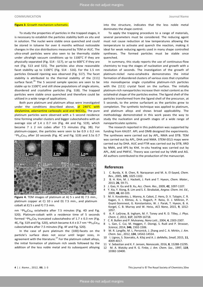
Both pure platinum and platinum alloys were investigated
under the conditions described above, at 240°C with
oleylamine, adamantine carboxylic acid and W(CO)6. The pure
platinum particles were observed with a 5 second residence
time forming smaller clusters and bigger cuboctahedra with an
average size of 1.4 ± 0.7 nm (Fig. 4A and Fig. S19), which
became 7 ± 2 nm cubes after 7.5 minutes (Fig. 3B). For
platinum-copper, the particles were seen to be 0.9 ± 0.2 nm
~Pt25Cu75 after 10 seconds (Fig. 4C and Fig. S19) and 3.5± 0.7
nm ~Pt60Cu40 octahedra after 7.5 minutes (Fig. 4D and Fig.
S20). Platinum-cobalt with a residence time of 5 seconds
formed ~Pt91Co9 truncated cuboctahedra of 1.7 ± 0.3 nm (Fig.
4E, Fig. S19 and Fig. S20), which became 4.4 ± 0.7 nm ~Pt70Co30
cuboctahedra after 7.5 minutes (Fig. 4F and Fig. S20).
In the case of pure platinum the {100}-facets on the
particle’s surface does not occur until larger sizes, in
agreement with the literature.27
For the platinum cobalt alloys,
the initial formation of platinum rich seeds followed by the
addition of the less noble metal and its subsequent alloying
into the structure, indicates that the less noble metal
dominates the shape control.
To apply the trapping procedure to a range of materials,
several parameters must be considered. The reducing agent
must not cause reduction at low temperatures allowing the
temperature to activate and quench the reaction, making it
ideal for weak reducing agents used in many shape controlled
syntheses. The formed particles must be stable once
quenched.
In summary, this study reports the use of continuous-flow
chemistry to trap the stages of nucleation and growth with a
resolution of seconds. The investigation of the growth of
platinum-nickel nano-octahedra demonstrates the initial
formation of disordered clusters of various sizes that crystallize
into monodisperse single crystalline platinum-rich particles
with the {111} crystal facet on the surface. The initially
platinum-rich nanoparticles increase their nickel content as the
octahedral shape of the particles evolve. The ligand shell of the
particles transformed from the ligand shell of the precursor, at
5 seconds, to the amine surfactant as the particles grew to
completion. The synthetic technique was applied to platinum,
and platinum alloys and shows broad applicability. The
methodology demonstrated in this work paves the way to
study the nucleation and growth stages of a wide range of
nanoparticulate systems.
The research reported in this publication was supported by
funding from KAUST. APL and OMB designed the experiments.
The syntheses were carried out by APL, NMA and DTB. TEM
was carried out by APL, DHA and NMA. STEM-EELS maps were
carried out by DHA. AUC and FTIR was carried out by DTB, XRD
by NMA, and XPS by KhK. In-situ heating was carried out by
APL, AAE and PMFJC. Theory was carried out by VMB and AG.
All authors contributed to the production of the manuscript.
References
1 C. Burda, X. B. Chen, R. Narayanan and M. A. El-Sayed, Chem. Rev., 2005, 105, 1025-1102.
2 B. H. Kim, M. J. Hackett, J. Park and T. Hyeon, Chem. Mater., 2013, 26, 59-71.
3 J. Gao, H. Gu and B. Xu, Acc. Chem. Res., 2009, 42, 1097-1107. 4 Y. Xia, Y. Xiong, B. Lim and S. E. Skrabalak, Angew. Chem. Int. Ed.,
2009, 48, 60-103. 5 M. V. Kovalenko, L. Manna, A. Cabot, Z. Hens, D. V. Talapin, C. R.
Kagan, V. I. Klimov, A. L. Rogach, P. Reiss, D. J. Milliron, P. Guyot-Sionnnest, G. Konstantatos, W. J. Parak, T. Hyeon, B. A. Korgel, C. B. Murray and W. Heiss, ACS Nano, 2015, 9, 1012-1057.
6 A. P. LaGrow, B. Ingham, M. F. Toney and R. D. Tilley, J. Phys. Chem. C, 2013, 117, 16709-16718.
7 C. R. Bullen and P. Mulvaney, Nano Lett., 2004, 4, 2303-2307. 8 L. Gan, C. Cui, M. Heggen, F. Dionigi, S. Rudi and P. Strasser,
Science, 2014, 346, 1502-1506. 9 M. R. Langille, M. L. Personick, J. Zhang and C. A. Mirkin, J. Am.
Chem. Soc., 2012, 134, 14542-14554. 10 I. Lignos, S. Stavrakis, A. Kilaj and A. J. deMello, Small, 2015, 11,
4009-4017. 11 V. Sebastian and K. F. Jensen, Nanoscale, 2016, 8, 15288-15295. 12 M. A. Watzky and R. G. Finke, J. Am. Chem. Soc., 1997, 119,
10382-10400.

Journal Name COMMUNICATION
This journal is © The Royal Society of Chemistry 20xx J. Name., 2013, 00, 1-3 | 5
Please do not adjust margins
Please do not adjust margins
13 J. Pan, A. a. O. El-Ballouli, L. Rollny, O. Voznyy, V. M. Burlakov, A. Goriely, E. H. Sargent and O. M. Bakr, ACS Nano, 2013, 7, 10158-10166.
14 H. Mehenni, L. Sinatra, R. Mahfouz, K. Katsiev and O. M. Bakr, RSC Adv., 2013, 3, 22397-22403.
15 N. M. AlYami, A. Lagrow, k. Joya, J. Hwang, K. Katsiev, D. H. Anjum, Y. Losovyj, L. Sinatra, J. Y. Kim and O. M. Bakr, Phys. Chem. Chem. Phys., 2016, 18, 16169-16178.
16 S. E. Skrabalak and R. L. Brutchey, Chem. Mater., 2016, 28, 1003-1005.
17 A. P. LaGrow, K. R. Knudsen, N. M. AlYami, D. H. Anjum and O. M. Bakr, Chem. Mater., 2015, 27, 4134-4141.
18 J. Zhang, H. Yang, J. Fang and S. Zou, Nano Lett., 2010, 10, 638-644.
19 L. Vitos, A. V. Ruban, H. L. Skriver and J. Kollár, Surf. Sci., 1998, 411, 186-202.
20 S. Y. Hwang, M. Zhang, C. Zhang, B. Ma, J. Zheng and Z. Peng, Chem. Commun., 2014, 50, 14013-14016.
21 V. M. Burlakov and L. Kantorovich, J. Chem. Phys., 2011, 134, 024521.
22 R. P. Carney, J. Y. Kim, H. Qian, R. Jin, H. Mehenni, F. Stellacci and O. M. Bakr, Nat. Commun., 2011, 2, 335.
23 J. Zhang and J. Fang, J. Am. Chem. Soc., 2009, 131, 18543-18547.
24 X. Li, L. An, X. Chen, N. Zhang, D. Xia, W. Huang, W. Chu and Z. Wu, Scientific Reports, 2013, 3, 3234.
25 A. M. Karim, N. Al Hasan, S. Ivanov, S. Siefert, R. T. Kelly, N. G. Hallfors, A. Benavidez, L. Kovarik, A. Jenkins, R. E. Winans and A. K. Datye, J. Phys. Chem. C, 2015, 119, 13257-13267.
26 Y. Wen, H. Fang, Z. Zhu and S. Sun, Phys. Lett. A, 2009, 373, 272-276.
27 H.-G. Liao, D. Zherebetskyy, H. Xin, C. Czarnik, P. Ercius, H. Elmlund, M. Pan, L.-W. Wang and H. Zheng, Science, 2014, 345, 916-919.