Embed Size (px)
Citation preview
1
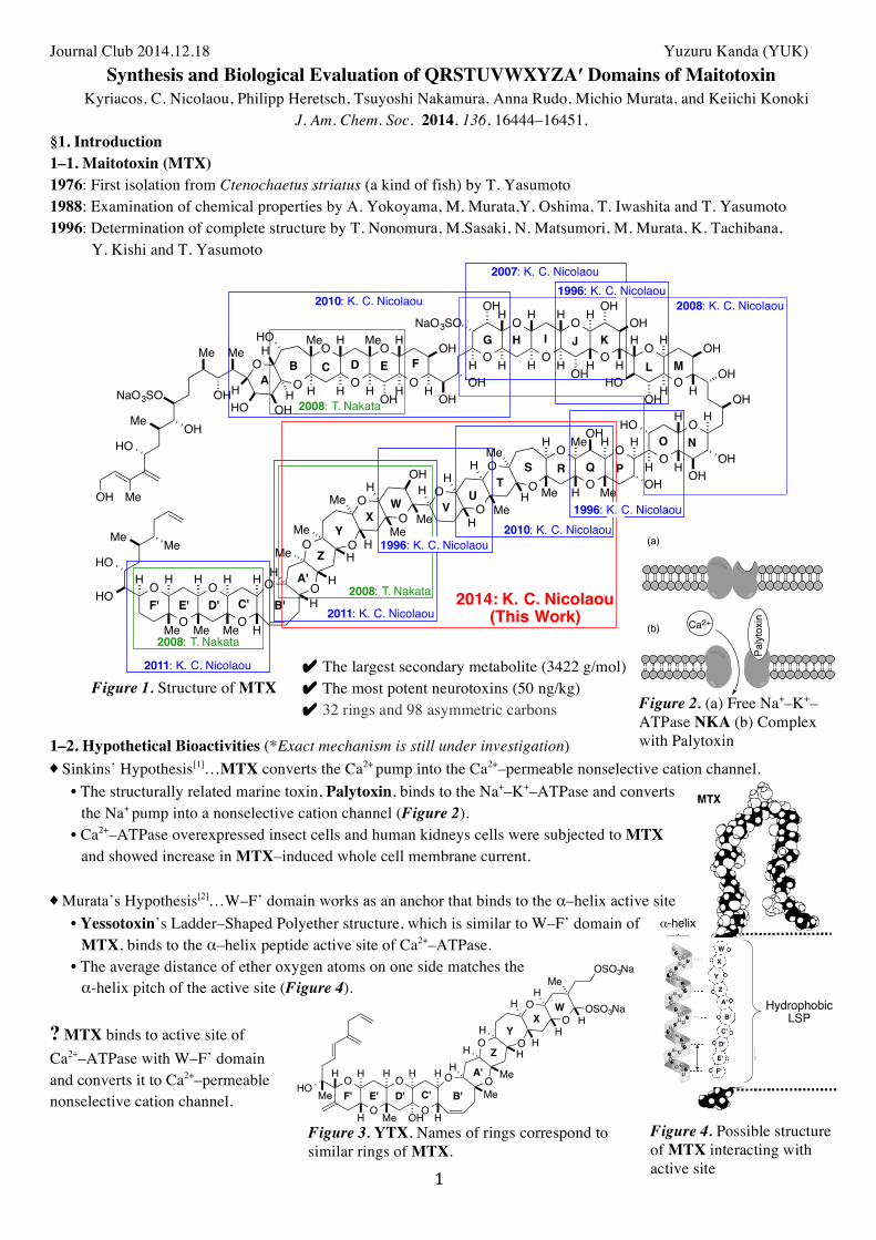
Journal Club 2014.12.18 Yuzuru Kanda (YUK) Synthesis and Biological Evaluation of QRSTUVWXYZA′ Domains of Maitotoxin
Kyriacos. C. Nicolaou, Philipp Heretsch, Tsuyoshi Nakamura, Anna Rudo, Michio Murata, and Keiichi Konoki J. Am. Chem. Soc. 2014, 136, 16444–16451.
§1. Introduction 1–1. Maitotoxin (MTX) 1976: First isolation from Ctenochaetus striatus (a kind of fish) by T. Yasumoto 1988: Examination of chemical properties by A. Yokoyama, M. Murata,Y. Oshima, T. Iwashita and T. Yasumoto 1996: Determination of complete structure by T. Nonomura, M.Sasaki, N. Matsumori, M. Murata, K. Tachibana,
Y. Kishi and T. Yasumoto
Figure 1. Structure of MTX
1–2. Hypothetical Bioactivities (*Exact mechanism is still under investigation) ♦ Sinkins’ Hypothesis[1]…MTX converts the Ca2+ pump into the Ca2+–permeable nonselective cation channel.
• The structurally related marine toxin, Palytoxin, binds to the Na+–K+–ATPase and converts the Na+ pump into a nonselective cation channel (Figure 2).
• Ca2+–ATPase overexpressed insect cells and human kidneys cells were subjected to MTX and showed increase in MTX–induced whole cell membrane current.
♦ Murata’s Hypothesis[2]…W–F’ domain works as an anchor that binds to the α–helix active site
• Yessotoxin’s Ladder–Shaped Polyether structure, which is similar to W–F’ domain of MTX, binds to the α–helix peptide active site of Ca2+–ATPase.
• The average distance of ether oxygen atoms on one side matches the α-helix pitch of the active site (Figure 4).
? MTX binds to active site of Ca2+–ATPase with W–F’ domain and converts it to Ca2+–permeable nonselective cation channel.
AB C D E F
G IH J K
L M
NO
PQRST
UVW
XY
A'
Z
B'C'D'E'F'
2007: K. C. Nicolaou
2008: K. C. Nicolaou1996: K. C. Nicolaou
2008: T. Nakata
2008: T. Nakata
2008: T. Nakata
2010: K. C. Nicolaou
2010: K. C. Nicolaou
2011: K. C. Nicolaou
2011: K. C. Nicolaou
1996: K. C. Nicolaou
1996: K. C. Nicolaou
O
OO
H
MeOH
O
O
O
O
O
O
O
O O
O
O
OO
O
O
OO
OO
OO
OO
OO
O
O
O
OHO
NaO3SO
Me Me
HO OH
HOOHMe
OH H H
HOH
Me
H
H Me
H
H
H
OH
H OHOHH H H H OH
NaO3SOOHH H H OH
H H
H OH
OH
H
HOH
H
HO
OH
H OH
OHH
H H
OH
HO
H
H
OH
HMeHMe
HH
H
HMe
H Me H MeOH
MeMe
HH
H
H
HMeMeMe
Me MeHO
H H H H H HMe
Me
MeH
OH
OH
2014: K. C. Nicolaou(This Work)
✔ The largest secondary metabolite (3422 g/mol) ✔ The most potent neurotoxins (50 ng/kg) ✔ 32 rings and 98 asymmetric carbons
Figure 2. (a) Free Na+–K+–ATPase NKA (b) Complex with Palytoxin
Palytoxin
Ca2+
(a)
(b)
Q–F' domainrich in ladder Poly Ether
F'
E'
D'
Z
A'
B'
C'
Y
X
W
HydrophobicLSP
α-helix
Figure 4. Possible structure of MTX interacting with active site
Figure 3. YTX, Names of rings correspond to similar rings of MTX.
OSO3NaO
O
OO
OO
O
O
O
O
HH
HH
Me
Me
HMeH
H H H H H HH
H
HH
WX
Y
A'
Z
B'C'D'E'F'HO Me
OH
MeOSO3Na
2
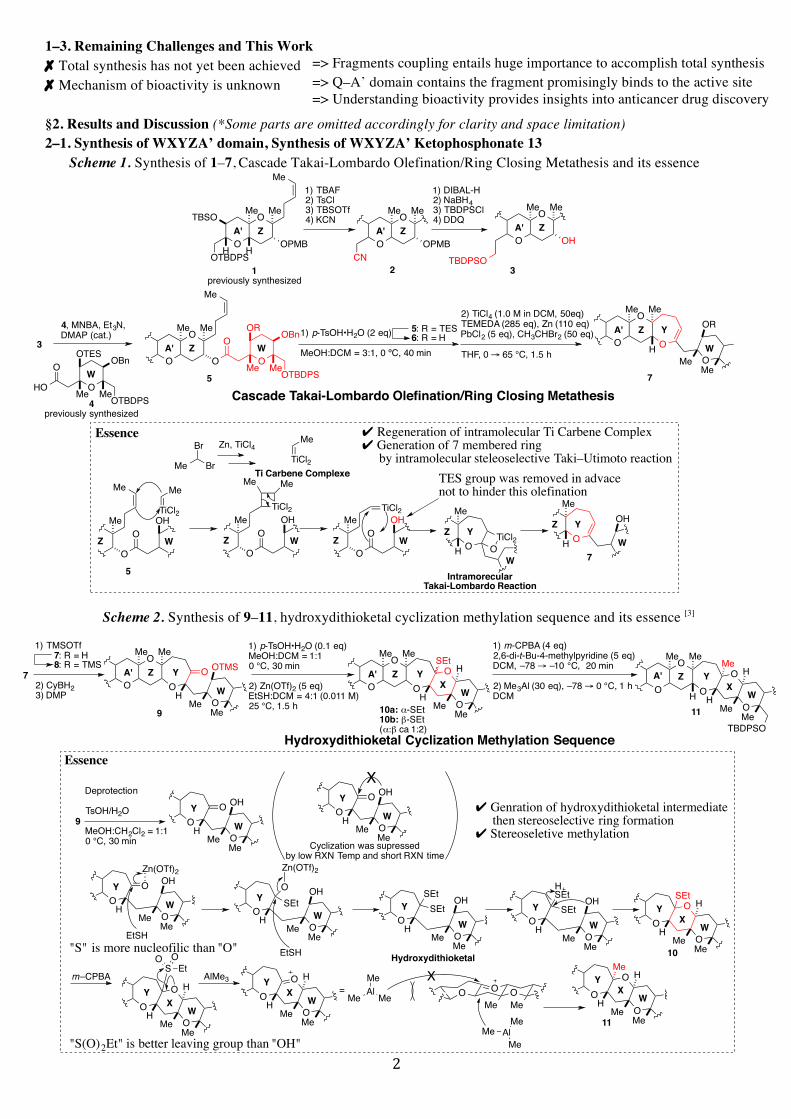
1–3. Remaining Challenges and This Work ✗ Total synthesis has not yet been achieved ✗ Mechanism of bioactivity is unknown §2. Results and Discussion (*Some parts are omitted accordingly for clarity and space limitation) 2–1. Synthesis of WXYZA’ domain, Synthesis of WXYZA’ Ketophosphonate 13
Scheme 1. Synthesis of 1–7, Cascade Takai-Lombardo Olefination/Ring Closing Metathesis and its essence
Scheme 2. Synthesis of 9–11, hydroxydithioketal cyclization methylation sequence and its essence [3]
O
OTBSO Me Me
Me
HHOTBDPSOPMB
1) TBAF2) TsCl3) TBSOTf4) KCN
1) DIBAL-H2) NaBH43) TBDPSCl4) DDQ
A' ZO
O
CN
A'O
OMe
OHA' Z
TBDPSO1
previously synthesized2 3
Z
Me
OPMB
Me Me
4, MNBA, Et3N,DMAP (cat.)
O
O Me
Me
OA' Z
OHO
OOTES
Me Me
OBn
OTBDPS
WO
OOR
Me Me
OBnW
1) p-TsOH•H2O (2 eq)
MeOH:DCM = 3:1, 0 ºC, 40 min
5: R = TES6: R = H
4previously synthesized
O
OA' Z
OTBDPS
O
Me
O
OR
MeMe
Y
WH
5 7
Cascade Takai-Lombardo Olefination/Ring Closing Metathesis
Me
Me2) TiCl4 (1.0 M in DCM, 50eq)TEMEDA (285 eq), Zn (110 eq)PbCl2 (5 eq), CH3CHBr2 (50 eq)
THF, 0 → 65 °C, 1.5 h3
Me Br
Br
TiCl2
Me
Me
Me
OZ
O
5
Me
Me
OZ
O
TiCl2
Me
Me
OZ
O
TiCl2Z
O
Me
Y
H7
TiCl2
Me
Ti Carbene Complexe
EssenceZn, TiCl4
IntramorecularTakai-Lombardo Reaction
ZO
Me
Y
H OTiCl2
OHOH OH OH
TES group was removed in advacenot to hinder this olefination
W W W
W
W
✔ Regeneration of intramolecular Ti Carbene Complex✔ Generation of 7 membered ring by intramolecular steleoselective Taki–Utimoto reaction
1) TMSOTf
2) CyBH23) DMP
7: R = H8: R = TMS
9
O
OA' Z
OO
OTMS
Me
Y
W
O
H10a: α-SEt10b: β-SEt(α:β ca 1:2)
O
OA' Z
OO
H
Me
Y
WH
OSEt
X
Hydroxydithioketal Cyclization Methylation Sequence
1) m-CPBA (4 eq)2,6-di-t-Bu-4-methylpyridine (5 eq)DCM, –78 → –10 °C, 20 min
2) Me3Al (30 eq), –78 → 0 °C, 1 hDCM
11
O
OA' Z
O
Me
O
H
Me
Y
WH H
OMe
X
Me Me
Me
Me Me Me
TBDPSOMe Me
1) p-TsOH•H2O (0.1 eq)MeOH:DCM = 1:10 °C, 30 min
2) Zn(OTf)2 (5 eq)EtSH:DCM = 4:1 (0.011 M)25 °C, 1.5 h
7
=> Fragments coupling entails huge importance to accomplish total synthesis => Q–A’ domain contains the fragment promisingly binds to the active site => Understanding bioactivity provides insights into anticancer drug discovery
OO
H
Me
Y
WH
O
S
X
Me
m–CPBA
OOEt
OO
H
Me
Y
WH
OX
Me
=MeAlMe
AlMe3
Essence
9 OO
OH
Me
O
HMe
Y
W
OO
OH
MeH
Me
Y
W
Zn(OTf)2
EtSH
O
OO
OH
MeH
Me
Y
W
OSEt
10
OO
H
Me
Y
WH
OSEt
X
Me
O OOMeMe
X
MeAlMe
Me
11O
H
Me
Y
WH
OMe
X
Me
OO
OH
MeH
Me
Y
W
SEtSEt
Hydroxydithioketal
Zn(OTf)2
EtSH
OO
OH
MeH
Me
Y
W
SEtSEt
H
Deprotection
TsOH/H2O
MeOH:CH2Cl2 = 1:10 °C, 30 min Cyclization was supressed
by low RXN Temp and short RXN time
OO
OH
Me
O
HMe
Y
W
X
"S" is more nucleofilic than "O"
✔ Genration of hydroxydithioketal intermediate then stereoselective ring formation✔ Stereoseletive methylation
MeO
"S(O)2Et" is better leaving group than "OH"
3
Scheme 3. Synthesis of 12–13, completion of W–A’ domain
2–2. Synthesis of QRSTU Aldehyde 18, Fragment Coupling and Completion of QRSTUVWXYZA’ Domain Scheme 4. Synthesis of 14–20, coupling of Q–U domain and W–A’ domain by Honor–Wadsworth Emmons Coupling
Scheme 5. Synthesis of 21–24, reductive hydroxyketone ring closure and its essence [4]
1) TPAP (cat.), NMO2) (MeO)2P(O)CH2Li3) DMP
13overall yield 8.1%
O
OTBSO Me
HH
A' Z
TBDPSO
O
Me
O
H
MeMe
OBnY
WH H
OMe
X
O
P(O)(OMe)2
1) DIBAL-H2) TBDPSCl
12O
OA' Z
OO
HO
Y
W
OX
MeMe
Me MeMe
11
OBnO
OO
O
OMe
HMeHH
H Me H MeH
Me
OBn
QR
STUO
SiO
t-But-Bu
OBn14
previously synthesized
OHO
OO
O
O
Me
Me Me
Me
OBn
QR
STU
OH15
Pd/C (cat.), H2
1) TEMPO (cat.),PhI(OAc)22) Ph3P=CH2Me
OHO
OO
O
O
Me
Me Me
Me
OBn
QR
STU
16
Me
1) TBAF2) TESOTf3) PPTS OTES
O
OO
O
OMe
Me
Me Me
Me
QR
STUTESO
X
1) TPAP cat.NMO2) 13 (1 eq)Ba(OH)2•8H2O (1.5 eq)then 18
THF: H2O = 6:125 °C, 4.5 h
17: X = O, H18: X = O
O
OO
O
OMe
Me
Me Me
Me
QR
STU
TESO
O
H
Me Me
OBn
HO
O
O
O
O
MeMeMe
TBSO
TBDPSO
HH
HA'Z
Y X W
19
Horner–Wadsworth–Emmons Coupling
17
O
OO
O
OMe
Me
Me MeH
Me
QR
STU
TESO
OMe MeO
O
O
O
O
MeMeMe
TBSO
TBDPSO
A'Z
Y X W
20
1) TBAF2) TESOTf[(PPh3)CuH]6
OTESO
OO
O
OMe
Me
Me Me
Me
QR
STUOMe Me
O
O
O
O
O
MeMeMe
TESO
TESO
A'Z
Y X W
21
TESO
BiBr3 (0.5 M in MeCN, 3 eq)TESH (50 eq)
MeCN:DCM = 4:1, –10 °C, 2 h
Me
O
OO
O
MeMe
Me
HO
HO
A'Z
Y X W
22OH
O
OO
O
OMe
Me
Me Me
Me
OBn
QR
STUO O
Me V
H
H
H
Reductive Hydroxyketone Ring Closure
Confirmed by NOE and 13C NMR
BiBr3
2HBr
H2O
BrBiO
U
O
O
W
21
Et3Si
O HEt3Si
HO
W
V
Br-
O H
HO
W
V
O HW
V
Br-
Et3SiBr
Et3SiBr
Et3SiOH
HBr
H2O
O HW
V
Et3SiOH
Et3SiOSiEt3
Et3SiH
Essence
✔ Stereoselective ring formation✔ TES deprotection, taking advantage of a byproduct HBr
22
U
U
U
U H
MeH
O
OO
O
MeMe
Me
HH
HA'Z
Y X W
23overall yield 31.3%
OHO
OO
O
OMe
HMeH
Me H MeH
Me
OBn
QR
STUO O
OBn
Me V
H
H
H
Me2C(OMe)2, CSA (cat.)
O
O
HMeMe
Pd(OH)2/C (cat.),
H
MeH
O
OO
O
MeMe
Me
HO
HO
HH
HA'Z
Y X W
24overall yield 37.8%
OHO
OO
O
OMe
HMeH
Me H MeH
Me
OH
QR
STUO O
OH
Me V
H
H
H
Me
4
2–3. Biological Evaluation • 19 different fragments, including previously synthesized A–E, A–G, Q–U, Q–A’, W–A’ and C’–F’ domains and its
analogs, were subjected to rat glioma C6 (a kind of cancer) cells (Figure 5) and human tumor cells. => Compound 24, 25 (Q–A’) and 26 (C’–F’) (Figure 6) gave a positive reaction to the Ca2+ influx examination. => Compound 24 exhibited significant growth inhibition against 10 different humane tumor cells.
Others, A–E, A–G, Q–U domains and its analogs were completely inactive or slightly active.
Figure 5. Process of the biological evaluation
Figure 6. Partial structures of MTX that induced Ca2+ influx
✔ Some domains bound to the active site, while all domains did not induce Ca2+ influx. => Binding domains and Ca2+ influx inducing domains are not the same. => More than 2 domains play a role of converting Ca2+–ATPase to Ca2+–permeable nonselective cation channel. ✔ Q–A’ domain and C’–F’ domain effectively bound to the active sites (consistent with Murata’s hypothesis). ✔ W–A’ domain nor S–U domain were not active (inconsistent with Murata’s Hypothesis). => Both S–U domain and W–A’ domain are necessary to bind to active sites. §3. Conclusion ✔ Succeeded in synthesizing QRSTUVWXYZA’ domains, which was remarkable advance towards total synthesis ✔ Evaluated bioactivity of different 19 domains and got new insight into the mechanism of bioactivity of MTX ✔ Promising anticancer activity was observed
§4. References [1] W. G. Sinkins et al. Am. J. Physiol. Cell Physiol. 2009, 297, C1533–C1543. [2] M. Murata et al. Bull. Chem. Soc. Jpn. 2008, 81, 307–319. [3] K. C. Nicolaou et al. J. Am. Chem. Soc. 1989, 111, 5321–5330. [4] P. A. Evans et al. J. Am. Chem. Soc. 2003, 125, 11456–11457. Abbreviations: MNBA = 2,6-methylnitrobenzoyl anhydride; TPAP = tetra-n-propylammonium perruthenate; NMO = N-methylmorpholine-N-oxide; PPTS = pyridineium p-toluene sulfonate; CAS = (±)-camphor-10-sulfonic acid
Rat glioma C6 cell1 mL (25000
cells/mL)
Compound of interest in MeOH
incubation buffer (250 mL)13 min
binding site "blocked" cell
45CaCl2 (50 µL, 6 µCi/mL)MTX
12 min
activecompound
inactivecompound
normal cell
binding site "blocked" cellRadio inactive
45Ca2+ containing cellRadioactive
Ca2+X
Ca2+
45CaCl2 (50 µL, 6 µCi/mL)MTX
12 min
Ca2+
O
O
OO
OO
OO
OO
O
MeHMe
HH
H
HMe
H Me H MeOH
MeMe
HH
H
H
HMe
Me
MeH
OH
QRST
UVW
XY
A'
Z
OH
O
O
MeMe
Me
O
O
O
OTBDPSO
MeMeMe
H H H H
C'D'E'F'OH
26previously synthesized
25This Work


![Synthesis of Novel Electrically Conducting Polymers: Potential ... · PPh3 + Br(CH2). CO2Me ..... > [Ph3P--CH2(CH2). i CO2Me]*Br* [phaP--CH2(CH2)n__CO2Mel*Br -Z--BuL>_phaP=CH (C H2)n_i](https://img.pdfslide.us/doc/110x75/5ebc39ab077be8135d1c1d2a/synthesis-of-novel-electrically-conducting-polymers-potential-pph3-brch2.jpg)