Embed Size (px)
Citation preview
Joint Hospital Surgical Grand Round18 July 2015Dr HUI Hon Cheung Rio
Princess Margaret Hospital
Can all early gastric cancer be treated equally?

Content Case Introduction Diagnosis and work up Management Conclusion
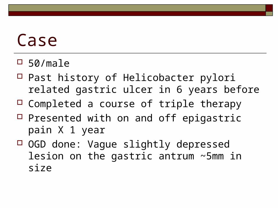
Case 50/male Past history of Helicobacter pylori related
gastric ulcer in 6 years before Completed a course of triple therapy Presented with on and off epigastric pain X 1
year OGD done: Vague slightly depressed lesion
on the gastric antrum ~5mm in size
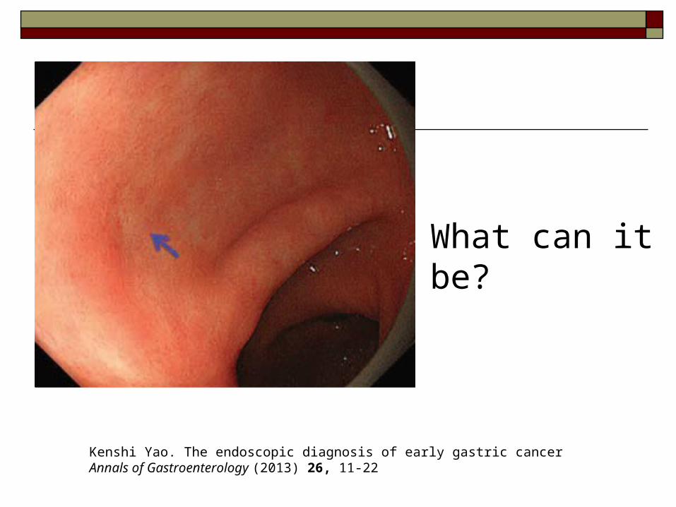
What can it be?
Kenshi Yao. The endoscopic diagnosis of early gastric cancer Annals of Gastroenterology (2013) 26, 11-22
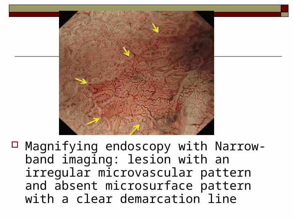
Magnifying endoscopy with Narrow-band imaging: lesion with an irregular microvascular pattern and absent microsurface pattern with a clear demarcation line
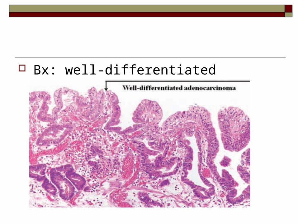
Bx: well-differentiated adenocarcinoma
What is the further Treatment?
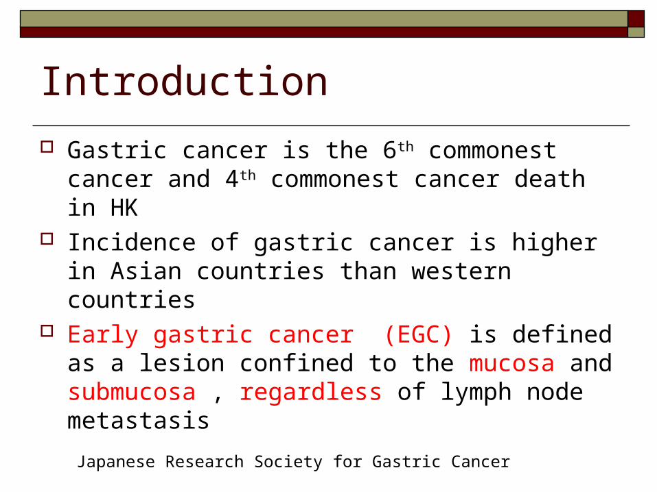
Introduction
Gastric cancer is the 6th commonest cancer and 4th commonest cancer death in HK
Incidence of gastric cancer is higher in Asian countries than western countries
Early gastric cancer (EGC) is defined as a lesion confined to the mucosa and submucosa , regardless of lymph node metastasis
Japanese Research Society for Gastric Cancer
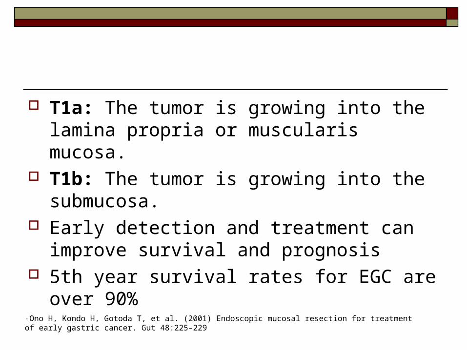
T1a: The tumor is growing into the lamina propria or muscularis mucosa.
T1b: The tumor is growing into the submucosa.
Early detection and treatment can improve survival and prognosis
5th year survival rates for EGC are over 90%
-Ono H, Kondo H, Gotoda T, et al. (2001) Endoscopic mucosal resection for treatment of early gastric cancer. Gut 48:225–229
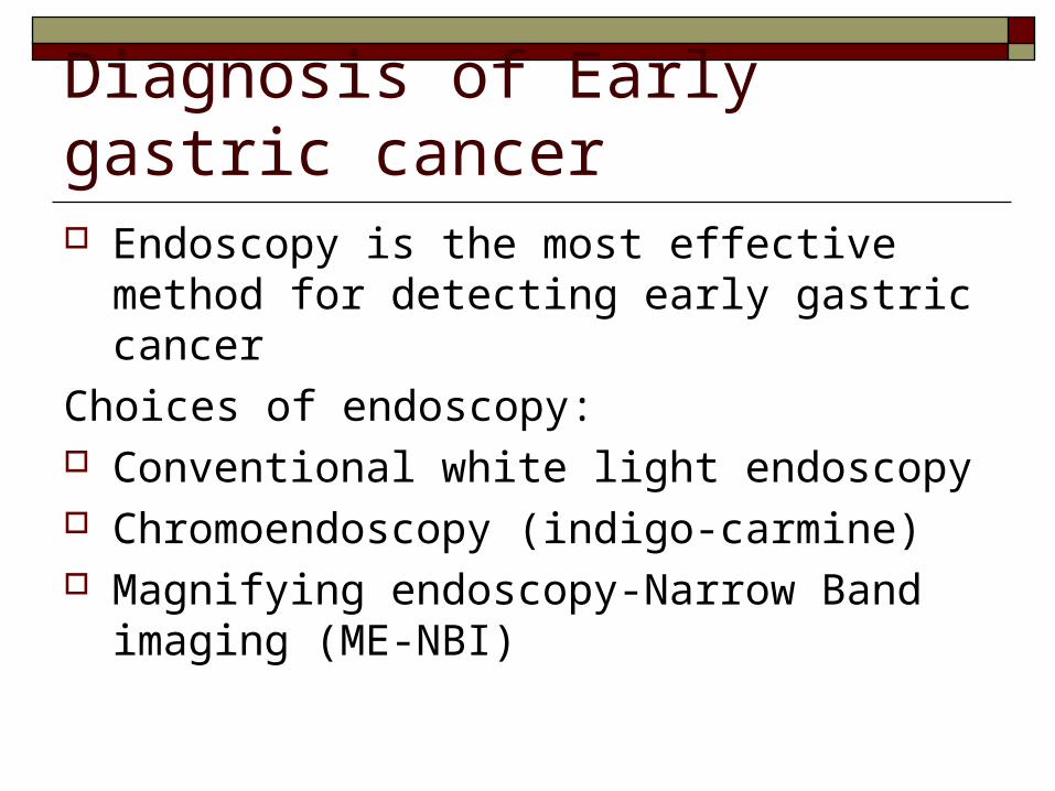
Diagnosis of Early gastric cancer Endoscopy is the most effective method for
detecting early gastric cancer
Choices of endoscopy: Conventional white light endoscopy Chromoendoscopy (indigo-carmine) Magnifying endoscopy-Narrow Band imaging
(ME-NBI)
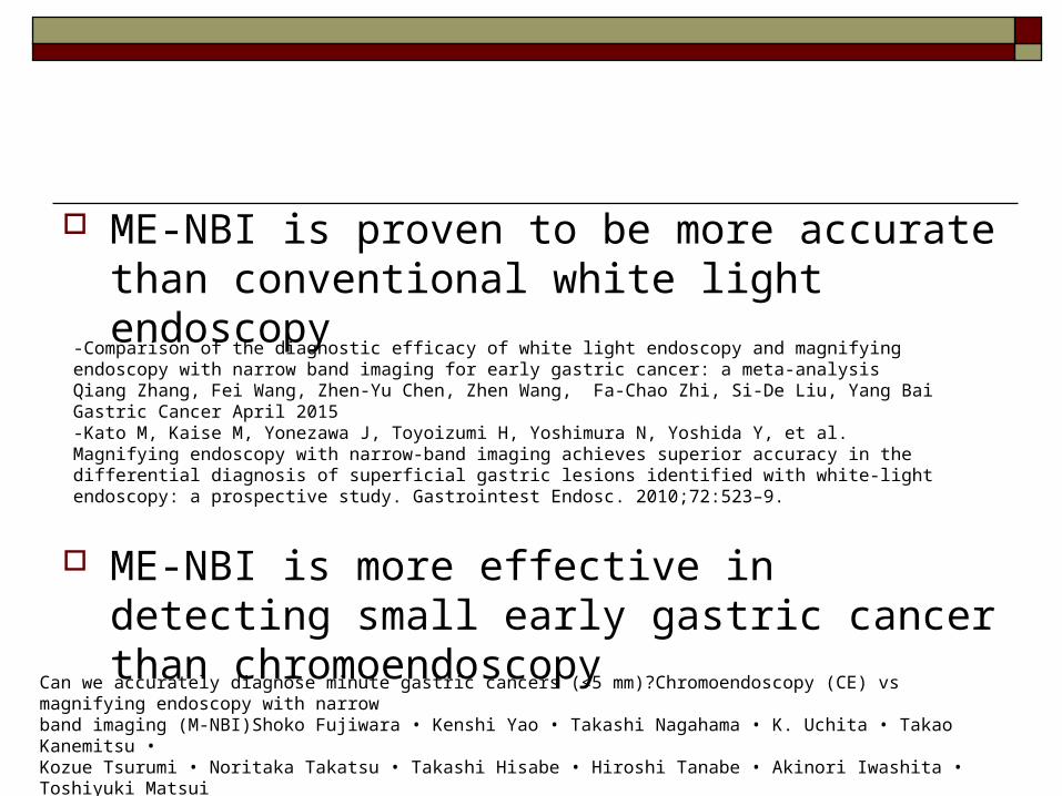
ME-NBI is proven to be more accurate than conventional white light endoscopy
ME-NBI is more effective in detecting small early gastric cancer than chromoendoscopy
Can we accurately diagnose minute gastric cancers (<5 mm)?Chromoendoscopy (CE) vs magnifying endoscopy with narrowband imaging (M-NBI)Shoko Fujiwara • Kenshi Yao • Takashi Nagahama • K. Uchita • Takao Kanemitsu •Kozue Tsurumi • Noritaka Takatsu • Takashi Hisabe • Hiroshi Tanabe • Akinori Iwashita • Toshiyuki MatsuiGastric Cancer 11 June 2014
-Comparison of the diagnostic efficacy of white light endoscopy and magnifying endoscopy with narrow band imaging for early gastric cancer: a meta-analysis Qiang Zhang, Fei Wang, Zhen-Yu Chen, Zhen Wang, Fa-Chao Zhi, Si-De Liu, Yang Bai Gastric Cancer April 2015-Kato M, Kaise M, Yonezawa J, Toyoizumi H, Yoshimura N, Yoshida Y, et al. Magnifying endoscopy with narrow-band imaging achieves superior accuracy in the differential diagnosis of superficial gastric lesions identified with white-light endoscopy: a prospective study. Gastrointest Endosc. 2010;72:523–9.
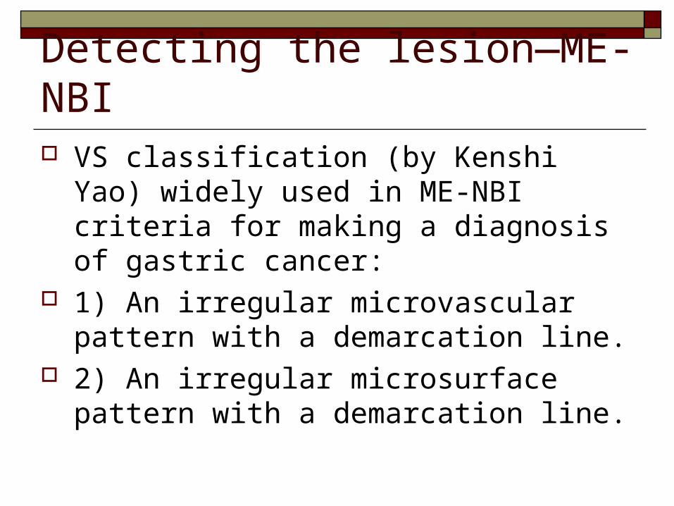
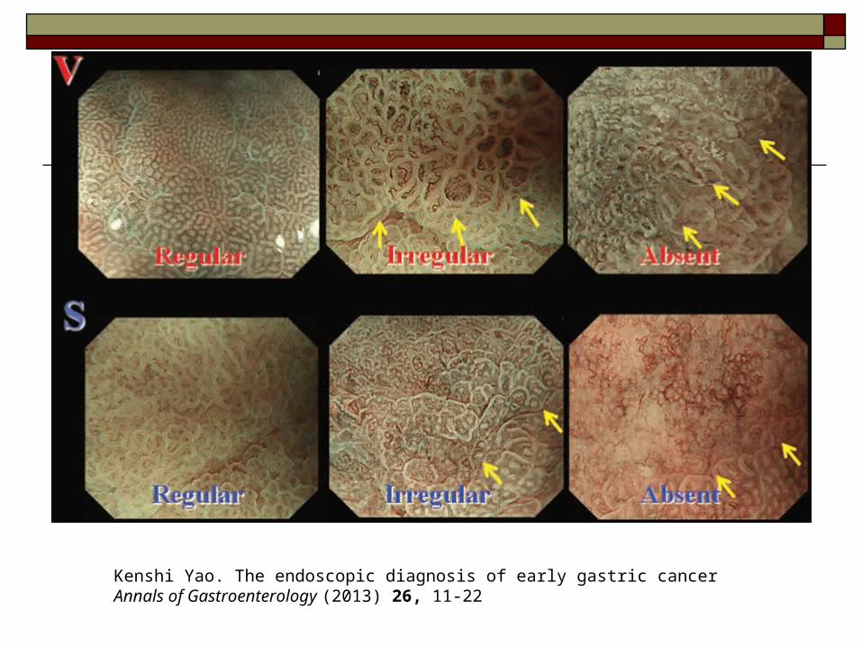
Detecting the lesion—ME-NBI VS classification (by Kenshi Yao) widely
used in ME-NBI criteria for making a diagnosis of gastric cancer:
1) An irregular microvascular pattern with a demarcation line.
2) An irregular microsurface pattern with a demarcation line.
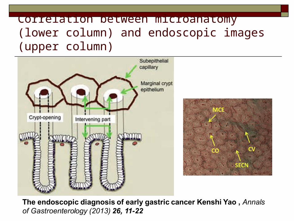
Correlation between microanatomy (lower column) and endoscopic images (upper column)
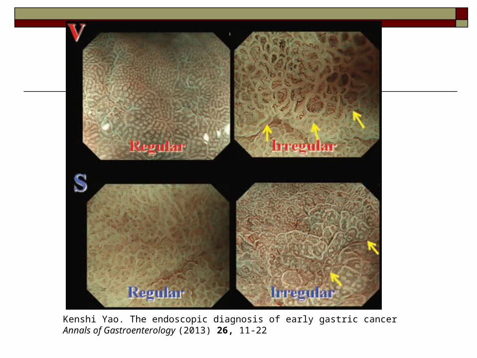
Kenshi Yao. The endoscopic diagnosis of early gastric cancer Annals of Gastroenterology (2013) 26, 11-22
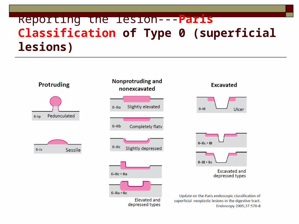
Reporting the lesion---Paris Classification of Type 0 (superficial lesions)
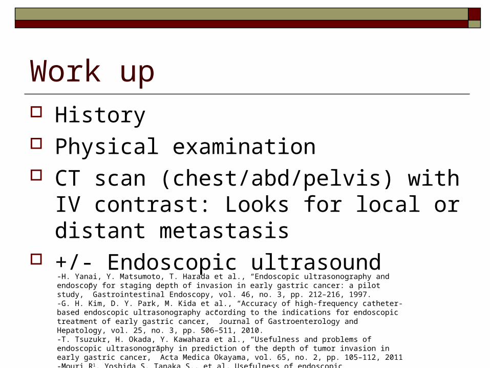
Work up History Physical examination CT scan (chest/abd/pelvis) with IV contrast:
Looks for local or distant metastasis +/- Endoscopic ultrasound
-H. Yanai, Y. Matsumoto, T. Harada et al., “Endoscopic ultrasonography and endoscopy for staging depth of invasion in early gastric cancer: a pilot study,” Gastrointestinal Endoscopy, vol. 46, no. 3, pp. 212–216, 1997. -G. H. Kim, D. Y. Park, M. Kida et al., “Accuracy of high-frequency catheter-based endoscopic ultrasonography according to the indications for endoscopic treatment of early gastric cancer,” Journal of Gastroenterology and Hepatology, vol. 25, no. 3, pp. 506–511, 2010. -T. Tsuzukr, H. Okada, Y. Kawahara et al., “Usefulness and problems of endoscopic ultrasonography in prediction of the depth of tumor invasion in early gastric cancer,” Acta Medica Okayama, vol. 65, no. 2, pp. 105–112, 2011-Mouri R1, Yoshida S, Tanaka S., et al, Usefulness of endoscopic ultrasonography in determining the depth of invasion and indication for endoscopic treatment of early gastric cancer. J Clin Gastroenterol. 2009 Apr;43(4):318-22
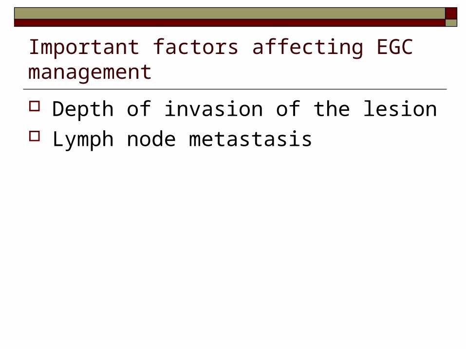
Important factors affecting EGC management
Depth of invasion of the lesion Lymph node metastasis

Incidence of lymph node metastasis 4.9% intramucosal cancers were associated with l
ymph node metastases 23.8% submucosal cancers were associated with l
ymph node metastases Independent risk factors for lymph node metastas
is:-tumor size which is 21 mm or more-lymphatic-vascular capillary involvement-submucosal penetration
Gotoda T, Yanagisawa A, Sasako M, et al. Incidence of lymph node metastasis from early gastric cancer: estimation with a large number of cases at two large centers. Gastric Cancer. 2000;3: 219–25
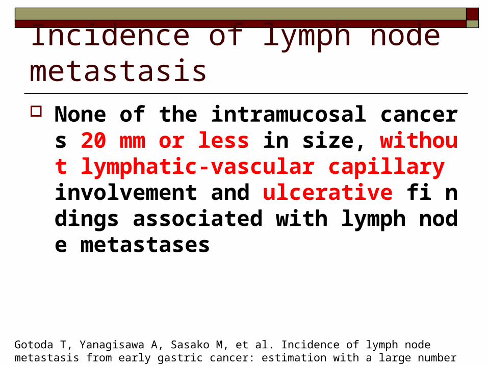
Incidence of lymph node metastasis None of the intramucosal cancers 20 mm or
less in size, without lymphatic-vascular capillary involvement and ulcerative fi ndings associated with lymph node metastases
Gotoda T, Yanagisawa A, Sasako M, et al. Incidence of lymph node metastasis from early gastric cancer: estimation with a large number of cases at two large centers. Gastric Cancer. 2000;3: 219–25
Management of early gastric cancer Management of gastric lesion Management of lymph node
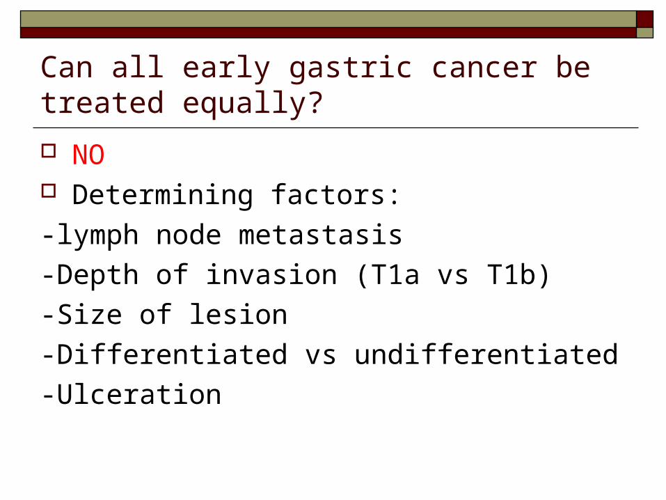
Can all early gastric cancer be treated equally?
NO Determining factors:
-lymph node metastasis
-Depth of invasion (T1a vs T1b)
-Size of lesion
-Differentiated vs undifferentiated
-Ulceration
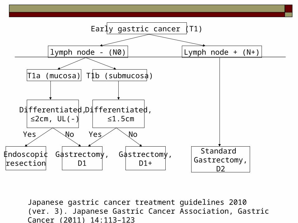
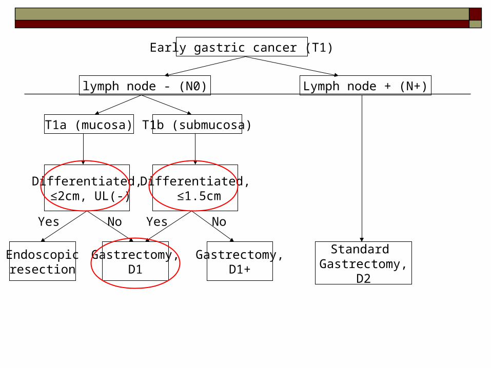
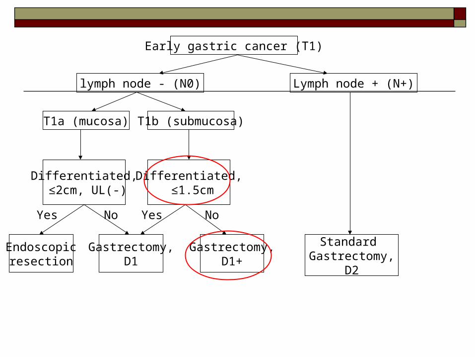
Early gastric cancer (T1)
lymph node - (N0) Lymph node + (N+)
T1b (submucosa)T1a (mucosa)
Differentiated, ≤2cm, UL(-)
Differentiated, ≤1.5cm
Endoscopicresection
Gastrectomy,D1
Gastrectomy,D1+
Standard Gastrectomy,
D2
Yes YesNo No
Japanese gastric cancer treatment guidelines 2010 (ver. 3). Japanese Gastric Cancer Association, Gastric Cancer (2011) 14:113–123
Early gastric cancer (T1)
lymph node - (N0) Lymph node + (N+)
T1b (submucosa)T1a (mucosa)
Differentiated, ≤2cm, UL(-)
Differentiated, ≤1.5cm
Endoscopicresection
Gastrectomy,D1
Gastrectomy,D1+
Standard Gastrectomy,
D2
Yes YesNo No
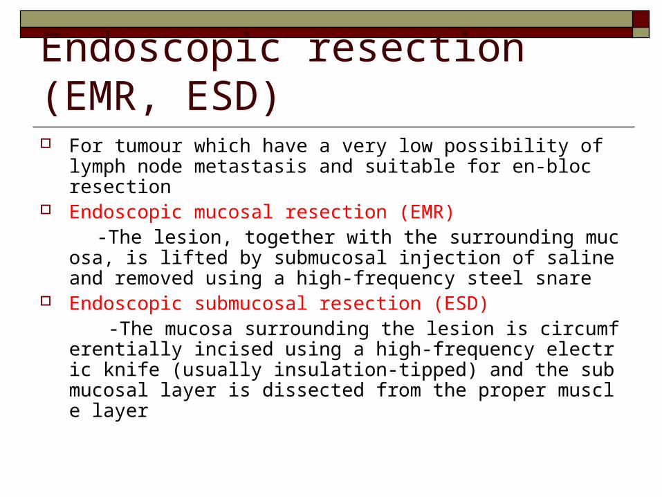
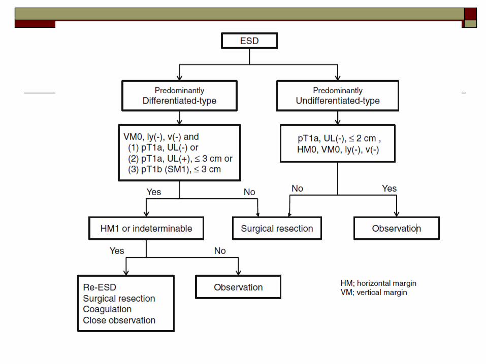
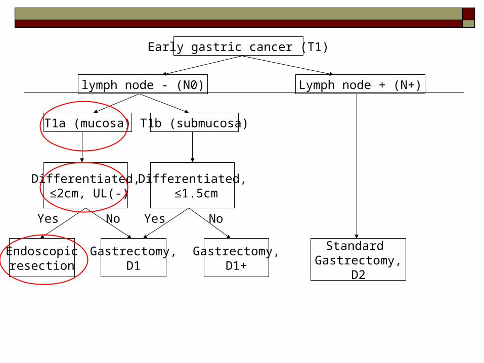
Endoscopic resection (EMR, ESD) For tumour which have a very low possibility of lymph node
metastasis and suitable for en-bloc resection Endoscopic mucosal resection (EMR) -The lesion, together with the surrounding mucosa, is lifted by
submucosal injection of saline and removed using a high-frequency steel snare
Endoscopic submucosal resection (ESD) -The mucosa surrounding the lesion is circumferentially incis
ed using a high-frequency electric knife (usually insulation-tipped) and the submucosal layer is dissected from the proper muscle layer
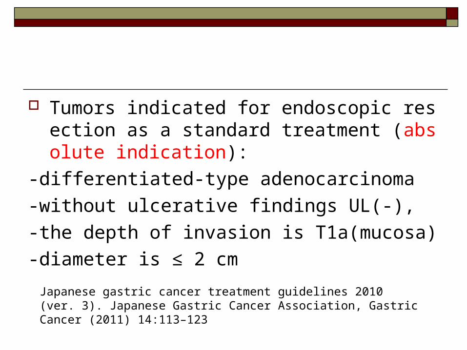
Tumors indicated for endoscopic resection as a standard treatment (absolute indication):
-differentiated-type adenocarcinoma
-without ulcerative findings UL(-),
-the depth of invasion is T1a(mucosa)
-diameter is ≤ 2 cm
Japanese gastric cancer treatment guidelines 2010 (ver. 3). Japanese Gastric Cancer Association, Gastric Cancer (2011) 14:113–123
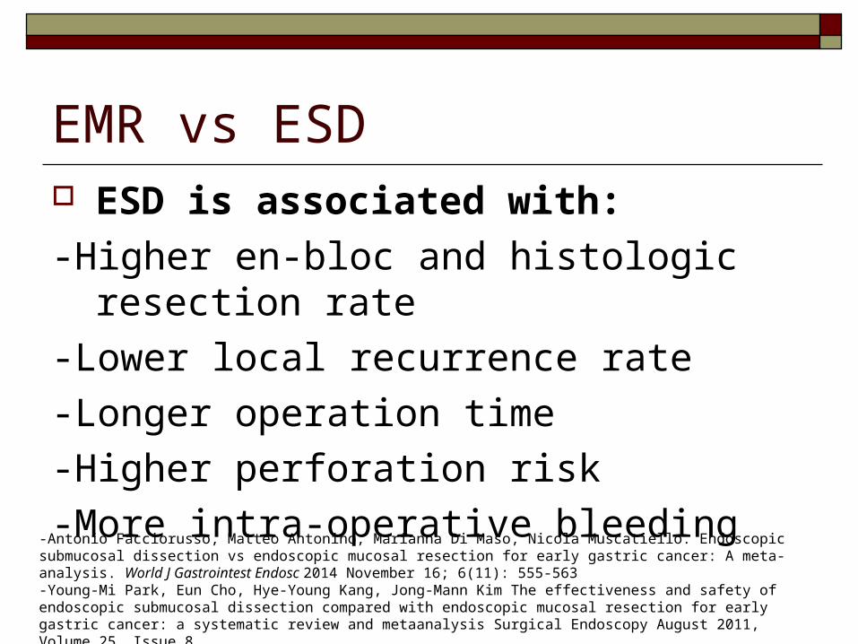
EMR vs ESD ESD is associated with:
-Higher en-bloc and histologic resection rate
-Lower local recurrence rate
-Longer operation time
-Higher perforation risk
-More intra-operative bleeding -Antonio Facciorusso, Matteo Antonino, Marianna Di Maso, Nicola Muscatiello. Endoscopic submucosal dissection vs endoscopic mucosal resection for early gastric cancer: A meta-analysis. World J Gastrointest Endosc 2014 November 16; 6(11): 555-563-Young-Mi Park, Eun Cho, Hye-Young Kang, Jong-Mann Kim The effectiveness and safety of endoscopic submucosal dissection compared with endoscopic mucosal resection for early gastric cancer: a systematic review and metaanalysis Surgical Endoscopy August 2011, Volume 25, Issue 8
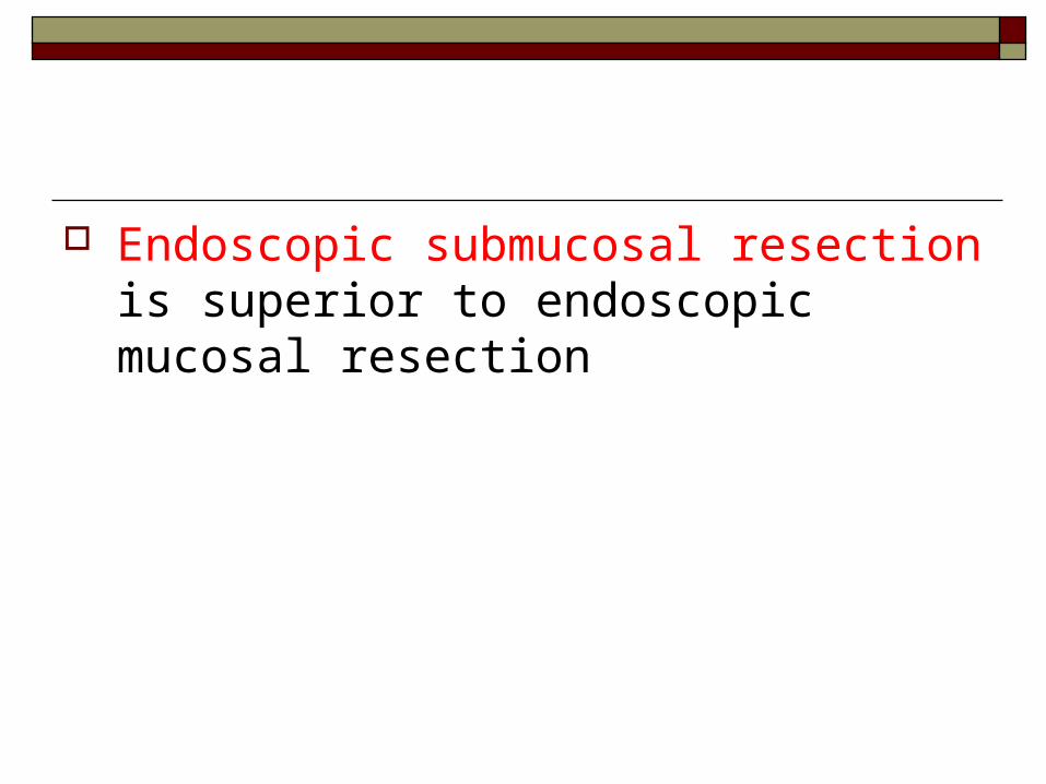
Endoscopic submucosal resection is superior to endoscopic mucosal resection
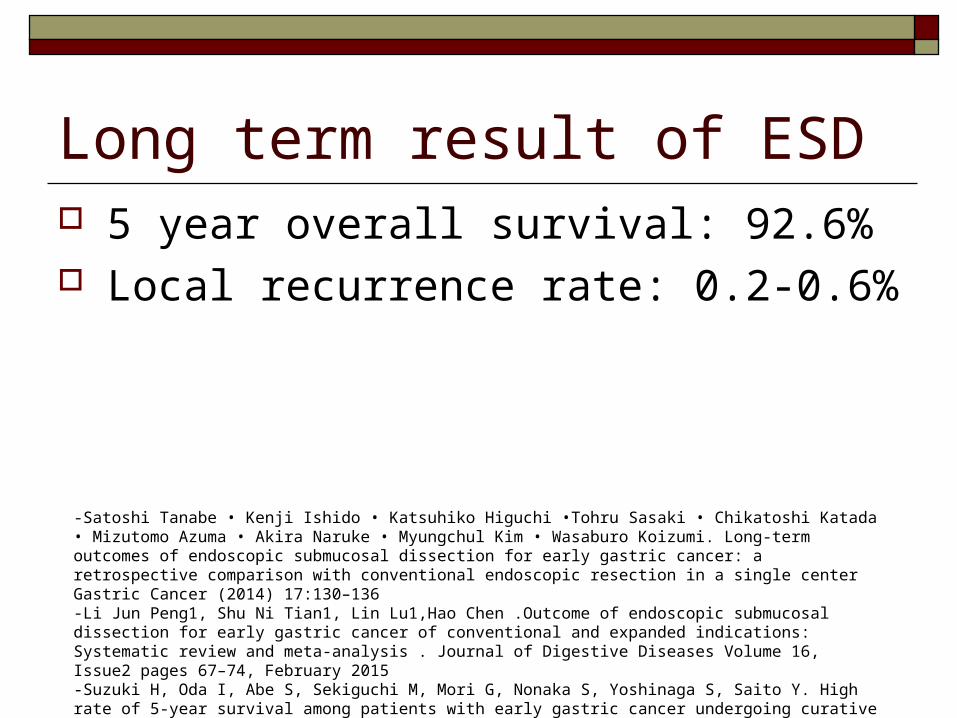
Long term result of ESD 5 year overall survival: 92.6% Local recurrence rate: 0.2-0.6%
-Satoshi Tanabe • Kenji Ishido • Katsuhiko Higuchi •Tohru Sasaki • Chikatoshi Katada • Mizutomo Azuma • Akira Naruke • Myungchul Kim • Wasaburo Koizumi. Long-term outcomes of endoscopic submucosal dissection for early gastric cancer: a retrospective comparison with conventional endoscopic resection in a single center Gastric Cancer (2014) 17:130–136-Li Jun Peng1, Shu Ni Tian1, Lin Lu1,Hao Chen .Outcome of endoscopic submucosal dissection for early gastric cancer of conventional and expanded indications: Systematic review and meta-analysis . Journal of Digestive Diseases Volume 16, Issue2 pages 67–74, February 2015-Suzuki H, Oda I, Abe S, Sekiguchi M, Mori G, Nonaka S, Yoshinaga S, Saito Y. High rate of 5-year survival among patients with early gastric cancer undergoing curative endoscopic submucosal dissection. Gastric Cancer. 2015 Jan 24.
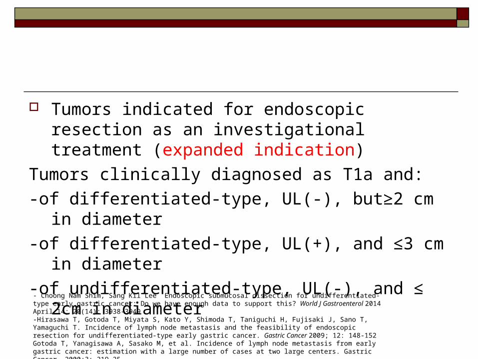
Tumors indicated for endoscopic resection as an investigational treatment (expanded indication)
Tumors clinically diagnosed as T1a and:
-of differentiated-type, UL(-), but≥2 cm in diameter
-of differentiated-type, UL(+), and ≤3 cm in diameter
-of undifferentiated-type, UL(-), and ≤ 2cm in diameter
- Choong Nam Shim, Sang Kil Lee Endoscopic submucosal dissection for undifferentiated-type early gastric cancer: Do we have enough data to support this? World J Gastroenterol 2014 April 14; 20(14): 3938-3949-Hirasawa T, Gotoda T, Miyata S, Kato Y, Shimoda T, Taniguchi H, Fujisaki J, Sano T, Yamaguchi T. Incidence of lymph node metastasis and the feasibility of endoscopic resection for undifferentiated-type early gastric cancer. Gastric Cancer 2009; 12: 148-152Gotoda T, Yanagisawa A, Sasako M, et al. Incidence of lymph node metastasis from early gastric cancer: estimation with a large number of cases at two large centers. Gastric Cancer. 2000;3: 219–25
Early gastric cancer (T1)
lymph node - (N0) Lymph node + (N+)
T1b (submucosa)T1a (mucosa)
Differentiated, ≤2cm, UL(-)
Differentiated, ≤1.5cm
Endoscopicresection
Gastrectomy,D1
Gastrectomy,D1+
Standard Gastrectomy,
D2
Yes YesNo No
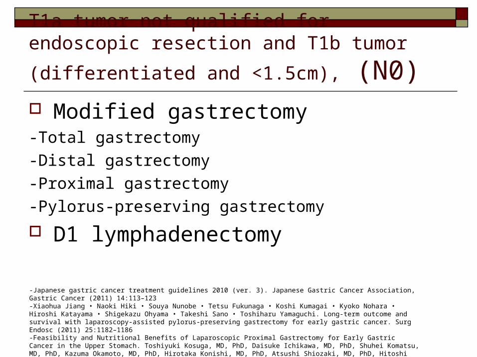
T1a tumor not qualified for endoscopic resection and
T1b tumor (differentiated and <1.5cm), (N0) Modified gastrectomy -Total gastrectomy
-Distal gastrectomy
-Proximal gastrectomy
-Pylorus-preserving gastrectomy
D1 lymphadenectomy
-Japanese gastric cancer treatment guidelines 2010 (ver. 3). Japanese Gastric Cancer Association, Gastric Cancer (2011) 14:113–123-Xiaohua Jiang • Naoki Hiki • Souya Nunobe • Tetsu Fukunaga • Koshi Kumagai • Kyoko Nohara • Hiroshi Katayama • Shigekazu Ohyama • Takeshi Sano • Toshiharu Yamaguchi. Long-term outcome and survival with laparoscopy-assisted pylorus-preserving gastrectomy for early gastric cancer. Surg Endosc (2011) 25:1182–1186-Feasibility and Nutritional Benefits of Laparoscopic Proximal Gastrectomy for Early Gastric Cancer in the Upper Stomach. Toshiyuki Kosuga, MD, PhD, Daisuke Ichikawa, MD, PhD, Shuhei Komatsu, MD, PhD, Kazuma Okamoto, MD, PhD, Hirotaka Konishi, MD, PhD, Atsushi Shiozaki, MD, PhD, Hitoshi Fujiwara, MD, PhD, and Eigo Otsuji, MD, PhD, Ann Surg Oncol DOI 10.1245/s10434-015-4590-4
Early gastric cancer (T1)
lymph node - (N0) Lymph node + (N+)
T1b (submucosa)T1a (mucosa)
Differentiated, ≤2cm, UL(-)
Differentiated, ≤1.5cm
Endoscopicresection
Gastrectomy,D1
Gastrectomy,D1+
Standard Gastrectomy,
D2
Yes YesNo No
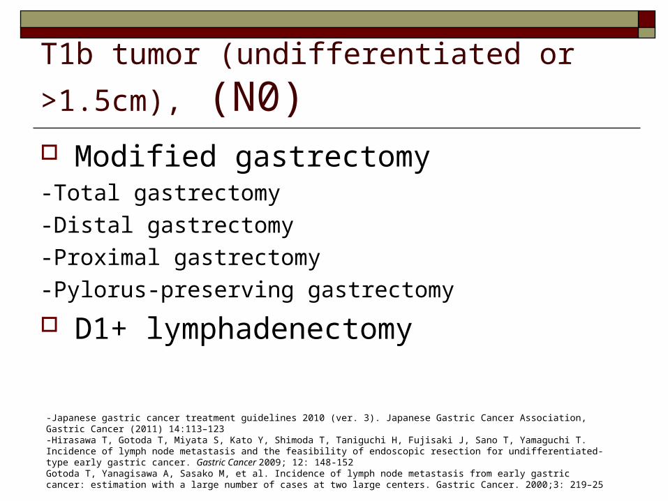
T1b tumor (undifferentiated or >1.5cm), (N0) Modified gastrectomy -Total gastrectomy
-Distal gastrectomy
-Proximal gastrectomy
-Pylorus-preserving gastrectomy
D1+ lymphadenectomy
-Japanese gastric cancer treatment guidelines 2010 (ver. 3). Japanese Gastric Cancer Association, Gastric Cancer (2011) 14:113–123-Hirasawa T, Gotoda T, Miyata S, Kato Y, Shimoda T, Taniguchi H, Fujisaki J, Sano T, Yamaguchi T. Incidence of lymph node metastasis and the feasibility of endoscopic resection for undifferentiated-type early gastric cancer. Gastric Cancer 2009; 12: 148-152Gotoda T, Yanagisawa A, Sasako M, et al. Incidence of lymph node metastasis from early gastric cancer: estimation with a large number of cases at two large centers. Gastric Cancer. 2000;3: 219–25
Early gastric cancer (T1)
lymph node - (N0) Lymph node + (N+)
T1b (submucosa)T1a (mucosa)
Differentiated, ≤2cm, UL(-)
Differentiated, ≤1.5cm
Endoscopicresection
Gastrectomy,D1
Gastrectomy,D1+
Standard Gastrectomy,
D2
Yes YesNo No
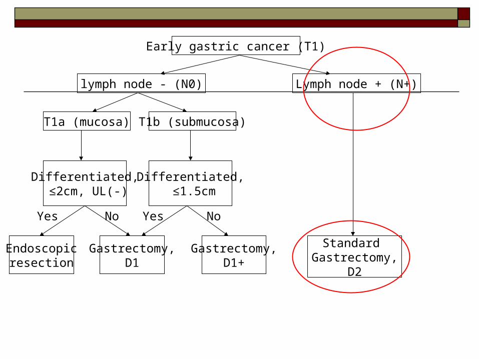
T1N+ tumour Standard gastrectomy + D2
lymphadenectomy
-Japanese gastric cancer treatment guidelines 2010 (ver. 3). Japanese Gastric Cancer Association, Gastric Cancer (2011) 14:113–123-Hirasawa T, Gotoda T, Miyata S, Kato Y, Shimoda T, Taniguchi H, Fujisaki J, Sano T, Yamaguchi T. Incidence of lymph node metastasis and the feasibility of endoscopic resection for undifferentiated-type early gastric cancer. Gastric Cancer 2009; 12: 148-152Gotoda T, Yanagisawa A, Sasako M, et al. Incidence of lymph node metastasis from early gastric cancer: estimation with a large number of cases at two large centers. Gastric Cancer. 2000;3: 219–25
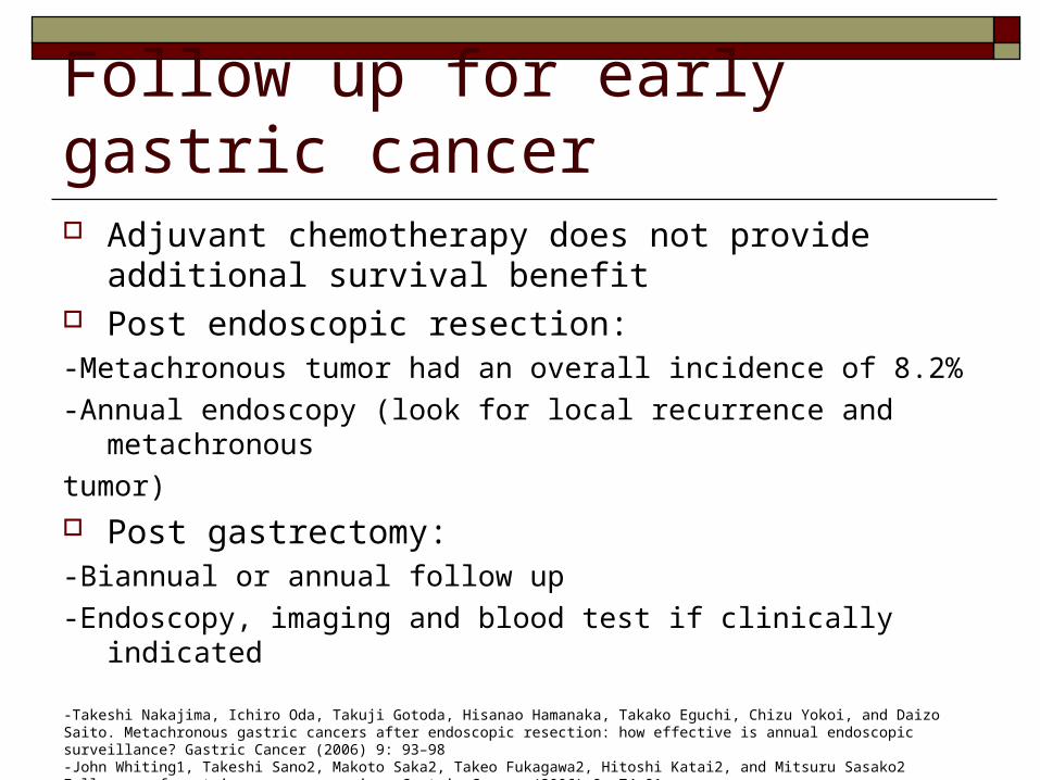
Follow up for early gastric cancer Adjuvant chemotherapy does not provide additional
survival benefit Post endoscopic resection: -Metachronous tumor had an overall incidence of 8.2%
-Annual endoscopy (look for local recurrence and metachronous
tumor) Post gastrectomy: -Biannual or annual follow up
-Endoscopy, imaging and blood test if clinically indicated
-Takeshi Nakajima, Ichiro Oda, Takuji Gotoda, Hisanao Hamanaka, Takako Eguchi, Chizu Yokoi, and Daizo Saito. Metachronous gastric cancers after endoscopic resection: how effective is annual endoscopic surveillance? Gastric Cancer (2006) 9: 93–98 -John Whiting1, Takeshi Sano2, Makoto Saka2, Takeo Fukagawa2, Hitoshi Katai2, and Mitsuru Sasako2 Follow-up of gastric cancer: a review. Gastric Cancer (2006) 9: 74–81
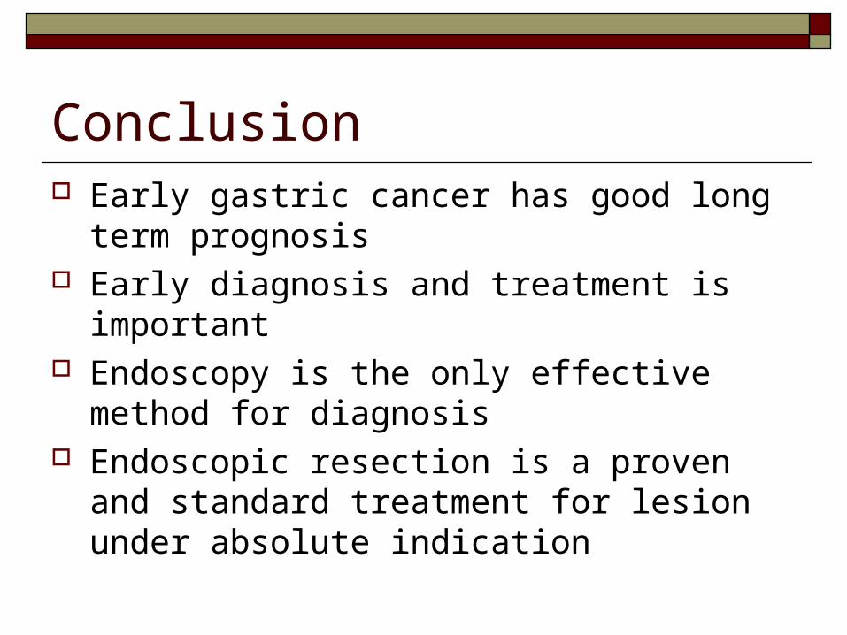
Conclusion Early gastric cancer has good long term
prognosis Early diagnosis and treatment is important Endoscopy is the only effective method for
diagnosis Endoscopic resection is a proven and standard
treatment for lesion under absolute indication
References -Ono H, Kondo H, Gotoda T, et al. (2001) Endoscopic mucosal resection for treatment of early gastric cancer. Gut 48:225–229 -Comparison of the diagnostic efficacy of white light endoscopy and magnifying endoscopy with narrow band imaging for early gastric cancer: a
meta-analysis Qiang Zhang, Fei Wang, Zhen-Yu Chen, Zhen Wang, Fa-Chao Zhi, Si-De Liu, Yang Bai Gastric Cancer April 2015 -Kato M, Kaise M, Yonezawa J, Toyoizumi H, Yoshimura N, Yoshida Y, et al. Magnifying endoscopy with narrow-band imaging achieves superior
accuracy in the differential diagnosis of superficial gastric lesions identified with white-light endoscopy: a prospective study. Gastrointest Endosc. 2010;72:523–9
.Can we accurately diagnose minute gastric cancers (<5 mm)?Chromoendoscopy (CE) vs magnifying endoscopy with narrow band imaging (M-NBI)Shoko Fujiwara • Kenshi Yao •Takashi Nagahama • K. Uchita • Takao Kanemitsu Kozue Tsurumi • Noritaka Takatsu • Takashi Hisabe • Hiroshi Tanabe • Akinori Iwashita • Toshiyuki MatsuiGastric Cancer 11 June 2014
Update on the Paris endoscopic classification of superficial neoplastic lesions in the digestive tract. Endoscopy 2005;37:570-8 H. Yanai, Y. Matsumoto, T. Harada et al., “Endoscopic ultrasonography and endoscopy for staging depth of invasion in
early gastric cancer: a pilot study,” Gastrointestinal Endoscopy, vol. 46, no. 3, pp. 212–216, 1997. -G. H. Kim, D. Y. Park, M. Kida et al., “Accuracy of high-frequency catheter-based endoscopic ultrasonography according
to the indications for endoscopic treatment of early gastric cancer,” Journal of Gastroenterology and Hepatology, vol. 25, no. 3, pp. 506–511, 2010.
-T. Tsuzukr, H. Okada, Y. Kawahara et al., “Usefulness and problems of endoscopic ultrasonography in prediction of the depth of tumor invasion in early gastric cancer,” Acta Medica Okayama, vol. 65, no. 2, pp. 105–112, 2011
Gotoda T., Yanagisawa A, Sasako M, et al. Incidence of lymph node metastasis from early gastric cancer: estimation with a large number of cases at two large centers. Gastric Cancer. 2000;3: 219–25
Facciorusso, Matteo Antonino, Marianna Di Maso, Nicola Muscatiello. Endoscopic submucosal dissection vs endoscopic mucosal resection for early gastric cancer: A meta-analysis. World J Gastrointest Endosc 2014 November 16; 6(11): 555-563
-Young-Mi Park, Eun Cho, Hye-Young Kang, Jong-Mann Kim The effectiveness and safety of endoscopic submucosal dissection compared with endoscopic mucosal resection for early gastric cancer: a systematic review and metaanalysis Surgical Endoscopy August 2011, Volume 25, Issue 8
-Satoshi Tanabe • Kenji Ishido • Katsuhiko Higuchi •Tohru Sasaki • Chikatoshi Katada • Mizutomo Azuma • Akira Naruke • Myungchul Kim • Wasaburo Koizumi. Long-term outcomes of endoscopic submucosal dissection for early gastric cancer: a retrospective comparison with conventional endoscopic resection in a single center Gastric Cancer (2014) 17:130–136
Li Jun Peng1, Shu Ni Tian1, Lin Lu1,Hao Chen .Outcome of endoscopic submucosal dissection for early gastric cancer of conventional and expanded indications: Systematic review and meta-analysis . Journal of Digestive Diseases Volume 16, Issue2 pages 67–74, February 2015
-Suzuki H, Oda I, Abe S, Sekiguchi M, Mori G, Nonaka S, Yoshinaga S, Saito Y. High rate of 5-year survival among patients with early gastric cancer undergoing curative endoscopic submucosal dissection. Gastric Cancer. 2015
-Japanese gastric cancer treatment guidelines 2010 (ver. 3). Japanese Gastric Cancer Association, Gastric Cancer (2011) 14:113–123
-Xiaohua Jiang • Naoki Hiki • Souya Nunobe • Tetsu Fukunaga • Koshi Kumagai • Kyoko Nohara • Hiroshi Katayama • Shigekazu Ohyama • Takeshi Sano • Toshiharu Yamaguchi. Long-term outcome and survival with laparoscopy-assisted pylorus-preserving gastrectomy for early gastric cancer. Surg Endosc (2011) 25:1182–1186
-Feasibility and Nutritional Benefits of Laparoscopic Proximal Gastrectomy for Early Gastric Cancer in the Upper Stomach. Toshiyuki Kosuga, MD, PhD, Daisuke Ichikawa, MD, PhD, Shuhei Komatsu, MD, PhD, Kazuma Okamoto, MD, PhD, Hirotaka Konishi, MD, PhD, Atsushi Shiozaki, MD, PhD, Hitoshi Fujiwara, MD, PhD, and Eigo Otsuji, MD, PhD, Ann Surg Oncol DOI 10.1245/s10434-015-4590-4

Thank you.


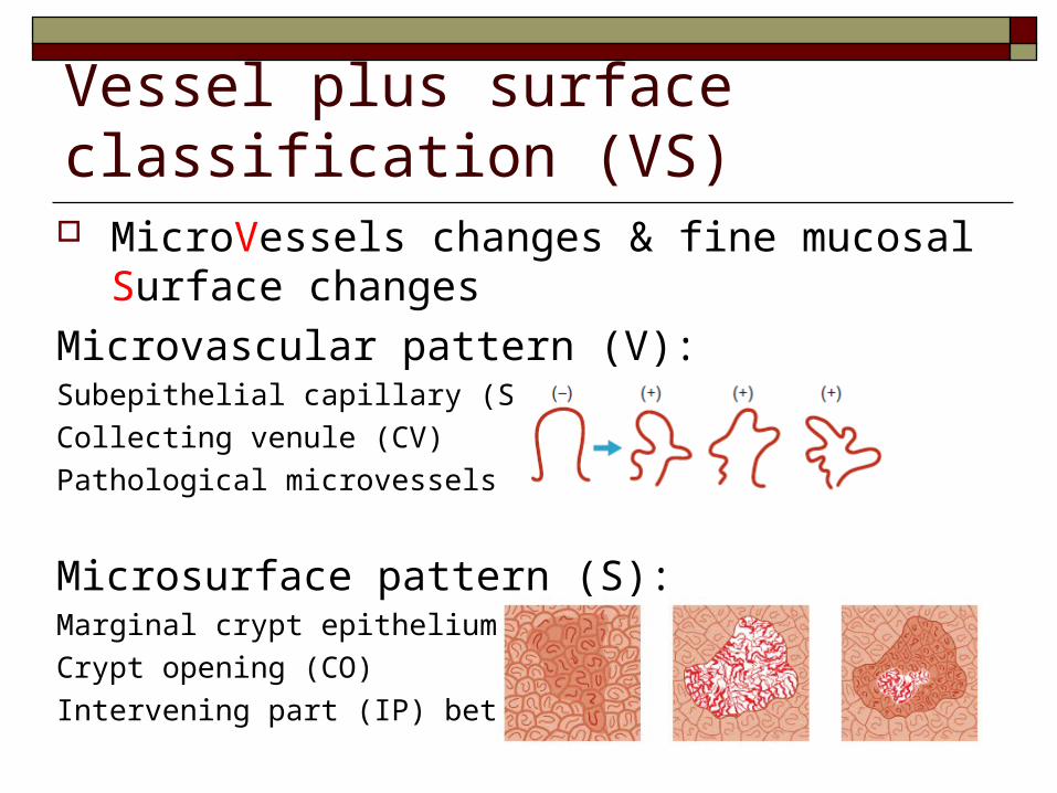
Vessel plus surface classification (VS) MicroVessels changes & fine mucosal Surface
changes
Microvascular pattern (V): Subepithelial capillary (SEC)
Collecting venule (CV)
Pathological microvessels (MV)
Microsurface pattern (S):Marginal crypt epithelium (MCE)
Crypt opening (CO)
Intervening part (IP) between crypts
Kenshi Yao. The endoscopic diagnosis of early gastric cancer Annals of Gastroenterology (2013) 26, 11-22
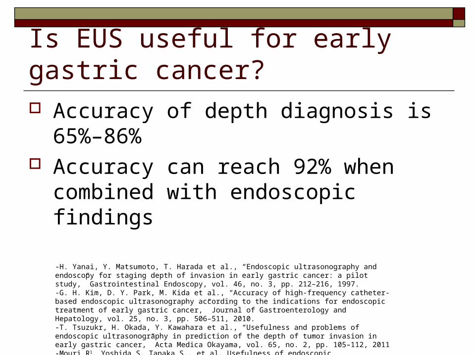
Is EUS useful for early gastric cancer? Accuracy of depth diagnosis is 65%–86% Accuracy can reach 92% when combined
with endoscopic findings
-H. Yanai, Y. Matsumoto, T. Harada et al., “Endoscopic ultrasonography and endoscopy for staging depth of invasion in early gastric cancer: a pilot study,” Gastrointestinal Endoscopy, vol. 46, no. 3, pp. 212–216, 1997. -G. H. Kim, D. Y. Park, M. Kida et al., “Accuracy of high-frequency catheter-based endoscopic ultrasonography according to the indications for endoscopic treatment of early gastric cancer,” Journal of Gastroenterology and Hepatology, vol. 25, no. 3, pp. 506–511, 2010. -T. Tsuzukr, H. Okada, Y. Kawahara et al., “Usefulness and problems of endoscopic ultrasonography in prediction of the depth of tumor invasion in early gastric cancer,” Acta Medica Okayama, vol. 65, no. 2, pp. 105–112, 2011-Mouri R1, Yoshida S, Tanaka S., et al, Usefulness of endoscopic ultrasonography in determining the depth of invasion and indication for endoscopic treatment of early gastric cancer. J Clin Gastroenterol. 2009 Apr;43(4):318-22
Is EUS useful for early gastric cancer? Objective method for determining depth of EGC Determine feasibility of endoscopic therapy Diagnostic accuracy decreased for:-depressed lesions
-undifferentiated cancers
-lesions with ulcers
-minute submucosal invasion in a large lesion
-type 0-I lesions
-lesions located in the upper-third of the stomach
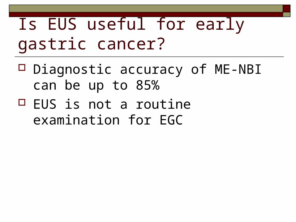
Is EUS useful for early gastric cancer? Diagnostic accuracy of ME-NBI can be up to
85% EUS is not a routine examination for EGC
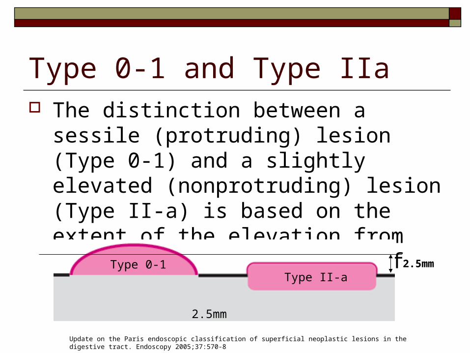
Type 0-1 and Type IIa The distinction between a sessile (protruding)
lesion (Type 0-1) and a slightly elevated (nonprotruding) lesion (Type II-a) is based on the extent of the elevation from the adjacent mucosa, cut off 2.5mm
2.5mm
Type 0-1Type II-a
Update on the Paris endoscopic classification of superficial neoplastic lesions in the digestive tract. Endoscopy 2005;37:570-8
2.5mm
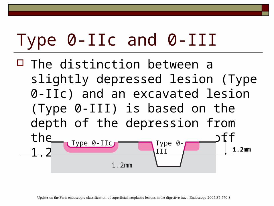
Type 0-IIc and 0-III The distinction between a slightly depressed
lesion (Type 0-IIc) and an excavated lesion (Type 0-III) is based on the depth of the depression from the adjacent mucosa, cut off 1.2mm
Type 0-IIc Type 0-III
1.2mm
1.2mm
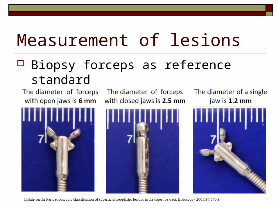
Measurement of lesions Biopsy forceps as reference standard