Embed Size (px)
Citation preview

John E. McMurry
http://www.cengage.com/chemistry/mcmurry
Richard Morrison • University of Georgia, Athens
Chapter 11 Structure Determination:
Nuclear Magnetic Resonance Spectroscopy

Many atomic nuclei behave as if they spin on an axis of rotation
• Nuclei are positively charged• These spinning nuclei generate tiny magnetic fields• Tiny magnets interact with an external magnetic
field, denoted
• Proton (1H) and carbon (13C) are the most important nuclear spins to organic chemists
11.1 Nuclear Magnetic Resonance Spectroscopy
Nuclear spins are oriented randomly in the absence (a) of an external magnetic field but have a specific orientation in the presence (b) of an external field,
• Some nuclear spins are aligned parallel to the external field• Lower energy orientation• More likely
• Some nuclear spins are aligned antiparallel to the external field• Higher energy orientation• Less likely
Nuclear Magnetic Resonance Spectroscopy

When nuclei that are aligned parallel with an external magnetic field are irradiated with the proper frequency of electromagnetic radiation the energy is absorbed and the nuclei “spin-flips” to the higher-energy antiparallel alignment• Nuclei that undergo “spin-flips” in response to applied
radiation are said to be in resonance with the applied radiation - nuclear magnetic resonance
• Frequency necessary for resonance depends on strength of external field and the identity of the nuclei
Nuclear Magnetic Resonance Spectroscopy

The energy difference E between nuclear spin states depends on the strength of the applied magnetic field
• Absorption of energy with frequency converts a nucleus from a lower to a higher spin state
• E = 8.0 x 10⁻⁵ kJ/mol for magnetic field strength of 4.7 T a• For field strength of 4.7 T a radiofrequency (rf) of = 200
MHz is required to bring 1H nuclei into resonance• For a field strength of 4.7 T a radiofrequency (rf) of = 50
MHz is required to bring 13C nuclei into resonance
Nuclear Magnetic Resonance Spectroscopy

Many nuclei exhibit NMR phenomenon• All nuclei with odd number
of protons • All nuclei with odd number
of neutrons• Nuclei with even numbers
of both protons and neutrons
do not exhibit NMR
phenomenon
Nuclear Magnetic Resonance Spectroscopy

The absorption frequency is not the same for all 1H and 13C nuclei• Nuclei in molecules are surrounded by electrons• Electrons set up tiny local magnetic fields that act in
opposition to the applied field, shielding the nucleus from the full effect of the external magnetic field
• The effective field actually felt by the nucleus is the applied field reduced by the local shielding effects
effective = applied – local
11.2 The Nature of NMR Absorptions

The absorption frequency is not the same for all 1H and 13C nuclei• Each chemically distinct nucleus in a molecule has a
slightly different electronic environment and consequently a different
effective field• Each chemically distinct 13C or 1H nucleus in a molecule
experiences a different effective field and will exhibit a distinct 13C or 1H NMR signal
The Nature of NMR Absorptions
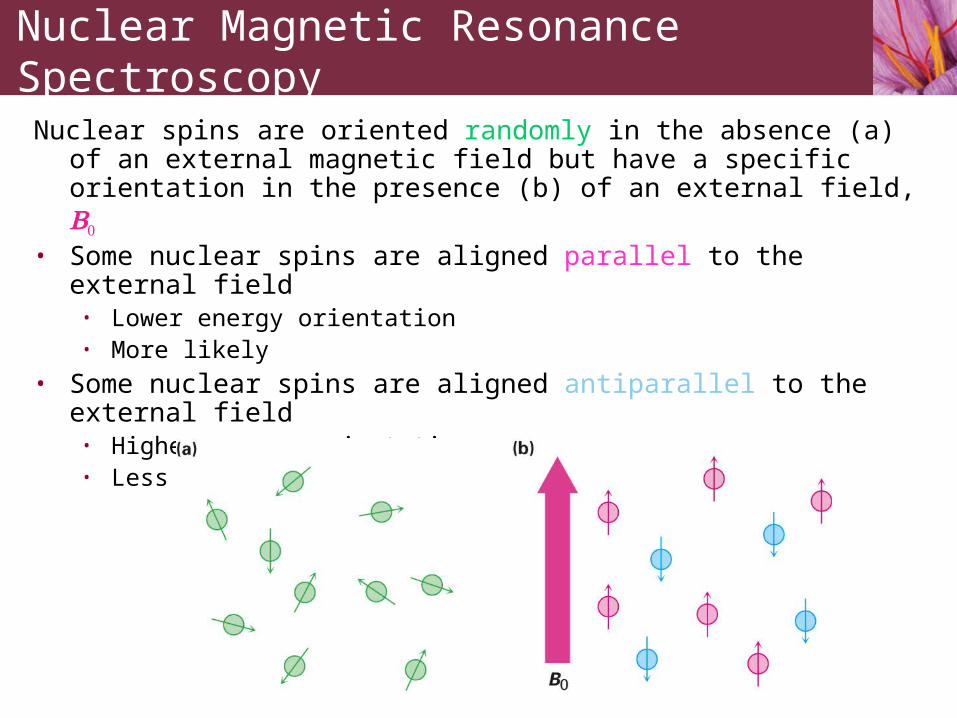
(a) 1H NMR spectrum and (b) 13C NMR spectrum of methyl acetate. Peak labeled “TMS” at far right is for calibration
The Nature of NMR Absorptions

Because the three hydrogens in each methyl group of methyl acetate have the same electronic environment they are shielded to the same extent and are said to be equivalent
• Chemically equivalent nuclei always show the same absorption
• The three hydrogens in each methyl group have the same 1H NMR signal
The Nature of NMR Absorptions

• The two methyl groups of methyl acetate are nonequivalent• The two sets of hydrogens absorb at different positions
• When the frequency of rf irradiation is held constant and the applied field strength is varied each nucleus in a molecule comes into resonance at a slightly different field strength, mapping the carbon-hydrogen framework of an organic molecule
The Nature of NMR Absorptions

The 13C spectrum of methyl acetate shows three peaks, one for each of the three chemically distinct carbon atoms in the molecule
The Nature of NMR Absorptions

Schematic operation of a basic NMR spectrometer
The Nature of NMR Absorptions
p. 547
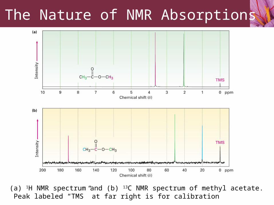
300 MHz NMR 900 MHz NMR

• NMR spectroscopy requires more time (about 10-3 s) compared to IR spectroscopy (about 10-13 s)
• If two rapidly interconverting species are present in a sample, IR spectroscopy will record spectra for both but the slower NMR spectroscopy will record a “time-averaged” spectrum
• “Time-averaging” of NMR spectra can be used to measure reaction rates and activation energies of very fast processes
The Nature of NMR Absorptions

The NMR ChartThe downfield, deshielded side is on the left, and requires a lower
field strength for resonanceThe upfield, shielded side is on the right, and requires a higher
field strength for resonance The tetramethylsilane (TMS) absorption is used as a reference point
11.3 Chemical Shifts

Chemical shift• Position on NMR chart at which a nucleus absorbs
• The chemical shift of TMS is set as zero point• Other absorptions normally occur downfield• NMR charts calibrated using delta () scale
• 1 = 1 part per million of operating frequency
• Chemical shift of an NMR absorption in units is constant, regardless of the operating frequency of the spectrometer
Chemical shift (number of Hz downfield from TMS)δ = Spectrometer frequency in MHz
Chemical Shifts

Narrow NMR absorption range• 0 to 10 for 1H NMR• 0 to 220 for 13C NMR
Higher magnetic field instruments have greater dispersion of NMR signals
• Two signals that are 20 Hz apart (0.1 ppm) at 200 MHz are 50 Hz (0.1 ppm) apart at 500 MHz
• Accidental overlap of nonequivalent signals decreases with increasing field strength
Chemical Shifts

Carbon-13 is only naturally occurring carbon isotope with a nuclear spin
• Natural abundance of 13C is 1.1%• Low abundance of 13C is overcome by signal averaging and
Fourier-transform NMR (FT-NMR)
11.4 13C NMR Spectroscopy: Signal Averaging and FT-NMR
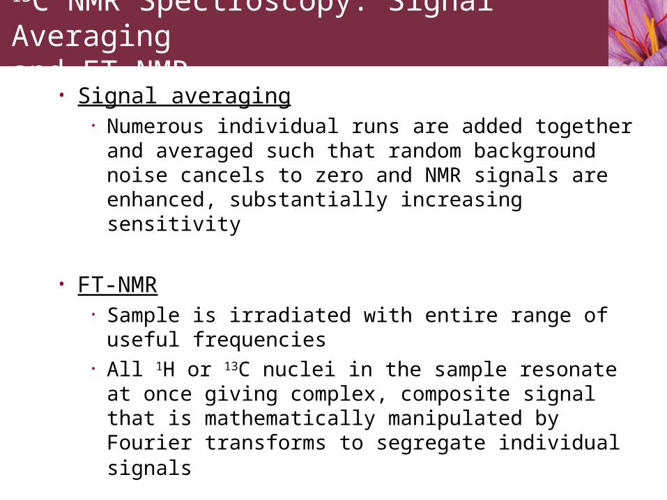
• Signal averaging• Numerous individual runs are added together and
averaged such that random background noise cancels to zero and NMR signals are enhanced, substantially increasing sensitivity
• FT-NMR• Sample is irradiated with entire range of useful frequencies• All 1H or 13C nuclei in the sample resonate at once giving
complex, composite signal that is mathematically manipulated by Fourier transforms to segregate individual signals
13C NMR Spectroscopy: Signal Averaging and FT-NMR

Carbon-13 NMR spectra of pentan-1-ol. Spectrum (a) is a single run, showing background noise. Spectrum (b) is an average of 200 runs
13C NMR Spectroscopy: Signal Averaging and FT-NMR

Factors that affect chemical shifts:1. Chemical shift affected by nearby electronegative atoms
• Carbons bonded to electronegative atoms absorb downfield from typical alkane carbons
2. Hybridization of carbon atoms• sp3-hybridized carbons generally absorb from 0 to 90 • sp2-hybridized carbons generally absorb from 110 to 220 • C=O carbons absorb from 160 to 220
11.5 Characteristics of 13C NMR Spectroscopy

13C spectrum for butan-2-one• Butan-2-one contains 4 chemically nonequivalent carbon
atoms• Carbonyl carbons (C=O) are always found at the low-field
end of the spectrum from 160 to 220
Characteristics of 13C NMR Spectroscopy

13C NMR spectrum of p-bromoacetophenone shows only six absorptions, even though the molecule contains eight carbons. A molecular plane of symmetry makes ring carbons 4 and 4′, and ring carbons 5 and 5′equivalent. Thus, six ring carbons show only four absorptions.
Characteristics of 13C NMR Spectroscopy

At what approximate positions would you expect ethyl acrylate, H2C=CHCO2CH2CH3, to show 13C NMR absorptions?
Worked Example 11.1
Predicting Chemical Shifts in 13C NMR Spectra

Strategy• Identify the distinct carbons in the molecule, and
note whether each is alkyl, vinylic, aromatic, or in a carbonyl group. Then predict where each absorbs, using Figure 11.7 as necessary.
Worked Example 11.1
Predicting Chemical Shifts in 13C NMR Spectra

Solution• Ethyl acrylate has five distinct carbons: two different C=C,
one C=O, one C(O)-C, and one alkyl C. From Figure 11.7, the likely absorptions are
• The actual absorptions are at 14.1, 60.5, 128.5, 130.3, and 166.0
Worked Example 11.1
Predicting Chemical Shifts in 13C NMR Spectra

Distortionless Enhancement by Polarization Transfer (DEPT-NMR) experiment
• Run in three stages1. Ordinary broadband-decoupled spectrum
• Locates chemical shifts of all carbons2. DEPT-90
• Only signals due to CH carbons appear3. DEPT-135
• CH3 and CH resonances appear positive
• CH2 signals appear as negative signals (below the baseline)
• Used to determine number of hydrogens attached to each carbon
11.6 DEPT 13C NMR Spectroscopy

Summary of signals in the three stage DEPT experiment
DEPT 13C NMR Spectroscopy

(a) Ordinary broadband-decoupled spectrum showing signals for all eight of 6-methylhept-5-en-2-ol
(b) DEPT-90 spectrum showing signals only for the two C-H carbons
(c) DEPT-135 spectrum showing positive signals for the two CH carbons and the three CH3 carbons and negative signals for the two CH2 carbons
DEPT 13C NMR Spectroscopy

Propose a structure for an alcohol, C4H10O, that has the following 13C NMR spectral data:• Broadband-decoupled 13C NMR: 19.0, 31.7, 69.5 • DEPT-90: 31.7 • DEPT-135: positive peak at 19.0 , negative peak at
69.5
Worked Example 11.2
Assigning a Chemical Structure from a 13C NMR Spectrum

Strategy• Begin by noting that the unknown alcohol has four carbon atoms,
yet has only three NMR absorption, which implies that two carbons must be equivalent
• Two of the absorptions are in the typical alkane region (19.0 and 31.7 ) while one is in the region of a carbon bonded to an electronegative atom (69.5 ) – oxygen in this instance
• The DEPT-90 spectrum tells us that the alkyl carbon at 31.7 is tertiary (CH); the DEPT-135 spectrum tells us that the alkyl carbon at 19.0 is a methyl (CH3) and that the carbon bonded to oxygen (69.5 ) is secondary (CH2)
• The two equivalent carbons are probably both methyls bonded to the same tertiary carbon, (CH3)2CH-
Worked Example 11.2
Assigning a Chemical Structure from a 13C NMR Spectrum

Solution• We can now put the pieces together to propose a structure:
Worked Example 11.2
Assigning a Chemical Structure from a 13C NMR Spectrum

13C NMR spectroscopy provides information about:• The number of nonequivalent carbons atoms in a
molecule• The electronic environment of each carbon• How many protons are bonded to each carbon
11.7 Uses of 13C NMR Spectroscopy
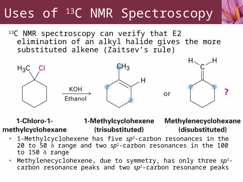
13C NMR spectroscopy can verify that E2 elimination of an alkyl halide gives the more substituted alkene (Zaitsev’s rule)
• 1-Methylcyclohexene has five sp3-carbon resonances in the 20 to 50 range and two sp2-carbon resonances in the 100 to 150 range
• Methylenecyclohexene, due to symmetry, has only three sp3-carbon resonance peaks and two sp2-carbon resonance peaks
Uses of 13C NMR Spectroscopy

The 13C NMR spectrum of the E2 reaction product from the treatment of 1-chloro-1-methylcyclohexane with a base. The
product is clearly identified as 1-methylcyclohexene.
Uses of 13C NMR Spectroscopy

1H NMR spectroscopy determines how many kinds of electronically nonequivalent hydrogens are present in a molecule
• Equivalence or nonequivalence of two protons determined by replacing each H by an X group
• Four possibilities:
1. Protons are chemically unrelated and thus nonequivalent
11.8 1H NMR Spectroscopy and Proton Equivalence
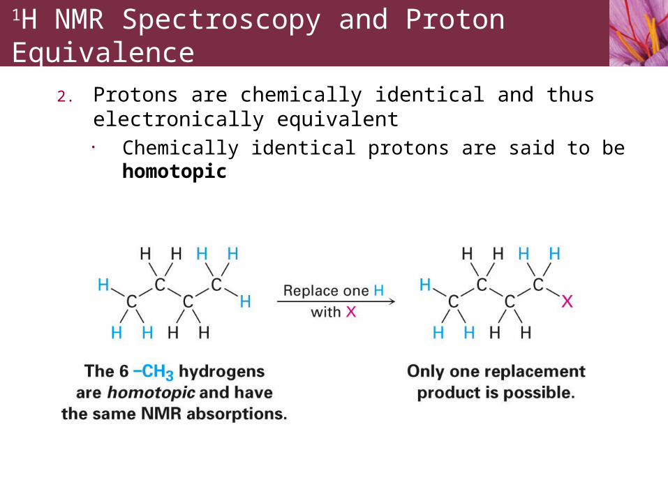
2. Protons are chemically identical and thus electronically equivalent
• Chemically identical protons are said to be homotopic
1H NMR Spectroscopy and Proton Equivalence

3. Protons are electronically equivalent but not identical• The two –CH2 – hydrogens on C2 of butane (as well as the
two C3 hydrogens) are not identical because replacing one or the other would lead to a new chirality center• Non-identical but electronically equivalent protons are said
to be enantiotopic• Different enantiomers would result if pro-R or pro-S
hydrogen were replaced• Prochiral hydrogens are electronically equivalent and thus
have the same NMR absorption
1H NMR Spectroscopy and Proton Equivalence
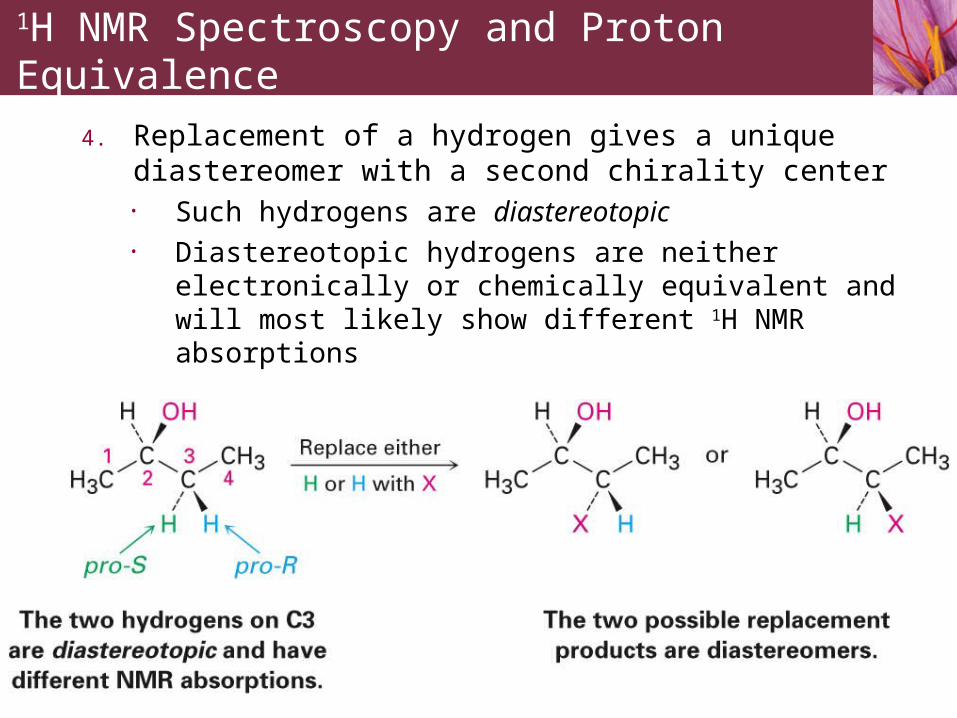
4. Replacement of a hydrogen gives a unique diastereomer with a second chirality center
• Such hydrogens are diastereotopic• Diastereotopic hydrogens are neither electronically or
chemically equivalent and will most likely show different 1H NMR absorptions
1H NMR Spectroscopy and Proton Equivalence

Most 1H NMR chemical shifts occur within the 0 to 10 range except for carboxylic acid O-H absorptions which usually occur within the 11-12 range
11.9 Chemical Shifts in 1H NMR Spectroscopy

Chemical Shifts in 1H NMR Spectroscopy

Methyl 2,2-dimethylpropanoate, (CH3)3CCO2CH3, has two peaks in its 1H NMR spectrum. What are their approximate chemical shifts?
Worked Example 11.3
Predicting Chemical Shifts in 1H NMR Spectra

Strategy• Identify the types of hydrogens in the molecule and note
whether each is alkyl, vinylic, or next to an electronegative atom. Then predict where each absorbs, using Table 11.3 if necessary
Worked Example 11.3
Predicting Chemical Shifts in 1H NMR Spectra

Solution
• The –OCH3 protons absorb around 3.5 to 4.0 because they are on carbon bonded to oxygen. The (CH3)3C- protons absorb near 1.0 because they are typical alkane-like protons
Worked Example 11.3
Predicting Chemical Shifts in 1H NMR Spectra

The area under each 1H NMR peak is proportional to the number of protons causing that peak
• Integrating (electronically measuring) the area under each peak makes it possible to determine the relative number of each kind of proton in a molecule
• Integrating the peaks of 2,2-dimethylpropanoate in a “stair-step” manner shows that they have 1:3 ratio, corresponding to the ratio of the numbers of protons (3:9)
11.10 Integration of 1H NMR Absorptions: Proton Counting

The absorption of a proton can split into multiple peaks called a multiplet
• 1H NMR spectrum of bromoethane shows four peaks (a quartet) at 3.42 for –CH2Br protons and three peaks (a triplet) at 1.68 for –CH3 protons
11.11 Spin-Spin Splitting in 1H NMR Spectra
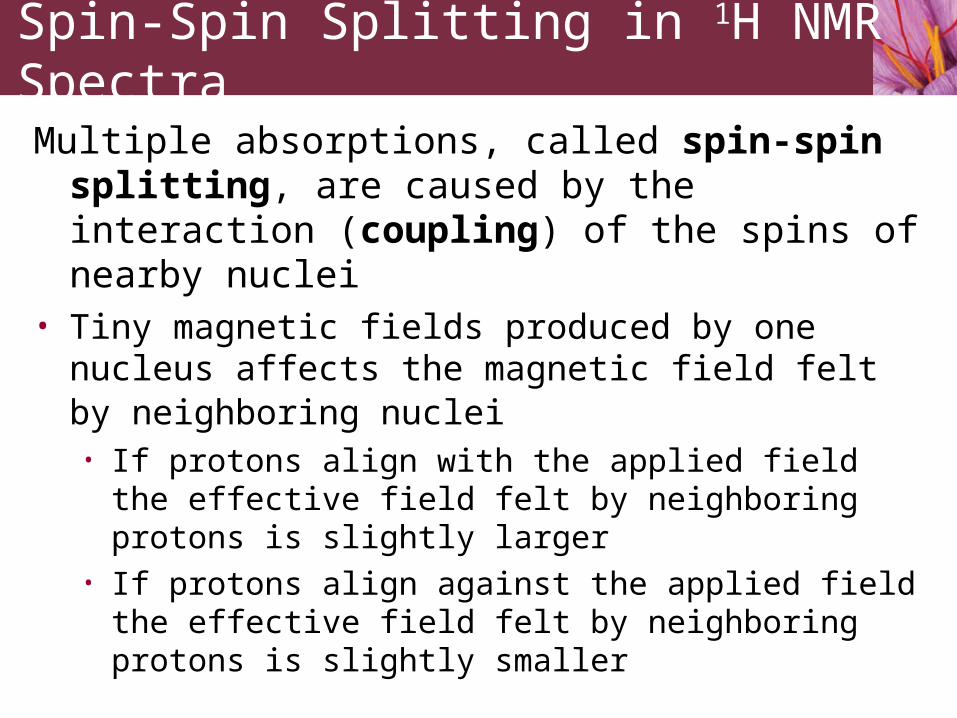
Multiple absorptions, called spin-spin splitting, are caused by the interaction (coupling) of the spins of nearby nuclei
• Tiny magnetic fields produced by one nucleus affects the magnetic field felt by neighboring nuclei • If protons align with the applied field the effective field felt
by neighboring protons is slightly larger • If protons align against the applied field the effective field
felt by neighboring protons is slightly smaller
Spin-Spin Splitting in 1H NMR Spectra

Each –CH2Br proton of CH3CH2Br has its own nuclear spin which can align either with or against the applied field, producing a small change in the effective field experienced by the –CH3 protons
• Three possible spin states (combinations)1. Both protons spin in alignment with applied field
• Effective field felt by neighboring –CH3 protons is larger
• Applied field necessary to cause resonance is reduced
2. One proton spin is aligned with and one proton spin is aligned against the applied field (two possible combinations)
• No effect on neighboring protons
3. Both proton spins align against applied field• Effective field felt by neighboring –CH3 is smaller
• Applied field necessary to cause resonance is increased
Spin-Spin Splitting in 1H NMR Spectra

The origin of spin-spin splitting in bromoethane. The nuclear spins of –CH2Br protons, indicated by horizontal arrows, align either with or against the applied field, causing the splitting of –CH3 absorptions into a triplet
Spin-Spin Splitting in 1H NMR Spectra

Each –CH3 proton of CH3CH2Br has its own nuclear spin which can align either with or against the applied field, producing a small change in the effective field experienced by the –CH2Br protons
• Four possible spin states (combinations)1. All proton spins are aligned with applied field
• Effective field felt by neighboring –CH2Br protons is larger
• Applied field necessary to cause resonance is reduced
2. Two proton spins are aligned with and one proton spin is aligned against the applied field (three possible combinations)
• Effective field felt by neighboring –CH2Br protons is slightly larger
• Applied field necessary to cause resonance is slightly reduced
Spin-Spin Splitting in 1H NMR Spectra

3. Two proton spins are aligned against and one proton spin is aligned with the applied field (three possible combinations)
• Effective field felt by neighboring –CH2Br protons is slightly smaller
• Applied field necessary to cause resonance is slightly increased
4. All proton spins align against applied field• Effective field felt by neighboring –CH3 is smaller
• Applied field necessary to cause resonance is increased
Spin-Spin Splitting in 1H NMR Spectra

The origin of spin-spin splitting in bromoethane. The nuclear spins of –CH3 protons, indicated by horizontal arrows, align either with or against the applied field, causing the splitting of –CH2Br absorptions into a quartet
Spin-Spin Splitting in 1H NMR Spectra

Coupling constant• The distance between peaks in a multiplet• Denoted J• Measured in hertz• Generally fall into range 0 to 18 Hz• Same coupling constant is shared by both groups
of hydrogens whose spins are coupled• Coupling constants are independent of
spectrometer field strength
Spin-Spin Splitting in 1H NMR Spectra

n + 1 rule• Protons that have n equivalent neighboring protons show n + 1 peaks in their 1H NMR spectrum
• The septet is caused by splitting of the –CHBr- proton signal at 4.28 by six equivalent neighboring protons on the two methyl groups (n = 6 leads to 6+1 = 7 peaks)
• The doublet at 1.71 is due to signal splitting of the six equivalent methyl protons by the single –CHBr- proton (n = 1 leads to 2 peaks)
Spin-Spin Splitting in 1H NMR Spectra

Spin-Spin Splitting in 1H NMR Spectra

Summary of spin-spin splitting in 1H NMR:1. Chemically equivalent protons do not show spin-spin splitting
2. The signal of a proton with n equivalent neighboring protons is split into a multiplet of n + 1 peaks with coupling constant J
Spin-Spin Splitting in 1H NMR Spectra

3. Two groups of protons coupled to each other have the same coupling constant, J
1H NMR spectrum of p-methoxypropiophenone• Downfield absorptions at 6.91 and 7.93 are due to four aromatic ring
protons of two kinds, each of which is split into a doublet by its neighbor
• –OCH3 signal is unsplit at 3.84 • –CH2- protons next to carbonyl appear as a quartet at 2.93 coupled
to neighboring –CH3 protons which appear as a triplet at 1.20
Spin-Spin Splitting in 1H NMR Spectra

Why no 13C NMR splitting • No carbon-carbon spin coupling because low
natural abundance of 13C makes it unlikely that two 13C nuclei will be adjacent in a molecule
• No carbon-hydrogen spin coupling because of broadband decoupling• Molecule is irradiated with a pulse of rf energy to cover
carbon and hydrogen resonance frequencies simultaneously
• Hydrogens spin-flip so rapidly that their local magnetic fields average to zero
Spin-Spin Splitting in 1H NMR Spectra

Propose a structure for a compound, C5H12O, that fits the following 1H NMR data:
• 0.92 (3 H, triplet, J = 7 Hz)• 1.20 (6 H, singlet)• 1.50 (2 H, quartet, J = 7 Hz)• 1.64 (1H, broad singlet)
Worked Example 11.4
Assigning a Chemical Structure from a 1H NMR Spectrum

Strategy• Look at each absorption individually.
• The three-proton absorption at 0.92 is due to a methyl group in an alkane-like environment, and the triplet splitting pattern implies that the –CH3 is next to a –CH2. Thus, our molecule contains an ethyl group, CH3CH2 –
• The six-proton singlet at 1.20 is due to two equivalent alkane-like methyl groups attached to a carbon with no hydrogens, (CH3)2C–, and the two-proton quartet at 1.50 is due to the –CH2 of the ethyl group
• All 5 carbons and 11 of the 12 hydrogens in the molecule are now accounted for. The remaining hydrogen, which appears as a broad one-proton singlet at 1.64 , is probably due to an –OH group, since there is no other way to account for it.
• Putting the pieces together gives the structure: 2-methylbutan-2-ol
Worked Example 11.4
Assigning a Chemical Structure from a 1H NMR Spectrum

Solution
Worked Example 11.4
Assigning a Chemical Structure from a 1H NMR Spectrum

Different kinds of hydrogens in a molecule may have accidentally overlapping signals
• The 1H NMR spectrum of toluene shows the accidental overlap of the five nonequivalent aromatic ring protons
11.12 More Complex Spin-Spin Splitting Patterns

One signal can be split by two or more nonequivalent kinds of protons
• Not predicted by n + 1 rule
More Complex Spin-Spin Splitting Patterns
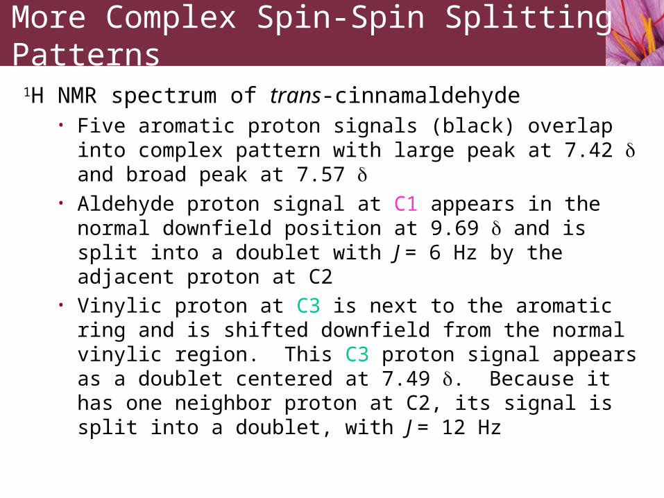
1H NMR spectrum of trans-cinnamaldehyde• Five aromatic proton signals (black) overlap into complex
pattern with large peak at 7.42 and broad peak at 7.57 • Aldehyde proton signal at C1 appears in the normal downfield
position at 9.69 and is split into a doublet with J = 6 Hz by the adjacent proton at C2
• Vinylic proton at C3 is next to the aromatic ring and is shifted downfield from the normal vinylic region. This C3 proton signal appears as a doublet centered at 7.49 . Because it has one neighbor proton at C2, its signal is split into a doublet, with J = 12 Hz
More Complex Spin-Spin Splitting Patterns

C2 proton of trans-cinnamaldehyde • Multiple coupling of C2 proton gives “doublet of doublets”• Splitting pattern illustrated by tree diagram
• C3 proton splits C2 proton signal into doublet (J2-3 = 12 Hz) which is split by C1 aldehyde proton into new doublets (J1-2 = 6 Hz) producing four-line spectrum
More Complex Spin-Spin Splitting Patterns
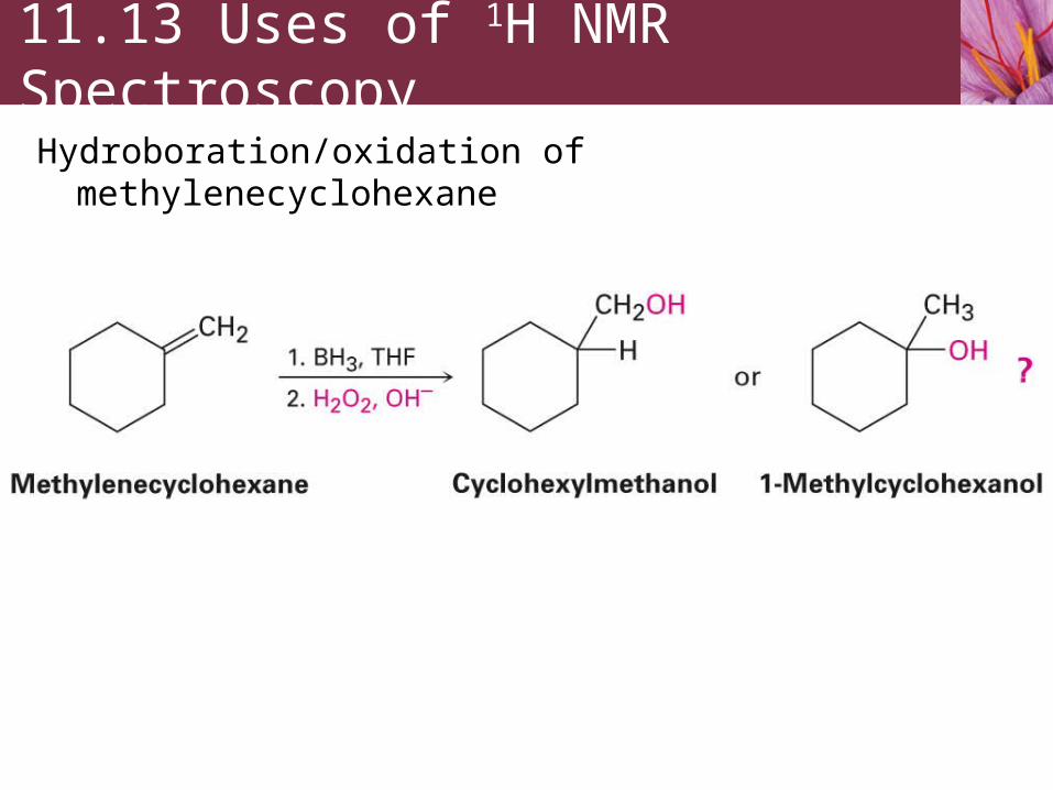
Hydroboration/oxidation of methylenecyclohexane
11.13 Uses of 1H NMR Spectroscopy

1H NMR of reaction product (a) shows a two-proton multiplet at 3.40 indicating the product has a –CH2- group bonded to an electronegative oxygen atom (–CH2OH)
Absence of quaternary –CH3 signal in 1H NMR of reaction product clearly shows that alternative reaction product (b) is not formed by the
hydroboration/oxidation of methylenecyclohexane
Reaction product
Alternative product
(not formed)
Uses of 1H NMR Spectroscopy

Magnetic Resonance Imaging (MRI)
Magnetic Resonance Imaging (MRI) is a diagnostic technique of enormous value to the medical community. MRI takes advantage of the magnetic properties of certain nuclei, typically hydrogen, and of the signals emitted when those nuclei are stimulated by radiofrequency energy. Signals detected by MRI vary with the density of hydrogen atoms and with the nature of their surroundings, allowing identification of different types of tissue and even allowing the visualization of motion.
MRI of this left knee shows thepresence of a ganglion cyst
Lagniappe


![Crossed McMurry Coupling Reactions for Porphycenic Macrocycles… · Crossed McMurry Coupling Reactions for Porphycenic Macrocycles modest yield.[1,11] Very few other reliable methods](https://img.pdfslide.us/doc/110x75/5f0856357e708231d4218219/crossed-mcmurry-coupling-reactions-for-porphycenic-macrocycles-crossed-mcmurry-coupling.jpg)