Embed Size (px)
Citation preview
1
Mutations in hemG mediate resistance to Salicylidene acylhydrazides: A novel link between 1
protoporphyrinogen oxidase (HemG) and Chlamydia trachomatis infectivity 2
3
Patrik Engström1,2,3, Bidong D. Nguyen7, Johan Normark1,2,3, Ingela Nilsson1,2,3, Robert J. 4
Bastidas7, Åsa Gylfe2,3,4, Mikael Elofsson2,3,5, Kenneth A. Fields6, Raphael H. Valdivia7, Hans 5
Wolf-Watz1,2,3 and Sven Bergström1,2,3* 6
7
1Department of Molecular Biology, 2Laboratory for Molecular Infection Medicine Sweden 8
(MIMS), 3Umeå Centre for Microbial Research (UCMR), 4Department of Clinical Microbiology, 9
5Department of Chemistry, Umeå University, Sweden. 10
6Department of Microbiology and Immunology, University of Miami Miller School of Medicine. 11
7Department of Molecular Genetics and Microbiology, Center for Microbial Pathogenesis, Duke 12
University. 13
*Correspondence: [email protected] 14
15
Running Title: Iron-saturated INP0341 targets C. trachomatis HemG 16
17
Copyright © 2013, American Society for Microbiology. All Rights Reserved.J. Bacteriol. doi:10.1128/JB.00506-13 JB Accepts, published online ahead of print on 12 July 2013
on October 5, 2018 by guest
http://jb.asm.org/
Dow
nloaded from
2
Abstract 18
Salicylidene acylhydrazides (SAHs) inhibit the type III secretion system (T3S) of Yersinia and 19
other Gram-negative bacteria. In addition, SAHs restrict the growth and development of 20
Chlamydia species. However, since the inhibition of Chlamydia growth by SAH is suppressed by 21
the addition of excess iron and SAHs have iron-chelating capacity their role as specific T3S 22
inhibitors is unclear. We investigated herein whether SAHs exhibit a function on C. trachomatis 23
that goes beyond iron chelation. We found that the iron-saturated (IS) SAH INP0341 specifically 24
affects C. trachomatis infectivity with reduced generation of infectious EB progeny. Selection 25
and isolation of spontaneous SAH-resistant mutant strains revealed that mutations in hemG 26
suppressed the reduced infectivity caused by IS-INP0341 treatment. Structural modeling of C. 27
trachomatis HemG predicts that the acquired mutations are located in the active site of the 28
enzyme, suggesting that IS-INP0341 inhibits this domain of HemG and that protoporphyrinogen 29
oxidase (HemG) and heme metabolism are important for C. trachomatis infectivity. 30
31
32
on October 5, 2018 by guest
http://jb.asm.org/
Dow
nloaded from

3
Introduction 33
Chlamydiae are Gram-negative bacteria that cause common respiratory diseases, sexually 34
transmitted diseases and the eye disease trachoma (1). Antibiotics are effective for treating most 35
chlamydial infections however new antibiotic drugs or treatment approaches may be needed to 36
combat future infections that limits the emergence of secondary infections (2). Persistent 37
chlamydial infections might exist in clinical practice, but have not yet been successfully 38
validated. However, “persistent” Chlamydia forms can be generated in the laboratory during 39
antibiotic-induced stress, viral co-infection, amino acid and iron limitation (3). 40
Chlamydiae are obligate intracellular pathogens with a biphasic developmental cycle. The 41
pathogen exists in two distinct forms; the environmentally stable and infectious elementary body 42
(EB) and the replicative reticulate body (RB). The EB form attaches to epithelial cells and enters 43
by endocytosis. Once inside the host cell, the EB transition to the metabolically active RB which 44
replicates within the confines of the pathogen-containing vacuole (“inclusion”). Midway through 45
the developmental cycle, the RB forms begin to transition back to EB forms in an asynchronous 46
manner (4). Finally, EBs are released either by cell lysis or by an inclusion extrusion mechanism 47
into the extracellular milieu (5, 6). At all stages of infection, chlamydiae manipulate the host cell 48
by secreting effector proteins that help establish a replicative niche and suppress innate immune 49
responses (7). Many of these effectors are likely substrates of type III secretion (T3S) and are 50
synthesized early, mid and late in the developmental cycle (8, 9). T3S systems are well-51
characterized delivery systems for virulence factors in Gram-negative bacteria, and are important 52
for bacterial avoidance of professional phagocytes, suppression of innate immunity and 53
on October 5, 2018 by guest
http://jb.asm.org/
Dow
nloaded from

4
promotion of uptake into epithelial cells (10). Structurally, T3S systems resemble “injection 54
needles” consisting of a basal apparatus that spans the inner and outer membrane (11). 55
A role for T3S in Chlamydia development has been proposed (12, 13) but formal proof has been 56
hampered by the lack of practical genetic tools. The use of small inhibitory molecules provides 57
an alternative approach to study the role played by T3S in Chlamydia development (14). Small 58
molecule screens performed by Kauppi et al., identified salicylidene acylhydrazides (SAHs) as 59
inhibitors of Yersinia pseudotuberculosis T3S (15, 16). Subsequent studies indicate that SAHs 60
block Chlamydia growth but not entry into cells (17-21), which supports the prevalent notion 61
that the T3S is essential during the Chlamydia mid and late developmental cycle (22). 62
Subsequent studies indicate that secretion or localization of predicted T3S-effectors is altered by 63
SAHs (23-25, 19, 26). Because the growth inhibition of Chlamydia by SAHs is reversed by 64
exogenous iron, it has been postulated that iron chelation by SAHs may be responsible for its 65
anti-chlamydial properties (27, 28). However, the SAH INP0406, which does not inhibit T3S, 66
retains iron-chelating properties yet it cannot inhibit Chlamydia growth (28). 67
In this study we have investigated how SAHs affect C. trachomatis development and secretion, 68
both in the presence and in the absence of exogenously added iron. We found that iron-saturated 69
INP0341 inhibits the generation of infectious C. trachomatis EBs and that mutations in hemG 70
mediate resistance to INP0341. 71
72
on October 5, 2018 by guest
http://jb.asm.org/
Dow
nloaded from
5
Materials and methods 73
Chemicals and INPs. The chemical compounds INP0010 (also known as ME0052) (16, 29) and 74
INP0341 (30) were synthesized and purified from commercially available hydrazides and 75
salicylaldehydes, as described previously (31). The SAHs were dissolved in dimethyl sulfoxide 76
(DMSO, Sigma) to a final concentration of 20 mM, stored at room temperature without exposure 77
to light for a maximum of 3 weeks. For the indicated experiments, FeSO4 (Merck), FeCl3 78
(Sigma) and Deferoxamine methanesulfonate (DFO, Sigma) were diluted in distilled water and 79
filter-sterilized (0.22 µm PES, Corning). For iron saturation experiments, the SAHs were diluted 80
in pre-warmed RPMI prior to supplementation of iron sulfate. Compound INP0341 was iron-81
saturated by mixing equal molar equivalents of compound and FeCl3. The mixture was incubated 82
for one hour at room temperature prior to supplementation to pre-warmed RPMI media. 83
84
Cells lines and Chlamydia strains. HeLa cell line (DSMZ) were grown in RPMI 1640 (Sigma) 85
supplemented with 10% heat inactivated fetal bovine serum (FBS, Sigma) and 20 mM HEPES 86
(pH 8.0). Vero cells (ATCC CCL-81) were grown in DMEM (GIBCO, Invitrogen) supplemented 87
with 10% FBS at 37°C (5% CO2). C. trachomatis serovar LGV-L2 434/Bu (ATTC VR902B) 88
was propagated in HeLa cells and purified as described by Caldwell et al., (32). Cells and 89
bacteria were negative for mycoplasma infection, as determined by a mycoplasma detection kit 90
(Stratagene). 91
92
Chlamydia infections and determination of infectivity. HeLa cells were infected by C. 93
trachomatis in Hank’s Balanced Salt Solution (HBSS) (GIBCO, Invitrogen) at a multiplicity of 94
on October 5, 2018 by guest
http://jb.asm.org/
Dow
nloaded from
6
infection (MOI) of 0.1-1 for one hour at 37°C in 5% CO2. HBSS was removed and RPMI 95
medium containing indicated treatments was added to the infected cells (lacking cycloheximide). 96
Infectivity was determined by harvesting infectious EB progeny at 44-48 hours post infection (h 97
p.i.). Harvested bacteria were diluted in HBSS and fresh HeLa cells were infected, ~40 hours 98
after re-infection were C. trachomatis inclusions counted. Data are represented as the relative 99
amount of EBs in treated infections compared to the amount of EBs in DMSO treated infections. 100
When multiple strains were analyzed, infectivity was determined by normalization to input IFUs 101
as previously described (33). 102
103
Immunofluorescence analysis. In samples fixed with methanol, C. trachomatis was detected 104
with a rabbit anti-MOMP antibody and an LRSC-conjugated anti-rabbit antibody (Jackson 105
ImmunoResearch laboratories). CPAF and IncA were detected in samples that were fixed with 106
4% PFA and permeabilized with 0.1% Triton-X (34). Thereafter, an anti-CPAF antibody (34) 107
was detected with an anti-rabbit LRSC-conjugated antibody and a mouse anti-IncA (gift from 108
Daniel D. Rockey, Oregon State University) antibody with an anti-mouse FITC-conjugated 109
antibody (both secondary antibodies from Jackson ImmunoResearch laboratories). Host and 110
bacterial DNA were stained with 200 nM 4′,6-diamidino-2-phenylindole (DAPI). Images were 111
acquired by laser scanning confocal microscopy (Nikon Eclipse C1 plus). The signal from 112
uninfected cells was used to set the background levels. Digital images were processed using 113
Adobe Photoshop C6 software (Adobe Systems Inc.). 114
115
on October 5, 2018 by guest
http://jb.asm.org/
Dow
nloaded from
7
Transmission electron microscopy (TEM). At 44 h p.i., infected cells were processed for TEM 116
as previously described (33). Briefly, cells were fixed with 2.5% glutaraldehyde/0.05% 117
malachite green (EMS) in 0.1 M sodium cacodylate buffer (pH 6.8) and then post-fixed with 118
0.5% osmium tetroxide/0.8% potassium ferricyanidein in 0.1 M sodium cacodylate, 1% tannic 119
acid and 1% uranyl acetate. Samples were dehydrated with graded amounts of ethanol and 120
embedded in Spur resin, subsequently imaged on a Tecnai G2 Twin microscope (FEI). 121
122
Determination of inclusion size. Infected cells were fixed at 44 h p.i., and stained with an anti-123
MOMP antibody as described above. Inclusion membranes were visualized with a 440 nm diode 124
laser and a specific detector to obtain transmission photographs (as seen in figure S1). 125
Subsequently, each inclusion area was measured using EZ-C1 software (Nikon). When C. 126
trachomatis strains were grown in the absence of the SAHs it was possible to determine 127
inclusion sizes by using the Cellomics ArrayScan Vti HCS automated fluorescent imaging 128
system (ThermoFisher) as previously described (33). 129
130
Selection of INP0341-resistant strains. HeLa cells grown in large flasks were infected with C. 131
trachomatis LGV-L2 at a MOI of 5 (~108 IFUs). Infected cells were treated with concentration 132
ranging from 20 to 35 µM of compound INP0341 and excess iron sulfate, or iron-saturated 133
INP0341 with concentration ranging from 25 to 40 µM, for serial passages. Infectious progeny 134
were collected at 44-48 h p.i., and subsequently used for re-infection (Fig. 5). During the first 4-6 135
passages the cells were infected at a MOI of 2 or higher in the presence of SAHs, thereafter the 136
selection continued with a MOI of 1 or lower. When mutant populations were passaged in the 137
on October 5, 2018 by guest
http://jb.asm.org/
Dow
nloaded from
8
absence of INP0341, the MOI was kept between 0.5 and 2 to allow genetic recombination. 138
Clonal strains were collected by a plaquing assay on cell monolayers as previously described 139
(33). Briefly, confluent Vero cells were grown in 6-well plates and infected with 100-10 IFUs 140
from the mutant populations for 2 hours and infected cells were overlaid with agarose/DMEM. 141
Plaques in the monolayers were picked 10-20 days after infection and propagated in HeLa cells. 142
143
Whole genome sequencing and genotyping. C. trachomatis genomic DNA prepared for whole 144
genome sequencing (WGS) was purified from density gradient purified bacteria with the DNeasy 145
Blood & Tissue Kit (Qiagen, purification of total DNA from Gram-negative bacteria) according 146
to the manufacturer’s instructions. DNA was concentrated by sodium acetate-ethanol 147
precipitation, and the pellets were dissolved in AE-buffer (Elution buffer for genomic DNA, 148
Qiagen) to avoid aggregation. For WGS, 1 μg of enriched chlamydial DNA was fragmented with 149
an Adaptive Focused Acoustics S220 instrument (Covaris, Inc. Woburn, MA), and DNA 150
sequencing libraries were prepared with a library construction kit (TruSeq DNA Sample 151
Preparation Kit v2, Illumina, Inc. San Diego, CA) according to the manufacturer’s instructions. 152
Libraries were sequenced on a MiSeq DNA Sequencing Platform (Illumina, Inc. San Diego, CA) 153
at Duke Universities IGSP DNA Core sequencing facility. Genome assembly and single 154
nucleotide variant (SNV) identification was performed with Geneious version 6 from Biomatters 155
(available at http://www.geneious.com/). The C. trachomatis L2 434/Bu genome (GenBank no, 156
NC_010287) was used as reference sequence. A mutation is herein defined as a nucleotide 157
variant present at a frequency above 15% and strand-bias below 80%, detectable both by WGS 158
and capillary sequencing in a bacterial population. In the second derived mutant population a 159
on October 5, 2018 by guest
http://jb.asm.org/
Dow
nloaded from
9
mutation in phosphoatidylcholine-hydrolyzing phospholipase D (CTL0413) was detected by 160
WGS at a variant frequency of 33% and strand-bias 75%. Attempts to read through this GC-rich 161
region were not possible by using conventional capillary sequencing (data not shown). 162
Therefore, it is possible that this mutant population contains a mutation in CTL0413. To identify 163
mutations by capillary sequencing templates were PCR amplified with primers presented in 164
Table S1. PCR products were sequenced by capillary sequencing (Big Dye; Applied biosystems). 165
The intensity of the fluorescence peaks detected by capillary DNA sequencing was used as 166
estimates of the relative abundance of any single nucleotide variant in the bacterial population. 167
Estimated frequencies were consistent with the obtained values from WGS (data not shown). 168
169
Structural prediction. Template candidates, structurally homologous to C. trachomatis HemG, 170
were identified through HMM-HMM-comparisons using the HHpred (35) toolkit with default 171
settings, MODELLER (36) and the MPI-toolkit were used with default settings to create the 3D-172
models. The STRAP (37) toolkit was used to make structural alignments and structure 173
superposition. Structural visualization was made in PyMOL (38). Montage of images was 174
prepared in Adobe Photoshop C6 (Adobe Systems Inc.). 175
176
on October 5, 2018 by guest
http://jb.asm.org/
Dow
nloaded from

10
Results 177
Salicylidene acylhydrazides inhibit C. trachomatis infectivity but not growth following 178
addition of excess iron. We recently described that salicylidene acylhydrazides (SAHs) act as 179
inhibitors of the Yersinia pseudotuberculosis T3S (15, 16). We further showed that the SAH 180
variants INP0341 and INP0010 also inhibit Chlamydia growth and development, observations 181
that have been corroborated by other groups (17, 18, 20, 28). These findings suggest that T3S 182
activity is essential for Chlamydia (22). It has previously been shown that excess iron in the cell 183
culture media can suppress the negative impact of SAHs on C. trachomatis growth, suggesting 184
that the iron chelation properties of SAH may be partially responsible for the observed growth 185
inhibition (28). However, INP0406, an SAH that does not inhibit T3S yet retains the capacity to 186
chelate iron similar to INP0341, does not restrict C. trachomatis growth (28). We compared the 187
antimicrobial activity of INP0341 and INP0010 on C. trachomatis and contrasted them to the 188
effects induced by the iron specific chelator DFO (39). HeLa cells were infected with C. 189
trachomatis for 1 hour, and the media was exchanged with media containing the indicated 190
inhibitors. At 44 h p.i., the infected cells were fixed, stained, and analyzed by laser scanning 191
confocal microscopy. Addition of INP0010 (10 and 30 µM) and INP0341 (10 and 30 µM) to 192
infected cells resulted in the formation of enlarged, aberrant C. trachomatis forms as had been 193
previously observed with iron chelators (39) (Fig. 1). Indeed, DFO treatment also induced the 194
formation of these aberrant forms, although significantly higher concentrations were required 195
(400 µM) (Fig. 1). Addition of 250 µM iron sulfate suppressed the effects of INP0341, INP0010 196
and DFO resulting in no visible aberrant C. trachomatis indicating that the iron limitation 197
induced by these compounds is reduced. However, in the presence of excess iron and INP0341, 198
on October 5, 2018 by guest
http://jb.asm.org/
Dow
nloaded from
11
the bacteria appeared to aggregate at the center of the inclusion (Fig. 1 and Fig S1). Addition of 199
INP0010 alone or DFO in the presence or absence of excess iron did not cause this redistribution 200
(Fig. 1). We next measured inclusions sizes, which can be used to indirectly assess bacterial 201
growth (40). Addition of excess iron to INP0010, INP0341 and DFO-treated infected cells 202
resulted in similar inclusion sizes however these inclusions were significantly larger than those 203
in iron free conditions (Fig. 2). These results indicate that excess iron, at least partly, suppresses 204
the growth inhibitory effect of SAHs and DFO. Interestingly, in the presence of excess iron, 205
INP0341 treatment resulted in detachment of bacteria from the inclusion membrane. Together, 206
these data indicate that INP0341 exhibits additional effect(s) that are distinguishable from its 207
iron chelating effects. 208
Next, we quantified the yield of infectious EBs after treating infected cells with INP0010, 209
INP0341 and DFO, in the presence or absence of excess iron. Compounds were added 210
immediately after infection and EBs were harvested at 44 h p.i. and titered by infecting a 211
monolayer HeLa cells. Addition of excess iron almost completely suppressed the effect of DFO 212
(45% of DMSO-control) while the effect by INP0010 and INP0341was partly suppressed (6% 213
and 0.15% of DMSO-control, respectively) for the generation of infectious EBs (Fig. 3). These 214
results show that the effect of DFO on Chlamydia infectivity is suppressed by excess iron while 215
the effect of treatment with SAHs is only partly suppressed. This further suggests that the SAHs, 216
particularly INP0341 have an effect on C. trachomatis development that is not linked to iron 217
chelation. The fact that compound INP0341 possesses a stronger effect compared to INP0010 on 218
C. trachomatis might be explained by more efficient uptake into host cells or binding to 219
molecular target(s). 220
on October 5, 2018 by guest
http://jb.asm.org/
Dow
nloaded from
12
221
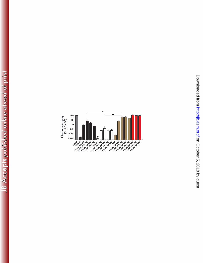
Secretion of CPAF or IncA is not inhibited by INP0341. The above results suggest that SAHs 222
restrict the generation of infectious C. trachomatis EBs. The simplest interpretation is that SAHs 223
target T3S in C. trachomatis because earlier studies identified INP0341 as a potent inhibitor of 224
T3S in Yersinia and other Gram-negative bacteria (41, 16). We therefore investigated the 225
localization of the secreted proteins IncA and CPAF after treatment with INP0341 or DFO 226
following excess addition of iron. IncA, is a T3S effector known to be secreted to the chlamydial 227
inclusion membrane during mid-development (42), CPAF, is a mid-late effector secreted via the 228
general secretion pathway to the host cell cytoplasm (34). We found that CPAF was secreted to 229
the host cell cytoplasm in the majority of infected cells after INP0341 or DFO treatment, 230
analogous to IncA, which was localized to the inclusion membrane independent of INP0341 231
treatment or not (Fig. 4A and B). These data suggest that INP0341 does not inhibit secretion of 232
IncA or CPAF which is consistent with the growth alleviation that follows addition of excess 233
iron to INP0341-treated C. trachomatis. Thus, these data suggests that the effect by INP0341 is 234
not related to secretion. 235
236
Isolation of SAH-resistant C. trachomatis mutant strains. To define the mechanism of action 237
of INP0341 we isolated spontaneous INP0341-resistant populations of C. trachomatis LGV-L2 238
by serial passage of bacteria through several rounds of infection in HeLa cells in the presence of 239
INP0341 and excess iron (Fig. 5). This strategy has been previously used to understand the mode 240
of action for new anti-bacterial compounds (43, 44). INP0341-resistant C. trachomatis began to 241
emerge after 6 passages and stable resistance was achieved by passage 12. Genomic DNA was 242
on October 5, 2018 by guest
http://jb.asm.org/
Dow
nloaded from
13
isolated from this mutant population and subjected to whole genome sequencing (WGS). Two 243
mutations were identified (35% variant frequency per mutation) after INP0341 selection. One 244
mutation was in ruvC, a gene that encodes the holiday junction resolvase known to facilitate the 245
last step in DNA recombination (45). The mutation in ruvC leads to a histidine to tyrosine 246
substitution at residue 31. The second mutation was in hemG, a gene encoding the 247
protoporphyrinogen oxidase, an enzyme that catalysis the aromatization of protoporphyrinogen 248
IX to protoporphyrin IX, a central step in heme biosynthesis (46). The mutation in hemG 249
changes a glycine to serine at residue 58. The genome of C. trachomatis encodes all the 250
components for heme biosynthesis indicating an important role for heme in oxidative catalysis 251
and respiration during C. trachomatis development (47). We next plaque purified clonal strains 252
from this mutant population (Fig. 5), and genotyped them for the hemG and ruvC mutant alleles. 253
All mutant strains contained both the HemGG58S and the RuvCH31Y substitutions (Table 1) 254
indicating that both mutations are present within individual SAH-resistant strains in this 255
population. 256
Because mutants in a population can be positively or negatively selected depending on the fitness 257
cost of the individual mutation, we decided to further passage our INP0341-resistant mutant 258
population for an additional eight passages in the presence or absence of INP0341. Interestingly, 259
in both populations, the RuvCH31Y substitution was negatively selected while the HemGG58S 260
substitution was retained (Fig 5), suggesting that the HemGG58S substitution is primarily 261
responsible for INP0341-resistance, and imply that the RuvCH31Y substitution had a high fitness 262
cost. Plaque purified strains were collected from the expanded mutant population that had been 263
grown in the absence of INP0341 (named expanded mutant population P20), plaque purified, and 264
on October 5, 2018 by guest
http://jb.asm.org/
Dow
nloaded from
14
clonal isolates were genotyped for the hemG and ruvC mutant alleles. We obtained strains 265
bearing only a mutation in hemG (Table 1). Next we characterized these mutant strains by 266
assessing growth and production of infectious EBs in the absence of the compound. In 267
comparison to the wild-type strain, the strain with the RuvCH31Y substitution delayed growth 268
with significantly smaller inclusions at 24 and 36 h p.i. The mutant strain carrying only the 269
HemGG58S substitution formed slightly smaller inclusions at 24 h p.i. while at later time points 270
the sizes were comparable to wild-type strains (Fig. S2A). The yields of infectious EBs were also 271
quantified and the double mutant produced significantly less infectious EBs at 36 and 48 h p.i., 272
when compared to both the wild type and the strain only carrying the HemGG58S substitution 273
(Fig. S2B). These data are consistent with the observation that the RuvCH31Y substitution has a 274
high fitness cost while the HemGG58S substitution has a relatively low cost. 275
Because SAHs chelate iron (28) yet some of its anti-chlamydial functions appear to be 276
independent from iron sequestration (Fig. 3), we decided to use a genetic approach to determine 277
if we could identify mutants resistant to iron-loaded SAHs. We first tested if INP0341 could be 278
iron-saturated prior addition to the cell culture media. We found that a ratio of 1:1 (Iron 279
chloride:INP0341) was needed to avoid precipitation of INP0341 and that 30 µM iron-saturated 280
INP0341 (IS-INP0341) affected the intra-inclusion distribution of C. trachomatis similarly as the 281
addition of excess iron to media containing 30 µM INP0341 (data not shown). This approach 282
was therefore employed to further investigate if INP0341 has an effect beyond iron chelation and 283
as well to avoid potential secondary effects by the addition of excess iron to cell culture media. 284
We started a new selection for C. trachomatis LGV-L2 variants that would be resistant to IS-285
INP0341 and were able to enrich for a resistant population by Passage 15. WGS of this 286
on October 5, 2018 by guest
http://jb.asm.org/
Dow
nloaded from
15
population identified two mutations present at 70% frequency (Fig. 5), including a new mutation 287
in hemG leading to an arginine to a cysteine change at residue 91. The second mutation was in 288
rpsF, a gene that encodes the 30S ribosomal protein S6 (Fig. 5), leading to an alanine to an 289
aspartic acid substitution at residue 54. Altogether, these data indicate that mutation in hemG is 290
strongly associated with resistance to IS-INP0341. 291
292
Mutations in hemG mediate IS-INP0341 resistance with increased infectivity. The above 293
data suggests that mutations in hemG mediate resistance to the effect by INP0341 separated from 294
the iron chelation feature, resulting in increased generation of infectious EBs. To verify this 295
resistance, we first tested the collected mutant strains (Table 1) for the generation of infectious 296
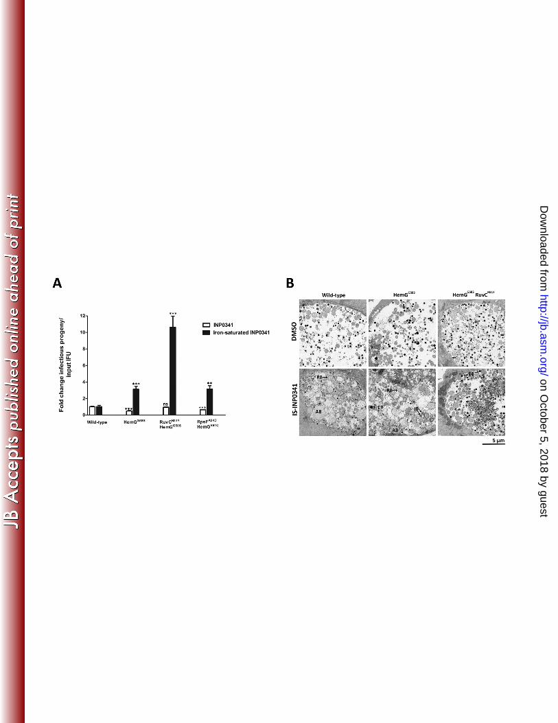
EBs in the presence of IS-INP0341. The strain with the HemGG58S substitution displayed a 3-fold 297
increased yield of infectious EBs. A similar increase was observed for the strain with the 298
HemGR91C and the RpsFA54D substitutions. The strain with both the HemGG58S and the RuvCH31Y 299
mutations resulted in a further increase (~10-fold) of infectious EBs (Fig. 6A). In the case of 300
iron-saturated INP0010, all the strains generated similar yield of infectious EBs (data not 301
shown). Next, we characterized the effect of IS-INP0341 on the mutant strains by transmission 302
electron microscopy and determined that IS-INP0341 treatment of the mutant strains did not 303
induce the formation of aberrant RBs to the same extent as in wild type C. trachomatis (Fig. 6B). 304
Together, these data suggest that IS-INP0341 inhibits some aspect of the RB-to-EB transition 305
and that the hemG mutant strains, at least partly, overcome this inhibitory effect with more IBs 306
and EBs observed (Fig. 6B). Finally, we tested the strains for sensitivity to iron limitation 307
induced by INP0341 by assessment of infectivity at 48 h p.i. In comparison with the wild-type 308
on October 5, 2018 by guest
http://jb.asm.org/
Dow
nloaded from
16
strain, all the strains displayed similar or slightly more sensitivity to iron limitation induced by 309
INP0341 (Fig. 6A). These data show that the mutant strains are not resistant to iron limitation 310
induced by INP0341 and therefore we conclude that INP0341 has an effect beyond iron 311
chelation. 312
313
Structural prediction of C. trachomatis HemG. The crystal structure of protoporphyrinogen 314
oxidase (PPO) from Myxococcus xantus (MxPPO) (48) was predicted to have the highest 315
structural homology to C. trachomatis HemG (CtHemG) and a structural model was built from 316
that template (Fig. 7A). Based on structural prediction, PPO from Bacillus subtilis (BsPPO) (49) 317
is also predicted to have a high structural homology to CtHemG (Fig. 7B), BsPPO has recently 318
been co-crystalized together with flavine-adenine dinucleotide (FAD) and the inhibitor 319
acifluorfen (AF), the latter is proposed to mimic half of the PPO substrate protoporphyrinogen 320
XI and therefore binds to the active site of BsPPO (48, 49). This structural information allowed 321
us to map the mutations that confer IS-INP0341 resistance onto a structural model of the 322
Chlamydia HemG. In this manner, we determined that mutation that confers IS-INP0341-323
resistance occurs in close vicinity to the putative catalytic site (Fig. 7C). 324
on October 5, 2018 by guest
http://jb.asm.org/
Dow
nloaded from

17
Discussion 325
New methods that enable the isolation and rapid mapping of mutations in Chlamydia genes, 326
combined with recent reports of successful transformation of C. trachomatis with plasmid DNA 327
(50, 51, 33, 52), indicate that genetic approaches will soon yield new insight into the biology of 328
this obligate intracellular pathogen. However these genetic approaches still remain somewhat 329
limited. Thus, the use of small organic molecules with inhibitory properties presents an 330
alternative approach to study the molecular function of Chlamydia gene products. By selecting 331
C. trachomatis mutant strains resistant to novel inhibitory compounds, the target or mode of 332
action of the compound may be revealed. In addition, this method can also lead to the isolation 333
of isogenic mutant strains with phenotypes separated from the drug-resistance. Thus, an 334
extension of this procedure can complement genetic approaches such as chemical mutagenesis. 335
Selected SAH compounds have been used to study T3S of Chlamydia and other Gram negative-336
bacteria (41, 53-55). These compounds inhibit chlamydial growth, development and secretion, 337
suggesting a role for T3S throughout the intracellular developmental cycle (17, 23, 56, 18, 24, 338
25, 19-21, 26). However, Slepenkin et al., reported that addition of excess iron restored 339
Chlamydia growth during SAH treatment, suggesting these compounds affect chlamydial growth 340
via iron chelation (28). On the other hand, Prantner et al., argued that the SAHs might have an 341
effect separated from iron chelation, possibly affecting late T3S activity in Chlamydia (27). 342
Since addition of excess iron to SAHs does not restore secretion of T3S-substrates in Yersinia 343
(data not shown) we wanted to determine if these compounds had an effect on Chlamydia 344
development beyond iron chelation. 345
on October 5, 2018 by guest
http://jb.asm.org/
Dow
nloaded from

18
In this report we confirm previously published data that excess iron suppresses the effect on 346
bacterial growth by SAHs (27, 28) (Fig. 1 and 2). However, despite the presence of iron, 347
compound INP0341 still caused distinct phenotypes including the accumulation of bacteria 348
within regions of the inclusion lumen (Fig. S1), and reduced yield of infectious EB progeny (Fig. 349
3). In addition, we found that INP0341 did not affect the secretion of IncA (T3S) or CPAF (Fig. 350
4A and B). In aggregate, our results suggest that INP0341 has an effect on C. trachomatis 351
beyond iron chelation that likely includes a partial block in the RB-to-EB transition. Such a 352
block in differentiation is consistent with our observation by electron microscopy where RB-to-353
EB transition appears to be impaired in IS-INP0341-treated infections (Fig. 6B). 354
We collected independent INP0341-resistant C. trachomatis populations and showed that 355
mutations in hemG primarily mediate the resistance observed (Fig. 5). This gene encodes 356
Protoporphyrinogen oxidase (PPO or HemG), an enzyme that catalyzes the second to last step of 357
heme biosynthesis (46, 57). Chlamydia codes for all components for the biosynthesis of heme 358
(47), an important cofactor of peroxidases, catalases, sensor molecules and cytochromes (46). 359
Cytochromes are essential for respiration and are the most abundant proteins that contain heme 360
in bacteria (58). Thus, HemG activity is indirectly coupled to respiration. In E. coli it has been 361
suggested that HemG activity also has a direct role in respiration (59, 60). At this stage, it is 362
unclear how the specific hemG mutations identified provide resistance to IS-INP0341. The 363
simplest interpretation is that the mutations in hemG impair a potential interaction between IS-364
INP0341 and HemG. Alternatively these are gain-of-function mutations that allow the bacteria 365
to cope with oxidative stress caused by IS-INP0341 or inhibited heme biosynthesis. Together, 366
on October 5, 2018 by guest
http://jb.asm.org/
Dow
nloaded from

19
this strongly suggests that HemG activity plays an important role in the generation of C. 367
trachomatis progeny at least in vitro and under these experimental conditions. 368
In order to isolate isogenic strains, we took advantage of the fact that the RuvCH31Y substitution 369
has a high fitness cost. Specifically, the mutant population that contained both the HemGG58S and 370
RuvCH31Y substitutions was grown in the absence of INP0341 for multiple passages. The clonal 371
strains collected from this expanded mutant population were isogenic for the mutation in hemG 372
(Table 1). This approach represents a promising strategy to collect panels of isogenic C. 373
trachomatis mutant strains. 374
Secretion of the mid T3S-effector IncA is not inhibited by IS-INP0341 suggesting that the 375
chlamydial T3S is not a direct target of this compound, however, a potential scenario is that the 376
chlamydial T3S has a role in control host iron homeostasis such that it feeds RB metabolism and 377
that addition of excess iron to SAHs would partially suppress this inhibitory effect. This scenario 378
is difficult to determine because both SAHs and DFO affect both IncA and CPAF secretion (data 379
not shown), thus all secretion appears to be impaired during iron chelation. Furthermore, IS-380
INP0341 affects HemG and late C. trachomatis development, and although speculative, IS-381
INP0341 might via an effect on HemG impair secretion of T3S-effectors that are expressed late 382
during infection. 383
By using a biochemical approach we identified WrbA in E. coli as a potential target of the SAHs 384
(54), both WrbA and C. trachomatis HemG contain a FAD-binding domain (Fig. 7D) which 385
could indicate that this domain plays a role in drug-target interaction. The fact that we acquire 386
resistant mutations in HemG from two independent selections and that these mutations are 387
on October 5, 2018 by guest
http://jb.asm.org/
Dow
nloaded from

20
located in close vicinity to the FAD-binding domain further suggests that HemG is the bona fide 388
target of IS-INP0341. 389
In summary, we successfully isolated C. trachomatis mutant strains that are resistant to IS-390
INP0341. Our data further shows that mutations in hemG primarily mediate a resistant phenotype 391
with increased infectivity. Altogether, our data suggest a novel link between HemG and C. 392
trachomatis infectivity. In addition, HemG and heme metabolism represent an interesting and 393
attractive target for the development of anti-bacterial therapeutics to treat chlamydial infections. 394
395
on October 5, 2018 by guest
http://jb.asm.org/
Dow
nloaded from

21
Acknowledgements 396
We thank A. Alfaro for critically reading the manuscript. S. Arreljung for technical assistance. 397
SB, ME and HWW were funded by the Swedish Research Council, ÅG by Västerbottens county 398
council, K.A.F. was supported by a Public Health Service grant (AI065530) from the National 399
Institutes of Health and R.H.V was supported by Public Health Service grants (AI100759 and 400
AI081694). 401
on October 5, 2018 by guest
http://jb.asm.org/
Dow
nloaded from

References 402
1. Horn M. 2008. Chlamydiae as symbionts in eukaryotes. Annu Rev Microbiol 62:113-131. 403
2. Valdivia RH. 2012. Thinking outside the box: new strategies for antichlamydial control. Future 404
Microbiol 7:427-429. 405
3. Wyrick PB. 2010. Chlamydia trachomatis persistence in vitro: an overview. J Infect Dis 201 406
Suppl 2:S88-95. 407
4. Fields KA, and Hackstadt T. 2002. The chlamydial inclusion: escape from the endocytic 408
pathway. Annu Rev Cell Dev Biol 18:221-245. 409
5. Cocchiaro JL, and Valdivia RH. 2009. New insights into Chlamydia intracellular survival 410
mechanisms. Cell Microbiol 11:1571-1578. 411
6. Hybiske K, and Stephens RS. 2007. Mechanisms of host cell exit by the intracellular bacterium 412
Chlamydia. Proc Natl Acad Sci U S A 104:11430-11435. 413
7. Betts HJ, Wolf K, and Fields KA. 2009. Effector protein modulation of host cells: examples in 414
the Chlamydia spp. arsenal. Curr Opin Microbiol 12:81-87. 415
8. Shaw EI, Dooley CA, Fischer ER, Scidmore MA, Fields KA, and Hackstadt T. 2000. Three 416
temporal classes of gene expression during the Chlamydia trachomatis developmental cycle. Mol 417
Microbiol 37:913-925. 418
9. Valdivia RH. 2008. Chlamydia effector proteins and new insights into chlamydial cellular 419
microbiology. Curr Opin Microbiol 11:53-59. 420
10. Cornelis GR. 2000. Type III secretion: a bacterial device for close combat with cells of their 421
eukaryotic host. Philos Trans R Soc Lond B Biol Sci 355:681-693. 422
11. Galan JE, and Wolf-Watz H. 2006. Protein delivery into eukaryotic cells by type III secretion 423
machines. Nature 444:567-573. 424
on October 5, 2018 by guest
http://jb.asm.org/
Dow
nloaded from
12. Hoare A, Timms P, Bavoil PM, and Wilson DP. 2008. Spatial constraints within the 425
chlamydial host cell inclusion predict interrupted development and persistence. BMC Microbiol 426
8:5. 427
13. Wilson DP, Timms P, McElwain DL, and Bavoil PM. 2006. Type III secretion, contact-428
dependent model for the intracellular development of chlamydia. Bull Math Biol 68:161-178. 429
14. Puri AW, and Bogyo M. 2009. Using small molecules to dissect mechanisms of microbial 430
pathogenesis. ACS Chem Biol 4:603-616. 431
15. Kauppi AM, Nordfelth R, Uvell H, Wolf-Watz H, and Elofsson M. 2003. Targeting bacterial 432
virulence: inhibitors of type III secretion in Yersinia. Chem Biol 10:241-249. 433
16. Nordfelth R, Kauppi AM, Norberg HA, Wolf-Watz H, and Elofsson M. 2005. Small-434
molecule inhibitors specifically targeting type III secretion. Infect Immun 73:3104-3114. 435
17. Bailey L, Gylfe Å, Sundin C, Muschiol S, Elofsson M, Nordström P, Henriques-Normark B, 436
Lugert R, Waldenström A, Wolf-Watz H, and Bergström S. 2007. Small molecule inhibitors 437
of type III secretion in Yersinia block the Chlamydia pneumoniae infection cycle. FEBS Lett 438
581:587-595. 439
18. Engström P, Bailey L, Önskog T, Bergström S, and Johansson J. 2010. A comparative study 440
of RNA and DNA as internal gene expression controls early in the developmental cycle of 441
Chlamydia pneumoniae. FEMS Immunol Med Microbiol 58:244-253. 442
19. Muschiol S, Bailey L, Gylfe Å, Sundin C, Hultenby K, Bergström S, Elofsson M, Wolf-443
Watz H, Normark S, and Henriques-Normark B. 2006. A small-molecule inhibitor of type III 444
secretion inhibits different stages of the infectious cycle of Chlamydia trachomatis. Proc Natl 445
Acad Sci U S A 103:14566-14571. 446
on October 5, 2018 by guest
http://jb.asm.org/
Dow
nloaded from
20. Muschiol S, Normark S, Henriques-Normark B, and Subtil A. 2009. Small molecule 447
inhibitors of the Yersinia type III secretion system impair the development of Chlamydia after 448
entry into host cells. BMC Microbiol 9:75. 449
21. Wolf K, Betts HJ, Chellas-Gery B, Hower S, Linton CN, and Fields KA. 2006. Treatment of 450
Chlamydia trachomatis with a small molecule inhibitor of the Yersinia type III secretion system 451
disrupts progression of the chlamydial developmental cycle. Mol Microbiol 61:1543-1555. 452
22. Peters J, Wilson DP, Myers G, Timms P, and Bavoil PM. 2007. Type III secretion a la 453
Chlamydia. Trends Microbiol 15:241-251. 454
23. Chellas-Gery B, Wolf K, Tisoncik J, Hackstadt T, and Fields KA. 2011. Biochemical and 455
localization analyses of putative type III secretion translocator proteins CopB and CopB2 of 456
Chlamydia trachomatis reveal significant distinctions. Infect Immun 79:3036-3045. 457
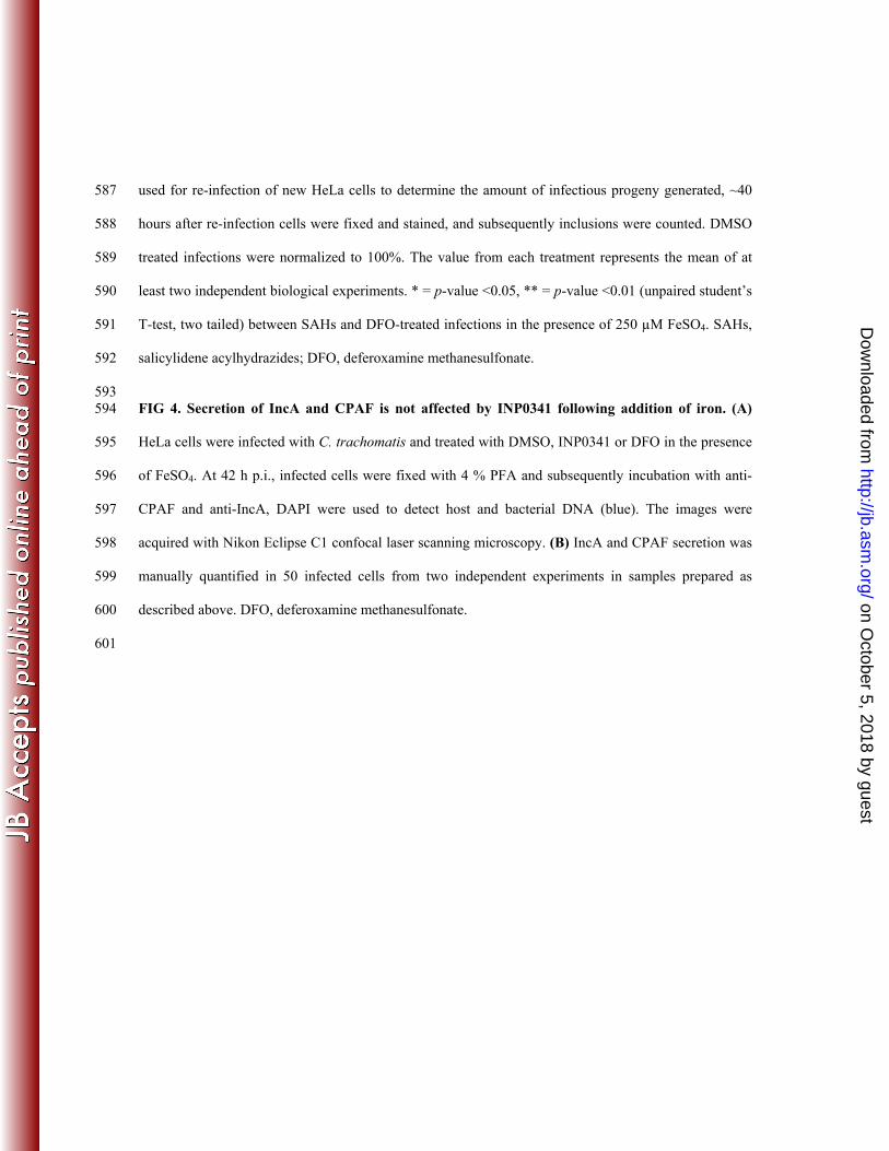
24. Gong S, Lei L, Chang X, Belland R, and Zhong G. 2011. Chlamydia trachomatis secretion of 458
hypothetical protein CT622 into host cell cytoplasm via a secretion pathway that can be inhibited 459
by the type III secretion system inhibitor compound 1. Microbiology 157:1134-1144. 460
25. Hobolt-Pedersen AS, Christiansen G, Timmerman E, Gevaert K, and Birkelund S. 2009. 461
Identification of Chlamydia trachomatis CT621, a protein delivered through the type III 462
secretion system to the host cell cytoplasm and nucleus. FEMS Immunol Med Microbiol 57:46-463
58. 464
26. Wu X, Lei L, Gong S, Chen D, Flores R, and Zhong G. 2011. The chlamydial periplasmic 465
stress response serine protease cHtrA is secreted into host cell cytosol. BMC Microbiol 11:87. 466
27. Prantner D, and Nagarajan UM. 2009. Role for the chlamydial type III secretion apparatus in 467
host cytokine expression. Infect Immun 77:76-84. 468
on October 5, 2018 by guest
http://jb.asm.org/
Dow
nloaded from
28. Slepenkin A, Enquist PA, Hägglund U, de la Maza LM, Elofsson M, and Peterson EM. 469
2007. Reversal of the antichlamydial activity of putative type III secretion inhibitors by iron. 470
Infect Immun 75:3478-3489. 471
29. Ur-Rehman T, Nordfelth R, Blomgren A, Zetterström CE, Elofsson M, and Gylfe Å. 2012. 472
Preliminary pharmacokinetics of the bacterial virulence inhibitor n'-(3,5-dibromo-2-hydroxy-473
benzylidenene)-nicotinic Acid hydrazide. Adv Exp Med Biol 954:349-356. 474
30. Slepenkin A, Chu H, Elofsson M, Keyser P, and Peterson EM. 2011. Protection of mice from 475
a Chlamydia trachomatis vaginal infection using a Salicylidene acylhydrazide, a potential 476
microbicide. J Infect Dis 204:1313-1320. 477
31. Dahlgren MK, Zetterström CE, Gylfe S, Linusson A, and Elofsson M. 2010. Statistical 478
molecular design of a focused salicylidene acylhydrazide library and multivariate QSAR of 479
inhibition of type III secretion in the Gram-negative bacterium Yersinia. Bioorg Med Chem 480
18:2686-2703. 481
32. Caldwell HD, Kromhout J, and Schachter J. 1981. Purification and partial characterization of 482
the major outer membrane protein of Chlamydia trachomatis. Infection and immunity 31:1161-483
1176. 484
33. Nguyen BD, and Valdivia RH. 2012. Virulence determinants in the obligate intracellular 485
pathogen Chlamydia trachomatis revealed by forward genetic approaches. Proc Natl Acad Sci U 486
S A 109:1263-1268. 487
34. Jorgensen I, Bednar MM, Amin V, Davis BK, Ting JP, McCafferty DG, and Valdivia RH. 488
2011. The Chlamydia protease CPAF regulates host and bacterial proteins to maintain pathogen 489
vacuole integrity and promote virulence. Cell Host Microbe 10:21-32. 490
35. Soding J, Biegert A, and Lupas AN. 2005. The HHpred interactive server for protein homology 491
detection and structure prediction. Nucleic Acids Res 33:W244-248. 492
on October 5, 2018 by guest
http://jb.asm.org/
Dow
nloaded from
36. Sali A, Potterton L, Yuan F, van Vlijmen H, and Karplus M. 1995. Evaluation of 493
comparative protein modeling by MODELLER. Proteins 23:318-326. 494
37. Gille C, and Frommel C. 2001. STRAP: editor for STRuctural Alignments of Proteins. 495
Bioinformatics 17:377-378. 496
38. DeLano WL, Warren, L.D. 2002. DeLano Scientific, San Carlos, California. Available 497
http://www.pymol.org. 498
39. Raulston JE. 1997. Response of Chlamydia trachomatis serovar E to iron restriction in vitro and 499
evidence for iron-regulated chlamydial proteins. Infect Immun 65:4539-4547. 500
40. Tietzel I, El-Haibi C, and Carabeo RA. 2009. Human guanylate binding proteins potentiate the 501
anti-chlamydia effects of interferon-gamma. PLoS One 4:e6499. 502
41. Keyser P, Elofsson M, Rosell S, and Wolf-Watz H. 2008. Virulence blockers as alternatives to 503
antibiotics: type III secretion inhibitors against Gram-negative bacteria. J Intern Med 264:17-29. 504
42. Subtil A, Parsot C, and Dautry-Varsat A. 2001. Secretion of predicted Inc proteins of 505
Chlamydia pneumoniae by a heterologous type III machinery. Mol Microbiol 39:792-800. 506
43. Balakrishnan A, Patel B, Sieber SA, Chen D, Pachikara N, Zhong G, Cravatt BF, and Fan 507
H. 2006. Metalloprotease inhibitors GM6001 and TAPI-0 inhibit the obligate intracellular human 508
pathogen Chlamydia trachomatis by targeting peptide deformylase of the bacterium. The Journal 509
of biological chemistry 281:16691-16699. 510
44. Sandoz KM, Eriksen SG, Jeffrey BM, Suchland RJ, Putman TE, Hruby DE, Jordan R, and 511
Rockey DD. 2012. Resistance to a novel antichlamydial compound is mediated through 512
mutations in Chlamydia trachomatis secY. Antimicrob Agents Chemother 56:4296-4302. 513
45. Connolly B, Parsons CA, Benson FE, Dunderdale HJ, Sharples GJ, Lloyd RG, and West 514
SC. 1991. Resolution of Holliday junctions in vitro requires the Escherichia coli ruvC gene 515
product. Proc Natl Acad Sci U S A 88:6063-6067. 516
on October 5, 2018 by guest
http://jb.asm.org/
Dow
nloaded from
46. Heinemann IU, Jahn M, and Jahn D. 2008. The biochemistry of heme biosynthesis. Arch 517
Biochem Biophys 474:238-251. 518
47. Stephens RS, Kalman S, Lammel C, Fan J, Marathe R, Aravind L, Mitchell W, Olinger L, 519
Tatusov RL, Zhao Q, Koonin EV, and Davis RW. 1998. Genome sequence of an obligate 520
intracellular pathogen of humans: Chlamydia trachomatis. Science 282:754-759. 521
48. Corradi HR, Corrigall AV, Boix E, Mohan CG, Sturrock ED, Meissner PN, and Acharya 522
KR. 2006. Crystal structure of protoporphyrinogen oxidase from Myxococcus xanthus and its 523
complex with the inhibitor acifluorfen. J Biol Chem 281:38625-38633. 524
49. Qin X, Sun L, Wen X, Yang X, Tan Y, Jin H, Cao Q, Zhou W, Xi Z, and Shen Y. 2010. 525
Structural insight into unique properties of protoporphyrinogen oxidase from Bacillus subtilis. J 526
Struct Biol 170:76-82. 527
50. Binet R, and Maurelli A. 2009. Transformation and isolation of allelic exchange mutants of 528
Chlamydia psittaci using recombinant DNA introduced by electroporation. Proc Natl Acad Sci U 529
S A 106:292-297. 530
51. Kari L, Goheen MM, Randall LB, Taylor LD, Carlson JH, Whitmire WM, Virok D, 531
Rajaram K, Endresz V, McClarty G, Nelson DE, and Caldwell HD. 2011. Generation of 532
targeted Chlamydia trachomatis null mutants. Proc Natl Acad Sci U S A 108:7189-7193. 533
52. Song L, Carlson JH, Whitmire WM, Kari L, Virtaneva K, Sturdevant DE, Watkins H, 534
Zhou B, Sturdevant GL, Porcella SF, McClarty G, and Caldwell HD. 2013. The Chlamydia 535
trachomatis plasmid-encoded Pgp4 is a transcriptional regulator of virulence associated genes. 536
Infect Immun. 537
53. Layton AN, Hudson DL, Thompson A, Hinton JC, Stevens JM, Galyov EE, and Stevens 538
MP. 2010. Salicylidene acylhydrazide-mediated inhibition of type III secretion system-1 in 539
on October 5, 2018 by guest
http://jb.asm.org/
Dow
nloaded from
Salmonella enterica serovar Typhimurium is associated with iron restriction and can be reversed 540
by free iron. FEMS Microbiol Lett 302:114-122. 541
54. Wang D, Zetterström CE, Gabrielsen M, Beckham KS, Tree JJ, Macdonald SE, Byron O, 542
Mitchell TJ, Gally DL, Herzyk P, Mahajan A, Uvell H, Burchmore R, Smith BO, Elofsson 543
M, and Roe AJ. 2011. Identification of bacterial target proteins for the salicylidene 544
acylhydrazide class of virulence-blocking compounds. J Biol Chem 286:29922-29931. 545
55. Veenendaal AK, Sundin C, and Blocker AJ. 2009. Small-molecule type III secretion system 546
inhibitors block assembly of the Shigella type III secreton. Journal of bacteriology 191:563-570. 547
56. Chin E, Kirker K, Zuck M, James G, and Hybiske K. 2012. Actin recruitment to the 548
Chlamydia inclusion is spatiotemporally regulated by a mechanism that requires host and 549
bacterial factors. PLoS One 7:e46949. 550
57. Mayfield JA, Dehner CA, and DuBois JL. 2011. Recent advances in bacterial heme protein 551
biochemistry. Curr Opin Chem Biol 15:260-266. 552
58. Panek H, and O'Brian MR. 2002. A whole genome view of prokaryotic haem biosynthesis. 553
Microbiology 148:2273-2282. 554
59. Boynton T, Daugherty L, Dailey T, and Dailey H. 2009. Identification of Escherichia coli 555
HemG as a novel, menadione-dependent flavodoxin with protoporphyrinogen oxidase activity. 556
Biochemistry 48:6705-6711. 557
60. Mobius K, Arias-Cartin R, Breckau D, Hannig AL, Riedmann K, Biedendieck R, Schroder 558
S, Becher D, Magalon A, Moser J, Jahn M, and Jahn D. 2010. Heme biosynthesis is coupled 559
to electron transport chains for energy generation. Proc Natl Acad Sci U S A 107:10436-10441. 560
561
562
563
on October 5, 2018 by guest
http://jb.asm.org/
Dow
nloaded from
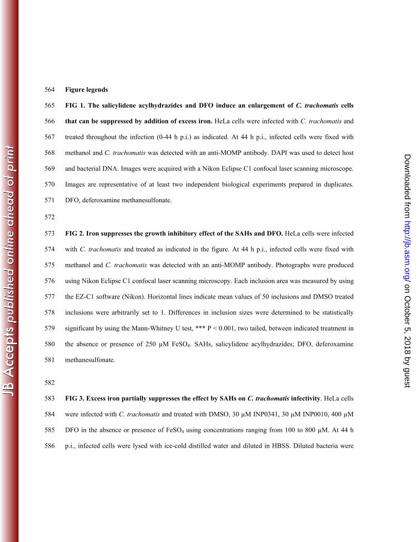
Figure legends 564
FIG 1. The salicylidene acylhydrazides and DFO induce an enlargement of C. trachomatis cells 565
that can be suppressed by addition of excess iron. HeLa cells were infected with C. trachomatis and 566
treated throughout the infection (0-44 h p.i.) as indicated. At 44 h p.i., infected cells were fixed with 567
methanol and C. trachomatis was detected with an anti-MOMP antibody. DAPI was used to detect host 568
and bacterial DNA. Images were acquired with a Nikon Eclipse C1 confocal laser scanning microscope. 569
Images are representative of at least two independent biological experiments prepared in duplicates. 570
DFO, deferoxamine methanesulfonate. 571
572
FIG 2. Iron suppresses the growth inhibitory effect of the SAHs and DFO. HeLa cells were infected 573
with C. trachomatis and treated as indicated in the figure. At 44 h p.i., infected cells were fixed with 574
methanol and C. trachomatis was detected with an anti-MOMP antibody. Photographs were produced 575
using Nikon Eclipse C1 confocal laser scanning microscopy. Each inclusion area was measured by using 576
the EZ-C1 software (Nikon). Horizontal lines indicate mean values of 50 inclusions and DMSO treated 577
inclusions were arbitrarily set to 1. Differences in inclusion sizes were determined to be statistically 578
significant by using the Mann-Whitney U test, *** P < 0.001, two tailed, between indicated treatment in 579
the absence or presence of 250 µM FeSO4. SAHs, salicylidene acylhydrazides; DFO, deferoxamine 580
methanesulfonate. 581
582
FIG 3. Excess iron partially suppresses the effect by SAHs on C. trachomatis infectivity. HeLa cells 583
were infected with C. trachomatis and treated with DMSO, 30 µM INP0341, 30 µM INP0010, 400 µM 584
DFO in the absence or presence of FeSO4 using concentrations ranging from 100 to 800 µM. At 44 h 585
p.i., infected cells were lysed with ice-cold distilled water and diluted in HBSS. Diluted bacteria were 586
on October 5, 2018 by guest
http://jb.asm.org/
Dow
nloaded from
used for re-infection of new HeLa cells to determine the amount of infectious progeny generated, ~40 587
hours after re-infection cells were fixed and stained, and subsequently inclusions were counted. DMSO 588
treated infections were normalized to 100%. The value from each treatment represents the mean of at 589
least two independent biological experiments. * = p-value <0.05, ** = p-value <0.01 (unpaired student’s 590
T-test, two tailed) between SAHs and DFO-treated infections in the presence of 250 µM FeSO4. SAHs, 591
salicylidene acylhydrazides; DFO, deferoxamine methanesulfonate. 592
593 FIG 4. Secretion of IncA and CPAF is not affected by INP0341 following addition of iron. (A) 594
HeLa cells were infected with C. trachomatis and treated with DMSO, INP0341 or DFO in the presence 595
of FeSO4. At 42 h p.i., infected cells were fixed with 4 % PFA and subsequently incubation with anti-596
CPAF and anti-IncA, DAPI were used to detect host and bacterial DNA (blue). The images were 597
acquired with Nikon Eclipse C1 confocal laser scanning microscopy. (B) IncA and CPAF secretion was 598
manually quantified in 50 infected cells from two independent experiments in samples prepared as 599
described above. DFO, deferoxamine methanesulfonate. 600
601
on October 5, 2018 by guest
http://jb.asm.org/
Dow
nloaded from
FIG 5. Strategy to select and isolate C. trachomatis mutant strains. (A) HeLa cells infected with 602
wild-type C. trachomatis LGV-L2 were treated with INP0341 in the presence of exogenously added iron 603
sulfate (250 µM). At 44-48 h p.i., infected cells were lysed with ice cold water to release infectious EB 604
progeny which were used to infect fresh HeLa cells for a new round of selection. This procedure was 605
repeated and selection was continued for up to 20 passages. Notably the selected C. trachomatis mutant 606
population had increased RB-to-EB transition in the presence of INP0341 and iron sulfate. 1) EB-to-RB 607
transition. 2) C. trachomatis growth. (B) Wild-type C. trachomatis LGV-L2 was serially passaged as 608
described above. Note that the first mutant population (passage 12) was further grown in both the 609
presence and absence of selective pressure for serial passages. At indicated mutant passages bacterial 610
DNA were purified and subjected for whole genome sequencing (WGS) or genotyped by capillary 611
sequence. Identified mutations via WGS were verified with capillary sequencing. Infectious EB progeny 612
from respective mutant populations were used to infect Vero cells for the formations of plaques. Formed 613
plaques were amplified in HeLa cells and the clonal C. trachomatis strains were subsequently genotyped 614
and characterized. Vf, variant frequency; evf, estimated variant frequency. 615
616 FIG 6. Mutant strains are resistant to IS-INP0341 but not to iron limitation induced by INP0341. 617
(A) HeLa cells were infected with indicated C. trachomatis strains at a MOI of 0.1 and subsequently 618
treated with 30 µM IS-INP0341 or 10 µM non-saturated INP0341, which inhibits C. trachomatis 619
development similarly as 30 µM IS-INP0341 and therefore was used to analyze the mutant strains in a 620
comparable susceptibility-assay. In parallel each strain was titrated to normalize the infectivity to the 621
exact number of inputs IFUs. Infectious progeny was collected at 48 h p.i., and subsequently used for re-622
infection. Data presented was acquired from an experiment performed in triplicate which is 623
representative of at least two independent experiments. The treated wild-type set was set to 1 for each 624
treatment. ** = p-value <0.01, *** = p-value <0.001 (unpaired student’s T-test, two tailed) between 625
on October 5, 2018 by guest
http://jb.asm.org/
Dow
nloaded from

treated wild-type and mutant strains, for each treatment. (B) Morphological analysis after treatment with 626
DMSO only (upper) or 30 µM IS-INP0341 (lower) using transmission electron microscopy. Infected 627
cells (MOI: 1) were fixed at 44 h p.i. and processed according to the description in the material and 628
methods. RB, reticulate bodies; EB, elementary bodies; IB, intermediate bodies; AB, aberrant bodies. 629
IS-INP0341, iron-saturated INP0341. 630
631
FIG 7. Predicted structure of C. trachomatis HemG. (A) Protoporphyrinogen oxidase (PPO) from 632
Myxococcus xantus was used as a template to model C. trachomatis HemG (CtHemG). (B) 633
Protoporphyrinogen oxidase (PPO) from Bacilus Subtilis (BsPPO) and the predicted structure of 634
CtHemG was overlayed. (C) BsPPO Co-crystalized with FAD and the inhibitor AF, the latter which 635
binds to the active site of BsPPO. Red residues in CtHemG indicate where IS-INP0341 resistant 636
substitutions are localized and corresponding residues in BsPPO are indicated in black. (D) Structural 637
alignment of CtHemG, BsPPO, MxPPO, and WrbA, regarded as a potential target of SAHs (54). 638
Notably all four proteins contain a FAD binding domain (the first ~55 amino acids). 639
on October 5, 2018 by guest
http://jb.asm.org/
Dow
nloaded from
Table 1. Isolated mutant strains
C. trachomatis straina
Amino acid substitution(s)
Mutants with indicated substitution(s)
Origin (Passage)
Mutant 1 HemGG58S, RuvCH31Y 5/6 Mutant population-1 (P12)
Mutant 2 HemGG58S 2/5 Expanded mutant population-1 (P20)
Mutant 3 HemGR91C, RpsFA54D 2/4 Mutant population-2 (P15) aPlaque-purified strains isolated from indicated mutant populations, genotyped by capillary sequencing.
on October 5, 2018 by guest
http://jb.asm.org/
Dow
nloaded from


![Cis-encoded sRNAs, a conserved mechanism for …jb.asm.org/content/early/2016/06/21/JB.00381-16.full.pdf · 3 39 ,psruwdqfh 40 7khkxpdqjxwlvfrorql]hge\d ghqvhplfurelrwdzklfklvhvvhq](https://img.pdfslide.us/doc/110x75/5b2f6bc77f8b9a91438cd637/cis-encoded-srnas-a-conserved-mechanism-for-jbasmorgcontentearly20160621jb00381-16fullpdf.jpg)