Embed Size (px)
Citation preview
Jack’s Complete Anatomy Essays
essentialgross anatomy,
histology & embryologyfor medicine year one
second edition
Jack’s Complete Anatomy Essays 0
Limbs Simple spinal reflex arc 1 Shoulder joint 1 Axillary nerve 3 Cubital fossa 3 Carpal tunnel 4 Median nerve 5 Ulnar nerve 6 Radial nerve 8 Hip joint 10 Avascular necrosis of femoral head 11 Sciatic nerve 12 Knee joint 13 Common peroneal nerve 14 Venous drainage of lower limb 16 Thorax Level of sternal angle 19 Lungs 20 Heart 21 Arch of aorta 25 Oesophagus 26 Ribs & Diaphragm 28 Abdomen Inguinal canal & Hernias 30 Stomach 32 Jejunum vs. Ileum 36 Small vs. Large intestines 37 Midgut & Superior mesenteric artery 38 Large intestine 39 Appendix 41 Liver 42 Porto-systemic anastomoses 43 Extrahepatic biliary tree 44 Kidney 46 Ureter 49
Pelvis & Perineum Pelvic diaphragm 50 Uterus 51 Vagina 53 Urinary bladder 54 Prostate gland 56 Male urethra 57 Rectum & Anal canal 58 Ischiorectal fossae 60 Pudendal nerve 61 Head & Neck Sternocleidomastoid 62 Thyroid gland 63 Parathyroid glands 65 Intracranial haemorrhage 66 Cavernous sinus 68 Third nerve palsy 69 Fourth nerve palsy 69 Sixth nerve palsy 70 Parotid gland 70 Facial nerve 71 Temporomandibular joint 73 Submandibular gland 74 Horner’s syndrome 76 Scalenus anterior 77 Nasal cavity 77 Cleft lip & Cleft palate 79 Nasopharynx 80 Larynx 82 Deglutition 83
-JJJ- First published 2004 Second edition 2006 The author is indebted to Miss Tan Poh Yong of Class 2005-2010 for reviewing the first edition and contributing to its revision. Any future revision of this essay collection, for the benefit of your juniors, depends on your valuable feedback. You may email your comments to [email protected].
Jack’s Complete Anatomy Essays 1
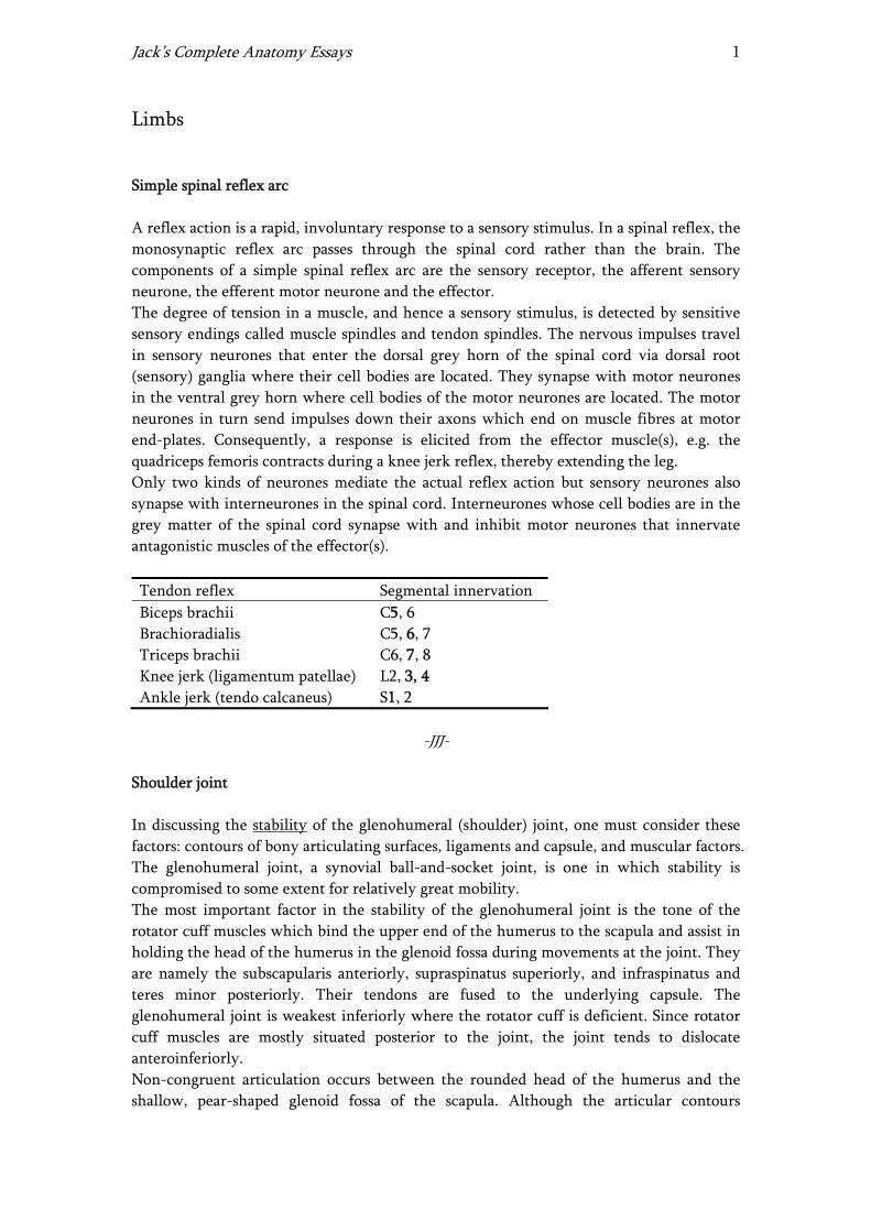
Limbs Simple spinal reflex arc A reflex action is a rapid, involuntary response to a sensory stimulus. In a spinal reflex, the monosynaptic reflex arc passes through the spinal cord rather than the brain. The components of a simple spinal reflex arc are the sensory receptor, the afferent sensory neurone, the efferent motor neurone and the effector. The degree of tension in a muscle, and hence a sensory stimulus, is detected by sensitive sensory endings called muscle spindles and tendon spindles. The nervous impulses travel in sensory neurones that enter the dorsal grey horn of the spinal cord via dorsal root (sensory) ganglia where their cell bodies are located. They synapse with motor neurones in the ventral grey horn where cell bodies of the motor neurones are located. The motor neurones in turn send impulses down their axons which end on muscle fibres at motor end-plates. Consequently, a response is elicited from the effector muscle(s), e.g. the quadriceps femoris contracts during a knee jerk reflex, thereby extending the leg. Only two kinds of neurones mediate the actual reflex action but sensory neurones also synapse with interneurones in the spinal cord. Interneurones whose cell bodies are in the grey matter of the spinal cord synapse with and inhibit motor neurones that innervate antagonistic muscles of the effector(s). Tendon reflex Segmental innervation Biceps brachii C5, 6 Brachioradialis C5, 6, 7 Triceps brachii C6, 7, 8 Knee jerk (ligamentum patellae) L2, 3, 4 Ankle jerk (tendo calcaneus) S1, 2
-JJJ-
Shoulder joint In discussing the stability of the glenohumeral (shoulder) joint, one must consider these factors: contours of bony articulating surfaces, ligaments and capsule, and muscular factors. The glenohumeral joint, a synovial ball-and-socket joint, is one in which stability is compromised to some extent for relatively great mobility. The most important factor in the stability of the glenohumeral joint is the tone of the rotator cuff muscles which bind the upper end of the humerus to the scapula and assist in holding the head of the humerus in the glenoid fossa during movements at the joint. They are namely the subscapularis anteriorly, supraspinatus superiorly, and infraspinatus and teres minor posteriorly. Their tendons are fused to the underlying capsule. The glenohumeral joint is weakest inferiorly where the rotator cuff is deficient. Since rotator cuff muscles are mostly situated posterior to the joint, the joint tends to dislocate anteroinferiorly. Non-congruent articulation occurs between the rounded head of the humerus and the shallow, pear-shaped glenoid fossa of the scapula. Although the articular contours
Jack’s Complete Anatomy Essays 2
contribute poorly to the stability of the joint, deepening of the glenoid fossa by the glenoid labrum, a fibrocartilaginous rim, increases stability. Weak ligaments provide little support to the glenohumeral joint. The superior, middle and inferior glenohumeral ligaments are three weak bands of fibrous tissue that strengthen the capsule anteriorly. The coracohumeral ligament strengthens the capsule superiorly while the coracoacromial ligament (an accessory ligament) protects the joint superiorly. The transverse humeral ligament strengthens the capsule and bridges the bicipital groove. Thin and lax, the capsule allows a wide range of movement at the joint. It surrounds the joint and is attached medially to the margin of the glenoid cavity outside the labrum and laterally to the anatomic neck of the humerus. Endoscopic features of the glenohumeral joint are:
- the cartilage-lined articular surfaces between the rounded head of the humerus and the shallow, pear-shaped glenoid fossa of the scapula
- the synovial membrane lining the capsule and attached to the margins of the articular cartilage
- the capsule and the opening in the capsule which leads into the subscapularis bursa. The synovial membrane extends through the anterior wall of the capsule to form this bursa.
- the tendon of the long head of biceps brachii from the supraglenoid tubercle of the scapula and around which the synovial membrane forms a tubular sheath, i.e., the tendon is intracapsular but extrasynovial
- the ligaments of the joint The movements at the glenohumeral joint are:
- Flexion is normally about 90o and is performed by the anterior fibres of deltoid, pectoralis major, biceps brachii and coracobrachialis.
- Extension is normally about 45o and is performed by the posterior fibres of deltoid, latissimus dorsi and teres major.
- Abduction of the upper limb occurs both at the shoulder joint and between the scapula and the thoracic wall. The supraspinatus initiates abduction and holds the head of the humerus against the glenoid fossa of the scapula; this latter function allows the middle fibres of deltoid to contract and abduct the humerus at the glenohumeral joint.
- Adduction is performed by pectoralis major, latissimus dorsi and teres major and minor. Normally, the upper limb can be swung 45o across the front of the chest.
- Lateral rotation is normally 40o to 45o and is performed by the infraspinatus, teres minor and the posterior fibres of deltoid.
- Medial rotation is normally about 55o and is performed by the subscapularis, latissimus dorsi, teres major and the anterior fibres of deltoid.
- Circumduction is a combination of the abovementioned movements. For every 3o of abduction of the arm, 2o occurs in the glenohumeral joint and 1o by rotation of the scapula. The supraspinatus initiates abduction for the first 15o and stabilises the head of the humerus against the glenoid fossa of the scapula. This allows the middle fibres of the deltoid to take over and continue the movement of abduction until an angle
Jack’s Complete Anatomy Essays 3
of 120o is reached. At this angle, the greater tuberosity of the humerus is jammed against the lateral edge of the acromion. Elevation of the arm to a fully vertical (180o) position above the head is accomplished by rotation of the scapula by the trapezius (superior and inferior fibres) and serratus anterior. If the supraspinatus tendon is ruptured but the arm is assisted passively for the first 15o of abduction, the deltoid can take over and complete the movement to a right angle.
-JJJ- Axillary nerve The axillary (circumflex) nerve arising from the posterior cord of the brachial plexus (C5 and 6) in the axilla passes backwards and enters the quadrangular space with the posterior circumflex humeral artery. As it passes through the space, it comes into close relationship with the inferior aspect of the capsule of the shoulder joint and with the medial side of the surgical neck of the humerus. It terminates by dividing into anterior and posterior branches. Its branches are:
- an articular branch to the shoulder joint - an anterior terminal branch which winds around the surgical neck of the humerus
beneath the deltoid and supplies the deltoid and the skin covering its lower half - a posterior terminal branch which gives off a branch to teres minor and a few
branches to deltoid, then emerges from the posterior border of the deltoid as the upper lateral cutaneous nerve of the arm
Injury in shoulder dislocations or fractures of the surgical neck of the humerus Motor: The deltoid and teres minor are paralysed. The paralysed deltoid wastes rapidly and the underlying greater tuberosity can be readily palpated. Since the supraspinatus is the only other abductor of the shoulder, abduction is much impaired. Paralysis of teres minor is not clinically recognisable. Sensory: Skin sensation is lost over the lower half of the deltoid, i.e., the lateral aspect of the upper arm (the ‘regimental badge’ area).
-JJJ- Cubital fossa The cubital fossa is a triangular depression lying in front of the elbow. Its boundaries are the brachioradialis laterally and the pronator teres medially. The base of the triangle is formed by an imaginary line between the two epicondyles of the humerus. The floor of the fossa is formed by the supinator laterally and the brachialis medially. The roof is formed by skin and fascia and is reinforced by the bicipital aponeurosis.

Jack’s Complete Anatomy Essays 4
The cubital fossa contains the following structures, enumerated from medial to lateral: the median nerve, the bifurcation of the brachial artery into the ulnar and radial arteries, the tendon of biceps brachii, and the radial nerve and its deep branch. Superficially, in the superficial fascia overlying the fossa are: the median cubital vein lying anterior to the brachial artery, and the median and lateral antebrachial cutaneous nerves related the basilic and cephalic veins respectively. The deep and superficial branches of the radial nerve are within the floor of the fossa.
-JJJ- Carpal tunnel The carpal tunnel, formed by the concave anterior surface of the carpal bones posteriorly and closed by the flexor retinaculum anteriorly, is tightly packed with the long flexor muscles of the digits with their surrounding synovial sheaths and the median nerve. The flexor retinaculum, a thickening of deep fascia, stretches across the front of the wrist; it is attached medially to the pisiform bone and the hook of the hamate, and laterally to the tubercle of the scaphoid and the trapezium bone. The median nerve passes beneath the flexor retinaculum in a restricted space between flexor digitorum superficialis and flexor carpi radialis. The four separate tendons of flexor digitorum superficialis are arranged in anterior and posterior rows, those to the middle and ring fingers lying anterior to those to the index and little fingers. At the lower border of the flexor retinaculum, they diverge and become arranged on the same plane. The tendons of flexor digitorum profundus are on the same plane and lie behind the superficialis tendons. All eight tendons of flexor digitorum superficialis and profundus invaginate a common synovial sheath from the lateral side. This allows the arterial supply to the tendons to enter them from the lateral side. The tendon of flexor pollicis longus runs through the lateral part of the tunnel in its own synovial sheath. The carpal tunnel syndrome is produced by the compression of the median nerve within the tunnel. The median nerve has two terminal sensory branches that supply the skin of the hand. Hence paraesthesia, hypoaesthesia or anaesthesia may occur in the lateral 3½ digits. The nerve also has one terminal motor branch which supplies the three thenar muscles. Progressive loss of coordination and strength in the thumb may occur due to weakness of abductor pollicis brevis and opponens pollicis. The patient may be unable to oppose the thumb. As the condition progresses, sensory changes radiate into the forearm and axilla. No paraesthesia occurs over the thenar eminence as this area of skin is supplied by the palmar cutaneous branch of the median nerve which arises proximal to the carpal tunnel and passes superficially to the flexor retinaculum.
-JJJ-
Jack’s Complete Anatomy Essays 5
Median nerve The lateral root of the median nerve is the direct continuation of the lateral cord of the brachial plexus. The medial root of the median nerve arises from the medial cord of the brachial plexus and crosses in front of the third part of the axillary artery to join the lateral root of the median nerve. The median nerve trunk (C5, 6, 7, 8, T1) passes downwards on the lateral sides of the axillary artery and its inferior continuation, the brachial artery. The median nerve has no branches in the axilla. Halfway down the upper arm, the median nerve crosses the brachial artery and continues on its medial side. The nerve, like the artery, is thus superficial but at the elbow, it is crossed by the bicipital aponeurosis. It has no branches in the upper arm, except for a small vasomotor nerve to the brachial artery. The median nerve leaves the cubital fossa by passing between the two heads of pronator teres. It continues downwards behind flexor digitorum superficialis and rests posteriorly on flexor digitorum profundus. In the anterior compartment of the forearm, the median nerve gives off:
- muscular branches in the cubital fossa to pronator teres, flexor carpi radialis, palmaris longus and flexor digitorum superficialis
- articular branches to the elbow joint - anterior interosseous nerve which has muscular branches to flexor pollicis longus,
pronator quadratus and the lateral half of flexor digitorum profundus - palmar cutaneous branch crossing anterior to the flexor retinaculum and
distributed to the skin over the lateral part of the palm At the wrist, the median nerve emerges from the lateral border of flexor digitorum superficialis and lies behind the tendon of palmaris longus. It enters the palm by passing behind the flexor retinaculum and through the carpal tunnel. The median nerve immediately divides into lateral and medial branches. The muscular branch supplies muscles of the thenar eminence, namely abductor pollicis brevis, flexor pollicis brevis and opponens pollicis, as well as the first lumbrical. The cutaneous branches supply the palmar aspect of the lateral 3½ digits and the distal half of the dorsal aspect of these digits; one of these branches also supplies the second lumbrical. Injury at the elbow Motor: The pronator muscles of the forearm and the long flexors of the wrist and digits, except flexor carpi ulnaris and the medial half of flexor digitorum profundus, are paralysed. The forearm is kept in the supine position; wrist flexion is weak and accompanied by adduction due to the paralysis of flexor carpi radialis and the unopposed action of flexor carpi ulnaris. The first two lumbricals are paralysed. The interphalangeal joints of the index and middle fingers cannot be flexed although weak flexion of the metacarpophalangeal joints of these fingers is attempted by the interossei. When the patient tries to make a fist, the index and, to a lesser extent, the middle fingers tend to remain straight whereas the ring and little fingers flex (but are weakened by paralysis of flexor digitorum superficialis).
Jack’s Complete Anatomy Essays 6
Flexion of the terminal phalanx of the thumb is lost due to paralysis of flexor pollicis longus. Muscles of the thenar eminence are paralysed and wasted so that the eminence is flattened. The thumb is laterally rotated and adducted. The hand looks flattened and ‘apelike’. Sensory: Skin sensation is lost on the lateral half or less of the palm of the hand, the palmar aspect of the lateral 3½ digits and the distal part of the dorsal surfaces of these digits. Vasomotor changes: The skin areas involved in sensory loss are warmer and drier than normal due to vasodilation and anhidrosis caused by loss of sympathetic vasoconstrictive and sudomotor control. Injury at the wrist Motor: Flexion of the terminal phalanx of the thumb is lost due to paralysis of flexor pollicis longus. Muscles of the thenar eminence are paralysed and wasted so that the eminence is flattened. The thumb is laterally rotated and adducted. The hand looks flattened and ‘apelike’. Opposition of the thumb and hence the delicate pincer-like action of the hand are lost following the paralysis of opponens pollicis. The first two lumbricals are paralysed; this is recognized clinically when the patient is asked to make a fist slowly, and the index and middle fingers tend to lag behind the ring and little fingers. Sensory: Skin sensation is lost on the lateral half or less of the palm of the hand, the palmar aspect of the lateral 3½ digits and the distal part of the dorsal surfaces of these digits. Vasomotor changes: The skin areas involved in sensory loss are warmer and drier than normal due to vasodilation and anhidrosis caused by loss of sympathetic vasoconstrictive and sudomotor control.
-JJJ- Ulnar nerve The ulnar nerve arising from the medial cord of the brachial plexus (C8 and T1) has no cutaneous or motor branches in the axilla or in the arm. In the axilla, it descends in the interval between the axillary artery and vein. It runs downwards on the medial side of the brachial artery as far as the middle of the arm. Here, at the insertion of the coracobrachialis, the nerve pierces the medial fascial septum, accompanied by the superior ulnar collateral artery. It enters the posterior compartment of the arm where it descends behind the septum covered posteriorly by the medial head of triceps brachii. The nerve passes in a groove behind the medial epicondyle of the humerus and crosses the medial ligament of the elbow joint. It has no branches in the anterior
Jack’s Complete Anatomy Essays 7
compartment of the upper arm but has an articular branch to the elbow joint in the posterior compartment. The nerve continues downwards to enter the forearm between the two heads of origin of flexor carpi ulnaris. It runs down the forearm between flexor carpi ulnaris and flexor digitorum profundus. In the distal two-thirds of the forearm, the ulnar artery lies on the lateral side of the ulnar nerve. At the wrist, the ulnar nerve becomes superficial and lies between the tendons of the flexor carpi ulnaris and flexor digitorum superficialis. The ulnar nerve enters the palm of the hand by passing in front of the flexor retinaculum and lateral to the pisiform bone. Here it has the ulnar artery lateral to it. In the anterior compartment of the forearm, the ulnar nerve gives off:
- muscular branches to flexor carpi ulnaris and medial half of the flexor digitorum profundus
- articular branches to the elbow joint - palmar cutaneous branch crossing anterior to the flexor retinaculum and
supplying skin over the hypothenar eminence - dorsal posterior cutaneous branch distributed to the posterior surface of the hand
and fingers As the ulnar nerve crosses the flexor retinaculum, it divides into superficial and deep terminal branches. The superficial branch gives off a muscular branch to palmaris brevis, and cutaneous branches to the palmar aspect of the medial side of the little finger and the adjacent sides of the little and ring fingers; it also supplies the distal half of the dorsal aspect of these fingers. The deep branch gives off muscular branches to the three muscles of the hypothenar eminence, namely abductor digiti minimi, flexor digiti minimi and opponens digiti minimi; all palmar and dorsal interossei; third and fourth lumbricals; and adductor pollicis. Injury at the elbow Motor: The flexor carpi ulnaris and the medial half of flexor digitorum profundus are paralysed and waste away, causing flattening of the medial border of the front of the forearm. Flexion of the wrist joint results in abduction due to the unopposed action of flexor carpi radialis. The profundus tendons to the ring and little fingers will be functionless and the terminal phalanges of these fingers cannot be markedly flexed. The small muscles of the hand, except the muscles of the thenar eminence and the first two lumbricals (supplied by median nerve), are paralysed. The patient is unable to adduct and abduct the fingers due to paralysis of the interossei, or adduct the thumb due to paralysis of adductor pollicis. If asked to grip a piece of paper between the thumb and index finger, he/she does so by strong contraction of the flexor pollicis longus and flexion of the terminal phalanx (Froment’s sign). Paralysis of the third and fourth lumbricals and the interossei, which normally flex the metacarpophalangeal joints and extend the interphalangeal joints through the extensor expansion, causes:
- the metacarpophalangeal joints to be hyper-extended, most prominently in the ring and little fingers
Jack’s Complete Anatomy Essays 8
- the interphalangeal joints to be flexed, more obviously in the ring and little fingers
In long-standing cases, the hand assumes the characteristic ‘claw’ deformity. Wasting of the paralysed muscles result in flattening of the hypothenar eminence and loss of the convex curve of the medial border of the hand. Wasting of the dorsal interossei causes hollowing between the metacarpal bones on the dorsum of the hand. Sensory: Skin sensation is lost over the anterior and posterior surfaces of the medial third of the hand and the medial 1½ fingers. Vasomotor changes: The skin areas involved in sensory loss are warmer and drier than normal due to vasodilation and anhidrosis caused by loss of sympathetic vasoconstrictive and sudomotor control. Injury at the wrist Motor: The small muscles of the hand, except the muscles of the thenar eminence and the first two lumbricals (supplied by median nerve), are paralysed. The claw hand is very obvious as the flexor digitorum profundus is not paralysed and marked flexion of the terminal phalanges occurs. This gives rise to the phenomenon known as ‘ulnar paradox’ whereby a lesion of the ulnar nerve at the wrist results in a more severe-looking claw hand than a lesion at the elbow or above (which gives straighter fingers), belying the fact that the wrist lesion paralyses fewer muscles. Sensory: The main ulnar nerve and its palmar cutaneous branch are usually severed while the dorsal posterior cutaneous branch arising from the ulnar nerve trunk above the pisiform bone escapes lesion. Loss of skin sensation is therefore confined to the palmar surface of the medial third of the hand and the medial 1½ fingers, and to the dorsal aspects of the middle and distal phalanges of the same fingers. Vasomotor changes: The skin areas involved in sensory loss are warmer and drier than normal due to vasodilation and anhidrosis caused by loss of sympathetic vasoconstrictive and sudomotor control.
-JJJ- Radial nerve The radial nerve (C5, 6, 7, 8, T1), the largest branch of the brachial plexus arising from the posterior cord, lies behind the axillary artery. It gives off muscular branches to the long and medial heads of triceps brachii, and the posterior cutaneous nerve of the arm which is distributed to the skin on the middle of the back of the arm.
Jack’s Complete Anatomy Essays 9
On leaving the axilla, the nerve winds around the back of the arm in the spiral groove on the back of the humerus between the heads of triceps brachii. In the spiral groove, the nerve is accompanied by the profunda brachii artery and lies directly in contact with the shaft of the humerus. Muscular branches are given to the lateral and medial heads of triceps brachii and to anconeus. The lower lateral cutaneous nerve of the arm supplies the skin over the lateral and anterior aspects of the lower part of the arm. The posterior cutaneous nerve of the forearm runs down the middle of the back of the forearm as far as the wrist. The radial nerve pierces the lateral fascial septum in the lower part of the arm and passes forwards into the cubital fossa. It then passes downwards in front of the lateral epicondyle of the humerus, lying between brachialis medially and brachioradialis and extensor carpi radialis longus laterally. At the level of the lateral epicondyle, it divides into superficial and deep branches. The radial nerve also gives off muscular branches to the lateral part of brachialis, brachioradialis and extensor carpi radialis longus, and articular branches to the elbow joint. The superficial branch is the direct continuation of the radial nerve and runs down under cover of brachioradialis on the lateral side of the radial artery. In the distal part of the forearm, it leaves the artery and passes backwards under the tendon of brachioradialis. It reaches the posterior surface of the wrist where it divides into terminal branches that supply the skin on the lateral two-thirds of the posterior surface of the hand and the posterior surface over the proximal phalanges of the lateral 3½ digits. The area of skin supplied by the superficial branch on the dorsum of the hand is variable. The deep branch pierces the supinator and winds around the lateral aspect of the neck of the radius in the substance of the muscle to reach the posterior compartment of the forearm. It descends in the interval between the superficial and deep groups of muscles, eventually reaching the posterior surface of the wrist joint. It gives off muscular branches to extensor carpi radialis brevis, supinator, extensor digitorum, extensor digiti minimi, extensor carpi ulnaris, abductor pollicis longus, extensor pollicis brevis and longus, and extensor indicis, as well as articular branches to the wrist and carpal joints. Injury in the axilla Motor: The triceps brachii, anconeus and long extensors of the wrist are paralysed. The patient cannot extend the wrist joint and digits. Wrist drop occurs due to the unopposed action of flexor muscles of the wrist. With wrist drop, one cannot flex the digits strongly to firmly grip an object. The brachioradialis and supinator muscles are also paralysed but supination is still performed well by the unaffected biceps brachii. Sensory: A small loss of skin sensation occurs down the posterior surface of the lower part of the arm and down a narrow strip on the back of the forearm. A variable area of sensory loss is present on the lateral part of the dorsum of the hand and the dorsal surface of the roots of the lateral 3½ digits.
Jack’s Complete Anatomy Essays 10
Injury in the spiral groove Motor: The anconeus and long extensors of the wrist are paralysed. The patient cannot extend the wrist joint and digits. Wrist-drop occurs due to the unopposed action of flexor muscles of the wrist. With wrist-drop, one cannot flex the digits strongly to firmly grip an object. Sensory: A variable area of sensory loss is present on the lateral part of the dorsum of the hand and the dorsal surface of the roots of the lateral 3½ digits.
-JJJ- Hip joint In discussing the stability of the hip joint, one must consider these factors: contours of bony articulating surfaces, ligaments and capsule, and muscular factors. The hip joint, a synovial ball-and-socket joint, is one in which mobility is compromised to some extent for relatively great stability. That said, the hip joint still has a wide range of movement albeit less than the glenohumeral joint. The most important factors in the stability of the hip joint are the congruent articulation between the bones taking part in the joint, and the strong ligaments. Articulation is between the head of the femur, shaped like two-thirds of a sphere, and the cup-shaped acetabulum of the hip (inominate) bone. Moreover, the deepening of the cavity of the acetabulum by the acetabular labrum, a fibrocartilaginous rim, enhances the stability of the joint. The horseshoe-shaped articular surface of the acetabulum is deficient inferiorly at the acetabular notch which is bridged by the transverse acetabular ligament. Hence the hip joint is more stable superiorly than inferiorly. Most of the ligaments of the hip joint limit certain movements at the joint. All the ligaments are listed below:
- the strong, inverted Y-shaped iliofemoral ligament which prevents overextension during standing and limits lateral rotation
- the triangular pubofemoral ligament which limits extension, abduction and lateral rotation
- the spiral-shaped ischiofemoral ligament which limits extension and medial rotation
- the transverse acetabular ligament formed by the acetabular labrum as it bridges the acetabular notch
- the ligament of the head of the femur (ligamentum teres) which lies within the joint and whose tension limits adduction. It is weak and of little importance in strengthening the joint.
The capsule encloses the hip joint and is attached medially to the acetabular labrum. Laterally, it is attached to the intertrochanteric line of the femur in front and halfway along the posterior aspect of the femoral neck behind. The capsule plays a minor role in stabilising the hip joint.

Jack’s Complete Anatomy Essays 11
Many muscles surround and move the hip joint but their role in stabilising the hip joint is subordinate to the primary role that the rotator cuff plays in stabilising the glenohumeral joint. Unlike the rotator cuff muscles, their tendons are not fused to the capsule of the hip joint. However, to stabilise the hip joint when a person stands on one leg with the foot of the opposite leg raised above ground, the glutei medius and minimus must be functional. Traumatic hip dislocation is typically posterior and happens when the joint is flexed and adducted, e.g. dashboard impact is transmitted up the femoral shaft of a car passenger involved in a road traffic accident. The femoral head is displaced posteriorly out of the acetabulum onto the gluteal surface of the ilium; in some cases, the posterior lip of the acetabulum is fractured. As such, the sciatic nerve which is closely related to the posterior surface of the hip joint is susceptible to injury. The patient will present with his/her hip held slightly flexed, adducted and internally rotated. The movements at the hip joint are:
- Flexion is performed by iliopsoas, sartorius, rectus femoris, pectineus, adductor longus and brevis, adductor fibres of adductor magnus, (tensor fasciae latae and gracilis).
- Extension (backward movement of flexed thigh) is performed by gluteus maximus, long head of biceps femoris, semitendinosus, semimembranosus and the hamstring portion of adductor magnus.
- Abduction is performed by glutei medius and minimus, and assisted by sartorius, tensor fasciae latae and piriformis.
- Adduction is performed by adductor longus and brevis, and the adductor fibres of adductor magnus, and assisted by gracilis, pectineus (and obturator externus).
- Lateral rotation is performed by piriformis, obturator internus and externus, superior and inferior gemelli, and quadratus femoris, and assisted by gluteus maximus (and sartorius).
- Medial rotation is performed by the anterior fibres of gluteus medius, gluteus minimus and tensor fasciae latae.
- Circumduction is a combination of the abovementioned movements.
-JJJ- Avascular necrosis of femoral head The sources of blood supply to the head of the femur are: retinacular vessels from the trochanteric anastomosis and artery to the head of the femur; bone marrow of the femur may also contribute to this blood supply. The trochanteric anastomosis lying near the trochanteric fossa provides the main blood supply to the femoral head, mainly via branches of the medial circumflex femoral artery. The superior and inferior gluteal arteries, and medial and lateral circumflex femoral arteries take part in the anastomosis. Nutrient arteries arising from the anastomosis pass along the femoral neck beneath the retinacular fibres of the hip joint capsule. Retinacular fibres are reflections from the attachment of the hip joint capsule along the femoral neck to the articular margin of the head.
Jack’s Complete Anatomy Essays 12
The artery to the head of the femur is a small branch of the obturator artery and enters the femoral head at the fovea capitis along the ligament of the head (ligamentum teres). In the adult, an anastomosis is established between these two sources of blood supply after the epiphyseal cartilage – whose presence in childhood intervenes between and separates the blood sources – disappears. An intracapsular fracture of the femoral neck necessarily ruptures the retinacular fibres and vessels. The scant blood flow along the artery to the head of femur may be insufficient to sustain the viability of the femoral head, and avascular necrosis gradually ensues.
-JJJ- Sciatic nerve The sciatic nerve is a branch of the sacral plexus (L4, 5, S1, 2, 3). It emerges from the pelvis through the inferior part of the greater sciatic foramen, appearing below the piriformis muscle. It then curves inferolaterally, lying consecutively on the root of ischial sppine, the superior gemellus, the obturator internus, the inferior gemellus and the quadratus femoris to reach the posterior aspect of adductor magnus. It is related posteriorly to the posterior cutaneous nerve of the thigh and gluteus maximus. The sciatic nerve leaves the gluteal region by passing deep to the long head of biceps femoris to enter the posterior aspect of the thigh. It descends in the midline of the thigh. At a variable site above the popliteal fossa, it divides into its two component nerves: the larger tibial nerve and the smaller common peroneal nerve which enter the popliteal fossa, the common peroneal nerve on the lateral side of the tibial nerve. The division of the sciatic nerve into its terminal branches often occurs in the lower third of the thigh, but occasionally in the upper part of the thigh, the gluteal region or even inside the pelvis. The sciatic nerve usually has no branches in the gluteal region. Its muscular branches arise from the tibial component of the sciatic nerve and run medially to innervate the long head of the biceps, semitendinosus, semimembranosus and the hamstring part of adductor magnus. Injury in the gluteal region Most sciatic nerve palsies are incomplete. Probably since the common peroneal nerve fibres lie most superficial in the sciatic nerve, the common peroneal component is preferentially affected in the vast majority of injuries to the sciatic nerve. Motor: The hamstring muscles, namely biceps femoris, semitendinosus and semimembranosus, are paralysed. However, knee flexion is possible by the action of sartorius (innervated by femoral nerve) and gracilis (innervated by obturator nerve). All muscles below the knee are paralysed too. The weight of the foot causes it to plantarflex, i.e., foot drop. Sensory: Skin sensation is lost below the knee, except for a narrow area down the medial side of the lower part of the leg and along the medial border of the foot as far as the ball of the

Jack’s Complete Anatomy Essays 13
big toes (supplied by saphenous nerve, a branch of femoral nerve). Anaesthesia in the sole of the foot invariably leads to the development of trophic foot ulcers.
-JJJ- Knee joint In discussing the stability of the knee joint, one must consider these factors: contours of bony articulating surfaces, ligaments and capsule, and muscular factors. The knee joint consists of a synovial hinge joint between the medial and lateral condyles of the femur and the corresponding tibial condyles, as well as a synovial plane gliding joint between the patella and the patellar surface of the femur (patellofemoral joint). Some degree of rotatory movement is possible at the knee joint. The most important factor in the stability of the knee joint is the tone of quadriceps femoris. Provided that this is well developed, it can stabilise the knee joint in the presence of torn ligaments. The quadriceps femoris muscles, namely rectus femoris and vasti medialis, lateralis and intermedius, have a common tendon of insertion into the patella and then, via the ligamentum patellae, into the tibial tuberosity. Some of the tendinous fibres of the vastus lateralis and medialis form retinacula that join the capsule of the knee joint and strengthen it. The lowest muscle fibres of the vastus medialis are almost horizontal and prevent the patella from being pulled laterally during contraction of the quadriceps femoris. The next most important factor is the strong ligaments that bind the femur to the tibia which can be extra- or intracapsular. The tension of all the major ligaments limits extension. The stability of the knee joint depends largely on the integrity of the collateral ligaments, next to the tone of quadriceps femoris. The collateral ligaments are extracapsular. All the extracapsular ligaments are listed below:
- the lateral collateral ligament which prevents excessive adduction of the tibia on
the femur - the medial collateral ligament which prevents excessive abduction of the tibia on
the femur. It is firmly attached to the edge of the medial meniscus, restricting the mobility of the meniscus.
- the ligamentum patellae which is a continuation of the quadriceps femoris tendon - the oblique popliteal ligament, a tendinous expansion of the semimembranosus,
strengthens the posterior aspect of the capsule The intracapsular ligaments are the cruciate ligaments and menisci. The cruciate ligaments which cross each other within the joint cavity are the main bonds between the two bones throughout the joint’s range of movement. The anterior cruciate ligament prevents posterior displacement of the femur on the tibia whereas the posterior cruciate ligament prevents anterior displacement of the femur on the tibia. Provided that quadriceps femoris and the collateral ligaments are intact, operative repair of isolated torn cruciate ligaments is not always attempted. The medial and lateral menisci deepen the articular surfaces of the tibial condyles to receive the convex femoral condyles; they also serve as cushions between the two bones. The medial meniscus is damaged more frequently than the lateral. This is probably so since its mobility is restricted by its firm adherence to the deep surface of the medial

Jack’s Complete Anatomy Essays 14
collateral ligament. Conversely, the lateral meniscus is separated from the lateral collateral ligament by the popliteus tendon and hence is more freely moveable. The capsule is attached to the margins of the articular surfaces and surrounds the sides and posterior aspect of the knee joint. By itself, it plays a minor role in stabilising the knee joint but it is strengthened by retinacula from tendons of vastus medialis and lateralis on each side of the patella, and by the oblique popliteal ligament posteriorly. Articulation occurs between the rounded condyles of the femur above and the tibial condyles below, with the menisci intervening. Anteriorly, the lower end of the femur articulates with the patella. The articular contours do not contribute significantly to the stability of the knee joint compared to the muscular and ligamentous factors. The movements at the knee joint are:
- Flexion is performed by biceps femoris, semitendinosus and semimembranosus, and assisted by gracilis, sartorius and popliteus.
- Extension is performed by quadriceps femoris (rectus femoris, and vasti medialis, lateralis and intermedius).
- Lateral rotation is performed by biceps femoris. - Medial rotation is performed by semitendinosus, semimembranosus, sartorius,
gracilis and popliteus. Physical examination is conducted to elicit for signs of ligamentous injury of the knee joint. On inspection, knee swelling (within the limits of the synovial membrane: 3-4 fingerbreaths above the patella, and laterally and medially beneath the aponeuroses of insertion of vastus lateralis and medialis respectively) or haemarthrosis (e.g. injury to cruciate ligaments) may be present. The anterior drawer test is performed to check the integrity of the anterior cruciate ligament. The test is positive when the tibia can be pulled excessively forward on the femur and denotes rupture of the anterior cruciate ligament. The posterior drawer test is used for the posterior cruciate ligament. The test is positive when an excessive posterior excursion of the tibia on the femur is detected, signifying rupture of the posterior cruciate ligament. (N.B. This is a simplified overview. You will learn about the details of these and other special tests during your Orthopaedic Surgery posting.)
-JJJ- Common peroneal nerve The common peroneal nerve (L4, 5, S1, 2) is the smaller of two terminal branches of the sciatic nerve (the other larger branch being the tibial nerve) in the lower third of the thigh, and occasionally, in the upper part of the thigh, the gluteal region or even inside the pelvis. The common peroneal nerve enters the popliteal fossa on the lateral side of the tibial nerve and descends through the fossa, closely following the medial border of biceps femoris. It leaves the fossa by crossing superficially the lateral head of gastrocnemius. It then passes behind the head of fibula, winds laterally around the neck of fibula, pierces peroneus longus and divides into two terminal branches in the substance of the muscle: superficial and deep peroneal nerves. As the nerve lies on the lateral aspect of the neck of
Jack’s Complete Anatomy Essays 15
fibula where it is vulnerable to injury, it is subcutaneous and can easily be rolled against the bone. The other branches of the common peroneal nerve are:
- cutaneous: the sural communicating branch descends and joins the sural nerve. The lateral cutaneous nerve of the calf supplies the skin on the lateral side of the back of the leg.
- muscular branch to the short head of biceps femoris which arises high up in the popliteal fossa
- articular branches to the knee joint The deep peroneal nerve enters the anterior compartment of the leg by piercing the anterior fascial septum. It then descends deep to extensor digitorum longus, first lying lateral, then anterior and finally lateral to the anterior tibial artery. It gives off muscular branches to tibialis anterior, extensor digitorum longus, peroneus tertius and extensor hallucis longus, and an articular branch to the ankle joint. The nerve enters the dorsum of the foot by passing behind the extensor retinacula on the lateral side of the dorsalis pedis artery. It divides into terminal, medial and lateral branches. The medial branch supplies the skin of the adjacent sides of the big and second toes. The lateral branch supplies extensor digitorum brevis. Both terminal branches give articular branches to the joints of the foot. The superficial peroneal nerve descends between peroneus longus and brevis and becomes cutaneous in the lower part of the leg. It gives off muscular branches to peroneus longus and brevis. Its medial and lateral cutaneous branches are distributed to the skin on the lower part of the front of the leg and the dorsum of the foot. In addition, branches supply the dorsal surfaces of the skin of all the toes, except the adjacent sides of the first and second toes and the lateral side of the little toe. Injury at the neck of fibula Motor: The muscles of the anterior and lateral compartments of the leg are paralysed, namely tibialis anterior, extensor digitorum longus and brevis, peroneus tertius, extensor hallucis longus (supplied by deep peroneal nerve), and peronei longus and brevis (supplied by superficial peroneal nerve). Therefore the antagonistic muscles, the plantar flexors of the ankle joint and invertors of the subtalar and transverse tarsal joints, cause the foot to be plantar flexed (foot drop) and inverted, an attitude referred to as equinovarus. Sensory: Skin sensation is lost down the anterior and lateral sides of the leg and the dorsum of the foot and toes, including the medial side of the big toe.
-JJJ-
Jack’s Complete Anatomy Essays 16
Venous drainage of lower limb The veins of the lower limb can be divided into three groups: superficial, deep and perforating. The deep veins responsible for most of the venous return from the lower limb are the venae comitantes to the anterior and posterior tibial arteries, the popliteal vein and the femoral veins and their tributaries. The popliteal vein is formed by the union of the venae comitantes of the anterior and posterior tibial arteries at the lower border of the popliteus muscle on the medial side of the popliteal artery in the popliteal fossa. As it ascends through the fossa, it crosses the popliteal artery to lie on its lateral side. The popliteal vein passes through the hiatus in adductor magnus to become the femoral vein in the subsartorial (adductor) canal. The femoral vein ascends through the thigh to reach the femoral triangle, lying at first lateral, then posterior and finally medial to the femoral artery. It leaves the thigh in the intermediate compartment of the femoral sheath, passing behind the inguinal ligament to become the external iliac vein. The superficial veins beneath the skin in the superficial fascia consist of the great and small saphenous veins and their tributaries. The great saphenous vein drains the medial end of the dorsal venous arch of the foot and passes upwards directly in front of the medial malleolus. It then ascends in company with the saphenous nerve over the medial side of the leg. It passes behind the knee and curves forwards around the medial side of the thigh. The great saphenous vein passes through the lower part of the saphenous opening in the deep fascia and joins the femoral vein below and lateral to the pubic tubercle. It possesses numerous valves and is connected to the small saphenous veins by one or two branches passing behind the knee. At the saphenous opening in the deep fascia, it usually receives three tributaries: the superficial circumflex iliac vein, superficial epigastric vein and superficial external pudendal vein. The small saphenous vein arises from the lateral part of the dorsal venous arch of the foot and ascends behind the lateral malleolus in company with the sural nerve. It follows the lateral border of the tendo calcaneus and then runs up the middle of the back of the leg. The vein then pierces the deep fascia and passes between the two heads of gastrocnemius in the lower part of the popliteal fossa to end in the popliteal vein. It has numerous valves along its course. The perforating veins are communicating vessels between the deep and superficial veins. Many of them are found particularly in the region of the ankle and the medial side of the lower part of the leg. They possess valves arranged to prevent blood flow from the deep to superficial veins. Within the closed fascial compartments of the lower limb, the venae comitantes are subjected to intermittent pressure at rest and during exercise. The contractions of the large muscles within the compartments during exercise, and to a lesser extent, pulsations of adjacent arteries, compress these deep veins and force blood up the limb. The great and small saphenous veins, except near their termination, lie within the superficial fascia and are not subjected to these compression forces. The valves in the perforating veins prevent the high-pressure deep venous blood from being forced outwards into the low-pressure superficial veins. Moreover, as the muscles within the closed fascial compartments relax, venous blood is sucked from the superficial into the deep via the perforating veins.
Jack’s Complete Anatomy Essays 17
The predisposing factors for the development of varicose veins in the lower limb are:
- hereditary weakness of venous walls and incompetent valves in perforating veins - elevated intra-abdominal pressure as a result of multiple pregnancies or abdominal
tumours - thrombophlebitis of the deep veins, resulting in the superficial veins becoming the
main venous pathway for the lower limb Every time the patient exercises, high-pressure deep venous blood escapes into superficial veins via perforating veins whose valves are incompetent. This produces a varicosity in a superficial vein which localised initially, may become more extensive and tortuous later, causing considerable discomfort and pain. Further, when the valves within the superficial vein itself are incompetent, the pull of gravity on the uninterrupted column of blood results in higher intraluminal pressure which also exacerbates varicosities. Thrombosis of the veins of the soleus gives rise to mild pain or tightness in the calf and calf muscle tenderness. However deep vein thrombosis can also occur without signs or symptoms. Should the thrombus become dislodged, it passes rapidly to the heart and lungs, causing often-fatal pulmonary embolism. The thromboembolus follows this course through the venous system: tributary of posterior tibial vein, posterior tibial vein, popliteal vein, femoral vein, external iliac vein, inferior vena cava, right atrium and ventricle of the heart, pulmonary trunk, pulmonary artery and its branches until it reaches a vessel whose calibre is too small to permit free passage. There it forms a plug, occluding the lumen and obstructing perfusion. The blockage results in mismatch of ventilation (present) and perfusion (absent) in a sector of lung, i.e., the phenomenon of ‘dead space’ occurs. When a large embolus occludes a pulmonary artery, the patient suffers acute respiratory distress due to a major decrease in the oxygenation of blood and may die within minutes from hypoxia. A medium-sized embolus may block an artery supplying a bronchopulmonary segment, producing a thrombotic infarct, an area of necrotic tissue. The three risk factors for the development of deep vein thrombosis form ‘Virchow’s triad’:
- Alteration in normal blood flow: Increased venous stasis arises from bed rest;
immobilisation (especially following orthopaedic surgery); low cardiac output states; pregnancy; obesity; hyperviscosity; local vascular damage (especially prior thrombosis with incompetent valves); and increasing age. Compared to other sites, deep veins of the leg which are high-capacity, low-flow veins have relatively sluggish blood flow.
- Endothelial injury or inflammation: The extremities are more susceptible to injury to the trunk and trauma causes blood vessel compression and injury.
- Hypercoagulability: This can be attributed to acquired (secondary) causes such as tissue injury (surgery, trauma, myocardial infarction); malignancy; presence of a lupus anticoagulant; nephrotic syndrome; and oral contraceptive use (especially oestrogen administration). Primary causes are genetic coagulation disorders such as factor V mutations, prothrombin mutation, anti-thrombin III deficiency, and protein C or S deficiency.
Jack’s Complete Anatomy Essays 18
Increased venous stasis causes the concentration of clotting factors to rise in slow-flowing blood, increasing the tendency of blood to clot. On the other hand, vascular damage disrupts the endothelial surface factors (e.g. smooth endothelium with its glycocalyx layer, prostacyclin secretion, and thrombomodulin bound with the endothelial membrane) that under normal circumstances inhibit blood clotting.
-JJJ-
Jack’s Complete Anatomy Essays 19
Thorax Level of sternal angle The sternal angle (angle of Louis) is the angle made by the manubriosternal joint. It lies opposite the intervertebral disc between the fourth and fifth thoracic vertebrae. The main structures identifiable from a transverse section at this level are:
- anteriorly: skin and fasciae of the anterior thoracic wall, the pectoralis major and minor muscles, the manubrium, the internal thoracic artery and vein on each side of the manubrium and the thymus gland
- centrally (the superior mediastinum) and enumerated in a anteroposterior sequence: the arch of aorta extending posteriorly towards the left from the centre; the superior vena cava on the right; pretracheal lymph node(s) in the centre; the trachea in the centre; the arch of azygos vein on the right; the left recurrent laryngeal nerve on the left between the trachea and the oesophagus; the oesophagus slightly left of the centre and the thoracic duct to its left. The plane passes through the concavity of the arch of aorta. At this level, the ascending aorta anteriorly becomes continuous with the arch of the aorta, which in turn becomes continuous with the descending aorta posteriorly at the same level. The openings of the branches of the aortic arch may be visible on its superior wall; they are, anteroposteriorly and towards the left sequentially, the brachiocephalic trunk, the left common carotid artery and the left subclavian artery. The arch of azygos vein opens into the posterior surface of the superior vena cava on this plane.
- laterally on the right and enumerated in an anteroposterior sequence: the superior lobe of the right lung, the right phrenic nerve, the right vagus nerve and the inferior lobe of the right lung. laterally on the left and enumerated in a anteroposterior sequence: the superior lobe of the left lung, the left phrenic nerve, the left vagus nerve, the superior intercostal vein and the inferior lobe of the left lung The superior and inferior lobes of each lung are separated by the oblique fissure. Medially, the lungs are covered with mediastinal pleura; where they are in contact with the chest wall, they are covered with costal pleura. Ribs, intercostal muscles and serratus anterior are seen along the costal surface of the lungs.
- posteriorly: the intervertebral disc between the fourth and fifth thoracic vertebrae, the spinal cord in the vertebral foramen, muscles of the back, the rhomboid and trapezius muscles, the scapulae on right and left sides with the subscapularis, teres major and minor and infraspinatus muscles
-JJJ-
Jack’s Complete Anatomy Essays 20
Lungs The larger mediastinal structures usually leave visible impressions in the cadaveric lungs. The main medial relations of the right lung with respect to its lung root (hilus) are:
- anteriorly: the right atrium covered by pericardium; the superior vena cava (SVC); the right phrenic nerve; the pericardiacophrenic artery and vein; the right brachiocephalic vein; the thymus gland and the fatty tissue of the anterior mediastinum The SVC and the right brachiocephalic vein form grooves on the mediastinal surface of the right lung.
- posteriorly: the azygos vein; the oesophagus and more inferiorly, the oesophageal plexus; the right vagus nerve The azygos vein and the oesophagus form grooves on the mediastinal surface of the right lung.
- superiorly: the arch of the azygos vein; the trachea; the right vagus nerve; the right subclavian artery The trachea and the right subclavian artery form grooves on the mediastinal surface of the right lung.
- inferiorly: the right atrium covered by pericardium; the inferior vena cava (IVC) The IVC forms a groove on the mediastinal surface of the right lung. The right atrium creates a cardiac impression anteroinferior to the hilus.
The main medial relations of the left lung with respect to its lung root (hilus) are:
- anteriorly: the left ventricle covered by pericardium; the left phrenic nerve; the pericardiacophrenic artery and vein; the thymus gland and the fatty tissue of the anterior mediastinum
- posteriorly: the descending thoracic aorta; the accessory hemiazygos vein; the oesophagus and more inferiorly, the oesophageal plexus; the left vagus nerve; the thoracic duct The aorta and the oesophagus form grooves on the mediastinal surface of the right lung.
- superiorly: the arch of the aorta; the left recurrent laryngeal nerve hooking under the lower border of ligamentum arteriosum; the trachea; the left brachiocephalic vein; the left vagus nerve; the left subclavian artery The arch of the aorta, the trachea, the left brachiocephalic vein and the left subclavian artery form grooves on the mediastinal surface of the right lung.
- inferiorly: the left ventricle covered by pericardium which creates a deeper cardiac notch on the mediastinal surface of the left lung than the cardiac impression on the right lung. The cardiac notch lies anteroinferior to the left lung hilus.
-JJJ-
Jack’s Complete Anatomy Essays 21
Heart The arterial supply of the heart is provided by the right and left coronary arteries which arise from the ascending aorta immediately above the aortic valve. The coronary arteries and their major branches are distributed over the surface of the heart, lying within subepicardial connective tissue. The right coronary artery arises from the right aortic sinus of the ascending aorta and runs forwards between the pulmonary trunk and the right auricle. It descends almost vertically in the right atrioventricular groove and at the inferior border of the heart, it continues posteriorly along the atrioventricular groove to anastomose with the left coronary artery in the posterior interventricular groove. The right coronary artery supplies all of the right ventricle (except for a small area to the right of the interventricular groove), the variable part of the diaphragmatic surface of the left ventricle, the posteroinferior third of the ventricular septum, the right atrium, part of the left atrium, the sinuatrial node, and the atrioventricular node and bundle. The left bundle branch also receives small branches. The branches of the right coronary artery are:
- The posterior interventricular (descending) artery runs towards the apex in the posterior interventricular groove. It gives off branches to the right and left ventricles, including its inferior wall. It supplies branches to the posterior part of the ventricular septum but not to the apical part which is supplied by the anterior interventricular branch of the left coronary artery. A large septal branch, the branch to the atrioventricular node, supplies the node.
- The right conus artery supplies the anterior surface of the pulmonary conus and the upper part of the anterior wall of the right ventricle.
- The anterior ventricular branches (two or three in number) supply the anterior surface of the right ventricle. The marginal branch which is the largest runs along the lower margin of the costal surface to reach the apex.
- The posterior ventricular branches (usually two in number) supply the diaphragmatic surface of the right ventricle.
- The atrial branches supply the anterior and lateral surfaces of the right atrium. A branch supplies the posterior surface of both the right and left atria. The artery of the sinuatrial node supplies the node, and the right and left atria.
The left coronary artery, usually larger than the right coronary artery, arises from the left aortic sinus of the ascending aorta, passing forwards between the pulmonary trunk and the left auricle. It then enters the atrioventricular groove and divides into an anterior interventricular branch and a circumflex branch. The left coronary artery supplies most of the left atrium and ventricle, a small part of the right ventricle to the right of the anterior interventricular groove, the anterior two-thirds of the ventricular septum, and the left and right bundle branches. The branches of the left coronary artery are:
- The anterior interventricular (descending) branch runs downwards in the anterior interventricular groove to the apex of the heart. In most individuals, it passes around the apex to enter the posterior interventricular groove and anastomoses with the terminal branches of the right coronary artery. The anterior interventricular artery ends at the apex of the heart in one-third of individuals. It supplies the right and left ventricles with numerous branches that also supply the
Jack’s Complete Anatomy Essays 22
anterior two-thirds of the ventricular septum. One of these ventricular branches (the left diagonal artery) may arise directly from the trunk of the left coronary artery. A small left conus artery supplies the pulmonary conus.
- The circumflex artery, the same size as the anterior interventricular artery, winds around the left margin of the heart in the atrioventricular groove. The left marginal artery is a large branch that supplies the left margin of the left ventricle down to the apex. Anterior ventricular and posterior ventricular branches supply the left ventricle. Atrial branches supply the left atrium.
Cardiac ischaemic pain is supposedly triggered by oxygen deficiency and build-up of metabolites that stimulate the sensory nerve endings in the myocardium. Afferent pain fibres from the heart ascend to the central nervous system with the cardiac branches of the sympathetic trunk, passing through the cardiac plexus en route. They reach and pass through the first four thoracic sympathetic ganglia to the spinal nerves via white rami communicantes. Their cell bodies lie in the dorsal root ganglia of T1 to T4. Thus pain is referred to T1 to T4 dermatomes. The second intercostal nerve (T2) is joined to the medial cutaneous nerve of the arm by a branch, the intercostobrachial nerve, and therefore supplies (together with T1) the skin of the armpit and that of the medial side of the upper arm. Hence pain is felt retrosternally and radiates down the inner aspect of the left arm in myocardial ischaemia. The electrocardiographic (ECG) changes expected of the various degrees of myocardial ischaemia are:
- Non-infarction subendocardial ischaemia (classic angina): transient ST depressions - Non-infarction transmural ischaemia: transient ST elevations or paradoxical T
wave normalisation, sometimes followed by T wave inversions - Non-ST elevation infarction: ST depressions or T wave inversions without Q
waves - ST elevation infarction: acute ST elevations followed by Q waves, ST segment
normalisation and T wave inversions - Old or established transmural infarction: persistent Q waves, less marked T wave
inversions One of the sequelae of longstanding hypertension is hypertensive heart disease; concentric left ventricular hypertrophy typically develops. On the ECG, left ventricular hypertrophy is characterised by, among other features, the summation of R waves in leads V5 or V6 plus S waves in lead V1 being greater than 35 mm. Atrial fibrillation may be detected on the ECG of a chronic hypertensive patient; this may be due to diastolic dysfunction caused by left ventricular hypertrophy or the effects of coronary heart disease. The right atrium consists of a main cavity and a small outpouching, the auricle. On the outside of the heart, at the junction between the right atrium and the right auricle, is a vertical groove, the sulcus terminalis which on the inside forms a ridge, the crista terminalis. The main part of the atrium lying posterior to the crista terminalis is smooth-walled and derived embryologically from the sinus venosus. The part of the atrium anterior to the crista terminalis, derived embryologically from the primitive atrium, is
Jack’s Complete Anatomy Essays 23
trabeculated by bundles of muscle fibres, the musculi pectinati, running from the crista terminalis to the auricle. The superior vena cava (SVC) opens into the upper part of the right atrium; it has no valve. It returns blood to the heart from the upper part of the body. The inferior vena cava (IVC) is larger than the SVC and opens into the lower part of the right atrium. Guarded by a rudimentary, non-functioning valve, it returns blood to the heart from the lower part of the body. The coronary sinus, draining most of the blood from the heart wall, opens into the right atrium between the IVC and atrioventricular orifice. It is guarded by a rudimentary, non-functioning valve. Many small orifices of small veins also drain the heart wall and open directly into the right atrium. The right atrioventricular orifice lies anterior to the IVC opening and is guarded by the tricupsid valve. The tricupsid valve consists of three valves formed by a fold of endocardium with some connective tissue enclosed. The anterior cusp lies anteriorly, the septal cusp lies against the ventricular septum and the inferior (posterior) cusp lies inferiorly. On the atrial septum separating the right and left atria, the fossa ovalis is a shallow depression at the site of the foramen ovale in the foetus. Its floor represents the persistent septum primum. The anulus ovalis, formed from the lower edge of the septum secundum, forms the upper margin of the fossa. The following is an account of how the primitive atrium becomes divided into two. First, the atrioventricular canal widens transversely. The canal then becomes divided into right and left halves by the appearance of superior and inferior atrioventricular endocardial cushions which fuse to form the septum intermedium. Meanwhile, another septum, the septum primum, grows down from the atrial roof to fuse with the septum intermedium, dividing the atrium into right and left parts. Prior to fusion, the opening between the lower edge of the septum primum and septum intermedium is referred to as the ostium primum. Before completion obliteration of the ostium primum has taken place, degenerative changes occur in the central portion of the septum primum; a foramen, the ostium secundum, appears so that the right and left atrial chambers again communicate. Another thicker septum, the septum secundum, grows down from the atrial roof to the right of the septum primum. The lower edge of the septum secundum overlaps the ostium secundum in the septum primum but does not reach the atrial floor or fuse with the septum intermedium. The space between the free margin of the septum secundum and the septum primum is known as the foramen ovale. Before birth, the foramen ovale allows oxygenated blood entering the right atrium from the inferior vena cava to pass into the left atrium, bypassing the pulmonary circulation. However the lower part of the septum primum serves as a flap-like valve preventing reflux from the left atrium into the right atrium. At birth, owing to raised blood pressure in the left atrium, the septum primum is pressed against the septum secundum and fuses with it, and the foramen ovale is closed. The two atria are therefore separated from each other. On the atrial septum viewed from the side of the right atrium, the fossa ovalis is a shallow depression at the site of the foramen ovale. Its floor represents the persistent septum primum. The anulus ovalis, formed from the lower edge of the septum secundum, forms the upper margin of the fossa.
Jack’s Complete Anatomy Essays 24
Congenital malformations of the atrial septum, usually in the form of incomplete closure of the foramen ovale, are referred to as atrial septal defects (ASDs). Large ASDs allow oxygenated blood from the lungs to be shunted from the left atrium through the ASD into the right atrium, causing enlargement of the right atrium, diastolic overloading of the right ventricle and dilation of the pulmonary trunk. This left-to-right shunt of blood overloads the pulmonary vascular system, resulting in hypertrophy of the right atrium and ventricle and pulmonary arteries. Whether performed using the femoral, brachial or radial approach, the left heart catheter is advanced under fluoroscopic guidance into the central aorta. Next the catheter is advanced in retrograde fashion across the aortic valve into the left ventricle. In the femoral approach, the catheter is introduced from the femoral artery and passes heartwards sequentially through the external iliac artery, common iliac artery, and the bifurcation of the abdominal aorta into the two common iliac arteries at the level of L4 vertebra. As the catheter travels cephalically along the descending abdominal aorta, the branches that it encounters are, in order, the five lateral abdominal wall branches (four lumbar arteries and inferior phrenic artery), three lateral visceral branches (testicular or ovarian, renal and suprarenal arteries) and three anterior visceral branches (inferior and superior mesenteric arteries, and coeliac trunk). The abdominal aorta becomes continuous with the descending thoracic aorta at the level of T12 vertebra as it passes through the aortic opening of the diaphragm. The branches of the descending thoracic aorta are small pericardial, oesophageal and bronchial arteries as well as subcostal and posterior intercostals arteries. At the level of the sternal angle (angle of Louis), the catheter starts making a curved passage from left to right and anterosuperiorly through the aortic arch. From the superior convexity of the aortic arch arise three branches which the catheter passes by serially: the left subclavian artery, left common carotid artery and brachiocephalic trunk. Finally the catheter is directed caudally to enter the ascending aorta, past the anterior and left posterior aortic sinuses (origins of right and left coronary arteries respectively), and the three cusps of the aortic valve into the left ventricle. In the radial approach, the catheter is introduced from the radial artery and passes proximally through the bifurcation of the brachial artery in the cubital fossa and the brachial artery. Major branches along the brachial artery include inferior ulnar collateral artery near the termination, superior ulnar collateral artery near the middle of the upper arm and profunda brachii artery near the origin. The brachial artery is continuous with the axilllary artery at the lower border of the teres major. The axillary artery gives off three branches (posterior and anterior circumflex humeral, and subscapular arteries) in its third part, two (lateral thoracic and thoracoacromial arteries) in its second part, and one (highest thoracic artery) in its first part. Medial to the lateral border of the first rib, the catheter enters the third part of the subclavian artery which usually has no branches. The first part of the subclavian artery gives off a branch, the thyrocervical trunk. On the left, the subclavian artery arises from the aortic arch so the catheter advances into the aortic arch and down the ascending aorta towards the left ventricle as described for the femoral approach. However, on the right, the subclavian artery is a branch of the brachiocephalic trunk which in turn is the first branch of the aortic arch. After passing through the brachiocephalic trunk into the ascending aorta, the catheter descends into the left ventricle likewise.
-JJJ-

Jack’s Complete Anatomy Essays 25
Arch of aorta Emerging from the pericardium, the ascending aorta approaches the manubrium and then at the level of the sternal angle (angle of Louis) becomes the arch which passes backwards over the left bronchus to reach the body of T4 vertebra just to the left of the midline. From its superior convexity, which reaches as high as the midpoint of the manubrium, arise three branches, from right to left and from in front to behind: the brachiocephalic trunk, left common carotid artery and left subclavian artery. These branches are crossed anteriorly by the left brachiocephalic vein. The arch is crossed on its left (anterior) side by the left phrenic and left vagus nerves as they pass downwards in front of and behind the left lung root respectively. Between them lie the sympathetic and vagus branches to the superficial part of the cardiac plexus. The left superior intercostal vein passes forwards across the arch into the left brachiocephalic vein. The ligamentum arteriosum, the remains of the ductus arteriosus, is a fibrous band connecting the bifurcation of the pulmonary trunk to the inferior concavity of the arch of aorta (where the superficial cardiac plexus lies). The left recurrent laryngeal nerve arises from the left vagus trunk as the nerve crosses the arch of aorta, and hooks around the lower border of the ligamentum arteriosum. It then passes upwards on the right side of the arch in the groove between the trachea and oesophagus. The pulmonary trunk also bifurcates into right and left pulmonary arteries in the inferior concavity of the arch. On the right (posterior) side of the arch lie the trachea and oesophagus. The deep cardiac plexus lies between the aorta and the trachea. The relations of the arch of aorta are:
- anteriorly, enumerated in an anteroposterior sequence: the left phrenic nerve, the inferior cervical cardiac branch of the left vagus, the superior cervical cardiac branch of the left sympathetic chain, the left vagus trunk and the superior intercostal vein These structures are separated from the chest wall by the left lung and pleura and the remains of the thymus gland.
- posteriorly: the trachea, the tracheobronchial lymph nodes, the deep cardiac plexus, the left recurrent laryngeal nerve, the oesophagus, the thoracic duct and the thoracic vertebral column The left recurrent laryngeal nerve here ascends in the groove between the trachea and the oesophagus on the left side after it has hooked around the ligamentum arteriosum.
- superiorly: the branches of the arch of aorta which, from right to left and from in front to behind, are the brachiocephalic trunk, left common carotid artery and left subclavian artery These branches are crossed anteriorly by the left brachiocephalic vein.
- inferiorly: the bifurcation of the pulmonary trunk, the left principal bronchus, the ligamentum arteriosum, the superficial cardiac plexus and the left recurrent laryngeal nerve
-JJJ-

Jack’s Complete Anatomy Essays 26
Oesophagus The oesophagus, a collapsible muscular tube, is continuous above with the laryngopharynx at the level of the lower border of the cricoid cartilage opposite the body of C6 vertebra. The greater part of it lies within the thorax. It passes through an opening in the right crus of the diaphragm at the level of T10 vertebra to join the stomach. The oesophagus commences in the midline but descending through the neck, it inclines to the left side. Its relations in the neck are:
- anteriorly: the trachea and the recurrent laryngeal nerves which ascend, one on each side, in the groove between the trachea and the oesophagus
- posteriorly: the prevertebral layer of deep cervical fascia, the longus colli muscle and the cervical vertebral column
- laterally: the lobes of the thyroid gland, one on each side, and the carotid sheath containing the carotid arteries, internal jugular vein and vagus nerves
In the thorax, it passes downwards and to the left through the superior and then posterior mediastinum. At the level of the sternal angle (angle of Louis), opposite the intervertebral disc between T4 and 5 vertebrae, the arch of aorta pushes the oesophagus over to the midline. The relations of the thoracic part of the oesophagus superoinferiorly are:
- anteriorly: the trachea and the left recurrent laryngeal nerve, the left principal bronchus which constricts it, and the pericardium which separates it from the posterior wall of the left atrium of the heart
- posteriorly: the bodies of the thoracic vertebrae, the thoracic duct, the azygos veins, the right posterior intercostal arteries and at its lower end, the descending thoracic aorta
- right side: the mediastinal pleura and the terminal part of the azygos vein - left side: the left subclavian artery, the arch of aorta, the thoracic duct and the
mediastinal pleura Inferiorly to the level of the roots of the lungs, the vagus nerves leave the pulmonary plexus and join the sympathetic nerves to form the oesophageal plexus. The left and the right vagi lie anterior and posterior to the oesophagus respectively. At the opening of the diaphragm, the oesophagus is accompanied by the two vagi, branches of the left gastric blood vessels and lymph vessels. Fibres from the right crus of the diaphragm pass around the oesophagus in the form of a sling. In the abdomen, the oesophagus descends for a short distance and enters the stomach on its right side. It is related to the posterior surface of the left lobe of the liver anteriorly and to the left crus of the diaphragm posteriorly. The oesophagus is constricted at three sites: where the laryngopharynx joins its upper end behind the cricoid cartilage of the larynx, where the left bronchus and arch of aorta cross its anterior surface and where it passes through the diaphragm into the stomach. The upper third of the oesophagus is supplied by the inferior thyroid artery, a branch of the thyrocervical trunk from the first part of the subclavian artery. The middle third is
Jack’s Complete Anatomy Essays 27
supplied by branches from the descending thoracic aorta, and the lower third by branches from the left gastric artery, a branch of the coeliac trunk. The veins of the upper third drain into the inferior thyroid vein which drain into the left brachiocephalic vein in the thorax. Those of the middle third drain into the azygos veins and those of the lower third, into the left gastric vein, a tributary of the hepatic portal vein. Under the light microscope, the oesophagus is seen to have four distinct functional layers, from luminal outwards: the mucosa comprising of epithelium, lamina propria and muscularis mucosae, the submucosa, the muscularis externa, and the adventitia or serosa depending on the site of origin of the section being observed. In the relaxed state, the oesophageal mucosa is deeply folded, allowing marked distension during the passage of a food bolus. The lumen is lined by a thick protective stratified squamous non-keratinised epithelium. The underlying lamina propria is quite narrow and contains scattered lymphoid aggregates. The submucosa is relatively loose with many elastic fibres, allowing for considerable distension during passage of a bolus. It also contains small seromucous glands, the oesophageal glands, which aid lubrication and are most prominent in the upper and lower thirds. The submucosal (Meissner’s) plexus is found here. Since the first stage of deglutition is under voluntary control, only fasciculi of striated muscle are found in the muscularis externa of the upper third of the oesophagus. In the middle third, the muscularis externa consists of a mixture of striated and smooth muscle fibres whereas in the lower third, there are only smooth muscle fibres. The myenteric (Auerbach’s) plexus lies in the muscularis externa. Only the portion of the oesophagus that is in the peritoneal cavity is covered by serosa. The serosa (visceral peritoneum) comprises of loose connective tissue conducting the major vessels and nerves and lined by a simple squamous mesothelium. The rest of the oesophagus is covered by a layer of loose connective tissue, the adventitia, which blends into the surrounding tissue. At the gastro-oesophageal junction, the mucosa undergoes an abrupt transition from stratified squamous non-keratinised epithelium to a tightly packed glandular secretory mucosa. The muscularis mucosae is continuous across the junction. The underlying submucosa and muscularis externa continue uninterrupted beneath the mucosal junction. The muscularis externa does not form a thick anatomical sphincter but rather, a physiological sphincter mechanism exists.
-JJJ-

Jack’s Complete Anatomy Essays 28
Ribs & Diaphragm During quiet inspiration, the diaphragm contracts, flattening its domes and pulling down its central tendon. The level of the diaphragm is thus lowered, increasing the vertical diameter of the thoracic cavity. As the diaphragm descends on inspiration, intra-abdominal pressure rises; this rise in pressure is accommodated by the reciprocal relaxation of the anterior abdominal wall musculature. However, a point is reached when no further abdominal relaxation is possible, and the liver and other upper abdominal viscera act as a platform resisting further diaphragmatic descent. On further contraction, the diaphragm will have its central tendon supported from below, and its shortening fibres will assist the intercostal muscles in raising the lower ribs. The external intercostal and interchondral part of the internal intercostal muscles also contract during quiet inspiration. Since the first rib is fixed by contraction of the scaleni muscles of the neck, the second to 12th ribs are drawn together and elevated towards the first rib. This leads to increases in the anteroposterior and transverse diameters of the thoracic cavity. The transverse diameter of the thoracic cavity increases slightly when the intercostal muscles contract, raising the middle (lateral-most parts) of the ribs, i.e., the ‘bucket-handle’ movement. The anteroposterior diameter of the thoracic cavity also increases considerably when these muscles contract; movement of the ribs (primarily second through sixth) as the costovertebral joints about an axis passing through the necks of the ribs causes the sternal ends of the ribs to rise, i.e., the ‘pump-handle’ movement. Because the ribs slope inferiorly, their elevation also results in anteroposterior movement of the sternum, especially its inferior end. Air is drawn into the bronchial tree and lungs as the result of positive atmospheric pressure exerted through the upper respiratory tract and the negative pressure on the outer surface of the lungs brought about by the increased capacity of the thoracic cavity. Quiet expiration is largely a passive phenomenon brought about by the elastic recoil of the lungs, the relaxation of the intercostal muscles and diaphragm and an increase in tone of the anterior abdominal wall musculature, forcing the relaxing diaphragm upwards. The capacity of the thoracic cavity is reduced, increasing intra-thoracic pressure and causing air to leave the lungs. In deep forced inspiration, a maximal increase in the capacity of the thoracic activity occurs. The accessory muscles of inspiration contribute to this effort. These are sternocleidomastoid which elevates the sternum, scaleni anterior and medius which elevate the first rib, and scalenus posterior which elevates the second rib. Others include the serratus anterior and pectoralis minor muscles which elevate the ribs when the scapulae are fixed (by the trapezius, levator scapulae and rhomboid muscles). If the upper limbs are supported by grasping a chair back or table, the sternal origin of the pectoralis major muscles can also assist in the process. Forced expiration is an active process brought about by the forcible contraction of the anterior abdominal musculature, namely rectus abdominis, external and internal oblique and transversus abdominis muscles, which depresses the lower ribs and by raising the intra-abdominal pressure, forces the diaphragm upwards. The quadratus lumborum also contracts and depresses the 12th ribs. Under such circumstances, the internal intercostal muscles (except their interchondral parts) may contract, pulling the ribs together and
Jack’s Complete Anatomy Essays 29
depressing them to the lowered 12th rib. The serratus posterior inferior and latissimus dorsi muscles are also accessory muscles of expiration that may play a minor role. The primordium of the diaphragm is first recognisable after folding of the germ disc as an incomplete mesodermal partition, the septum transversum, on the ventral aspect of the embryo just caudal to the developing heart. Being first located close to the neck region of the embryo, the nerve supply is derived from the cervical spinal nerves (C3, 4 and 5 forming the phrenic nerve). The incompleteness of the septum transversum is due to two openings dorsal to it, one on each side, which allow communication between pleural and peritoneal cavities. These are the pleuroperitoneal canals. Pleuroperitoneal folds develop from the dorsolateral body wall growing ventromedially to fuse with the septum transversum and the dorsal meso-oesophagus, thereby forming the pleuroperitoneal membranes. The developing lungs growing caudally, especially at the periphery, probably help add the peripheral portions of the diaphragm from the body wall as well as create the dome shape. Hence the periphery of the diaphragm shares nerve supply with the thoracic wall. Progressive caudal migration of the diaphragm, possibly from the disproportionate growth between the cephalic and caudal parts of the body, results in the phrenic nerve taking a course more in line with the body axis. Failure of closure of a pleuroperitoneal canal (more common on the left side) caused by failure of fusion of the septum transversum with a pleuroperitoneal membrane may result in a congenital diaphragmatic defect that can lead to diaphragmatic herniation. Some abdominal viscera or contents herniate into the thoracic cavity owing to a higher intra-abdominal pressure compared to the intra-thoracic pressure. In neonates, the herniating contents compress on the lung, inhibiting its development and inflation and leading to life-threatening respiratory distress and cyanosis.
-JJJ-
Jack’s Complete Anatomy Essays 30
Abdomen Inguinal canal & Hernias The inguinal canal is an oblique passage through the lower part of the abdominal wall, extending from the deep inguinal ring downwards and medially to the superficial inguinal ring. It lies parallel to and immediately above the inguinal ligament. The deep inguinal ring, an oval opening in the fascia transversalis, lies above the inguinal ligament midway between the anterior superior iliac spine and the symphysis pubis. Related to it medially are the inferior epigastric vessels which pass upwards from the external iliac vessels. The margins of the ring give attachment to the internal spermatic fascia. The superficial inguinal ring, a triangular-shaped defect in the external oblique aponeurosis, lies immediately above and medial to the pubic tubercle. The margins (crura) of the ring give attachment to the external spermatic fascia. The anterior wall of the canal is formed along its entire length by the external oblique aponeurosis and is reinforced by the origin of the internal oblique muscle from the inguinal ligament in its lateral third. This wall is therefore strongest where it lies opposite the weakest part of the posterior wall, namely the deep inguinal ring. The posterior wall is formed along its entire length by the fascia transversalis and is reinforced by the conjoint tendon in its medial third. This wall is therefore strongest where it lies opposite the weakest part of the anterior wall, namely the superficial inguinal ring. The conjoint tendon, formed by the lowest tendinous fibres of the internal oblique and transversus abdominis muscles, is attached medially to the linea alba but has a lateral free border. The inferior wall or floor is formed by the rolled-under inferior edge of the external oblique aponeurosis, i.e., the inguinal ligament, and at its medial end, the lacunar ligament. The superior wall or roof is formed by the arching lowest fibres of the internal oblique and transversus abdominis muscles. Owing to the obliquity of the inguinal canal, the deep and superficial inguinal rings do not coincide and lie some distance apart. So increases in intra-abdominal pressure act on the deep inguinal ring, forcing the posterior wall of the canal against the anterior wall. This strengthens this potentially weak part of the inferior portion of the anterior abdominal wall. During standing, there is a continuous contraction of the internal oblique and transverse abdominis muscles in the inguinal region. During coughing and straining, as in micturition, defaecation and parturition, the raised intra-abdominal pressure threatens to force some of the abdominal contents through the canal producing an indirect inguinal hernia. But the arching lowest fibres of the internal oblique and transversus abdominis muscles contract, flattening out the arched roof so that it is lowered towards the floor. The roof may actually compress the contents of the canal against the floor without damaging the spermatic cord so that the canal is virtually closed. This half-sphincteric action tends to prevent indirect inguinal herniation.
Jack’s Complete Anatomy Essays 31
The superficial inguinal ring has the conjoint tendon immediately posterior to it; the rectus abdominis muscle is posterior to the conjoint tendon. When intra-abdominal pressure rises, the flat muscles of the abdomen all contract, forcing the external oblique aponeurosis against the conjoint tendon which in turn pushes against the rectus abdominis. Hence the conjoint tendon and rectus abdominis reinforce the posterior surface of the superficial ring, i.e., the medial part of the posterior wall of the inguinal canal, tending to prevent direct inguinal herniation of abdominal contents through it. A direct inguinal hernia is acquired and results from weakness of the anterior abdominal wall, e.g. of the fascia transversalis with atrophy of the conjoint tendon. There is no known embryological basis for this type of hernia. As the inferior intercostal nerves, and the iliohypogastric and ilioinguinal nerves from L1 supply the abdominal musculature, injury to any of them during surgery or an accident can result in weakening of muscles in the inguinal region, predisposing to development of direct inguinal hernia. The ilioinguinal nerve also gives motor branches to the fibres of the internal oblique muscle which are inserted into the lateral border of the conjoint tendon. Division of this nerve paralyses these fibres and relaxes the conjoint tendon; this may result in a direct inguinal hernia. There are two types of indirect inguinal hernia: congenital, whereby the potential for herniation is present at birth but the herniation itself may occur during infancy, childhood or even adulthood, and acquired. The embryological basis of a congenital indirect inguinal hernia is the persistence of the processus vaginalis, a diverticulum of the peritoneum which pushes through the abdominal wall and forms the inguinal canal in preparation for later descent of the testis through it. The processus vaginalis evaginates all layers of the abdominal wall before it, and in males, these become the coverings of the spermatic cord. In females, the entire processus vaginalis normally disappears, whereas in males, the inferior part persists and becomes the tunica vaginalis. A patent processus vaginalis predisposes to indirect inguinal hernia by creating a weakness in the anterior abdominal wall, and a hernial sac into which abdominal contents may herniate if the intra-abdominal pressure becomes very high, as occurs during straining while an adult male lifts a heavy object, or crying of a male infant. The embryological basis of an acquired indirect inguinal hernia is the incomplete obliteration of the stalk of the processus vaginalis, even though the processus vaginalis has closed after birth. In males, the hernia therefore resects the stalk and enters the scrotum, but does not enter the tunica vaginalis. In females, the hernia resects the stalk and enters the labium majus. In the male, the inguinal canal allows structures of the spermatic cord to pass to and from the testis to the abdomen; it also transmits the ilioinguinal nerve. The constituents of the spermatic cord are the:
- vas deferens, a muscular tube conveying spermatozoa from the epididymis to the ejaculatory duct
- testicular artery arising from the abdominal aorta and supplying the testis and epididymis
- pampiniform venous plexus draining into the right or left testicular vein - cremasteric artery arising from the inferior epigastric artery - artery of the vas deferens arising from the inferior vesical artery
Jack’s Complete Anatomy Essays 32
- autonomic nerves, namely sympathetic fibres on arteries, and sympathetic and parasympathetic fibres on the vas deferens
- genital branch of the genitofemoral nerve supplying the cremaster muscle - lymph vessels draining the testis and closely associated structures and passing to
the lumbar lymph nodes - remains of processus vaginalis, a diverticulum of the peritoneum which pushes
through the abdominal wall and forms the inguinal canal in preparation for later descent of the testis through it
In the female, the smaller inguinal canal permits the passage of the round ligament of the uterus from the uterus to the labium majus, and transmits the ilioinguinal nerve and a few lymph vessels which convey a small amount of lymph from the body of the uterus to the superficial inguinal nodes. Direct and indirect inguinal hernias can be distinguished by the relationship of the respective hernial sac to the inferior epigastric artery whose pulsations can usually be felt in the inguinal canal. In direct inguinal hernia, the neck of the hernial sac is in the inguinal (Hesselbach’s) triangle and lies medial to the inferior epigastric artery, whereas in indirect inguinal hernia, it is in the deep inguinal ring and lies lateral to the same artery. Inguinal and femoral hernias can be distinguished by the fact that the sac of an inguinal hernia, as it passes through the superficial inguinal ring, lies above and medial to the pubic tubercle, whereas that of a femoral hernia lies below and lateral to the tubercle.
-JJJ- Stomach Anterior relations (superficial to deep): anterior abdominal wall, left costal margin, left lung and pleura, diaphragm, left lobe of liver Additional notes:
- The lesser omentum suspends the lesser curvature of the stomach from the fissure of the ligamentum venosum and the porta hepatitis on the inferior surface of the liver.
- A penetrating ulcer of the anterior stomach wall may result in the escape of stomach contents into the greater sac, producing diffuse peritonitis. The anterior wall may adhere to the liver and the chronic ulcer may penetrate the liver substance.
The posterior relations of the stomach, superoinferiorly, are: the lesser sac, the diaphragm, the spleen, the splenic artery, the pancreas, the left suprarenal gland, the upper part of left kidney, the transverse mesocolon and the transverse colon. Additional notes:
- The lesser sac is the smaller of the two parts of the peritoneal cavity and communicates freely with the greater sac, the main and larger compartment, through the epiploic foramen.
- The gastrosplenic ligament connects the greater curvature of the stomach to the hilus of the spleen, carrying the short gastric and left gastroepiploic vessels. The
Jack’s Complete Anatomy Essays 33
splenicorenal ligament connects the hilus of the spleen to the left kidney, carrying the splenic vessels and the tail of the pancreas.
- The splenic artery, the largest branch of the coeliac trunk, has a tortuous course as it runs along the upper border of the pancreas. It then divides into about six branches which enter the spleen at the hilus.
o A gastric ulcer on the posterior wall of the stomach may perforate into the lesser sac or become adherent to the pancreas. Erosion of the splenic artery may produce fatal haemorrhage.
- The left suprarenal gland is crescentic in shape and extends along the medial border of the left kidney from the upper pole to the hilus.
- The transverse mesocolon suspends the transverse colon from the anterior border of the pancreas and is attached to the superior border of the transverse colon. Due to the length of the transverse mesocolon, the position of the transverse colon is variable and may sometimes reach down as far as the pelvis.
The arterial supply of the stomach is derived from branches of the coeliac trunk, a branch of the abdominal aorta. The left gastric artery arises from the coeliac trunk. It passes upwards and to the left to reach the oesophagus and then descends along the lesser curvature of the stomach. It supplies the lower third of the oesophagus and the upper right part of the stomach. The right gastric artery arises from the common hepatic artery (branch of the coeliac trunk) at the upper border of the pylorus and passes to the left along the lesser curvature. It supplies the lower right part of the stomach. The short gastric arteries and left gastroepiploic artery arise from the splenic artery (branch of the coeliac trunk) at the hilus of the spleen and passes forwards in the gastrosplenic ligament to supply the fundus of the stomach and the stomach along the upper part of the greater curvature respectively. The right gastroepiploic artery arises from the gastroduodenal branch of the common hepatic artery and passes to the left to supply the stomach along the lower part of the greater curvature. The gastroepiploic arteries run between the layers of the greater omentum and anastomose with each other. They also supply the greater omentum through omental branches. Because the anastomoses of the various arteries of the stomach provide a good collateral circulation, one or more of the major arteries may be ligated without seriously affecting its blood supply. During a partial gastrectomy (e.g. excision of the pyloric antrum), the greater omentum is incised inferior to the right gastroepiploic artery. Even though all the omental branches of this artery are ligated, the greater omentum does not degenerate due to the still intact omental branches of the left gastroepiploic artery. The lymph vessels draining the stomach follow the left and right gastric arteries, the left and right gastroepiploic arteries and the short gastric artery into similarly-named lymph nodes. Gastric lymphatic drainage eventually passes to the coeliac nodes situated around the root of the coeliac artery on the posterior abdominal wall. N.B. It is possible for gastric carcinoma to involve different parts of the stomach away from the primary tumour owing to the lymphatic continuity between the mucous membrane and submucosa. Gastric carcinoma also frequently metastases to regional lymph nodes via, or bypassing, the local nodes. Total gastrectomy is a surgical option for
Jack’s Complete Anatomy Essays 34
the treatment of gastric carcinoma, depending on the extent of malignant spread. It involves radical en bloc excision of the stomach as well as dissection of its lymphatic field, with an oesophago-jejunal anastomosis to re-establish continuity of the gastrointestinal tract. During this operation, the distal oesophagus and the first part of the duodenum; the spleen, and the gastrosplenic and splenicorenal ligaments with their associated nodes; the splenic vessels; the pancreatic tail and body with their associated nodes; and the nodes along the greater and lesser curvatures of the stomach along with the greater omentum are removed. Under the light microscope, the stomach is seen to have four distinct functional layers, from luminal outwards: the mucosa comprising of epithelium, lamina propria and muscularis mucosae, the submucosa, the muscularis externa, and the serosa. The mucosa and submucosa of the undistended stomach lie in longitudinally directed folds (rugae). When the stomach is filled with food, the rugae flatten out. The mucosa is made up of a simple columnar epithelium lining the luminal surface, a supporting lamina propria and a thin smooth muscle layer, the muscularis mucosae. The numerous small circular or ovoid invaginations of the epithelium are the openings of the gastric pits. The epithelium lines the pits which extend to various depths into the lamina propria, and all its cells secrete mucus which forms a thick layer protecting them against gastric acid. Emptying into the gastric pits are branched, tubular glands (cardiac, gastric and pyloric glands) characteristic of each region of the stomach: the cardia, fundus and body, and pylorus respectively. Most of the secretory cells of the cardiac glands produce mucus and lysozyme. The distribution of the epithelial cells in gastric glands is not uniform. Surface mucous cells cover the luminal surface and line the pits. The neck of the glands consists of stem, mucous neck and parietal cells while the base contains parietal, chief and enteroendocrine cells. Neck mucous cells are squeezed between the parietal cells in the neck of the glands. They have basal nuclei and apical secretory granules. Parietal cells are distributed along the length of the glands but tend to be most numerous in the isthmus. These large rounded cells, having an extensive eosinophilic cytoplasm and a central nucleus, secrete hydrochloric acid and intrinsic factor. Chief cells are recognized by their condensed, basal nuclei and strongly basophilic granular cytoplasm. These are the pepsin-secreting cells. The pylorus has deep, long gastric pits and short, coiled glands. G cells which release gastrin are intercalated among the mucous cells of the pyloric glands. Other enteroendocrine (D) cells secrete somatostatin which inhibits insulin, glucagon, gastrin and growth hormone secretion. The submucosa is a layer of loose connective tissue supporting the mucosa and containing the larger blood vessels, lymphatics and the submucosal (Meissner’s) plexus. The muscularis externa is composed of smooth muscle fibres oriented in three main directions. The longitudinal fibres are the most superficial and are the most concentrated along the curvatures. The middle circular fibres encircle the body of the stomach and are greatly thickened at the pylorus to form the pyloric sphincter. Few circular fibres are found in the region of the fundus. The oblique fibres form the innermost muscle coat and loop over the fundus and pass along the anterior and posterior walls, running parallel with the lesser curvature. The myenteric (Auerbach’s) plexus lies between the outer longitudinal and inner circular layers of the muscularis externa.
Jack’s Complete Anatomy Essays 35
The serosa (visceral peritoneum) is the outermost layer comprising of loose connective tissue conducting the major vessels and nerves and lined by a simple squamous mesothelium. Loss of vagal stimulation results in loss of secretomotor input to the gastric glands, motor input to the muscular wall of the stomach and inhibitory input to the pyloric sphincter. The stomach wall relaxes, giving rise to gastric distension, while the pyloric sphincter contracts due to unopposed sympathetic motor input and remains closed. Parietal cells of gastric glands in the mucous membrane of the fundus and body are responsible for acid secretion. Hence in the surgical treatment of chronic gastric and duodenal ulcers, attempts are made to reduce the amount of acid secretion by sectioning the vagus nerves (vagotomy). Removal of a large part of the stomach results in vitamin B12 deficiency. Parietal cells of gastric glands in the mucous membrane of the fundus and body produce intrinsic factor, a glycoprotein that binds avidly to vitamin B12. The vitamin B12-intrinsic factor complex is absorbed into the cells in the ileum. Hence loss of intrinsic factor-producing parietal cell leads to the diminished ability of the body to absorb vitamin BB12. No other significant nutritional deficiencies occurs as the stomach stores primarily and mixes the food that has been ingested, emptying chyme into the duodenum at a rate consistent with the ability of the small intestine to process its constituents and neutralise gastric acid. The physiological consequences of total gastrectomy are:
- Vitamin B12 deficiency occurs secondary to the loss of gastric intrinsic factor. This may manifest as macrocytic anaemia or subacute combined degeneration of the spinal cord, but is preventable by regular hydroxycobalamin injections.
- Patients may experience cramps, nausea, dizziness and/or discomfort shortly after eating when food and liquid enter the small intestine. These symptoms recognised collectively as “dumping syndrome” can often be relieved if the patient eats smaller, more frequent meals and if he/she eats a diet low in carbohydrates (which exacerbate this problem) and high in fat and proteins. Medicines may also help. Often the problem disappears over a period of several months but is permanent in some patients. Most patients, however, are able to adjust with dietary changes.
- Episodic bilious vomiting results due to the reflux of bile into the oesophagus from the small intestine. This can be alleviated with medications.
-JJJ-

Jack’s Complete Anatomy Essays 36
Jejunum vs. Ileum Jejunum
Ileum
Lies coiled in the upper part of the infracolic component of the peritoneal cavity below the left side of the transverse colon
In the lower part of the infracolic compartment and in the pelvis
Wider bored, thicker walled and redder - wall feels thicker since permanent
infoldings of the mucous membrane (plicae circulares) are larger, more numerous and closely set
Plicae circulares are smaller and more widely separated and in the lower part, they are absent
Mesentery attached to the posterior abdominal wall above and to the left of the aorta
Mesentery attached below and to the right of the aorta
Mesenteric vessels form one or two arcades with long, infrequent branches running to the intestinal wall Windows between vasa recta running to the intestinal wall are high and narrow
Receives numerous, short terminal vessels from a series of three or four or even more arcades Windows between vasa recta running to the intestinal wall are low and broad
At the jejunal end of the mesentery, fat is deposited near the root and is scanty near the intestinal wall
At the ileal end of the mesentery, fat is deposited throughout so that it extends from the root to the intestinal wall
Peyer’s patches are absent Aggregates of lymphoid tissue (Peyer’s patches) are found in the mucous membrane of the lower ileum along the anti-mesenteric border. In the living, these may be visible through the wall of the ileum from the outside.
-JJJ-
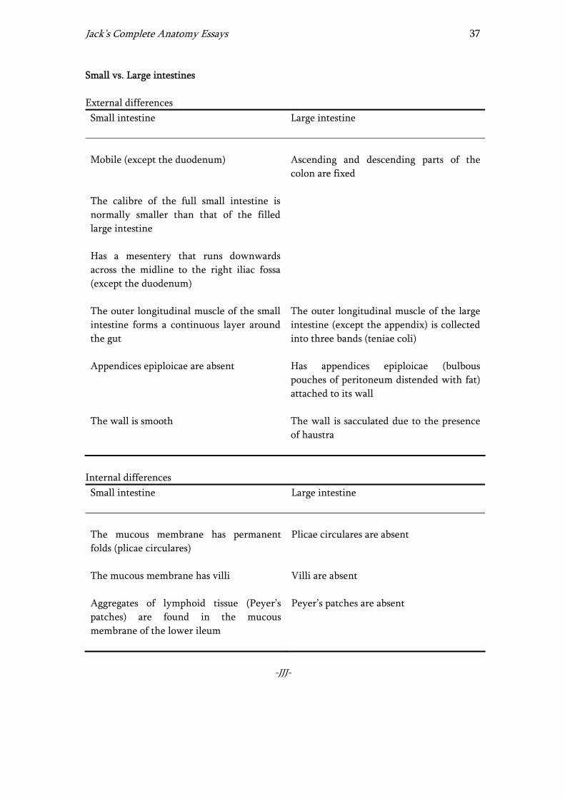
Jack’s Complete Anatomy Essays 37
Small vs. Large intestines External differences Small intestine
Large intestine
Mobile (except the duodenum)
Ascending and descending parts of the colon are fixed
The calibre of the full small intestine is normally smaller than that of the filled large intestine
Has a mesentery that runs downwards across the midline to the right iliac fossa (except the duodenum)
The outer longitudinal muscle of the small intestine forms a continuous layer around the gut
The outer longitudinal muscle of the large intestine (except the appendix) is collected into three bands (teniae coli)
Appendices epiploicae are absent Has appendices epiploicae (bulbous pouches of peritoneum distended with fat) attached to its wall
The wall is smooth The wall is sacculated due to the presence of haustra
Internal differences Small intestine
Large intestine
The mucous membrane has permanent folds (plicae circulares)
Plicae circulares are absent
The mucous membrane has villi
Villi are absent
Aggregates of lymphoid tissue (Peyer’s patches) are found in the mucous membrane of the lower ileum
Peyer’s patches are absent
-JJJ-
Jack’s Complete Anatomy Essays 38
Midgut & Superior mesenteric artery The midgut is the short portion of the primitive gut that is located opposite the yolk stalk. Its derivatives are the distal part of the duodenum, the jejunum, the ileum, the caecum, the appendix, the ascending colon and the proximal two-thirds of the transverse colon. Rapid elongation of the midgut during the embryonic period within the limited space of the abdominal cavity results in the following:
1. Loop formation 2. Coiling of the cephalic segment of the loop 3. Physiological hernia with an associated 90o counterclockwise rotation. The
herniation occurs into the umbilical cord which contains a space that is the continuation of the primitive peritoneal cavity.
With progressive increase in the size of the abdominal cavity, the midgut undergoes the final important steps of its morphological development comprising of:
1. Return to the abdominal cavity with a further 180o counterclockwise rotation, resulting in the superior mesenteric artery occupying a position between the duodenum and the transverse colon (behind the transverse colon and in front of the duodenum). This occurs at about the 10th week of development.
2. Fixation whereby parts of the original mesentery become attached to the posterior abdominal wall thereby rendering the associated segments of gut retroperitoneal. Some parts like the small intestine and the transverse colon retain a significant mesentery and hence mobility.
Failure to return leads to a hernia in the umbilical region. Inadequate fixation gives abnormal mobility to the gut which predisposes it to a volvulus (a loop getting twisted around itself and getting strangulated). Visceral pain is produced by spasm of the smooth muscle coat of the affected segment. Afferent pain fibres accompany sympathetic nerves through the superior mesenteric plexus, the lesser splanchnic nerve and the thoracic sympathetic trunk to enter the spinal cord at the T10 segment through the dorsal root. Vague referred pain is felt in the region of the umbilicus (T10 dermatome). The superior mesenteric artery supplies the derivatives of the midgut. It arises from the front of the abdominal aorta just below the coeliac trunk, and passes downwards and to the left behind the neck of the pancreas and in front of the third part of the duodenum. It continues downwards to the right between the layers of the mesentery of the small intestine and ends by anastomosing with the ileal end of its own ileocolic branch. Its branches are:
- The inferior pancreaticoduodenal artery passes to the right as a single or double branch along the upper border of the third part of the duodenum and the head of the pancreas. It supplies the pancreas and the adjoining part of the duodenum.
Jack’s Complete Anatomy Essays 39
- The middle colic artery passes forwards in the transverse mesocolon. It supplies the proximal two-thirds of the transverse colon and divides into right and left branches.
- The right colic artery is often a branch of the ileocolic artery. It passes to the right to supply the ascending colon and divides into ascending and descending branches.
- The ileocolic artery passes downwards and to the right. It gives rise to a superior branch which anastomoses with the right colic artery and an inferior branch which anastomoses with the end of the superior mesenteric artery. The inferior branch gives rise to anterior and posterior caecal branches; the appendicular artery is a branch of the posterior caecal artery.
- The jejunal and ileal arteries number 12 to 15 and arise from the left side of the superior mesenteric artery. Each artery divides into two vessels which unite with adjacent branches to form a series of arcades. Branches from the arcades divide and unite to form the second, third and fourth series of arcades. Fewer arcades supply the jejunum than supply the ileum. From the terminal arcades, short, straight vessels supply the intestine.
-JJJ-
Large intestine The large intestine, up to and including the mucous membrane of the rectum, receives its arterial supply from branches of the superior and inferior mesenteric arteries, both of which arise from the front of the abdominal aorta just below the coeliac trunk and above its bifurcation respectively. Blood from the large intestine, up to and including the mucous membrane of the rectum, eventually drains into the superior and inferior mesenteric veins, both of which are tributaries of the hepatic portal vein. The inferior mesenteric vein joins the splenic vein behind the body of the pancreas while the superior mesenteric and splenic veins unite behind the neck of the pancreas to form the portal vein. Anterior and posterior caecal arteries from the ileocolic artery, a branch of the superior mesenteric artery, supply the caecum. Anterior and posterior caecal veins drain blood from the caecum into the ileocolic vein, a tributary of the superior mesenteric vein. The appendicular artery is a branch of the posterior caecal artery, passing to the tip of the appendix in the mesoappendix. The appendicular vein drains into the posterior caecal vein. The ileocolic and right colic branches of the superior mesenteric artery supply the ascending colon. Ileocolic and right colic veins drain blood from the ascending colon into the superior mesenteric vein. The proximal two-thirds of the transverse colon is supplied by the middle colic artery, a branch of the superior mesenteric artery. The middle colic vein, a tributary of the superior mesenteric vein, drains blood from this segment. The distal one-third of the transverse colon is supplied by the left colic artery, a branch of the inferior mesenteric artery. The left colic vein, a tributary of the inferior mesenteric vein, drains blood from this segment. The left colic and the sigmoid branches of the inferior mesenteric artery supply the descending colon. Left colic and sigmoid veins drain blood from the descending colon into the inferior mesenteric vein.
Jack’s Complete Anatomy Essays 40
Sigmoid branches of the inferior mesenteric artery supply the sigmoid colon. Sigmoid veins drain blood from the sigmoid colon into the inferior mesenteric vein. The anastomosis of the colic arteries around the concave margin of the large intestine forms a single arterial trunk called the marginal artery. This begins at the ileocaecal junction, where it anastomoses with the ileal branches of the superior mesenteric artery, and it ends where it anastomoses less freely with the superior rectal artery. The superior, middle and inferior rectal arteries supply the rectum. The superior, middle and inferior rectal veins drain blood from the rectum. The superior rectal artery is a direct continuation of the inferior mesenteric artery and is the chief artery supplying the mucous membrane. It enters the pelvis by descending in the root of the sigmoid mesocolon and divides into the right and left branches which at first lie behind the rectum and then pierce the muscular coat and supply the mucous membrane. They anastomose with one another and with the middle and inferior rectal arteries. The superior rectal vein drains blood from the mucous membrane into the inferior mesenteric vein. The middle rectal artery is a small branch of the internal iliac artery, a branch of the common iliac artery which in turn arises from the bifurcation of the abdominal aorta. It runs forwards and medially to the rectum, to be distributed mainly to the muscular coat. The inferior rectal artery is a branch of the internal pudendal artery in the perineum, a branch of the anterior division of the internal iliac artery. It anastomoses with the middle rectal artery at the recto-anal junction. The middle and inferior rectal veins drain into the internal iliac and internal pudendal veins respectively. The three rectal veins form an important porto-systemic anastomosis since the superior rectal vein drains ultimately into the portal vein, and the middle and inferior veins, into the inferior vena cava. The superior and inferior rectal arteries supply the upper and lower halves of the anal canal respectively. The upper half is drained by the superior rectal vein into the inferior mesenteric vein and the lower half is drained by the inferior rectal vein into the internal pudendal vein. Under the light microscope, the large intestine is seen to have four distinct functional layers, from luminal outwards: the mucosa comprising of epithelium, lamina propria and muscularis mucosae, the submucosa, the muscularis externa, and the adventitia or serosa depending on the segment of colon being observed. The mucosa is folded in the non-distended state but does not exhibit distinct plicae circulares and villi like those of the small intestine. Consistent with its functions of water absorption and faecal lubrication, the mucosa consists of absorptive and mucus-secreting goblet cells. These are arranged in closely packed, straight tubular glands or crypts which extend to the muscularis mucosae. Goblet cells predominate in the base of the crypts while the luminal surface is almost entirely lined by tall columnar absorptive cells with basal nuclei. As faeces pass along the colon and become progressively dehydrated, the mucus becomes increasingly important in protecting the mucosa from trauma. The lamina propria fills the space between the crypts and contains numerous blood and lymph vessels into which water is absorbed by passive diffusion. It is also rich in lymphocytes and lymphoid aggregates (smaller than Peyer’s patches in the terminal ileum) that frequently extend into the submucosa. The richness in lymphoid tissue is related to the abundant bacterial population of the large intestine.
Jack’s Complete Anatomy Essays 41
The submucosa is a layer of loose connective tissue supporting the mucosa and containing the larger blood vessels, lymphatics and the submucosal (Meissner’s) plexus. As in the rest of the gastrointestinal tract, the muscularis externa of the colon consists of inner circular and outer longitudinal layers but, except in the appendix and the rectum, the longitudinal fibres congregate in three thick longitudinal bands called teniae coli. The myenteric (Auerbach’s) plexus lies between the outer longitudinal and inner circular layers of the muscularis externa. In the intraperitoneal portions of the colon, except the appendix, the serosa is characterised by small, pendulous protuberances composed of adipose tissue, the appendices epiploicae. The serosa (visceral peritoneum) comprises of loose connective tissue conducting the major vessels and nerves and lined by a simple squamous mesothelium. The rest of the colon is covered by a layer of loose connective tissue, the adventitia. The rectal mucosa is similar to the rest of the colon except that it has even more goblet cells. The mucous membrane of the rectum, together with the circular layer of the muscularis externa, forms semi-circular permanent folds called the transverse folds of the rectum. The teniae coli of the sigmoid colon come together to form a continuous longitudinal layer. At the recto-anal junction, the mucosa undergoes an abrupt transition to become stratified squamous non-keratinised epithelium in the anal canal, whereas the muscularis mucosae becomes discontinuous. Immediately above the anal valves, the mucosa forms a series of longitudinal folds, the columns of Morgagni. Branched tubular circumanal glands also open at the recto-anal junction into small pits at the distal ends of the columns of Morgagni. In this region, the lamina propria contains a plexus of large veins that, when excessively dilated and varicose, produce internal haemorrhoids. The anal canal is surrounded by voluntary muscle which forms the external anal sphincter. Near the anus, the stratified squamous epithelium of the non-keratinised variety undergoes a gradual transition to that of the keratinised variety as part of the perianal skin which also contains sebaceous glands, hair follicles and large apocrine sweat glands.
-JJJ- Appendix Visceral pain is produced by luminal distension of the appendix or spasm of its smooth muscle coat. Afferent pain fibres accompany sympathetic nerves through the superior mesenteric plexus, the lesser splanchnic nerve and the thoracic sympathetic trunk to enter the spinal cord at the T10 segment through the dorsal root. Hence vague referred pain is felt in the region of the umbilicus (T10 dermatome). Subsequently, if the inflammatory process involves the parietal peritoneum, pain shifts to where the inflamed appendix irritates the parietal peritoneum. The severe somatic pain is localised precisely in the right lower quadrant. Under the light microscope, the appendix is seen in its entirety and to have four distinct functional layers, from luminal outwards: the mucosa comprising of epithelium, lamina propria and muscularis mucosae, the submucosa, the muscularis externa, and the serosa. It is characterised by a relatively small, narrow and irregular lumen that is caused by the

Jack’s Complete Anatomy Essays 42
presence of abundant lymphoid follicles in the lamina propria and which extend into the upper submucosa. The epithelium is formed by simple columnar cells. The mucosa contains fewer and shorter straight tubular intestinal glands or crypts compared to the rest of the large intestine. The submucosa is a layer of loose connective tissue supporting the mucosa and containing the larger blood vessels, lymphatics and the submucosal (Meissner’s) plexus. As in the rest of the gastrointestinal tract, the muscularis externa of the colon consists of inner circular and outer longitudinal layers. The teniae coli of the caecum converge to form a continuous longitudinal layer at the base of the appendix. The myenteric (Auerbach’s) plexus lies between the outer longitudinal and inner circular layers of the muscularis externa. The serosa (visceral peritoneum) is the outermost layer comprising of loose connective tissue conducting the major vessels and nerves and lined by a simple squamous mesothelium. The serosa of the appendix lacks the appendices epiploicae of the large intestine. Part of the mesoappendix containing the appendicular blood and lymph vessels and nerves may be seen in continuity with the outermost serosal layer. The mesoappendix attaches the appendix to the lower layer of the mesentery of the small intestine.
-JJJ- Liver Under the light microscope, structural units of the liver called classic liver lobules are seen. The liver lobule is formed of a polygonal mass of tissue. There are three to six portal triads per lobule, each with a venule (a branch of the hepatic portal vein), an arteriole (a branch of the hepatic artery) and a bile duct (part of the bile duct system) and lymphatic vessels. The portal triads occupy portal spaces present at the corners of the lobules. The hepatocytes in the liver lobule are radially disposed. They form layers one or two cells thick; these cellular plates are directed from the periphery of the lobule to its centre and anastomose freely, forming a labyrinthine and sponge-like structure. The space between the plates contains liver sinusoids, irregularly dilated capillaries composed solely of a discontinuous layer of fenestrated endothelial cells. They are surrounded and supported by a delicate sheath of reticular fibres. The sinusoids arise in the periphery of the lobule and run in the direction of the lobular centre where they drain into the central vein. The endothelial cells are separated from the underlying hepatocytes by a subendothelial space known as the space of Disse which contains microvilli of the hepatocytes. Blood fluids readily percolate through the endothelial wall and make intimate contact with the surface of the hepatocytes, permitting an easy exchange of macromolecules from the sinusoidal lumen to the hepatocytes and vice versa. The sinusoids also contain Kupffer cells. These are phagocytic cells found on the luminal surface of the endothelial cells. Part of the mononuclear phagocyte system, Kupffer cells function mainly to metabolise spent erythrocytes, digest haemoglobin and secrete proteins related to immunologic processes. Bile is secreted into a network of minute bile canaliculi situated between the plasma membranes of adjacent hepatocytes but these are far too small to be seen at high
Jack’s Complete Anatomy Essays 43
magnification. The canaliculi drain into bile collecting ducts lined by simple cuboidal or columnar epithelium, the canals of Hering, which in turn drain into the bile ductules located in the portal triads. Bile flow therefore progresses in a direction opposite of sinusoidal blood flow, i.e., from the centre of the classic liver lobule to its periphery. The liver receives 70% of blood from the hepatic portal vein which carries oxygen-poor, nutrient-rich blood from the abdominal viscera, and 30% from the hepatic artery which supplies oxygen-rich blood. The arterial and venous blood is conducted to the central vein of each liver lobule by the liver sinusoids. The central veins drain into the right and left hepatic veins which leave the posterior surface of the liver and open directly into the inferior vena cava.
-JJJ- Porto-systemic anastomoses Porto-systemic anastomoses exist as such:
- At the lower third of the oesophagus, the oesophageal tributaries of the left gastric vein (portal tributary) anastomose with the oesophageal tributaries of the azygos veins (systemic tributaries) draining the middle third of the oesophagus.
- Halfway down the anal canal, the superior rectal vein (portal tributary) anastomoses with the middle and inferior rectal veins (systemic tributaries) which are tributaries of the internal iliac and internal pudendal veins respectively.
- The paraumbilical veins (portal tributaries) connect the superficial veins of the anterior abdominal wall (systemic tributaries) through the umbilicus to the left branch of the portal vein. The paraumbilical veins travel in the falciform ligament and accompany the ligamentum teres. The superficial veins of the anterior abdominal wall form a network radiating out from the umbilicus. Above, the network is drained into the axillary vein via the lateral thoracic vein and below, into the femoral vein via the superficial epigastric and great saphenous veins.
- In the retroperitoneal space, the veins of the ascending colon, descending colon, duodenum, pancreas and liver (portal tributaries) anastomose with the renal, lumbar and phrenic veins (systemic tributaries).
Portal hypertension resulting from portal vein obstruction (as occurs in cirrhosis of liver) causes dilation and varicosity of porto-systemic anastomoses:
- The oesophageal varices may rupture, causing severe and fatal vomiting of blood (haematemesis).
- The superficial veins around the umbilicus and the paraumbilical veins also become grossly distended. The distended subcutaneous veins radiate out from the umbilicus, producing in severe cases the clinical picture referred to as caput medusae.
- Dilated rectal veins give rise to internal haemorrhoids. The tributaries of the superior rectal vein lie in the anal columns at the 3-, 7- and 11-o’clock positions when the patient is viewed in the lithotomy position and are particularly liable to become varicosed. Anatomically, an internal haemorrhoid is a fold of mucous
Jack’s Complete Anatomy Essays 44
membrane and submucosa containing a varicosed tributary of the superior rectal vein and a terminal branch of the superior rectal artery.
-JJJ-
Extrahepatic biliary tree The major bile ducts outside the liver are collectively called the extrahepatic biliary tree. These ducts, enumerated distally from the porta hepatis, are the right and left hepatic ducts, the common hepatic duct, the cystic duct which drains the gallbladder, and the bile duct. The intrahepatic bile collecting system merges to from the right and left hepatic ducts. They emerge from the porta hepatis and drain the right and left lobes of the liver respectively. The porta hepatis, or hilus of the liver, is a region of the visceral (inferior) surface of the liver for the passage of the hepatic pedicle; the hepatoduodenal part of the lesser omentum also attaches here. The components of the hepatic pedicle, namely right and left hepatic ducts, right and left hepatic arteries, and right and left branches of the portal vein, exhibit a triple-Y arrangement. Sympathetic and parasympathetic nerve fibres and a few hepatic lymph nodes also lie in the porta hepatis. After a short course, the right and left hepatic ducts unite to form a single, large common hepatic duct which descends within the right free margin of the lesser omentum. The common hepatic duct is joined on the right side by the cystic duct from the gallbladder to form the bile duct. The gallbladder is a pear-shaped sac, lying on the under-surface of the liver, having a capacity of 30 to 50 ml and storing bile which it concentrates by absorbing water. Its rounded fundus usually projects below the inferior margin of the liver where it comes in contact with the anterior abdominal wall at the level of the tip of the ninth right costal cartilage, i.e., the junction of the right linea semilunaris and the costal margin. The body of the gallbladder lies in contact with the visceral surface of the liver and is directed upwards, backwards and to the left. The neck becomes continuous with the cystic duct. The cystic duct connects the neck of the gallbladder to the common hepatic duct to from the (common) bile duct. It is usually somewhat S-shaped, has spiral valves (of Heister) and descends for a variable distance in the right free margin of the lesser omentum. In the first part of its course, the bile duct lies in the right free margin of the lesser omentum in front of the opening into the lesser sac, the epiploic foramen (foramen of Winslow). Here it lies anterior to the right margin of the portal vein and to the right of the hepatic artery. In the second part, it is situated behind the first part of the duodenum to the right of the gastroduodenal artery. In the third part, it lies in a groove on the posterior surface of the head of the pancreas where it comes into contact with the main pancreatic duct. The bile duct ends below by piercing the medial wall of the second part of the duodenum about halfway down the latter’s length. It is usually joined by the main pancreatic duct and together they open into a small ampulla in the duodenal wall, the hepatopancreatic ampulla (ampulla of Vater). The ampulla opens into the lumen of the duodenum by means of a small papilla, the major duodenal papilla. The terminal parts of both ducts and ampulla are surrounded by circular muscle, the sphincter of the hepatopancreatic ampulla (sphincter of Oddi). Occasionally, the bile and main pancreatic ducts open separately into the duodenum.

Jack’s Complete Anatomy Essays 45
Under the light microscope, the wall of the gallbladder is seen to consist of, from luminal outwards: a simple columnar epithelium, a submucosa or lamina propria, a smooth muscular layer, and an adventitial and/or serosal coat. The epithelial cells are tall and columnar with basal nuclei; their numerous short, irregular microvilli account for the unevenness of the luminal surface. The lining cells concentrate bile by an active process, the resulting water passing into the lymphatics in the lamina propria/submucosa. In the neck of the gallbladder, the epithelium invaginates into the lamina propria/submucosa, forming tubuloacinar mucous glands with wide lumens. Mucus produced may provide a protective surface film for the biliary tract. The fibres of the muscular layer are arranged in longitudinal, transverse and oblique orientations but do not form distinct layers. A thick connective tissue layer (adventitia) binds the superior surface of the gallbladder to the liver. The opposite surface is covered by serosa (visceral peritoneum). Gallstones (cholelithiasis) can cause obstruction of biliary ducts and blockage of bile flow, resulting in cholestasis, i.e., failure to secrete bile into the duodenum. Cholestasis results in a failure to solubilise dietary lipids and fat-soluble vitamins (vitamins A, D, E and K), leading to malabsorption and deficiency states respectively. Steatorrhoea (greasy stools) consequently occurs. In cholestasis secondary to cholelithiasis, endogenous substances that are normally excreted via the biliary tract can accumulate to high levels. One such substance is bilirubin, a product of haem metabolism. Conjugated hyperbilirubinaemia occurs to cause obstructive (cholestatic) jaundice (icterus) manifested as yellow discoloration of the scleras and skin. Bile salts also accumulate in the plasma, eventually deposited in the skin. This is believed to cause intense itching (pruritus). Biliary colic is usually caused by spasm of the smooth muscle of the wall of the gallbladder in an attempt to expel a gallstone. Afferent pain fibres accompany sympathetic nerves through the coeliac plexus, the greater splanchnic nerve and the thoracic sympathetic trunk to enter the spinal cord at the thoracic segments through the dorsal roots. Referred pain is felt in the right upper quadrant or epigastrium (T7, 8 and 9 dermatomes).
-JJJ-

Jack’s Complete Anatomy Essays 46
Kidney Anterior relations of the right kidney: suprarenal gland, liver, second part of duodenum, right colic flexure Additional notes:
- Only the liver is separated from the right kidney by the visceral layer of peritoneum whereas the second part of the duodenum and right colic flexure are not.
- The right suprarenal gland is pyramid-shaped and caps the upper pole of the right kidney. It is surrounded by renal fascia but separated from the kidney by perirenal fat.
Anterior relations of the left kidney: suprarenal gland, spleen, stomach, pancreas, left colic flexure, coils of jejunum Additional notes:
- The spleen, stomach and jejunum are separated from the left kidney by the visceral layer of peritoneum whereas the pancreas and left colic flexure are not.
- The left suprarenal gland is crescentic in shape and extends along the medial border of the left kidney from the upper pole to the hilus. It is surrounded by renal fascia but separated from the kidney by perirenal fat.
The posterior relations of each kidneys, anteroposteriorly, are: the diaphragm, the costodiaphragmatic recess of pleura, the psoas and quadratus lumborum muscles, the aponeurosis of origin of the transversus abdominis muscle. The subcostal (T12) and iliohypogastric and ilioinguinal (L1) nerves run downwards and laterally behind each kidney. In addition, the 12th right rib is posterior to the right kidney but the 11th and 12th left ribs are posterior to the left kidney. This difference is attributed to the left kidney being slightly higher than the right kidney (due to the large size of the right liver lobe). Additional notes:
- Viewed from the side, the diaphragm appears to be an inverted ‘J’, the long limb extending up from the vertebral column (it is bounded posteriorly by the body of the T12 vertebra) and the short limb extending forwards to the xiphoid process. Hence the kidney lies anterior to the long limb of this inverted ‘J’. The diaphragm is the primary muscle of inspiration, and separates the thoracic and abdominal cavities.
- The costodiaphragmatic recesses are slit-like spaces between the costal and diaphragmatic parietal pleura that are separated only by a capillary layer of pleural fluid. The lower margins of the lungs descend into these recesses during inspiration.
- The three muscles are arranged as such, from the midline laterally: the psoas, quadratus lumborum and transversus abdominis.
o The psoas muscle arises from the roots of the transverse processes, the sides of the vertebral bodies and the intervertebral discs from T12 to L5 vertebrae. Its fibres run downwards and laterally, leaving the abdomen to enter the thigh and inserting into the lesser trochanter of the femur. The psoas flexes the thigh on the trunk.
o The quadratus lumborum is a flat, quadrilateral-shaped muscle lying alongside the vertebral column. It arises from the iliolumbar ligament, the

Jack’s Complete Anatomy Essays 47
adjoining part of the iliac crest and the tips of the transverse processes of the lower lumbar vertebrae. The fibres run upwards and medially to be inserted into the 12th rib and the transverse processes of the upper 4 lumbar vertebrae. It fixes the 12th rib during inspiration and depresses it during forced expiration; it also laterally flexes the vertebral column the same side.
o The transversus abdominis muscle is the deepest muscle of the anterior abdominal wall, its fibres running horizontally forwards. It arises from the lower 6 costal cartilages, the lumbar fascia, the iliac crest and the inguinal ligament, and is inserted into the xiphoid process, the linea alba and the symphysis pubis. It compresses abdominal contents when contracted.
- The subcostal nerve supplies the skin and muscles of the anterior abdominal wall and the parietal peritoneum. It pierces the posterior wall of the rectus sheath to supply the rectus abdominis muscle and the pyramidalis and terminates by piercing the anterior wall of the sheath and supplying the skin.
- The iliohypogastric nerve pierces the external oblique aponeurosis above the superficial inguinal ring while the ilioinguinal nerve emerges through the ring. They end by supplying the skin above the skin just above the inguinal ligament and symphysis pubis.
The structures of the renal pedicle at the hilus of the kidney, anteroposteriorly, are: the renal vein, two branches of the renal artery, the ureter and the third branch of the renal artery. Lymph vessels and sympathetic fibres, forming the renal plexus around the artery, also pass through the hilus. The developing kidneys are initially located in the pelvic cavity. They ‘ascend’ up the posterior abdominal wall due to diminution of the body curvature and growth of the body in lumbar and sacral regions. The kidneys eventually reach their final position opposite the second lumbar vertebra, with the right kidney slightly lower than the left (due to the large size of the right lobe of the liver). Arrest in some part of the normal ascent leads to a pelvic kidney which is usually found at pelvic brim. N.B. The ureteric bud (outgrowth of the mesonephric duct close to its entrance into the cloaca) develops into the collecting system of the kidney (the ureter, renal pelvis, major and minor calyces, and collecting tubules). The metanephric tissue cap (appearing in the embryo after the pronephros and mesonephros) forms the excretory system (proximal and distal convoluted tubules, and loops of Henle). The renal cortex forms a continuous smooth outer zone which extends down between the medullary pyramids. Under the light microscope, the cortex is seen to contain numerous renal corpuscles which appear to be arranged in parallel rows at right angles to the capsule. Each renal corpuscle consists of a tuff of capillaries, the glomerulus, surrounded by a double-walled epithelial capsule, the Bowman’s capsule. Between the inner visceral layer and outer parietal layers of the Bowman’s capsule is the urinary space which receives the glomerular ultrafiltrate. Each renal corpuscle has a vascular pole, where the afferent arteriole enters and divides into the glomerular capillaries and where the efferent arteriole leaves, and a urinary pole where the proximal convoluted tubule (PCT) begins.
Jack’s Complete Anatomy Essays 48
The parietal layer of Bowman’s capsule consists of a simple squamous epithelium supported by a basal lamina. The cells of the visceral layer, the podocytes, have a cell body from which arise several primary processes. Each primary process gives rise to numerous secondary processes called pedicels that embrace the glomerular capillaries and are periodically in contact with the basal lamina. However the cell bodies of the podocytes and their primary processes do not touch the basal lamina. The pedicels from one podocyte embrace more than one capillary; on a single capillary, the pedicels of two podocytes alternate in position next to the basal lamina. The interdigitation of these pedicels give rise to filtration slits. Besides fenestrated endothelial cells and podocytes, the glomerular capillaries have mesangial cells adhering to their walls in places where the basal lamina forms a sheath shared by two or more capillaries. Mesangial cells act as phagocytes. Between the fenestrated endothelial cells of the glomerular capillaries and the podocytes that cover their external surfaces is a thick basement membrane derived from the fusion of capillary- and podocyte-produced basal laminae. This basement membrane, together with the capillary endothelium and filtration slits between pedicels of podocytes, forms the filtration barrier separating the urinary space and glomerular blood. Most of the cortical parenchyma surrounding the renal corpuscles consists of cortical tubules: mainly PCTs with smaller numbers of distal convoluted tubules (DCTs) and collecting tubules. PCTs are lined by simple cuboidal epithelium whose cell apices have abundant microvilli, forming a brush border. The basal portions of the epithelial cells have lateral interdigitations with neighbouring cells. Mitochondria are concentrated at the base of the cells, arranged parallel to their long axis. The mitochondrial location and the increase in the area of cell membrane at the base of the cells are characteristic of cells engaged in active ion transport. Indeed PCTs function to absorb water, chloride, calcium, phosphate, glucose and amino acids from the glomerular filtrate. DCTs are lined by simple cuboidal epithelium. Cells of DCTs are flatter and smaller than those of DCTs; they lack the brush borders of PCTs and are less acidophilic. The lumens of DCTs are larger and more nuclei are seen in the DCT than in the PCT in the same histologic section. Along its path in the cortex, the DCT establishes contact with the vascular pole of the renal corpuscle. In this juxtaglomerular region, DCT cells become columnar and their nuclei are closely packed together. Acting as chemoreceptors (detecting low sodium ionic concentration in distal tubular fluid), cells of this modified DCT segment called the macula densa promotes renin secretion from juxtaglomerular cells. Juxtaglomerular cells are specialised smooth muscle cells of the wall of the afferent arteriole which secrete renin in response to a drop in systemic blood pressure. Also part of the juxtaglomerular apparatus are extraglomerular mesangial (lacis) cells of unknown function. These cells are bounded by the afferent and efferent arterioles and make contact with the macula densa. The smaller collecting tubules are lined by cuboidal epithelium. As they penetrate deeper into the medulla, their cells increase in height until they become columnar.
-JJJ-
Jack’s Complete Anatomy Essays 49
Ureter The two ureters are muscular tubes extending from the kidneys to the posterior surface of the urinary bladder. Each ureter has three constrictions along its course: where the renal pelvis joins the ureter, where it is kinked as it crosses the pelvic brim and where it pierces the bladder wall. Ureteric stones (calculi) can be located at one or more of these constrictions in a plain abdominal X-ray. The renal pelvis is the funnel-shaped expanded upper end of the ureter and lies within the hilus of the kidney, receiving urine from the major calyces. The ureter emerges from the hilus of the kidney and runs vertically downwards behind the parietal peritoneum (adherent to it) on the psoas muscle which separates it from the tips of the transverse processes of the lumbar vertebrae. It enters the pelvis by crossing the bifurcation of the common iliac artery in front of the sacroiliac joint. In the male, each ureter then runs down the lateral wall of the pelvis in front of the internal iliac artery to the region of the ischial spine and turns forwards to enter the lateral angle of the bladder. Near its termination, it is crossed by the vas deferens. The ureter passes obliquely through the bladder wall for a short distance before opening into the bladder. In the female, upon entering the pelvis, each ureter runs downwards and backwards in front of the internal iliac artery and behind the ovary until it reaches the region of the ischial spine. It then turns forwards and medially beneath the base of the broad ligament where it is crossed by the uterine artery. The ureter then runs forwards, lateral to the lateral fornix of the vagina, to enter the bladder. In ureteric colic, strong peristaltic contraction waves pass down the ureter in an attempt to pass the ureteric stone onwards. The spasm of the smooth muscle causes an agonising colicky visceral pain which travels in afferent nerve fibres that accompany sympathetic nerves through the renal plexus around the renal artery. They ascend through the lowest splanchnic nerve and the thoracic sympathetic trunk, entering the spinal cord at segments T11, T12, L1 and L2 through the dorsal roots. Pain is referred along the distribution of the 11th intercostal nerve (T11), subcostal nerve (T12), iliohypogastric and ilioinguinal nerves (L1) and L2 to the flank, loin and groin. Hence the pain is often described as ‘loin-to-groin’. N.B. Sometimes ureteral pain is referred along the femoral branch of the genitofemoral nerve (L1 and 2) so that pain is experienced in the front of the thigh.
-JJJ-
Jack’s Complete Anatomy Essays 50
Pelvis & Perineum Pelvic diaphragm The funnel-shaped, musculofascial pelvic diaphragm forms the inferior pelvic wall or pelvic floor which supports the pelvic viscera. It is in turn formed by levatores ani and coccygeus muscles as well as the fasciae covering the superior and inferior aspects of these muscles. The pelvic diaphragm stretches between the pubis and coccyx in the anteroposterior direction, and from one lateral pelvic wall to the other. From their origin, the muscle fibres on either side slope posteroinferiorly to the midline, creating a gutter that slopes anteroinferiorly. It is incomplete anteriorly to permit passage of the male urethra, or the female urethra and vagina. The levator ani, a broad, thin muscular sheet, is the larger and more important muscle of the pelvic floor. Its origin is extensive: from the back of the body of the pubis, a tendinous arch formed by a thickening of the pelvic fascia covering obturator internus, and the ischial spine. Component fibres are named according to their attachments, and sweep inferomedially to their insertions:
- Anterior fibres: The levator prostatae or sphincter vaginae passes around the side of the prostate or vagina respectively to insert into the fibrous perineal body, in front of the anal canal. Both levator postatae and sphincter vaginae stabilise the perineal body; the former supports the prostate while the latter constricts the vagina.
- Intermediate fibres: The puborectalis consists of continuous fibres passing from one pubic bone to the other around the posterior aspect of the recto-anal junction, forming a U-shaped muscular sling. The pubococcygeus passes posteriorly and inserts into a small fibrous mass, the anococcygeal body (raphe), between the anal canal and the tip of the coccyx.
- Posterior fibres: The iliococcygeus arises from the inner surface of the ilium. Thin and often poorly developed, it is inserted into the anococcygeal body and coccyx.
Contraction of the components of levatores ani counteracts any rise in intra-abdominal pressure produced by contraction of the diaphragm and anterolateral abdominal wall muscles, e.g. during coughing and straining. In so doing, they support and maintain the pelvic viscera in position. The levatores ani also have important functions in voluntary control of micturition, faecal continence (via puborectalis which pulls the recto-anal junction anterosuperiorly) and support of the uterus (via sphincter vaginae). The coccygeus is a small triangular muscle originating from the ischial spine, and inserted into the inferior end of the sacrum and into the coccyx. The two coccygeus muscles assist the levatores ani in resisting increases of intra-abdominal pressure and supporting pelvic viscera. If the pelvic diaphragm was injured, e.g., during difficult parturition (vaginal delivery), the position of the bladder neck and urethra may be altered, producing urinary stress
Jack’s Complete Anatomy Essays 51
incontinence. This condition is characterised by dribbling of urine during rises in intra-abdominal pressure. Other possible sequelae are: uterine, vaginal and/or rectal prolapse secondary to the loss of support, and bladder herniation (cystocele).
-JJJ- Uterus In most women, the long axis of the uterus is bent forward on the long axis of the vagina, i.e., the uterus is anteverted. Furthermore, the long axis of body of the uterus is bent forward at the level of the internal os with the long axis of the cervix, i.e., the uterus is also anteflexed. Thus in the erect position and with the bladder empty, the uterus lies in an almost horizontal plane. The uterus is supported mainly by the tone of the levatores ani muscles and the condensations of the pelvic fascia which form three important ligaments. The levatores ani muscles form a broad muscular sheet stretching across the pelvic cavity and, together with the pelvic fascia, they effectively support the pelvic viscera and resist the intra-abdominal pressure transmitted downwards through the pelvis. The medial edges of the anterior parts of the levatores ani muscles are attached to the cervix of the uterus by the pelvic fascia. Some of the fibres of levator ani are inserted into a fibromuscular structure, the perineal body, which is important in maintaining the integrity of the pelvic floor; if it is damaged during childbirth, prolapse of pelvic viscera may occur. The perineal body lies in the perineum between the vagina and the anal canal. It is slung up to the pelvic walls by the levatores ani and thus supports the vagina and, indirectly, the uterus. The transverse cervical ligament (cardinal ligament), pubocervical and sacrocervical ligaments are subperitoneal condensations of pelvic fascia on the upper surface of the levatores ani muscles. They are attached to the cervix and the vault of the vagina and play an important part in supporting the uterus and keeping the cervix in its correct position. The cardinal ligaments are fibromuscular condensations of pelvic fascia that pass to the cervix and the upper end of the vagina from the lateral walls of the pelvis. The pubocervical ligaments consist of two firm bands of connective tissue that pass to the cervix from the posterior surface of the pubis. They are positioned on either side of the neck of the bladder, to which they give some support (pubovesical ligaments). The sacrocervical ligaments consist of two firm fibromuscular bands of pelvic fascia that pass to the cervix and the upper end of the vagina from the lower end of the sacrum. They form two ridges, one on either side of the rectouterine pouch (pouch of Douglas). The broad ligaments and the round ligaments of the uterus are lax structures and the uterus can be pulled up or pushed down for a considerable distance before they become taut. Clinically, they are considered to play a minor role in supporting the uterus. The round ligament of the uterus, representing the remains of the lower half of the gubernaculum, extends between the superolateral angle of the uterus, through the deep inguinal ring and the inguinal canal, to the subcutaneous tissue of the labium magus. It helps keep the uterus anteverted and anteflexed but is considerably stretched during pregnancy.
Jack’s Complete Anatomy Essays 52
The broad ligaments are two-layered folds of peritoneum that extend across the pelvic cavity from the lateral margins of the uterus to the lateral pelvic walls. Superiorly, the two layers are continuous and form the upper free edge. Inferiorly, at the base of the ligament, the layers separate to cover the pelvic floor. The ovary is attached to the posterior layer by the mesovarium. The part of the broad ligament that lies lateral to the attachment of the mesovarium forms the suspensory ligament of the ovary. The part of the ligament between the uterine tube and the mesovarium is called the mesosalpinx. At the base of the broad ligament, the uterine artery crosses the ureter. Each broad ligament contains the uterine tube in the upper free border, the round ligament of the ovary and the round ligament of the uterus (representing the remains of the gubernaculum), the uterine and ovarian blood and lymph vessels and nerves, the epoophoron (a vestigial structure above the attachment of the mesovarium, representing the remains of the mesonephros) and the paroophoron (a vestigial structure just lateral to the uterus and which is a mesonephros remnant). Under the light microscope, the wall of the body of the uterus (corpus uteri) is seen to be relatively thick and is formed of three layers. Depending on the part of the uterus, there is either an outer serosa (connective tissue lined by mesothelium) or adventitia (connective tissue). The other uterine layers are the myometrium, a thick tunic of smooth muscle, and the endometrium, the mucosa of the uterus. The myometrium, the thickest tunic of the uterus, is composed of bundles of smooth muscle fibres in three poorly-defined layers separated by connective tissue. The inner and outer layers are composed mainly of fibres disposed longitudinally. The middle circular layer contains the larger blood vessels. Under the light microscope, the smooth muscle layers appear to run in a spiral fashion, crisscrossing each other. The endometrium consists of simple columnar epithelium and lamina propria containing simple tubular glands that sometimes branch in their deeper portions (near the myometrium). Its covering epithelial cells are a mixture of ciliated and secretory simple columnar cells. The epithelium of the uterine glands is similar to the superficial epithelium but ciliated cells are rare within the glands. The connective tissue of the lamina propria is rich in fibroblasts and contains abundant ground substance. Connective tissue fibres are mostly reticular. The endometrium can be divided into two zones: the stratum functionalis which constitutes the portions sloughed off at menstruation and replaced during each menstrual cycle, and the stratum basalis, the portion retained after menstruation that subsequently proliferates and provides a new epithelium and lamina propria for endometrial renewal. The bases of the uterine glands which lie deep within the stratum basalis are the source of the cells that divide and migrate over the exposed connective tissue of the menstrual-phase endometrium, thereby providing for the new epithelial lining of the uterus after menstruation. The stratum functionalis, in turn, comprises of two layers: the thinner superficial layer having a compact stromal appearance (stratum compactum) and the broad deeper layer characterised by a spongy stromal appearance (stratum spongiosum). The arrangement of the arterial supply of the endometrium has important influences on the menstrual cycle. Arcuate arteries are circumferentially oriented in the middle layers of the myometrium. From these vessels, two sets of arteries arise to supply blood to the endometrium. Straight arteries are short and pass a short distance into the endometrium, then bifurcating to form a plexus supplying the stratum basalis. Spiral arteries are long, coiled and thick-walled and pass to the surface of the endometrium, giving off numerous
Jack’s Complete Anatomy Essays 53
branches which give rise to a capillary plexus around the glands in the stratum compactum. Unlike the straight arteries, the spiral arteries are responsive to the hormonal changes of the menstrual cycle. The withdrawal of progesterone secretion at the end of the cycle causes the spiral arteries to constrict and this precipitates an ischaemic phase immediately preceding menstruation. Under the light microscope, the endocervical canal is seen to be lined by a tall simple columnar mucus-secreting epithelium. Where the cervix is exposed to the more hostile environment of the vagina, the ectocervix, it is lined by thick stratified squamous epithelium as in the rest of the vagina. The cells of the ectocervix often have clear cytoplasm due to their high glycogen content. The junction between the vaginal and endocervical epithelium (squamo-columnar junction) is quite abrupt and is normally located at the external os, the point at which the endocervical canal opens into the vagina. The cervix has few smooth muscle fibres and consists mainly of dense connective tissue. Beneath the squamo-columnar junction, the cervical stroma is often infiltrated with leucocytes forming part of the defence against ingress of micro-organisms. The mucus-secreting epithelial lining of the endocervical canal is thrown into deep furrows and tunnels, giving the appearance in two dimensions of branched tubular glands; hence the rather inaccurate term ‘endocervical glands’. During the menstrual cycle, the endocervical glands undergo cyclic changes in secretory activity.
-JJJ- Vagina Under the light microscope, the wall of the vagina is seen to be devoid of glands and consists of three layers: a mucosa, a muscular layer and an adventitia. The epithelium of the mucosa is stratified squamous of the non-keratinised type. Under the stimulus of oestrogen, the vaginal epithelium synthesizes and accumulates a large quantity of glycogen which is deposited in the lumen of the vagina when the vaginal cells desquamate. Bacteria in the vagina metabolise the glycogen, forming lactic acid which is responsible for the usually low pH of the vagina. The acidic vaginal environment protects against some pathogenic microorganisms. The lamina propria of the vaginal mucosa is composed of loose connective tissue that is very rich in elastic fibres. Among the cells present are lymphocytes and neutrophils in relatively large quantities. During certain phases of the menstrual cycle, these two types of leucocytes invade the epithelium and pass into the lumen of the vagina. Although the lamina propria lacks glands, it exhibits a rich vascularisation that is the source of the fluid exudates that seeps through the squamous epithelium during sexual stimulation. The vaginal mucosa is virtually devoid of sensory nerve endings and the few naked nerve endings that do exist are probably pain fibres. The muscular layer of the vagina is composed mainly of longitudinal bundles of smooth muscle fibres. There are some circular bundles, especially in the innermost part (next to the mucosa). Outside the muscular layer, a coat of dense connective tissue, the adventitia, rich in thick elastic fibres, unites the vagina with the surrounding tissues. The great elasticity of the vagina is related to the large number of elastic fibres in the connective tissues of its wall.
Jack’s Complete Anatomy Essays 54
In this connective tissue are an extensive venous plexus, nerve bundles and groups of nerve cells.
-JJJ- Urinary bladder The urinary bladder lies immediately posterior to the pubic bones within the pelvis. It is pyramidal and lies entirely within the true pelvis when empty; but it is ovoid with its superior wall rising into the false pelvis, up to the hypogastric (suprapubic) region, as it distends with urine. The vesical apex points anteriorly and lies behind the superior edge of the symphysis pubis. It is joined to the umbilicus by the median umbilical ligament. The fundus or base (posterior surface) of the bladder faces posteriorly and is triangular; the ureters pierce externally through its superolateral angles and the urethra arises from its inferior angle. In the male, the two vasa deferentia lie abreast on the base of the bladder and separate the seminal vesicles from each other. The superior portion of the fundus is covered by peritoneum which forms the anterior wall of the rectovesical pouch. The inferior portion of the fundus is separated from the rectum by the vasa deferentia, seminal vesicles and rectovesical fascia. In the female, the fundus is separated by the vagina from the rectum. In the male, the superior vesical surface is lined by peritoneum and related to coils of ileum or sigmoid colon. Along its lateral margins, the peritoneum is reflected onto the lateral pelvic walls. In the female, the superior surface is related to the uterovesical pouch of peritoneum and to the corpus uteri. The two inferolateral surfaces are related anteriorly to the retropubic fat pad and pubic bones. More posteriorly, they lie in contact with obturator internus superiorly and levator ani inferiorly. The neck of the bladder is situated at the inferior convergence of the fundus and inferolateral surfaces. It rests on the superior prostatic surface in the male, and directly on the superior aspect of the urogenital diaphragm in the female. The smooth muscle fibres of the bladder wall here merge together with the fibromuscular tissue of the prostate. The neck is held in place by thickenings of the pelvic fascia, the puboprostatic (male) or pubovaginal (female) ligaments. The mucous membrane of the greater part of the empty bladder is thrown into trabeculae that disappear with bladder distension. Only one area of mucous membrane remains constantly smooth, even when the bladder is empty: that covering the internal surface of the vesical base. This is because the mucous membrane over this so-called trigone adheres firmly to the underlying muscular coat. The superior angles of the trigone correspond to the ureteric orifices and the inferior angle, to the internal urethral orifice. The ureters pierce the bladder wall obliquely and inferomedially, providing a valve-like action which prevents retrograde vesicoureteric reflux of urine. The trigone is bounded superiorly by a muscular ridge, the interureteric ridge, which spans between the two ureteric orifices. The uvula vesicae, produced by the underlying median lobe of the prostate, is a slight projection of the male bladder located immediately posterior to the internal urethral orifice.
Jack’s Complete Anatomy Essays 55
The muscular coat of the bladder comprises of smooth muscle arranged as three layers of interlacing bundles known as the detrusor muscle. At the male bladder neck, the circular component of the muscle coat is thickened as the involuntary sphincter vesicae. The innervation to the urinary bladder is from the inferior hypogastric plexuses. The parasympathetic preganglionic fibres arise as the pelvic splanchnic nerves from S2, 3 and 4. They pass through the inferior hypogastric plexuses to reach the bladder wall where they synapse with postganglionic neurones. The sympathetic postganglionic fibres originate in the first two lumbar ganglia and descend to the bladder via the hypogastric plexuses. Most afferent sensory fibres from the bladder reach the S2, 3 and 4 segments of the spinal cord via the pelvic splanchnic nerves. Some afferent fibres travel with sympathetic nerves via the hypogastric plexuses and enter L1 and 2 segments of the spinal cord. Parasympathetic nerves, which provide the main motor innervation of the bladder, stimulate contraction of the detrusor muscle of the bladder wall and inhibit the action of sphincter vesicae. The sympathetic nerves are motor to the sphincter vesicae and are now thought to be mostly vasomotor and probably inhibitory to the detrusor muscle. The onset of the simple reflex action of micturition occurs when bladder wall stretch receptors are stimulated in response to bladder distension by about 300 ml of urine. Afferent impulses enter the central nervous system mostly via the pelvic splanchnic nerves. Parasympathetic preganglionic fibres transmit the efferent parasympathetic impulses from the sacral cord (conus medullaris) to the bladder wall in the manner previously described; within the wall, they synapse with postganglionic fibres. By means of this sacrovesical reflex arc, the detrusor muscle is contracted whereas the sphincter vesicae is relaxed. Efferent impulses also pass to the sphincter urethrae via the pudendal nerves (S2, 3 and 4) to effect relaxation. This is accentuated by positive feedback to the cord from the stimulation of stretch receptors of the urethra as urine flows through. Voluntary control of micturition develops during the second or three year of life and involves contraction of the sphincter urethrae (closing the urethra) aided by the sphincter vesicae (compressing the bladder neck). When the bladder is half-full, afferents from the bladder travelling in the pelvic splanchnic nerves also relay this information (through the spinoreticular tract) to the pontine centre which responds by inhibiting the sacrovesical reflex arc and hence the micturition reflex. When the bladder is full and the time is opportune, the pontine centre releases the sacral cord from inhibition. However, if the time is inopportune, the premotor cortex can defer voiding by reinforcing the inhibitory function of the pontine centre via corticospinal tracts. Disruption of the process of micturition by spinal cord injuries may produce the following types of bladder:
- The atonic bladder occurs during the phase of spinal shock, immediately post-injury and may last from days to weeks. The detrusor muscle is relaxed, the sphincter vesicae tightly contracted, and the sphincter urethrae relaxed. Therefore there is difficulty initiating micturition and bladder distension with overflow. Depending on the level of cord injury, the patient may or may not be aware that the bladder is full.
Jack’s Complete Anatomy Essays 56
- The automatic reflex bladder follows recovery from spinal shock, provided that the cord lesion levels above the level of the parasympathetic outflow. The bladder fills and empties reflexly every 1-4 hours.
- The autonomous bladder occurs if the conus medullaris is destroyed, leaving the bladder without external reflex control. The bladder wall is flaccid and the vesical capacity is considerably increased. The bladder fills to capacity and overflows, giving rise to continual dribbling.
Under the light microscope, the mucosa of the urinary bladder is seen to comprise of transitional epithelium (urothelium) and a lamina propria of loose-to-dense connective tissue. The stratified transitional epithelium is thrown into many folds in the relaxed state. The basal cells are compact and cuboidal or columnar in shape whereas cells of the intermediate layers are more polygonal. When the bladder is empty, the epithelium is in the relaxed state and five to six cells thick. The surface cells, umbrella cells, are large and ovoid with round nuclei and abundant eosinophilic cytoplasm; some of them are binucleate. The apical outline is characterised by scalloping, and the superficial cytoplasm is indistinct and more intensely stained than the underlying layers. When distension of the bladder stretches the epithelium, the epithelium becomes only three to four cells thick with squamous umbrella cells. Umbrella cells confer the epithelium with an impermeable nature. The lamina propria carries capillaries. Surrounding the lamina propria is the wall of the bladder consisting of three loosely arranged layers (inner longitudinal, middle circular and outer longitudinal) of smooth muscle and elastic fibres. Save for the superior part of the bladder which is covered by serous peritoneum, the bladder is mostly covered by adventitia. The outer adventitial coat contains blood vessels and lymphatics.
-JJJ- Prostate gland The prostate is a fibromuscular glandular organ surrounding the prostatic urethra and lying between the neck of the bladder above and the urogenital diaphragm below. The prostate is surrounded by a fibrous capsule. Outside the capsule is a fibrous sheath which is part of the visceral layer of pelvic fascia. The somewhat conical prostate has a base which lies against the bladder neck superiorly and a neck which lies against the urogenital diaphragm inferiorly. The two ejaculatory ducts pierce the upper part of the posterior surface of the prostate to open into the prostatic urethra at the lateral margins of the prostatic utricle. Superiorly, the base of the prostate is continuous with the bladder neck, the smooth muscle passing without interruption from one organ to the other. The urethra enters the centre of the base of the prostate. Inferiorly, the apex lies on the upper surface of the urogenital diaphragm. The urethra leaves the prostate just above the apex on the anterior surface. The anterior surface of the prostate is related to the pubic symphysis, separated from it by the extraperitoneal fat in the retropubic space (cave of Retzius). The fibrous sheath of the prostate is connected to the posterior aspect of the pubic bones anteriorly by the
Jack’s Complete Anatomy Essays 57
puboprostatic ligaments. These ligaments lie one on either side of the midline and are condensations of pelvic fascia. The posterior surface of the prostate is closely related to the anterior surface of the rectal ampulla and is separated from it by the rectovesical septum (fascia of Denonvillier). The lateral surfaces of the prostate are embraced by the anterior fibres of the levatores ani as they run posteriorly from the pubis. Under the light microscope, the prostate gland is seen to be a fibromuscular glandular organ. It comprises of a collection of branched tubuloalveolar glands whose ducts empty into the prostatic urethra. It produces prostatic fluid and stores it in its interior for expulsion during ejaculation. The prostate is surrounded by a fibroelastic capsule rich in smooth muscle. Septa from this capsule penetrate the gland and divide it into lobes that are indistinct in adult men. An exceptionally rich fibromuscular stroma surrounds the glands. Small spherical bodies of glycoproteins, often calcified, are frequently observed in the lumen of prostatic glands. They are called prostatic concretions or corpora amylacea. Their significance is not understood but their number increases with age.
-JJJ- Male urethra The male urethra extends from the neck of the bladder to the external meatus on the glans penis. It is divided into three parts, proximal to distal:
- The prostatic urethra begins at the neck of the bladder and passes through the prostate gland from base to apex. It is the widest and most dilatable portion of the whole urethra. On its posterior surface is a longitudinal ridge called the urethral crest. On each side of the ridge is a groove, the prostatic sinus, into which the prostatic glands open. On the summit of the urethral crest is a depression, the prostatic utricle, which is the male analogue of the female vagina and uterus. On the edge of the mouth of the utricle are openings for the two ejaculatory ducts.
- The membranous urethra lies within the urogenital diaphragm, surrounded by the sphincter urethrae muscle. It is the least dilatable portion of the urethra.
- The penile urethra is enclosed in the bulb and corpus spongiosum of the penis. The external meatus is the narrowest part of the urethra. The part of the penile urethra that lies within the glans penis is dilated to form the fossa terminalis (navicular fossa). The bulbourethral glands of Cowper open into the penile urethra below the urogenital diaphragm.
Traumatic rupture of the penile urethra (most commonly in the bulb of the penis, just below the perineal membrane) results in superficial or subcutaneous extravasation of urine into the superficial perineal pouch (i.e., deep to the membranous layer of the superficial pelvic fascia but superficial to the perineal membrane) when micturition is attempted. The attachments of the perineal fascia determine the direction of flow of the extravasated urine. Hence urine passes inferiorly into the loose connective tissue of the scrotum beneath the membranous layer of superficial fascia (Colles’ fascia), anteriorly into and around the penis, and superiorly into the potential space of the lower anterior
Jack’s Complete Anatomy Essays 58
abdominal wall between the membranous layer of superficial fascia (Scarpa’s fascia) and the aponeurosis of the external oblique muscle. The perineal membrane and the subcutaneous tissue of the perineum are firmly attached to the ischiopubic rami. So the urine cannot pass posteriorly into the anal triangle because the superficial and deep layers of perineal fascia are continuous with each other around the superficial perineal muscles and with the posterior edge of the perineal membrane between them. Similarly, because the two layers are connected to the rami of the pubis and ischium, the urine cannot extend laterally. Neither can it extend into the lesser pelvis since the opening of this cavity is closed by the perineal membrane. Urine cannot pass into the thigh because the Scarpa’s fascia of the anterior abdominal wall blends with the fascia lata, just distal to the inguinal ligament.
-JJJ- Rectum & Anal canal The relations of the rectum are:
- Posteriorly, the rectum is in contact with the sacrum and coccyx, the piriformis, coccygeus and levatores ani muscles, the sacral plexus and the sympathetic trunks.
- Anteriorly, the upper two-thirds of the rectum which is covered by peritoneum is related to the sigmoid colon and coils of ileum that occupy the rectovesical pouch in males but the rectouterine pouch (pouch of Douglas) in females. In males, the lower third of the rectum which is devoid of peritoneum is related to the posterior surface of the bladder, to the termination of the vas deferens and the seminal vesicles on each side, and to the prostate. These abovementioned structures are embedded in visceral pelvic fascia. In females, the lower third of the rectum which is devoid of peritoneum is related to the posterior surface of vagina.
The superior, middle and inferior rectal arteries supply the rectum. The superior, middle and inferior rectal veins drain blood from the rectum. The superior rectal artery is a direct continuation of the inferior mesenteric artery and is the chief artery supplying the mucous membrane. It enters the pelvis by descending in the root of the sigmoid mesocolon and divides into the right and left branches which at first lie behind the rectum and then pierce the muscular coat and supply the mucous membrane. They anastomose with one another and with the middle and inferior rectal arteries. The middle rectal artery is a small branch of the internal iliac artery, a branch of the common iliac artery which in turn arises from the bifurcation of the abdominal aorta. It runs forwards and medially to the rectum, to be distributed mainly to the muscular coat. The inferior rectal artery is a branch of the internal pudendal artery in the perineum, a branch of the anterior division of the internal iliac artery. It anastomoses with the middle rectal artery at the recto-anal junction. The superior rectal vein drains blood from the mucous membrane into the inferior mesenteric vein. The middle and inferior rectal veins drain into the internal iliac and internal pudendal veins respectively. The three rectal veins form an important porto-
Jack’s Complete Anatomy Essays 59
systemic anastomosis since the superior rectal vein drains ultimately into the portal vein, and the middle and inferior veins, into the inferior vena cava. The lymph vessels of the rectum drain into the pararectal nodes. Lymph vessels then accompany the superior rectal artery to the inferior mesenteric nodes. Lymph vessels from the lower part of the rectum follow the middle rectal artery to the internal iliac nodes. Sympathetic and parasympathetic nerves from the inferior hypogastric plexuses supply the rectum. The rectum is sensitive only to stretch. The superior and inferior rectal arteries supply the upper and lower halves of the anal canal respectively. The upper half is drained by the superior rectal vein into the inferior mesenteric vein and the lower half is drained by the inferior rectal vein into the internal pudendal vein. The lymph vessels of the upper half of the anal canal drain into the pararectal nodes. Lymph vessels then accompany the superior rectal artery to the inferior mesenteric nodes. Lymph vessels from the lower half of the anal canal drains into the medial group of superficial inguinal nodes. The mucous membrane of the upper half of the anal canal is sensitive to stretch and is innervated by sensory fibres that ascend through the hypogastric plexuses. The lower half is sensitive to pain, temperature, touch and pressure and is innervated by the inferior rectal nerves. The involuntary internal anal sphincter is supplied by sympathetic fibres from the inferior hypogastric plexuses. The voluntary external anal sphincter is supplied by the inferior rectal nerve from the pudendal nerve and the perineal branch of S4. Under the light microscope, the rectum is seen to have four distinct functional layers, from luminal outwards: the mucosa comprising of epithelium, lamina propria and muscularis mucosae, the submucosa, the muscularis externa, and the adventitia or serosa depending on the segment being observed. The mucosa is folded in the non-distended state but does not exhibit distinct plicae circulares and villi like those of the small intestine. Consistent with its functions of water absorption and faecal lubrication, the mucosa consists of absorptive and abundant mucus-secreting goblet cells. These are arranged in closely packed, straight tubular glands or crypts which extend to the muscularis mucosae. Goblet cells predominate in the base of the crypts while the luminal surface is almost entirely lined by tall columnar absorptive cells with basal nuclei. As faeces pass along the colon and become progressively dehydrated, the mucus becomes increasingly important in protecting the mucosa from trauma. The lamina propria fills the space between the crypts and contains numerous blood and lymph vessels into which water is absorbed by passive diffusion. It is also rich in lymphocytes and lymphoid aggregates (smaller than Peyer’s patches in the terminal ileum) that frequently extend into the submucosa. The richness in lymphoid tissue is related to the abundant bacterial population of the large intestine. The submucosa is a layer of loose connective tissue supporting the mucosa and containing the larger blood vessels, lymphatics and the submucosal (Meissner’s) plexus. The muscularis externa of the rectum consists of inner circular and outer longitudinal layers. The teniae coli of the sigmoid colon converge to form a continuous longitudinal layer. The myenteric (Auerbach’s) plexus lies between the outer longitudinal and inner circular layers of the muscularis externa.
Jack’s Complete Anatomy Essays 60
The mucous membrane of the rectum, together with the circular layer of the muscularis externa, forms semi-circular permanent folds called the transverse folds of the rectum. Adventitia or serosa is the outermost layer of the rectum depending on the segment being observed. The upper third of the rectum has its anterior and lateral surfaces covered with peritoneum (which appears under the light microscope as serosa, or loose connective tissue lined by simple squamous mesothelium); the middle third has only its anterior surface covered with peritoneum; the lower third is devoid of peritoneum (and so is surrounded externally by loose connective tissue known as adventitia). The serosa, where it is present, lacks the appendices epiploicae of the large intestine. At the recto-anal junction, the mucosa undergoes an abrupt transition to become stratified squamous non-keratinised epithelium in the anal canal, whereas the muscularis mucosae becomes discontinuous. Immediately above the anal valves, the mucosa forms a series of longitudinal folds, the columns of Morgagni. Branched tubular circumanal glands also open at the recto-anal junction into small pits at the distal ends of the columns of Morgagni. In this region, the lamina propria contains a plexus of large veins that, when excessively dilated and varicose, produce internal haemorrhoids. The anal canal is surrounded by voluntary muscle which forms the external anal sphincter. Near the anus, the stratified squamous epithelium of the non-keratinised variety undergoes a gradual transition to that of the keratinised variety as part of the perianal skin which also contains sebaceous glands, hair follicles and large apocrine sweat glands.
-JJJ- Ischiorectal fossae The ischiorectal fossae around the wall of the anal canal are large fascia-lined, wedge-shaped spaces between the skin of the anal region and the pelvic diaphragm. The apex of each fossa lies superiorly where the levator ani muscle arises from the obturator fascia. The ischiorectal fossa, wide inferiorly and narrow superiorly, are filled with fat and loose connective tissue. The two ischiorectal fossae communicate by means of the deep post-anal space over the anococcygeal ligament (body), a fibrous mass located between the anal canal and the tip of the coccyx. Each ischiorectal fossa is bounded:
- laterally by the ischium and the inferior part of the obturator internus covered with obturator fascia
- medially by the anal canal to which the levator ani descends and which the external anal sphincter surrounds
- posteriorly by the sacrotuberous ligament and gluteus maximus - anteriorly by the external urethral sphincter and deep transverse perineal muscles
and their fasciae. These parts of the fossae, superior to the perineal membrane, are called the anterior recesses of the ischiorectal fossae.
The ischiorectal fossae are traversed by tough, fibrous bands and are filled with fat, forming the fat bodies of the ischiorectal fossae. These fat bodies support the anal canal

Jack’s Complete Anatomy Essays 61
but they are readily displaced to permit expansion of the anal canal during the passage of faeces. The lateral wall of the ischiorectal fossa is lined by the pudendal canal containing the internal pudendal vessels and pudendal nerve. Posteriorly, these vessels and the nerve give rise respectively to the inferior rectal vessels and nerve which cross the ischiorectal fossa and become superficial as they supply the external anal sphincter and the perianal skin. The other cutaneous nerves, the perforating branch of S2 and S3 and the perineal branch of S4, also pass through the ischiorectal fossae.
-JJJ- Pudendal nerve The pudendal nerve is the main nerve of the perineum and the chief sensory nerve of the external genitalia. A branch of the sacral plexus, it is derived from the anterior divisions of the ventral rami of S2, 3 and 4. It supplies structures in the perineum, muscular branches to perineal muscles, the external urethral and external anal sphincters, and the skin of the perineum. The pudendal nerve accompanies the internal pudendal artery and leaves the main pelvic cavity through the greater sciatic foramen between the piriformis and coccygeus muscles. It takes a brief course in the gluteal region of the lower limb, hooking around the ischial spine and the sacrospinous ligament and entering the perineum through the lesser sciatic foramen. It then passes forwards in the pudendal canal of the ischiorectal fossa. At the posterior end of the pudendal canal, it gives off its inferior rectal branch which runs medially across the ischiorectal fossa and supplies the external anal sphincter, the mucous membrane of the lower half of the anal canal and the perianal skin. The pudendal nerve then divides within the pudendal canal into its terminal branches, the dorsal nerve of the penis (or clitoris) and the perineal nerve. Both enter the deep perineal pouch, running forwards above and below the internal pudendal artery respectively. The dorsal nerve of the penis (or clitoris) appears to be the direct continuation of the pudendal nerve. It pierces the anterior angle of the perineal membrane between the deep and dorsal arteries of the penis and then accompanies the dorsal artery on its lateral side. It supplies the skin of the penis and the glans and gives branches to the corpora cavernosum. It has no branches in the deep perineal pouch. The perineal nerve, the larger terminal branch of the pudendal nerve, gives muscular branches to the superficial and deep perineal muscles, and to the external urethral sphincter. The branch to the bulbospongiosus supplies the sensory fibres to the mucous membrane of the urethra. Either before or just after entering the deep perineal pouch, the perineal nerve gives off the posterior scrotal branches which run forwards superficial to the perineal membrane to supply scrotal skin.
-JJJ-
Jack’s Complete Anatomy Essays 62
Head & Neck Sternocleidomastoid The origin of the sternocleidomastoid is by a rounded tendon from the front of upper part of the manubrium sterni and by a muscular head from the medial third of the upper surface of the clavicle. The two heads join one another and the muscle is inserted into the lateral surface of the mastoid process of the temporal bone and the lateral part of the superior nuchal line of the occipital bone. Both muscles acting together extend the head at the atlanto-occipital joint and flex the cervical part of the vertebral column. The contraction of one muscle pulls the ear down to the tip of the shoulder on the same side and rotates the head so that the face looks upwards to the opposite side, i.e., it pulls the mastoid process of the same side down towards the sternum. If the head is fixed by contracting the prevertebral and postvertebral muscles, the two muscles can act as accessory muscles of inspiration. If the sternocleidomastoid is shortened, the mastoid process is pulled down towards the sternoclavicular joint of the same side. The cervical spine is flexed and the face looks upwards to the opposite side. If left untreated, asymmetrical growth changes occur in the face and the cervical vertebrae may become wedge-shaped. The spinal root of the accessory nerve (CN XI) is the motor supply to the sternocleidomastoid; it pierces the deep surface of the muscle and emerges from its posterior border. The anterior rami of C2 and C3 are believed to be the sensory (proprioceptive) supply to the muscle. The relations of the sternocleidomastoid are:
- anteriorly: skin and cutaneous nerves, fascia and platysma, the anterior triangle of the neck and its contents. The common carotid artery bifurcates into the internal and external carotid arteries within the anterior triangle. Other contents of the anterior triangle anterior to the sternocleidomastoid include the carotid sheath; branches of the external carotid artery; tributaries of the internal jugular vein; the vagus nerve; the hypoglossal nerve with its descending branch; the internal and external laryngeal nerves; the accessory nerve; the submandibular gland; the lower part of the parotid gland; the facial artery and vein; and the submandibular and submental lymph nodes. The suprahyoid and infrahyoid muscles, and the superior belly of the omohyoid are also found in the anterior triangle. Deep to the floor of the muscular triangle lie the thyroid gland, the larynx, the trachea and the oesophagus.
- posteriorly: skin and cutaneous nerves, fascia and posterior triangle of the neck containing the third part of the subclavian artery; the superficial cervical artery; the suprascapular artery; the occipital artery; tributaries of the external jugular vein; the brachial plexus; the spinal root of the accessory nerve; and branches of the cervical plexus.
Jack’s Complete Anatomy Essays 63
The muscular floor of the posterior triangle is formed by the semispinalis capitis, splenius capitis, levator scapulae and scalenus medius muscles from above downwards. The inferior belly of the omohyoid is also found in the posterior triangle.
- laterally: skin and cutaneous nerves, fascia and over the lower part of the sternocleidomastoid, the platysma. The external jugular vein (EJV) descends obliquely across the sternocleidomastoid, piercing the deep fascia and draining into the subclavian vein just above the clavicle in the posterior triangle. The superficial cervical lymph nodes lie along the EJV superficial to the sternocleidomastoid.
- medially: the cervical plexus, the internal jugular vein (IJV), part of the chain of deep cervical lymph nodes along the anterolateral surface of IJV, the intermediate tendon of the omohyoid and the scalenus anterior. The IJV tends to be overlapped by the anterior border of the sternocleidomastoid and is therefore hidden from view. Similarly, the scalenus anterior is usually overlapped and hidden by the sternocleidomastoid.
-JJJ-
Thyroid gland The relations of the lobe of the thyroid gland are:
- anterolaterally: the sternothyroid, the superior belly of the omohyoid, the sternohyoid and the anterior border of the sternocleidomastoid
- posterolaterally: the carotid sheath with the common carotid artery, the internal jugular vein and the vagus nerve
- medially: the larynx, the trachea, the pharynx and the oesophagus. Associated with these structures are the cricothyroid muscle and its nerve supply, the external laryngeal nerve. In the groove between the oesophagus and the trachea is the recurrent laryngeal nerve.
- posteriorly: The rounded posterior border of each lobe is related posteriorly to the superior and inferior parathyroid glands and the anastomosis between the superior and inferior thyroid arteries.
The relations of the isthmus of the thyroid gland are:
- anteriorly: the sternothyroids, the sternohyoids, the anterior jugular veins, fascia and skin
- posteriorly: the second, third and fourth rings of the trachea - superiorly: The terminal branches of the superior thyroid arteries anastomose
along its upper border. During a thyroidectomy, care must be taken to avoid injury to the external laryngeal and recurrent laryngeal nerves. The external laryngeal nerve (external laryngeal branch of the superior laryngeal branch of the vagus) is immediately behind the superior thyroid artery as the artery descends to the summit of the upper pole of the thyroid gland. To avoid injury to the external
Jack’s Complete Anatomy Essays 64
laryngeal nerve, the superior thyroid artery is ligated right at the upper pole of the gland where it is not as closely related to the nerve. Section of the external laryngeal nerve produces hoarseness of the voice because the cricothyroid muscle is paralysed and cannot tense the vocal fold. Near the inferior pole of the thyroid gland, the recurrent laryngeal nerve is intimately related to the inferior thyroid artery and its branches. This nerve may cross anterior or posterior to the artery, or may pass between its branches. Due to this close relationship, the inferior thyroid artery is ligated well lateral to the thyroid gland where it is not close to the nerve and before it begins to divide into its terminal branches. Since the recurrent laryngeal nerve innervates the muscles moving the vocal fold except the cricothyroid, paralysis of the vocal fold results when the nerve is injured. The voice is poor since the paralysed vocal fold cannot meet the normal one. When bilateral paralysis of the vocal folds occurs, the voice is almost absent since the vocal folds cannot be adducted sufficiently to produce tone. Breathing is also impaired as the rima glottidis is partially closed. (N.B. Refer to standard textbooks for the discussion of differences between uni- and bilateral sections, as well as between partial and complete sections of the recurrent laryngeal nerve.) The arteries to the thyroid gland are the superior and inferior thyroid arteries, and sometimes the thyroidea ima. The arteries anastomose profusely with one another over the surface of the gland. The superior thyroid artery, a branch of the external carotid artery, descends to the upper pole of each lobe, accompanied by the external laryngeal nerve. The inferior thyroid artery, a branch of the thyrocervical trunk (branch of the first part of the subclavian artery), ascends behind the gland to the level of the cricoid cartilage. It then turns medially and downwards to reach the posterior border of the gland. The recurrent laryngeal nerve crosses either in front of or behind the artery, or may pass between its branches. The thyroidea ima, if present, may arise from the brachiocephalic trunk or the arch of aorta. It ascends in front of the trachea to the isthmus. The veins from the thyroid gland are the superior and middle thyroid veins which drain into the internal jugular vein and the inferior thyroid vein. The inferior thyroid vein receives its tributaries from the isthmus and the lower poles of the gland and anastomose with its fellow of the contralateral side as they descend in front of the trachea. They drain into the left brachiocephalic vein in the thorax. Under the light microscope, the parenchyma of the thyroid gland is seen to be composed of follicles of varying diameters. Thyroid follicles are spheroidal structures consisting of simple epithelial cells bounded by a basement membrane. The role of these epithelial cells is the synthesis and secretion of thyroid hormones, thyroxine (T4) and tri-iodothyronine (T3). The follicular lumen contains homogenous gelatinous colloid which stores thyroid hormones prior to secretion and is stained pink in an H & E preparation. The morphology of thyroid follicles depends upon the functional activity of the gland. Thyroid-stimulating hormone (thyrotropin) increases the height of follicular epithelium, and decreases the quantity of colloid (with the effect of liberating T4 and T3) and follicular size. Hence actively secreting thyroid tissue consists of smaller follicles lined by tall
Jack’s Complete Anatomy Essays 65
cuboidal or columnar epithelium. Conversely, hypoactive tissue is characterised by distended follicles lined by squamous cells. Another cell type in the thyroid gland is the parafollicular, or C (clear), cell, responsible for the synthesis and secretion of calcitonin in response to hypercalcaemia. It is found as single cells scattered among epithelial cells within the follicular basement membrane or as isolated clusters between follicles. Parafollicular cells are somewhat larger than follicular cells and stain less intensely with resultant pale, granular cytoplasm. The gland is enveloped by a fibrous outer capsule from which fine connective tissue septa extends inwards, dividing the gland into lobules. The septa convey blood vessels, lymphatics and nerves. The thyroid gland is a highly vascularised organ, as evident by an extensive blood and lymphatic capillary network surrounding the follicles. Fenestration of the capillary endothelial cells facilitates passage of thyroid hormones into the bloodstream. The thyroid gland begins to develop during the third week of development as an endodermal thickening in the midline in the floor of the pharynx between the tuberculum impar and the copula. Later, this thickening becomes a diverticulum that grows inferiorly into the underlying mesenchyme and is called the thyroglossal duct. As development continues, the duct elongates and its distal end becomes bilobed. Soon the duct becomes a solid cord of cells and as a result of epithelial proliferation, the bilobed terminal swellings expand to form the thyroid gland. The thyroid gland then migrates inferiorly in the neck and passes either anterior to, posterior to or through the developing body of the hyoid bone. By the seventh week, it reaches its final position in relation to the larynx and trachea. Meanwhile, the solid cord connecting the thyroid gland to the tongue fragments and disappears. The site of origin of the thyroglossal duct on the tongue remains as a pit, the foramen caecum. The thyroid gland is subsequently divided into a small median isthmus and two large lateral lobes. The solid mass of the thyroid gland becomes broken up into plates and cords and finally into clusters of cells due to invasion by the surrounding vascular mesenchymal tissue. Each of the centres of these clusters will later be filled with colloid such that follicles are formed. The ventral component of the fourth pharyngeal pouch (4 + 5) becomes incorporated into the thyroid as the parafollicular cells. The descent of the thyroid may be arrested at any point between the base of the tongue and the trachea. Lingual thyroid is the most common form of incomplete descent. The thyroglossal cyst develops as a result of a small amount of epithelium (persistent thyroglossal duct) that continues to secrete mucus.
-JJJ- Parathyroid glands Endodermal cells of both dorsal and ventral components of the third pharyngeal pouch on each side proliferate to become solid and detach from the pharynx to migrate ventrocaudally. The two components separate during migration, with the dorsal developing into the inferior parathyroid gland and the ventral developing into the thymus. Before separation occurs, the descending thymus pulls the inferior parathyroid with it so that the inferior parathyroid finally comes to rest on the posterior surface of the lateral lobe of the thyroid gland near its lower pole. The thymus migrates further down to reach the superior mediastinum.
Jack’s Complete Anatomy Essays 66
The fourth pharyngeal pouch (4 + 5) undergoes proliferation, detachment from the pharyngeal pouch and migration in a similar fashion as the third pouch. The dorsal component becomes the superior parathyroid gland while the ventral component becomes incorporated into the thyroid as the parafollicular cells. The superior parathyroid gland takes up its final position on the posterior surface of the lateral lobe of the thyroid gland. The close relationship between the inferior parathyroids and the developing thymus explains the frequent finding of parathyroid tissue ectopically in the superior mediastinum of the thorax. If the dorsal and ventral components of the third pouch do not separate, the inferior parathyroid may be pulled inferiorly into the lower part of the neck or thoracic cavity, or adopt variable positions in relation to the lower poles of the lateral lobes of the thyroid gland. The elevation of circulating parathyroid hormone in (primary and tertiary) hyperparathyroidism usually leads to hypercalcaemia and hypophosphataemia. There is a profound tendency to develop nephrolithiasis in hyperparathyroidism since the excess calcium and phosophate absorbed from the intestines or mobilised from the bones are excreted by the kidneys so that their urine concentration levels are proportionately increased. Calcium phosphate crystals precipitate in supersaturated urine, forming renal calculi. Overexcretion of calcium (hypercalcuria) also causes precipitation of calcium oxalate crystals and hence development of calcium oxalate calculi even in the presence of normal oxalate levels. While many patients remain asymptomatic, the classical signs of hyperparathyroidism are embodied in this adage: ‘bones, stones, groans (abdominal pain) and psychic moans (altered mental status)’. The condition can produce pathologic deposits of calcium in several organs, e.g. kidneys (nephrocalcinosis) and arteries. The bone disease caused by hyperparathyroidism, osteitis fibrosa cystica, is characterised by an increased number of osteoclasts and multiple bone cavities. Bones of patients with osteitis fibrosa cystica are less resistant and prone to fractures. Reduced neurotransmission secondary to hypercalcaemia causes muscle weakness, easy fatigue, decreased gastrointestinal motility and impaired mentation. The cardiac manifestation is a shortened QT interval detected on an electrocardiogram.
-JJJ- Intracranial haemorrhage The brain is surrounded by three meninges: dura mater, arachnoid mater and pia mater. The dura mater has two layers, namely endosteal and meningeal, which are closely united except along certain lines where they separate to form dural venous sinuses. The endosteal layer is the ordinary periosteum covering the inner surface of the skull bones and is most adherent to the bones over the base of the skull. The meningeal layer is the dura mater proper: this dense, strong, fibrous membrane covering the brain is continuous through the foramen magnum with the dura mater of the spinal cord (the endosteal layer is not). The meningeal layer of dura provides tubular sheaths for the cranial nerves as they pass through the foramina of the skull. It also sends inwards four septa, namely falx cerebri, tentorium cerebelli, falx cerebelli and diaphragma sellae, that divide the cranial cavity into freely communicating spaces lodging the subdivisions of the brain.
Jack’s Complete Anatomy Essays 67
The arachnoid mater is a delicate, impermeable membrane covering the brain and lying between the pia mater internally and the dura mater externally. It is separated from the dura by a potential space, the subdural space, and from the pia by the subarachnoid space which is filled with cerebrospinal fluid (CSF). In certain areas, especially along the superior sagittal sinus, the arachnoid projects into the venous sinuses to form arachnoid villi, aggregations of which are called arachnoid granulations; the main site of CSF absorption into the venous system is through these granulations. Structures passing to and from the brain to the skull or its foramina must pass through the subarachnoid space. All the cerebral arteries and veins and cranial nerves lie in the space. The pia mater is a vascular membrane that closely invests the brain, covering the gyri and descending into the deepest sulci. The cerebral arteries entering the substance of the brain carry a sheath of pia with them. Three meningeal spaces are related to the intracranial haemorrhages. The dura-skull interface (extradural or epidural space) is normally only a potential one between the cranial bones and the endosteal layer of dura since the dura is attached to the bones. It becomes a real space only pathologically, e.g. when blood from torn meningeal vessels pushes the periosteum from the skull and accumulates. Likewise, the subdural space is normally a potential space that may develop in the dural border cell layer (internal part of the meningeal dura layer) after a blow to the head. The subarachnoid space is an actual space between the arachnoid and pia containing CSF and cerebral blood vessels. Intracranial haemorrhage can be of four varieties: extradural (epidural), subdural, subarachnoid and cerebral. In extradural haemorrhage, blood collects between the endosteal layer of dura and the internal surface of the calvaria. In subdural haemorrhage, blood accumulates in the potential subdural space that forms abnormally when trauma separates the dura and arachnoid. Subarachnoid haemorrhage occurs within the subarachnoid space while cerebral haemorrhage, in the substance of the brain. There are four types of intracranial haemorrhage: extradural (epidural), subdural, subarachnoid and cerebral. Extradural (epidural) haemorrhage is caused by injuries to the meningeal vessels. The most common artery to be damaged is the anterior (frontal) division of the middle meningeal artery, e.g. when it is lacerated by a traumatic skull fracture in the region of the pterion. Blood collects between the endosteal dura layer and the calvaria and forms an extradural haematoma. The intracranial pressure rises and the enlarging blood clot exerts local pressure on the underlying motor area in the precentral gyrus. Typically, a brief cerebral concussion occurs in extradural haemorrhage, followed by a lucid interval of some hours due to the slow formation of the extradural haematoma. This kind of space-occupying intracranial lesion can be tolerated for a short time, especially if some blood and CSF are squeezed out of the calvaria through lacerated veins. Later, because the cranium is non-expansible, the intracranial pressure rises, producing drowsiness and then coma. Subdural haemorrhage is typically venous in origin, commonly resulting from tearing of the superior cerebral veins at their point of entrance into the superior sagittal sinus. Although the dura and arachnoid are normally adjacent and usually encountered as two surfaces of a single membrane, blood accumulates in the abnormal space, subdural space, that forms when trauma separates them. Acute and chronic forms of the clinical condition occur, depending on the speed of accumulation of fluid in the subdural space. For example, if the patient starts to vomit, the venous pressure will rise due to a rise in intra-thoracic
Jack’s Complete Anatomy Essays 68
pressure. Under these circumstances, the subdural blood clot will increase rapidly in size and produce acute symptoms. In the chronic form, over a course of several months, the small, hypertonic blood clot will attract fluid by osmosis so that a haemorrhagic cyst is formed which gradually expands and produces pressure symptoms. Subarachnoid haemorrhage results from the leakage or rupture of a congenital aneurysm on the circle of Willis or, less commonly, from an angioma. Most are due to the rupture of a saccular berry aneurysm of an intracranial artery. These thin-walled outpouchings or evaginations occur chiefly at bifurcations of the arteries at the base of the brain where two pulse waves meet (e.g. where the wave from the internal carotid artery meets that from the posterior cerebral artery). Blood in the CSF causes meningeal irritation which produces symptoms severe in onset, including severe headache, stiffness of neck and loss of consciousness. Since blood escaping from the ruptured aneurysm will enter the subarachnoid space around the spinal cord, diagnosis of a subarachnoid haemorrhage is established by withdrawing heavily blood-stained CSF through a lumbar puncture (spinal tap). Cerebral haemorrhage is generally caused by rupture of the thin-walled lenticulostriate artery, a branch of the middle cerebral artery going to the corpus striatum. It is frequent in hypertensive patients. The haemorrhage involves the vital corticobulbar and corticospinal fibres in the internal capsule, i.e., interrupts motor pathways from the motor cortex to the brain stem and spinal cord, producing hemiplegia (paralytic stroke) on the contralateral side of the body. The patient immediately loses consciousness and the paralysis is evident when consciousness is regained.
-JJJ- Cavernous sinus The cavernous sinus, a dural venous sinus, is an endothelium-lined space between the endosteal and meningeal layers of the dura mater; it forms where the dural septa attach. It is situated bilaterally on each side of the hypophysial fossa (sella turcica) on the upper surface of the body of the hollow sphenoid bone which contains the sphenoidal air sinus. Each sinus consists of a venous plexus of extremely thin-walled veins extending from the superior orbital fissure anteriorly to the apex of the petrous part of the temporal bone posteriorly. Inside each sinus is the internal carotid artery (ICA) with its small branches, surrounded by the carotid plexus of sympathetic nerves, and the abducent nerve (CN VI). The ICA and the nerves are separated from the blood by an endothelial covering. Superoinferiorly, its lateral wall incorporates the oculomotor nerve (CN III), trochlear nerve (CN IV), and ophthalmic and maxillary divisions of the trigeminal nerve (CN V1 and CN V2). They lie between the endothelial lining and the dura mater. Each cavernous sinus receives blood from the superior and inferior ophthalmic veins, superficial middle cerebral vein, sphenoparietal sinus and central vein of the retina. The venous channels in the sinuses communicate with each other by means of the anterior and posterior intercavernous sinuses running in the diaphragma sellae in front and behind the infundibulum. The cavernous sinuses drain posteroinferiorly through the superior and inferior petrosal sinuses (into the transverse sinuses and internal jugular veins respectively) and emissary veins to the pterygoid venous plexuses within the infratemporal fossa.
Jack’s Complete Anatomy Essays 69
The facial vein makes a clinically important connection with the cavernous sinus through the superior ophthalmic vein via one of its tributaries, the supraorbital vein. The supraorbital vein unites with the supratrochlear vein to form the facial vein at the medial angle of the eye. Cavernous sinus thrombosis usually results from infections in the orbit, nasal sinuses and superior part of the face (the danger triangle from the upper lip to the nose bridge). Blood from the medial angle of the eye, nose and lips usually drain inferiorly through the facial vein. Since the facial vein has no valves, blood may pass through it in the opposite direction; consequently, venous blood from the face may enter the cavernous sinus. In patients with thrombophlebitis of the facial vein, infected thromboemboli may extend into and produce thrombophlebitis of the cavernous sinus. The infection usually involves one sinus initially but may spread to the opposite side through the intercavernous sinuses. Cavernous sinus thrombosis can lead to meningitis, and cerebral oedema or encephalitis.
-JJJ- Third nerve palsy A lesion that interrupts the oculomotor nerve (CN III) fibres causes paralysis of all extraocular muscles except the superior oblique and lateral rectus, i.e., the levator palpebrae superioris, inferior oblique, superior rectus, medial rectus and inferior rectus. The sphincter pupillae in the iris and the ciliary muscle in the ciliary body are also paralysed. Characteristic signs of complete third nerve palsy are:
- The eyeball cannot be moved upwards, downwards or inwards. Instead, it is abducted (external strabismus) due to the unopposed action of the lateral rectus and directed slightly inferiorly due to the unopposed action of the superior oblique. The patient sees double (diplopia).
- Drooping of the upper eyelid (ptosis) occurs due to paralysis of the levator palpebrae superioris.
- The pupil is widely dilated caused by the interruption of parasympathetic fibres to the sphincter pupillae, leaving the dilator pupillae (supplied by sympathetic fibres) unopposed. Consequently, there is no pupillary reflex in the affected eye.
- There is no accommodation of the lens since the ciliary muscle is paralysed.
-JJJ- Fourth nerve palsy A lesion of the trochlear nerve (CN IV) causes paralysis of the superior oblique muscle. The patient complains of diplopia on looking straight downwards since the eye turns medially as the inferior rectus pulls the eye downwards, i.e., the direction of gaze is different for the two eyes when an attempt is made to look downwards.
-JJJ-

Jack’s Complete Anatomy Essays 70
Sixth nerve palsy Complete paralysis of the abducent nerve (CN VI) causes paralysis of the lateral rectus muscle. The affected eye is fully adducted (internal strabismus) due to the unopposed action of the medial rectus, rendering the patient unable to abduct the eye. Diplopia is present in all ranges of movement of the eyeball, except on gazing to the side contralateral to the lesion.
-JJJ- Parotid gland The parotid gland is situated below the external auditory meatus and lies in a deep hollow behind the ramus of the mandible and in front of the sternocleidomastoid. Seen from the superficial surface, it is roughly wedge-shaped, with its apex posterior to the angle of the mandible and its base related to the zygomatic arch. The superior margin of the gland extends upwards behind the temporomandibular joint (TMJ) into the posterior part of the mandibular fossa. The anterior margin extends forwards superficial to the masseter muscle. The deep part of the gland may extend forwards between the medial pterygoid muscle and the ramus of the mandible. The relations of the parotid gland are:
- superiorly: the external auditory meatus and the posterior surface of the TMJ - superficially: the parotid lymph nodes, fascia, the great auricular nerve and skin - posteromedially: the mastoid process of the temporal bone; the
sternocleidomastoid; the posterior belly of the digastric; the styloid process of the temporal bone and its attached muscles (styloglossus, stylohyoid and stylopharyngeus); the carotid sheath with the internal carotid artery; the internal jugular vein; and the vagus, glossopharyngeal, accessory, hypoglossal and facial nerves
- anteromedially: the posterior border of the ramus of the mandible, the TMJ, the masseter and medial pterygoid muscles
- At the union of the anteromedial and posteromedial surfaces, the gland lies in contact with the pharyngeal wall.
Parasympathetic secretomotor fibres from the inferior salivary nucleus of the glossopharyngeal nerve (CN IX) supply the parotid gland. The nerve fibres pass to the otic ganglion via the tympanic branch of CN IX and then its continuation from the tympanic plexus, the lesser petrosal nerve. Postganglionic parasympathetic fibres reach the parotid gland via the auriculotemporal nerve (branch of CN V3) which lies in contact with the deep surface of the gland. Postganglionic sympathetic fibres from the superior cervical ganglia reach the gland through the external carotid nerve plexus on the external carotid artery. N.B. The structures within the parotid gland, from lateral to medial, are the facial nerve, retromandibular vein (formed within the gland by union of the superficial temporal and maxillary veins) and external carotid artery (divides into superficial temporal and
Jack’s Complete Anatomy Essays 71
maxillary arteries at the level of the neck of the mandible). Some members of the parotid group of lymph nodes are also located within the gland. Under the light microscope, the parotid gland is seen to be divided into numerous lobules, each containing many secretory units. Connective tissue septa radiate between the lobules from an outer capsule and convey blood vessels, nerves and large excretory ducts. The parotid gland consists almost exclusively of serous secretory units which are darkly stained in an H & E preparation. It produces a thin watery secretion rich in enzymes and antibodies. The serous secretory unit consists of a terminal branched tubulo-acinar structure. The serous cells have plentiful strongly stained apical cytoplasmic zymogen granules and basal nuclei. Myoepithelial cells, recognisable in section only by their large flattened nuclei lying within the basement membrane surrounding the acinus, embrace the secretory units; they contract to help expel the secretory product. The terminal secretory units merge to form small intercalated ducts lined by cuboidal secretory cells. They drain into larger striated ducts whose striated appearance of the basal cytoplasm results from the presence of numerous interdigitations of basal cytoplasmic processes of adjacent cells. Striated ducts are lined by tall columnar cells with large apical nuclei. The striated ducts of each lobule converge and drain into interlobular or excretory ducts in the septa. Excretory ducts are initially lined by stratified cuboidal epithelium but the more distal parts are lined by stratified columnar epithelium.
-JJJ- Facial nerve Following a circuitous course through the temporal bone, the facial nerve (CN VII) emerges from the skull through the stylomastoid foramen located between the mastoid and styloid processes of the temporal bone. Branches of CN VII immediately before it enters the parotid gland are a muscular branch to the posterior belly of the digastric and the stylohyoid, and the posterior auricular nerve which supplies the posterior and superior auricular muscles and the occipital belly of the occipitofrontalis. The main trunk of CN VII runs anteriorly and enters the parotid gland. As it runs forwards in the substance of the gland superficial to the retromandibular vein and the external carotid artery, it divides into five terminal branches which leave the gland on its anteromedial surface:
- The temporal branch emerges from the superior border of the parotid gland and crosses the zygomatic arch to supply the anterior and superior auricular muscles, the frontal belly of the occipitofrontalis, the superior part of the orbicularis oculi and corrugator supercilii.
- The zygomatic branch emerges from the anterior border of the gland and supplies the inferior part of the orbicularis oculi.
- The buccal branch emerges from the anterior border of the gland below the parotid duct (Stensen’s duct) and external to the buccinator to supply this muscle and the muscles of the upper lip (upper part of orbicularis oris) and nostril.

Jack’s Complete Anatomy Essays 72
- The (marginal) mandibular branch supplies the risorius and the muscles of the lower lip (lower part of orbicularis oris) and chin. It emerges from the inferior border of the gland and crosses the inferior border of the mandible deep to the platysma to reach the face.
- The cervical branch passes inferiorly from the inferior border of the gland and runs posterior to the mandible to supply the platysma. It may cross the lower margin of the body of the mandible to supply the depressor anguli oris muscle.
CN VII is the nerve of the second pharyngeal arch and supplies all the muscles of facial expression. It also supplies the stylohyoid, the posterior belly of the digastric and the stapedius. It does not supply the skin but its branches communicate with branches of the trigeminal nerve (CN V). It is believed that the proprioceptive nerve fibres of the facial muscles leave CN VII in these communicating branches and pass to the central nervous system via CN V. The sensory root of CN VII carries taste fibres from the anterior two-thirds of the tongue (except the circumvallate papillae), the floor of the mouth and the palate. The parasympathetic secretomotor fibres supply the submandibular and sublingual glands, lacrimal gland and glands of the nasal cavity and palate. Corticobulbar fibres from motor cortical areas (upper motor neurones) control the activity of lower motor neurones located in the facial motor nuclei which directly innervate the skeletal muscles of facial expression. Those controlling motor neurones that supply the muscles of the upper face, e.g., frontalis, superior portion of orbicularis oculi, are distributed bilaterally. Those which control the motor neurones supplying the muscles of the lower face are entirely decussated (crossed). An upper motor neurone (supranuclear) lesion occurs above the level of the facial motor nucleus. This is characterised by weakness of the lower two-thirds of the face with preservation of the upper third, i.e., the forehead is spared, owing to bilateral cortical representation of the latter muscles. A lower motor neurone (infranuclear) lesion is at the level of the facial nerve nucleus or nerve root. All muscles of facial expression are paralysed on the side of the lesion. Lesion of the zygomatic branch causes loss of tone of the orbicularis oculi; the lower eyelid falls away from the surface of the eyeball so that the cornea on the affected side is not adequately hydrated with lacrimal fluid, making it vulnerable to ulceration. Lesion of the buccal branch weakens or paralyses the buccinator and orbicularis oris, preventing the emptying of food from the vestibule of the cheeks. Food lodges in the vestibule and cannot be maintained in position between the teeth for mastication. Injury to the mandibular branch results in an unsightly drooping of the corner of the mouth produced by the unopposed contraction of contralateral facial muscles. Food and saliva dribble out of the side of the mouth. A complete interruption of the facial nerve at the stylomastoid foramen paralyses all muscles of facial expression. The angle of the mouth sags, the creases and skin folds are effaced, the forehead is unfurrowed, and the eyelids will not close. Upon attempted closure of the lids, the eye on the paralysed side rolls upward (Bell’s phenomenon). The lower lid sags and falls away from the conjunctiva, permitting tears to spill over the cheek. Food collects between the teeth and lips, and saliva may dribble from the angle of the
Jack’s Complete Anatomy Essays 73
mouth. The patient complains of a heaviness or numbness in the face, but sensory loss is rarely demonstrable and taste is intact. In addition to the abovementioned functional deficits, injury of the facial nerve at the internal acoustic meatus (the bottom of which is the entry point of the nerve into the facial canal) also results in the following:
- Taste is lost over the anterior two-thirds of the tongue (excluding the circumvallate papillae), the floor of the mouth and the palate ipsilaterally. In addition, ipsilateral salivation and lacrimation are diminished. These consequences are attributable to the damage of the facial nerve proximal to the point where the greater petrosal nerve and chorda tympani are given off as branches of CN VII.
- There is hyperacusis (sensitivity to loud sounds) because the fibres constituting another branch, the nerve to the stapedius, are also interrupted.
- N.B. Lesions at the internal auditory meatus may affect the adjacent vestibular and cochlear nerves, causing deafness, tinnitus or dizziness.
-JJJ-
Temporomandibular joint The temporomandibular joint (TMJ) is a modified hinge type of synovial joint, allowing movement in three planes. Articulation occurs between the articular tubercle and the anterior part of the mandibular fossa of the temporal bone superiorly and the head (condyloid process) of the mandible inferiorly. The articular surfaces are covered with fibrocartilage as opposed to the hyaline cartilage of a classical synovial joint. A fibrocartilaginous articular disc separates the joint cavity into upper and lower synovial compartments. Its upper surface is concavo-convex anteroposteriorly and its lower surface, concave to match the shape of the respective articular surfaces. It moves anteriorly and posteriorly with the head of the mandible during protraction and retraction of the mandible respectively. The fibrous capsule attaches to the margins of the articular area on the temporal bone and around the neck of the mandible. The thick part of the articular capsule forms the intrinsic lateral temporomandibular ligament which reinforces the TMJ laterally and prevents posterior dislocation of the joint. Besides this, two extrinsic ligaments also connect the mandible to the cranium. The stylomandibular ligament extends from the styloid process to the angle of the mandible while the sphenomandibular ligament runs from the spine of the sphenoid to the lingula of the mandibular foramen. The movements at the TMJ are produced chiefly by muscles of mastication:
- Depression is performed by digastrics, geniohyoids and mylohyoids, and facilitated by lateral pterygoids since protrusion must occur for all but minimal depression. As the mouth is opened, the head of the mandible rotates on the inferior aspect of the articular disc around a horizontal axis. The mandible is protruded so as to avoid impingement of the angle of the jaw upon the parotid gland and sternocleidomastoid; the lateral pterygoid pull forward the neck of the mandible and the articular disc so that the latter moves onto the articular tubercle.

Jack’s Complete Anatomy Essays 74
The anterior movement of the disc is restricted by tension of the fibroelastic tissue which tethers the disc to the temporal bone posteriorly.
- Elevation is performed by temporalis, masseter and medial pterygoids. This movement closes the mouth and is the strongest action at the TMJ. The head of the mandible and the disc move posteriorly followed by rotation of the head on the inferior aspect of the disc. The head of the mandible is pulled posteriorly by the posterior fibres of the temporalis whereas the articular disc, by the aforementioned fibroelastic tissue.
- Protrusion is performed by lateral pterygoids bilaterally, and assisted by medial pterygoids (and only the oblique [superficial] fibres of masseter). The lower teeth are drawn anteriorly over the upper teeth. The articular disc is pulled anteriorly onto the articular tubercle, carrying the head of the mandible with it, i.e., all movement during protrusion of the TMJ occurs in the superior synovial cavity.
- Retraction is performed by posterior [nearly horizontal] (and oblique [superficial] fibres) of temporalis, (and assisted by only the vertical [deep] fibres of masseter). The lower teeth are drawn posteriorly over the upper teeth. The articular disc and the head of the mandible are drawn posteriorly into the mandibular fossa.
- Lateral chewing movements are performed by alternating protrusion and retraction of the mandible on each side. For this to occur, a certain amount of rotation is effected and the muscles responsible on both sides work alternately, not synchronously.
-JJJ-
Submandibular gland The submandibular gland lies partly under cover of the body of the mandible and is made up of a large superficial part and a small deep part which are continuous with each other around the posterior border of the mylohyoid muscle. The superficial part of the submandibular gland lies in the digastric triangle, reaching upwards under cover of the body of the mandible. Its relations are:
- anteriorly: the anterior belly of the digastric - posteriorly: the stylohyoid, the posterior belly of the digastric and the parotid
gland - medially: the mylohyoid, the hyoglossus, and the lingual and hypoglossal nerves - laterally: The gland lies in contact with the submandibular fossa on the medial
surface of the mandible. Inferolaterally, it is covered by the investing layer of deep cervical fascia, the platysma and skin. It is crossed by the mandibular branch of the facial nerve and facial vein. The submandibular lymph nodes also lie lateral to it.
- The facial artery is related to the posterior and superior aspects of the superficial part of the gland.
The deep part of the submandibular gland extends forwards in the interval between the mylohyoid below and laterally, and the hyoglossus and styloglossus medially. Its posterior end is continuous with the superficial part of the gland around the posterior border of the mylohyoid muscle; its anterior end reaches as far as the sublingual gland. Its relations are:
Jack’s Complete Anatomy Essays 75
- anteriorly: the sublingual gland - posteriorly: the stylohyoid, the posterior belly of the digastric and the parotid
gland - medially: the hyoglossus and styloglossus - laterally: the mylohyoid and the superficial part of the gland - superiorly: the lingual nerve and the submandibular ganglion. It is covered by the
mucous membrane of the floor of the mouth. - inferiorly: the hypoglossal nerve
The lingual nerve crosses the lateral surface of the submandibular duct (Wharton’s duct) and then, winding below it, passes upwards and forwards on its side. The nerves that are closely related to the submandibular gland and may be damaged during surgical excision of the gland are:
- The (marginal) mandibular branch of the facial nerve (CN VII) crosses the
superficial part of the submandibular gland laterally. The skin incision is made at least 2.5 cm inferior to the angle of the mandible to avoid this nerve. Injury to it results in an unsightly drooping of the corner of the mouth produced by the unopposed contraction of contralateral facial muscles. Since food and saliva will dribble out of the side of the mouth, difficulty is experienced with chewing.
- The lingual nerve (branch of CN V3) is medial to the superficial part and superior to the deep part of the submandibular gland. It also crosses the lateral surface of the submandibular duct and then, winding below it, passes upwards and forwards on its side. Injury to it causes loss of parasympathetic secretomotor input to the submandibular and sublingual glands, and loss of sensation (general sensation and taste) over the mucous membrane of the anterior two-thirds of the tongue (except the circumvallate papillae), the floor of the mouth and lingual gingivae. The formation of food boluses may be less amenable with diminished salivation.
- The hypoglossal nerve (CN XII) lies inferior to the deep part of the submandibular gland, to the submandibular duct and to the lingual nerve. Injury to it paralyses the genioglossus muscle on ipsilateral half of the tongue, causing a protruded tongue to deviate towards the paralysed side due to the unopposed action of its fellow on the normal side of the tongue. In patients with long-standing paralysis, the muscles on the affected side are wasted and the tongue appears wrinkled on that side. Furthermore, the styloglossus which is involved in the first stage of deglutition is also paralysed.
Parasympathetic secretomotor fibres from the superior salivary nucleus of the facial nerve (CN VII) supply the submandibular gland. The nerve fibres pass to the submandibular ganglion and other small ganglia close to the duct via the nervus intermedius, the geniculate ganglion, the facial canal, the chorda tympani nerve (branch of CN VII) and then the lingual nerve (branch of CN V3). Postganglionic parasympathetic fibres reach the submandibular gland either directly or along the duct. Postganglionic sympathetic fibres from the superior cervical ganglion reach the gland as a plexus of nerves around the facial and lingual arteries.
Jack’s Complete Anatomy Essays 76
Under the light microscope, the submandibular gland is seen to be divided into numerous lobules, each containing many secretory units. Connective tissue septa radiate between the lobules from an outer capsule and convey blood vessels, nerves and large excretory ducts. The submandibular gland consists of both serous and mucous secretory cells, often found in the form of mixed seromucous secretory units (pure serous and pure mucous glands are also found). It produces a secretion of consistency intermediate between those of the parotid (watery) and sublingual (viscid) glands. The secretory units consist of a terminal branched tubulo-acinar structure. In the mixed secretory units, serous cells form semi-lunar caps called serous demilunes surrounding the terminal part of the mucous acini. In an H & E preparation, mucigen granules within the mucous acini are poorly stained whereas zymogen granules of the serous acini are strongly stained. The nuclei of mucous cells are characteristically condensed and flattened against the basement membrane whereas the nuclei of serous cells are rounded with dispersed chromatin and usually occupy a more central position within the cell. Myoepithelial cells, recognisable in section only by their large flattened nuclei lying within the basement membrane surrounding the acinus, embrace the secretory units; they contract to help expel the secretory product. The terminal secretory units merge to form small intercalated ducts lined by cuboidal secretory cells. They drain into larger striated ducts whose striated appearance of the basal cytoplasm results from the presence of numerous interdigitations of basal cytoplasmic processes of adjacent cells. Striated ducts are lined by tall columnar cells with large apical nuclei. The striated ducts of each lobule converge and drain into interlobular or excretory ducts in the septa. Excretory ducts are initially lined by stratified cuboidal epithelium but the more distal parts are lined by stratified columnar epithelium.
-JJJ- Horner’s syndrome A lesion of the cervical part of the sympathetic trunk results in Horner’s syndrome which is characterised by:
- drooping of the upper eyelid (ptosis) due to paralysis of the smooth (tarsal) muscle intermingled with the striated muscle of the levator palpebrae superioris
- pupillary constriction caused by the interruption of sympathetic fibres to the dilator pupillae, leaving the sphincter pupillae (supplied by CN III) unopposed
- depression of the eyeball into the orbital cavity (enophthalmos) possibly due to paralysis of the orbitalis muscle in the floor of the orbit
- warmer (and flushing) and drier than normal skin on the face and neck due to vasodilation and anhidrosis caused by loss of sympathetic vasoconstrictive and sudomotor control
-JJJ-
Jack’s Complete Anatomy Essays 77
Scalenus anterior The scalenus anterior arises from the transverse processes of the third, fourth, fifth and sixth cervical vertebrae. Its fibres pass downwards and laterally to be inserted into the scalene tubercle on the inner border of the first rib and into a ridge on the upper surface of the first rib. Its relations are:
- anteriorly: the prevertebral layer of deep cervical fascia which binds the phrenic nerve down to the anterior surface of the muscle; the superficial cervical and suprascapular arteries which cross the phrenic nerve; and the internal jugular and subclavian veins
- posteriorly: the subclavian artery, the brachial plexus and the cervical dome of the pleura
- medially: the vertebral artery and vein passing through the foramina transversaria of the transverse processes of C1-C6 and C1-C7 vertebrae respectively; the inferior thyroid artery; the thyrocervical trunk from the first part of the subclavian artery; the sympathetic trunk; and on the left side, the thoracic duct
- laterally: The roots of the phrenic nerve (C3, 4, 5 of the cervical plexus) unite at the lateral border of the muscle at the level of the cricoid cartilage before the nerve starts to descend on its anterior surface. The roots of the brachial plexus (C5-8, T1) and the subclavian artery emerge from behind the lateral border of the muscle to enter the posterior triangle of the neck.
-JJJ-
Nasal cavity The nasal cavity extends from the nostrils anteriorly to the choanae posteriorly and is divided into right and left halves by the nasal septum. Each half has a floor, roof, and medial and lateral walls. The floor is formed by the palatine process of the maxilla and the horizontal plate of the palatine bone, i.e., the upper surface of the hard palate. The roof is narrow and is formed anteroposteriorly by the nasal bone and the nasal cartilages, the frontal bone, the cribiform plate of the ethmoid and the body of the sphenoid. The medial wall (nasal septum) is an osteocartilaginous partition covered by adherent mucous membrane. The upper part is formed by the vertical plate of the ethmoid and its posterior part, by the vomer. The anterior portion is formed by the septal cartilage. Only rarely does it lie in the median plane. The lateral wall is marked by three projections, the superior, middle and inferior conchae (turbinates). The area below each concha is referred to as a meatus.
- The sphenoethmoidal recess is a small area that lies above the superior concha and in front of the body of the sphenoid bone. It receives the opening of the sphenoidal air sinus.
- The superior meatus lies below and lateral to the superior concha. It receives the opening of the posterior ethmoidal sinuses.

Jack’s Complete Anatomy Essays 78
- The middle meatus lies below and lateral to the middle concha. It has on its lateral wall a rounded prominence, the bulla ethmoidalis, caused by the bulging of the underlying middle ethmoidal sinuses which open on its upper border. A curved cleft, the hiatus semilunaris, lies immediately below the bulla. The anterior end of the hiatus leads into a funnel-shaped channel, the infundibulum. The maxillary sinus opens into the middle meatus via the hiatus semilunaris. The frontal sinus opens into and is continuous with the infundibulum. The anterior ethmoidal sinuses also open into the infundibulum. The middle meatus is continuous in front with a depression called the atrium which is limited above by a ridge, the agger nasi. Below and in front of the atrium, and just within the nostril, is the vestibule which is lined by modified skin and possesses vibrissae.
- The inferior meatus lies below and lateral to the inferior concha and receives the opening of the nasolacrimal duct. A fold of mucous membrane forms an imperfect valve guarding the opening.
Structure Site of drainage on the lateral wall of the nasal cavity Sphenoidal sinus Sphenoethmoidal recess Posterior ethmoidal sinuses Superior meatus Middle ethmoidal sinuses Middle meatus on or above bulla ethmoidalis Anterior ethmoidal sinuses Infundibulum and into middle meatus Frontal sinus Middle meatus via infundibulum Maxillary sinus Middle meatus through hiatus semilunaris Nasolacrimal duct Inferior meatus
The mucous membrane lines the nasal cavity except the vestibule which is lined by modified skin. There are two types of mucous membrane:
- The olfactory mucous membrane lines the upper surface of the superior concha and the sphenoethmoidal recess, a corresponding area on the nasal septum, and the roof. Its function is the reception of olfactory stimuli for which it possesses specialised olfactory nerve cells. The central axons of these cells (the olfactory nerve fibres) pass through openings in the cribiform plate of the ethmoid and end in the olfactory bulbs. The surface of the mucous membrane is kept moist by the secretions of numerous serous glands.
- The respiratory mucous membrane lines the lower part of the nasal cavities. Its function is to warm, moisten and clean, i.e., condition, the inspired air. Warming is by the venous plexus in the submucosa; moisture is derived from mucus production by glands and goblet cells; inspired dust particles are removed from the air by the moist, sticky surface of the mucous membrane. The contaminated mucus is continually being moved backwards by the ciliary action of the pseudostratified ciliated columnar epithelium. On reaching the pharynx, the mucus is swallowed.
The olfactory nerves (CN I) arise from the special olfactory cells in the olfactory membrane. They ascend through the cribiform plate of the ethmoid bone to reach the olfactory bulbs.
Jack’s Complete Anatomy Essays 79
The nerves of general sensation are derived from the ophthalmic and maxillary divisions of the trigeminal nerve (CN V1 and CN V2). The nerve supply to the anterior part of the nasal cavity comes from the anterior ethmoidal nerve, a branch of the nasociliary nerve from CN V1. The nerve supply to the posterior part comes from the nasal, nasopalatine and palatine branches of the pterygopalatine ganglion. N.B. The arterial supply to the nasal cavity is dual. It is derived from the internal carotid artery via branches of the ophthalmic artery (anterior and posterior ethmoidal arteries), as well as from the external carotid artery via branches of the (internal) maxillary artery (sphenopalatine and greater palatine arteries). The most important artery, sphenopalatine artery, supplies the mucosa over the conchae and meatuses, and much of the septum. It anastomoses with the ethmoidal arteries, the septal branch of the superior labial artery (branch of facial artery), and the ascending branch of the greater palatine artery on the anteroinferior part of the septum (Little’s area). This so-called Kiesselbach’s plexus is the common site for epistaxis. (Woodruff’s plexus, another possible site of epistaxis, is located more posteriorly in the nasal cavity.) The anterior and posterior ethmoidal arteries supply the roof and anterior part of the lateral wall.
-JJJ- Cleft lip & Cleft palate The maxillary process grows out from the upper end of each first pharyngeal arch and passes medially. The lateral and medial nasal processes form swellings around the nasal placode on each side. The two medial nasal swellings fuse to form the intermaxillary segment. The intermaxillary segment has three components: the bony primary palate, the alveolar component carrying the upper four incisors, and the labial component which will form the centre (philtrum) of the upper lip. The upper lip is formed by the growth medially of the maxillary processes on each side. Ultimately, the maxillary processes meet in the midline and fuse with each other and with the intermaxillary segment. Thus the lateral parts of the upper lip are formed by the maxillary processes; and the philtrum, from the medial nasal processes, with contribution from the maxillary processes. A median cleft upper lip is very rare and is caused by the failure of the medial nasal swellings to fuse in the midline. A unilateral cleft lip is caused by the failure of the maxillary process to fuse with the medial nasal process. A bilateral cleft lip is caused by the failure of both maxillary processes to fuse with the intermaxillary segment. In early foetal life, the nasal and oral cavities are in communication but later, they become separated by the development of the palate. Posterior to the primary palate of the intermaxillary segment, the maxillary process on each side sends medially a horizontal palatine shelf. The shelves fuse to form the secondary palate and also unite with the primary palate and the developing nasal septum. The fusion occurs anteroposteriorly. The primary and secondary palates will later form the hard palate. The interval between the primary and secondary palates is represented by the incisive foramen. Two folds grow posteriorly from the posterior edge of the palatine shelves to create the soft palate so that the uvula is the last structure to be formed by union of the two folds. The cleft palate may involve only the uvula, giving it a fish-tail appearance, or it may extend through the soft and hard palates. In severe cases associated with cleft lip, the cleft
Jack’s Complete Anatomy Essays 80
palate extends through the alveolar process of the maxilla and the lips on both sides. The embryological basis of cleft palate is failure of the palatine shelves to meet and fuse with each other, with the nasal septum and/or with the posterior margin of the primary palate. An infant born with a severe cleft palate presents a difficult feeding problem since it cannot suck effectively. It often receives in the mouth some milk which is then regurgitated through the nose or aspirated into the lungs, leading to respiratory infection. If left uncorrected by surgical means, the cleft palate impairs proper speech in the older child.
-JJJ- Nasopharynx The nasopharynx lies behind the nasal cavities, above the soft palate. It has a roof, a floor, an anterior wall, a posterior wall and lateral walls. The roof is supported by the body of the sphenoid and the basilar part of the occipital bone. A collection of lymphoid tissue, the pharyngeal tonsil (adenoid), is present in the submucosa in this region. The floor is formed by the sloping upper surface of the soft palate. The pharyngeal isthmus is the opening in the floor between the free edges of the soft palate and the posterior pharyngeal wall. During swallowing, this communication between the nasopharynx and oropharynx is closed by the elevation of the soft palate and the pulling forward of the posterior wall of the pharynx. The anterior wall is formed by the two choanae separated by the posterior edge of the nasal septum. The posterior wall forms a continuous sloping surface with the roof. It is supported by the anterior arch of the atlas. The lateral wall, on each side, has the pharyngeal orifice of the Eustachian tube. The posterior margin of the tube forms an elevation, the tubal elevation (Eustachian cushion). The salpingopharyngeus muscle, which is attached to the inferior margin of the tube, produces a vertical fold of mucous membrane called the salpingopharyngeal fold. The pharyngeal recess (fossa of Rosenmüller) is a small depression in the lateral wall behind the tubal elevation (N.B. Nasopharyngeal carcinoma is believed to arise from here). A collection of lymphoid tissue in the submucosa behind the orifice of the Eustachian tube is called the tubal tonsil. The abundant lymphoid tissue in the pharynx forms an incomplete tonsillar ring about the superior part of the pharynx, the Waldeyer’s ring. The relations of the nasopharynx are:
- superiorly: the body of the sphenoid (containing the sphenoidal air sinuses) and the basilar part of the occipital bone which support its roof, the middle cranial fossa lodging the temporal lobes of the cerebral hemispheres, the cavernous and intercavernous sinuses surrounding the infundibulum of the pituitary gland which lies within the hypophysial fossa in the body of the sphenoid
- inferiorly: the sloping upper surface of the soft palate which forms its floor, oropharynx and laryngopharynx
- anteriorly: two choanae separated by the posterior edge of the nasal septum and nasal cavities
Jack’s Complete Anatomy Essays 81
- posteriorly: muscles of the posterior pharyngeal wall and the anterior arch of the atlas
- laterally: the Eustachian tube connecting it to the middle ear N.B. Nasopharyngeal carcinoma often starts in the pharyngeal recess. Structures adjacent to the nasopharynx, such as nerves and vessels, facilitate infiltration of the tumour through foramina and fissures, from extra- to intracranial spaces. For instance, perineural spread intracranially can occur through the foramina lacerum, spinosum and ovale; hypoglossal canal; jugular foramen; and carotid canal. Another mechanism of disease extension is direct invasion into the surrounding tissues in five directions: anteriorly, posteriorly, laterally, inferiorly or superiorly (the body of the sphenoid and the basilar part of the occipital bone). Neurological complaints tend to occur late in the disease with spread of tumour through the skull base via foramen lacerum or from parapharyngeal involvement of the last four cranial nerves. Direct continuity through the foramen lacerum or sphenoid sinus can result in tumour invasion of the cavernous sinus, encroachment on sympathetic nerves and involvement of CN VI, IV and III. Parapharyngeal infiltration results from lateral spread either through the pharyngobasilar fascia or along Eustachian tube. This may involve the glossopharyngeal nerve, which carries sensory fibres to the middle ear (tympanic plexus formed primarily by the tympanic branch of CN IX, Jacobson’s nerve), thus giving rise to occasional otolgia. Typically, patients with nasopharyngeal carcinoma present with a cervical mass from metastatic spread to a deep cervical lymph node. The presentation may also include blood-stained saliva or sputum, deafness (due to otitis media with effusion secondary to Eustachian tube dysfunction), epistaxis, nasal obstruction or unilateral tinnitus. Advanced nasopharyngeal carcinoma causes cranial nerve infiltration with resultant palsy (CN VI, V, XII, IX/X in descending order of frequency). Under the light microscope, the nasopharynx is seen to be lined by pseudostratified ciliated columnar epithelium with numerous goblet cells (respiratory epithelium). All cells of the respiratory epithelium touch the basement membrane. Ciliated columnar cells constitute the most abundant type. Coordinated, wave-like beating of their apical cilia propels mucus with trapped particles towards the oropharynx where is swallowed. The next most abundant cells are the mucous goblet cells characterised by the presence of numerous large, lightly-stained granules containing strongly hydrophilic glycoproteins known as mucin. Secretory granules fill their extensive apical poles and their nuclei are usually located in the cell bases. Small particles in inspired air are trapped in a thin layer of surface mucus which they secrete. Basal cells are small rounded cells lying on the basal lamina but do not extend to the luminal surface of the epithelium. These generative cells undergo mitosis and subsequently differentiate into the other cell types.
-JJJ-
Jack’s Complete Anatomy Essays 82
Larynx The cavity of the larynx extends from the inlet to the lower border of the cricoid cartilage. It is divided into three parts: the upper vestibule, the middle part and the lower infraglottic cavity. The vestibule extends from the inlet to the level of the two vestibular folds which are pink, project medially and separated by a gap, the rima vestibuli. The vestibular ligament lying within each vestibular fold is the thickened lower edge of the quadrangular membrane. The middle part extends from the level of the vestibular folds to the level of the vocal folds. The vocal folds are white and contain the vocal ligaments, each being the thickened upper edge of the cricothyroid ligament. The rima glottidis is the gap between the vocal folds anteriorly and the vocal processes of the arytenoids cartilages posteriorly. Between the vestibular and vocal folds on each side is a small recess, the sinus (ventricle) of the larynx. It is lined with mucous membrane and from it, a small diverticulum, the saccule of the larynx, passes upwards between the vestibular fold and thyroid cartilage. The infraglottic cavity extends from the level of the vocal folds to the lower border of the cricoid cartilage. Its walls are formed by the inner surface of the cricothyroid ligament and the cricoid cartilage. The mucous membrane of the larynx lines the cavity and is covered with pseudostratified ciliated columnar epithelium (respiratory epithelium) except on the vocal folds where the epithelium is of the stratified squamous non-keratinised type. The sensory nerve supply to the mucous membrane of the larynx above the vocal folds is from the internal laryngeal branch of the superior laryngeal branch of the vagus. Below the level of the vocal folds, the mucous membrane is supplied by the recurrent laryngeal nerve. The motor nerve supply to the intrinsic muscles of the larynx is the recurrent laryngeal nerve, except the cricothyroid muscle which is supplied by the external laryngeal branch of the superior laryngeal branch of the vagus. Paralysis of the superior laryngeal nerve causes anaesthesia of the superior laryngeal mucosa supplied by its internal laryngeal branch. Consequently, the protective mechanism designed to keep foreign bodies out of the larynx is inactive and foreign bodies can easily enter the larynx. Section of the external laryngeal nerve produces hoarseness of the voice because the cricothyroid muscle is paralysed and cannot tense the vocal fold. Since the recurrent laryngeal nerve innervates the muscles moving the vocal fold except the cricothyroid, paralysis of the vocal fold results when the nerve is injured. The voice is poor since the paralysed vocal fold cannot meet the normal one. When bilateral paralysis of the vocal folds occurs, the voice is almost absent since the vocal folds cannot be adducted sufficiently to produce tone. Breathing is also impaired as the rima glottidis is partially closed. (N.B. Refer to standard textbooks for the discussion of differences between uni- and bilateral sections, as well as between partial and complete sections of the recurrent laryngeal nerve.)
-JJJ-
Jack’s Complete Anatomy Essays 83
Deglutition Deglutition (swallowing) occurs in three stages:
- The first stage is voluntary. The food bolus is pushed upwards and backwards against the under-surface of the hard palate by the contraction of styloglossus muscles which pull the root of the tongue upwards and backwards. Then the palatoglossus muscles contract and squeeze the bolus posteriorly into the oropharynx.
- The second stage is involuntary and rapid. The soft palate is elevated by levatores veli palatini while the posterior pharyngeal wall is pulled forward by the upper fibres of the superior constrictor muscle. The palatopharyngeus muscle also contracts. As such, the pharyngeal isthmus is closed, sealing off the nasopharynx from the oropharynx. The pharynx is wide and short to receive the food bolus. Thyrohyoid, palatopharyngeus, stylopharyngeus and salpingopharyngeus muscles contract, pulling the larynx and laryngopharynx (hypopharynx) upwards. The elevation of the main part of the larynx to the posterior surface of the epiglottis effectively closes off the laryngeal entrance.
- The third stage is also involuntary. The bolus moves downwards over the epiglottis. Sequential contraction of the superior, middle and inferior constrictor muscles forces it inferiorly into the oesophagus. The lower fibres of the inferior constrictor muscle (cricopharyngeus) also relax to allow the bolus to enter the oesophagus. The bolus then moves down the oesophagus by peristaltic contraction.
-JJJ-