Embed Size (px)
Citation preview

Jo!1l71i11of J. Nwr.,l Transmission Si, 83-96 (J981) Neural Transmission
© by Springcr- Verlag 1981
N europeptides in Striato-Nigral Pathways
A. C. CueUo, M. Dei Fiacco, G. Paxinos, P. Somogyi, and J. V. Priestley
University Depanmenls of Pharmacology and Human Anacomy, South Parks Road, Oxford, England
With 5 Figures
Recclved December 29, 1980
Summary
The existence of a neuronal pathway comainmg Leu-enkephalin and connecting the neostriatum with the globus pallidus has been confirmed comblning immunohistochemistry with microinjections of neurotOxic ageIHs (binic acid, colchicine) and discrete knife lesions.
The presence of sllbstance P in nerve terminals of the substantia nigra was demonstrated by [he application of a mo[\oc.lon~l antibody agains[ this peptide. Electron microscopic sw dies revealed immunoreactive sires for substance P in nerve terminals establishing symmetric and asymmetric sylupses, mainly over dendritic profiles.
The possible peptide-conuining neuronal pathway> in the nigro-striatal system are discussed.
Introduction
Peprides with neurotransmitter characcerisncs have been found in a variety of eNS nuclei (Cuello el ai, 1978; Emson, 1979; H6kfell, 1980; Snyder, 1980). Among these peptides. substance P and enkephalins are prominent in the basal ganglia and substantia nigra (EIde ec aI., 1976; Hong et ai, 1976; Kanazawa et al.. 1976; Hok/elt et aI., 1977; \Val5on et al., 1977; Brownstein et al., 1977; Simanlov et aL., 1977; Hong et al., 1977; Gale et al., 1977; Cuello et al., 1978; }esseJl et al., 1978; Sar et al., 1978; Ljungdahl et ai., 1978; Larsson et al., 1979; Uhl et al., 1979).
0300-9564/81/0051/0083/$ 02.80

84 J\. C. Cucllo er al. :
Acrempts have also been made co correlate the presence of these peptides with clearly defined neuronal pathways. Here \Ve reVlew experimental studies aimed at establishing sllch correlations.
Materials and Methods
ExperimelHs were carried Out on male adult WjSt;t( and SflragueDawley rats (250-300 g). Both imlTILlI1ohistofluorescence for lig],! microscopy and PAP technique for electron microscopy were used for the detection of pep tides in the corpus striatum and substantia nigr;l.
For "enkephalin" immunoreactivity all 3mi-Leu-enkcphalin serum developed by Dr. Richard Miller (USA.) and colbborators was milized. Tbe characteristics of this antiserum have been reponed elsewhere (Mille) el aL., 1978).
Substance P immunoreactivity was detected with a rat X mouse 1110noclonal antibody, which recognizes the C-terminal of this peptide (Cucllo et al., 1979).
for the immuLlofluorescence, cryostat sections 10,unl thick were processed following the indirect technique of Coons and colleagues (Coons et al., 1950), either after unilateral in vivo microinjcctiolls of colchicine or kainic acid (Sigma) or discrete unilateral knife cues at cJifferent levels of the corpus striatum. Derails of rhe surgical procedures will be rel?oned in :t
separate paper (Dd Fiaao, Paxinos, and Cuello, in prepar;ttion). For electron microscopy rhe PAP unlabelled antibody procedure of Slemberger (1970) was used as previously described (Cuello el al., 1980). Staining was carried out on 40 It VibratOme cut seCtions of tissue fixed with 4 % p"raformaldehyde and 0.05 % glutaraldehyde. Stained sections were 1110unted in Durcupan (Fluka) on glass slides and photOgraphed in the light microscope prior to re-embedding and e.m. processing (SomogYI, 1978).
Results
The mlcrolll)eCtlOn of small amounts of kainic acid in various regions of the caudate putamen (neostriatum) resulted in a loss of Leu-enkephalin immunoreactivity in equivalent areas of the globus pallidus. Thus, injections in the more rostra I portion of the caudate putamen prevented the appearance of immunofluorescence ill the more medial and rostral portions of the globus pallidus. More posterior injections of kainic acid in the neostriarum produced, correspondingly, more posterior losses of immunoreactivity in the globus pallidus. Fig. 1 illustrates the loss of immunoreactivity in the globus pallidus following injections of this neurotoxic compound intO the al1teromedial portion of the neostriatum. Injections of kainic acid imo the globus pallidus failed to produce th.ese effects.
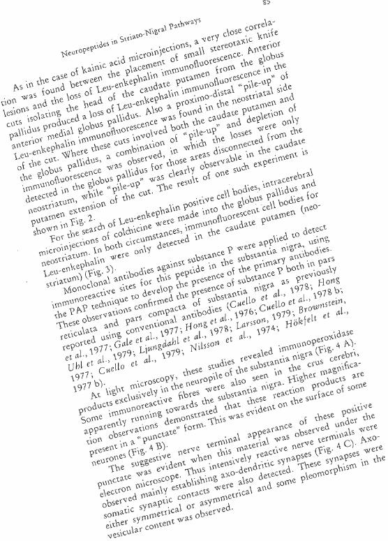
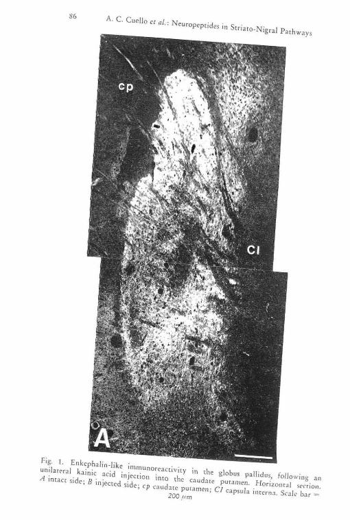
Fig. I. Enkephalin-Uke immuno<cactivity in the globu, pallidu" fOllowing an unila",,1 kainic acid injec<ioll into the caudate pUtamen. Ho,izontal ""ion. A intac< side, B injected side, cp cauda" putamen; Cl cap,ula inte,"a. Scale bac ~
200 ."m


Fig. 2. "Pile."p" of "kephalin.immuno"a',ive matccial (mows) in the tOst,,1 side of a COtonal knife c.'I. involving the ant".iot pan of the glob", pallid", and the adjacent caudate pUtamen. Note the intense n"ot<scencc in th e ponio
n of the globu, pallid"s tow.1 to the '''t.
cp caudate putamen; gp glob" pallid"" dashed line indicates the knife C"t. Scale b" = lOO!,,"

A. C. CuelJo et al.: Neuropeptides in S(riaco-Nigra\ Pathw~ys 89
Fig. 3. Enkephalin-immtlnoreactive cell bodies in the caudate putamen after intrapallidal colchicine (arrows). Arrowheads indicate bundles of EK-immunoreactivc
nerve fibers Scale bar = SO ft,m

ril·' . A Low ""'<"ioe"io" ",i"og"ph of ,he "" .'''bsr'n,i, nig", "'caling '"ban" J> i"'''',,,,ore.,,,i,,,y bo," in pars 'omp<>C<n '"d ,wi" '''a . Ne I / J
4
H L p,i",acy ''1 'ibod y d"clopCd '" i,h ,he PNo, id."., n ,ip<,o., id"" t<ehn'q "'. CC en" ccr, bri. Se"e h", ,', 200 Um. /J fligh" . m'g"j O'ntio
n
miccog"ph {"om ,h, "no< prep.1 ""ion .H A. No'e P"'o , id"". "'.crio" P"od"", ; n ,he ne" ropile. "'''Ound i ng n
o
,,.,.«<,,; 'e ncuron,l cell bodi" in ,be Pars cOmpacta. Sea le h .. · ,~ 100 J'm. C £1 ""on '" k"ng "'ph of ,he ,ubsta 0 ,i, ni g<a ' ho '" iog n ",b"'nee P im'nuno""'''ive ne"" ""oi,,,oI ''1 P'rs '·"i,ul." . Note ",,,ion P"od",,, '«PtiMed With '''1,11 and I"g, 'Yn'p,i, vesd"
Sc;de b(lr :~ l,um
:>
o () c ~ 0" ~
~
\0 o
Neuropeptides in Striato-Nigral Pathways 91
Discussion
The highest concentrations of enkephalins are found in the corpus striatum (fIde et al., 1975; Simantov et al., 1977, Say et al., 1978; W'atson et al., 1977; Del Fiacco et al., in preparation). From immunohisrochemical studies carried out on untreated animals it appears evident (hac (hese peptides are largely restricted to the globus pallidus (CuelLo and Paxino5, 1978; fide et al., 1978). Biochemcial data indicate that substantial amounts of enkephalins are also presem in (he neostriatum (Miller et al., 1978 a, b; Simanlov et al., 1976; Simanlov et al., 1977; Young et al., 1977). Funhermorc, cnkephalin containIng neurones, as well as nerve terminals, have been demonstrated within (hc caudate putamen (Elde et al., 1976; Simantov et al., 1977; \Vatson et al., 1977, Sar et al., 1978; HokfeLt et ai., 1978; Fratla et al., 1977; Pickel el al., 1980). This, however, does nOt clarify whether they are inrerneurones or long cirCUit elements within the scriawf\1. le has been proposed that this immunoreactivity represented to Cl long-en kephalincrgic pathway connecting thc caudate putamen with che globus pallidus on the basis of deafferentation studies and electrolytic lesions (CHello et al., 1978 c). These contemions have been challenged for at le2.si: one group (Correa et al., 1979). In some swdies enkephalincontaining cells are referred to as imcmeurones wiLhin the corpus striatum without clarifying their location in (he neostriatum Or globus paJiidus. These circumstances prompted us (0 re-examine (he anatomical orgonization of the "enkephalinergic" neurones within the corpus striatum. The results here summarized give further suppon (0
the concept that the "enkephalinergic" neurones in the strjawm are definitely organized as a "long-pathway", connecting the neostriatum (caudate putamen) with (he globus pallidus. This is indicated by the fact thar kainic acid, which is reported w cause a neuronal loss without damaging the fibre "en passage" in Ihis nuclear complex (Coyle et al., 1978) produces a depletion in rhe enkephalin-immunoreactivity of the globus pallidus only whcn mjected into the caudate pu("men. Similarly, che unilaceral mlcroknife CUtS in the corpus striatum only produced loss of immunofluorescent Leu-enkephalin material in the globus pallidus when they interrupted the connections between the caudate pmamen and the globus pallidus. The results obtained also indicate a radial concentric tOpographic innervation of the globus pallidus by these "enkephalinergic" neurones. This would be consistent with the presence of such neurones as revealed by conventional ncuroanawmical tracing tedmiques (Kemp et al., 1971; Carpenter, 1976). It is also consistent with the presence of enkephalinpositive cell bodies in the caudate putamen following incravenmcular

92 A. C. Cllel]o et at. :
injections of colchicine (Say et al.) 1978; H ok/ell et al., 1977). It could be argued chat intraventricular injections of colchicine would only affect (he axonal transport in neurOl1es located in (he caudate ,Pu[llrnen, because of their proximity to the ventricular surface. To overcome such criticism, we made intracerebral microinjections ot colchicine) inclependendy, into the caudate pu£amcn and irll:o [he globus pallidus itself . The bce chat positive cell bodies wtre found only in the former division of the sU'ianJn1, fLHthel' reinforces the argument of a long enkephalinergic p~thway .
The presence of this enkeph<1linergic link between [he caudate plllamen and the globus pallidus does not preclude [he possible ex i Stcnce of <.\xonaJ coil a [era Is of 1 he same cells, branching wi ch in the ljmits of the caudaTe putamen. In this direction Picket et al. (1980) have found enkephalin-imnwnoreactive material in axons and
rep ~ I G '
I
GP
fig. ~ . SC.h 21ll.:11ic rct>reselH;ll iol\ of (he postulaled syn(lp,ic conn~clions of pC~)lidc. col'tca,ining t\(~lll"()n<?~ in the srrio- nig r: !\ syw;m, Bent arrows ind ic.i.tc po ss ible ;\XOdend nllc. ;'\xo-;'\xonic and dend rO' .1.XOll it IllfcraCl iOl1s, J1CC nude ',l '; ;'ICCUI))\)CIIS;
er> C<Hlcbce pU[;I[)len; GP globus pallidusj SN Sllbsr~l)(i;) nigr;\ ; VT A VCIHl'O tegm~l\t<ll ar(!:\; Drl dopam ;n<:: CCK colecis(okln;n; G ;' -:1lTl inoblllyric ;\ cid
(GABA); EK cnkephnlin; 5P 511bHrtnC~ P

Neuropeptides in Stria to-Nigra I Pachways 93
nerve terminals in the globus pallidus as well as in neuronal perikarya and dendrites in the neostriatum. Our preliminary EM observations basically agree with this possibility. A high density of opiate receptors has been described in the neostriatum (Atweh and Kuhar, 1977; Simantov et al., 1977). Nevertheless [he incidence of enkephalinimmunoreactive elements in the caudate putamen is incomparably inferior to that of the globus pallidus. Therefore, the abundant "opiate" recepwrs described in the caudate putamen may respond to either clenciriticaJly or axonally (coHaterals) released enkephalins.
The position of these enkephalinergic neurones in the corpus srriarum are schematically represented in Fig. 5.
The immunocytochemical application of monoclonal antibodies against substance P in the substantia nigra confirmed the presence of this peptide in this area of tne rat brain (Cuello et al., 1978 b, c; Ljungdahl et al., 1978; Hong et al., 1977; Nilsson et al., 1974; Brownstein et al., 1976; Kanazawa et al., 1976).
The analysis of the electron microscopic material would suggest the existence of more than one population of substance P-conraining neurones. If confirmed, this would be a very inceresting phenomenon in the light of the co-existence of classical neurorransmitters with neurally active peptides. A case for such neurones has been advanced by Hokfelt et al. (1980) for dopamine-CCK containing neurones, connecting the ViA and substantia nigra with the nucleus accumbens.
The geometry of these neurones, containing putative neurotransmitter peptides, and the substance P-containing strio-nigral fibers are schematicaUy represented in Fig. 5, in relation to other transmitter-specific neurones.
Acknow ledgement
This work was made possible with grants (rom The Medical Research Council, U.K., The Royal Society and The Wellcome Trust. The technical assistance of Mr. 5teven BramweIl and Miss Sara Patel is acknowledged along with the secretarial help of Mrs. EBa IIes.
References
Atweh, S. F., Kuhar, M. J.: Autoradiographic localization of opiate recepwTs in rat brain. Ill. The telencephalon. Brain Res. 134, 393-405 ( 1977).
BYownslein, M. J., Mroz, E. A., Kizer, ]. 5., Palkovits, M., Leeman, S. E.: Regional distribution of substance P in the brain of the cat. Brain Res. 116, 299-305 (1976).

A. C. Cuello
,Ad. j., Mroz, E. A, Tappal, M. L., Leeman, S. E.: On the Orlglll of substance P and glutamic acid decarboxylase (GAD) in thc substantia nigo Brain Res. 135,315-323 (1977).
Carpenter, M. B.: Anaromy of [he basal ganglia and related nuclei: A review. In: Advances in Neurology, Vol. 14 (ELdridge, R., Fahn, 5., eds.), pp. 7-48. New York: Raven Press. 1976.
Coons, A. H., Kaplan, M. H.: Localization of antigens ill tissue cells. n. Improvemellts in a method for the detection of antigcns by means of
ilnribody. J. Exp. Med . . Innis, R. E., Hesccr,
of the source of cut, electrolytic and kit
(USA.), Vol. 5, . M. E., Kuhar,
. R., Snyder, S. H.: ['at globus ~allidus h Meeting Soc. for
of kainic acid: i bodies while sparing
axolls of passage. J. Comp. Neurol. 180,301 (1978). CI/eilo, A. c., Emson, P. c., Del Fiacco, M., Gale, }., !vcysen, L L,
jessell, 7. M., J(anazawa, I., Paxinos, G., Quik, M.: Distribution and release of substance P in the central nervous system Ill: Cenually Actil)g Peptides (H.tghes, )., cd.), pp. 135-1S6. London: Macmill;)1l Press. 1978 a.
Cuello, A. C., Gal/re, G., Mi/stein, c.: Detection of sul)S[:t!1CC P in tile ccnrral nervous system by a monOC 10Ji8]
3532-3S36 (1979). , Kanazawa, I.: The
ill the rat central (1978b).
, ;',filslein, c., Priestley, tochemistry with specia I
system Res. Bull. 5, 575-587 (
Proc. N;ltl. Acad.
substance P immuno; Comp. Neurol 178,
,,,onoci.onai antibodies in l he central nel"VOUS
Cuclio, A. c., Paxinos, G.' Evidence for a long leu-enkephalin strio-lJallidal pathway in rat brain. Nature. 271, 178-180 (1978 c).
Elde, R., Hok/elt, 7., johansson, 0., Terenil.ts, L.: Immunohistochemiul studies using annbodies to leucine-enkephalin: initial observations OIl
the nervous system of the rat. Neurosci. 1,349-351 (1976). Emson, P. c.: Peptides as neurotransmitter candidates in the mammalian
CNS. Prog. in NeurobioL 1],61-116 (1979). H.-Y.7., Hong,
content in brain strucru rats. Nature 268,
. j. S., Guidolti, A.: to-nigra I neurons. Brain
R., }ohansson, D., i ke immunoreactivity
Stabil iry of Metmonph.'ne-dependent or fOOl
tce P and GABA in 75(1977).
, L.: Distribution of ual nervous system.
1. Cell bodies. Neurosci. Letrs. 5, 25-31 (1977 a).
Hok/elt, 7., }ohansson, 0., Kellerth, f.-O., Ljungdahl, A., Ni/sson, G, Nygards, A., Pernow, E.: Immunohistochemical distribution of sub-
NeuropepLldes III Striaro-Nigra! Pathways 95
stance P. In: Substance P. Nobel Symposium 37 (von Euler, U., Pernow, B., eds.), pp. 117-145. New York: Raven Press. 1977.
Hokfeit, T., Johansson, 0., Ljungdahl, A., Lundberg. j. M., Schultzbel'g, M.: Peptidergic neurones. Nature 284, 515-521 (1980).
HokJelt, 7., Rehfeld, }. F., Skirboll, L., /vemark, B., Goldstein, M., Markey, K.: Evidence for co-exisrence of dopamine and CCK in mesolimbic neurones. Nature 285.476-478 (1980).
Hong, }. S., Yang, H.-Y. T., Racagni. C., Costa, E.: Projections of substance P containing neurons from neostriatum to substantia nigra. Brain Res. 122,541-544 (1977).
Kanazawa, I., Bird, E., O'Connell, R., Powell, D.: Evidence for a decrease in substance P content of substantia nigra in Huntington's chorea. Brain Res. 120,387-392 (1977 b).
Kanazawa, J., Emson, P. C., CueUo, A. C.: Evidence for the existence of substance P-containing fibres in striato-nigral and pallido-nigral pathways in rat brain. Brain Res. 119,447-453 (1977 a).
Kanazawa, I., lessell, T. M.: Post-mortem changes and regional distribution of substance P in the rat and mouse nervous system. Brain Res. 117, 362-367 (1976).
Kemp. ]. M., Powell, T. P. S.: The connections of the striatum and globus pallidus: synthesis and speculations. Phil. Trans. R. Soc. (Lond.) 262, 441-457 (1971).
Lamon, L.-I., Childers, 5., Snyder, S. H.: Met- and Leu-enkephalin immunoreactivity in separate neurones. Nature 1$2, 407-4'10 (1979).
Ljtmgdahl, A., Hokfelt, T., Nilsson, G.: Distribution of substance P-like immunoreactivity in the central nervous system of the rat. 1. Cell bodies and nerve terminals. Neurosci. J, 861-943 (1978).
Miller, R.}., Chang, K.-]., Cooper, B., Cuatrecasas, P.: Radioimmunoassay and characterization of enkephalins in rar rissue. J. BioI. Chem. 253, 53\-538 (1978 a).
Miller, R.]., Chang. K.-}., Cuatrecasas, P., Wilkinson, 5., Lowe, L., Beddel, C, Follen/ant, c.: Distribution and pharmacology of enkephalins and related opiate peptides. In: Centrally Acting Peprides (Hughes, j., ed.), pp. 195-213. New York: Macmillan Press. 197B b.
Mroz, E. A., Brownstein, M.]., Leemo.n, S. E.: Evidence for substance P in the stria!o-nigral tract. Brain Res. 125, 305-311 (1977).
Nilsson, G., Hokfeit, T., Pernow, B.: Distribution of substance P-like immunoreactivity in the rat central nervous system as revealed by immunohistochemistry. Med. BioI. 52, 424-427 (1974).
Pickel, V. M., Sunw.l, K. K., Beckley, S. C., Miller, R. l., Reis, D. j.: ImmunocY[Qchemical localization of enkephalin in the neostriatum of the rat brain: a light and electron microscopy study. ]. Comp. Neurol. 189,721-740 (1980).
Sar, M., Stump!, E., Miller, R. j., Cuatrecasas, P.: ImmunohistOchemical localizafion of enkephalin in rat brain and spinal cord. J. Comp. Neurol. 182, 17-38 (L 978).

96 A. C. CllelJo Cl al.: Nct1ropepticles in S(riaco-Nigr:\! P;"Hhw~ys
SimanlDV, R. ., /(uhar, M. )., Pastcrrurk, C. W ., Snyde l" , S. H .: The reglO]);"!! ctisrriburion of a morphin -like (acwr enkepha! :n in mon\{ey br~in. Br .. in Res . 106, 187-197 (1976) .
Simancov , R ., T<1.1har, M. j., Uhl , G .> Snyder, S, H.: Opioid peptide cnkephClIIIl: immunohistochemical mapping il1 rH central nerVOLlS s>,scem. Proc . N~tJ. ACAci. Sei. US.A , 7') ,2167-2171 (1977).
Snyd.cr, S. n.: Brain peprides :l$ neurorransmitcers. Science 29, 976--983 (19S0) .
Somogyi, P.: The study of Golgi stained cells find of experimental dtgener;1-(ion under the e!eclfOI) microscope : a direct method for che idenrificl(jo'l in rhe visual cortex of three successive links in a neuron chain . Ncu(osci . ),167-180 (1978) .
Slernbcrge-r, L. A ' I Hardy, P. H., Cu(ulis , J. j., Meycr, H. G .: The unlabelled anribody enz.yme method of immunohiswchemiscry: pl·epJfJ.tlon and propenies of soluble ant;gen~an{ibody complex (hol's€radlSh peroxidase) and ifS use in inenc;{lcl(ioll of spirochetcs. j. Hiscochem . Cyrochem . 18) 315-333 (1970).
Warson, S. j., Akiil H., Sullivan, S., Barchas, j. D.; I mlnunocytOchcmiol !ocaliz.aTion of melhiollille-enkeph;din : preliminary obser"J [ions . Life 56.21,733-738 (1977).
Yang l H, Y. , Hong , j.-5., Cos/tt , E. : Regional distribution of Leu- ~Ild Mecenkephalin In rar brain. Neuroph;nmacol. 16 1 30}-)07 (1977).
Authors' address: Dr . A . C. Cuelio, University Dep:lnmenr of Pharmtlcology, South Parks Ro;'\d, Oxford , Eng !>\))d.
Printed il) AIIS!ri.1