Embed Size (px)
Citation preview
fphys-09-01167 August 22, 2018 Time: 9:40 # 1
REVIEWpublished: 23 August 2018
doi: 10.3389/fphys.2018.01167
Edited by:Giselle Melendez,
Wake Forest University, United States
Reviewed by:Owen Llewellyn Woodman,
Baker Heart and Diabetes Institute,Australia
Wei Kong,Peking University, China
*Correspondence:Kristen J. Bubb
Specialty section:This article was submitted to
Integrative Physiology,a section of the journalFrontiers in Physiology
Received: 30 April 2018Accepted: 03 August 2018Published: 23 August 2018
Citation:Lo CCW, Moosavi SM and Bubb KJ
(2018) The Regulation of PulmonaryVascular Tone by Neuropeptides
and the Implications for PulmonaryHypertension. Front. Physiol. 9:1167.
doi: 10.3389/fphys.2018.01167
The Regulation of PulmonaryVascular Tone by Neuropeptides andthe Implications for PulmonaryHypertensionCharmaine C. W. Lo1, Seyed M. Moosavi1,2 and Kristen J. Bubb1*
1 Kolling Institute of Medical Research, University of Sydney, St Leonards, NSW, Australia, 2 School of Life Sciences,University of Technology Sydney, Ultimo, NSW, Australia
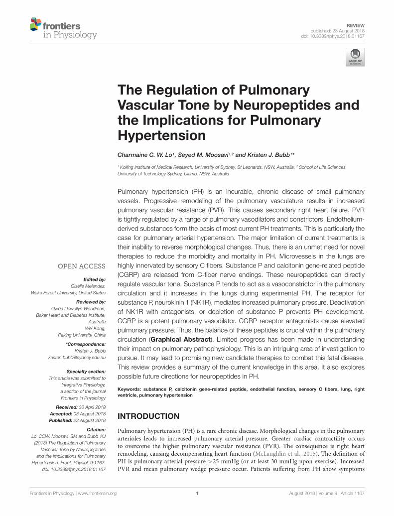
Pulmonary hypertension (PH) is an incurable, chronic disease of small pulmonaryvessels. Progressive remodeling of the pulmonary vasculature results in increasedpulmonary vascular resistance (PVR). This causes secondary right heart failure. PVRis tightly regulated by a range of pulmonary vasodilators and constrictors. Endothelium-derived substances form the basis of most current PH treatments. This is particularly thecase for pulmonary arterial hypertension. The major limitation of current treatments istheir inability to reverse morphological changes. Thus, there is an unmet need for noveltherapies to reduce the morbidity and mortality in PH. Microvessels in the lungs arehighly innervated by sensory C fibers. Substance P and calcitonin gene-related peptide(CGRP) are released from C-fiber nerve endings. These neuropeptides can directlyregulate vascular tone. Substance P tends to act as a vasoconstrictor in the pulmonarycirculation and it increases in the lungs during experimental PH. The receptor forsubstance P, neurokinin 1 (NK1R), mediates increased pulmonary pressure. Deactivationof NK1R with antagonists, or depletion of substance P prevents PH development.CGRP is a potent pulmonary vasodilator. CGRP receptor antagonists cause elevatedpulmonary pressure. Thus, the balance of these peptides is crucial within the pulmonarycirculation (Graphical Abstract). Limited progress has been made in understandingtheir impact on pulmonary pathophysiology. This is an intriguing area of investigation topursue. It may lead to promising new candidate therapies to combat this fatal disease.This review provides a summary of the current knowledge in this area. It also explorespossible future directions for neuropeptides in PH.
Keywords: substance P, calcitonin gene-related peptide, endothelial function, sensory C fibers, lung, rightventricle, pulmonary hypertension
INTRODUCTION
Pulmonary hypertension (PH) is a rare chronic disease. Morphological changes in the pulmonaryarterioles leads to increased pulmonary arterial pressure. Greater cardiac contractility occursto overcome the higher pulmonary vascular resistance (PVR). The consequence is right heartremodeling, causing decompensating heart function (McLaughlin et al., 2015). The definition ofPH is pulmonary arterial pressure >25 mmHg (or at least 30 mmHg upon exercise). IncreasedPVR and mean pulmonary wedge pressure occur. Patients suffering from PH show symptoms
Frontiers in Physiology | www.frontiersin.org 1 August 2018 | Volume 9 | Article 1167
fphys-09-01167 August 22, 2018 Time: 9:40 # 2
Lo et al. Neuropeptides in Pulmonary Hypertension
GRAPHICAL ABSTRACT |
such as dyspnea, fatigue, chest pain, near syncope, syncope, leg orperipheral edema, angina, palpitations, and abdominal distension(Rich et al., 1987; Barst et al., 2004). PH is hard to diagnose asinitial symptoms are non-specific. Fatigue, malaise, and exerciseintolerance are often misdiagnosed as the patient being unfit.Asthma is also a common misdiagnosis due to the presence ofdyspnoea. In fact, PH diagnosis often takes up to 2 years aftersymptom onset. Confirmation of PH sometimes occurs only afterthe progression of right heart failure.
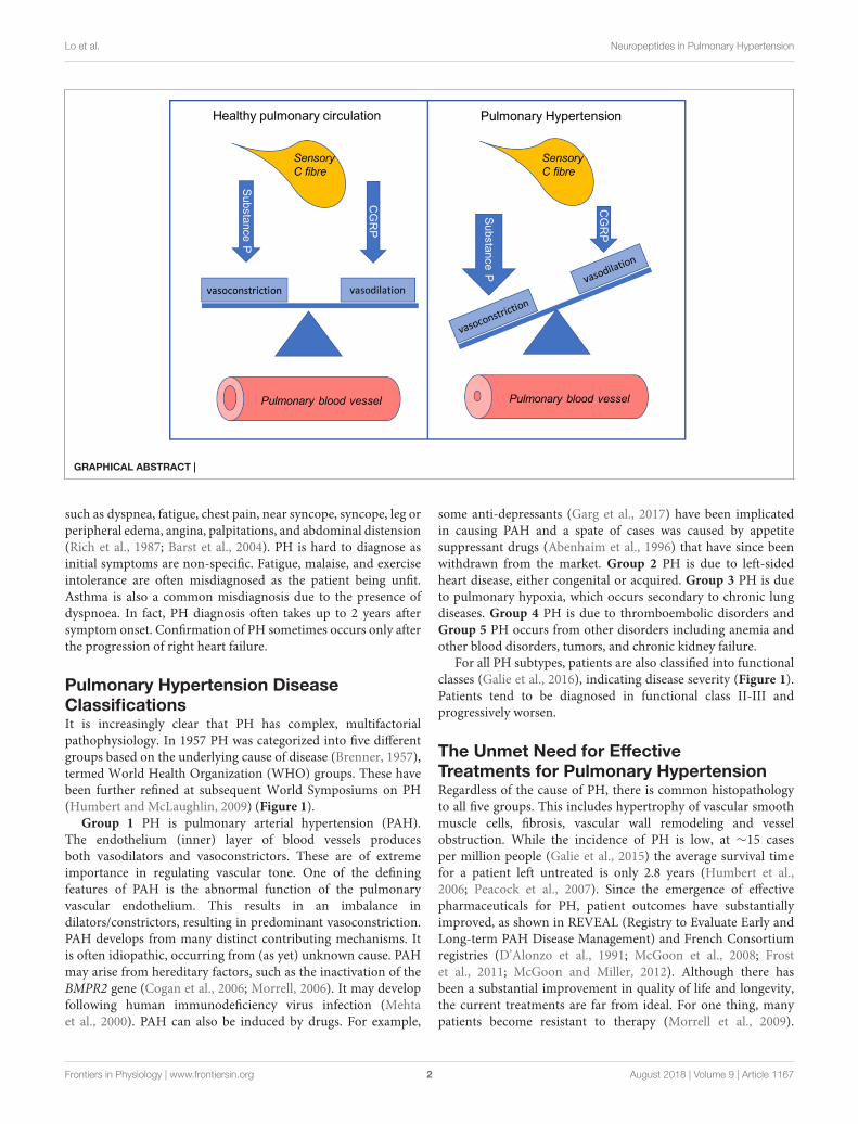
Pulmonary Hypertension DiseaseClassificationsIt is increasingly clear that PH has complex, multifactorialpathophysiology. In 1957 PH was categorized into five differentgroups based on the underlying cause of disease (Brenner, 1957),termed World Health Organization (WHO) groups. These havebeen further refined at subsequent World Symposiums on PH(Humbert and McLaughlin, 2009) (Figure 1).
Group 1 PH is pulmonary arterial hypertension (PAH).The endothelium (inner) layer of blood vessels producesboth vasodilators and vasoconstrictors. These are of extremeimportance in regulating vascular tone. One of the definingfeatures of PAH is the abnormal function of the pulmonaryvascular endothelium. This results in an imbalance indilators/constrictors, resulting in predominant vasoconstriction.PAH develops from many distinct contributing mechanisms. Itis often idiopathic, occurring from (as yet) unknown cause. PAHmay arise from hereditary factors, such as the inactivation of theBMPR2 gene (Cogan et al., 2006; Morrell, 2006). It may developfollowing human immunodeficiency virus infection (Mehtaet al., 2000). PAH can also be induced by drugs. For example,
some anti-depressants (Garg et al., 2017) have been implicatedin causing PAH and a spate of cases was caused by appetitesuppressant drugs (Abenhaim et al., 1996) that have since beenwithdrawn from the market. Group 2 PH is due to left-sidedheart disease, either congenital or acquired. Group 3 PH is dueto pulmonary hypoxia, which occurs secondary to chronic lungdiseases. Group 4 PH is due to thromboembolic disorders andGroup 5 PH occurs from other disorders including anemia andother blood disorders, tumors, and chronic kidney failure.
For all PH subtypes, patients are also classified into functionalclasses (Galie et al., 2016), indicating disease severity (Figure 1).Patients tend to be diagnosed in functional class II-III andprogressively worsen.
The Unmet Need for EffectiveTreatments for Pulmonary HypertensionRegardless of the cause of PH, there is common histopathologyto all five groups. This includes hypertrophy of vascular smoothmuscle cells, fibrosis, vascular wall remodeling and vesselobstruction. While the incidence of PH is low, at ∼15 casesper million people (Galie et al., 2015) the average survival timefor a patient left untreated is only 2.8 years (Humbert et al.,2006; Peacock et al., 2007). Since the emergence of effectivepharmaceuticals for PH, patient outcomes have substantiallyimproved, as shown in REVEAL (Registry to Evaluate Early andLong-term PAH Disease Management) and French Consortiumregistries (D’Alonzo et al., 1991; McGoon et al., 2008; Frostet al., 2011; McGoon and Miller, 2012). Although there hasbeen a substantial improvement in quality of life and longevity,the current treatments are far from ideal. For one thing, manypatients become resistant to therapy (Morrell et al., 2009).
Frontiers in Physiology | www.frontiersin.org 2 August 2018 | Volume 9 | Article 1167
fphys-09-01167 August 22, 2018 Time: 9:40 # 3
Lo et al. Neuropeptides in Pulmonary Hypertension
FIGURE 1 | PH World Health Organization classification and functional class. Five different major sub-types of PH according to World Health Organizationclassification and functional classes are out-lined. Adapted from (Humbert and McLaughlin, 2009; Galie et al., 2016).
In addition, PH remains a progressive and terminal disease.Current treatments have shown limited ability to reverse vascularand cardiac remodeling (Bubb et al., 2015). Thus, the search fornovel and breakthrough treatments for PH continues in earnest.
In order to establish new PH treatments, it is importantto determine how existing treatments can be improved. Thefirst part of this review outlines the current treatments of PH.Subsequently, we review the effects of the sensory C-fiber-derivedneuropeptides on the lung circulation. Several neuropeptidesshow promising effects and are being investigated for PHtreatment. These include neuropeptides released from C-fibers:substance P and calcitonin gene-related peptide (CGRP).Both are important regulators of the pulmonary circulation.Modulation of either of the C-fiber-derived neuropeptides canreverse progression of experimental PH. Yet, their use has notprogressed beyond pre-clinical research. We also discuss theirpotential as novel treatments for PH.
CURRENT TREATMENTS FORPULMONARY HYPERTENSION
Current treatment options for all forms of PH include primarytherapies directed at treating the underlying cause of thedisease and broad therapies that alleviate the symptoms. Generaltreatment prescribed at the discretion of the primary carephysician includes the use of warfarin, diuretics and oxygen.The aims of these therapies are to alleviate volume and
viscosity-induced pressure within the pulmonary circulation,reduce hypoxia, and treat right heart failure. Non-pharmaceuticalapproaches include lung transplant and double heart-lungtransplant. Surgical approaches can be utilized in some cases,often when all other options have been exhausted. The primarypharmaceutical therapies that are approved for PH treatment arethe main focus of this review and are detailed in subsequentsections.
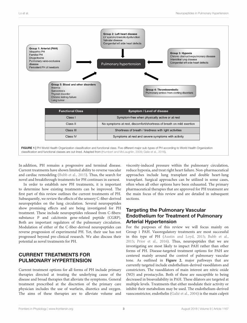
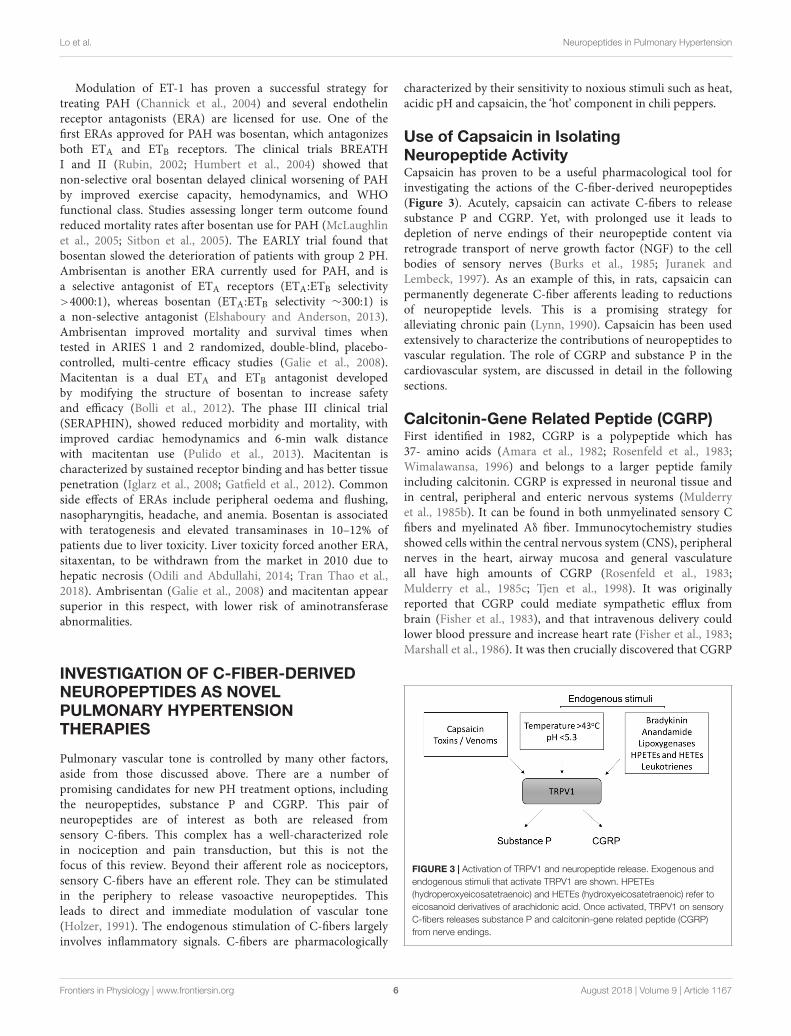
Targeting the Pulmonary VascularEndothelium for Treatment of PulmonaryArterial HypertensionFor the purposes of this review we will focus mainly onGroup 1 PAH. Vasoregulatory treatments are most successfulin this type of PH (Austin and Loyd, 2015; Bubb et al.,2015; Prior et al., 2016). Thus, neuropeptides that we areinvestigating are most likely to impact PAH rather than otherforms of PH. Disease-targeted treatment options for PAH arecentered mainly around the control of pulmonary vasculartone. As outlined in Figure 2, major pathways that arecurrently targeted include endothelium-derived vasodilators andconstrictors. The vasodilators of main interest are nitric oxide(NO) and prostacyclin. Both of these are susceptible to beingdecreased in bioavailability in PAH. These dilators are targeted atmultiple levels. Treatments that either modulate their activity orinhibit their metabolism may be used. The endothelium-derivedvasoconstrictor, endothelin (Galié et al., 2004) is the main culprit
Frontiers in Physiology | www.frontiersin.org 3 August 2018 | Volume 9 | Article 1167
fphys-09-01167 August 22, 2018 Time: 9:40 # 4
Lo et al. Neuropeptides in Pulmonary Hypertension
FIGURE 2 | Summary of current PAH treatments. Currently licensed treatments for PAH are shown inside the circles with broken lines indicating their mode of actionwithin vascular signaling pathways. AC, adenylate cyclase; cAMP, cyclic adenosine monophosphate; cGMP, cyclic guanosine monophosphate; COX,cyclooxygenase; ECE, endothelin converting enzyme; eNOS, endothelial nitric oxide synthase; ET-1, endothelin; ERA,B, endothelin receptor A,B; GTP, guanosinetriphosphate; IP, inositol monophosphate; NO nitric oxide; PDE5, phosphodiesterase 5; PGI2, prostacyclin; sGC soluble guanylate cyclase.
causing excessive vasoconstriction. Antagonists of the endothelinreceptor/s are in clinical use for PAH. Calcium-channel blockersare also used to inhibit the calcium-dependent smooth musclecontraction.
Endothelium-Dependent Pulmonary VasodilationThe reduction in pulmonary pressure by stimulating pulmonaryendothelium-dependent vasodilation resulted in the firstapproved treatment for PAH. This continues to be the basisbehind the development of the majority of PAH treatments.
Nitric oxide-cyclic guanosine monophosphate signalingNO and the associated signaling pathways have a majorrole in promoting pulmonary vasodilation. The NO signalingpathway can be summarized as follows: NO activates solubleguanylate cyclase (sGC); this increases the second messenger,cyclic guanosine monophosphate (cGMP); cGMP then activatesprotein kinase G (PKG); PKG mediates vasodilation bydecreasing intracellular calcium by a number of mechanisms.NO also mediates anti-inflammatory and anti-coagulatory effects.The low bioavailability of NO is a major occurrence in patientswith PH. This is replicated in pre-clinical models of the disease(Michelakis, 2003). Upregulation of NO, either exogenously orendogenously, has been the primary aim of many decades oftrials. Inhaled NO (Steiner et al., 2005) is useful in neonatessuffering from persistent PH of the newborn. However, in adultsit tends to result in methemoglobinemia, increased pulmonaryoedema and a potentially life-threatening hypertensive reboundwhen therapy is ceased. It also suffers a short half-life and
is prohibitively expensive and cumbersome for patients toadminister (Michelakis, 2003; Steiner et al., 2005; Baliga et al.,2011). NO donors were developed in an attempt to improve thedelivery of NO. Nebulised NONOates (Vanderford et al., 1994;Hampl et al., 1996) and inhaled glyceryl trinitrate (Goyal et al.,2006) effectively lower PVR. Yet they notoriously lack specificityfor the pulmonary circulation (Baliga et al., 2011). Thus, targetingof the intermediates in the NO signaling pathway is a moreattractive option for improving vasodilation.
In vascular endothelial cells, L-arginine is converted toNO (and the by-product L-citrulline) by endothelial NOsynthase (eNOS) in a two-step oxidation reaction. Thisreaction utilizes nicotinamide adenine dinucleotide phosphate(NADPH), molecular oxygen and the essential cofactor,tetrahydrobiopterin (BH4) (Palmer et al., 1988; Andrew andMayer, 1999; Forstermann, 2010). Phosphorylation of eNOS is animportant regulatory mechanism for NO generation. It can alsooccur in response to shear stress, estrogen, vascular endothelialgrowth factor, and insulin. In normally functioning arteriesthe major stimulus for eNOS phosphorylation is increasedintracellular calcium. This proceeds by binding of calcium tocalmodulin in the oxygenase domain of eNOS, increasing the rateof NADPH electron transfer within the domains. In PAH (likeother cardiovascular diseases) eNOS can exist in an ‘uncoupledstate.’ Usually the substrate L-arginine or co-factor BH4become rate-limiting. In this scenario, eNOS transfers electronsto molecular oxygen, rather than oxidizing L-arginine andsuperoxide is produced. Superoxide is a reactive oxygen speciesthat is prevalent in vascular dysfunction (Verhaar et al., 2004).
Frontiers in Physiology | www.frontiersin.org 4 August 2018 | Volume 9 | Article 1167
fphys-09-01167 August 22, 2018 Time: 9:40 # 5
Lo et al. Neuropeptides in Pulmonary Hypertension
Supplementation with BH4 improves systemic hypertension(Landmesser et al., 2003). Evidence suggests that this strategymay also be effective in PAH (Baliga et al., 2011). Stimulatingthe coupling of eNOS and BH4 to promote NO rather thansuperoxide production has proven beneficial in experimental (Jinet al., 1992) and clinical PH (Saadjian et al., 1998). Increasingthe substrate for NO by L-arginine administration can likewiseattenuate PH in the experimental setting (Mitani et al., 1997)and acutely decreases PVR in PH patients (Mehta et al., 1995).Despite the extensive research, no therapeutic targets have beenlicensed that target NO generation.
Drugs that target the receptor for NO, sGC have provento be more successful. Activation of sGC by NO is dependenton binding of NO to a heme prosthetic group. This stimulatesa conformational change which activates the enzyme. Changesto the redox state of sGC, which can occur in PAH, canresult in modulation of sGC expression or activity (Privieroand Webb, 2010; Baliga et al., 2011). Mice lacking sGC aresensitive to experimentally induced PH (Vermeersch et al., 2007).This makes sGC an important target in PH. Pharmacologicalstimulation of sGC is complicated and drugs fall into twodistinct classes. sGC activators, (e.g., cinaciguat) are effective atactivating sGC when it is in an oxidized or heme-free state.sGC stimulators (e.g., riociguat) stabilize the enzyme in itsactive configuration and upregulate sGC activity. There is anadvantageous synergistic relationship of sGC stimulators and NOwhen they are co-administered (Stasch et al., 2011). ExperimentalPH was ameliorated after treatment with either sGC activatorsor stimulators (Dumitrascu et al., 2006; Stasch et al., 2011; Langet al., 2012). Both classes of drugs are promising, but, sGCstimulators have progressed to the clinic first. The PATENT trialsdemonstrated improved clinical outcome for patients with PAHtreated with riociguat (Ghofrani et al., 2012). Riociguat was firstFDA-approved for treatment of PAH in 2013 and is now licensedin many countries. It is also the only drug licensed to treat group4 PH after success in the CHEST trial (Lian et al., 2017).
The other major class of drugs to successfully treatPAH by targeting the NO-cGMP signaling pathway arephosphodiesterase (PDE) 5 inhibitors. PDEs metabolize cyclicnucleotides to their inactive form and some, including PDE5,are specific to cGMP. PDE5 is substantially upregulated in mostforms of experimental and clinical PH (Murray et al., 2002;Sebkhi et al., 2003; Wharton et al., 2005; Schermuly et al.,2007) and this led to extensive testing of the efficacy of PDE5inhibition for PH. Pre-clinical studies have provided insight intothe therapeutic mechanisms of PDE5 inhibitors. It has beenwell-established that sildenafil is effective in inhibiting PH inexperimentally induced models (Zhao et al., 2001; Schermulyet al., 2004; Steiner et al., 2005), and this is cGMP-dependent.Both sildenafil (Galie et al., 2005) and tadalafil (Galie et al., 2009)are effective in improving symptoms and outcomes of PH inpatients.
ProstacyclinProstacyclin was first identified in 1976 (Moncada et al.,1976) and is the main eicosanoid produced by vascularendothelial cells. It is synthesized by cyclooxygenase-dependent
conversion from arachidonic acid. Like NO, prostacyclin isalso a classic endothelium-dependent vasodilator and alsopossesses anti-inflammatory and anti-coagulatory properties.Prostacyclin acts predominantly by binding with cell-surface IPreceptors which stimulates adenylyl cyclase and cyclic adenosinemonophosphate (cAMP) production with downstream effectsmediated by protein kinase A (Mitchell et al., 2008). It isa particularly potent vasodilator in the lung circulation andPAH patients often exhibit decreased prostacyclin levels and/orreduced lung prostacyclin synthases (Mitchell et al., 2014).Prostacyclin analogs are a mainstay of PAH treatment but aregenerally restricted to functional class III and IV patients duemainly to limitations with their administration as outlined below.Prior to the use of epoprostenol (therapeutic prostacyclin) in1995 (Barst et al., 1996), there was no therapy for PAH, andpatients had a 1-year survival of 69% and a 5-year survival of38% (D’Alonzo et al., 1991). When first introduced, epoprostenolwas the only treatment available for PAH. It was widely usedeven though administration was challenging and side-effects weremarked. It has been largely superseded by the next generationof therapeutic prostacyclins, iloprost and treprostinil. Theseare available in inhalers or by subcutaneous or intravenousroutes. Recently the first non-prostacyclin that activates thecAMP signaling pathway has been developed. Selexipag is an IPreceptor agonist, which has the benefit of being available in oralformulation (Mitchell et al., 2014). It has proven successful inPhase III trials (Sitbon et al., 2015) and has recently been licensedfor PAH treatment.
Pulmonary Vasoconstriction by EndothelinThere are at least three isoforms of endothelin: endothelin(ET)-1, ET-2, and ET-3, of which ET-1 is the most potentand abundant in lungs (Matsumoto et al., 1989; Giaid et al.,1993a). First discovered in 1988 (Hickey et al., 1985; Yanagisawaet al., 1988), ET-1 is a peptide mainly produced by vascularendothelial cells. A number of physical factors affect ET-1production, including shear stress, epinephrine, angiotensin II,growth factors, cytokines and free radicals (Marasciulo et al.,2006). ET-1 activity is mediated by ET receptors, ETA andETB, both expressed on vascular smooth muscle cells (Seoet al., 1994). ET-1 is produced by ET converting enzyme (ECE)from a precursor molecule of ET (La and Reid, 1995), bigET-1. ET-1 induces increased intracellular calcium via ETA andETB receptor-mediated activation of phospholipase C (PLC)(Pollock et al., 1995) and increases mitogenesis by ETB-mediatedactivation of protein kinase C (Ohlstein et al., 1992), leading toboth vasoconstriction and cell hyperplasia. ETB, which is alsofound on endothelial cells, stimulates local vasodilators such asNO and prostaglandins. It also plays a role in the clearanceof ET-1, a feature unique to the lungs (Fukuroda et al., 1994;Kelland et al., 2010). PAH is associated with increased circulatingET-1 levels (Miyauchi et al., 1993). Expression of ET-1 has beenfound in plexiform lesions of lungs, where higher levels of ET-1correlate with increased PVR and structural abnormalities (Giaidet al., 1993b; Bressollette et al., 2001). PAH patients may alsohave reduced clearance of ET-1 in the lung (Stewart et al., 1991),contributing to vasoconstriction.
Frontiers in Physiology | www.frontiersin.org 5 August 2018 | Volume 9 | Article 1167
fphys-09-01167 August 22, 2018 Time: 9:40 # 6
Lo et al. Neuropeptides in Pulmonary Hypertension
Modulation of ET-1 has proven a successful strategy fortreating PAH (Channick et al., 2004) and several endothelinreceptor antagonists (ERA) are licensed for use. One of thefirst ERAs approved for PAH was bosentan, which antagonizesboth ETA and ETB receptors. The clinical trials BREATHI and II (Rubin, 2002; Humbert et al., 2004) showed thatnon-selective oral bosentan delayed clinical worsening of PAHby improved exercise capacity, hemodynamics, and WHOfunctional class. Studies assessing longer term outcome foundreduced mortality rates after bosentan use for PAH (McLaughlinet al., 2005; Sitbon et al., 2005). The EARLY trial found thatbosentan slowed the deterioration of patients with group 2 PH.Ambrisentan is another ERA currently used for PAH, and isa selective antagonist of ETA receptors (ETA:ETB selectivity>4000:1), whereas bosentan (ETA:ETB selectivity ∼300:1) isa non-selective antagonist (Elshaboury and Anderson, 2013).Ambrisentan improved mortality and survival times whentested in ARIES 1 and 2 randomized, double-blind, placebo-controlled, multi-centre efficacy studies (Galie et al., 2008).Macitentan is a dual ETA and ETB antagonist developedby modifying the structure of bosentan to increase safetyand efficacy (Bolli et al., 2012). The phase III clinical trial(SERAPHIN), showed reduced morbidity and mortality, withimproved cardiac hemodynamics and 6-min walk distancewith macitentan use (Pulido et al., 2013). Macitentan ischaracterized by sustained receptor binding and has better tissuepenetration (Iglarz et al., 2008; Gatfield et al., 2012). Commonside effects of ERAs include peripheral oedema and flushing,nasopharyngitis, headache, and anemia. Bosentan is associatedwith teratogenesis and elevated transaminases in 10–12% ofpatients due to liver toxicity. Liver toxicity forced another ERA,sitaxentan, to be withdrawn from the market in 2010 due tohepatic necrosis (Odili and Abdullahi, 2014; Tran Thao et al.,2018). Ambrisentan (Galie et al., 2008) and macitentan appearsuperior in this respect, with lower risk of aminotransferaseabnormalities.
INVESTIGATION OF C-FIBER-DERIVEDNEUROPEPTIDES AS NOVELPULMONARY HYPERTENSIONTHERAPIES
Pulmonary vascular tone is controlled by many other factors,aside from those discussed above. There are a number ofpromising candidates for new PH treatment options, includingthe neuropeptides, substance P and CGRP. This pair ofneuropeptides are of interest as both are released fromsensory C-fibers. This complex has a well-characterized rolein nociception and pain transduction, but this is not thefocus of this review. Beyond their afferent role as nociceptors,sensory C-fibers have an efferent role. They can be stimulatedin the periphery to release vasoactive neuropeptides. Thisleads to direct and immediate modulation of vascular tone(Holzer, 1991). The endogenous stimulation of C-fibers largelyinvolves inflammatory signals. C-fibers are pharmacologically
characterized by their sensitivity to noxious stimuli such as heat,acidic pH and capsaicin, the ‘hot’ component in chili peppers.
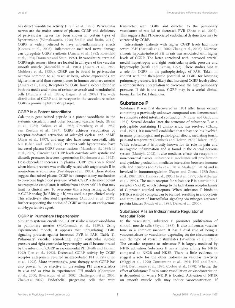
Use of Capsaicin in IsolatingNeuropeptide ActivityCapsaicin has proven to be a useful pharmacological tool forinvestigating the actions of the C-fiber-derived neuropeptides(Figure 3). Acutely, capsaicin can activate C-fibers to releasesubstance P and CGRP. Yet, with prolonged use it leads todepletion of nerve endings of their neuropeptide content viaretrograde transport of nerve growth factor (NGF) to the cellbodies of sensory nerves (Burks et al., 1985; Juranek andLembeck, 1997). As an example of this, in rats, capsaicin canpermanently degenerate C-fiber afferents leading to reductionsof neuropeptide levels. This is a promising strategy foralleviating chronic pain (Lynn, 1990). Capsaicin has been usedextensively to characterize the contributions of neuropeptides tovascular regulation. The role of CGRP and substance P in thecardiovascular system, are discussed in detail in the followingsections.
Calcitonin-Gene Related Peptide (CGRP)First identified in 1982, CGRP is a polypeptide which has37- amino acids (Amara et al., 1982; Rosenfeld et al., 1983;Wimalawansa, 1996) and belongs to a larger peptide familyincluding calcitonin. CGRP is expressed in neuronal tissue andin central, peripheral and enteric nervous systems (Mulderryet al., 1985b). It can be found in both unmyelinated sensory Cfibers and myelinated Aδ fiber. Immunocytochemistry studiesshowed cells within the central nervous system (CNS), peripheralnerves in the heart, airway mucosa and general vasculatureall have high amounts of CGRP (Rosenfeld et al., 1983;Mulderry et al., 1985c; Tjen et al., 1998). It was originallyreported that CGRP could mediate sympathetic efflux frombrain (Fisher et al., 1983), and that intravenous delivery couldlower blood pressure and increase heart rate (Fisher et al., 1983;Marshall et al., 1986). It was then crucially discovered that CGRP
FIGURE 3 | Activation of TRPV1 and neuropeptide release. Exogenous andendogenous stimuli that activate TRPV1 are shown. HPETEs(hydroperoxyeicosatetraenoic) and HETEs (hydroxyeicosatetraenoic) refer toeicosanoid derivatives of arachidonic acid. Once activated, TRPV1 on sensoryC-fibers releases substance P and calcitonin-gene related peptide (CGRP)from nerve endings.
Frontiers in Physiology | www.frontiersin.org 6 August 2018 | Volume 9 | Article 1167
fphys-09-01167 August 22, 2018 Time: 9:40 # 7
Lo et al. Neuropeptides in Pulmonary Hypertension
has direct vasodilator activity (Brain et al., 1985). Perivascularnerves are the major source of plasma CGRP and deficiencyof perivascular nerves has been shown in certain types ofhypertension (Wimalawansa, 1996; Smillie and Brain, 2011).CGRP is widely believed to have anti-inflammatory effects(Gomes et al., 2005). Inflammation-mediated nerve damagecan upregulate CGRP synthesis (Amara et al., 1982; Rosenfeldet al., 1984; Donnerer and Stein, 1992). In vasculature, terminalCGRPergic sensory fibers are located in all layers of the vascularsmooth muscle (Rosenfeld et al., 1983) (Amara et al., 1985;Mulderry et al., 1985a). CGRP can be found in perivascularneurons common to all vascular beds, where expressions arehigher in arterial than venous tissues in human coronary arteries(Amara et al., 1985). Receptors for CGRP have also been found inboth the media and intima of resistance vessels and in endothelialcells (Mulderry et al., 1985a; Hagner et al., 2002). The widedistribution of CGRP and its receptor in the vasculature makesCGRP a promising future drug target.
CGRP Is a Potent VasodilatorCalcitonin gene-related peptide is a potent vasodilator in thesystemic circulation and other localized vascular beds. (Brainet al., 1985; Kubota et al., 1985; Greenberg et al., 1987;van Rossum et al., 1997). CGRP achieves vasodilation byreceptor-mediated activation of adenylyl cyclase and cAMP(Aiyar et al., 1997), and may also have some cross-talk withNO (Chen and Guth, 1995). Patients with hypertension haveincreased plasma CGRP concentrations (Masuda et al., 1992; Liet al., 2009). Circulating CGRP levels correlate with systolic anddiastolic pressures in severe hypertension (Edvinsson et al., 1992).Dose-dependent increases in plasma CGRP levels were foundwhen blood pressure was artificially raised with angiotensin II innormotensive volunteers (Portaluppi et al., 1993). These studiessuggest that raised plasma CGRP is a compensatory mechanismto overcome high blood pressure. While CGRP is the most potentneuropeptide vasodilator, it suffers from a short half-life that maylimit its clinical use. To overcome this a long lasting acylatedα-CGRP analog (half-life ≥ 7 h) was used in a pre-clinical study.This effectively alleviated hypertension (Aubdool et al., 2017),further supporting the notion of CGRP acting as an endogenousanti-hypertensive agent.
CGRP in Pulmonary HypertensionSimilar to systemic circulation, CGRP is also a major vasodilatorin pulmonary arteries (McCormack et al., 1989a). Usingexperimental models, it appears that upregulating CGRPsignaling protects against increased PVR in PAH (Table 1).Pulmonary vascular remodeling, right ventricular systolicpressure and right ventricular hypertrophy can all be amelioratedby the infusion of CGRP in experimental PH (Keith and Ekman,1992; Tjen et al., 1992). Decreased CGRP activity, by CGRPreceptor antagonism resulted in exacerbated PH in rats (Tjenet al., 1992). More interestingly, gene therapy with CGRP hasalso proven to be effective in preventing PH characteristicsin vivo and in vitro in experimental PH models (Championet al., 2000; Bivalacqua et al., 2002; Chattergoon et al., 2005;Zhao et al., 2007). Endothelial progenitor cells that were
transfected with CGRP and directed to the pulmonaryvasculature of rats led to decreased PVR (Zhao et al., 2007).This suggests that PH-associated endothelial dysfunction may becorrected by CGRP.
Interestingly, patients with higher CGRP levels had moresevere PAH (Bartosik et al., 2002; Zhang et al., 2006). Likewise,chronic hypoxia-induced PH in rats was associated with higherlevels of CGRP. The latter correlated with increased arterialmedial hypertrophy and right ventricular systolic pressure andhypertrophy (Keith and Ekman, 1992). These studies hint ata role for CGRP in the pathophysiology of PAH. Taken incontext with the therapeutic potential of CGRP for loweringpulmonary pressure, it is likely that increased CGRP levels reflecta compensatory upregulation to overcome the high pulmonarypressure. If this is the case, CGRP may be a useful clinicalbiomarker for PAH diagnosis.
Substance PSubstance P was first discovered in 1931 after tissue extractcontaining a previously unknown compound was demonstratedto stimulate rabbit intestinal contraction (V Euler and Gaddum,1931). Several decades later the structure of substance P, as apolypeptide containing 11 amino acids, was resolved (Changet al., 1971). It is now well established that substance P is involvedin many physiological and pathological effects, mediating touch,pain and temperature (Lembeck and Holzer, 1979; Holzer, 1988).While substance P is mostly known for its role in pain andneurogenic inflammation and is found in the central nervoussystem (Mantyh, 2002), it also mediates effects via receptors innon-neuronal tissues. Substance P modulates cell proliferationand cytokine production, mediates interaction between immunecells and neurons (de Avila et al., 2014; Mashaghi, 2016) and isinvolved in immunoregulation (Payan and Goetzl, 1985; Steadet al., 1987, 1989; Haines et al., 1993; Ho et al., 1997; Schratzbergeret al., 1997). The main receptor for substance P is neurokinin 1receptor (NK1R), which belongs to the tachykinin receptor familyof G protein-coupled receptors. When substance P binds toNK1R a scaffold complex can be formed, resulting in endocytosisand stimulation of intracellular signaling via mitogen activatedprotein kinases (Grady et al., 1995; DeFea et al., 2000).
Substance P Is an Indiscriminate Regulator ofVascular ToneIn the vasculature, substance P promotes proliferation ofsmooth muscle cells (Payan, 1985). It also influences vasculartone in a complex manner. It has a dual role of being avasoconstrictor or vasodilator, depending on the circumstancesand the type of vessel it stimulates (Worthen et al., 1985).The vascular response to substance P is largely mediated byNK1R activation. Substance P has a higher affinity for NK1Rcompared to NK2R and NK3R. There is little evidence tosuggest a role for the other isoforms in vascular reactivity(Maggi et al., 1990; Constantine et al., 1991; Hall and Brain,1994; Berthiaume et al., 1995; Corboz et al., 1998). Whether theeffect of Substance P is to cause vasodilation or vasoconstrictionis dependent on where NK1R is located. Activation of NK1Ron smooth muscle cells may induce vasoconstriction. If
Frontiers in Physiology | www.frontiersin.org 7 August 2018 | Volume 9 | Article 1167
fphys-09-01167 August 22, 2018 Time: 9:40 # 8
Lo et al. Neuropeptides in Pulmonary Hypertension
TABLE 1 | Role of neuropeptides in experimental models of PH.
Species PH model Treatment Effect Reference
TRPV1
Rat Monocrotaline Capsaicin ↓ PH, ↓RVH Zhou and Lai, 1993; Katzman and Lai, 2000
Rat Pulmonary banding Capsaicin ↓ PH, ↓RVH Xu et al., 2017
Rat Hypoxia Capsaicin ↑ PH, ↑RVH Tjen et al., 1998
Rat Perinatal hypoxia/ monocrotaline Capsaicin ↓ PAP Chen et al., 2012
CGRP
Rat Post-natal hypoxia ↓ CGRP ↑ PH Keith et al., 2000
Mouse Hypoxia CGRP gene transfer ↓ PH, ↓RVH Champion et al., 2000; Bivalacqua et al., 2002
Rat Hypoxia CGRP ↓ PH, ↓RVH Tjen et al., 1992
Rat Hypoxia CGRP receptor impaired ↑ PH Qing and Keith, 2003
Rat Hypoxia CGRP infusion ↓ PH, ↓RVH Qing and Keith, 2003
Substance P
Rat Perinatal hypoxia/ monocrotaline ↑ Substance P Chen et al., 2012
Rat Monocrotaline ↑ Substance P Zhou and Lai, 1993
Rat Hypoxia NK1R antagonist ↓ PAP Chen et al., 1999
Rat Hypoxia NK1R activation ↑ PAP Chen et al., 1999
CGRP, calcitonin gene-related peptide; NK1R, neurokinin 1 receptor; PH, pulmonary hypertension; RVH, right ventricular hypertrophy.
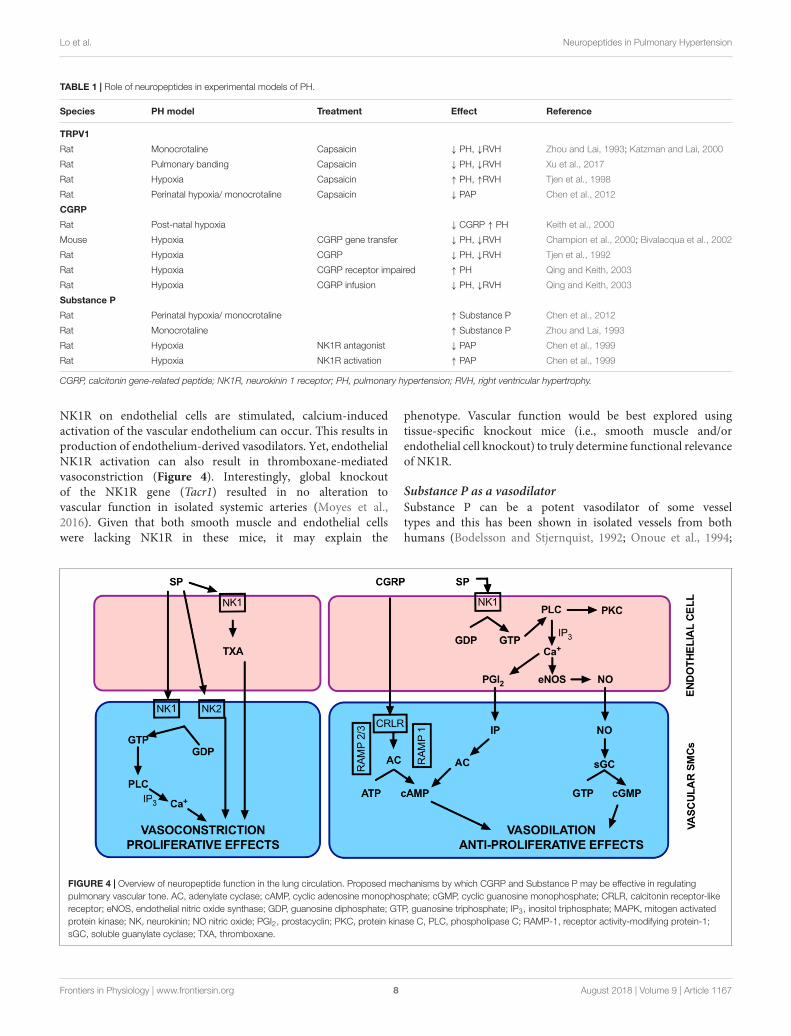
NK1R on endothelial cells are stimulated, calcium-inducedactivation of the vascular endothelium can occur. This results inproduction of endothelium-derived vasodilators. Yet, endothelialNK1R activation can also result in thromboxane-mediatedvasoconstriction (Figure 4). Interestingly, global knockoutof the NK1R gene (Tacr1) resulted in no alteration tovascular function in isolated systemic arteries (Moyes et al.,2016). Given that both smooth muscle and endothelial cellswere lacking NK1R in these mice, it may explain the
phenotype. Vascular function would be best explored usingtissue-specific knockout mice (i.e., smooth muscle and/orendothelial cell knockout) to truly determine functional relevanceof NK1R.
Substance P as a vasodilatorSubstance P can be a potent vasodilator of some vesseltypes and this has been shown in isolated vessels from bothhumans (Bodelsson and Stjernquist, 1992; Onoue et al., 1994;
FIGURE 4 | Overview of neuropeptide function in the lung circulation. Proposed mechanisms by which CGRP and Substance P may be effective in regulatingpulmonary vascular tone. AC, adenylate cyclase; cAMP, cyclic adenosine monophosphate; cGMP, cyclic guanosine monophosphate; CRLR, calcitonin receptor-likereceptor; eNOS, endothelial nitric oxide synthase; GDP, guanosine diphosphate; GTP, guanosine triphosphate; IP3, inositol triphosphate; MAPK, mitogen activatedprotein kinase; NK, neurokinin; NO nitric oxide; PGI2, prostacyclin; PKC, protein kinase C, PLC, phospholipase C; RAMP-1, receptor activity-modifying protein-1;sGC, soluble guanylate cyclase; TXA, thromboxane.
Frontiers in Physiology | www.frontiersin.org 8 August 2018 | Volume 9 | Article 1167
fphys-09-01167 August 22, 2018 Time: 9:40 # 9
Lo et al. Neuropeptides in Pulmonary Hypertension
Wallerstedt and Bodelsson, 1997) and animals (D’Orleans-Juste et al., 1985; Bolton and Clapp, 1986; McCormack et al.,1989b; Toda et al., 1991) and in vivo (McEwan et al., 1988;Beattie et al., 1993; Hall and Brain, 1994; Strobel et al., 1996).Vasodilation induced by substance P via NK1R can occur throughboth endothelium-dependent (D’Orleans-Juste et al., 1985;Bolton and Clapp, 1986; Onoue et al., 1994) and -independentmechanisms (Enokibori et al., 1994). Endothelium-dependentvasodilation (Figure 4) after substance P stimulation can bevia any of the major endothelium-dependent pathways; NO(Rosenblum et al., 1993; Holton et al., 2010), prostacyclin(Bodelsson and Stjernquist, 1994) or endothelium-dependenthyperpolarization of the smooth muscle membranes (Peterssonet al., 1995; Wallerstedt and Bodelsson, 1997). This can alsooccur through NK1R in isolated pulmonary vessels (Corboz et al.,1998; Pedersen et al., 2000). Substance P-induced pulmonaryvasodilation tends to occur at low concentrations (McCormacket al., 1989b) and can be transient in nature (Maxwell, 1968).
Substance P as a vasoconstrictorWhilst it is often considered a systemic vasodilator, substanceP can also induce a myogenic ‘response,’ or pressure-inducedconstriction (Scotland et al., 2004). When substance P release isstimulated, it can induce coronary vasoconstriction and increasedblood pressure (Bubb et al., 2013) and it may have a role in thepathogenesis of hypertension (Faulhaber et al., 1987; Kohlmannet al., 1997). Activation of the NK1R on smooth muscle cellsresults in activation of phospholipase C. This creates a transientincrease of inositol 1,4,5 triphosphate (Takeda et al., 1991) andincreased intracellular calcium (Krause et al., 1992), resulting invasoconstriction. Substance P primarily causes vasoconstrictionin pulmonary arteries (Selig et al., 1988; McCormack et al.,1989b; Shirahase et al., 1995), which can be mediated viaNK1R via thromboxane or NK2R (Figure 4). This appearsto be largely pulmonary-specific (Selig et al., 1988) and likelyinvolves complex receptor interactions. At higher substanceP concentrations, pulmonary vasoconstriction dominates overvasodilation (McCormack et al., 1989b).
Substance P in Pulmonary HypertensionThe complexities around the dual vasodilator/vasoconstrictorrole of substance P in the pulmonary circulation (Figure 4) makeit difficult to predict what role it has (if any) in PH. Infusionof substance P has minimal vasodilating effect on patients withPAH (Uren et al., 1992; Brett et al., 1996; Cailes et al., 1998).Thus, is has been postulated that substance P dysfunction is anunderlying cause of PAH. On the other hand, the dominant roleof substance P as a pulmonary–specific vasoconstrictor led tothe hypothesis that substance P overactivity is causative in PAH.Pre-clinical models of PH are associated with increased lungsubstance P (Zhou and Lai, 1993; Chen et al., 2012). Pulmonarypressure can be decreased by depleting substance P (Table 1) orby using NK1R antagonists (Zhou and Lai, 1993; Chen et al.,1999, 2012). Likewise, activation of NK1R can induce increasedpulmonary pressure (Chen et al., 1999). Substance P is involvedin lung vascular remodeling, possibly due to increased oxidativestress (Springer and Fischer, 2003). Increased PVR has also
been attributed to inflammatory stimulation of mitogen-activatedprotein kinase pathway by substance P. This is alleviated bycapsaicin-induced depletion (Xu et al., 2017). In model systemsusing hypoxia to simulate PH, substance P release is increased(Lindefors et al., 1986). Based upon this body of literature itwould appear that substance P is indeed a promising candidatefor reducing PVR.
Further research should be conducted to pursue the intricatemechanisms of substance P in PAH. As NK1R antagonistscan lower pulmonary pressure in rats, they may be thebest pharmacological target to consider for humans. NK1Rantagonists are in clinical use as anti-emetics. They are usuallyprescribed to take prior to chemotherapy or surgery. Aprepitantis available as an oral formulation and is well tolerated(Martinez and Philipp, 2016). It has been investigated for use asan anti-inflammatory agent. Interestingly, reported side-effectsinclude low blood pressure. If subsequent pre-clinical studies arepositive, a clinical trial in PH could conceivably proceed usingthis safety-approved formulation.
Transient Receptor Potential VanilloidType 1 (TRPV1) in Vascular RegulationThere are a number of signaling intermediates in C-fiber-derivedneuropeptide release. One of the best characterized in thevasculature is TRPV1. TRPV1 activation results in substance Pand/or CGRP release from peripheral nerve terminals (Holzer,1991; Gibbons et al., 2010). TRPV1 receptors are non-selectivecation channels located predominantly on sensory nerve endings(Tominaga and Julius, 2000) but they also reside on cells ofmany peripheral tissue types. They are present in the entirerespiratory tract (Zholos, 2015) and located on both smoothmuscle and endothelial cells of the vasculature. TRPV1 receptorsare nociceptors that are highly responsive to pro-inflammatorystimuli and are activated by a range of endogenous andexogenous factors (Figure 3). These include temperature, pH,bradykinin, anandamide, arachidonic acid metabolites such as20-hydroxyeicosatetraenoic acid (Wen et al., 2012), spider toxins,and most famously, capsaicin, (Caterina et al., 1997; Devesa et al.,2011). TRPV1 receptors have a binding site for capsaicin. Thecomplicated mechanism for activation is still being unraveledand great progress has been made since 2013 when breakthroughstructural information was first reported (Yang and Zheng, 2017).TRPV1 activation is involved in a variety of cardiovascularpathologies, including the modulation of atherosclerosis (Li et al.,2014; Xiong et al., 2016), myogenic tone (Scotland et al., 2004;Bubb et al., 2013), systemic arterial pressure (Bubb et al., 2013)and hypertension (Hao et al., 2011), congestive heart failure(Gao et al., 2014; Lang et al., 2015), vascular remodeling (Chenet al., 2010), haemorrhagic shock (Akabori et al., 2007) and sepsis(Chen et al., 2018).
Evidence of a Role for TRPV1 in PulmonaryHypertensionPulmonary hypertension is characterized by pulmonary smoothmuscle cell hyperproliferation causing remodeling of the smoothmuscle cell layer and impacting on PVR. Although it is likelythat TRPV1 activation in sensory nerves is the key to modulation
Frontiers in Physiology | www.frontiersin.org 9 August 2018 | Volume 9 | Article 1167
fphys-09-01167 August 22, 2018 Time: 9:40 # 10
Lo et al. Neuropeptides in Pulmonary Hypertension
of pulmonary circulation, it is possible that some direct effectsin smooth muscle cells could contribute to the PH phenotype.Activation of TRPV1 on cultured pulmonary smooth musclecells (SMC) results in enhanced proliferation (Randhawa andJaggi, 2017), increased intracellular calcium and stimulates cellmigration (Martin et al., 2012). Under laboratory conditions, PHcan be simulated by exposing cells to chronic hypoxia. Using thismodel, TRPV1 expression can increase (Wang et al., 2008) withcorresponding increased intracellular calcium and reorganizationof cytoskeletal architecture. TRPV1 blockade with capsazepinecan abolish these effects (Parpaite et al., 2016). There are anumber of studies assessing the role of TRPV1 by using capsaicinto desensitize it (Table 1). These studies are further evidence ofthe importance of TRPV1 in PH. The contribution of individualneuropeptides downstream of TRPV1 activation is complex andhas been discussed in the relevant sections of this review.
DIRECT CARDIAC EFFECTS OFSENSORY C-FIBER NEUROPEPTIDES
The increased PVR in all groups of PH generally leads toright ventricular cardiac remodeling due to the increased right
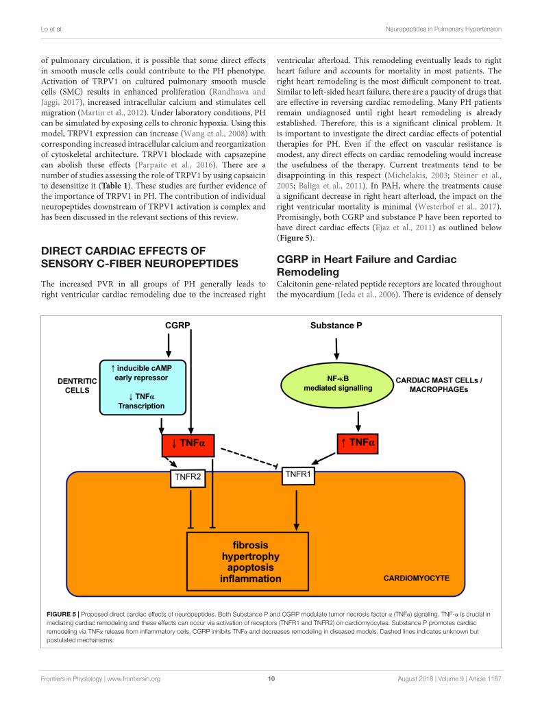
ventricular afterload. This remodeling eventually leads to rightheart failure and accounts for mortality in most patients. Theright heart remodeling is the most difficult component to treat.Similar to left-sided heart failure, there are a paucity of drugs thatare effective in reversing cardiac remodeling. Many PH patientsremain undiagnosed until right heart remodeling is alreadyestablished. Therefore, this is a significant clinical problem. Itis important to investigate the direct cardiac effects of potentialtherapies for PH. Even if the effect on vascular resistance ismodest, any direct effects on cardiac remodeling would increasethe usefulness of the therapy. Current treatments tend to bedisappointing in this respect (Michelakis, 2003; Steiner et al.,2005; Baliga et al., 2011). In PAH, where the treatments causea significant decrease in right heart afterload, the impact on theright ventricular mortality is minimal (Westerhof et al., 2017).Promisingly, both CGRP and substance P have been reported tohave direct cardiac effects (Ejaz et al., 2011) as outlined below(Figure 5).
CGRP in Heart Failure and CardiacRemodelingCalcitonin gene-related peptide receptors are located throughoutthe myocardium (Ieda et al., 2006). There is evidence of densely
FIGURE 5 | Proposed direct cardiac effects of neuropeptides. Both Substance P and CGRP modulate tumor necrosis factor α (TNFα) signaling. TNF-α is crucial inmediating cardiac remodeling and these effects can occur via activation of receptors (TNFR1 and TNFR2) on cardiomyocytes. Substance P promotes cardiacremodeling via TNFα release from inflammatory cells. CGRP inhibits TNFα and decreases remodeling in diseased models. Dashed lines indicates unknown butpostulated mechanisms.
Frontiers in Physiology | www.frontiersin.org 10 August 2018 | Volume 9 | Article 1167
fphys-09-01167 August 22, 2018 Time: 9:40 # 11
Lo et al. Neuropeptides in Pulmonary Hypertension
located CGRP-containing nerve fibers around the coronaryarteries, papillary muscles, sinoatrial and atrioventricular nodes(Mulderry et al., 1985b). CGRP protects against cardiachypertrophy in a pre-clinical model of heart failure (Li et al.,2010). CGRP levels can be increased in heart failure (Hsuet al., 2005). Whether this is causative or compensatory isunknown. However, there is evidence to support the latter notion.Infusion of CGRP increases myocardial contractility (Gennariet al., 1990) and improves circulation in coronary disease(Gennari et al., 1990; Dubois-Rande et al., 1992; Stevensonet al., 1992). On a cellular level, CGRP inhibits generation ofpro-inflammatory tumor necrosis factor (TNF)-α and preventsinterstitial and perivascular fibrosis (Li et al., 2013). CGRP alsoameliorates cardiomyocyte apoptosis via the Bcl-2/Bax pathway(Ma et al., 2013) and has direct anti-proliferative effects oncardiac fibroblasts (Li et al., 2016). CGRP has positive cardiacionotropic effects that are mostly abolished by both a CGRPantagonist and a PI3 kinase inhibitor (Al-Rubaiee et al., 2013).A CGRP analog alleviated pre-clinical diabetes-induced heartfailure and remodeling due to reduced α-smooth muscle actinand transforming growth factor (TGF)-β1 (Aubdool et al.,2017). Therefore, CGRP appears to be cardioprotective. Evenmore promising for PH, is the recent finding that rutacarpine,which stimulates CGRP release, was able to reverse right heartremodeling in experimental PH and this was attributed top27-dependent signaling (Li et al., 2016).
Substance P in Heart Failure and CardiacRemodelingIn the human heart, substance P has been found in intrinsiccardiac ganglia (Wharton et al., 1990; Hoover et al., 2009),coronary vessels (Laine et al., 2000), and within myocardium(Weihe et al., 1981; Rechardt et al., 1986). Substance P appearsto have a damaging role in heart disease (Dehlin and Levick,2014), hinting at a direct effect of substance P on myocardialtissue. Pro-substance P (a stable surrogate for substance P)is an independent predictor of recurrent acute myocardialinfarction, heart failure and cardiac mortality (Ng et al., 2014).Levels of substance P are increased in congestive heart failure(Valdemarsson et al., 1991). Substance P promotes hypertrophyin isolated cardiomyocytes, suggesting this effect is direct andindependent of systemic factors (Church et al., 1996). Severalpre-clinical models have demonstrated a role for substance Pin mediating heart failure. Cardiac hypertrophy, apoptosis anddilated cardiomyopathy were absent from substance P knockoutmice (D’Souza et al., 2007). Genetic deletion of TAC1, whichencodes substance P was protective against increased cardiacmast cell density and TNFα upregulation and associated cardiacremodeling (Melendez et al., 2011). Inhibition of NK1R alsoimproved cardiac function and ameliorated cardiac hypertrophyin models of heart failure (Melendez et al., 2011; Robinsonet al., 2015). Substance P appears to be a mediator of cardiactoxicity induced by doxorubicin, which is a chemotherapy agentthat causes cardiomyocyte death. Inhibition of substance Presulted in lower cardiomyocyte apoptosis after doxorubicintreatment to an isolated cell line (Robinson et al., 2016).
Taken together, these findings are clinically exciting, as it suggeststhat substance P-antagonist based therapies may be able todirectly improve right ventricular parameters above simplyrelieving right ventricular afterload for PAH treatment.
WHAT IS NOT KNOWN? FUTUREPROSPECTS FOR NEUROPEPTIDES ASA TREATMENT STRATEGY FORPULMONARY ARTERIALHYPERTENSION
From a relatively small pool of research we can conclude thatthe neuropeptides, substance P and CGRP, have the ability toregulate both systemic and pulmonary arterial pressure. We havealso provided evidence that these neuropeptides regulate vascularsmooth and cardiac muscle cell remodeling under diseasedconditions. However, there is not yet a comprehensive answeron whether these neuropeptides are strong signaling targets fortreating PAH. CGRP is a proven potent dilator of systemic andpulmonary vasculature and appears to have favorable effectson cardiac remodeling. Based on what we know, stimulationof endogenous CGRP signaling or exogenous administration ofstable analogs should be good candidates for the treatment ofPAH. Yet only a handful of studies have investigated this andit has not gone further than pre-clinical investigation. The nextstep would be to thoroughly characterize the potential for CGRPin reversing all aspects of PAH. It is important to show this inthree or more different PH models, before embarking on a PhaseII trial.
The clinical appeal of substance P is more distant as there arestill inconsistencies in identifying the precise vascular regulatoryfunction of this neuropeptide. From the pulmonary literature, itappears that the over-arching role of substance P is likely to beas a vasoconstrictor. This is contrary to its effect in the systemiccirculation, where it seems to predominantly cause vasodilation.If this is proven to be the case, it is a very attractive property forPAH treatment. A major adverse effect for some PAH therapieshas been the unacceptable lowering of systemic arterial pressure.This is an important factor to consider for PAH treatment,especially in regard to using combination therapies.
Combination therapies are emerging as a promisingtreatment regimen for PAH, while we wait for development ofnovel compounds. An effective PVR-lowering combination hasinvolved upregulation of cGMP generation with concurrentinhibition of cGMP metabolism. However, this is not alwayspossible. For example, the use of sildenafil and long-actingnitrates can cause life-threatening hypotension (Cheitlin et al.,1999). Using pre-clinical models, an alternative strategy hasemerged. Stimulation of a different ‘pool’ of cGMP, derived fromparticulate guanylate cyclase activity rather than sGC (Baligaet al., 2008), and combining this with sildenafil, has been asuccess. This has resulted in a far more pulmonary-specificvasodilation, negating the hypotensive effects. A similarapproach could apply for substance P. Antagonism of NK1Rwould presumably block the pulmonary vasoconstriction of
Frontiers in Physiology | www.frontiersin.org 11 August 2018 | Volume 9 | Article 1167
fphys-09-01167 August 22, 2018 Time: 9:40 # 12
Lo et al. Neuropeptides in Pulmonary Hypertension
substance P, but also prevent excessive systemic dilation to somedegree. A hypothetical treatment strategy could involve usingNK1R antagonists alongside already approved PAH therapiessuch as PDE5 inhibition or sGC stimulation. This may result ina pulmonary-specific dilation, as substance P-induced systemicvasodilation would be prevented at the same time as pulmonaryvasoconstriction is lowered. Pre-clinical studies should focus oninvestigating the substance P axis alongside current treatments,or in combination with other promising candidates. This couldidentify new strategies that are truly able to target multipleetiologies of PAH and are an essential starting point. A similarstrategy should be utilized when investigating CGRP for PAHtherapy. It is likely that CGRP-based pharmaceuticals may havesignificant and unacceptable blood pressure lowering effects,similar to the organic nitrates. Therefore dose-selection andcombination approaches would be essential in establishingCGRP as a therapy.
As TRPV1 activation results in release of both neuropeptides,it may also make an interesting target. If antagonism of TRPV1lowers substance P, this may be beneficial for PAH. It would alsopresumably lower CGRP, which may not be beneficial. It probablydepends on the release pattern of CGRP/substance P in PH lungsand little is known about that. The majority of studies so far havefound that depleting the C-fiber nerve endings by using capsaicinhas generally resulted in improvements in PH models (Table 1).This suggests that inhibiting sensory C-fibers predominantlyimpairs substance P-induced pulmonary constriction withouta major influence on CGRP-induced dilation. This may bereflective of the mechanisms at play in regards to the roleof TRPV1 in PAH. Future research should focus on whethercomplete inhibition of TRPV1 could be effective as a PH therapy,or whether the balance could be shifted more toward CGRP-production with inhibition of substance P. Of course, with anystrategy, consideration must be given to the consequences forother organ systems. Complete inhibition of either TRPV1 orsubstance P may elicit unexpected effects and this should bethoroughly investigated in PH models. Use of targeted therapyto the pulmonary vasculature and/or heart would help toelucidate the role of both neuropeptides. Also, receptor-specifictherapy could be considered. One study has investigated all threeneurokinin receptors, but it indicated no role of NK2R and NK3R(Corboz et al., 1998). The little that has been done suggeststhat NK1R is likely to be the primary mediator of pulmonaryvasoconstriction, but the other neurokinins should not yet beruled out.
Another interesting area to explore could be related to neutralendopeptidases (NEP), which are the most important enzymesin the degradation of tachykinins, including substance P (Skidgelet al., 1984). Interestingly, there is a decent body of evidence tosuggest that NEP inhibitors could be a promising PAH therapy,and they are currently in clinical trial. In addition to tachykinins,NEPs also degrade a selection of other peptides, includingpulmonary vasodilating natriuretic peptides and pulmonaryvasoconstrictors such as ET-1 (Abassi et al., 1992). NEPs appearto have an underlying role in the pathogenesis of PH (Dempseyet al., 2009). NEP inhibitors have produced promising resultsin models of PH, both as monotherapy (Klinger et al., 1993;
Thompson et al., 2012) or in combination with PDE5 inhibition(Baliga et al., 2008). This is largely due to their ability to increasecirculating atrial natriuretic peptide. However, in addition toincreasing pulmonary vasodilators, NEP inhibitors may resultin increased circulating substance P (in addition to othervasoconstrictors such as ET-1 and angiotensin II) and thiswould likely counteract their beneficial effects on the pulmonarycirculation. An alternative strategy could be to introduce NEPinhibition in combination with an NK1R antagonist, for example.A similar strategy has been trialed for hypertension, usingcombined NEP/ angiotensin II inhibition with promising results(Cuculi and Erne, 2011).
Another exciting avenue worth pursuing is the interactionof neuropeptides with ET-1. CGRP inhibits the interaction ofET-1 with ETA on vascular smooth muscle cells. This candecrease smooth muscle contraction and is reversible with CGRPantagonists (De Mey and Vanhoutte, 2014). This suggests thatintact CGRP may be important in preventing ET-1 activity.Upregulation of CGRP may act as an endogenous ERA inhibitorand attenuate ET-1 over-activity in PAH. There are also emergingfindings that substance P may directly interact with ET-1. In non-cardiovascular (melanocyte) cells, substance P can stimulate ET-1(Park et al., 2015). An NK1R antagonist was able to prevent anincrease in ET-1 expression seen in spontaneously hypertensiverats, which was associated with reduced cardiac fibrosis (Dehlinet al., 2013). If the relationship between substance P and ET-1is proven, this could also have important implications for PAHtreatment, given that ET-1 is already a prime target for licensedtherapies. Taken together, it would seem that selectivity inregulating neuropeptide release/ activity is of prime importance.Most of the in vivo studies in this area have investigated broad-spectrum C-fiber depletion. This is unlikely to give meaningfulinformation on the mechanisms at play in PH. Many of thesestudies were conducted years ago, prior to the advancementsin the efficiency of genome manipulation. It is worth revisitingthese neuropeptides in PH. The use of elegant and well-designedstudies, utilizing advanced technology, should fully interrogatethe vascular interactions of substance P and CGRP in thepulmonary circulation.
SUMMARY
Pulmonary arterial hypertension is a fatal disease that afflictspeople of any age and causes substantial reduction in quality oflife. It tends to be more prevalent in young people, particularlywomen. While treatments developed in the past few decadesimproved the prognosis for PAH patients, mortality rates stillremain high. Current treatment methods are primarily centeredon enhancing pulmonary vasodilation. They are not effectiveat reducing mortality. There is great potential to develop newtreatments that target both cardiopulmonary re-modeling andPVR. Altered release of neuropeptides such as substance P andCGRP have been implicated in the pathophysiology of PAH.Selective control of the balance on these neuropeptides in thepulmonary circulation is a promising approach to combating thisfatal disease.
Frontiers in Physiology | www.frontiersin.org 12 August 2018 | Volume 9 | Article 1167
fphys-09-01167 August 22, 2018 Time: 9:40 # 13
Lo et al. Neuropeptides in Pulmonary Hypertension
AUTHOR CONTRIBUTIONS
CL and KB created the figures. CL, SM, and KB researched theliterature, prepared the table, and drafted the manuscript.
FUNDING
KB is funded by Rebecca L. Cooper Medical Research Foundationand University of Sydney Kickstart Grants related to this work.
REFERENCESAbassi, Z., Golomb, E., and Keiser, H. R. (1992). Neutral endopeptidase inhibition
increases the urinary excretion and plasma levels of endothelin. Metabolism 41,683–685. doi: 10.1016/0026-0495(92)90303-R
Abenhaim, L., Moride, Y., Brenot, F., Rich, S., Benichou, J., Kurz, X., et al. (1996).Appetite-suppressant drugs and the risk of primary pulmonary hypertension.International Primary Pulmonary Hypertension Study Group. N. Engl. J. Med.335, 609–616. doi: 10.1056/NEJM199608293350901
Aiyar, N., Disa, J., Siemens, I. R., and Nambi, P. (1997). Differential effects ofguanine nucleotides on [125I]-hCGRP(8-37) binding to porcine lung andhuman neuroblastoma cell membranes. Neuropeptides 31, 99–103. doi: 10.1016/S0143-4179(97)90028-7
Akabori, H., Yamamoto, H., Tsuchihashi, H., Mori, T., Fujino, K., Shimizu, T.,et al. (2007). Transient receptor potential vanilloid 1 antagonist, capsazepine,improves survival in a rat hemorrhagic shock model. Ann. Surg. 245, 964–970.doi: 10.1097/01.sla.0000255577.80800.e1
Al-Rubaiee, M., Gangula, P. R., Millis, R. M., Walker, R. K., Umoh, N. A., Cousins,V. M., et al. (2013). Inotropic and lusitropic effects of calcitonin gene-relatedpeptide in the heart. Am. J. Physiol. Heart Circ. Physiol. 304, H1525–H1537.doi: 10.1152/ajpheart.00874.2012
Amara, S. G., Arriza, J. L., Leff, S. E., Swanson, L. W., Evans, R. M., andRosenfeld, M. G. (1985). Expression in brain of a messenger RNA encoding anovel neuropeptide homologous to calcitonin gene-related peptide. Science 229,1094–1097. doi: 10.1126/science.2994212
Amara, S. G., Jonas, V., Rosenfeld, M. G., Ong, E. S., and Evans, R. M. (1982).Alternative RNA processing in calcitonin gene expression generates mRNAsencoding different polypeptide products. Nature 298, 240–244. doi: 10.1038/298240a0
Andrew, P. J., and Mayer, B. (1999). Enzymatic function of nitric oxide synthases.Cardiovasc. Res. 43, 521–531. doi: 10.1016/S0008-6363(99)00115-7
Aubdool, A. A., Thakore, P., Argunhan, F., Smillie, S. J., Schnelle, M., Srivastava, S.,et al. (2017). A novel alpha-calcitonin gene-related peptide analogue protectsagainst end-organ damage in experimental hypertension, cardiac hypertrophy,and heart failure. Circulation 136, 367–383. doi: 10.1161/CIRCULATIONAHA.117.028388
Austin, E. D., and Loyd, J. E. (2015). Toward precision medicine in pulmonaryarterial hypertension. Am. J. Respir. Crit. Care Med. 192, 1272–1274. doi: 10.1164/rccm.201508-1607ED
Baliga, R. S., MacAllister, R. J., and Hobbs, A. J. (2011). New perspectives forthe treatment of pulmonary hypertension. Br. J. Pharmacol. 163, 125–140.doi: 10.1111/j.1476-5381.2010.01164.x
Baliga, R. S., Zhao, L., Madhani, M., Lopez-Torondel, B., Visintin, C., Selwood, D.,et al. (2008). Synergy between natriuretic peptides and phosphodiesterase 5inhibitors ameliorates pulmonary arterial hypertension. Am. J. Respir. Crit. CareMed. 178, 861–869. doi: 10.1164/rccm.200801-121OC
Barst, R. J., McGoon, M., Torbicki, A., Sitbon, O., Krowka, M. J., Olschewski, H.,et al. (2004). Diagnosis and differential assessment of pulmonary arterialhypertension. J. Am. Coll. Cardiol. 43(12 Suppl. S), 40S–47S. doi: 10.1016/j.jacc.2004.02.032
Barst, R. J., Rubin, L. J., Long, W. A., McGoon, M. D., Rich, S., Badesch, D. B., et al.(1996). A comparison of continuous intravenous epoprostenol (prostacyclin)with conventional therapy for primary pulmonary hypertension. N. Engl. J.Med. 334, 296–301. doi: 10.1056/nejm199602013340504
Bartosik, I., Eskilsson, J., Ekman, R., Akesson, A., and Scheja, A. (2002).Correlation between plasma concentrations of calcitonin gene related peptideand pulmonary pressure in patients with systemic sclerosis. Ann. Rheum. Dis.61, 261–263. doi: 10.1136/ard.61.3.261
Beattie, D. T., Stubbs, C. M., Connor, H. E., and Feniuk, W. (1993). Neurokinin-induced changes in pial artery diameter in the anaesthetized guinea-pig. Br. J.Pharmacol. 108, 146–149. doi: 10.1111/j.1476-5381.1993.tb13454.x
Berthiaume, N., Claing, A., Regoli, D., Warner, T. D., and D’Orleans-Juste, P.(1995). Characterization of receptors for kinins and neurokinins in the arterialand venous mesenteric vasculatures of the guinea-pig. Br. J. Pharmacol. 115,1319–1325. doi: 10.1111/j.1476-5381.1995.tb15043.x
Bivalacqua, T. J., Hyman, A. L., Kadowitz, P. J., Paolocci, N., Kass, D. A., andChampion, H. C. (2002). Role of calcitonin gene-related peptide (CGRP) inchronic hypoxia-induced pulmonary hypertension in the mouse. Influence ofgene transfer in vivo. Regul. Pept. 108, 129–133. doi: 10.1016/S0167-0115(02)00100-3
Bodelsson, G., and Stjernquist, M. (1992). Smooth muscle dilatation in thehuman uterine artery induced by substance P, vasoactive intestinal polypeptide,calcitonin gene-related peptide and atrial natriuretic peptide: relationto endothelium-derived relaxing substances. Hum. Reprod. 7, 1185–1188.doi: 10.1093/oxfordjournals.humrep.a137823
Bodelsson, G., and Stjernquist, M. (1994). Endothelium-dependent relaxation tosubstance P in human umbilical artery is mediated via prostanoid synthesis.Hum. Reprod. 9, 733–737. doi: 10.1093/oxfordjournals.humrep.a138580
Bolli, M. H., Boss, C., Binkert, C., Buchmann, S., Bur, D., Hess, P.,et al. (2012). The discovery of N-[5-(4-bromophenyl)-6-[2-[(5-bromo-2-pyrimidinyl)oxy]ethoxy]-4-pyrimidinyl]-N’-p ropylsulfamide (Macitentan), anorally active, potent dual endothelin receptor antagonist. J. Med. Chem. 55,7849–7861. doi: 10.1021/jm3009103
Bolton, T. B., and Clapp, L. H. (1986). Endothelial-dependent relaxant actionsof carbachol and substance P in arterial smooth muscle. Br. J. Pharmacol. 87,713–723. doi: 10.1111/j.1476-5381.1986.tb14589.x
Brain, S. D., Williams, T. J., Tippins, J. R., Morris, H. R., and MacIntyre, I. (1985).Calcitonin gene-related peptide is a potent vasodilator. Nature 313, 54–56.doi: 10.1038/313054a0
Brenner, O. (1957). The lungs in heart disease. Br. J. Tuberc. Dis. Chest 51, 209–222.doi: 10.1016/S0366-0869(57)80076-8
Bressollette, E., Dupuis, J., Bonan, R., Doucet, S., Cernacek, P., and Tardif, J. C.(2001). Intravascular ultrasound assessment of pulmonary vascular disease inpatients with pulmonary hypertension. Chest 120, 809–815. doi: 10.1378/chest.120.3.809
Brett, S. J., Simon, J., Gibbs, R., Pepper, J. R., and Evans, T. W. (1996).Impairment of endothelium-dependent pulmonary vasodilation in patientswith primary pulmonary hypertension. Thorax 51, 89–91. doi: 10.1136/thx.51.1.89
Bubb, K. J., Hobbs, A. J., and Klinger, J. R. (2015). “Modulation of cGMP Synthesisand Metabolism,” in Diagnosis and Management of Pulmonary Hypertension.New York, NY: Springer, 355–375. doi: 10.1007/978-1-4939-2636-7_15
Bubb, K. J., Wen, H., Panayiotou, C. M., Finsterbusch, M., Khan, F. J.,Chan, M. V., et al. (2013). Activation of neuronal transient receptorpotential vanilloid 1 channel underlies 20-hydroxyeicosatetraenoic acid-induced vasoactivity: role for protein kinase A. Hypertension 62, 426–433.doi: 10.1161/HYPERTENSIONAHA.111.00942
Burks, T. F., Buck, S. H., and Miller, M. S. (1985). Mechanisms of depletion ofsubstance P by capsaicin. Fed. Proc. 44, 2531–2534.
Cailes, J., Winter, S., du Bois, R. M., and Evans, T. W. (1998). Defectiveendothelially mediated pulmonary vasodilation in systemic sclerosis. Chest 114,178–184. doi: 10.1378/chest.114.1.178
Caterina, M. J., Schumacher, M. A., Tominaga, M., Rosen, T. A., Levine, J. D., andJulius, D. (1997). The capsaicin receptor: a heat-activated ion channel in thepain pathway. Nature 389, 816–824.
Champion, H. C., Bivalacqua, T. J., Toyoda, K., Heistad, D. D., Hyman, A. L.,and Kadowitz, P. J. (2000). In vivo gene transfer of prepro-calcitonin gene-related peptide to the lung attenuates chronic hypoxia-induced pulmonaryhypertension in the mouse. Circulation 101, 923–930. doi: 10.1161/01.CIR.101.8.923
Chang, M. M., Leeman, S. E., and Niall, H. D. (1971). Amino-acid sequence ofsubstance P. Nat. New Biol. 232, 86–87. doi: 10.1038/newbio232086a0
Frontiers in Physiology | www.frontiersin.org 13 August 2018 | Volume 9 | Article 1167
fphys-09-01167 August 22, 2018 Time: 9:40 # 14
Lo et al. Neuropeptides in Pulmonary Hypertension
Channick, R. N., Sitbon, O., Barst, R. J., Manes, A., and Rubin, L. J. (2004).Endothelin receptor antagonists in pulmonary arterial hypertension. J. Am.Coll. Cardiol. 43(12 Suppl.), S62–S67. doi: 10.1016/j.jacc.2004.02.042
Chattergoon, N. N., D’Souza, F. M., Deng, W., Chen, H., Hyman, A. L., Kadowitz,P. J., et al. (2005). Antiproliferative effects of calcitonin gene-related peptide inaortic and pulmonary artery smooth muscle cells. Am. J. Physiol. Lung Cell. Mol.Physiol. 288, L202–L211. doi: 10.1152/ajplung.00064.2004
Cheitlin, M. D., Hutter, A. M. Jr., Brindis, R. G., Ganz, P., Kaul, S., Russell,R. O. Jr., et al. (1999). Use of sildenafil (Viagra) in patients with cardiovasculardisease. Technology and practice executive committee. Circulation 99, 168–177.doi: 10.1161/01.CIR.99.1.168
Chen, J., Hamers, A. J. P., Finsterbusch, M., Massimo, G., Zafar, M., Corder, R.,et al. (2018). Endogenously generated arachidonate-derived ligands for TRPV1induce cardiac protection in sepsis. FASEB J. 32, 3816–3831. doi: 10.1096/fj.201701303R
Chen, K. H., Lai, Y. L., and Chen, M. J. (2012). Oxygen radicals and substanceP in perinatal hypoxia-exaggerated, monocrotaline-induced pulmonaryhypertension. Chin. J. Physiol. 55, 82–90. doi: 10.4077/cjp.2012.amm103
Chen, L. W., Chen, C. F., and Lai, Y. L. (1999). Chronic activation of neurokinin-1receptor induces pulmonary hypertension in rats. Am. J. Physiol. 276(5 Pt 2),H1543–H1551. doi: 10.1152/ajpheart.1999.276.5.H1543
Chen, R. Y., and Guth, P. H. (1995). Interaction of endogenous nitric oxide andCGRP in sensory neuron-induced gastric vasodilation. Am. J. Physiol. 268(5 Pt1), G791–G796. doi: 10.1152/ajpgi.1995.268.5.G791
Chen, Y.-S., Lu, M.-J., Huang, H.-S., and Ma, M.-C. (2010). Mechanosensitivetransient receptor potential vanilloid type 1 channels contribute to vascularremodeling of rat fistula veins. J. Vasc. Surg. 52, 1310–1320. doi: 10.1016/j.jvs.2010.05.095
Church, D. J., Arkinstall, S. J., Vallotton, M. B., Chollet, A., Kawashima, E., andLang, U. (1996). Stimulation of atrial natriuretic peptide release by neurokininsin neonatal rat ventricular cardiomyocytes. Am. J. Physiol. 270(3 Pt 2),H935–H944. doi: 10.1152/ajpheart.1996.270.3.H935
Cogan, J. D., Pauciulo, M. W., Batchman, A. P., Prince, M. A., Robbins,I. M., Hedges, L. K., et al. (2006). High frequency of BMPR2 exonicdeletions/duplications in familial pulmonary arterial hypertension. Am. J.Respir. Crit. Care Med. 174, 590–598. doi: 10.1164/rccm.200602-165OC
Constantine, J. W., Lebel, W. S., and Woody, H. A. (1991). Inhibition of tachykinin-induced hypotension in dogs by CP-96,345, a selective blocker of NK-1receptors. Naunyn Schmiedebergs Arch. Pharmacol. 344, 471–477.
Corboz, M. R., Rivelli, M. A., Ramos, S. I., Rizzo, C. A., and Hey, J. A. (1998).Tachykinin NK1 receptor-mediated vasorelaxation in human pulmonaryarteries. Eur. J. Pharmacol. 350, R1–R3. doi: 10.1016/S0014-2999(98)00310-0
Cuculi, F., and Erne, P. (2011). Combined neutral endopeptidase inhibitors. ExpertOpin. Investig. Drugs 20, 457–463. doi: 10.1517/13543784.2011.556617
D’Alonzo, G. E., Barst, R. J., Ayres, S. M., Bergofsky, E. H., Brundage, B. H., Detre,K. M., et al. (1991). Survival in patients with primary pulmonary hypertension.Results from a national prospective registry. Ann. Intern. Med. 115, 343–349.doi: 10.7326/0003-4819-115-5-343
de Avila, E. D., de Molon, R. S., de Godoi Goncalves, D. A., and Camparis, C. M.(2014). Relationship between levels of neuropeptide Substance P in periodontaldisease and chronic pain: a literature review. J. Investig. Clin. Dent. 5, 91–97.doi: 10.1111/jicd.12087
De Mey, J. G. R., and Vanhoutte, P. M. (2014). End O’ The Line Revisited: movingon from nitric oxide to CGRP. Life Sci. 118, 120–128. doi: 10.1016/j.lfs.2014.04.012
DeFea, K. A., Vaughn, Z. D., O’Bryan, E. M., Nishijima, D., Dery, O., andBunnett, N. W. (2000). The proliferative and antiapoptotic effects of substanceP are facilitated by formation of a beta -arrestin-dependent scaffoldingcomplex. Proc. Natl. Acad. Sci. U.S.A. 97, 11086–11091. doi: 10.1073/pnas.190276697
Dehlin, H. M., and Levick, S. P. (2014). Substance P in heart failure: the good andthe bad. Int. J. Cardiol. 170, 270–277. doi: 10.1016/j.ijcard.2013.11.010
Dehlin, H. M., Manteufel, E. J., Monroe, A. L., Reimer, M. H. Jr., and Levick,S. P. (2013). Substance P acting via the neurokinin-1 receptor regulates adversemyocardial remodeling in a rat model of hypertension. Int. J. Cardiol. 168,4643–4651. doi: 10.1016/j.ijcard.2013.07.190
Dempsey, E. C., Wick, M. J., Karoor, V., Barr, E. J., Tallman, D. W., Wehling, C. A.,et al. (2009). Neprilysin null mice develop exaggerated pulmonary vascular
remodeling in response to chronic hypoxia. Am. J. Pathol. 174, 782–796.doi: 10.2353/ajpath.2009.080345
Devesa, I., Planells-Cases, R., Fernández-Ballester, G., González-Ros, J. M., Ferrer-Montiel, A., and Fernández-Carvajal, A. (2011). Role of the transient receptorpotential vanilloid 1 in inflammation and sepsis. J. Inflamm. Res. 4, 67–81.doi: 10.2147/jir.s12978
Donnerer, J., and Stein, C. (1992). Evidence for an increase in the release of CGRPfrom sensory nerves during inflammation. Ann. N. Y. Acad. Sci. 657, 505–506.doi: 10.1111/j.1749-6632.1992.tb22814.x
D’Orleans-Juste, P., Dion, S., Mizrahi, J., and Regoli, D. (1985). Effects of peptidesand non-peptides on isolated arterial smooth muscles: role of endothelium. Eur.J. Pharmacol. 114, 9–21. doi: 10.1016/0014-2999(85)90515-1
D’Souza, M., Garza, M. A., Xie, M., Weinstock, J., Xiang, Q., and Robinson, P.(2007). Substance P is associated with heart enlargement and apoptosisin murine dilated cardiomyopathy induced by Taenia crassiceps infection.J. Parasitol. 93, 1121–1127. doi: 10.1645/GE-596R1.1
Dubois-Rande, J. L., Merlet, P., Benvenuti, C., Sediame, S., Macquin-Mavier, I.,Chabrier, E., et al. (1992). Effects of calcitonin gene-related peptide on cardiaccontractility, coronary hemodynamics and myocardial energetics in idiopathicdilated cardiomyopathy. Am. J. Cardiol. 70, 906–912. doi: 10.1016/0002-9149(92)90736-I
Dumitrascu, R., Weissmann, N., Ghofrani, H. A., Dony, E., Beuerlein, K.,Schmidt, H., et al. (2006). Activation of soluble guanylate cyclase reversesexperimental pulmonary hypertension and vascular remodeling. Circulation113, 286–295. doi: 10.1161/CIRCULATIONAHA.105.581405
Edvinsson, L., Erlinge, D., Ekman, R., and Thulin, T. (1992). Sensory nerveterminal activity in severe hypertension as reflected by circulating CalcitoninGene-Related Peptide (CGRP) and substance P. Blood Press. 1, 223–229. doi:10.3109/08037059209077667
Ejaz, A., LoGerfo, F. W., and Pradhan, L. (2011). Diabetic neuropathy and heartfailure: role of neuropeptides. Expert Rev. Mol. Med. 13, e26. doi: 10.1017/s1462399411001979
Elshaboury, S. M., and Anderson, J. R. (2013). Ambrisentan for the treatmentof pulmonary arterial hypertension: improving outcomes. Patient Prefer.Adherence 7, 401–409. doi: 10.2147/PPA.S30949
Enokibori, M., Okamura, T., and Toda, N. (1994). Mechanism underlyingsubstance P-induced relaxation in dog isolated superficial temporal arteries. Br.J. Pharmacol. 111, 77–82. doi: 10.1111/j.1476-5381.1994.tb14026.x
Faulhaber, H. D., Oehme, P., Baumann, R., Enderlein, J., Rathsack, R., Rostock, G.,et al. (1987). Substance P in human essential hypertension. J. Cardiovasc.Pharmacol. 10(Suppl. 12), S172–S176.
Fisher, L. A., Kikkawa, D. O., Rivier, J. E., Amara, S. G., Evans, R. M., Rosenfeld,M. G., et al. (1983). Stimulation of noradrenergic sympathetic outflow bycalcitonin gene-related peptide. Nature 305, 534–536. doi: 10.1038/305534a0
Forstermann, U. (2010). Nitric oxide and oxidative stress in vascular disease.Pflugers Arch. 459, 923–939. doi: 10.1007/s00424-010-0808-2
Frost, A. E., Badesch, D. B., Barst, R. J., Benza, R. L., Elliott, C. G., Farber,H. W., et al. (2011). The changing picture of patients with pulmonary arterialhypertension in the United States: how REVEAL differs from historic andnon-US Contemporary Registries. Chest 139, 128–137. doi: 10.1378/chest.10-0075
Fukuroda, T., Fujikawa, T., Ozaki, S., Ishikawa, K., Yano, M., and Nishikibe, M.(1994). Clearance of circulating endothelin-1 by ETB receptors in rats. Biochem.Biophys. Res. Commun. 199, 1461–1465. doi: 10.1006/bbrc.1994.1395
Galie, N., Brundage, B. H., Ghofrani, H. A., Oudiz, R. J., Simonneau, G., Safdar, Z.,et al. (2009). Tadalafil therapy for pulmonary arterial hypertension. Circulation119, 2894–2903. doi: 10.1161/CIRCULATIONAHA.108.839274
Galie, N., Ghofrani, H. A., Torbicki, A., Barst, R. J., Rubin, L. J., Badesch, D., et al.(2005). Sildenafil citrate therapy for pulmonary arterial hypertension. N. Engl.J. Med. 353, 2148–2157. doi: 10.1056/NEJMoa050010
Galie, N., Humbert, M., Vachiery, J. L., Gibbs, S., Lang, I., Torbicki, A., et al.(2015). 2015 ESC/ERS Guidelines for the diagnosis and treatment of pulmonaryhypertension: the joint task force for the diagnosis and treatment of pulmonaryhypertension of the European Society of Cardiology (ESC) and the EuropeanRespiratory Society (ERS): endorsed by: Association for European Paediatricand Congenital Cardiology (AEPC), International Society for Heart and LungTransplantation (ISHLT). Eur. Respir. J. 46, 903–975. doi: 10.1183/13993003.01032-2015
Frontiers in Physiology | www.frontiersin.org 14 August 2018 | Volume 9 | Article 1167
fphys-09-01167 August 22, 2018 Time: 9:40 # 15
Lo et al. Neuropeptides in Pulmonary Hypertension
Galie, N., Humbert, M., Vachiery, J. L., Gibbs, S., Lang, I., Torbicki, A., et al.(2016). 2015 ESC/ERS Guidelines for the diagnosis and treatment of pulmonaryhypertension: the joint task force for the diagnosis and treatment of pulmonaryhypertension of the European Society of Cardiology (ESC) and the EuropeanRespiratory Society (ERS): endorsed by: Association for European Paediatricand Congenital Cardiology (AEPC), International Society for Heart and LungTransplantation (ISHLT). Eur. Heart J. 37, 67–119. doi: 10.1093/eurheartj/ehv317
Galié, N., Manes, A., and Branzi, A. (2004). The endothelin system in pulmonaryarterial hypertension. Cardiovasc. Res. 61, 227–237. doi: 10.1016/j.cardiores.2003.11.026
Galie, N., Olschewski, H., Oudiz, R. J., Torres, F., Frost, A., Ghofrani, H. A., et al.(2008). Ambrisentan for the treatment of pulmonary arterial hypertension:results of the ambrisentan in pulmonary arterial hypertension, randomized,double-blind, placebo-controlled, multicenter, efficacy (ARIES) study 1 and 2.Circulation 117, 3010–3019. doi: 10.1161/circulationaha.107.742510
Gao, F., Liang, Y., Wang, X., Lu, Z., Li, L., Zhu, S., et al. (2014). TRPV1 ActivationAttenuates High-Salt Diet-Induced Cardiac Hypertrophy and Fibrosis throughPPAR-δ. Upregulation. PPAR Res. 2014:491963. doi: 10.1155/2014/491963
Garg, L., Akbar, G., Agrawal, S., Agarwal, M., Khaddour, L., Handa, R., et al. (2017).Drug-induced pulmonary arterial hypertension: a review. Heart Fail. Rev. 22,289–297. doi: 10.1007/s10741-017-9612-9
Gatfield, J., Mueller Grandjean, C., Sasse, T., Clozel, M., and Nayler, O. (2012). Slowreceptor dissociation kinetics differentiate macitentan from other endothelinreceptor antagonists in pulmonary arterial smooth muscle cells. PLoS One7:e47662. doi: 10.1371/journal.pone.0047662
Gennari, C., Nami, R., Agnusdei, D., and Fischer, J. A. (1990). Improved cardiacperformance with human calcitonin gene related peptide in patients withcongestive heart failure. Cardiovasc. Res. 24, 239–241. doi: 10.1093/cvr/24.3.239
Ghofrani, H. A., Galie, N., Grimminger, F., Humbert, M., Keogh, A., Langleben, D.,et al. (2012). Riociguat for the treatment of pulmonary arterial hypertension:a randomised, double-blind, placebo-controlled study (PATENT-1). Chest142:1027A. doi: 10.1378/chest.1462799
Giaid, A., Michel, R. P., Stewart, D. J., Sheppard, M., Corrin, B., and Hamid, Q.(1993a). Expression of endothelin-1 in lungs of patients with cryptogenicfibrosing alveolitis. Lancet 341, 1550–1554.
Giaid, A., Yanagisawa, M., Langleben, D., Michel, R. P., Levy, R., Shennib, H.,et al. (1993b). Expression of endothelin-1 in the lungs of patients withpulmonary hypertension. N. Engl. J. Med. 328, 1732–1739. doi: 10.1056/nejm199306173282402
Gibbons, C. H., Wang, N., and Freeman, R. (2010). Capsaicin induces degenerationof cutaneous autonomic nerve fibers. Ann. Neurol. 68, 888–898. doi: 10.1002/ana.22126
Gomes, R. N., Castro-Faria-Neto, H. C., Bozza, P. T., Soares, M. B., Shoemaker,C. B., David, J. R., et al. (2005). Calcitonin gene-related peptide inhibits localacute inflammation and protects mice against lethal endotoxemia. Shock 24,590–594. doi: 10.1097/01.shk.0000183395.29014.7c
Goyal, P., Kiran, U., Chauhan, S., Juneja, R., and Choudhary, M. (2006). Efficacyof nitroglycerin inhalation in reducing pulmonary arterial hypertension inchildren with congenital heart disease. Br. J. Anaesth. 97, 208–214. doi: 10.1093/bja/ael112
Grady, E. F., Garland, A. M., Gamp, P. D., Lovett, M., Payan, D. G., and Bunnett,N. W. (1995). Delineation of the endocytic pathway of substance P andits seven-transmembrane domain NK1 receptor. Mol. Biol. Cell 6, 509–524.doi: 10.1091/mbc.6.5.509
Greenberg, B., Rhoden, K., and Barnes, P. (1987). Calcitonin gene-relatedpeptide (CGRP) is a potent non-endothelium-dependent inhibitor of coronaryvasomotor tone. Br. J. Pharmacol. 92, 789–794. doi: 10.1111/j.1476-5381.1987.tb11382.x
Hagner, S., Stahl, U., Knoblauch, B., McGregor, G., and Lang, R. (2002).Calcitonin receptor-like receptor: identification and distribution in humanperipheral tissues. Cell Tissue Res. 310, 41–50. doi: 10.1007/s00441-002-0616-x
Haines, K. A., Kolasinski, S. L., Cronstein, B. N., Reibman, J., Gold, L. I., andWeissmann, G. (1993). Chemoattraction of neutrophils by substance P andtransforming growth factor-beta 1 is inadequately explained by current modelsof lipid remodeling. J. Immunol. 151, 1491–1499.
Hall, J. M., and Brain, S. D. (1994). Inhibition by SR 140333 of NK1 tachykininreceptor-evoked, nitric oxide-dependent vasodilatation in the hamster cheekpouch microvasculature in vivo. Br. J. Pharmacol. 113, 522–526. doi: 10.1111/j.1476-5381.1994.tb17020.x
Hampl, V., Tristani-Firouzi, M., Hutsell, T. C., and Archer, S. L. (1996). Nebulizednitric oxide/nucleophile adduct reduces chronic pulmonary hypertension.Cardiovasc. Res. 31, 55–62. doi: 10.1016/S0008-6363(95)00172-7
Hao, X., Chen, J., Luo, Z., He, H., Yu, H., Ma, L., et al. (2011). TRPV1 activationprevents high-salt diet-induced nocturnal hypertension in mice. Pflugers Arch.461, 345–353. doi: 10.1007/s00424-011-0921-x
Hickey, K. A., Rubanyi, G., Paul, R. J., and Highsmith, R. F. (1985).Characterization of a coronary vasoconstrictor produced by culturedendothelial cells. Am. J. Physiol. 248(5 Pt 1), C550–C556. doi: 10.1152/ajpcell.1985.248.5.C550
Ho, W. Z., Lai, J. P., Zhu, X. H., Uvaydova, M., and Douglas, S. D. (1997). Humanmonocytes and macrophages express substance P and neurokinin-1 receptor.J. Immunol. 159, 5654–5660.
Holton, M., Mohamed, T. M., Oceandy, D., Wang, W., Lamas, S., Emerson, M.,et al. (2010). Endothelial nitric oxide synthase activity is inhibited by the plasmamembrane calcium ATPase in human endothelial cells. Cardiovasc. Res. 87,440–448. doi: 10.1093/cvr/cvq077
Holzer, P. (1988). Local effector functions of capsaicin-sensitive sensory nerveendings: involvement of tachykinins, calcitonin gene-related peptide and otherneuropeptides. Neuroscience 24, 739–768. doi: 10.1016/0306-4522(88)90064-4
Holzer, P. (1991). Capsaicin: cellular targets, mechanisms of action, and selectivityfor thin sensory neurons. Pharmacol. Rev. 43, 143–201.
Hoover, D. B., Isaacs, E. R., Jacques, F., Hoard, J. L., Page, P., and Armour,J. A. (2009). Localization of multiple neurotransmitters in surgically derivedspecimens of human atrial ganglia. Neuroscience 164, 1170–1179. doi: 10.1016/j.neuroscience.2009.09.001
Hsu, J. H., Yeh, J. L., Dai, Z. K., Chen, I. J., and Wu, J. R. (2005). Increasedcirculating calcitonin gene-related peptide in congestive heart failure caused bycongenital heart disease. Int. Heart J. 46, 867–875. doi: 10.1536/ihj.46.867
Humbert, M., Barst, R. J., Robbins, I. M., Channick, R. N., Galie, N., Boonstra, A.,et al. (2004). Combination of bosentan with epoprostenol in pulmonary arterialhypertension: BREATHE-2. Eur. Respir. J. 24, 353–359. doi: 10.1183/09031936.04.00028404
Humbert, M., and McLaughlin, V. V. (2009). The 4th World Symposium onPulmonary Hypertension. Introduction. J. Am. Coll. Cardiol. 54 (1 Suppl.),S1–S2. doi: 10.1016/j.jacc.2009.04.013
Humbert, M., Sitbon, O., Chaouat, A., Bertocchi, M., Habib, G., Gressin, V., et al.(2006). Pulmonary arterial hypertension in France: results from a nationalregistry. Am. J. Respir. Crit. Care Med. 173, 1023–1030. doi: 10.1164/rccm.200510-1668OC
Ieda, M., Kanazawa, H., Ieda, Y., Kimura, K., Matsumura, K., Tomita, Y., et al.(2006). Nerve growth factor is critical for cardiac sensory innervation andrescues neuropathy in diabetic hearts. Circulation 114, 2351–2363. doi: 10.1161/circulationaha.106.627588
Iglarz, M., Binkert, C., Morrison, K., Fischli, W., Gatfield, J., Treiber, A.,et al. (2008). Pharmacology of macitentan, an orally active tissue-targetingdual endothelin receptor antagonist. J. Pharmacol. Exp. Ther. 327, 736–745.doi: 10.1124/jpet.108.142976
Jin, H., Yang, R. H., and Oparil, S. (1992). Cicletanine blunts the pulmonarypressor response to acute hypoxia in rats. Am. J. Med. Sci. 304, 14–19.doi: 10.1097/00000441-199207000-00005
Juranek, I., and Lembeck, F. (1997). Afferent C-fibres release substance P andglutamate. Can. J. Physiol. Pharmacol. 75, 661–664. doi: 10.1139/y97-058
Katzman, N. J., and Lai, Y. L. (2000). Capsaicin pre- and post-treatment on ratmonocrotaline pneumotoxicity. Chin. J. Physiol. 43, 171–178.
Keith, I. M., and Ekman, R. (1992). Dynamic aspects of regulatory lung peptidesin chronic hypoxic pulmonary hypertension. Exp. Lung Res. 18, 205–224.doi: 10.3109/01902149209031681
Keith, I. M., Tjen, A. L. S., Kraiczi, H., and Ekman, R. (2000). Three-week neonatalhypoxia reduces blood CGRP and causes persistent pulmonary hypertensionin rats. Am. J. Physiol. Heart Circ. Physiol. 279, H1571–H1578. doi: 10.1152/ajpheart.2000.279.4.H1571
Kelland, N. F., Kuc, R. E., McLean, D. L., Azfer, A., Bagnall, A. J., Gray, G. A., et al.(2010). Endothelial cell-specific ETB receptor knockout: autoradiographic and
Frontiers in Physiology | www.frontiersin.org 15 August 2018 | Volume 9 | Article 1167
fphys-09-01167 August 22, 2018 Time: 9:40 # 16
Lo et al. Neuropeptides in Pulmonary Hypertension
histological characterisation and crucial role in the clearance of endothelin-1.Can. J. Physiol. Pharmacol. 88, 644–651. doi: 10.1139/y10-041
Klinger, J. R., Petit, R. D., Warburton, R. R., Wrenn, D. S., Arnal, F., and Hill, N. S.(1993). Neutral endopeptidase inhibition attenuates development of hypoxicpulmonary hypertension in rats. J. Appl. Physiol. 75, 1615–1623. doi: 10.1152/jappl.1993.75.4.1615
Kohlmann, O., Cesaretti, M. L., Ginoza, M., Tavares, A., Zanella, M. T., Ribeiro,A. B., et al. (1997). Role of substance P in blood pressure regulation insalt-dependent experimental hypertension. Hypertension 29(1 Pt 2), 506–509.doi: 10.1161/01.HYP.29.1.506
Krause, J. E., Takeda, Y., and Hershey, A. D. (1992). Structure, functions, andmechanisms of substance P receptor action. J. Invest. Dermatol. 98(6 Suppl.),S2–S7. doi: 10.1111/1523-1747.ep12462082
Kubota, M., Moseley, J. M., Butera, L., Dusting, G. J., MacDonald, P. S., and Martin,T. J. (1985). Calcitonin gene-related peptide stimulates cyclic AMP formationin rat aortic smooth muscle cells. Biochem. Biophys. Res. Commun. 132, 88–94.doi: 10.1016/0006-291X(85)90992-1
La, M., and Reid, J. J. (1995). Endothelin-1 and the regulation of vascular tone. Clin.Exp. Pharmacol. Physiol. 22, 315–323. doi: 10.1111/j.1440-1681.1995.tb02008.x
Laine, P., Naukkarinen, A., Heikkila, L., Penttila, A., and Kovanen, P. T. (2000).Adventitial mast cells connect with sensory nerve fibers in atheroscleroticcoronary arteries. Circulation 101, 1665–1669. doi: 10.1161/01.CIR.101.14.1665
Landmesser, U., Dikalov, S., Price, S. R., McCann, L., Fukai, T., Holland, S. M., et al.(2003). Oxidation of tetrahydrobiopterin leads to uncoupling of endothelialcell nitric oxide synthase in hypertension. J. Clin. Invest. 111, 1201–1209.doi: 10.1172/JCI14172
Lang, H., Li, Q., Yu, H., Li, P., Lu, Z., Xiong, S., et al. (2015). Activation of TRPV1attenuates high salt-induced cardiac hypertrophy through improvement ofmitochondrial function. Br. J. Pharmacol. 172, 5548–5558. doi: 10.1111/bph.12987
Lang, M., Kojonazarov, B., Tian, X., Kalymbetov, A., Weissmann, N.,Grimminger, F., et al. (2012). The soluble guanylate cyclase stimulatorriociguat ameliorates pulmonary hypertension induced by hypoxia and SU5416in rats. PLoS One 7:e43433. doi: 10.1371/journal.pone.0043433
Lembeck, F., and Holzer, P. (1979). Substance P as neurogenic mediator ofantidromic vasodilation and neurogenic plasma extravasation. NaunynSchmiedebergs Arch. Pharmacol. 310, 175–183. doi: 10.1007/BF00500282
Li, D., Chen, B. M., Peng, J., Zhang, Y. S., Li, X. H., Yuan, Q., et al. (2009).Role of anandamide transporter in regulating calcitonin gene-related peptideproduction and blood pressure in hypertension. J. Hypertens. 27, 1224–1232.doi: 10.1097/HJH.0b013e328329bbd7
Li, J., Carnevale, K. A., Dipette, D. J., and Supowit, S. C. (2013). Renal protectiveeffects of alpha-calcitonin gene-related peptide in deoxycorticosterone-salthypertension. Am. J. Physiol. Renal Physiol. 304, F1000–F1008. doi: 10.1152/ajprenal.00434.2012
Li, J. Z., Peng, J., Xiao, L., Zhang, Y. S., Liao, M. C., Li, X. H., et al. (2010). Reversalof isoprenaline-induced cardiac remodeling by rutaecarpine via stimulationof calcitonin gene-related peptide production. Can. J. Physiol. Pharmacol. 88,949–959. doi: 10.1139/y10-067
Li, L., Wang, F., Wei, X., Liang, Y., Cui, Y., Gao, F., et al. (2014). Transientreceptor potential vanilloid 1 activation by dietary capsaicin promotes urinarysodium excretion by inhibiting epithelial sodium channel α subunit–mediatedsodium reabsorption. Hypertension 64, 397–404. doi: 10.1161/hypertensionaha.114.03105
Li, W. Q., Li, X. H., Du, J., Zhang, W., Li, D., Xiong, X. M., et al. (2016).Rutaecarpine attenuates hypoxia-induced right ventricular remodeling in rats.Naunyn Schmiedebergs Arch. Pharmacol. 389, 757–767. doi: 10.1007/s00210-016-1240-8
Lian, T. Y., Jiang, X., and Jing, Z. C. (2017). Riociguat: a soluble guanylate cyclasestimulator for the treatment of pulmonary hypertension. Drug Des. Dev. Ther.11, 1195–1207. doi: 10.2147/DDDT.S117277
Lindefors, N., Yamamoto, Y., Pantaleo, T., Lagercrantz, H., Brodin, E., andUngerstedt, U. (1986). In vivo release of substance P in the nucleus tractussolitarii increases during hypoxia. Neurosci. Lett. 69, 94–97. doi: 10.1016/0304-3940(86)90421-0
Lynn, B. (1990). Capsaicin: actions on nociceptive C-fibres and therapeuticpotential. Pain 41, 61–69. doi: 10.1016/0304-3959(90)91110-5
Ma, Y. X., Guo, Z., and Sun, T. (2013). CGRP inhibits norepinephrine inducedapoptosis with restoration of Bcl-2/Bax in cultured cardiomyocytes of rat.Neurosci. Lett. 549, 130–134. doi: 10.1016/j.neulet.2013.05.028
Maggi, C. A., Patacchini, R., Perretti, F., Tramontana, M., Manzini, S., Geppetti, P.,et al. (1990). Sensory nerves, vascular endothelium and neurogenic relaxationof the guinea-pig isolated pulmonary artery. Naunyn Schmiedebergs Arch.Pharmacol. 342, 78–84.
Mantyh, P. W. (2002). Neurobiology of substance P and the NK1 receptor. J. Clin.Psychiatry 63(Suppl. 11), 6–10.
Marasciulo, F. L., Montagnani, M., and Potenza, M. A. (2006). Endothelin-1:the yin and yang on vascular function. Curr. Med. Chem. 13, 1655–1665.doi: 10.2174/092986706777441968
Marshall, I., Al-Kazwini, S. J., Roberts, P. M., Shepperson, N. B., Adams, M., andCraig, R. K. (1986). Cardiovascular effects of human and rat CGRP comparedin the rat and other species. Eur. J. Pharmacol. 123, 207–216. doi: 10.1016/0014-2999(86)90661-8
Martin, E., Dahan, D., Cardouat, G., Gillibert-Duplantier, J., Marthan, R., Savineau,J.-P., et al. (2012). Involvement of TRPV1 and TRPV4 channels in migrationof rat pulmonary arterial smooth muscle cells. Pflugers Arch. 464, 261–272.doi: 10.1007/s00424-012-1136-5
Martinez, A. N., and Philipp, M. T. (2016). Substance P and antagonists of theneurokinin-1 receptor in neuroinflammation associated with infectious andneurodegenerative diseases of the central nervous system. J. Neurol. Neuromed.1, 29–36. doi: 10.29245/2572.942X/2016/2.1020
Mashaghi, A. (2016). Neuropeptide Substance P and the Immune Response. Cell.Mol. Life Sci. 73, 4249–4264. doi: 10.1007/s00018-016-2293-z
Masuda, A., Shimamoto, K., Mori, Y., Nakagawa, M., Ura, N., and limura, O.(1992). Plasma calcitonin gene-related peptide levels in patients with varioushypertensive diseases. J. Hypertens. 10, 1499–1504. doi: 10.1097/00004872-199210120-00010
Matsumoto, H., Suzuki, N., Onda, H., and Fujino, M. (1989). Abundance ofendothelin-3 in rat intestine, pituitary gland and brain. Biochem. Biophys. Res.Commun. 164, 74–80. doi: 10.1016/0006-291X(89)91684-7
Maxwell, G. M. (1968). Actions of substance P on the general, pulmonary, andcoronary haemodynamics and metabolism of intact dogs. Br. J. Pharmacol.Chemother. 32, 514–522. doi: 10.1111/j.1476-5381.1968.tb00452.x
McCormack, D. G., Mak, J. C., Coupe, M. O., and Barnes, P. J. (1989a). Calcitoningene-related peptide vasodilation of human pulmonary vessels. J. Appl. Physiol.67, 1265–1270. doi: 10.1152/jappl.1989.67.3.1265
McCormack, D. G., Salonen, R. O., and Barnes, P. J. (1989b). Effect of sensoryneuropeptides on canine bronchial and pulmonary vessels in vitro. Life Sci. 45,2405–2412.
McEwan, J. R., Benjamin, N., Larkin, S., Fuller, R. W., Dollery, C. T., andMacIntyre, I. (1988). Vasodilatation by calcitonin gene-related peptide and bysubstance P: a comparison of their effects on resistance and capacitance vesselsof human forearms. Circulation 77, 1072–1080. doi: 10.1161/01.CIR.77.5.1072
McGoon, M. D., Krichman, A., Farber, H. W., Barst, R. J., Raskob, G. E., Liou, T. G.,et al. (2008). Design of the REVEAL registry for US patients with pulmonaryarterial hypertension. Mayo Clin. Proc. 83, 923–931. doi: 10.4065/83.8.923
McGoon, M. D., and Miller, D. P. (2012). REVEAL: a contemporary US pulmonaryarterial hypertension registry. Eur. Respir. Rev. 21, 8–18. doi: 10.1183/09059180.00008211
McLaughlin, V. V., Shah, S. J., Souza, R., and Humbert, M. (2015). Managementof pulmonary arterial hypertension. J. Am. Coll. Cardiol. 65, 1976–1997.doi: 10.1016/j.jacc.2015.03.540
McLaughlin, V. V., Sitbon, O., Badesch, D. B., Barst, R. J., Black, C., Galie, N., et al.(2005). Survival with first-line bosentan in patients with primary pulmonaryhypertension. Eur. Respir. J. 25, 244–249. doi: 10.1183/09031936.05.00054804
Mehta, N. J., Khan, I. A., Mehta, R. N., and Sepkowitz, D. A. (2000). HIV-Relatedpulmonary hypertension: analytic review of 131 cases. Chest 118, 1133–1141.doi: 10.1378/chest.118.4.1133
Mehta, S., Stewart, D. J., Langleben, D., and Levy, R. D. (1995). Short-term pulmonary vasodilation with L-arginine in pulmonary hypertension.Circulation 92, 1539–1545. doi: 10.1161/01.CIR.92.6.1539
Melendez, G. C., Li, J., Law, B. A., Janicki, J. S., Supowit, S. C., and Levick, S. P.(2011). Substance P induces adverse myocardial remodelling via a mechanisminvolving cardiac mast cells. Cardiovasc. Res. 92, 420–429. doi: 10.1093/cvr/cvr244
Frontiers in Physiology | www.frontiersin.org 16 August 2018 | Volume 9 | Article 1167
fphys-09-01167 August 22, 2018 Time: 9:40 # 17
Lo et al. Neuropeptides in Pulmonary Hypertension
Michelakis, E. D. (2003). The role of the NO axis and its therapeutic implicationsin pulmonary arterial hypertension. Heart Fail. Rev. 8, 5–21. doi: 10.1023/A:1022150819223
Mitani, Y., Maruyama, K., and Sakurai, M. (1997). Prolonged administrationof L-arginine ameliorates chronic pulmonary hypertension and pulmonaryvascular remodeling in rats. Circulation 96, 689–697. doi: 10.1161/01.CIR.96.2.689
Mitchell, J. A., Ahmetaj-Shala, B., Kirkby, N. S., Wright, W. R., Mackenzie, L. S.,Reed, D. M., et al. (2014). Role of prostacyclin in pulmonary hypertension. Glob.Cardiol. Sci. Pract. 2014, 382–393. doi: 10.5339/gcsp.2014.53
Mitchell, J. A., Ali, F., Bailey, L., Moreno, L., and Harrington, L. S. (2008).Role of nitric oxide and prostacyclin as vasoactive hormones released by theendothelium. Exp. Physiol. 93, 141–147. doi: 10.1113/expphysiol.2007.038588
Miyauchi, T., Yorikane, R., Sakai, S., Sakurai, T., Okada, M., Nishikibe, M.,et al. (1993). Contribution of endogenous endothelin-1 to the progression ofcardiopulmonary alterations in rats with monocrotaline-induced pulmonaryhypertension. Circ. Res. 73, 887–897. doi: 10.1161/01.RES.73.5.887
Moncada, S., Gryglewski, R., Bunting, S., and Vane, J. R. (1976). An enzyme isolatedfrom arteries transforms prostaglandin endoperoxides to an unstable substancethat inhibits platelet aggregation. Nature 263, 663–665. doi: 10.1038/263663a0
Morrell, N. W. (2006). Pulmonary hypertension due to BMPR2 mutation: a newparadigm for tissue remodeling? Proc. Am. Thorac. Soc. 3, 680–686. doi: 10.1513/pats.200605-118SF
Morrell, N. W., Adnot, S., Archer, S. L., Dupuis, J., Jones, P. L., MacLean, M. R.,et al. (2009). Cellular and molecular basis of pulmonary arterial hypertension.J. Am. Coll. Cardiol. 54(1 Suppl.), S20–S31. doi: 10.1016/j.jacc.2009.04.018
Moyes, A. J., Stanford, S. C., Hosford, P. S., Hobbs, A. J., and Ramage, A. G.(2016). Raised arterial blood pressure in neurokinin-1 receptor-deficient mice(NK1R(-/-) ): evidence for a neural rather than a vascular mechanism. Exp.Physiol. 101, 588–598. doi: 10.1113/ep085347
Mulderry, P. K., Ghatei, M., Rodrigo, J., Allen, J., Rosenfeld, M., Polak, J., et al.(1985a). Calcitonin gene-related peptide in cardiovascular tissues of the rat.Neuroscience 14, 947–954.
Mulderry, P. K., Ghatei, M. A., Bishop, A. E., Allen, Y. S., Polak, J. M., and Bloom,S. R. (1985b). Distribution and chromatographic characterisation of CGRP-likeimmunoreactivity in the brain and gut of the rat. Regul. Pept. 12, 133–143.
Mulderry, P. K., Ghatei, M. A., Rodrigo, J., Allen, J. M., Rosenfeld, M. G., Polak,J. M., et al. (1985c). Calcitonin gene-related peptide in cardiovascular tissues ofthe rat. Neuroscience 14, 947–954.
Murray, F., MacLean, M. R., and Pyne, N. J. (2002). Increased expression ofthe cGMP-inhibited cAMP-specific (PDE3) and cGMP binding cGMP-specific(PDE5) phosphodiesterases in models of pulmonary hypertension. Br. J.Pharmacol. 137, 1187–1194. doi: 10.1038/sj.bjp.0704984
Ng, L. L., Sandhu, J. K., Narayan, H., Quinn, P. A., Squire, I. B., Davies, J. E.,et al. (2014). Pro-substance p for evaluation of risk in acute myocardialinfarction. J. Am. Coll. Cardiol. 64, 1698–1707. doi: 10.1016/j.jacc.2014.05.074
Odili, A. N., and Abdullahi, B. (2014). “Chapter 20 - Antihypertensive Drugs,” inSide Effects of Drugs Annual, ed. S. D. Ray (New York, NY: Elsevier), 279–287.
Ohlstein, E. H., Arleth, A., Bryan, H., Elliott, J. D., and Sung, C. P. (1992). Theselective endothelin ETA receptor antagonist BQ123 antagonizes endothelin-1-mediated mitogenesis. Eur. J. Pharmacol. 225, 347–350. doi: 10.1016/0922-4106(92)90109-9
Onoue, H., Kaito, N., Tomii, M., Tokudome, S., Nakajima, M., and Abe, T. (1994).Human basilar and middle cerebral arteries exhibit endothelium-dependentresponses to peptides. Am. J. Physiol. Heart Circ. Physiol. 267, H880–H886.doi: 10.1152/ajpheart.1994.267.3.H880
Palmer, R. M., Ashton, D. S., and Moncada, S. (1988). Vascular endothelial cellssynthesize nitric oxide from L-arginine. Nature 333, 664–666. doi: 10.1038/333664a0
Park, P. J., Lee, T. R., and Cho, E. G. (2015). Substance P stimulates endothelin 1secretion via endothelin-converting enzyme 1 and promotes melanogenesis inhuman melanocytes. J. Invest. Dermatol. 135, 551–559. doi: 10.1038/jid.2014.423
Parpaite, T., Cardouat, G., Mauroux, M., Gillibert-Duplantier, J., Robillard, P.,Quignard, J.-F., et al. (2016). Effect of hypoxia on TRPV1 and TRPV4 channelsin rat pulmonary arterial smooth muscle cells. Pflugers Arch. 468, 111–130.doi: 10.1007/s00424-015-1704-6
Payan, D. G. (1985). Receptor-mediated mitogenic effects of substance P oncultured smooth muscle cells. Biochem. Biophys. Res. Commun. 130, 104–109.doi: 10.1016/0006-291X(85)90388-2
Payan, D. G., and Goetzl, E. J. (1985). Modulation of lymphocyte function bysensory neuropeptides. J. Immunol. 135(2 Suppl.), 783s–786s.
Peacock, A. J., Murphy, N. F., McMurray, J. J., Caballero, L., and Stewart, S. (2007).An epidemiological study of pulmonary arterial hypertension. Eur. Respir. J. 30,104–109. doi: 10.1183/09031936.00092306
Pedersen, K. E., Buckner, C. K., Meeker, S. N., and Undem, B. J. (2000).Pharmacological examination of the neurokinin-1 receptor mediatingrelaxation of human intralobar pulmonary artery. J. Pharmacol. Exp. Ther. 292,319–325.
Petersson, J., Zygmunt, P. M., Brandt, L., and Hogestatt, E. D. (1995).Substance P-induced relaxation and hyperpolarization in human cerebralarteries. Br. J. Pharmacol. 115, 889–894. doi: 10.1111/j.1476-5381.1995.tb15893.x
Pollock, D. M., Keith, T. L., and Highsmith, R. F. (1995). Endothelin receptors andcalcium signaling. FASEB J. 9, 1196–1204. doi: 10.1096/fasebj.9.12.7672512
Portaluppi, F., Vergnani, L., and degli Uberti, E. C. (1993). Atrial natriureticpeptide and circadian blood pressure regulation: clues from a chronobiologicalapproach. Chronobiol. Int. 10, 176–189. doi: 10.3109/07420529309073886
Prior, D. L., Adams, H., and Williams, T. J. (2016). Update on pharmacotherapy forpulmonary hypertension. Med. J. Aust. 205, 271–276. doi: 10.5694/mja16.00468
Priviero, F. B., and Webb, R. C. (2010). Heme-dependent and independent solubleguanylate cyclase activators and vasodilation. J. Cardiovasc. Pharmacol. 56,229–233. doi: 10.1097/FJC.0b013e3181eb4e75
Pulido, T., Adzerikho, I., Channick, R. N., Delcroix, M., Galiè, N., Ghofrani, H.-A., et al. (2013). Macitentan and morbidity and mortality in pulmonary arterialhypertension. N. Engl. J. Med. 369, 809–818. doi: 10.1056/NEJMoa1213917
Qing, X., and Keith, I. M. (2003). Targeted blocking of gene expression for CGRPreceptors elevates pulmonary artery pressure in hypoxic rats. Am. J. Physiol.Lung Cell Mol. Physiol. 285, L86–L96. doi: 10.1152/ajplung.00356.2002
Randhawa, P. K., and Jaggi, A. S. (2017). TRPV1 channels in cardiovascular system:a double edged sword? Int. J. Cardiol. 228, 103–113. doi: 10.1016/j.ijcard.2016.11.205
Rechardt, L., Aalto-Setala, K., Purjeranta, M., Pelto-Huikko, M., and Kyosola, K.(1986). Peptidergic innervation of human atrial myocardium: an electronmicroscopical and immunocytochemical study. J. Auton. Nerv. Syst. 17, 21–32.doi: 10.1016/0165-1838(86)90041-X
Rich, S., Dantzker, D. R., Ayres, S. M., Bergofsky, E. H., Brundage, B. H., Detre,K. M., et al. (1987). Primary pulmonary hypertension. A national prospectivestudy. Ann. Intern. Med. 107, 216–223. doi: 10.7326/0003-4819-107-2-216
Robinson, P., Kasembeli, M., Bharadwaj, U., Engineer, N., Eckols, K. T.,and Tweardy, D. J. (2016). Substance P receptor signaling mediatesdoxorubicin-induced cardiomyocyte apoptosis and triple-negative breastcancer chemoresistance. Biomed. Res. Int. 2016:1959270. doi: 10.1155/2016/1959270
Robinson, P., Taffet, G. E., Engineer, N., Khumbatta, M., Firozgary, B., Reynolds, C.,et al. (2015). Substance P receptor antagonism: a potential novel treatmentoption for viral-myocarditis. Biomed. Res. Int. 2015:645153. doi: 10.1155/2015/645153
Rosenblum, W. I., Shimizu, T., and Nelson, G. H. (1993). Endothelium-dependenteffects of substance P and calcitonin gene-related peptide on mouse pialarterioles. Stroke 24, 1043–7; discussion 1047–8. doi: 10.1161/01.STR.24.7.1043
Rosenfeld, M. G., Amara, S. G., and Evans, R. M. (1984). Alternative RNAprocessing: determining neuronal phenotype. Science 225, 1315–1320. doi: 10.1126/science.6089345
Rosenfeld, M. G., Mermod, J. J., Amara, S. G., Swanson, L. W., Sawchenko,P. E., Rivier, J., et al. (1983). Production of a novel neuropeptide encoded bythe calcitonin gene via tissue-specific RNA processing. Nature 304, 129–135.doi: 10.1038/304129a0
Rubin, L. J. (2002). Therapy of pulmonary hypertension: the evolution fromvasodilators to antiproliferative agents. Am. J. Respir. Crit. Care Med. 166,1308–1309. doi: 10.1164/rccm.2208008
Saadjian, A., Philip-Joet, F., Paganelli, F., Arnaud, A., and Levy, S. (1998). Long-term effects of cicletanine on secondary pulmonary hypertension. J. Cardiovasc.Pharmacol. 31, 364–371. doi: 10.1097/00005344-199803000-00006
Frontiers in Physiology | www.frontiersin.org 17 August 2018 | Volume 9 | Article 1167
fphys-09-01167 August 22, 2018 Time: 9:40 # 18
Lo et al. Neuropeptides in Pulmonary Hypertension
Schermuly, R. T., Kreisselmeier, K. P., Ghofrani, H. A., Yilmaz, H., Butrous, G.,Ermert, L., et al. (2004). Chronic sildenafil treatment inhibits monocrotaline-induced pulmonary hypertension in rats. Am. J. Respir. Crit. Care Med. 169,39–45. doi: 10.1164/rccm.200302-282OC
Schermuly, R. T., Pullamsetti, S. S., Kwapiszewska, G., Dumitrascu, R., Tian, X.,Weissmann, N., et al. (2007). Phosphodiesterase 1 upregulation in pulmonaryarterial hypertension: target for reverse-remodeling therapy. Circulation 115,2331–2339. doi: 10.1161/CIRCULATIONAHA.106.676809
Schratzberger, P., Reinisch, N., Prodinger, W. M., Kahler, C. M., Sitte, B. A.,Bellmann, R., et al. (1997). Differential chemotactic activities of sensoryneuropeptides for human peripheral blood mononuclear cells. J. Immunol. 158,3895–3901.
Scotland, R. S., Chauhan, S., Davis, C., De Felipe, C., Hunt, S., Kabir, J., et al. (2004).Vanilloid receptor TRPV1, sensory C-fibers, and vascular autoregulation: anovel mechanism involved in myogenic constriction. Circ. Res. 95, 1027–1034.doi: 10.1161/01.RES.0000148633.93110.24
Sebkhi, A., Strange, J. W., Phillips, S. C., Wharton, J., and Wilkins, M. R. (2003).Phosphodiesterase type 5 as a target for the treatment of hypoxia-inducedpulmonary hypertension. Circulation 107, 3230–3235. doi: 10.1161/01.CIR.0000074226.20466.B1
Selig, W. M., Burhop, K. E., Garcia, J. G., and Malik, A. B. (1988). SubstanceP-induced pulmonary vasoreactivity in isolated perfused guinea pig lung. Circ.Res. 62, 196–203. doi: 10.1161/01.RES.62.2.196
Seo, B., Oemar, B. S., Siebenmann, R., von Segesser, L., and Luscher, T. F. (1994).Both ETA and ETB receptors mediate contraction to endothelin-1 in humanblood vessels. Circulation 89, 1203–1208. doi: 10.1161/01.CIR.89.3.1203
Shirahase, H., Kanda, M., Kurahashi, K., Nakamura, S., Usui, H., and Shimizu, Y.(1995). Endothelium-dependent contraction in intrapulmonary arteries:mediation by endothelial NK1 receptors and TXA2. Br. J. Pharmacol. 115,1215–1220. doi: 10.1111/j.1476-5381.1995.tb15028.x
Sitbon, O., Channick, R., Chin, K. M., Frey, A., Gaine, S., Galie, N., et al. (2015).Selexipag for the treatment of pulmonary arterial hypertension. N. Engl. J. Med.373, 2522–2533. doi: 10.1056/NEJMoa1503184
Sitbon, O., McLaughlin, V. V., Badesch, D. B., Barst, R. J., Black, C., Galie, N.,et al. (2005). Survival in patients with class III idiopathic pulmonary arterialhypertension treated with first line oral bosentan compared with an historicalcohort of patients started on intravenous epoprostenol. Thorax 60, 1025–1030.doi: 10.1136/thx.2005.040618
Skidgel, R. A., Engelbrecht, S., Johnson, A. R., and Erdos, E. G. (1984). Hydrolysis ofsubstance p and neurotensin by converting enzyme and neutral endopeptidase.Peptides 5, 769–776. doi: 10.1016/0196-9781(84)90020-2
Smillie, S. J., and Brain, S. D. (2011). Calcitonin gene-related peptide (CGRP) andits role in hypertension. Neuropeptides 45, 93–104. doi: 10.1016/j.npep.2010.12.002
Springer, J., and Fischer, A. (2003). Substance P-induced pulmonary vascularremodelling in precision cut lung slices. Eur. Respir. J. 22, 596–601. doi: 10.1183/09031936.03.00027903
Stasch, J. P., Pacher, P., and Evgenov, O. V. (2011). Soluble guanylate cyclase asan emerging therapeutic target in cardiopulmonary disease. Circulation 123,2263–2273. doi: 10.1161/CIRCULATIONAHA.110.981738
Stead, R. H., Dixon, M. F., Bramwell, N. H., Riddell, R. H., and Bienenstock, J.(1989). Mast cells are closely apposed to nerves in the human gastrointestinalmucosa. Gastroenterology 97, 575–585. doi: 10.1016/0016-5085(89)90627-6
Stead, R. H., Tomioka, M., Quinonez, G., Simon, G. T., Felten, S. Y., andBienenstock, J. (1987). Intestinal mucosal mast cells in normal and nematode-infected rat intestines are in intimate contact with peptidergic nerves. Proc. Natl.Acad. Sci. U.S.A. 84, 2975–2979. doi: 10.1073/pnas.84.9.2975
Steiner, M. K., Preston, I. R., Klinger, J. R., and Hill, N. S. (2005). Pulmonaryhypertension: inhaled nitric oxide, sildenafil and natriuretic peptides. Curr.Opin. Pharmacol. 5, 245–250. doi: 10.1016/j.coph.2004.12.008
Stevenson, R. N., Roberts, R. H., and Timmis, A. D. (1992). Calcitonin gene-relatedpeptide: a haemodynamic study of a novel vasodilator in patients with severechronic heart failure. Int. J. Cardiol. 37, 407–414. doi: 10.1016/0167-5273(92)90274-7
Stewart, D. J., Levy, R. D., Cernacek, P., and Langleben, D. (1991). Increased plasmaendothelin-1 in pulmonary hypertension: marker or mediator of disease? Ann.Intern. Med. 114, 464–469. doi: 10.7326/0003-4819-114-6-464
Strobel, W. M., Luscher, T. F., Simper, D., Linder, L., and Haefeli, W. E.(1996). Substance P in human hand veins in vivo: tolerance, efficacy, potency,and mechanism of venodilator action. Clin. Pharmacol. Ther. 60, 435–443.doi: 10.1016/s0009-9236(96)90200-3
Takeda, Y., Chou, K. B., Takeda, J., Sachais, B. S., and Krause, J. E. (1991).Molecular cloning, structural characterization and functional expression of thehuman substance P receptor. Biochem. Biophys. Res. Commun. 179, 1232–1240.doi: 10.1016/0006-291X(91)91704-G
Thompson, J. S., Sheedy, W., and Morice, A. H. (2012). Neutral endopeptidase(NEP) inhibition in rats with established pulmonary hypertension secondaryto chronic hypoxia. Br. J. Pharmacol. 113, 1121–1126. doi: 10.1111/j.1476-5381.1994.tb17112.x
Tjen, A. L. S., Ekman, R., Lippton, H., Cary, J., and Keith, I. (1992).CGRP and somatostatin modulate chronic hypoxic pulmonary hypertension.Am. J. Physiol. 263(3 Pt 2), H681–H690. doi: 10.1152/ajpheart.1992.263.3.H681
Tjen, A. L. S., Kraiczi, H., Ekman, R., and Keith, I. M. (1998). Sensory CGRPdepletion by capsaicin exacerbates hypoxia-induced pulmonary hypertensionin rats. Regul. Pept. 74, 1–10. doi: 10.1016/S0167-0115(98)00007-X
Toda, N., Kawakami, M., Yamazaki, M., and Okamura, T. (1991). Comparison ofendothelium-dependent responses of monkey cerebral and temporal arteries.Br. J. Pharmacol. 102, 805–810. doi: 10.1111/j.1476-5381.1991.tb12256.x
Tominaga, M., and Julius, D. (2000). Capsaicin receptor in the pain pathway. Jpn.J. Pharmacol. 83, 20–24. doi: 10.1254/jjp.83.20
Tran Thao, T., Brinker Allen, D., and Muñoz, M. (2018). Serious liver injuryassociated with macitentan: a case report. Pharmacotherapy 38, e22–e24.doi: 10.1002/phar.2078
Uren, N. G., Ludman, P. F., Crake, T., and Oakley, C. M. (1992). Response ofthe pulmonary circulation to acetylcholine, calcitonin gene-related peptide,substance P and oral nicardipine in patients with primary pulmonaryhypertension. J. Am. Coll. Cardiol. 19, 835–841. doi: 10.1016/0735-1097(92)90528-U
V Euler, U. S., and Gaddum, J. H. (1931). An unidentified depressor substance incertain tissue extracts. J. Physiol. 72, 74–87. doi: 10.1113/jphysiol.1931.sp002763
Valdemarsson, S., Edvinsson, L., Ekman, R., Hedner, P., and Sjoholm, A. (1991).Increased plasma level of substance P in patients with severe congestive heartfailure treated with ACE inhibitors. J. Intern. Med. 230, 325–331. doi: 10.1111/j.1365-2796.1991.tb00452.x
van Rossum, D., Hanisch, U. K., and Quirion, R. (1997). Neuroanatomicallocalization, pharmacological characterization and functions of CGRP, relatedpeptides and their receptors. Neurosci. Biobehav. Rev. 21, 649–678. doi: 10.1016/S0149-7634(96)00023-1
Vanderford, P. A., Wong, J., Chang, R., Keefer, L. K., Soifer, S. J., andFineman, J. R. (1994). Diethylamine/nitric oxide (NO) adduct, an NO donor,produces potent pulmonary and systemic vasodilation in intact newborn lambs.J. Cardiovasc. Pharmacol. 23, 113–119. doi: 10.1097/00005344-199401000-00016
Verhaar, M. C., Westerweel, P. E., van Zonneveld, A. J., and Rabelink, T. J.(2004). Free radical production by dysfunctional eNOS. Heart 90, 494–495.doi: 10.1136/hrt.2003.029405
Vermeersch, P., Buys, E., Pokreisz, P., Marsboom, G., Ichinose, F., Sips, P.,et al. (2007). Soluble guanylate cyclase-alpha1 deficiency selectively inhibits thepulmonary vasodilator response to nitric oxide and increases the pulmonaryvascular remodeling response to chronic hypoxia. Circulation 116, 936–943.doi: 10.1161/CIRCULATIONAHA.106.677245
Wallerstedt, S. M., and Bodelsson, M. (1997). Endothelium-dependent relaxationby substance P in human isolated omental arteries and veins: relativecontribution of prostanoids, nitric oxide and hyperpolarization. Br. J.Pharmacol. 120, 25–30. doi: 10.1038/sj.bjp.0700879
Wang, Y. X., Wang, J., Wang, C., Liu, J., Shi, L. P., Xu, M., et al. (2008).Functional expression of transient receptor potential vanilloid-related channelsin chronically hypoxic human pulmonary arterial smooth muscle cells.J. Membr. Biol. 223, 151–159. doi: 10.1007/s00232-008-9121-9
Weihe, E., Reinecke, M., Opherk, D., and Forssmann, W. G. (1981). Peptidergicinnervation (substance P) in the human heart. J. Mol. Cell. Cardiol. 13, 331–333.doi: 10.1016/0022-2828(81)90321-7
Wen, H., Ostman, J., Bubb, K. J., Panayiotou, C., Priestley, J. V., Baker, M. D.,et al. (2012). 20-Hydroxyeicosatetraenoic acid (20-HETE) is a novel activator
Frontiers in Physiology | www.frontiersin.org 18 August 2018 | Volume 9 | Article 1167
fphys-09-01167 August 22, 2018 Time: 9:40 # 19
Lo et al. Neuropeptides in Pulmonary Hypertension
of transient receptor potential vanilloid 1 (TRPV1) channel. J. Biol. Chem. 287,13868–13876. doi: 10.1074/jbc.M111.334896
Westerhof, B. E., Saouti, N., van der Laarse, W. J., Westerhof, N., andVonk Noordegraaf, A. (2017). Treatment strategies for the right heart inpulmonary hypertension. Cardiovasc. Res. 113, 1465–1473. doi: 10.1093/cvr/cvx148
Wharton, J., Polak, J. M., Gordon, L., Banner, N. R., Springall, D. R., Rose, M., et al.(1990). Immunohistochemical demonstration of human cardiac innervationbefore and after transplantation. Circ. Res. 66, 900–912. doi: 10.1161/01.RES.66.4.900
Wharton, J., Strange, J. W., Moller, G. M., Growcott, E. J., Ren, X., Franklyn, A. P.,et al. (2005). Antiproliferative effects of phosphodiesterase type 5 inhibition inhuman pulmonary artery cells. Am. J. Respir. Crit. Care Med. 172, 105–113.doi: 10.1164/rccm.200411-1587OC
Wimalawansa, S. J. (1996). Calcitonin gene-related peptide and its receptors:molecular genetics, physiology, pathophysiology, and therapeutic potentials.Endocr. Rev. 17, 533–585. doi: 10.1210/edrv-17-5-533
Worthen, G. S., Gumbay, R. S., Tanaka, D. T., and Grunstein, M. M. (1985).Opposing hemodynamic effects of substance P on pulmonary vasculature inrabbits. J. Appl. Physiol. 59, 1098–1103. doi: 10.1152/jappl.1985.59.4.1098
Xiong, S., Wang, P., Ma, L., Gao, P., Gong, L., Li, L., et al. (2016). Amelioratingendothelial mitochondrial dysfunction restores coronary function via transientreceptor potential vanilloid 1-mediated protein kinase A/uncoupling protein2 pathway. Hypertension 67, 451–460. doi: 10.1161/hypertensionaha.115.06223
Xu, Y., Gu, Q., and Qu, C. (2017). Capsaicin pretreatment reversed pulmonaryarterial hypertension by alleviating inflammation via p38MAPK pathway. Exp.Lung Res. 43, 8–18. doi: 10.1080/01902148.2016.1271481
Yanagisawa, M., Kurihara, H., Kimura, S., Tomobe, Y., Kobayashi, M., Mitsui, Y.,et al. (1988). A novel potent vasoconstrictor peptide produced by vascularendothelial cells. Nature 332, 411–415. doi: 10.1038/332411a0
Yang, F., and Zheng, J. (2017). Understand spiciness: mechanism of TRPV1channel activation by capsaicin. Protein Cell 8, 169–177. doi: 10.1007/s13238-016-0353-7
Zhang, Z.-W., Shi, S.-S., Li, J.-H., Lin, R., Hu, J., and Wang, X. (2006). Levelsof plasma endothelin and calcitonin gene-related peptide in children withcongenital heart disease and pulmonary hypertension. World J. Pediatr. 2,139–142.
Zhao, L., Mason, N. A., Morrell, N. W., Kojonazarov, B., Sadykov, A., Maripov, A.,et al. (2001). Sildenafil inhibits hypoxia-induced pulmonary hypertension.Circulation 104, 424–428. doi: 10.1161/hc2901.093117
Zhao, Q., Liu, Z., Wang, Z., Yang, C., Liu, J., and Lu, J. (2007). Effect ofprepro-calcitonin gene-related peptide-expressing endothelial progenitor cellson pulmonary hypertension. Ann. Thorac. Surg. 84, 544–552. doi: 10.1016/j.athoracsur.2007.03.067
Zholos, A. V. (2015). TRP channels in respiratory pathophysiology: the role ofoxidative, chemical irritant and temperature stimuli. Curr. Neuropharmacol. 13,279–291. doi: 10.2174/1570159X13666150331223118
Zhou, K. R., and Lai, Y. L. (1993). Capsaicin pretreatment attenuatesmonocrotaline-induced ventilatory dysfunction and pulmonary hypertension.J. Appl. Physiol. 75, 2781–2788. doi: 10.1152/jappl.1993.75.6.2781
Conflict of Interest Statement: The authors declare that the research wasconducted in the absence of any commercial or financial relationships that couldbe construed as a potential conflict of interest.
Copyright © 2018 Lo, Moosavi and Bubb. This is an open-access article distributedunder the terms of the Creative Commons Attribution License (CC BY). The use,distribution or reproduction in other forums is permitted, provided the originalauthor(s) and the copyright owner(s) are credited and that the original publicationin this journal is cited, in accordance with accepted academic practice. No use,distribution or reproduction is permitted which does not comply with these terms.
Frontiers in Physiology | www.frontiersin.org 19 August 2018 | Volume 9 | Article 1167


![Design, Synthesis and Biological Evaluation of ...elpub.bib.uni-wuppertal.de/servlets/DerivateServlet/...Figure 2: Overview on functions of insect neuropeptides[8] Neuropeptides and](https://img.pdfslide.us/doc/110x75/602c9862fd38af6cb12ca3b8/design-synthesis-and-biological-evaluation-of-elpubbibuni-figure-2-overview.jpg)