Embed Size (px)
Citation preview
8/8/2019 J. Biol. Chem.-2003-Ayyagari-1618-25
http://slidepdf.com/reader/full/j-biol-chem-2003-ayyagari-1618-25 1/8
Okazaki Fragment Maturation in Yeast
I. DISTRIBUTION OF FUNCTIONS BETWEEN FEN1 AND DNA2*
Received for publication, September 24, 2002, and in revised form, October 30, 2002Published, JBC Papers in Press, November 6, 2002, DOI 10.1074/jbc.M209801200
Rao Ayyagari‡§, Xavier V. Gomes‡¶, Dmitry A. Gordenin, and Peter M. J. Burgers‡**
From the ‡ Department of Biochemistry and Molecular Biophysics, Washington University School of Medicine, St. Louis, Missouri 63110, Laboratory of Molecular Genetics, NIEHS, National Institutes of Health, Research Triangle Park, North Carolina 27709, and § Lindenwood University, St. Charles, Missouri 63301
In the presence of proliferating cell nuclear antigen,yeast DNA polymerase (Pol ) replicated DNA at a rateof 40–60 nt/s. When downstream double-stranded DNAwas encountered, Pol paused, but most replicationcomplexes proceeded to carry out strand-displacementsynthesis at a rate of 1.5 nt/s. In the presence of the flapendonuclease FEN1 (Rad27), the complex carried outnick translation (1.7 nt/s). The Dna2 nuclease/helicasealone did not efficiently promote nick translation, nor
did it affect nick translation with FEN1. Maturation inthe presence of DNA ligase was studied with variousdownstream primers. Downstream DNA primers, RNAprimers, and small 5-flaps were efficiently matured byPol and FEN1, and Dna2 did not stimulate maturation.However, maturation of long 5-flaps to which replica-tion protein A can bind required both DNA2 and FEN1.The maturation kinetics were optimal with a slight mo-lar excess over DNA of Pol , FEN1, and proliferatingcell nuclear antigen. A large molar excess of DNA ligasesubstantially enhanced the rate of maturation andshortened the nick-translation patch (nucleotides ex-cised past the RNA/DNA junction before ligation) to 4 – 6nt from 8–12 nt with equimolar ligase. These resultssuggest that FEN1, but not DNA ligase, is a stable com-
ponent of the maturation complex.
In eukaryotic cells, Okazaki fragments are efficiently ma-
tured during elongation of DNA replication. Earlier models of
this process, based on in vitro replication studies of simian
virus 40 DNA, implicated the FEN1 5-FLAP-exo/endonuclease
and RNase H1 as the main degradative enzymes to remove the
RNA moiety of the Okazaki fragment and provide an appropri-
ate gap for filling by a DNA polymerase and sealing by DNA
ligase I (for reviews, see Refs. 1 and 2). Recent studies of the
mutator phenotypes of RAD27 (encoding FEN1), RNH35 (en-
coding RNase H1), and the double mutants suggest that the
two enzymes function in separate pathways (3). Rather, genetic
studies have suggested that an essential nuclease/helicase,
Dna2, may be an important component of the lagging-strandreplication apparatus based on several criteria, including syn-
thetic lethality of temperature-sensitive mutations in DNA2
with a deletion mutation of the RAD27 gene (4, 5). DNA2 shows
genetic interactions with DNA polymerase alpha and alpha-
accessory proteins (6), and the temperature sensitivity of S.
pombe DNA2 mutants is suppressed by overexpression of each
of several genes playing a role in the elongation and matura-
tion of Okazaki fragments, including those encoding FEN1,
DNA ligase, and DNA polymerase (Pol )1 (7). Subsequent to
the demonstration of a rather inefficient helicase activity in
Dna2, a 53 3-endonuclease activity was characterized, and it
is the nuclease rather than the helicase that confers the essen-
tial phenotype (8–10). In addition, DNA2 is required for theproper maintenance of telomeres (11, 12).
Beyond its demonstrated function in Okazaki-fragment mat-
uration, FEN1 plays an important role in several other DNA
metabolic processes. In DNA repair, FEN1 is required for long-
patch base-excision repair (13–15). Repetitive sequences are
destabilized in RAD27 deletion strains, and such strains are
strong mutators (16–18). Genetic and biochemical studies in-
dicate that FEN1 preferentially restricts recombination be-
tween short repeated sequences (19–21). Lesions that accumu-
late in the absence of FEN1 require homologous recombination
for repair as RAD27 deletion results in poor growth or lethality
in recombination-defective backgrounds (22, 23). Some, but not
all of the rad27- phenotypic defects are suppressed by over-
expression of the EXO1 gene that encodes a related nuclease(11, 24). In addition, the temperature sensitivity of a rad27-
mutant is suppressed by overexpression of the DNA2 gene (5).
In many if not all of these processes, the activity of FEN1
depends on its interaction with the replication clamp PCNA
(proliferating cell nuclear antigen), and synthetic lethality is
observed between RAD27 and POL30 (encoding PCNA) mu-
tants (25, 26).
FEN1 is a structure-specific nuclease that cleaves substrates
containing unannealed 5-flaps (reviewed in ref. 27). Biochem-
ical and structural studies are consistent with a model in which
FEN1 loads by sliding onto the 5-unannealed strand of the
flap. The crystal structure of archeabacterial FEN1 shows a
long flexible loop near the active site, which forms a hole large
enough to accommodate the DNA substrate (28, 29). Slidingoccurs most efficiently across single-stranded DNA; double-
stranded DNA flaps and protein-bound flaps poorly support
loading of FEN1 (30). Cleavage removes the flap at or near the
point of annealing. The favored substrate for the FEN1 class of
nucleases is actually a double-flap structure containing a 1-nt
3-tail on the upstream primer adjacent to the 5-flap. With this
double-flap substrate, the site of cleavage is one nucleotide into* This work was supported by National Institutes of Health Grant
GM58534. The costs of publication of this article were defrayed in partby the payment of page charges. This article must therefore be herebymarked “advertisement” in accordance with 18 U.S.C. Section 1734solely to indicate this fact.¶ Present Address: 454 Corporation,20 Commercial St., Branford, CT
06405.** To whom correspondence should be addressed. Tel.: 314-362-3872
Fax: 314-362-7183; E-mail: [email protected].
1 The abbreviations used are: Pol , DNA polymerase ; RFC, repli-cation factor C; RPA, replication protein A; PCNA, proliferating cellnuclear antigen; ss, single-stranded; nt, nucleotide; SSB, single-stranded DNA-binding protein.
THE JOURNAL OF BIOLOGICAL CHEMISTRY Vol. 278, No. 3, Issue of January 17, pp. 1618 –1625, 2003 Printed in U.S.A.
This paper is available on line at http://www.jbc.org1618
8/8/2019 J. Biol. Chem.-2003-Ayyagari-1618-25
http://slidepdf.com/reader/full/j-biol-chem-2003-ayyagari-1618-25 2/8
the double-stranded region, thereby providing a suitable nick
for closure by DNA ligase (27, 31).
Recent studies have provided new insights in the process of
Okazaki-fragment maturation in the eukaryotic cell. These
studies have illuminated three key components of this process.
First, the combined action of Pol and FEN1 is able to remove
the RNA primer of an Okazaki fragment by a process called
nick translation (32–35). Presumably, the process proceeds via
strand-displacement synthesis by the polymerase followed by
flap cutting by FEN1. Second, in the presence of the single-stranded DNA-binding protein RPA, long strand displacement
products, i.e. with long 5-flaps, cannot be cleaved by FEN1, but
rather the nuclease activity of Dna2 is required to partially
degrade the flap and allow accessibility of FEN1 (34, 36). Third,
the nicks propagated during nick translation are substrates for
DNA ligase (34, 37). However, several important questions
remain. Do the polymerase and the accessory factors function
as a stable complex in which all reactions are coupled? To
address this question, a study of the stoichiometry of the proc-
ess is important. Second, is the generation of long flaps during
maturation a prerequisite, requiring the obligatory presence of
Dna2 in the complex, or is it an exception? Third, how far and
how efficient does nick translation proceed before ligation?
Fourth, does the 3 3 5-exonuclease activity of Pol performa function during maturation? And finally, are the efficiency
and kinetics of maturation consistent with that of a process
that by necessity has to occur rapidly in vivo? In this paper, we
present kinetic studies of the maturation process and show
that the function of Dna2 is limited to situations where the
activity of FEN1 has been compromised. In a companion second
paper, we provide both genetic and biochemical evidence for the
importance of the 3 3 5-exonuclease of Pol in this process.
EXPERIMENTAL PROCEDURES
Materials—Pol , Dna2, and DNA ligase I were purified from yeast
overproduction strains (4, 38). The CDC9 gene (DNA ligase I; a gift of Dr. Lee Johnston) was cloned into vector pRS424-GAL to give pBL176
(Bluescript 2 M ori TRP1 GAL10-CDC9). The DNA2 gene was overex-
pressed from a derivative of plasmid pBM2 (a gift from Tim Formosa),such that the N terminus of the DNA2 gene contained successive His
7
and hemagglutinin tags. Cell growth, induction, and extract prepara-
tion were as described (39). DNA ligase was purified to homogeneity by
chromatography over successive Affi-Gel blue, heparin agarose, monoS,and MonoQ columns. Dna2 was purified to homogeneity by chromatog-
raphy over successive heparin agarose, nickel agarose, MonoQ, andphenyl superose columns. Five g of Dna2 were subjected to SDS-PAGE
followed by a Western analysis with antibodies to FEN1. No contami-nation by FEN1 (detection limit 2 ng) was detected. Replication factor
C (RFC), PCNA, replication protein A (RP-A), FEN1, and fen1-ga (with
a FF346,347GA mutation in the PCNA-binding motif) were purifiedfrom Escherichia coli overproduction strains as described (26, 40– 42). A
truncated form of RFC, in which residues 2–273 from Rfc1p was de-leted, was used in this study (41). E. coli single-stranded DNA-binding
protein (SSB) was a gift from Dr. T. Lohman of this department (Wash-ington University, St. Louis, MO).
All oligonucleotides were obtained from Integrated DNA Technolo-gies and purified by polyacrylamide electrophoresis or HPLC before
use. The 107-nt 5- and 3-biotinylated template Bio-V5 (Bio-5-AGT-
GGGTTGGTTTTGGGT30
CTCCCTTCTTCTCCTCCCTCTCCCTTCCC-T31
-Bio) was prepared by hybridizing two half-oligonucleotides to a
bridging primer C12 (5-AGGGAAGGGAGAGGGAGGAGAAGAAGGG- AG) followed by ligation with T4 DNA ligase and purification by pre-
parative urea-PAGE. The template was primed again with C12, and theprimer extended for 2 nt with [-32P]dATP and an exo form of DNApolymerase I, Klenow fragment. After phenol extraction, the labeled
primed template was purified over a G50 column, and the downstreamprimer, either dc10 (5-CCCAAAACCAACCCAC), dc11 (5-C
13 AAAAC-
CAACCCAC), or rc18 (5-p-rArCrCrCrArArArArCCAACCCAC) hybrid-ized to it at 50 °C. Streptavidin was added in 2-fold molar excess where
indicated.Single-stranded Bluescript SKII DNA was obtained by superinfec-
tion of E. coli DH5 containing the plasmid with helper phage M13K07
(43). After purification of the phage by polyethylene glycol precipitationand banding in a CsCl gradient, phage DNA was isolated by proteinase
K digestion, phenol/chloroform extraction, and ethanol precipitation(44). The preparation was contaminated with 5% M13K07 DNA.However, as none of the primers used hybridized to the helper phage
and all DNA synthesis was primer-dependent, no signals were gener-ated from the helper phage DNA.
Bluescript SKII plasmid DNA was digested with EcoRI, 3-end
labeled with carrier-free [-32P]dATP and 10 M dTTP by DNA polym-erase I Klenow fragment and further digested with ScaI. The labeled
1.14-kb fragment was isolated by preparative agarose gel electrophore-sis and hybridized to SKII ssDNA.Primers used for maturation assays were either SKdc10 (5-p-ACGA-
CGTTGTAAAACGACGGCCAGTGAGCG), SKdc11 (T10
ACGACGTTGT-
AAAACGACGGCCAGTGAGCG), SKdc12 (T30
ACGACGTTGTAAAACG- ACGGCCAGTGAGCG), or SKrc14 (5-p-rArCrGrArCrGrUrU-GTAAAA-CGACGGCCAGTGAGCGC). To measure nick translation patch length,oligonucleotides SKrc14 –14 (5-p-rArCrGrArCrGrUrU-GTAAAA), SKrc-
14–20 (5-p-rArCrGrArCrGrUrU-GTAAAACGACGG), or SK14 –30 (5-p-rArCrGrArCrGrUrU-GTAAAACGACGGCCAGTGAGCG) were hybrid-ized to SKII ssDNA, extended with carrier-free [-32P]dCTP by a 2-fold
molar excess of Exo DNA polymerase I Klenow fragment, followed by achase with1 mM each dNTPs for 30 s to fix the label and extend the primerby 20 –50 nt (determined by 7 M urea/12% PAGE).
Replication Assays—Standard 30-l assays contained 20 mM Tris-HCl 7.8, 1 mM dithiothreitol, 100 g/ml bovine serum albumin, 8 mM
MgAc2
, 1 mM ATP, 100 M each dNTPs, 100 mM NaCl, 100 fmol of
primed template, 400 fmol (for oligonucleotides) or 10 pmol (for SKIIDNA) of RPA, and 150 fmol of all other enzymes (RFC, Pol , FEN1,DNA2, ligase) unless indicated otherwise. In general, the DNA was
preincubated with RPA, PCNA, and RFC for 1 min at 30 °C, and thereaction was started by adding the other proteins in a mix. Incubationswere performed at 30 °C. Radiolabel was either incorporated in the
primers by extension with a single radiolabeled [-32P]dNTP (300 Ci/ mmol), as appropriate (see above), or added as [-32P]dATP during thereplication assay. In the latter case, the concentration of non-radioac-tive dATP was lowered to 20 M. Reaction products were analyzed by
electrophoresis on a 12% polyacrylamide/7 M urea gel, a 1% alkalineagarose gel, or a 1% agarose gel in the presence of 0.5 g/ml ethidiumbromide (43). The gels were dried and analyzed on a PhosphoImager.
Quantitation was carried out using ImageQuant software. The imagesin the figures were contrast-enhanced for visualization purposes.
RESULTS
Kinetic Analysis of Strand Displacement and Nick Transla-
tion—To study the individual steps of Okazaki-fragment matu-
ration, we used two different primer-template systems. A circular
system was used to investigate coupling of replication to matu-
ration, whereas a linear oligonucleotide-based system was used
to allow high resolution analysis of replication products. The
replication clamp PCNA serves as a key organizing and stabiliz-
ing factor of the maturation complex as it specifically interacts
not only with Pol but also with FEN1 and with DNA ligase (25,
45– 48). However, because PCNA tends to slide off of linear DNA
substrates, we used an anchoring method with biotin-streptavi-
din blocks previously devised for the analogous T4 system to
stabilize occupancy of PCNA on oligonucleotides (49 –51). In this
oligonucleotide-based system, displacement synthesis of a down-
stream primer by Pol was shown to depend not only on thepresence of PCNA and the clamp loader RFC, but also on the
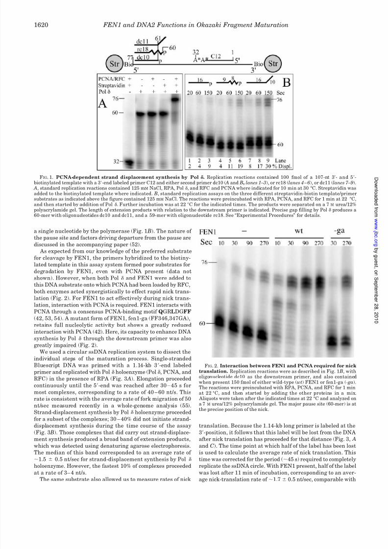
presence of the streptavidin blocks (Fig. 1 A). Control experi-
ments showed that the downstream primer was displaced and
not degraded (data not shown).
When the strand-displacement assay was carried out at
22 °C rather than 30 °C and early time points were taken, rates
of displacement of various downstream primers could be deter-
mined. The rate of strand-displacement synthesis was highest
if the primer to be displaced already contained a 5-unannealed
strand (Fig. 1 B). Fully hybridized RNA-DNA primers were also
more readily displaced than the analogous DNA primers. In all
cases, two prominent pause sites were observed. These pause
sites were at the site of the nick, corresponding to precise gap
filling, and at the 1 position, corresponding to displacement of
FEN1 and DNA2 Functions in Okazaki Fragment Maturation 1619
8/8/2019 J. Biol. Chem.-2003-Ayyagari-1618-25
http://slidepdf.com/reader/full/j-biol-chem-2003-ayyagari-1618-25 3/8
a single nucleotide by the polymerase (Fig. 1 B). The nature of
the pause site and factors driving departure from the pause are
discussed in the accompanying paper (52).
As expected from our knowledge of the preferred substrate
for cleavage by FEN1, the primers hybridized to the biotiny-
lated template in this assay system formed poor substrates for
degradation by FEN1, even with PCNA present (data not
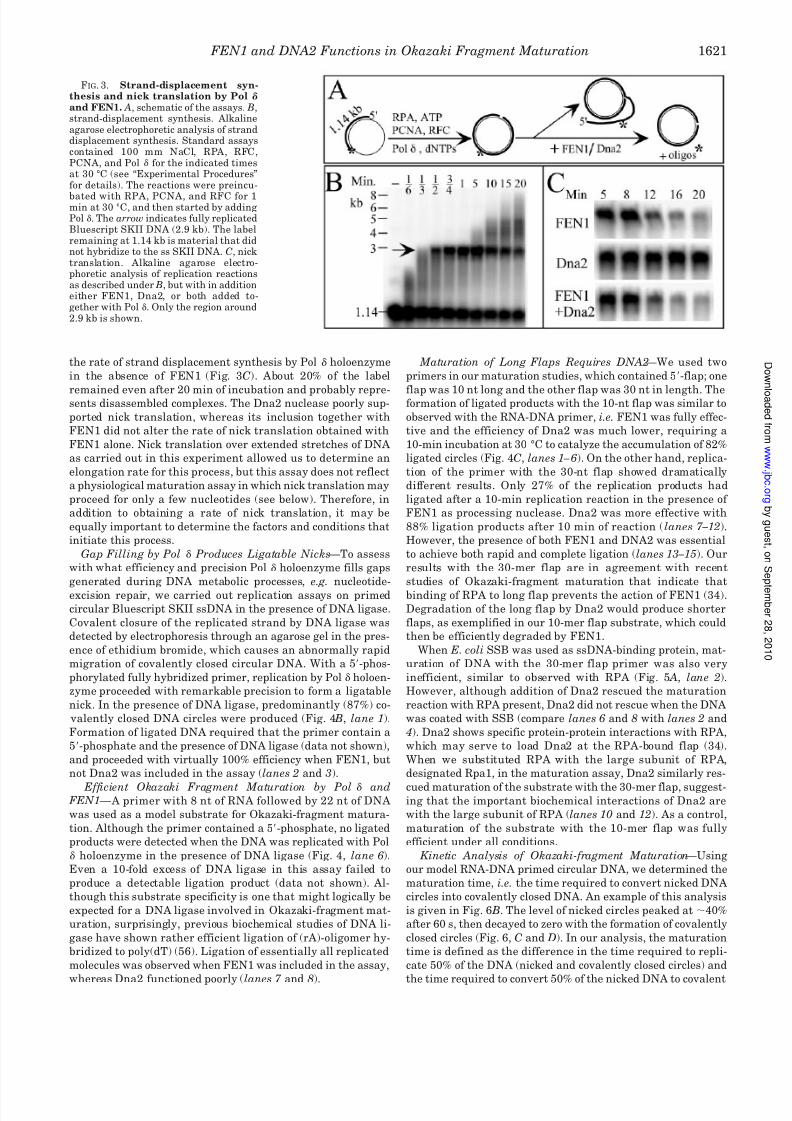
shown). However, when both Pol and FEN1 were added to
this DNA substrate onto which PCNA had been loaded by RFC,
both enzymes acted synergistically to effect rapid nick trans-
lation (Fig. 2). For FEN1 to act effectively during nick trans-
lation, interaction with PCNA is required. FEN1 interacts with
PCNA through a consensus PCNA-binding motif QGRLDGFF
(42, 53, 54). A mutant form of FEN1, fen1-ga (FF346,347GA),
retains full nucleolytic activity but shows a greatly reduced
interaction with PCNA (42). Here, its capacity to enhance DNA
synthesis by Pol through the downstream primer was also
greatly impaired (Fig. 2).
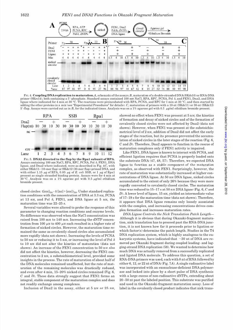
We used a circular ssDNA replication system to dissect the
individual steps of the maturation process. Single-stranded
Bluescript DNA was primed with a 1.14-kb 3-end labeled
primer and replicated with Pol holoenzyme (Pol , PCNA, and
RFC) in the presence of RPA (Fig. 3 A). Elongation proceeded
continuously until the 5-end was reached after 30 – 45 s formost complexes, corresponding to a rate of 40 – 60 nt/s. This
rate is consistent with the average rate of fork migration of 50
nt/sec measured recently in a whole-genome analysis (55).
Strand-displacement synthesis by Pol holoenzyme proceeded
for a subset of the complexes; 30 – 40% did not initiate strand-
displacement synthesis during the time course of the assay
(Fig. 3 B). Those complexes that did carry out strand-displace-
ment synthesis produced a broad band of extension products,
which was detected using denaturing agarose electrophoresis.
The median of this band corresponded to an average rate of
1.5 0.5 nt/sec for strand-displacement synthesis by Pol
holoenzyme. However, the fastest 10% of complexes proceeded
at a rate of 3– 4 nt/s.
The same substrate also allowed us to measure rates of nick
translation. Because the 1.14-kb long primer is labeled at the
3-position, it follows that this label will be lost from the DNA
after nick translation has proceeded for that distance (Fig. 3, A
and C). The time point at which half of the label has been lost
is used to calculate the average rate of nick translation. This
time was corrected for the period (45 s) required to completely
replicate the ssDNA circle. With FEN1 present, half of the label
was lost after 11 min of incubation, corresponding to an aver-
age nick-translation rate of 1.7 0.5 nt/sec, comparable with
FIG. 2. Interaction between FEN1 and PCNA required for nicktranslation. Replication reactions were as described in Fig. 1 B, witholigonucleotide dc10 as the downstream primer, and also containedwhen present 150 fmol of either wild-type (wt) FEN1 or fen1-ga (-ga).The reactions were preincubated with RPA, PCNA, and RFC for 1 minat 22 °C, and then started by adding the other proteins in a mix.
Aliquots were taken after the indicated times at 22 °C and analyzed ona 7 M urea/12% polyacrylamide gel. The major pause site (60-mer) is atthe precise position of the nick.
FIG. 1. PCNA-dependent strand displacement synthesis by Pol . Replication reactions contained 100 fmol of a 107-nt 3- and 5-biotinylated template with a 3-end labeled primer C12 and either second primer dc10 ( A and B, lanes 1 – 3), or rc18 (lanes 4 – 6), or dc11 (lanes 7 –9).
A, standard replication reactions contained 125 mM NaCl, RPA, Pol , and RFC and PCNA where indicated for 10 min at 30 °C. Streptavidin was
added to the biotinylated template where indicated. B, standard replication assays on the three different streptavidin-biotin template/primersubstrates as indicated above the figure contained 125 mM NaCl. The reactions were preincubated with RPA, PCNA, and RFC for 1 min at 22 °C,and then started by addition of Pol . Further incubation was at 22 °C for the indicated times. The products were separated on a 7 M urea/12%polyacrylamide gel. The length of extension products with relation to the downstream primer is indicated. Precise gap filling by Pol produces a60-mer with oligonucleotides dc10 and dc11, and a 59-mer with oligonucleotide rc18. See “Experimental Procedures” for details.
FEN1 and DNA2 Functions in Okazaki Fragment Maturation1620
8/8/2019 J. Biol. Chem.-2003-Ayyagari-1618-25
http://slidepdf.com/reader/full/j-biol-chem-2003-ayyagari-1618-25 4/8
the rate of strand displacement synthesis by Pol holoenzyme
in the absence of FEN1 (Fig. 3C). About 20% of the label
remained even after 20 min of incubation and probably repre-sents disassembled complexes. The Dna2 nuclease poorly sup-
ported nick translation, whereas its inclusion together with
FEN1 did not alter the rate of nick translation obtained with
FEN1 alone. Nick translation over extended stretches of DNA
as carried out in this experiment allowed us to determine an
elongation rate for this process, but this assay does not reflect
a physiological maturation assay in which nick translation may
proceed for only a few nucleotides (see below). Therefore, in
addition to obtaining a rate of nick translation, it may be
equally important to determine the factors and conditions that
initiate this process.
Gap Filling by Pol Produces Ligatable Nicks —To assess
with what efficiency and precision Pol holoenzyme fills gaps
generated during DNA metabolic processes, e.g. nucleotide-
excision repair, we carried out replication assays on primed
circular Bluescript SKII ssDNA in the presence of DNA ligase.
Covalent closure of the replicated strand by DNA ligase was
detected by electrophoresis through an agarose gel in the pres-
ence of ethidium bromide, which causes an abnormally rapid
migration of covalently closed circular DNA. With a 5-phos-
phorylated fully hybridized primer, replication by Pol holoen-
zyme proceeded with remarkable precision to form a ligatable
nick. In the presence of DNA ligase, predominantly (87%) co-
valently closed DNA circles were produced (Fig. 4 B, lane 1).
Formation of ligated DNA required that the primer contain a
5-phosphate and the presence of DNA ligase (data not shown),
and proceeded with virtually 100% efficiency when FEN1, but
not Dna2 was included in the assay (lanes 2 and 3).
Efficient Okazaki Fragment Maturation by Pol and FEN1 — A primer with 8 nt of RNA followed by 22 nt of DNA
was used as a model substrate for Okazaki-fragment matura-
tion. Although the primer contained a 5-phosphate, no ligated
products were detected when the DNA was replicated with Pol
holoenzyme in the presence of DNA ligase (Fig. 4, lane 6).
Even a 10-fold excess of DNA ligase in this assay failed to
produce a detectable ligation product (data not shown). Al-
though this substrate specificity is one that might logically be
expected for a DNA ligase involved in Okazaki-fragment mat-
uration, surprisingly, previous biochemical studies of DNA li-
gase have shown rather efficient ligation of (rA)-oligomer hy-
bridized to poly(dT) (56). Ligation of essentially all replicated
molecules was observed when FEN1 was included in the assay,
whereas Dna2 functioned poorly (lanes 7 and 8).
Maturation of Long Flaps Requires DNA2 —We used two
primers in our maturation studies, which contained 5-flap; one
flap was 10 nt long and the other flap was 30 nt in length. Theformation of ligated products with the 10-nt flap was similar to
observed with the RNA-DNA primer, i.e. FEN1 was fully effec-
tive and the efficiency of Dna2 was much lower, requiring a
10-min incubation at 30 °C to catalyze the accumulation of 82%
ligated circles (Fig. 4C, lanes 1 – 6). On the other hand, replica-
tion of the primer with the 30-nt flap showed dramatically
different results. Only 27% of the replication products had
ligated after a 10-min replication reaction in the presence of
FEN1 as processing nuclease. Dna2 was more effective with
88% ligation products after 10 min of reaction ( lanes 7 –12).
However, the presence of both FEN1 and DNA2 was essential
to achieve both rapid and complete ligation (lanes 13 –15). Our
results with the 30-mer flap are in agreement with recent
studies of Okazaki-fragment maturation that indicate that
binding of RPA to long flap prevents the action of FEN1 (34).
Degradation of the long flap by Dna2 would produce shorter
flaps, as exemplified in our 10-mer flap substrate, which could
then be efficiently degraded by FEN1.
When E. coli SSB was used as ssDNA-binding protein, mat-
uration of DNA with the 30-mer flap primer was also very
inefficient, similar to observed with RPA (Fig. 5 A, lane 2).
However, although addition of Dna2 rescued the maturation
reaction with RPA present, Dna2 did not rescue when the DNA
was coated with SSB (compare lanes 6 and 8 with lanes 2 and
4). Dna2 shows specific protein-protein interactions with RPA,
which may serve to load Dna2 at the RPA-bound flap (34).
When we substituted RPA with the large subunit of RPA,
designated Rpa1, in the maturation assay, Dna2 similarly res-
cued maturation of the substrate with the 30-mer flap, suggest-ing that the important biochemical interactions of Dna2 are
with the large subunit of RPA (lanes 10 and 12). As a control,
maturation of the substrate with the 10-mer flap was fully
efficient under all conditions.
Kinetic Analysis of Okazaki-fragment Maturation —Using
our model RNA-DNA primed circular DNA, we determined the
maturation time, i.e. the time required to convert nicked DNA
circles into covalently closed DNA. An example of this analysis
is given in Fig. 6 B. The level of nicked circles peaked at 40%
after 60 s, then decayed to zero with the formation of covalently
closed circles (Fig. 6, C and D). In our analysis, the maturation
time is defined as the difference in the time required to repli-
cate 50% of the DNA (nicked and covalently closed circles) and
the time required to convert 50% of the nicked DNA to covalent
FIG. 3. Strand-displacement syn-thesis and nick translation by Pol
and FEN1. A, schematic of the assays. B,strand-displacement synthesis. Alkalineagarose electrophoretic analysis of stranddisplacement synthesis. Standard assayscontained 100 mm NaCl, RPA, RFC,PCNA, and Pol for the indicated timesat 30 °C (see “Experimental Procedures”for details). The reactions were preincu-
bated with RPA, PCNA, and RFC for 1min at 30 °C, and then started by addingPol . The arrow indicates fully replicatedBluescript SKII DNA (2.9 kb). The labelremaining at 1.14 kb is material that didnot hybridize to the ss SKII DNA. C, nicktranslation. Alkaline agarose electro-phoretic analysis of replication reactionsas described under B, but with in additioneither FEN1, Dna2, or both added to-gether with Pol . Only the region around2.9 kb is shown.
FEN1 and DNA2 Functions in Okazaki Fragment Maturation 1621
8/8/2019 J. Biol. Chem.-2003-Ayyagari-1618-25
http://slidepdf.com/reader/full/j-biol-chem-2003-ayyagari-1618-25 5/8
closed circles: t[ccc]50 - t([nc][ccc])50. Under standard replica-
tion conditions with the concentration of DNA at 3.3 nM, PCNA
at 13 nM, and Pol , FEN1, and DNA ligase at 5 nM, the
maturation time was 22–25 s.
Several variables were altered to probe the response of this
parameter to changing reaction conditions and enzyme levels.
No difference was observed when the NaCl concentration was
raised from 100 mM to 140 mM. Increasing the dNTP concen-
tration from 100 M to 400 M each resulted in a higher rate of
formation of nicked circles. However, the maturation time re-
mained the same as covalently closed circles also accumulatedmore rapidly (data not shown). Increasing the levels of PCNA
to 50 nM or reducing it to 5 nM, or increasing the level of Pol
to 10 nM did not alter the kinetics of maturation (data not
shown). An increase of the FEN1 concentration to 50 n M also
did not affect the kinetics, however, decreasing the FEN1 con-
centration to 2 nM, a substoichiometrical level, provided some
insights in the process. The rate of maturation of about half of
the DNA molecules remained unaffected, but the rate of mat-
uration of the remaining molecules was drastically reduced,
and even after 4 min, 15–20% nicked circles remained (Fig. 6,
C and D). These data strongly suggest that FEN1 forms an
integral, stable component of the maturation complex and does
not readily exchange among complexes.
Inclusion of Dna2 in the assay, either at 5 nM or 25 nM,
showed no effect when FEN1 was present at 5 nM; the kineticsof formation and decay of nicked circles and of the formation of
covalently closed circles were not affected by Dna2 (data not
shown). However, when FEN1 was present at the substoichio-
metrical level of 2 nM, addition of Dna2 did not affect the early
stages of the reaction, but its presence prevented the accumu-
lation of nicked circles in the later stages of the reaction (Fig. 6,
C and D). Therefore, Dna2 appears to function in the rescue of
maturation complexes only if FEN1 activity is impaired.
Like FEN1, DNA ligase is known to interact with PCNA, and
efficient ligation requires that PCNA is properly loaded onto
the substrate DNA (47, 48, 57). Therefore, we expected DNA
ligase to function as a stable component of the maturation
complex, as observed with FEN1. Surprisingly, however, the
rate of maturation was substantially increased at higher con-centrations of DNA ligase. At 50 nM DNA ligase, nicked circles
accumulated to the extent of only 26% because they were more
rapidly converted to covalently closed circles. The maturation
time was reduced to 15–17 s at 50 nM DNA ligase (Fig. 6, C and
D). A lower level of ligase, 15 nM, yielded an intermediate value
of 17–19 s for the maturation time (data not shown). Therefore,
it appears that DNA ligase remains only loosely associated
with the complex, and increasing concentrations drives com-
plex formation and increases maturation rates.
DNA Ligase Controls the Nick Translation Patch Length —
Although it is obvious that during Okazaki-fragment matura-
tion, nick translation has to proceed past the RNA-DNA junc-
tion, it is not known how far it proceeds prior to ligation or
which factor(s) determine the patch length. Studies in the T4DNA replication system, which is highly analogous to the eu-
karyotic systems, have indicated that 30 nt of DNA are re-
moved per Okazaki fragment during coupled leading- and lag-
ging-strand DNA replication (58). We wanted to determine how
much DNA was actually removed from a successfully replicated
and ligated DNA molecule. To address this question, a set of
RNA-DNA primers was used, each with 8 nt of RNA followed by
either 6, 12, or 22 nt of DNA (Fig. 7 A). A single radioactive label
was incorporated with an exonuclease-deficient DNA polymer-
ase and locked into place by a short pulse of DNA synthesis
with a large excess of non-radioactive dNTPs, extending about
20 –50 nt past the labeled position. This substrate was purified
and used in the Okazaki-fragment maturation assay. Loss of
label in the covalently closed product indicates that nick trans-
FIG. 4. Coupling DNA replication to maturation. A, schematic of the assays. B, maturation of a double-stranded DNA(SKdc10) or RNA-DNAprimer (SKrc14), both containing a 5-phosphate. Standard assays contained 100 mM NaCl, RPA, RFC, PCNA, Pol , and FEN1, Dna2, and DNAligase where indicated for 4 min at 30 °C. The reactions were preincubated with RPA, PCNA, and RFC for 1 min at 30 °C, and then started byadding the other proteins in a mix (see “Experimental Procedures” for details). C, maturation of primers with a 10-nt (SKdc11) or 30-nt (SKdc12)5-flap. Assays were carried out as in B, for the indicated times. Analysis was on a 1% agarose gel with 0.5 g/ml ethidium bromide present.
FIG. 5. DNA2 directed to the flap by the Rpa1 subunit of RPA. Assays containing 100 mM NaCl, RPA, RFC, PCNA, Pol , FEN1, DNAligase, and Dna2 where indicated, were as described in Fig. 4 C, witheither SKdc11- (10-mer flap) or SKdc12 (30-mer flap)-primed DNA, and
with either 1.15 g of RPA, 0.85 g of E. coli SSB, or 1 g of Rpa1present as single stranded binding protein. Assays were for 4 min at30 °C. Analysis was on a 1% agarose gel with 0.5 g/ml ethidiumbromide present.
FEN1 and DNA2 Functions in Okazaki Fragment Maturation1622
8/8/2019 J. Biol. Chem.-2003-Ayyagari-1618-25
http://slidepdf.com/reader/full/j-biol-chem-2003-ayyagari-1618-25 6/8
lation proceeded past the labeled position prior to ligation. Fig.
7 B shows the results obtained when the label was inserted
after the sixth nucleotide past the RNA-DNA junction, showing
several controls and the effect of varying levels of FEN1, Dna2,
and DNA ligase on the percent retention of label in the co-
valently closed DNA. The same assays were carried out withprimers in which the label was introduced at positions 12 or 22
after the RNA-DNA junction (data not shown). For each exper-
iment, the percent retention of label was plotted as a function
of the position of the label in nucleotides past the RNA/DNA
junction, and the level of 50% retention of label was determined
by interpolation or extrapolation to obtain the average nick
translation patch length (Fig. 7C).
This type of analysis indicates that under our standard rep-
lication and maturation conditions, the average nick transla-
tion patch is 8 –12 nt (the range of three independent experi-
ments) past the RNA-DNA junction, with or without Dna2
present. However, in the presence of 50 nM of DNA ligase, the
patch size is reduced to only 4 – 6 nt. A lower concentration of
DNA ligase (15 nM) was almost as effective (Fig. 7 B, lane 5).
Very high levels of FEN1 (50 nM) slightly increased the patch
size to 10 –15 nt from 8 –12nt.
DISCUSSION
Our kinetic analysis of gap filling and Okazaki-fragment
synthesis has yielded a picture of a remarkably efficient inter-
action between Pol and FEN1. PCNA is the organizing force
in this coupling between synthesis and degradation, as exem-
plified by the observation that a FEN1 mutant that is only
deficient for interaction with PCNA, is unable to carry out nick
translation with Pol (Fig. 2). All of our studies were carried
out at 100 –140 mM NaCl, which lends specificity to the reac-
tions by inhibiting binding of the enzymes to the DNA when
PCNA is not loaded (Figs. 1 and 2).
When Pol encounters downstream double-stranded DNA, it
pauses at the position of a precise nick with high frequency as
follows from the observation that in the presence of DNA li-
FIG. 6. Rates of Okazaki fragment maturation. A, schematic of
the assay. The primer was SKrc14 with 8 nt of RNA followed by 22 ntof DNA. B, time course of maturation under standard conditions with3.3 nM DNA, 5 nM Pol , 5 nM of FEN1, and 5 nM of DNA ligase. Thereactions were preincubated with RPA, PCNA, and RFC for 1 min at30 °C, and then started by adding the other proteins in a mix (see“Experimental Procedures” for details). Analysis was on a 1% agarosegel with 0.5 g/ml ethidium bromide present. C, accumulation anddisappearance of nicked circles under the indicated experimental con-ditions. D, accumulation of covalently closed circles.
FIG. 7. Nick translation patch length during maturation. A,
schematic of the assay. The starred position indicates the position of thelabel in each of three individual primers. B, assays containing 100 mM
NaCl, RPA, RFC, PCNA, Pol , and FEN1, DNA ligase, or Dna2 asindicated, were carried out with SKII ssDNA primed with 3-end la-beled SKrc14–14 (5-p-rArCrGrArCrGrUrU-GTAAAA*CN
20–50; see
“Experimental Procedures”). The reactions were preincubated withRPA, PCNA, and RFC for 1 min at 30 °C and then started by adding theother proteins in a mix. Incubation was for 4 min at 30 °C and analysiswas on a 1% agarose gel with 0.5 g/ml ethidium bromide present. C,quantitation of label remaining for assays similar to described in B butcarried out with either 3-end labeled SKrc14 –14, SKrc14 –20 (5-p-rArCrGrArCrGrUrU-GTAAAACGACGG*CN
20–50), or SKrc14–30
(5-p-rArCrGrArCrGrUrU-GTAAAACGACGGCCAGTGAGCG*CN20–50
)as primer.
FEN1 and DNA2 Functions in Okazaki Fragment Maturation 1623
8/8/2019 J. Biol. Chem.-2003-Ayyagari-1618-25
http://slidepdf.com/reader/full/j-biol-chem-2003-ayyagari-1618-25 7/8
8/8/2019 J. Biol. Chem.-2003-Ayyagari-1618-25
http://slidepdf.com/reader/full/j-biol-chem-2003-ayyagari-1618-25 8/8
to the cell, we cannot address whether the essential nature of
DNA2 is due to its constitutively required presence at each Oka-
zaki fragment, or to the rescue of a small number of stalled
Okazaki fragments which otherwise would be lethal. However,
our in vitro results do suggest the latter as a reasonable scenario
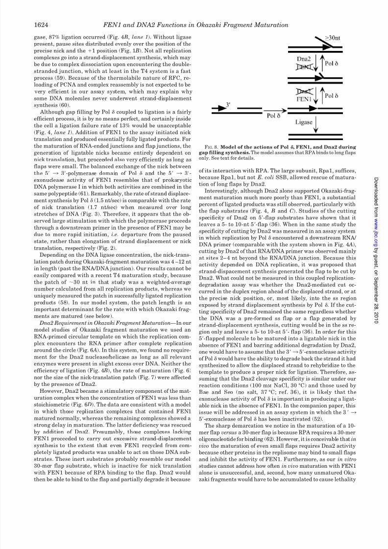
(Fig. 8). Our data are not in disagreement with a recent study by
Bae et al. (34). Because in that study all maturation experiments
with DNA ligase were performed on substrates with long 5-flaps,
an absolute requirement for Dna2 was measured, as we did with
our 30-mer flap substrate. DNA Ligase Is Loosely Associated with the Maturation Com-
plex —The biphasic response resulting from the maturation stud-
ies with substoichiometrical concentrations of FEN suggest that
FEN1 forms a stable integral component of the maturation com-
plex. The same is not the case for DNA ligase. Even though DNA
ligase has a PCNA-binding domain similar to FEN1, and the
interaction between PCNA and DNA ligase I is critical for joining
Okazaki fragments and for long-patch base-excision repair in
mammalian cells, we observed a titratable response which satu-
rated at an15-fold molar excess of DNA ligase (47). Perhaps, on
the DNA, the interaction between PCNA and DNA ligase is less
strong, or alternatively, FEN1 and/or Pol compete with DNA
ligase for the same binding site(s) on PCNA. Presumably, other
factor(s) may be required in vivo to keep DNA ligase positioned inthe maturation complex.
Efficiency of Okazaki Fragment Maturation — Although Oka-
zaki-fragment maturation in our in vitro system is very effi-
cient, at least as measured by end-product formation, the rates
of maturation may still be incompatible with cellular metabo-
lism. Assuming that the average length of an Okazaki frag-
ment is 150 nt, it would take 3– 4 s for elongation to be
complete at a fork rate of 50 nt/sec (55). No information is
available regarding the rate of initiation, i.e. priming by DNA
polymerase -primase. However, maturation under our most
favorable conditions, with high concentrations of DNA ligase,
still takes an average of 15–17 s. Most of this period is taken up
by nick translation through an RNA-DNA stretch of 15 nt at
1.7 nt/s. Under these conditions, one would expect Okazaki
fragments to accumulate inside the cell, which appears not to
be the case (63). Therefore, to improve maturation rates one
would either need to improve the rate of nick translation or
bypass extensive nick translation through helicase action to
rapidly displace the RNA-DNA section to be matured. Dna2,
which has helicase activity, did not accelerate maturation (Fig.
6) (4, 36). Furthermore, the essential activity of Dna2 is the
nuclease, rather than the helicase (9). Therefore, another DNA
helicase may be involved in this process.
Acknowledgments —We thank Kim Gerik and Carrie Welch for tech-nical assistance and John Majors for critical discussions during thecourse of this work.
REFERENCES
1. Bambara, R. A., Murante, R. S., and Henricksen, L. A. (1997) J. Biol. Chem.272, 4647– 4650
2. MacNeill, S. A., and Burgers, P. M. (2000) in The Yeast Nucleus: Frontiers in Molecular Biology (Fantes, P., and Beggs, J., eds), pp. 19 –57, Oxford Uni- versity Press, Oxford
3. Chen, J. Z., Qiu, J., Shen, B., and Holmquist, G. P. (2000) Nucleic Acids Res.28, 3649 –3656
4. Budd, M. E., Choe, W.-C., and Campbell, J. (1995) J. Biol. Chem. 270,26766 –26769
5. Budd, M. E., and Campbell, J. L. (1997) Mol. Cell. Biol. 17, 2136 –21426. Formosa, T., and Nittis, T. (1999) Genetics 151, 1459 –14707. Kang, H. Y., Choi, E., Bae, S. H., Lee, K. H., Gim, B. S., Kim, H. D., Park, C.,
MacNeill, S. A., and Seo, Y. S. (2000) Genetics 155, 1055–10678. Bae, S. H., Choi, E., Lee, K. H., Park, J. S., Lee, S. H., and Seo, Y. S. (1998)
J. Biol. Chem. 273, 26880 –268909. Lee, K. H., Kim, D. W., Bae, S. H., Kim, J. A., Ryu, G. H., Kwon, Y. N., Kim,
K. A., Koo, H. S., and Seo, Y. S. (2000) Nucleic Acids Res. 28, 2873–288110. Budd, M. E., Choe, W., and Campbell, J. L. (2000) J. Biol. Chem. 275,
16518 –1652911. Parenteau, J., and Wellinger, R. J. (1999) Mol. Cell. Biol. 19, 4143– 4152
12. Choe, W., Budd, M., Imamura, O., Hoopes, L., and Campbell, J. L. (2002) Mol.Cell. Biol. 22, 4202– 4217
13. Klungland, A., and Lindahl, T. (1997) EMBO J. 16, 3341–334814. Kim, K., Biade, S., and Matsumoto, Y. (1998) J. Biol. Chem. 273, 8842–884815. Matsumoto, Y. (2001) Progr. Nucleic Acids Res. Mol. Biol. 68, 129–13816. Johnson, R. E., Kovvali, G. K.,Prakash, L., andPrakash,S. (1995) Science 269,
238 –24017. Tishkoff, D. X., Filosi, N., Gaida, G. M., and Kolodner, R. D. (1997) Cell 88,
253–26318. Kokoska, R. J., Stefanovic, L., Tran, H. T., Resnick, M. A., Gordenin, D. A., and
Petes, T. D. (1998) Mol. Cell. Biol. 18, 2779 –278819. Negritto, M. C., Qiu, J., Ratay, D. O., Shen, B., and Bailis, A. M. (2001) Mol.
Cell. Biol. 21, 2349 –235820. Xie, Y., Liu, Y., Argueso, J. L., Henricksen, L. A., Kao, H. I., Bambara, R. A.,and Alani, E. (2001) Mol. Cell. Biol. 21, 4889 – 4899
21. Henricksen, L. A., Veeraraghavan, J., Chafin, D. R., and Bambara, R. A. (2002) J. Biol. Chem. 277, 22361–22369
22. Johnson, R. E., Kovvali, G. K., Prakash, L., and Prakash, S. (1998) Curr.Genet. 34, 21–29
23. Symington, L. S. (1998) Nucleic Acids Res. 26, 5589 –559524. Tishkoff, D. X., Boerger, A. L., Bertrand, P., Filosi, N., Gaida, G. M., Kane,
M. F., and Kolodner, R. D. (1997) Proc. Natl. Acad. Sci. U. S. A. 94,7487–7492
25. Li, X., Li, J., Harrington, J., Lieber, M. R., and Burgers, P. M. (1995) J. Biol.Chem. 270, 22109 –22112
26. Eissenberg, J. C., Ayyagari, R., Gomes, X. V., and Burgers, P. (1997) Mol. Cell. Biol. 17, 6367– 6378
27. Kao, H. I., Henricksen, L. A., Liu, Y., and Bambara, R. A. (2002) J. Biol. Chem.277, 14379 –14389
28. Hosfield, D. J., Mol, C. D., Shen, B., and Tainer, J. A. (1998) Cell 95, 135–14629. Hwang, K. Y., Baek, K., Kim, H. Y., and Cho, Y. (1998) Nat. Struct. Biol. 5,
707–713
30. Murante, R. S., Rust, L., and Bambara, R. A. (1995) J. Biol. Chem. 270,30377–30383
31. Xu, Y., Potapova, O., Leschziner, A. E., Grindley, N. D., and Joyce, C. M. (2001) J. Biol. Chem. 276, 30167–30177
32. Rigby, P. W., Dieckmann, M., Rhodes, C., and Berg, P. (1977) J. Mol. Biol. 113,
237–25133. Turchi, J. J., and Bambara, R. A. (1993) J. Biol. Chem. 268, 15136 –1514134. Bae, S. H., Bae, K. H., Kim, J. A., and Seo, Y. S. (2001) Nature 412, 456 –46135. Maga, G., Villani, G., Tillement, V., Stucki, M., Locatelli, G. A., Frouin, I.,
Spadari, S., and Hubscher, U. (2001) Proc. Natl. Acad. Sci. U. S. A. 98,
14298 –1430336. Bae, S. H., and Seo, Y. S. (2000) J. Biol. Chem. 275, 38022–3803137. Waga, S., Bauer, G., and Stillman, B. (1994) J. Biol. Chem. 269, 10923–1093438. Burgers, P. M., and Gerik, K. J. (1998) J. Biol. Chem. 273, 19756 –1976239. Burgers, P. M. (1999) Methods 18, 349 –35540. Henricksen, L. A., Umbricht, C. B., and Wold, M. S. (1994) J. Biol. Chem. 269,
11121–1113241. Gomes, X. V., Gary, S. L., and Burgers, P. M. (2000) J. Biol. Chem. 275,
14541–1454942. Gomes, X. V., and Burgers, P. M. J. (2000) EMBO J. 19, 3811–3821
43. Sambrook, J., Fritsch, E. F., and Maniatis, T. (1989) Molecular Cloning: A Laboratory Manual, Cold Spring Harbor Laboratory, Cold Spring Harbor, NY
44. Yamamoto, K. R., Alberts, B. M., Benzinger, R., Lawhorne, L., and Treiber, G.(1970) Virology. 40, 734 –744
45. Montecucco, A., Rossi, R., Levin, D. S., Gary, R., Park, M. S., Motycka, T. A.,Ciarrocchi, G., Villa, A., Biamonti, G., and Tomkinson, A. E. (1998) EMBO J. 17, 3786 –3795
46. Mossi, R., Ferrari, E., and Hubscher, U. (1998) J. Biol. Chem. 273,
14322–1433047. Levin, D. S., McKenna, A. E., Motycka, T. A., Matsumoto, Y., and Tomkinson,
A. E. (2000) Curr. Biol. 10, 919 –92248. Tom, S., Henricksen, L. A., Park, M. S., and Bambara, R. A. (2001) J. Biol.
Chem. 276, 24817–2482549. Burgers, P. M. J., and Yoder, B. L. (1993) J. Biol. Chem. 268, 19923–1993650. Podust, L. M., Podust, V. N., Floth, C., and Hubscher, U. (1994) Nucleic Acids
Res. 22, 2970 –297551. Kaboord, B. F., and Benkovic, S. J. (1993) Proc. Natl. Acad. Sci. U. S. A. 90,
10881–1088552. Jin, Y. H., Ayyagari, R., Resnick, M. A., Gordenin, D. A., and Burgers, P. M.
(2003) J. Biol. Chem., 278, 1626 –163353. Warbrick, E. (1998) Bioessays 20, 195–19954. Gary, R., Park, M. S., Nolan, J. P., Cornelius, H. L., Kozyreva, O. G., Tran,
H. T., Lobachev, K. S., Resnick, M. A., and Gordenin, D. A. (1999) Mol. Cell. Biol. 19, 5373–5382
55. Raghuraman, M. K., Winzeler, E. A., Collingwood, D., Hunt, S., Wodicka, L.,Conway, A., Lockhart, D. J., Davis, R. W., Brewer, B. J., and Fangman,W. L. (2001) Science 294, 115–121
56. Tomkinson, A. E., Tappe, N. J., and Friedberg, E. C. (1992) Biochemistry. 31,
11762–1177157. Matsumoto, Y., Kim, K., Hurwitz, J., Gary, R., Levin, D. S., Tomkinson, A. E.,
and Park, M. S. (1999) J. Biol. Chem. 274, 33703–3370858. Bhagwat, M., and Nossal, N. G. (2001) J. Biol. Chem. 276, 28516 –2852459. Hacker, K. J., and Alberts, B. M. (1994) J. Biol. Chem. 269, 24221–2422860. Yoder, B. L., and Burgers, P. M. J. (1991) J. Biol. Chem. 266, 22689 –2269761. Ma, W. P., Kaiser, M. W., Lyamicheva, N., Schaefer, J. J., Allawi, H. T.,
Takova, T., Neri, B. P., and Lyamichev, V. I. (2000) J. Biol. Chem. 275,
24693–2470062. Kim, C., Snyder, R. O., and Wold, M. S. (1992) Mol. Cell. Biol. 12, 3050 –305963. Bielinsky, A. K., and Gerbi, S. A. (1999) Mol. Cell 3, 477– 486
FEN1 and DNA2 Functions in Okazaki Fragment Maturation 1625


![Arch. Biol. Sci., Belgrade, 66 (4), 1617-1631, 2014 DOI:10 ... · 1618 KEMAL KURT Turkey or the Anatolian Plateau. Th e European por- ... Turkey [Nosek 1905; Kurt et al., 2010]. Egaenus](https://img.pdfslide.us/doc/110x75/5af6a5ba7f8b9a9e598fa61f/arch-biol-sci-belgrade-66-4-1617-1631-2014-doi10-kemal-kurt-turkey.jpg)