Embed Size (px)
Citation preview

IVIS | Lumina II
Quantitative Fluorescentand Bioluminescent ImagingThe IVIS Lumina II from Caliper Life Sciences provides an expandable,
sensitive imaging system that is easy to use for both fluorescent and
bioluminescent imaging in vivo. The system includes a highly sensitive
CCD camera, light-tight imaging chamber and complete automation
and analysis capabilities. As the leading optical imaging platform for
in vivo analysis, IVIS systems include a range of practical accessories
developed through experience in research laboratories worldwide.

IVIS LUMINA II
FluorophoresStandard High Resolution
Excitation Filter Set (Built-in)Emission Filter Options
GFP, YFP and PKH26
430, 465, 500, 535, 570, 605,
640, 675, 710, 745
*500 Series500, 520, 540, 560, 580, 600 and 620
Cy 5.5, DsRed, dTomato and XenoFluor 680
*600 Series580, 600, 620, 640, 660, 680 and 700
Indocyanine Green and XenoFluor 750
*700 Series720, 740, 760, 780, 800, 820, and 840
Multiple Fluorophores Spanning 500-900 nm Broad Imaging Solution
Standard Emission Filter Set515-575, 575-650, 695-770, 810-875
* Median wavelength band path 20nm on emission filters
Field of View
The IVIS Lumina II Imaging System provides 5 fields of view.Standard Lens FOV New XFOV-24 Upgrade
Ex Vivo In Vivo
In Vivo Molecular ImagingQuantitative Flexible Expandable
An adjustable field of view from 5 – 12.5 cm and an optional 24cm lens allows imaging of up to 5 mice or 2 medium size rats. The Lumina II can also accommodate Petri dishes or micro-titer plates for in vitro imaging. The system includes premium animal handling features such as a heated stage, gas anesthesia connections and ECG monitoring.
High resolution, sharp cut-off filters are simply interchangeable to achieve the highest performance, sensitivity and spectral unmixing in fluorescence imaging.
Superior Imaging Results The IVIS Lumina II is capable of imaging both fluorescent and bioluminescent reporters. The system is equipped with up to 21 filter sets that can be used to image reporters that emit from green to near-infrared. Superior spectral unmixing can be achieved by Lumina II’s optional high resolution short cut off filters. Absolute calibration affords you consistent and reproducible results independent of magnification, filter selection from one instrument to any another IVIS instrument within an organization or around the world. The Living Image software yields high-quality, reproducible, quantitative results incorporating instrument calibration, background subtraction and the image algorithms.
Customize the IVIS Lumina II with your own Filter Combinations

IVIS LUMINA II
Triple Reporter ImagingThigh Infection with Klebsiella pneumoniae expressing luxCDABE with optimized GFP or RFP. Approximately 108 CFU per thigh. Courtesy of the University of Glasgow.
Dual Reporter Imaging - High Resolution Ex Vivo ApplicationsBacterial luc (500nm) and GFAP (620nm) brain imaging from mice with Pneumococcal Meningitis. Ex Vivo Kadurugamuwa et al., Infection and Immunity, 2005.
Spectral Unmixing of Xenofluor 680/750Subcutaneous injections of 1014 molecules of XenoFluor 680 (scruff) and 1014 molecules of XenoFluor 750 (lower dorsal region) 605nm excitation filter.
Living Image Software with IVIS Lumina II SystemThe wide range of IVIS system instru-ment settings, combined with abso-lute calibration of each setting, allows users to track signals during longitu-dinal studies that vary by many orders of magnitude. In this drug study, tu-mor signals vary by three orders of magnitude during the course of a 35 day experiment. The capability of Liv-ing Image Software makes this type of analysis simple for the user in both flu-orescent and bioluminescent modes.
Imaging Results - Living Image Software with High Resolution Filters
Corporate Headquarters68 Elm StreetHopkinton, MA 01748-1668
Tel: 1.508.435.9500Email: [email protected]
©2008 Caliper Life Sciences, Inc. All rights reserved. Caliper, the Caliper logo, IVIS, Lumina, Xenogen,
XenoFluor and Living Image are tradenames and/or trademarks of Caliper Life Sciences, Inc. All other
names are trademarks of their respective companies.
LUM-BR-02 Jun 08
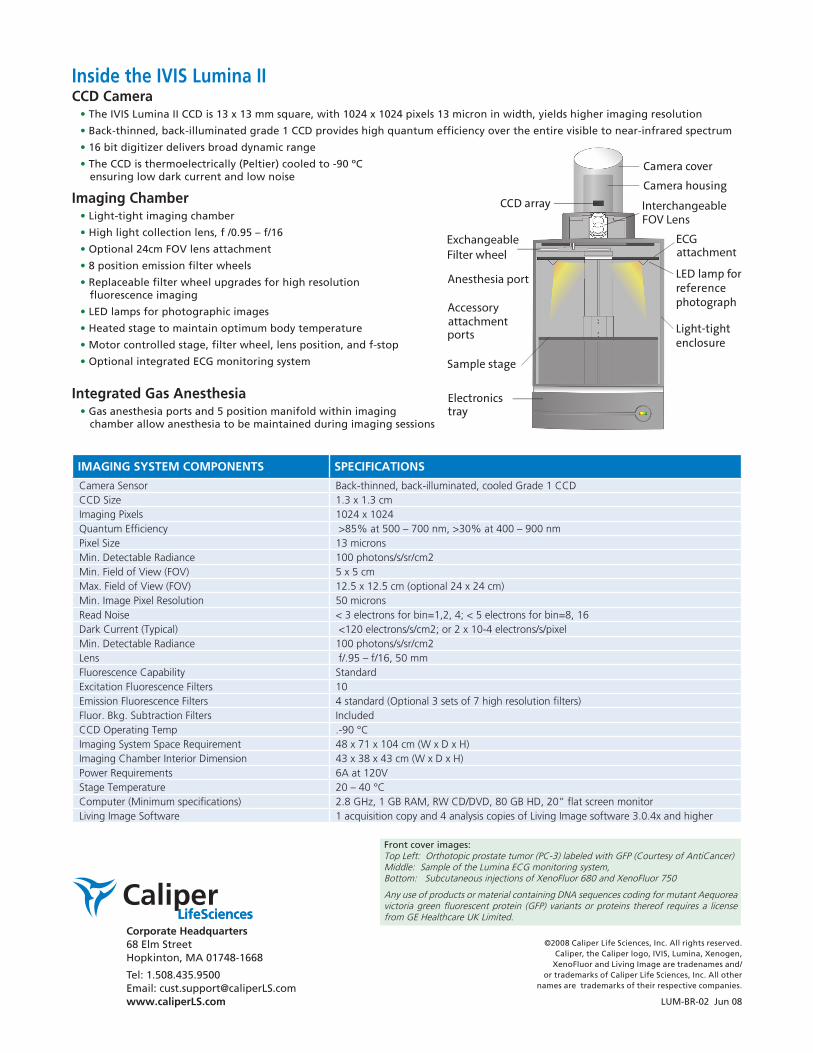
Inside the IVIS Lumina IICCD Camera• The IVIS Lumina II CCD is 13 x 13 mm square, with 1024 x 1024 pixels 13 micron in width, yields higher imaging resolution
• Back-thinned, back-illuminated grade 1 CCD provides high quantum efficiency over the entire visible to near-infrared spectrum
• 16 bit digitizer delivers broad dynamic range
• The CCD is thermoelectrically (Peltier) cooled to -90 ºC ensuring low dark current and low noise
Imaging Chamber• Light-tight imaging chamber
• High light collection lens, f /0.95 – f/16
• Optional 24cm FOV lens attachment
• 8 position emission filter wheels
• Replaceable filter wheel upgrades for high resolution fluorescence imaging
• LED lamps for photographic images
• Heated stage to maintain optimum body temperature
• Motor controlled stage, filter wheel, lens position, and f-stop
• Optional integrated ECG monitoring system
Integrated Gas Anesthesia• Gas anesthesia ports and 5 position manifold within imaging
chamber allow anesthesia to be maintained during imaging sessions
Front cover images:Top Left: Orthotopic prostate tumor (PC-3) labeled with GFP (Courtesy of AntiCancer) Middle: Sample of the Lumina ECG monitoring system, Bottom: Subcutaneous injections of XenoFluor 680 and XenoFluor 750
Any use of products or material containing DNA sequences coding for mutant Aequorea victoria green fluorescent protein (GFP) variants or proteins thereof requires a license from GE Healthcare UK Limited.
IMAGING SYSTEM COMPONENTS SPECIFICATIONS
Camera Sensor Back-thinned, back-illuminated, cooled Grade 1 CCDCCD Size 1.3 x 1.3 cmImaging Pixels 1024 x 1024Quantum Efficiency >85% at 500 – 700 nm, >30% at 400 – 900 nmPixel Size 13 micronsMin. Detectable Radiance 100 photons/s/sr/cm2Min. Field of View (FOV) 5 x 5 cmMax. Field of View (FOV) 12.5 x 12.5 cm (optional 24 x 24 cm)Min. Image Pixel Resolution 50 micronsRead Noise < 3 electrons for bin=1,2, 4; < 5 electrons for bin=8, 16Dark Current (Typical) <120 electrons/s/cm2; or 2 x 10⁻⁴ electrons/s/pixelMin. Detectable Radiance 100 photons/s/sr/cm2Lens f/.95 – f/16, 50 mmFluorescence Capability StandardExcitation Fluorescence Filters 10Emission Fluorescence Filters 4 standard (Optional 3 sets of 7 high resolution filters)Fluor. Bkg. Subtraction Filters IncludedCCD Operating Temp .-90 °CImaging System Space Requirement 48 x 71 x 104 cm (W x D x H)Imaging Chamber Interior Dimension 43 x 38 x 43 cm (W x D x H)Power Requirements 6A at 120VStage Temperature 20 – 40 °CComputer (Minimum specifications) 2.8 GHz, 1 GB RAM, RW CD/DVD, 80 GB HD, 20” flat screen monitorLiving Image Software 1 acquisition copy and 4 analysis copies of Living Image software 3.0.4x and higher