Embed Size (px)
Citation preview

VOLUME 16
IVF and embryo transfer: historicalorigin and developmentby John D. Biggers, DSc, PhD
The Importance of pH MeasurementWithin the IVF Laboratoryby Jason Swain, PhD, HCLD
The Use of global® forTime-lapse Videographic Analysis
of Human Embryo Developmentby Don Rieger, PhD
ASRM 2013, Boston, MA
Thoughts on embryo culture conditionsby John D. Biggers, DSc, PhD

global®
® ’.
Let the Embryo Choose! ®
’
®’.
®

“We are extremely happy with Global medium combined with the Embryoscope. Nice blastocyst
formation and our pregnancy rates have been over 70% with Day 5 transfer. We use it as a continuous
culture medium from Day 1 onwards with no media exchange.”
“We transitioned from using it from D1 thru 6 with a half change on Day 3 to using it for “uninterrupted
culture” from Day 1-Day 6. Equivalent results both ways.”
®
1. Uninterrupted culture of human embryos in global® or global® total®
Reprod Biomed Online 26
Human Reprod 28 Suppl. 1
Fertil Steril 99 Supplement
Reprod Biomed Online 25
Reprod Biomed Online 24 Suppl 1.
Hum Reprod 28
Eur J Obstet Gynecol Reprod
Biol 168
2. Time-lapse culture of human embryos in global® or global® total®

Fertility Magazine The First Magazine In FertilityTM
IVF and embryo transfer: historical origin and development by John D Biggers, DSc, PhD ......................................................................... 5
The Importance of pH Measurement Within the IVF Laboratoryby Jason E. Swain, PhD, HCLD .......................................................................16
The Use of global®
Embryo Development by Don Rieger, PhD .................................................21
Thoughts on embryo culture conditions by John D Biggers, DSc, PhD ...........................................................................34
DON RIEGER, PHD
JOHN D. BIGGERS, DSC, PHD

Instructions to Contributors
Fertility Magazine and all its associates ©2013, All Rights Reserved. Covers, contents, images, ads in print or web form are copryight protected and reprinting or reproduction of any kind
is expressly prohibited without the written permission of Fertility Magazine. Fertility Magazine does not knowingly accept false or misleading advertising, articles, opinions or editorial, nor
does the publisher assume any responsibility for the consequences that occur should any such material appear, and assumes no responsibility for content, text, opinions or artwork of
advertisements appearing in Fertility Magazine in print or web form. Some of the views expressed by contributors may not be the representative views of the publisher.
Editor in Chief: Monica Mezezi, MBAEditor(s): Don Rieger, PhD
Michael WestDesign: Debrah Frank
IVFonline, 24 Norwich St. E.Guelph, Ontario, Canada, N1H 2G6
T: 800-720-6375T: 519-826-5800F: 519-826-6947E: [email protected]
by Carole Lawrence
by Ali Farid Mohammed Ali, MD
56 Conferences

HAPPY “90th” BIRTHDAY
John BiggersDear Dr. Biggers,
It LifeGlobal team, I would like to thank you for all you contributions and strong support in paving the way for single culture medium. Your knowledge and wisdom have helped make global® a tremendous success world
Thank you again for believing in global®
With love and Respect,Monica Mezezi

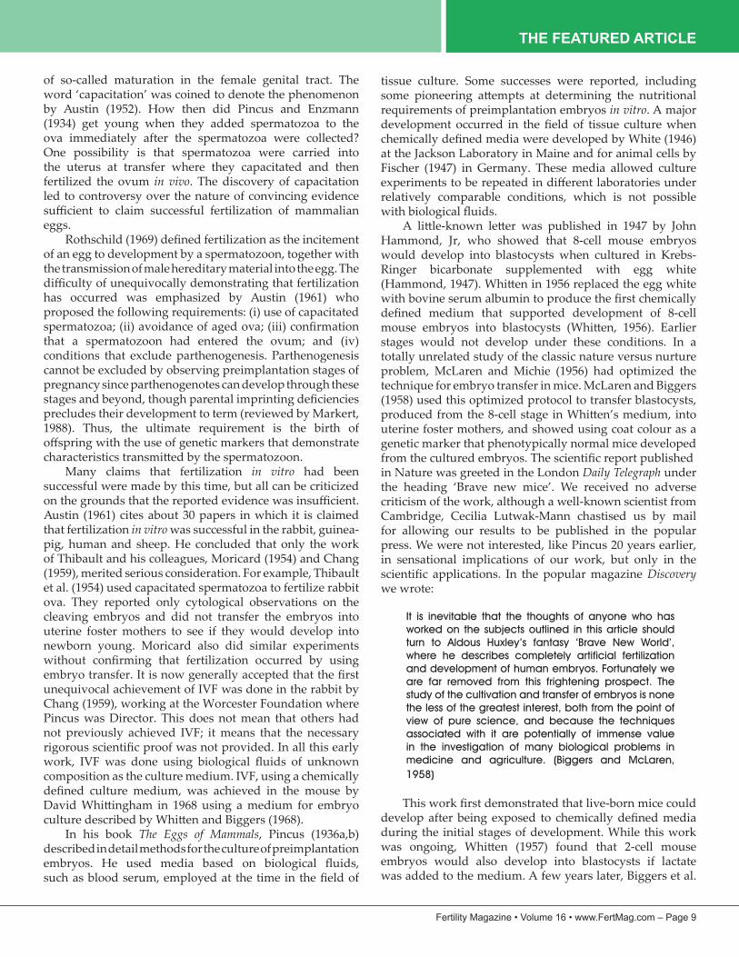
THE FEATURED ARTICLE
IVF and embryo transfer: historical origin and
developmentJohn D Biggers
Department of Cell Biology, 240 Longwood Avenue, Harvard Medical School, Boston, MA 02115, USAE-mail address: [email protected]
John Biggers, DSc, PhD is professor of cell biology at Harvard Medical School. His current research interests are evaporative drying of spermatozoa, vitrification, embryo culture, embryo assessment and the biography of Walter Heape. He is a former Commonwealth Fellow of St John’s College, Cambridge, past President of the Society of Reproduction, former Editor in Chief Biology of Reproduction, Chief Scientific Advisor to the Ethics Committee, US Department of Health, Education and Welfare that made recommendations on IVF and embryo transfer, Fellow of the American Association for the Advancement of Science, Hartman Award of the Society of Reproduction, Pioneer Award of the International Embryo Transfer Society, Marshall Medal of the Society for the Study of Fertility and a Life Member of the New England Fertility Society and the Society for the Study of Reproduction.
Abstract IVF and embryo transfer for the treatment of human infertility has now resulted in the birth of over 4 million babies. The technique did not arise as a quantum event but was built on the efforts of many earlier workers in the fields of reproductive endocrinology and development. One should remember the famous saying of Isaac Newton: ‘If I have seen further than most, it is because I have stood on the shoulder’s of giants’. Ethical and moral issues have always arisen when investigators study early mammalian development, particularly human development. This paper documents these earlier studies and also draws attention to the ethical and moral arguments that inevitably arose.
©2012, Reproductive Healthcare Ltd. Published by Elsevier Ltd. All rights reserved.KEYWORDS: ethical issues, history, in-vitro fertilization, IVF, Gregory Pincus, John Rock
This article was published in Reproductive BioMedicine Online, Vol 25, 2012, p118-127, ‘IVF and embryo transfer: historical origin and development’. Copyright Elsevier. It is reprinted here with permission.
JOHN D. BIGGERS, DSC, PHD
Introduction
The 2010 Nobel Prize for Physiology or Medicine was awarded to Robert Edwards for developing IVF and embryo transfer (IVF/ET) to treat infertility in women with non-patent oviducts. His work resulted in the birth of the
about 33 years, more than 4 million babies have been born using IVF/ET, and a new specialty of assisted reproduction has been established with its own professional societies. The history of IVF/ET is extensive and it has been recently documented in part by Johnson (2011) and on the web (www.IVF-Worldwide. com). The technique did not arise as
development. One should remember the famous quotation
and moral issues have always arisen when investigators study early mammalian development, particularly human development. This paper documents these early studies
that inevitably arose.IVF/ET in mythology
The idea of transferring a human fetus from one mother to another can be found in a story from the Jain religion about the religious leader Mahavira. The following account is a brief summary of the full story in the Sacred Books of the East (Jaina Sutra, 1964). At one of his reincarnations the ruler of all the gods of heaven and earth, Sakra, realized that the Mahavira had been conceived by Devananda, a woman of an unacceptable caste. Sakra then commanded that the embryo carried by Devananda be transferred into the womb of Trisala, a woman of an acceptable caste, and
story has been memorialized at least until the 15th century in sculpture and painting.
Treatment of non-patent oviducts prior to IVF/ET
Clinicians knew, by the middle of the 19th century, that blocked oviducts resulted in sterility (Churchill, 1846).

THE FEATURED ARTICLE
infertility on the dispersal of family wealth, and since that time several surgical procedures have been tried (reviewed
the oviduct in a patient by passing a whalebone bougie through the tube (Smith, 1849). Colleagues greeted the procedure with skepticism and it was never adopted. The demonstration at the end of the 19th century that the ovaries could be transplanted to ectopic sites paved the way for the discovery of the ovarian hormones. The fact that the ovary maintains its function when placed in an ectopic site led Morris (1895), an American gynaecologist, to treat patients with blocked oviducts by grafting ovarian tissue into the oviduct or uterus below the obstruction. The method was never successful. In 1909, Estes, another American gynaecologist, introduced an operation in which the ovary was inserted through the uterine wall, keeping its pedicle containing blood vessels and nerves intact, where it was hoped ovulation would occur (Estes, 1909). The so-called Estes operation was seldom successful, although it was used until the middle of the 20th century. While IVF/ET was
cut ends of the oviduct after removal of an obstruction. By
microsurgery procedures showed promise of being an alternative procedure (Eddy, 1981; Winston, 1981). Since then other improvements in technique have occurred so that the suggestion has been made that IVF/ET and surgery should be regarded as complimentary techniques for the treatment of tubal infertility (Gomel and McComb, 2006; Schippert et al., 2010a,b).
technologies
IVF/ET involves four main aspects: (i) acquisition in
mature ova; (ii) fertilization of these mature ova in vitro; (iii) culture of preimplantation embryos; and (iv) embryo replacement within a mother. Initially, these areas were largely studied independently and it was only later that they were linked to create the technical procedure called IVF/ET. Unfortunately, there is a tendency in historical accounts of IVF/ET, particularly among clinicians, to focus almost exclusively on the actual process of fertilization in vitro, ignoring the history of the other three components.
Walter Heape reported in 1891 that he had successfully transferred mammalian (rabbit) embryos from one mother to another (Heape, 1891). He was a gentleman scientist who studied under Francis Balfour in Cambridge. Although he never earned a degree, he was later appointed a demonstrator in the embryology course where students recovered and observed living rabbit embryos (Foster and Balfour, 1883). Heape transferred two ova from an Angora doe rabbit into the Fallopian tube of a Belgian Hare recipient and obtained six young; two had Angora phenotypes and
four had Belgian hare phenotypes.
by Schenk (1887) at a time when the essential biological featuresof fertilization were being worked out independently by Van Beneden, Hertwig and Folin in rabbits, sea urchins
unconvincing.
mammalian embryos were made in 1913 by Albert Brachet, Director of the Brussels School of Embryology at the Warocqué Institute of Anatomy (Brachet, 1913). He studied the expansion of the rabbit blastocyst in vitro. His
tissue culture in which nerve cells were grown by Harrison
if any, was paid to the manipulation of mammalian development in vitro during the next 18 years other than
the geneticist and biochemist JBS Haldane to the Heretics Society at the University of Cambridge. The main objective of this society was to needle religious dogma. Haldane made the tongue in cheek prediction that ectogenesis would be perfected by 1960. He imagined what a student might write in the 1960s, as follows:
It was 1951 that Dupont and Schwartz produced the
first ectogenetic child . . . France was the first country
to adopt ectogenesis officially, and by 1968 was producing 60,000 children annually by this method. In
most countries the opposition was far stronger, and was
intensified by the Papal Bull ‘Nunquam prius audito’,
and by the similar ‘fetwa’ of the Khalif, both of which
appeared in 1960. (Haldane, 1923)
A few years later, the notion of ectogenesis became widely disseminated by the book by Aldous Huxley called Brave New World, published in 1932 (Huxley, 1932). The techniques imagined by Huxley were remarkably realistic. Elsewhere I have speculated that his ideas may have resulted from conversations with Gregory Pincus, who was doing research on fertilization and development using rabbits in Cambridge at the time (Biggers, 1991).
because of his major contributions to the development of
obtaining his Doctor of Science degree in the Department of Biology, Harvard University in 1927, Pincus was awarded a National Research Council Fellowship for 3 years, the
Cambridge in England under the reproductive biologist John Hammond and at the Kaiser Wilhelm Institute in Berlin

THE FEATURED ARTICLE
under the geneticist R Goldschmidt. He was appointed Assistant Professor of Biology in 1931, soon after his return
work he had done in Cambridge, England, which included
fertilize rabbit ova in vitro. Three important papers in the
In 1934, he and Enzmann reported in Proceedings of the National Academy of Sciences, USA that they had successfully produced newborn rabbits following IVF. These results
shall be discussed, until the 1950s when further information on the nature of fertilization became available. The results
often with topics in reproductive biology. The results were well received by some commentators, who suggested they may eventually help in the solution of human problems. Others were critical, saying that the scientists were playing God. A year later, in 1935, Pincus and Enzmann showed
follicle and placed in culture they would resume meiosis spontaneously, passing from the arrested dictyate stage to the metaphase-II stage. Four years later, in 1939, Pincus and Saunders demonstrated that isolated human oocytes would also complete meiosis in vitro, although they underestimated the duration possibly by using partially matured oocytes. This phenomenon is exploited in current IVF protocols. In
of mammalian development. He used methods practised at
Pincus and Enzmann reported that they had successfully activated rabbit ova, causing them to begin the cleavage
Laurence, writing in New York Times on 27 March 1936, speculated on some of the possible consequences of the research Pincus had done over the previous few years as follows:
As rabbits and men belong to the mammalian group, the work is viewed as pointing toward the possibility
of human children being brought into the world by a
‘host-mother’ not related by blood to the child.
It is reasoned that eventually women capable of having children whose health does not permit them to do so
may ‘hire’ other women to bear their children for them,
children actually their own flesh and blood.
To one who desires to speculate at this point the Harvard experiment offers another possibility. Theoretically, at
least, it may become possible for a woman so inclined,
particularly in a country influenced by eugenic
considerations, to bring into the world twelve children a
year by ‘hiring’ twelve ‘host-mothers’ to bear their test-
tube-conceived children for them.
Advocates of ‘race betterment’ might urge such
procedures for men and women of special aptitudes,
physical, mental or spiritual (Laurence, 1936).
The following day, an emotionally negative editorial was published in the New York Times
article for Collier’s Magazine
rabbits that had not been produced parthenogenetically, and commented:
In the resulting world man’s value would shrink. It is
conceivable that the process would not even produce
males. The mythical land of the Amazons would then
come to life. A world where woman would be self-
sufficient; man’s value precisely zero.
In 1937, Pincus was granted sabbatical leave by Harvard to spend another year in Cambridge, UK, but was informed that his assistant professorship would not be renewed on his return to Harvard. Thus, when he returned to the USA he was unemployed. The sensational publicity may have
Many conservative academics probably believed that
other causes. The President of Harvard, James B Conant,
of Biology, with someone more oriented to a molecular
1937 was also a year when new ideas changed clinical approaches to infertility. In 1937 the following anonymous editorial was published in New England Journal of Medicine:
Conception in a watch glass
Contemplating this new discovery, one’s mind travels
much farther. Lewis and Hartman have isolated a
fertilized monkey ovum and photographed its early cleavage in vitro. Pincus and Enzmann have started
one step earlier with the rabbit, isolating an ovum,
fertilizing it in a watch glass, and re-implanting it
in a doe other than the one that furnished the egg,
and have thus successfully inaugurated pregnancy in an unmated animal. If such an accomplishment
with rabbits were to be duplicated in human beings,
we should, in the words of ‘flaming youth’, be ‘going
places’. The difficulty with human ova has been that
those recovered from tubes have regressed beyond the possibility of fertilization in vitro. But by utilizing the
electrical sign we may be able to obtain them from

THE FEATURED ARTICLE
the follicle at the peak of their maturity. If the new
peritoneoscope can be developed along the lines of the operating cystoscope, laparotomy may even be
dispensed with. What a boon for the barren woman
with closed tubes!
Who was this anonymous writer and what is the
Hospital for Women, Boston and Harvard Medical School,
Ronner, 2008). In the same issue of New England Journal of Medicine, Rock described the use of a potentiometer to detect the time of ovulation (Rock et al., 1937). At this time it was not known with certainty whether ovulation was associated with menstruation or whether it occurred mid-cycle between two menstrual periods. The technique would facilitate the collection of living human oocytes shortly after their release from the ovary.
The following year, Rock embarked on two parallel lines of research. One was the collection and study of the earliest stages of human development, and the other was to fertilize human eggs in vitro. He hired Miriam Menkin, who had
at Harvard, working on the isolation of the two pituitary hormones FSH and LH (McLaughlin, 1982). While there
work on early human development continued for about 15 years in collaboration with Arthur Hertig, a pathologist at Harvard Medical School, and led to the famous collection of early human stages frequently referred to in nearly all textbooks on human embryology (Hertig et al., 1956). Their prodigious work on the fertilization of human ova led to the claim that human ova had been fertilized in vitro (Rock and Menkin, 1944; Menkin and Rock, 1948). Nearly 800 human follicular eggs were obtained from women undergoing surgery and 138 of these eggs were exposed to spermatozoa. During this time Pincus was consulted. Eventually two ova that had divided into 2-cell stages were obtained and one 3-cell stage. The claim was made that fertilization had occurred in vitro. The claims of Menkin and Rock were widely accepted for a few years and are frequently cited in more recent times. Although published
page of Boston Globe. The newspaper viewed the work favourably, stating that it would help towards treating serious problems of infertility. Some, however, objected to the work. Rock, however, had antagonists at Harvard and
on human embryos (McLaughlin, 1982).
Columbia, claimed to have fertilized a human ovum in vitro
dated 6 June 1954 from Carl Hartman, then Director of the Ortho Research Foundation, to John Rock. He wrote:
I don’t believe you ever got in vitro fertilization . . . Have
a dozen reasons to question your conclusions, chief of
which is the simultaneous and independent discovery by Chang, Austin and Blandau [Braden?] that ‘raw’
sperms won’t fertilize any egg even in vivo! Sperms must
be ‘capacitated’ (Austin) in the female tract, either in
the uterus or the tube.
Furthermore, Hartman encouraged Rock to continue his work, for he continued:
Now, I want you to go back to the problem and clean
it up and really immortalize yourself. Inject 50,000,000 sperm into a woman’s uterus. In 2 h take out the sperms
and add to the ovarian egg (but only from a 16–18
mm. follicle, eggs in lesser ones are N.G.). I’m betting
heavy odds on the outcome of this experiment.
Rock had ceased working on IVF/ET. Perhaps he was
to achieve IVF. He was ahead of his time because, in the next two decades, important advances in reproductive biology made success much more likely. Nevertheless,
of infertility remained because, 4 years later after a paper
Society, he commented:
The time may be rapidly approaching when the poor
woman whose tubes had been excised, yet who still wants a baby, will rejoice that Dr Shettles will be able
to extract an ovum from her ovary, probably not by
laparotomy, but through an operating telescope
(which can be done – we have done it); then fertilize
the egg in vitro by the husband’s spermatozoa; and finally put it back in the uterus. Thus will he impregnate
the woman in spite of the fact that she has no tubes.
(Shettles, 1958)
There is ample evidence that Rock came under pressure to discontinue work on early human embryos both locally by his colleagues at Harvard and by the Catholic Church. Meanwhile he had become associated with Pincus in
the oral contraceptive pill. It seems likely that he could not defend two contentious issues simultaneously.
The sixth and seventh decades of the 20th century produced advances in understanding the physiology of fertilization and preimplantation development. Before this period, many investigators were convinced that fertilization in vitro had been achieved by Pincus in the rabbit and Menkin and Rock in the human. The claims were questioned after it was discovered in 1951 by two independent groups, Austin (1951) in Sydney, Australia and Chang (1951) in Worcester,
not immediately fertilize an egg since they require a period
THE FEATURED ARTICLE
of so-called maturation in the female genital tract. The
by Austin (1952). How then did Pincus and Enzmann (1934) get young when they added spermatozoa to the
One possibility is that spermatozoa were carried into the uterus at transfer where they capacitated and then fertilized the ovum in vivo. The discovery of capacitation led to controversy over the nature of convincing evidence
eggs.
of an egg to development by a spermatozoon, together with the transmission of male hereditary material into the egg. The
has occurred was emphasized by Austin (1961) who proposed the following requirements: (i) use of capacitated
that a spermatozoon had entered the ovum; and (iv) conditions that exclude parthenogenesis. Parthenogenesis cannot be excluded by observing preimplantation stages of pregnancy since parthenogenotes can develop through these
precludes their development to term (reviewed by Markert, 1988). Thus, the ultimate requirement is the birth of
Many claims that fertilization in vitro had been successful were made by this time, but all can be criticized
Austin (1961) cites about 30 papers in which it is claimed that fertilization in vitro was successful in the rabbit, guinea-pig, human and sheep. He concluded that only the work of Thibault and his colleagues, Moricard (1954) and Chang (1959), merited serious consideration. For example, Thibault et al. (1954) used capacitated spermatozoa to fertilize rabbit ova. They reported only cytological observations on the cleaving embryos and did not transfer the embryos into uterine foster mothers to see if they would develop into newborn young. Moricard also did similar experiments
unequivocal achievement of IVF was done in the rabbit by Chang (1959), working at the Worcester Foundation where Pincus was Director. This does not mean that others had not previously achieved IVF; it means that the necessary
composition as the culture medium. IVF, using a chemically
In his book The Eggs of Mammals, Pincus (1936a,b) described in detail methods for the culture of preimplantation
tissue culture. Some successes were reported, including
requirements of preimplantation embryos in vitro. A major
at the Jackson Laboratory in Maine and for animal cells by Fischer (1947) in Germany. These media allowed culture
relatively comparable conditions, which is not possible
Hammond, Jr, who showed that 8-cell mouse embryos would develop into blastocysts when cultured in Krebs-Ringer bicarbonate supplemented with egg white
stages would not develop under these conditions. In a totally unrelated study of the classic nature versus nurture problem, McLaren and Michie (1956) had optimized the technique for embryo transfer in mice. McLaren and Biggers (1958) used this optimized protocol to transfer blastocysts,
uterine foster mothers, and showed using coat colour as a genetic marker that phenotypically normal mice developed
in Nature was greeted in the London Daily Telegraph under
criticism of the work, although a well-known scientist from Cambridge, Cecilia Lutwak-Mann chastised us by mail for allowing our results to be published in the popular press. We were not interested, like Pincus 20 years earlier, in sensational implications of our work, but only in the
Discovery we wrote:
It is inevitable that the thoughts of anyone who has
worked on the subjects outlined in this article should
turn to Aldous Huxley’s fantasy ‘Brave New World’, where he describes completely artificial fertilization
and development of human embryos. Fortunately we
are far removed from this frightening prospect. The
study of the cultivation and transfer of embryos is none
the less of the greatest interest, both from the point of view of pure science, and because the techniques
associated with it are potentially of immense value
in the investigation of many biological problems in
medicine and agriculture. (Biggers and McLaren,
1958)
during the initial stages of development. While this work
embryos would also develop into blastocysts if lactate was added to the medium. A few years later, Biggers et al.

THE FEATURED ARTICLE
pyruvate in the medium. These observations established the basis for the design of several media, including medium KSOM/AA that many use to culture mouse embryos today
available medium for the culture of human embryos (Global: IVFOnline, Guelph, Ontario, Canada).
Successful IVF requires the availability of ova ready for fertilization. Pincus and Enzmann (1935) in the rabbit and Pincus and Saunders (1939) in the human had shown that oocytes isolated at the germinal vesicle stage would proceed to the metaphase-II stage spontaneously when placed in culture. In the next 30 years, the phenomenon was demonstrated in several more species: mouse, rat, hamster, rabbit, sheep, cow, pig and monkey (reviewed by Biggers, 1972). Particularly important contributions on larger animals with longer maturation times were made by Edwards (1962, 1965a), who observed the phenomenon using serum-supplemented cell-culture media. Another advance was made by Kennedy and Donahue (1969), who showed that human oocytes could complete meiosis in several
available for cell culture. These media varied in complexity, from F10, which contains many components, to simple
pyruvate-supplemented Krebs-Ringer bicarbonate.Pincus (1940a,b), in another pioneer paper using the
as large a number of follicles as possible and to determine
The technique was needed to increase numbers of ova and early embryos for experimental study. He succeeded in devising a technique, using crude extracts of the pituitary
to describe the technique of superovulation widely used today. By this time, it was known that the maturation of
and LH (reviewed by Lunenfeld, 2004). An important study by Fowler and Edwards (1957) worked out the protocol used to this day for the production of synchronous mouse oocytes capable of fertilization and development to term, using the sequential injection of pregnant mare serum gonadotrophin and human chorionic gonadotrophin. By the time Edwards began his work on the maturation of human oocytes in vitro, human menopausal gonadotrophin (a mixture of FSH and LH) and chorionic gonadotrophin (mainly LH) were available to stimulate follicular growth (Edwards et al., 1970).
Another technique that has played an important role in human IVF is laparoscopy for the recovery of oocytes
1967). Its potential usefulness to recover human oocytes was recognized by Rock in his editorial in New England Journal of
Medicine in 1937. A cystoscope employed in urology, similar
single human follicle in France by Klein and Palmer (1961).
Interest in human in IVF/ET recurred in the 1960s largely due to the work of Robert Edwards. In the course of a few years, Edwards and his colleagues published the following key papers that paved the way for the birth of the
(1) ‘Maturation in vitro of human ovarian oocytes’ in
The Lancet (Edwards, 1965b).
(2) ‘Early stages of fertilization in vitro of human
oocytes matured in vitro’ in Nature (Edwards et al.,
1969).(3) ‘Fertilization and cleavage in vitro of preovular
human oocytes’ in Nature (Edwards et al., 1970).
(4) ‘Laparoscopic recovery of preovulatory
human oocytes after priming of ovaries with gonadotrophins’ in The Lancet (Steptoe and
Edwards, 1970).
establishment of pregnancy arose from cytogenetic studies on oocyte maturation (reviewed by Edwards, 2001; Johnson, 2011). He extended work done in the 1930s by Pincus and his colleagues by showing that spontaneous maturation
medium 199 supplemented with 15% serum. Edwards began his initial studies on the maturation of human oocytes by incubating them for only 12 h, following Pincus and Saunders (1939). This period of incubation was too short, and it took some time before Edwards found that the required time was between 36 and 43 h after laparoscopy.
To get access to further material, Edwards spent 6
Baltimore with Howard Jones and Georgeana Seeger Jones. This rewarding visit resulted in key paper 1, which was published from Johns Hopkins Hospital. The availability of meiotically mature human ova that resulted from this work opened the door to needed experimental work on the fertilization of human eggs in vitroto fertilize these in-vitro matured oocytes with capacitated spermatozoa consistently failed (Edwards et al., 1966).
Fertilization in vitro of matured human oocytes
serendipitously. Bavister was working on the capacitation of hamster spermatozoa in a laboratory adjacent to
in vitro had been observed in the hamster by Yanagamachi and Chang (1963)
outcomes that he showed were due to poor pH control.

THE FEATURED ARTICLE
pH was strictly controlled at 7.6. When in-vitro matured human oocytes were incubated for 6 hours with human spermatozoa in this medium, spermatozoa were observed inside the oocytes, and one oocyte contained two pronuclei.
Nature, Rothschild (1969) pointed out that these experiments demonstrated only the initial stages of fertilization and did not satisfy the stringent requirements of Austin (1961); the results provided no proof that normal
showed that human ova penetrated by a spermatozoon
media through the subsequent cleavage divisions to
sodium and potassium concentrations. A second, simple,
complete culture of mouse embryos from the zygote to the blastocyst thus overcoming the 2-cell block. This medium
(1968) for his successful studies on IVF in the mouse, and it was subsequently passed on to Edwards and his colleagues.
serum. Pyruvate was included in all of these media after the demonstration in the mouse that it is required for
et al., 1967). Some penetration of spermatozoa into the ova, pronuclear formation and cleavage were observed with all media, although at low rates. These experiments did not rule out the possibility of parthenogenesis. Thus, it was not
A cohort of oocytes recovered from an ovary are
describes the use of two gonadotrophins given sequentially to synchronize as closely as possible a cohort of oocytes at a stage normally reached just before the expected time of ovulation. The two hormones were human menopausal gonadotrophin (HMG) and human chorionic gonadotrophin (HCG). The preovulatory oocytes were then harvested for IVF/ET by puncturing the follicles with a laparoscope and aspirating them with a specially designed piece of equipment.
baby conceived by IVF was born in July 1978 (Steptoe and Edwards, 1978). In all but one case, the patients failed to become pregnant after embryos produced by IVF were transferred to the mother. The one exception resulted in an ectopic pregnancy. Various reasons for the failure of transferred cleavage-stage embryos to develop were considered. For example, such transfers in mice to the uterus rather than to the oviduct were rarely successful due to the inappropriate hormonal priming. However, work
on the transfer of cleaving human embryos to the uterus was encouraged by the report of Marston et al. (1977) who showed that a 5-cell rhesus monkey embryo could develop into a newborn baby after transfer into the uterus of its mother. Another potential contributory cause of the failures
in human patients was the trauma involved in transferring the embryos into the uterus. A further possible cause was the endocrine disturbances elicited by the HMG and HCG used for ovarian stimulation, leading to what Edwards called luteal weakness (Edwards, 2001). After trying various hormonal supplements, Edwards and Steptoe turned to the recovery of a single oocyte from a
ovarian stimulation using clomiphene and HCG were not
1981).
IVF in the USA, particularly that of Howard Jones and
Hopkins Hospital. Others who became interested in the
Nashville (Soupart and Morgenstern, 1973; Soupart and Strong, 1974) and Melvin Taymor at the Brigham Hospital in Boston (Berger et al., 1975). Soupart, in fact, had a grant application approved by a study section at the National Institutes of Health to support human IVF, an application which, in an unusual step, had been referred to an Ethics Panel. Unfortunately Soupart died before approval was given.
By the end of the 1960s, Edwards decided to seek large-scale support for his work, and in 1971 he and Steptoe applied to the British Medical Research Council for a grant to set up a clinical research programme, but was rejected, a great disappointment to Edwards. In retrospect it may seem that those who decided the fate of the application were short sighted. Recently, Martin Johnson of the University of Cambridge and colleagues reviewed the deliberations within the Medical Research Council (Johnson et al., 2010).
to the MRC approved of the science, although concern was expressed over the potential of producing birth defects. One
in monkeys. Rejection was due to an excessive budget, concern over what was perceived as the use of patients in
Edwards and Steptoe, with some hints of politicking by aspirant Directors of other MRC-supported organizations who vied for the clinical research programme in their own
his application by insisting that if the clinic was located at a site other than Cambridge he was not interested. In 1979, after the birth of Louise Brown, Edwards applied to the
THE FEATURED ARTICLE
National Health System for state support to establish an
clinic, which was opened in September 1980 located on an estate near Cambridge. Clinical research proceeded rapidly in this new environment and it included the development of successful treatment regimens for overcoming the luteal weakness that plagued the early work. Securing
second application for research funding on IVF to the Medical Research Council also failed. Nevertheless, over
A controversial claim was made in India, soon after the birth of Louise Brown, that a baby girl had been born following IVF/ET. The team that made this claim were two physicians (Subhas Mukerji, leader of the team, and
Mukerji had done research on hormone assaying at the University of Edinburgh, and Mukherjee was well trained in cryogenics at Cornell University. The case was not described in professional journals but was widely reported with considerable hoopla in the Indian press (Kumar, 1997). A six-page detailed report appeared in the popular Indian magazine Sunday
3 October 1978 and thus, if the claim is true, her mother became pregnant about 6 months before Louise Brown was
International Congress of Hormonal Steroids held in New Delhi in the late fall of 1978. Dr Kenneth Ryan, Harvard
Subhas Mukerji in the Ashoka Hotel on 2 November 1978. He gave us full details of the protocol that was followed. We were informed that we could not see the baby since her parents wished to remain anonymous. The protocol used seemed to follow the practices used at the time so we were not inclined to dismiss the claim out of hand. Nevertheless, the Indian doctors we talked to unanimously regarded the claim as fraudulent. During the next 20 years, the case
in 1997, Anand Kumar, a prominent Indian gynaecologist (Metha, 2010), re-examined the case in minute detail. He had
Mukerji and his team had indeed succeeded in producing a baby by IVF/ET and that bureaucratic interference had prevented Mukerji from justifying his claim in front of his
Health Services of the West Bengal Government, and on 28
Permission to accept an invitation to present his case in
soon afterwards deprived himof any further opportunity to justify his claim.
The birth of Louise Brown was widely reported in the US press, often in a sensational manner. The negative reception received by John Rock 34 years earlier was repeated with greater intensity. Based on my own experiences, I can provide some understanding of the environment that scientists and physicians encountered in the USA.
In 1978, I was director of a programme project at Harvard, supported by the Institute of Child Health and Human Development, on the biology of early pregnancy. One section of the project, headed by Dr Melvin Taymor
with trying to mature human oocytes in vitro. The day the birth of Louise Brown was announced, I received
clear that some members of Congress were upset by the demonstration that a new individual could be created in a test tube. Soon after, Joseph Califano, Secretary of the then Department of Health, Education and Welfare, activated a dormant Ethics Board to advise him and President Carter on whether it was ethically acceptable for the Federal Government to support work on IVF/ET. I was appointed
and two lawyers. The members of the Board were a very talented group of people representative of many walks of life. Several hearings open to the public were held by the Board, which eventually recommended that the Federal Government could support work on IVF/ET subjected to certain restrictions. In retrospect, it is interesting that one provision made by the Board was that in any experiment the spermatozoon and egg must be donated by a married couple. The recommendations of the Board were never accepted by the Government, however, and as far as I know the report still sits in some government pigeon hole (Biggers, 1978, 1981). In 1980, the National Institute of Child Health and Human Development invited me and Luigi Mastroianni, Chairman of Obstetrics and Gynecology at the University of Pennsylvania, to organize a small conference on the Bethesda Campus about IVF/ET as practised in animals, with the explicit instructions that discussion of human IVF/ET be disallowed (Mastroianni and Biggers, 1981). I was chairing a session when Barry Bavister jumped
in the audience, I adjourned the meeting for lunch, with the
When the proceedings were published by Plenum Press, we were required not to acknowledge that the NICHD

THE FEATURED ARTICLE
by the possible reactions of some politicians in Congress.Soon after the birth of Louise Brown, the President of the
Eastern Virginia School of Medicine invited Howard Jones and Georgeanna Seeger Jones to come out of retirement and rekindle their interest in IVF to form an IVF/ET clinic. A
having a public hearing. Howard Jones invited me to testify at this hearing. It turned out to be a horrendous experience.
The audience had seated themselves in two groups on opposite sides of the hall, one group supporting IVF/ET, including couples who had not been able to conceive, and the other group, largely right-to-lifers, strongly opposing IVF/ET. The entire meeting turned out to be a shouting match, the two groups hurling insults at each other. I particularly remember a right-to-life activist from Chicago
the motives of Georgeanna Seeger Jones, one of the most dedicated clinicians you could ever meet. Fortunately, the
clinic continued to be made in the local media and this resulted in a lawsuit, which the Medical School won. The
the USA. Soon after the clinic was formed, the Virginia Bar Association sponsored a 1-day symposium on IVF/ET. I was
of IVF to the audience of lawyers. The meeting was held at Virginia Beach, which is also the home of the Christian Broadcasting Network run by Pat Robertson. His network organized a protest outside the hotel where the Conference was to take place. As a result, all speakers were taken to the
Public policy makers like to make the distinction
a procedure done for research. In some situations including
participating in a long, tedious conference call with Dr
rules developed other than all issues should be decided on a case-by-case basis.
Conclusion
IVF and embryo transfer in the human is built upon extensive basic research done by many investigators in reproductive biology for over a century. Robert Edwards
contributions. Equally important was his dogged resistance to those who opposed IVF/ET on ethical and moral grounds. His unwavering enthusiasm and perseverance led to a revolution in the treatment of human infertility and the establishment of a new branch of medicine. John Rock
was a visionary who was ahead of his time because the
statistics on IVF around the world. At the latest count, there are at least 3221 clinics located in almost all countries in the world and almost 4 million IVF babies have been born.
This article is based on a plenary lecture given to the Society for the Study of Reproduction on 1 August 2011, in Portland, Oregon.
Williams for help in researching and preparing this paper.
References
Dr Subhas Mukerji (16 January 1931 to 19 July 1981). Curr.
Austin, C.R., 1961. The Mammalian Egg. Blackwell, Oxford.
Nature 170, 326.Austin, C.R., 1951. Observations on the penetration of the sperm
Berger, M.J., Smith, D.M., Taymor, M.L., Thompson, R.S., 1975. Laparoscopic recovery of mature human oocytes. Fertil.
Biggers, J.D., 1991. Walter Heape, FRS: a pioneer in reproductive biology. Centenary of his embryo transfer experiments. J.
Biggers, J.D., 1984. In vitro fertilization and embryo transfer in historical perspective. In: Trounson, A., Wood, C. (Eds.), In Vitro Fertilization and Embryo Transfer. Churchill
Biggers, J.D., 1981. In vitro fertilization and embryo transfer in
Biggers, J.D., 1978. In vitro fertilization, embryo culture and embryo transfer in the human. Prepared for the Ethics Advisory Board of the United States Department of Health, Education and Welfare, September 15.
Biggers, J.D., 1972. Oogenesis and ovum maturation. In: Segal, S.J.,Crozier, R., Corfman, P.A. (Eds.), The Regulation of Mammalian
of energy metabolism in the mouse oocyte and zygote. Proc.
Brachet, A., 1913. Recherches sur la de´terminisme he´re´ditaire de
ve´sicules blastodermiques de lapin. Arch. Biol. (Lie´ge) 28,
Chang, M.C., 1959. Fertilization of rabbit ova in vitro. Nature 184,
Chang, M.C., 1951. Fertilizing capacity of spermatozoa deposited

THE FEATURED ARTICLE
Churchill, F., 1846. On the Theory and Practice of Midwifery, second American ed. Lea and Blanchard, Philadelphia.
Editorial, 1937. Conception in a watch glass. N. Engl. J. Med. 217, 678.
Editorial, 1936. Brave New World. New York Times, March 28.Edwards, R.G., 2001. The bumpy road to human in vitro
Edwards, R.G., Steptoe, P.C., Purdy, J.M., 1970. Fertilization and cleavage in vitro of preovular human oocytes. Nature 227,
Edwards, R.G., Bavister, B.D., Steptoe, P.C., 1969. Early stages of fertilization in vitro of human oocytes matured in vitro.
Edwards, R.G., Donahue, R.P., Baramki, T.A., Jones, H.W., 1966.
Edwards, R.G., 1965a. Maturation in vitro of mouse, sheep, cow, pig, rhesus monkey and human ovarian oocytes. Nature 208,
Edwards, R.G., 1965b. Maturation in vitro of human ovarian
Edwards, R.G., 1962. Meiosis in ovarian oocytes of adult mammals. Nature 196, 446.
Estes, W.L., 1909. A method of implanting ovarian tissue in order
Fischer, A., 1947. Biology of Tissue Cells. Cambridge University Press.
Foster, M., Balfour, F.M., 1883. In: Sedgewick, A., Heape, W. (Eds.), The Elements of Embryology, second ed. MacMillan, London, p. 462.
Fowler, R.E., Edwards, R.G., 1957. Induction of superovulation and pregnancy in mature mice by gonadotrophins. J. Endocrinol.
Gomel, V., McComb, P.F., 2006. Microsurgery for tubal infertility.
and Co., New York.Hammond Jr., J., 1947. Recovery and culture of tubal mouse ova.
Harrison, R., 1907. Observations on the living developing nerve
Heape, W., 1891. Preliminary note on the transplantation and growth of mammalian ova within a uterine foster mother.
Hertig, A.T., Rock, J., Adams, E.C., 1956. A description of 34
Ingle, D.J., 1971. Gregory Goodwin Pincus. Biogr. Mem. Natl. Acad. Sci., Washington, DC.
Jaina Sutra Translated Jacobi, H., 1964. In: Muller, M.F. (Ed.), Sacred Books of the East, vol. 22. Motilala Banarsidass, Delhi,
Johnson, M.H., 2011. Robert Edwards: the path to IVF. Reprod.
2010. Why the Medical Research Council refused Robert Edwards and Patrick Steptoe support for research on human
Kennedy, J.F., Donahue, R.P., 1969. Human oocytes: maturation in
humains par ponction folliculaire sous coelioscope. C. R. Biol.
York Times, 27 March.Lunenfeld, B., 2004. Historical perspectives in gonadotrophin
Markert, C.L., 1988. Imprinting of genome precludes parthenogenesis, but uniparental embryos can be rescued to
Marsh, M., Ronner, W., 2008. The Fertility Doctor. John Rock and the Reproductive Revolution. Johns Hopkins Press, Baltimore, Maryland.
Marston, J.H., Penn, R., Sivelle, P.C., 1977. Sussessful autotransfer
Mastroianni, L., Biggers, J.D., 1981. Fertilization and Embryonic Development In Vitro. Plenum Press, New York.
McLaren, A., Biggers, J.D., 1958. Successful demonstration and birth of mice cultivated in vitro as early embryos. Nature 182,
McLaren, A., Michie, D., 1956. Studies on the transfer of fertilized
implantation and survival of native and transferred eggs. J.
Brown and Co., Boston.Menkin, M.F., Rock, J., 1948. In vitro fertilization and cleavage of
Moricard, R., 1954. Observation of in vitro fertilization in the rabbit. Nature 173, 1140.
Pincus, G.G., Saunders, B., 1939. The comparative behavior of mammalian eggs in vivo and in vitro: VI. The maturation of
Pincus, G., Enzmann, E.V., 1936. The comparative behavior of mammalian eggs in vivo and in vitro: II. The activation of
Pincus, G.G., 1936a. The Eggs of Mammals. Macmillan, New York.Pincus, G.G., 1936b. The experimental activation of rabbit eggs.
Am. J. Physiol. 116, 121.Pincus, G.G., Enzmann, E.V., 1935. The comparative behavior of
mammalian eggs in vivo and in vitro: I. The activation of
Pincus, G., Enzmann, E.V., 1934. Can mammalian eggs undergo normal development in vitro. Proc. Natl. Acad. Sci. USA 20,
Pincus, G.G., 1930. Observations on the living eggs of the rabbit.
Rock, J., Menkin, M.F., 1944. In vitro fertilization and cleavage in
Rock, J., Reboul, J., Wiggers, H.C., 1937. The detection and

THE FEATURED ARTICLE
measurement of the electrical concomitant of human ovulation by the use of the vacuum-tube potentiometer. N.
S.L., 1887. Das Sa¨gethieri ku¨nstlich befruchter ausserhalb
Garcia-Rocha, G., 2010a. Reconstructive, organ-preserving microsurgery in tubal infertility: still an alternative to in vitro
Garcia-Rocha, G.J., 2010b. Reconstructive, organ-preserving microsurgery in tubal infertility: still an alternative to in vitro
Smith, W.T., 1849. On a new method of treating sterility, by the
531.Soupart, P., Strong, P.A., 1974. Ultrastructural observation on
Soupart, P., Morgenstern, L.L., 1973. Human sperm capacitation
Publishing Inc., Portland, Oregon.Steptoe, P.C., Edwards, R.G., 1978. Birth after the reimplantation
of a human embryo. Lancet 2, 366.Steptoe, P.C., Edwards, R.G., 1970. Laparoscopic recovery of
preovulatory human oocytes after priming of ovaries with
Steptoe, P.C., 1967. Laparoscopy in Gynecology. Livingstone, Edinburgh.
Thibault, C., Dauzier, L., Wintenberger, S., 1954. E´tude
Trounson, A.O., Leeton, J.F., Wood, C., Webb, J., Wood, J., 1981. Pregnancies in humans by fertilization in vitro and embryo
682.White, P.R., 1946. Cultivation of animal tissues in vitro in nutrients
vitro of the preimplantation stages of the mouse in a simple
Winston, R.M., 1981. Progress in tubal surgery. Clin. Obstet.
Yanagamachi, R., Chang, M.C., 1963. Fertilization of hamster eggs
Biologie und Klinik des Hypophysenvorderlappenhormons
Declaration: The author is a consultant to IVFOnline, Guelph, Ontario, Canada.
LifeGuard® Oil
LifeGuard® Oil is a high viscosity, quality-tested human embryo culture oil
Prescreened for purity and batch controlled
Thoroughly washed to remove the endotoxins and harmful chemicals
LifeGuard® Oil protects against temperature fluctuation
Protects from pH drift and impact from toxins
Processed and sourced specifically for IVF and ART embryo culture
Sterile filtered (SAL 10–3)
1-cell MEA and Endotoxin Tested
Optimal viscosity for embryo culture
24 month minimum shelf life

The Importance of pH Measurement Within the IVF Laboratory
by Jason E. Swain, PhD, HCLD, Assistant Professor, Department of Obstetrics &
Gynecology, Scientific Director ART Laboratories, University of Michigan, Ann Arbor, MI, USA
You can contact Jason Swain at [email protected]
JASON E. SWAIN, PHD, HCLDOptimizing growth conditions within the IVF lab is a common goal amongst embryologists. Paramount in this endeavor is minimizing detrimental
environmental stressors, which is often achieved through
lab parameters and measuring these variables on a regular basis. As ranges or thresholds are exceeded, corrective action is taken to return parameters to acceptable limits. This hopefully results in a consistent and stable laboratory environment suitable for embryo growth.
One example of an environmental variable that should be measured within the IVF lab is the pH of the culture medium (pHo). During routine handling and processing during common laboratory procedures, gametes and embryos can be especially sensitive to perturbations in pHo, which can lead to alteration in internal pH (pHi) and ultimately impact function and development (see review by Swain 2012). Thus, a proper and stable pHo is crucial.
pHo within the IVF laboratory is primarily the result of equilibrium reached between the CO2 concentration within the incubator and the bicarbonate concentration of the culture medium, though other factors, such as protein, can also impact pHo. Furthermore, specialized media
incubator. Because most laboratories purchase their media, pHo is adjusted most practically by altering incubator CO2 concentration. This is an inverse relationship, with pHo decreasing as CO2 increases.
While the optimum pHo is debatable (Swain 2012), and how regularly pHo should be measured can be disputed, most would agree that at some point the pHo should be measured. Simply relying on CO2 measurements from the laboratory incubator is not prudent.
To begin to illustrate the need to measure pHo, two independent devices, Fyrite and an automated CO2 infrared sensor (IR), were used for daily CO2 measurements and compared against the infrared incubator CO2 reading. Additionally, daily pHo measurements were recorded over 13 days. Importantly, Fryite (saturated KOH) was fresh, the IR device was new, and all devices/equipment were calibrated prior to use. Both Fyrite and IR readings were more variable than the incubator. The IR sensor readings
readings, p<0.05 (Figure 1). Importantly, pHo was stable and within acceptable limits during the duration of the study. Thus, if CO2 measurements alone were relied upon
and incubators adjusted based on one of the independent measuring units, then pHo may have fallen out of range. Furthermore, daily CO2 adjustment is tedious and likely unwise, as pHo remained stable. This demonstrates that equipment readings vary, that all equipment should be validated prior to implementation in the lab, and begins
2 value, especially trying
is problematic. In this scenario, at a minimum, an initial pHo should be measured when an incubator is set up to determine which CO2 reading from a particular device
2 reading, and it remains relatively stable, one may assume that the pHo is also stable. However, instrument drift over time and other variables likely dictate more routine pHo measuring.
As another example of the importance of pHo measurement, rather than simply relying on a CO2 value, not all media contain the same concentration of bicarbonate. As a result, the same CO2 concentration used to achieve the desired pHo of one medium may not be the same CO2
concentration required for another. For example, some companies purposely alter bicarbonate concentrations to adjust pHo to permit use within the same incubator. This is often done to give a high-low-high pHo paradigm for use during fertilization-cleavage-blastocyst stages. This is
However, without measuring pHo, it would be impossible to verify.
Additionally, another common scenario that results in
is protein supplementation. When performed in the lab, this supplementation of liquid protein dilutes the concentrations of all media components, including bicarbonate. However, if purchasing media that is pre-supplemented with protein, depending on company policy, some manufacturers volumize after protein supplementation, thus resulting in a higher bicarbonate concentration than if adding protein within the lab. As a result, the pHo is higher in the pre-supplemented media, despite use of the exact same protein and exact same concentration (Figure 2).
Also important to consider is that the amount of protein
ARTICLES

ARTICLES
impacts pHo. Not only does adding more protein dilute the bicarbonate concentration, but protein supplements tend to be slightly acidic. Both mechanisms result in a lower pHo. A doubling of a protein concentration, from 5% to 10% v/v, often leads to a ~0.03-0.05 decrease in pHo (unpublished observations). This should again make it readily apparent that selecting a single CO2
protein or equipment is unwise.This importance of measuring pHo due to media
on the formulation of a particular company, the same basal
Again, without measuring the pHo of the medium, the lab would never know that they may be exposing their cells to conditions outside of a set “acceptable range”.
The above scenarios suggest that measuring pHo is prudent for a variety of reasons. Measurement can validate functioning of the incubator and help determine reliability of CO2 measurement, while providing insight into a
Furthermore measuring pHo can help track variation in media formulation that may occur over time as recipes are
equipment and installing a culture system. It should then likely be monitored following any major maintenance or media change, perhaps even with new media lots. Daily pHo measures may be performed, especially if this is used to replace daily CO2 measurements required by some accrediting agencies. However, dailyadjustment of CO2 should likely be avoided, as this could prove an exercise in
pHo shifts that are trending out of range.
References
Swain JE Is there an optimum pH for culture media used in Hum Reprod Update 201218(3): 333-9
Figure 1.
1 2 3 4 5 6 7 8 9 10 11 12 13
Fyrite CO2
Viasensor CO2
Incubator CO2
pH
Fyrite CO2 Avg. = 5.9 ± 0.042a
IR CO2 Avg. = 5.6 ± 0.036b
Incubator CO2 Avg. = 6.0 ± 0.0a
% C
O2
or
pH
Day
IR CO2

ARTICLES
Figure 2.
Figure 3.
7.15
7.2
7.25
7.3
7.35
7.4
7.45
6.0% CO2 6.5% CO2
Media #1 Media #2
Med
ia p
H
Pre-supplemented Post-supplemented
7
7.05
7.1
7.15
7.2
7.25
7.3
#1 #2 #3 #4 #5
aa
a
b b
Me
dia
pH
Media #

®
®
®
®
®
Key Features and Big Benefits
Our 4-well GPS dishes have been specifically designed and manufactured for IVF to meet the human ART needs for a high quality and easy to work 4-well dish.
GPS® Dish for IVF…

A Member of the global® Family Based on global® medium
LifeGlobal® is the Media of the Future.
The Future of ART Culture!
global® Fast Freeze®
www.LifeGlobal.com
Easy To Use and a High Performance System
global® Blastocyst Fast Freeze® Kit Easy three-step Fast Freeze®
global® Blastocyst Fast Freeze®
embryo GPS®
Universal GPS®
embryo corral®
® Dish
4-Well GPS®

ARTICLES
The Use of global® for Time-lapse Videographic Analysis of Human Embryo Developmentby Don Rieger, PhDVice President, Research and Development, LifeGlobal, [email protected]
DON RIEGER, PHD
Research
“As a matter of fact we can safely say that the motion picture
originated in the biological laboratory.” (Rosenberger 1929)
Most people would tend to think of motion pictures as primarily an entertainment medium that is occasionally used for educational purposes and, even less often, for
analysis of movement of humans and other animals, in particular the work of Eadweard J. Muybridge and Étienne-Jules Marey. (See also, Ruddock 2001)
Muybridge eccentric. His interest in biology was sparked (and
tycoon and, later, governor of the state of California, who wanted to know whether the four feet of the running horse
invented a multi-camera apparatus to photograph
the same time (Muybridge 1882). The history of this work
to apply his technique to a wide variety of studies of the movement of humans and other animals at the University of Pennsylvania (Marks et al. 1888), and is recognized as the “Father of the Motion Picture.” It is perhaps interesting to note that Stanford later established Stanford University, the site of recent videographic studies of human embryo development (Wong et al. 2010; Chavez et al. 2012).
Marey, by contrast, was a highly-accomplished physiologist and educator at the Collège de France, in Paris. The major focus of his career was in developing techniques and devices to make objective and graphic measurements of a wide variety of physiological processes (Marey 1876; 1886). Marey (1879) used his “graphic method” to demonstrate that all four feet of a horse were
own photographic equipment for the study of movement (Marey 1882; 1884).
Following these early studies, cinematography was applied to numerous aspects of animal biology and medicine including x-ray cinematography of the heart, diaphragm, stomach and joints (Groedel 1909), nystagmus
1912), and medical (Anonymous (BMJ) 1910;Taylor 1918) and public health (Moree 1916) education.
Ruddock (2001) notes that photography was developed in 1839, and was quickly applied to medical science, especially for photomicrography (within the same year). It is therefore not at all surprising that medical scientists
for the study of living specimens (micro-cinematography). Early among these was Comandon (1909), who combined
and polynuclear blood cells. Comandon was concerned
and of ambient vibration on the images, which will strike a chord with modern embryologists. Micro-cinematography was subsequently applied to the study of ciliary motion (Buytendijk 1912; Gray 1930), bacterial penetration through
(Crawford & Rosenberger 1926a; b), and bacterial growth
Alexis Carrel, a pioneer of mammalian cell culture (Carrel & Burrows 1911; Carrel 1912) used micro-cinematography to study the locomotion of the macrophage (Carrel & Ebeling 1926).
Comandon red blood cells. They took exposures of one frame every 2
at the normal rate of 16 frames/second, resulting in an acceleration of 36.8 or 80 times normal speed. They referred to this as “chronophotographie,” what we call time-lapse photography. They described the advantages of time-lapse cinematography as follows (my translation):
“Marey also indicated the use of time-lapse photography in the study of phenomena which, because of their extreme
slowness, are difficult to appreciate by direct observation:
attention wearies, the eye tires, and the changes are
imperceptible. …Sometimes the movement is too rapid,
sometimes it is too slow. …With cinematographic projection, the movement can be accelerated and rendered
perceptible to the eye.”

2. Time-lapse Development
In essence, time-lapse photography serves two functions, to capture and determine the timing of discrete events that
allow the visualization of processes that would otherwise seem to be unconnected events. Both of these functions are highly useful for the study of early embryonic development, as was shown by Lewis and Gregory (1929) in their seminal time-lapse study of development of the rabbit embryo.The rabbit was a good choice for this work because it is an induced ovulator, and therefore the time of fertilization is closely related to the time of mating. They collected zygotes
mating. The embryos were cultured in blood plasma or
stage in a warm box. The zygotes divided to 2 cells by
Table 1.
Reference Species Observation(s)
et al.
et al.
et al.
et al.
et al.
et al.
et al.
et al.
et al.
et al.
et al.
et al.
et al.
et al.
et al.
39-42 hours, after mating. The very narrow spans of time for the various cleavages are remarkably similar to those seen in modern-day time-lapse micro-videographic studies of human embryo development (see below). The embryos collected at the morula stage survived for as long as 10 days in culture and developed into blastocysts, expanded, and herniated through the zona pellucida. The blastocysts went through (a process of) repeated cycles of expansion and contraction prior to and during herniation through the
(Massip & Mulnard 1980) , mouse (Bin & Mulnard 1980), hamster (Gonzales & Bavister 1995), and horse and human (Gonzales et al. 1996) blastocysts. The pioneering study of Lewis and Gregory (1929) was followed by numerous time-lapse studies of embryo development in various species. An extensive review of the literature is beyond the scope of this paper, but a number of salient studies are listed in Table 1.
3. Time-lapse Evaluation of Human Embryo Development
Comparisons of culture of human embryos under time-lapse videography with conventional culture have shown
development to blastocyst, blastocyst quality or ongoing pregnancy rate (Cruz et al. 2011; Barrie et al. 2012; Kirkegaard
et al. 2012b). These reports indicate that time-lapse videographic monitoring of human embryo development is
et al.
embryos cultured in a time-lapse incubator than for those
to both a more stable culture environment and the use of
ARTICLES

morphokinetic parameters for embryo selection for transfer. a temporally demonstrable change
in shape or form” (Daneo-Moore & Higgins 1972).Much of the interest in time-lapse videography of human
embryos is as an approach to predicting development in vitro, and, ultimately, after transfer. A number of studies have shown that various morphokinetic measurements are related to subsequent in-vitro development. Development to the blastocyst stage and blastocyst quality have been shown to be related to the time of syngamy and timing of the early cleavage divisions (Wong et al. 2010; Cruz et al. 2012; Dal Canto et al. 2012; Hashimoto et al. 2012; McEvoy et al. 2012). Iwata et al. (2010) found that compaction before the 8-cell stage was associated with developmental arrest and multinucleation. Yumoto et al. (2012) observed that, for previously frozen-thawed embryos, blastocyst collapse was detrimental to hatching.
Clinical outcome following transfer has also been related to morphokinetic measurements. Azzarello et al. (2012) observed that no live births resulted from embryos that experienced early pronuclear breakdown. Ramirez et al. (2012) found that early appearance of two pronuclei was associated with multinucleation and reduced implantation. Implantation has also been shown to be related to the timing of early cleavage events (Rubio et al. 2012; Chamayou et al. 2013). It is not at all surprising that successful clinical outcome can be related to the timing and other characteristics of early cleavage because they set the stage for subsequent development. However, in view of the fact that the major onset of activation of the human embryonic genome does not occur until the 4-8-cell stage (Telford et al. 1990), it would seem unwise to suggest that early cleavage events, alone, should be used for embryo selection for transfer. Evaluation of morphokinetics to the blastocyst stage would include evaluation of events controlled by expression of the
viability.Time-lapse
for fundamental studies of the early human embryo. For example, both the dose of FSH (Munoz et al. 2012) and the type of GnRH analogue used (Munoz et al. 2013) have been related to the morphokinetics of the early embryo,
development. Mio et al. (2012) )have proposed a novel mechanism for the block to polyspermy, based on time-lapse observations of fertilization. Freour et al. (2013) have shown that maternal smoking is related to delays in early cleavage. Conversely, Bellver et al.
et al. (2012c) showed that cleavage-stage blastomere biopsy resulted in delayed compaction and a change in the mechanism of hatching. Chavez et al. (2012) found that the timing of early cleavage was disturbed in aneuploid embryos. Conversely, Semeniuk et al. (2013) found no relationship between ploidy and the timing of early cleavage events. Campbell et al. (2013) similarly found that ploidy was not related to early cleavage events, but aneuploidy was associated with delayed compaction and blastocyst formation.
The
atmospheric oxygen concentration (20%) on cell-cycle et al. 2003), mouse (Wale and
Gardner 2010), and human (Kirkegaard et al. 2013) embryos is a particularly notable example of the potential value of time-lapse videography. Despite overwhelming evidence to
embryo development and viability, culture under reduced oxygen is not yet universally practiced in human ART (Gardner 2005; Bontekoe et al. 2012). Perhaps the highly objective and precise observations from time-lapse studies
of culture of human embryos under 20% oxygen.More extensive reviews of the literature are provided by
Meseguer et al. (2012a), Kirkegaard et al. (2012a), Herrero and Meseguer (2013) and Wong et al. (2013).
4. The Use of global® for Time-lapse Evaluation of Human Embryo Development
As noted by Herrero and Meseguer (2013), time-
advantage over standard culture because the embryos can be monitored without removing them from the stable gas and temperature conditions. This, in essence, is consistent with the philosophy of the use of global® medium and the global® family of media in which stress on the embryo is minimized by maintaining it in the same chemical background throughout culture and other ART procedures (see Biggers and Summers 2008). Given the extensive history of the success of global® for human embryo culture from the zygote to the blastocyst stage, time-lapse imaging of embryos in global®
The results of a number of time-lapse imaging studies are described below in which the embryos were cultured in global®. Unless otherwise indicated, the medium was renewed or refreshed on Day 3. As previously discussed (Rieger 2012), our general recommendation is that embryos be moved to fresh medium under fresh oil on Day 3 (2-step culture), in order minimize the possibility of exposure to volatile organic contaminants. It is certainly possible to culture human embryos from the zygote to the blastocyst stage without renewing the medium (1-step culture), providing that the environmental and other conditions in the laboratory are appropriate. (Reed et al. 2009; 2010; Keskintepe 2012; Singh et al. 2012) In this regard, time-lapse culture may be particularly suitable for 1-step culture because there is
embryos to environmental VOCs compared with culture in conventional incubators.
Cruz et al. (2012) cultured embryos from the zygote stage in global® in a time-lapse incubator for 5 days, and then compared the morphokinetic data between embryos that developed to the blastocyst stage (66.2%) with those that did not. 33.8%). Development to 2, 3, 4 and 5-cells, and to morula
blastocyst. Of the 247 blastocysts that were transferred, 136 (49.6%) implanted (Figure 1).
Silva et al. (2012) incubated embryos from donor oocytes in global® in a time-lapse incubator up to the blastocyst stage, and then performed laser-assisted hatching
ARTICLES

t2 t3 t4 t5 tM
0
24
48
72
96
120
Me
an
tim
e (
h)
Morphokinetic Event
Developed to blast (N=552)
Did not develop to blast (N=282)
Blastocysts Impl. Rate
0
20
40
60
80
% o
f E
mb
ryo
s
Figure 1.® et al.
Clin. Preg. Rate Impl. Rate Miscarriage Rate
0
20
40
60
80
% o
f E
mb
ryo
s o
r T
ran
sfe
rs
t2 t3 t4 t5 s2 Cc2 Cc3
0
20
40
60
Me
an
tim
e (
h)
Obese Infertile
Normal Wt Infertile
Normal Wt Fertile
Figure 2. ® in
et al.
Figure 3.
®
et al.
before transfer. As shown in Figure 2, this resulted in an implantation rate of 52.4%.
Bellver et al. (2013) compared the development of embryos from obese infertile, normal weight infertile, and normal weight fertile women during culture in global® in a time-lapse incubator over 5 days. The timing of cleavage of
and normal weight infertile women, but was slower than cleavage of those from normal weight fertile women (Figure 3).
Munoz et al. (2013) compared the development of embryos derived from donor cycles after ovarian stimulation using GnRH agonists with hCG triggering, or GnRH antagonists with GnRH agonist triggering. The embryos were cultured to Day 3 or Day 5 before transfer. As shown in Figure 4, early cleavage events were delayed in the GnRH agonists/hCG triggering group. The implantation rate was greater in the GnRH antagonists/GnRH agonist triggering
Costa-Borges et al. (2013) evaluated embryo development in a time-lapse incubator during culture in global total®
for 5 days. The medium was either renewed on Day 3 (2-step
any of the morphokinetic parameters measured, blastocyst development or quality, or implantation rate between 2-step and 1-step culture (Figure 5).
Campbell et al. (2013) cultured embryos from the zygote stage until the blastocyst stage in global® in a time-lapse incubator and then performed trophectoderm biopsy in order to determine ploidy by comparative genomic hybridization. Morphokinetic parameters were compared between embryos determined to be euploid, or to have
in the timing of the early cleavage events among the three
formation was delayed in the single or multiple aneuploidy groups compared with the euploid group (Figure 6).
Semeniuk et al. (2013) cultured embryos from the zygote stage until the blastocyst stage in global® in a 1-step protocol in a time-lapse incubator, and then performed trophectoderm biopsy in order to determine ploidy by comparative genomic
the early cleavage events between euploid and aneuploid embryos (Figure 7).
ARTICLES

t2 t3 t4 t5 t6 t7 t8 t9+ tM tB tEB
0
24
48
72
96
120
Me
an
tim
e (
h)
Morphokinetic Event
GnRH Agonist
GnRH Antagonist
0
10
20
30
40
% o
f P
ati
en
ts o
r e
mb
ryo
s
Implantation
Rate
Miscarriage
Rate
Figure 4.
in ® et al.
t2 t3 t4 t5 t9+ tM tCM tB tEB tHB
0
24
48
72
96
120
Me
an
tim
e (
h)
Morphokinetic Event
2-step
1-step
Blastocysts G.Q. Blasts Impl Rate
0
20
40
60
80
% o
f E
mb
ryo
s
Figure 5.® et al.
t2 t3 t5 t8 tSC tM tSB tB tEB tHB
0
24
48
72
96
120
Me
dia
n t
ime
(h
)
Morpholokinetic Event
Euploid
Mult. Aneuploid
t2 t3 t4 t5
0
12
24
36
48
60
Me
an
tim
e (
h)
Morphokinetic Event
Euploid Aneuploid
Figure 6.®
et al.
Figure 7.®
et al.
ARTICLES
5. Discussion and Conclusions
1. Time-lapse videography of embryos throughout early development has been shown to have no
of the embryos or on clinical outcomes after transfer. Conversely, time-lapse culture may, in itself, be advantageous because the embryos can be monitored without removing them from the incubator.
2. The measurement of various morphological
techniques for selection of a single embryo for transfer with the maximum potential to produce a healthy baby. However, this will almost certainly require morphokinetic analysis up to and including the blastocyst stage.
3. Time-lapse videography of embryo development is also potentially an important tool for the study of more fundamental aspects of ART, including ovarian stimulation, fertilization, culture, and
environmental oxygen concentrations (20%) on the morphokinetics of the early embryo is one notable example.
4. global® medium has been shown to be safe and
Given appropriate air quality, 1-step culture in
human embryos. 5. global® is particularly suitable for time-lapse
embryo culture. References
Anonymous (BMJ) (1910) Special Correspondence: Berlin, Brit. Med. J. , 598.
Azzarello A, Hoest T and Mikkelsen AL (2012) The impact of pronuclei morphology and dynamicity on live birth outcome after time-lapse culture. Hum Reprod 27, 2649-57.
Barrie AJ, Kingsland C and Troup S (2012) Undisturbed culture
Bayne-Jones Photomicrography of the Growth of Bacteria. J Bacteriol , 157-73.
Bellver J, Mifsud A, Grau N, Privitera L and Meseguer M (2013)
obese and normoweight infertile women: a time-lapse study. Hum Reprod 28, 794-800.
Biggers JD and Summers MC (2008) Choosing a culture medium: making informed choices. Fertil. Steril. , 473-483.
Bin L and Mulnard J (1980) [Cinematographic and morphometric analysis of in vitro behavior of blastocysts of the mouse]. Arch Biol (Liege) , 37-48.
Bontekoe S, Mantikou E, van Wely M, Seshadri S, Repping S and Mastenbroek S (2012) Low oxygen concentrations for embryo culture in assisted reproductive technologies. Cochrane Database Syst Rev 7, CD008950.
BG (1972) In vitro fertilization of rabbit ove: time
Buytendijk FJJ (1912) On the ciliary movement in the gills of the mussel. Proceedings of the Royal Netherlands Academy of Arts and Sciences , 1138-1148.
Campbell
human embryos using non-invasive morphokinetics. Reprod Biomed Online In press.
Carrel A (1912) On the Permanent Life of Tissues Outside of the Organism. J Exp Med , 516-28.
Carrel A and Burrows MT (1911) Cultivation of Tissues in Vitro and Its Technique. J Exp Med , 387-96.
Carrel A and Ebeling AH (1926) The Fundamental Properties of the Fibroblast and the Macrophage : Ii. The Macrophage. J Exp Med 44, 285-305.
Cassini Bolletino di zoologia 28, 253-
259.Chamayou S, Patrizio P, Storaci G, Tomaselli V, Alecci C,
Ragolia C, Crescenzo C and Guglielmino A (2013) The use of morphokinetic parameters to select all embryos with full capacity to implant. J Assist Reprod Genet In press.
Chavez SL, Loewke KE, Han J, Moussavi F, Colls P, Munne S, Behr B and Reijo Pera RA (2012) Dynamic blastomere behaviour
Nat Commun 3, 1251.
Cohen J, Wiemer KE and Wright G (1988) Prognostic value of morphologic characteristics of cryopreserved embryos: a study using videocinematography. Fertil Steril , 827-34.
Cole RJ (1967) Cinemicrographic observations on the trophoblast and zona pellucida of the mouse blastocyst. J Embryol Exp Morphol , 481-90.
Comandon microbes vivantes et des particules mobiles. Comptes rendus hebdomadaires des séances de l’Académie des sciences , 938-940.
Comandon J and Jolly J (1917) Étude cinématographique de la division cellulaire. Journ. de Physiol. Path. gén, 573-589.
Costa-Borges N, Bellés M, Herreros J, Teruel J, Ballesteros A, Pellicer A and Calderón G (2013) Single medium culture in a time-lapse incubator until the blastocyst stage with or without medium renewal on Day-3: a prospective randomised study with donor oocytes. (Accepted for presentation at the 2013 ESHRE Congress, London, U.K.)
Crawford JH and Rosenberger H (1926a) Studies on human capillaries, I. An apparatus for cinematographic observation of human capillaries. J Clin Invest. 2, 343-349.
Crawford JH and Rosenberger H (1926b) Studies on human capillaries, II. Observations on the capillary circulation in normal subjects. J Clin Invest. 2, 351-364.
Cruz M, Gadea B, Garrido N, Pedersen KS, Martinez M, Perez-Cano I, Munoz M and Meseguer M (2011) Embryo quality, blastocyst and ongoing pregnancy rates in oocyte donation patients whose embryos were monitored by time-lapse imaging. J Assist Reprod Genet 28, 569-73.
Cruz M, Garrido N, Herrero J, Perez-Cano I, Munoz M and
ARTICLES

Meseguer M (2012) Timing of cell division in human cleavage-stage embryos is linked with blastocyst formation and quality. Reprod Biomed Online 25, 371-381.
Dal Canto M, Coticchio G, Mignini Renzini M, De Ponti E, Novara PV, Brambillasca F, Comi R and Fadini R (2012) Cleavage kinetics analysis of human embryos predicts development to blastocyst and implantation. Reprod Biomed Online
Daneo-Moore L and Higgins ML (1972) Morphokinetic reaction of Streptococcus faecalis (ATCC 9790) cells to the
condensation on the inhibition of protein synthesis. J Bacteriol , 1210-20.
Freour Comparison of embryo morphokinetics after in vitro fertilization-intracytoplasmic sperm injection in smoking and nonsmoking women. Fertil Steril In press.
Gardner
Gendelman on gene expression, cleavage timing, and developmental competence of bovine preimplantation embryos. Reproduction
, 73-82.Gonzales
hamster blastocysts in vitro is delayed and morphologically Biol Reprod 52,
470-80.Gonzales DS, Jones JM, Pinyopummintr T, Carnevale EM, Ginther
OJ, Shapiro SS and Bavister BD (1996) Trophectoderm
implantation of bovine, equine and human blastocysts. Hum Reprod , 2739-45.
Gonzales DS, Pinheiro JC and Bavister BD (1995) Prediction of the developmental potential of hamster embryos in vitro by precise timing of the third cell cycle. J Reprod Fertil , 1-8.
Gray J (1930) The Mechanism of Ciliary Movement.--VI. Photographic and Stroboscopic Analysis of Ciliary Movement. Proceedings of the Royal Society of London. Series B, Containing Papers of a Biological Character , 313-332.
Groedel FM (1909) Roentgen cinematography and its importance in medicine. Br Med J , 1003.
Hashimoto S, Kato N, Saeki K and Morimoto Y (2012) Selection of high-potential embryos by culture in poly(dimethylsiloxane) microwells and time-lapse imaging. Fertil Steril , 332-7.
Herrero J and Meseguer M (2013) Selection of high potential embryos using time-lapse imaging: the era of morphokinetics. Fertil Steril , 1030-4.
Holm P, Booth PJ and Callesen H (2002) Kinetics of early in vitro development of bovine in vivo- and in vitro-derived zygotes
containing media. Reproduction , 553-65.Holm P, Shukri NN, Vajta G, Booth P, Bendixen C and Callesen H
in vitro produced embryos in relation to their in vitro viability and sex. Theriogenology , 1285-99.
Iwata K, K. Yumoto, Mochida T, Miura Y, Iba Y and Mio. Y (2010) Analysis of compaction initiation in human embryos using time-lapse cinematography. Fertil Steril ., S88 (Abstract O-299).
Keskintepe L (2012) Human embryo culture dilemma continues: “Back to Nature” or “Let the Embryo Choose”. J Fertiliz In Vitro 2, e112.
Kirkegaard K, Agerholm IE and Ingerslev HJ (2012a) Time-lapse monitoring as a tool for clinical embryo assessment. Hum Reprod 27, 1277-85.
Kirkegaard K, Hindkjaer JJ, Grondahl ML, Kesmodel US and Ingerslev HJ (2012b) A randomized clinical trial comparing embryo culture in a conventional incubator with a time-lapse incubator. J Assist Reprod Genet , 565-72.
Kirkegaard K, Hindkjaer JJ and Ingerslev HJ (2012c) Human embryonic development after blastomere removal: a time-lapse analysis. Hum Reprod 27, 97-105.
Kirkegaard concentration on human embryo development evaluated by time-lapse monitoring. Fertil Steril , 738-44.
Lequarre AS, Marchandise J, Moreau B, Massip A and Donnay I (2003) Cell cycle duration at the time of maternal zygotic
oxygen tension and transcription inhibition. Biol Reprod , 1707-13.
Lewis WH and Gregory PW (1929) Cinematographs of living developing rabbit-eggs. Science , 226-9.
Lopes AS, Lane M and Thompson JG (2010) Oxygen consumption and ROS production are increased at the time of fertilization and cell cleavage in bovine zygotes. Hum Reprod 25, 2762-73.
VF (1912) Review of recent Italian eye literature. California State Journal of Medicine , 469-70.
Marey E-J (1876) Lectures on the Graphic Method in the Experimental Sciences, and on its Special Application to Medicine. Br Med J , 1-3.
Marey E-J (1882) Le fusil photographique. La nature: revue des
22, 326-330.Marey E-J (1884) Analyse des mouvements par la photographie. J.
Phys. Theor. Appl. 3, 199-203.Marey É-J (1879) A study in locomotion. Nature (Lond.) , 438-43,
464-7, 488-9.Marey
Marks The Muybridge work at the University of Pennsylvania,
Philadelphia) Massip A and Mulnard J (1980) Time-lapse cinematographic
analysis of hatching of normal and frozen-thawed cow blastocysts. J Reprod Fertil 58, 475-8.
Massip A, Mulnard J, Vanderzwalmen P, Hanzen C and Ectors F (1982) The behaviour of cow blastocyst in vitro: cinematographic and morphometric analysis. J Anat , 399-405.
Massip Atypical hatching of a cow blastocyst leading to separation of complete twin half blastocysts. Vet Rec , 301.
Mateusen B, Van Soom A, Maes DG, Donnay I, Duchateau L and Lequarre AS (2005) Porcine embryo development and fragmentation and their relation to apoptotic markers: a cinematographic and confocal laser scanning microscopic study. Reproduction , 443-52.
McEvoy K, Critchlow D, Carroll M, Keramari M and Brison D (2012) Can blastocyst formation be predicted by events
ARTICLES

Meseguer M, Kruhne U and Laursen S (2012a) Full in vitro fertilization laboratory mechanization: toward robotic
Fertil Steril , 1277-86.Meseguer M, Rubio I, Cruz M, Basile N, Marcos J and Requena
A (2012b) Embryo incubation and selection in a time-lapse monitoring system improves pregnancy outcome compared with a standard incubator: a retrospective cohort study. Fertil Steril , 1481-9.
Mio Y, Iwata K, Yumoto K, Kai Y, Sargant HC, Mizoguchi C, Ueda M, Tsuchie Y, Imajo A, Iba Y and Nishikori K (2012) Possible mechanism of polyspermy block in human oocytes observed by time-lapse cinematography. J Assist Reprod Genet , 951-6.
Mio Y and Maeda K (2008) Time-lapse cinematography of dynamic changes occurring during in vitro development of human embryos. Am J Obstet Gynecol , 660 e1-5.
Moree EA (1916) Public health publicity: the art of stimulating and focussing public opinion: II. Organizing for health publicity. Am J Public Health (N Y) 6, 269-83.
Mudd S and Mudd EB (1924) The Penetration of Bacteria through Capillary Spaces : Iv. A Kinetic Mechanism in Interfaces. J Exp Med , 633-45.
Munoz M, Cruz M, Humaidan P, Garrido N, Perez-Cano I and Meseguer M (2012) Dose of recombinant FSH and oestradiol
kinetics. Reprod Biomed Online 25, 382-9.Munoz M, Cruz M, Humaidan P, Garrido N, Perez-Cano I and
Meseguer M (2013) The type of GnRH analogue used during
developmental kinetics: a time-lapse study. Eur J Obstet Gynecol Reprod Biol In press.
Muybridge J (1882) The horse in motion. Nature (Lond.) 25, 605.Payne
Preliminary observations on polar body extrusion and pronuclear formation in human oocytes using time-lapse video cinematography. Hum Reprod , 532-41.
Peippo J, Kurkilahti M and Bredbacka P (2001) Developmental
sex, glucose and exposure to time-lapse environment. Zygote , 105-13.
Ramirez JM, Fernandez FG, Bueno AS, Brandt M, Fernandez JAG and Lopez EG (2012) Importance of multinucleation at 2-cell stage: study in a time-lapse incubator. Fertil Steril
, S-169 (Abstract P-196).Reed ML, Hamic A, Thompson DJ and Caperton CL (2009)
Continuous uninterrupted single medium culture without medium renewal versus sequential media culture: a sibling embryo study. Fertil Steril , 1783-6.
Reed ML, Hamic A, Thompson DJ and Caperton CL (2010) Challenging traditional embryo culture techniques with
J. Clin. Embryol. , 33-41.
Rieger D (2012) The Use of global® for 1-Step (Uninterrupted)
pp. 5-11. (IVFonline: Guelph, Canada) Rogers
bacterial cultures. J Bacteriol , 181-90.Rosenberger H (1929) Micro-cinematography in medical research.
J. Dental Res , 343-352.Rubio I, Kuhlmann R, Agerholm I, Kirk J, Herrero J, Escriba
MJ, Bellver J and Meseguer M (2012) Limited implantation
success of direct-cleaved human zygotes: a time-lapse study. Fertil Steril , 1458-63.
Ruddock
Selwood L and Smith D (1990) Time-lapse analysis and normal stages of development of cleavage and blastocyst formation in the marsupials the brown antechinus and the stripe-faced dunnart. Mol Reprod Dev 26, 53-62.
Semeniuk (2013) Time-lapse and aCGH, is there any connection between ploidy and embryo cleavage timing on early stages of embryo
Fertil Steril , S6 (Abstract O-5).Silva MM, Llanos BA, David Gumbao, Marcos J, Sanchez A, Nicolas
M, Olmedilla LF and Gutierrez JL (2012) Optimization of clinical outcomes in an oocyte donation programme. Reprod Biomed Online ., S7 (Abstract PP-6).
Singh SR, Johnston-MacAnanny E, Yalcinkaya T and Carrillo AJ (2012) Blastocyst (blast) development of sibling embryos in a single continuous extended culture (CEC) vs. single culture media renewed on Day 3 (CMR). Fertil Steril ., S106 (Abstract O-357).
Taylor RT (1918) The moving picture as a method of teaching; especially adaptable to postgraduates. J Bone Joint Surg Am
, 414-418.Telford
from maternal to embryonic control in early mammalian development: a comparison of several species. Molec. Reprod. Devel. 26, 90-100.
Van Blerkom J, Davis P and Alexander S (2001) A microscopic and biochemical study of fragmentation phenotypes in stage-appropriate human embryos. Hum Reprod , 719-29.
Wale PL and Gardner DK (2010) Time-lapse analysis of mouse embryo development in oxygen gradients. Reprod Biomed Online , 402-10.
Weisenburg TH (1912) Moving picture illustrations in medicine with special reference to nervous and mental diseases. JAMA
, 2310-2313.Wong C, Chen AA, Behr B and Shen S (2013) Time-lapse
microscopy and image analysis in basic and clinical embryo development research. Reprod Biomed Online 26, 120-9.
Wong CC, Loewke KE, Bossert NL, Behr B, De Jonge CJ, Baer TM and Reijo Pera RA (2010) Non-invasive imaging of human embryos before embryonic genome activation predicts development to the blastocyst stage. Nat Biotechnol 28, 1115-21.
Yumoto K, Iwata K, Sargent CH, Kai Y, Imajyo A, Iba Y and Mio Y (2012) Dynamic Analysis of the Relationship Between the Collapse of Blastulation and Hatching in Human Blastocysts Using Time-Lapse Cinematography. Fertil Steril ., S30 (Abstr P58).
(2005) The secret lives of human preembryos: from fertilization to hatched blastocyst. Fertil Steril , S476 (Abstract VP-3).
ARTICLES

The Consistency of global® Medium
global®
global®
global®
global® global®

Subscribe today to Fertility Magazine …”The First Magazine In FertilityTM”
By joining us today you will receive the latest in:
All the information needed to keep you up-to-date in Fertility.Join Now …Join TodayCall US/Canada: 800-720-6375, International: 519-826-5800, email: [email protected], or Fax the form below to: 519-826-6947.
Receive Fertility Magazine FREE …”The First Magazine In FertilityTM”
Name:
Address:
City: State: Country:
Email address:
Send it to a Friend at email:

New Design Features®
Embryo Culture Dish
Safe, Effective and Easy to Use
® Dishware
New Breathable Packaging for all GPS® Dishware
embryo GPS®Universal GPS® embryo corral®
running droplets
droplets mixing
poor temperature control
losing track of marked droplet
sealing of dish lid & increased pH
micro wells designed to enhance embryo culture no droplets collapsing and mixing save time and money by reducing set-up
time, observation time, testing and handling
enhanced safety easily locate and observe embryos
standardization of culture, uniform volumes 1-cell MEA and LAL testing by independent company
Traditional Petri Dish embryo GPS® Dish
Standardize ART and other sensitive culture by using GPS® Dishware
4-Well GPS®
The best dish for PGD cases.

Effect of Coda® Air Filtration onChemical and Clinical Pregnancy Rates
(Battaglia et al., Fertil Steril 75, Suppl. 1, 6S, 2001)
ECOTM 1500 ECOTM 1200 CodaAir® 800 Aero® 700CodaAir® 900

253390
Effect of Follicular Phase Particulate AirPollution on Pregnancy Loss
(Perin et al., Fertil. Steril., 2009, in press)
Q4 (High Air Pollution)Q1-3 (Low Air Pollution)
P = 0.001
40
30
20
10
0
Fir
st
Tri
me
ste
r L
os
s (
%)
Natural Conception IVF
P = 0.047
Carbon-activated gas filtration during in vitroculture increased pregnancy rate following
transfer of in vitro-produced bovine embryos
(Merton et. al., Theriogenology 67, 1233-8, 2007)
0
10
20
30
40
50
% o
f E
mb
ryo
s o
r T
ran
sfe
rs Control
Coda
Blastocysts
on Day 8
Grade 1
Embryos
Pregnancy
Rate
N.S.
N.S.P < 0.05
Aero® 500 CodaAir® PositivePressure Unit Coda®2 Unit Coda® Filters
Coda® Regular &Xtra Inline® Filters
Aero® 400
Coda® Unit
Coda®2 SP
Coda® SP

ARTICLES
Thoughts on embryo culture conditions
John D. BiggersDepartment of Cell Biology, Harvard Medical School, Boston, MA 02115, USACorrespondence: Tel. +1 617 432 2080; Fax: +1 617 432 2229;e-mail: [email protected]
This article was previously published in the journal of Reproductive BioMedicine Online. It is reprinted here with permission from Reproductive Healthcare Ltd.
Biggers JG, McGinnis LK, Lawitts JA 2004 Enhanced effect of glycyl-L-glutamine on mouse preimplantation embryos in vitro. Reproductive BioMedicine Online 2004 Vol. 9 (1), 59-69.
Abstract This review discusses three topics: (i) the ‘back to nature’ and empirical optimization approaches to the design of chemically defined media for the culture of preimplantation embryos, (ii) the evolution of the simplex optimized family of media, and (iii) adaptation and stress in preimplantation embryos when placed in chemically defined media.
©2001, Reproductive Healthcare Ltd. Published by Elsevier Ltd. All rights reserved.KEYWORDS: culture media, gene expression, osmolality, preimplantation embryo, stress
This article was published in Reproductive BioMedicine Online, Vol 4, Suppl. 1, 2001, p30-38, ‘Thoughts on embryo culture conditions’. Copyright Elsevier. It is reprinted here with permission.
JOHN D. BIGGERS, DSC, PHD
Introduction
Two broad classes of media have been recognized since the pioneering phase of tissue culture that began with the work of Harrison (1907). These are biological media and
and are therefore of unknown chemical composition.
set of known chemical constituents, not necessarily pure, dissolved in water. Nowadays, the preferred media are
by Lewis and Lewis (1911a,b), who argued that such media:
manner, (iii) are free of unknown enzyme activities, and hormones and growth factors, which may interfere with the
These reasons are purely pragmatic in nature and are not founded on any physiological principles.
were recommended in the pioneering phases of human IVF and the culture of human preimplantation embryos.
et al., 1981), both of which had been developed for other purposes. They were supplemented, however, with maternal serum thus converting them to biological media. An important contribution was made by Menezo et al. (1984), who showed
the development of human preimplantation embryos without the need of a serum supplement.
used in human IVF needs to be improved. The design of media is complicated (reviews; Biggers, 1987, 1993, 1998;
Gardner and Lane, 1999; Loutradis et al., 2000; Lane, 2001). The components mush be selected, and their concentrations determined in order to minimize the inevitable stress cultured embryos experience in a foreign environment. Some of the problems involved in the design of chemically
the culture of mouse zygotes in SOM (Simplex optimized media) family of media, are discussed in this paper.
Theoretical model of preimplantation development
Biggers (1981) proposed a theoretical model summarizing the complex network of physiological processes that are involved in the initial states of pregnancy (of energy metabolism and transport systems of the embryo itself (amicroenvironment (bmother and the embryonic microenvironment (c). It was recognized that these processes may change as the embryo passes from the ampullary region of the oviduct to the

ARTICLES
the local environment of the embryo (d). Later, on the basis of this model, Biggers (1991) asked the questions: “Can a single culture medium be developed which supports development maximally throughout the preimplantation
to correspond with the physiological changes which occur
(i) the compounds to include in the medium and (ii) the concentrations of each of the selected compounds.
The Venn diagram shown in Figure 2classes of compounds that could be included in a chemically
embryos. These are (i) compounds found naturally only in the oviduct/uterus, (ii) compounds that occur naturally in the body, and (iii) non-natural compounds. Although
to time (Kane et al., 1997; Buhi et al., 2000), none of these has been routinely incorporated in media for preimplantation embryo culture. A few non-natural compounds have also been included, such as EDTA (Abramczuk et al.,1977), anti-oxidants (review: Johnson and Nasr-Esfahani, 1993), and PVP or PVA (review: Biggers et al., 1997). As indicated in Figure 2, the bulk of constituents in preimplantation embryo culture media are compounds found in many parts
In general, the media that have been used for the culture of preimplantation embryos can be arbitrarily
than 12 components. The existence of these two groups is the result of historical quirks. Simple media, with less than
who showed that 8-cell mouse ova could be cultured to
and Henseleit, 1932) supplemented with glucose and bovine serum albumin. The need for complex media (>12
(1961), who showed that rabbit zygotes would not develop
in the more complex medium described by Eagle (1959). Since this early work, many simple and complex media have been described for the culture of preimplantation embryos. Some investigators use media sequentially: mouse (Lane and Gardner, 1997); human (Gardner, 1994; Gardner et al., 1996); bovine (Gandhi et al., 2000).
The choice of the concentrations of the compounds
a simple issue. An intuitive approach is to use concentrations similar to those found in the genital tract. This approach
principle. An alternative approach is to seek experimentally optimized concentrations (Biggers et aland Biggers, 1991).
Back to nature
the composition quantitatively of the microenvironments in which the mammalian preimplantation embryo develops. However, our knowledge about these environments, particularly the oviduct, is far from complete (review: Leese, 1987). A major experimental problem is the collection
it produced very slowly in minute volumes. Restall (1966) listed the problems with the techniques that had been
Figure 1.
b
c
d
Figure 2.

ARTICLES
Table 1.
used previously. Ligation inhibits the secretion of tubal
tract precludes the determination of concentrations; the
are likely to be altered by rapid ionic shifts. Accordingly, Restall adopted a cannulation technique which allowed
microchemical analytical methods allows sampling of
(mouse: Roblero et al., 1976; Borland et al., 1977; Gardner and Leese, 1990; human: Borland et al., 1980; Gardner et al., 1996). Micropuncture techniques allowed the real time
coitum (Borland et alconcentration of NA+, Cl , K+, Ca2+, Mg2+ were observed.
preimplantation embryos has limitations, since sampling from the oviduct of normal women when preimplantation embryos are present cannot be done for ethical reasons. Instead, a heuristic approach has been adopted by sampling
and assuming that the results obtained from individual
oviductal composition should embryos be present. Thus, Borland et al., (1980) used a microsampling technique to
patients undergoing hysterectomy and bilateral salpingo-oophorectomy for menometrorrhagia (Borland et al., 1980). High concentrations of K+ and Cl and low concentrations
of Ca2+ were found, relative to serum concentrations. The concentrations of Na+ and Mg2+
serum were the same. Microsampling techniques were used by Gardner et al
for infertility. They found that the concentrations of pyruvate and lactate were higher in the ampullary region of the oviduct during the midcycle than in the uterus during the luteal phase. In contrast, the concentration of glucose was considerably lower in the ampullary region of the oviduct during the midcycle than in the uterus during the luteal phase.
Unfortunately, using the concentration of a substance found in the oviduct does not guarantee conditions in which preimplantation development can occur. This fact is well
of K+ on the development of the mouse preimplantation embryo in vitromicropunture, has a very high concentration of K+, ~25 mmol/l (Roblero et al., 1976; Borland et al., 1977). Earlier, Wales (1970) had reported that 2-cell mouse embryos would develop into blastocysts in potassium concentrations in
(1986) reported that 25 mmol/l potassium supported the preimplantation development of the mouse. In contrast,
mmol/l. Further, Wiley et al., (1986) observed that mouse embryos developed best in a medium containing <6 mmol/l potassium.
Three media for the culture of preimplantation embryos have been claimed to mimic the concentrations of the
et

ARTICLES
Figure 3.
al(Quinn et alfor the mouse (Gardner and Leese, 1990). Unfortunately, the names given to three of these media, synthetic oviduct
potentially misleading, since the media only marginally mimic the natural oviduct environments. Quinn et al. based their medium on analyses of several components
et al. (1972) and Lopata et al. (1976). These analyses are compared with the composition of HTF in . Only in three out of eight of the components is there a close correspondence between
et al. (1972) based their medium on the analyses of sheep
published by Restall and Wales and the concentrations of the compounds used in SOF. Medium MTF was produced by Gardner and Leese by replacing the concentrations of
1971) by the concentrations of these compounds measured
of K+ and Cl in M16 do not closely correspond with the
by micropuncture (Borland et al., 1977).
lactate and glucose found in the oviduct and uterus of intertile patients with normal menstral cycles led Gardner et al. (1996) to conclude that as the human preimplantation embryo passes from the ampullary region of the oviduct to the uterus, it encounters decreases in the concentrations of pyruvate and lactate and an increase in the concentration of glucose. This can only be a tentative conclusion, since the patients were not pregnant. Nevertheless, these observations provided the basis for the introduction of two media (G1 and G2) which were used sequentially for the culture of human preimplantation embryos from the zygote to the
use of two culture media, Gardner (1998) stressed that “in order to support development of a competent zygote to the viable blastocyst stage, one needs to use more than one
in embryo physiology and metabolism which occur during the preimplantation period”. Many of these changes are well documented in the mammalian preimplantation embryo, such as the increase and decrease in the utilization of pyruvate and glucose, respectively, at about the morula stage (review: Leese, 1995). Such physiological changes, however, provide no rigorous proof that the microenvironments of the developing preimplantation embryo alters even if the changes in the embryo correlate in time with possible changes in the environments in the female genital tract.
Empirical optimization
An alternative approach to media design is to optimize the concentrations of the components by bioassay (Biggers et al., 1957). The problem of optimizing a mixture, however, is not simple, and cannot be done by using the intuitive approach of varying the composition of each component one at a time, keeping the concentrations of the other components constant. An understanding of the problems involved needs consideration of a concentration response surface, a mathematical model introduced by
the processes involved in the large scale manufacture of chemical products. For simplicity, consider the case of the two components in which the concentrations of each
concentrations can be represented in a three-dimensional graph by a concentration response surface (Figure 3). It is clear that the properties of the concentration response line for compound A is dependent on the concentration of compound B. Thus the common practice of varying the concentration of a compound while holding constant the concentraton of the other compound gives only partial knowledge of their joint actions. This partial knowledge can
be detected when the concentrations of both components are varied in combination to explore the concentration response surface. These surfaces can be explored using a variety of experimental designs, such as factorial, fractional factorial, rotatable and shell designs, provided the number of levels of each components is >2.
When more than two components are involved, the regression model is a surface in (nwhere n is the number of components. An example is a 33
SOM, which can be represented in four-dimensional space. The exploration of such a surface by the designs listed above rapidly becomes impossible as n increases. Thus

ARTICLES
Figure 4
a factorial experiment involving 12 components each at three concentrations would require the comparison of 312
in detail is to seek the coordinates of a useful point on the surface. An obvious choice is the maximum response,
must be emphasized, however, that the maximum response is not necessarily the natural response.
simplex optimization (Spendley et al., 1962; Walters et al.,
Biggers (1991) to produce medium SOM for the culture of
by Karlsson et al., (1996) to optimize procedures for the cryopreservation of mouse oocytes. A bonus from this work was that SOM also favoured the subsequent development of the embryos into blastocysts, although the response used to optimize the medium was passage through the two-cell block.
The Simplex optimized family of media
that there is now a family of these media (Figure 4). The
concentrations of Na+ and K+ in 2-cell mouse embryos exposed SOM, which shoed a very low K+/Na+ ratio (Biggers et al., 1993). This led to the formulation of KSOM
Table 2). Subsequently, KSOM et al. (1995) showed that the
addition of amino acids (AA) improved the development of mouse zygotes to the blastocyst stage. Further properties of this medium called KSOMAA, have been recently described by Biggers et al. (2000). Summers et al. (1995) showed that KSOM would support IVF in the mouse provided the concentration of glucose was raised to the normal concentration found in blood. This medium was denoted mKSOM. Very recently, it has been shown that the results of IVF could be improved by adding AA to mKSOM (denoted mKSOMAA) to the fertilization medium (Summers et al., 2000). The concentrations of AA used in our current
et al., 2000) (Table 3).
with AA is the improved development of the inner cell mass in the blastocyst (Figure 5). Biggers et al. (2000) found
of cells that develop in the inner cell mass of embryos cultured from the zygote stage for 144 h post-human chorionic gonadotrophin (HCG) from a median of 11 to a median of 20. There was also an increase in the number of cells in the trophectoderm, although less spectacular, from a median of 63 to a median of 85. Further the extracellular matrix that separates the primitive endoderm and primitive
organized (Figure 5).KSOM will not support the fertilization of mouse
ova in vitro. Fertilization does occur in KSOM, however, if the glucose concentration is raised from 0.2 mmol/l to 5.56 mmol/l (Summers et al., 1995, 2000). Mouse zygotes produced by IVF in KSOM also develop into blastocysts in the presence of 5.56 mmol/l glucose. The result was surprising, since there was considerable previous evidence that glucose inhibited the early development of mammalian zygotes in vitro (review: Biggers and McGinnis, 2000). Glucose can, however, inhibit the development of mouse zygotes in other culture media for reasons that are still not clear.
has now converged (Figure 4). Summers et al. (2000) have shown that the addition of AA to mKSOM (mKSOMAA), to provide a fertilization medium, increases the percentage of blastocysts that hatch, increases the number of cells in the blastocyts particularly in the inner cell mass and supports in the blastocysts a more organized extracellular matrix.
The concentrations of AA added to KSOM are half of those used by Eagle (1959). These concentrations give almost the same responses as the full strength concentrations. These concentrations of AAs have not been optimized. To do so would involve a prodigious amount of testing which
Pathophysiology of preimplantation development in vitro
exposes them to stress, as it is inevitable that a chemically

ARTICLES
Table 2.
Table 3.
of the natural environment the preimplantation embryo requires to develop. In order for the embryo to survive and
contain or be able to synthesize essential compounds not provided by the medium, and adapt to the abnormal
(Biggers, 1993, 1998; Leese, 1995; Lane, 2001).
Abnormal gene expression in preimplantation mouse
embryos in vitro
It has now been established that the expression of some
the medium used to support preimplantation development in vitro. Ho et al. (1995) examined the expression of nine genes in blastocysts cultured from the 2-cell stage in a
AA. The
G3PDH, Na+/K+-ATPase, Spl, TATA box binding protein) were the same in blastocysts that developed in both media. In contrast, the mRNA from the other four genes (IGF-I, IGF-IR, IGF-II, IGF-IIR) was more abundant in blastocyts produced using the AA supplemented KSOM. In a further study, Doherty et al. (2000) have compared the expression of the H19 gene, which is preferentially expressed in the maternal allele, in blastocysts produced from the 2-cell
AA. The gene was abnormally expressed in both the maternal and paternal
and was normally expressed in the maternal allele only in blastocysts produced in KSOMAA. It is possible that the
for in-vitro studies. Sensitive molecular biological methods have now established the occurrence of gene expression at several stages between the zygote and blastocyst of the mouse preimplantation development. (Ko et al., 2000; Latham et al., 2000). Ko et al. mapped 798 new genes on the mouse genome and reached the following conclusions:
genes co-expressed in the same stage tend to cluster in the genome, and (iii) the expressed genes include cohorts
view that early preimplantation development merely involved a sequence of cell divisions is clearly not the case.
culture conditions on later development will depend on the redundancies in the genetic programme, a topic on which we are largely ignorant.
Adaptation to osmotic stress
To adapt, an embryo must make use of innate physiological mechanisms. As an example we consider the response of preimplantation embryos to changes in tonicity. The normal osmolality found in the mouse oviduct
the environment of cells has been explained soley in terms of the properties of semi-permeable membranes. It is now known that all cells have more complex, delicately balanced mechanisms which continually function to maintain the normal cell volume (reviews: Lang et al., 1998; Lange,

ARTICLES
Figure 5.
et al
cell cytoplasm in which the production and loss of small molecules continually occurs. Three responses may occur in sequence when a cell is placed in a hypertonic solution depending on the duration of the osmotic stress (Figure 6).
cell causing it to shrink. This shrinkage is followed by the uptake of ions and organic osmolytes from the environment,
is not restored, a slow response occurs in which genes are activated in the cell leading to the synthesis of organic osmolytes. If a cell is placed in a hypotonic solution the reverse of these processes occur. This new understanding of the importance of maintaining normal cell volume was triggered by the discover of organic osmolytes (Yancey et
al., 1982).The results summarized in Figure 7 show that the
development of preimplantation mouse embryos is particularly sensitive to hyperosmolality (Dawson and
Further, glutamine, a weak organic osmolyte, protects the preimplantation embryo developing in vitro in KSOM
other organic osmolytes are now known to protect mouse preimplantation embryos against hypertonic solutions: glycine, betaine, proline, alanine, and hypotaurine (Van Winkle et al., 1990; Biggers et al., 1993; Dawson and
culture media may be due in part to the fact that some are organic osmolytes. At present it is not known whether

ARTICLES
Figure 6.
Figure 7.
the prolonged exposure of preimplanation embryos to hypertonic conditions in the absence of external organic osmolytes activates the expression of genes to stimulate the production of endogeneous organic osmolytes.
Hsp70 and preimplantation development
The response of preimplantation embryos to stress has
the heat shock proteins (reviews: Luft and Dix, 1999). The
protein is produced constitutively in the mouse from the zygote to the 8-cell stage, and can be induced in response to stress, such as heat or arsenite, only after the 4-cell stage is reached. Dix et al. (1998) showed that the transfection of antisense oligonucleotides to hsp70 into 4-cell mouse embryos inhibited development into blastocysts cultured in KSOM, establishing that hsp70 subserves a normal function for part of preimplantation in the mouse. Hsp70 acts as a molecular chaperone ensuring the normal tertiary structure of the new proteins as they are synthesized. It seems likely that hsp70 would be particularly important at the time of the zygotic transition when the genes contributed by the fertilizing sperm begin to be translated. Dix et al. (1998) have also shown that inhibition of hsp70 by its complimentary antisense oligonucleotide sensitizes the 4-cell embryo developing in vitro in KSOM to the toxicity
due to the inhibition of the hsp70 produced constitutively or inhibition of inducible hsp70.
Other aspects of stress
The study of the molecular mechanisms involved in
at the present time (reviews: Mathias et al., 1998; Ronai, 1999; Hannun and Luberto, 2000). For example, several types of stress can cause the accumulation of ceramide, a compound involved in sphingolipid metabolism. Ceramide has been shown to be involved in cell-cycle arrest and apoptosis. Could this type of mechanism, still not studied in preimplantation embryos, be induced by adverse culture
Conclusion
Two approaches have been used to determine the
and empirical optimization. Both methods provide only a subset of the compounds found in the natural environments in which the embryo develop. As a result cultured embryos are subjected to continual stresses. They will only be able to develop if they can adapt to these stresses. Our knowledge of the responses of preimplantation embryos to stress is still very limited. It is suggested that the empirical optimization approach to media design will compensate for these putative stresses, thereby providing a favourable environment in which embryos can adapt and develop.
Acknowledgements
I thank Dr Betsey S Williams for helpful criticism of the manuscript.

ARTICLES
References
of EDTA on the development of mouse one-cell embryos in Developmental Biology , 378-383.
the metabolism of preimplantation embryos and their Molecular Reproduction and
Development 43, 105-133.Bavister BD 1995 Culture of preimplantation embryos: facts and
artifacts. Human Reproduction Update , 91-148.Biggers JD 1981 Introduction: cell biology of the developing egg.
In Glasser SR, Bullock W (eds) Cellular and Molecular Aspects of Implantation. Plenum, New York, pp. 39-42.
Biggers JD 1987 Pioneering mammalian embryo culture. In Bavister BD (ed.) The Mammalian Preimplantation Embryo. Plenum, New York, pp. 3-15.
Biggers JD 1991 Changing physiological systems in the preimplantation embryo. In Verlinsky Y, Kuliev A (eds) Preimplantation Genetics. Plenum, New York, pp. 25-35.
Biggers JD 1993 the culture of the preimplantation mammalian embryo. In Gianaroli L, Campana A, Trounson AO (eds) Implantation in mammals. Raven Press, new York, pp. 123-136.
embryo. International Journal of Developmental Biology 42, 879-884.
Biggers JD, McGinnis LK 2000 Evidence that glucose is not always an inhibitor of mouse preimplantation development in vitro. Human Reproduction , 153-163.
Biggers JD, Rinaldini LM, Webb M 1957 The study of growth factors in tissue culture. Symposia of the Society for Experimental Biology , 264-297.
of mouse embryos in vitro. In Danial JC Jr (ed.) Methods in Mammalian Embryology. Freeman, San Francisco, pp. 86-116.
mouse embryos in vitro. Molecular Reproduction and Development 34, 380-390.
Biggers JD, Summers MC, McGinnis LK 1997 Polyvinyl alcohol and amino acids as substitutes for bovine serum albumin in culture media for mouse preimplantation embryos. Human Reproduction Update 3, 125-135.
preimplantation development of the mouse in protein-free potassium simplex optimized medium. Biology of Reproduction 63, 281-293.
Borland RM, Hazra S, Biggers JD et al. 1977 The elemental composition of the environments of the gametes and preimplantation embryo during the initiation of pregnancy. Biology of Reproduction , 147-157.
Borland RM, Biggers JD, Lechene CP et al. 1980 Elemental Journal of
Reproduction and Fertility 58, 479-482.Box GEP 1957 Evolutionary operation: a method for increasing
industrial productivity. Applied Statistics 6, 3-22.Buhi WC, Alvarez IM, Kouba AJ 2000 Secreted proteins of the
oviduct. Cells Tissues Organs , 165-179.
tonicity based on osmotic responses of embryos. Biology of Reproduction , 1188-1193.
David A, Serr DM, Czernobilsky B 1973 Chemical composition of Fertility and Sterility 24, 435-439.
Biology of Reproduction 56, 1550-1558.
Dix DJ, Garges JB, Hong RL 1998 Inhibition of hsp70-1 and hsp70-3 expression disrupts preimplantation embryogenesis and heightens embryo sensitivity to arsenic. Molecular Reproduction and Development , 373-380.
Doherty AS, Mann MR, Tremblay KD et alof culture on imprinted H19 expression in the preimplantation mouse embryo. Biology of Reproduction 62, 1526-1535.
Eagle H 1959 Amino acid metabolism in mammalian cell cultures. Science , 432-437.
Edwards RG 1981 Test-tube babies, 1981. Nature , 253-256.Edwards RG, Purdy JM, Steptoe PC et al. 1981 The growth of
human preimplantation embryos in vitro. American Journal of Obstetrics and Gynecology , 408-416.
Introduction to Optimization Methods and Their Application in Statistics. Chapman and Hall, London, pp. 1-88.
Gandhi AP, Lane M, Gardner DK et al. 2000 A simple medium supports development of bovine embryos throughout maturation, fertilization and culture, Human Reproduction , 395-401.
Gardner DK 1994 Mammalian embryo culture in the absence of serum or somatic cell support. Cell Biology International , 1163-1179.
Gardner DK 1998 Development of serum-free media for the culture and transfer of human blastocysts. Human Reproduction (suppl. 4), 218-225.
Gardner DK, Lane M 1999 Embryo culture systems. In Tounson AO, Gardner DK (eds) Handbook of In vitro Fertilization, 2nd edn. CRC Press, Boca Raton, pp. 205-264.
Gardner DK, Leese HJ 1990 Concentrations of nutrients in mouse
metabolism in vitro. Journal of Reproduction and Fertility 88, 361-368.
Gardner DK, lane M, Calderon I et al. 1996 Environment of the preimplantation human embryo in vivo: metabolite analysis
cells. Fertility and Sterility 65, 349-353.
Proceedings of the Society of Experimental Biology and Medicine 4, 140-143.
Ho V, Wigglesworth K, Eppig JJ et al. 1995 Preimplantation development of mouse embryos in KSOM: augmentation by amino acids and analysis of gene expression. Molecular Reproduction and Development , 232-238.
Hannun YA, Luberto C 2000 Ceramide in the eukaryotic stress response. Trends in Cell Biology , 73-80.
Johnson MH, Nasr-Esfahani MH 1993 Radical solutions and cultural problems: could free oxygen radicals be responsible for the impaired development of preimplantation mammalian embryos in vitro. BioEssays , 31-38.
Kane MT, Morgan PN, Coonan C 1997 Peptide growth factors and preimplantation development. Human Reproduction Update 3, 137-157.
Karlsson JO, Eroglu A, Toth TL et al. 1996 Fertilization and development of mouse oocytes cryopreserved using a theoretically optimized protocol. Human Reproduction , 1296-1305.
Ko MSH, Kitchen JR, Wang X et al. 2000 Large-scale cDNA analysis reveals phased gene expression during preimplantation

ARTICLES
mouse development. Development , 1737-1749.Krebs HA, Henseleit K 1932 Untersuchungen über die
Zietschrift für Physikalische Chemie , 33-66.
Lane M 2001 Mechanisms for managing cellular and homestatic stress in vitro. Theriogenology 55, 225-236.
embryo development and viability by amino acids. Journal of Reproduction and Fertility , 153-164.
et alcell volume regulatory mechanisms. Physiological Reviews 78, 247-306.
Lange K 2000 Regulation of cell volume via microvillar ion channels. Journal of Cellular Physiology , 21-35.
expression during preimplantation development. In Tuan RS, Lo CW (eds) Developmental Biology Protocols. Vol. II. Humana Press, Totowa, New Jersey, pp. 315-331.
culture media using simplex methods. Journal of Reproduction and Fertility , 543-556.
glutamine, and glucose in mouse preimplantation culture media. Molecular Reproduction and Development , 189-194.
Methods in Enzymology 225, 153-164.Journal
of Reproduction and Fertility 82, 843-856.Leese HJ 1995 Metabolic control during preimplantation
mammalian development. Human Reproduction Update , 63-72.
Leese HJ 1998 Human embryo culture: back to nature. Journal of Assisted Reproduction and Genetics , 466-468.
Lewis MR, Lewis WH 1911a The growth of embryonic chick Johns Hopkins
Hospital Bulletin 22, 126-127.Lewis MR, Lewis WH 1911b The cultivation of tissues from chick
embryos in solutions of NaCl, CaCl2 KCl and NaHCO3. Anatomical Record 5, 277-293.
Lippes J, Enders RG, Prgay DA et al. 1972 The collection and Contraception 5, 85-
103.Lopata A, Patullo MJ, Chang A et al. 1976 A method for collecting
motile spermatozoa from human semen. Fertility and Sterility 27, 677-684.
Loutradis D, Drakakis P, Kallanidis K et al. 2000 Biological factors in in vitro fertilization, preimplantation
embryo development and implantation. Annals of the New York Academy of Sciences , 325-335.
Luft JC, Dix DJ 1999 Hsp 70 expression and function during embryogenesis. Cell Stress Chaperones 4, 162-170.
Mathias S, Peña LA, Kolesnick RN 1998 Signal transduction of stress via ceramide. Biochemical Journal 335, 465-480.
Menezo Y, Testart J, Perrone D 1984 Serum is not necessary in human in vitro fertilization, early embryo culture, and transfer. Fertility and Sterility 42, 750-755.
In vitro culture of mammalian eggs. Anatomical Record , 51-55.
Quinn P, Kerin JF, Warnes GM 1985 Improved pregnancy rate in human in vitro fertilization with use of a medium based on
Fertility and Sterility 44, 493-498.
Restall BJ 1966 The Fallopian tube of the sheep. I. Cannulation of the Fallopian tube. Australian Journal of Biological Sciences , 181-186.
Restall BJ, Wales RG 1966 The Fallopian tube of the sheep. III. The
Australian Journal of Biological Sciences , 687-698.
preimplantation development of mouse embryos in vitro. Fertility and Sterility 45, 412-416.
Roblero L, Biggers JD, Lechene CP 1976 Electron probe analysis of the elemental microenvironment of oviductal mouse embryos. Journal of Production and Fertility 46, 431-434.
stressful task. Oncogene , 6084-6086.Spendley W, Hext GR, Himsworth FR 1962 Sequantial application
of simplex designs in optimization and evolutionary operation. Technometrics 4, 441-461.
et al. 1995 Fertilization in vitro of mouse ova from inbred and outbred strains: complete preimplantation embryo development in glucose-supplemented KSOM. Biology of Reproduction 53, 431-437.
et al. 2000 IVF of mouse ova in a simplex optimized medium supplemented with amino acids. Human Reproduction , 1791-1801.
in vitro Journal of Reproduction and Fertility , 493-497.
Van Winkle LJ, Haghighat N, Campione AL 1990 Glycine protects preimplantation mouse conceptusers from a detrimental
Journal of Experimental Zoology 253, 215-219.
preimplantation mouse embryo in vitro. Australian Journal of Biological Sciences 23, 421-429.
Walters FH, Parker LR Jr, Morgan SL et al. 1991 Sequential Simplex Optimization. CRC Press, Boca Raton, pp. 1-325.
Nature , 96.
preimplantation embryos in vitro. Advances in Biosciences 6, 129-141.
Journal of Reproduction and Fertility Supplement , 7-21.
storage of mammalian ova in vitro. In Balls M, Wild AE (eds) The Early Development of Mammals. Cambridge University Press, Cambridge, pp. 1-24.
concentration, type of protein supplement, and embryo density on mouse preimplantation development in vitro. Fertility and Sterility 45, 111-119.
Yancey PH, Clark ME, Hand SC et al. 1982 Living with water stress: evolution of osmolyte systems. Science , 1214-1222.

A Unique Microsurgical Sperm Extraction
Program in a Non-hospital Based IVF Clinic
– a Report on our Experience
by Carole Lawrence, Laboratory Director, Pacific Centre for Reproductive Medicine, Burnaby, British Columbia, Canada
You can contact Carole Lawrence at [email protected]
CAROLE LAWRENCEThe introduction of intra cytoplasmic sperm injection (ICSI), to assisted reproductive technologies (ART), revolutionised the ability of couples experiencing
achieving pregnancy with ART. There is a group of patients however, limited by the severity of the male factor, who cannot achieve pregnancy even with conventional surgical sperm extraction procedures such as MESA, PESA, TESA or TESE (3). Most of the patients in this category are those with non-obstructive azoospermia (NOA) and decreased spermatogenesis. This situation can be the result of various conditions such as, but not limited exclusively to; decreased testicular volume, sertoli cell only syndrome, maturation arrest, hypospermatogenesis and Klinefelter syndrome (1,2,5).
Various experienced male infertility centres have developed an approach whereby the testes can be micro-surgically dissected, with the patient under general anaesthesia, to reveal small areas of localised sperm production or random testicular tubules, showing the appearance of possible active spermatogenesis (8,9). This technique, microsurgical sperm extraction (mTESE), was
well described in the literature. This technique allows for broader access of the NOA male patient to ART therapy. Because of the requirement for general anaesthesia and an operating microscope, mTESE is generally limited to ART programs located within hospitals or very close to a hospital. Ideally, the samples, as extracted, should be checked by a member of the embryology laboratory during the surgery. This enables the Urologist performing the micro surgical testicular sperm extraction (mTESE) immediate feedback
decisions can be made regarding the length and extent of the surgical procedure (6,7). This feedback is not available if the laboratory is not directly on site and especially if the sample must be transportedfrom the hospital site to a nearby IVF clinic. The mTESE surgery itself, for the worst cases, can take up to 5 hours and requires very good microsurgical skills (4). The ensuing search for sperm cells, in the laboratory, can take from many hours to days, thus
overall (7). Therefore a surgical team and a laboratory team working together at the same location co-ordinates the
(PCRM), has been able to perform mTESE procedures, very successfully in-house, in a non-hospital based ART program. The procedure room, which is used for oocyte
suite, with general anaesthesia capabilities. The mTESE procedure, for the male partner, is booked with the Urologist
carefully so that the oocyte retrieval is scheduled to occur approximately 24 to 48 hours after the mTESE surgery. One week prior to the mTESE procedure, the male patient is asked to provide an ejaculated sample for assessment. This is an important consideration because the NOA male patient can oscillate between azoospermia and cryptozoospermia and can occasionally have sperm cells found in the ejaculate.
is cryopreserved and the mTESE surgery is cancelled. The oocyte retrieval proceeds as scheduled the following week and the cryopreserved sperm sample is used for the insemination by ICSI. If there are no sperm cells found in the ejaculated sample, the mTESE procedure proceeds as scheduled and the resulting sperm obtained are kept in fresh culture, with a change of medium every 24 hours, until needed for ICSI insemination. Generally the numbers of sperm cells found are very low and do not permit cryopreservation of the sample, but where ever possible, sperm cryopreservation is performed after ICSI.
The successful sperm retrieval rate for mTESE has been reported to be between 35 to 77% (1,4,6,8,11) and the success varies with the underlying patient diagnosis and the experience of the Urologist. The preliminary successful sperm retrieval rate at PCRM is 15 of 23 patients (65%). Between 2009 and 2012, a total of 26 patients were initiated into the mTESEprogram and 3 patients had the mTESE surgery cancelled due to sperm cells obtained from the ejaculated sample one week prior to the surgery.
The fertilization rates can be lower, per injected oocyte, with mTESE than with other sperm retrieval methods. This is may be due to the possibility of higher sperm
in these patients (3). Current data is low, compared to other forms of ART, due to the number of mTESE cases
ARTICLES

ARTICLES
pregnancy rates, implantation rates and overall health of
the pregnancy and implantation rates for the patients in the mTESE program and will be reporting on these rates in the future. PCRM happily reports that they have a late ongoing pregnancy in the partner of a non-mosaic Klinefelter syndrome patient who had mTESE at the program. The baby, when born, will be one of only a possible 100-200 born worldwide from a father with Klinefelter Syndrome (1,2).
related child that otherwise would have been impossible.
non-hospital based program, further enhances the ability of patients to access these highly complicated and technical ART services.
References
1. Bakircioglu ME, Ulug U, Erden HF, Tosun S, Bayram A, Ciray N et al. Klinefelter syndrome: does it confer a bad prognosis
2011;95:1696-9.2. EmreBakircioglu M, Erden HF, Kaplancan T, Ciray N, Bener
recovery in patients with Klinefelter syndrome. Urology 2006;68:1082-6.
3. Esteves SC, Miyaoka R, Agarwal A. Sperm retrieval techniques for assisted reproduction. IntBraz J Urol 2011;37:570-83.
4. Ishikawa T, Nose R, Yamaguchi K, Chiba K, Fujisawa M. Learning curves of microdissection testicular sperm extraction for nonobstructive azoospermia. FertilSteril 2010;94:1008-11.
GD, Schlegel PN. High serum FSH levels in men with
microdissection testicular sperm extraction. FertilSteril 2009;92:590-3.
6. Ramasamy R, Padilla WO, Osterberg EC, Srivastava A, Reifsnyder JE, Niederberger C et al. A comparison of models for predicting sperm retrieval before microdissection testicular sperm extraction in men with nonobstructiveazoospermia. J Urol 2013;189:638-42.
Cook CA et al. Role of tissue digestion and extensive sperm search after microdissection testicular sperm extraction. FertilSteril 2011;96:299-302.
8. Ramasamy R, Reifsnyder JE, Husseini J, Eid PA, Bryson C, Schlegel PN. Localization of sperm during microdissection testicular sperm extraction in men with nonobstructiveazoospermia. J Urol 2013;189:643-6.
9. Reifsnyder JE, Ramasamy R, Husseini J, Schlegel PN. Role of optimizing testosterone before microdissection testicular sperm extraction in men with nonobstructiveazoospermia. J Urol 2012;188:532-6.
10. Schlegel PN. Testicular sperm extraction: microdissection improves sperm yield with minimal tissue excision. Hum Reprod 1999;14:131-5.
11. Schlegel PN. Nonobstructiveazoospermia: a revolutionary surgical approach and results. SeminReprod Med 2009; 27:165-70.
global® DMSO Blastocyst
Vitrification Warming Kitglobal® DMSO Blastocyst
Vitrification Kit

Soft, Flexible Tips for IVF• for the transfer or manipulation of oocytes and embryos.
• flexible, soft, clean, unbreakable, non-scratch, precision tips.
TPLG
-125
125
μmLA
L <
0.03
MEA >
80%
TPLG-135
135 μm
LAL < 0.03
MEA > 80%
TPLG-150
150 μm
LAL < 0.03
MEA > 80%
TPLG-175175 μmLAL < 0.03
MEA > 80%
TPLG-275
275 μm
LAL < 0.03
MEA > 80%
μPipetsTM are precision crafted from borosilicate glass for
all ICSI, holding, biopsy and hatching applications.
• Precision Tools
• All popular tools: Injection ICSI, Holding, Assisted Hatching,
Blastomere Biopsy, PZD, Polar Body Biopsy
• 10 per package, 6 cm in length
• Washed and heat treated to prevent sticking
• Easy access packaging reduces breakage and injury
• Specialized production methods ensure quality and
consistency
• Produced in a Class 7 clean room
• Individually sterile packed
• Fits all injection holders
• Gamma Irradiated
• FDA 510(k) Cleared
• CE and ISO Registered
• Patent Pending
• 1-cell MEA and < 0.03 LAL Tested
ICSI
Holding
Hatching
Blastomere
Biopsy
PZD
Polar Body
Biopsy

ReproductiveBioMedicine
Online
www.rbmonline.com
An international journal devotedto biomedical research on human conception and the welfare of the human embryo
VISIT OUR
RESOURCE CENTERS on www.rbmonline.com
IFFS-UIT Resource Center
Serono Symposia International Foundation Resource Center
Bob Edwards Resource Center
Alpha (Scientists inReproductive Medicine)
American College of Embryology(ACE)
The Global Chinese Associationfor Reproductive Medicine
(GCARM)
International Society forFallopian Tubes and
Reproductive Surgery (ISFT-RS)
International Society for In Vitro Fertilization (ISIVF)
Mediterranean Society for Reproductive Medicine (MSRM)
Preimplantation GeneticDiagnosis International Society
(PGDIS)
Turkish Society of Reproductive Medicine (TSRM)
Affiliated Societies

Fertility Treatment of Aged Women By
Laparoscopic Intra Ovarian Injection of Peripheral Blood Mononucler Cell (PBMNC) a New Modality
Ali Farid Mohammed Ali
Professor and Exchairman of Department of Obstetrics and Gynecology, Ein Shams University, Cairo Egypt. Head of Heliopolis Research Reproductive Center.13 El Montazah Street, Heliopolis Square, Cairo, EgyptEmail: [email protected]
Ibrahim Khalil, Ali Mohammed
Professor of Clinical Pathology Faculty of Medicine, Ein Shams University, Cairo, Egypt.Email: [email protected]
LailaFarid Ali
Lecturer Department of Obstetrics and Gynecology Faculty of Medicine, Cairo, Egypt.13 El Montazah Street, Heliopolis Square, Cairo, EgyptEmail: [email protected]
ALI FARID MOHAMMED ALI, MD
ARTICLES
Funding: Non
Details of ethical approval: The study was approved by local ethical committee of Heliopolis hospital and written informed consent was signed by the patient before being enrolled in the study.
Contribution to the authorship: Ali Farid conducted the study and analysed the data, Ibrahim Khalil prepared PBMNC, and Laila Farid did laparoscopy.
to use laparoscopic intra ovarian injection of peripheral blood mononucler cells (PBMNC) in the treatment of infertility in ages women.
Design: case report infertile forty nine years old.
Heliopolis Hospital Cairo Egypt.
Method: preparation of peripheral blood mononuclear cells (PBMNC) laparoscopic intra ovarian injection.
Main outcome measures: return of menstruation and pregnancy.
Result: delivery of female fetus.
Conclusion: laparoscopic intra ovarian injection of peripheral blood mononuclear cells is a new modality of treatment of infertility in aged women.
Key words: Aged women, vascular endothelial growth factor, laparoscopy, intra ovarian injection, PBMNC
Introduction
Ovarian and endometrial factors are the main problem of infertility in aged women, endometrial factors incudes thin endometrium, loss of endometrial receptivity. Ovarian factors including decreased number of oocytes, (1) chromosomal abnormalities (2) and injury of mitochondrial (3) the decreased number of oocyte result from loss of the mechanism for the transition from primordial follicles to primary follicle rather than the depletion of primordial follicle. (4)
Apoptosis in the ovary account for the loss of 90% of original oocyte. (5) It was suggested the potential presence of a female germ line stem cell population which provide a continual supply of primordial follicle, (6) and it was reported that mitotically active oogonial stem cells (OSCs)
on the cell surface from adult mouse ovaries and human
aged women may possess rare mitotically active germ cells that can be propagated in vitro as well as generate oocytes in vitro and in vivo (8)
The therapies include IVF, DHEA, gonadotropins, estrogens, GnRH analogue, oral contraceptive, corticosteroids, co enzyme Q10 and a combination of all these if nothing succeeds egg donation. (9),(10) So in a

ARTICLES
search of an agent which stimulate female germ line stem cell population for formation of primary follicle and agent to reduce or stop apoptosis in the ovary and to activate the mechanism for the assembly of primordial follicles and transition from primordial follicle to primary follicle, hence come peripheral blood mononuclear cell (PBMNC). (11) So the aim of this work is to use (PBMNC) in the treatment of infertility in aged women after failure of all the previous lines of treatment. In searching for a reference no reference in the world literature dealt with this aspect.
and Methods
49 year old women, married for 30 years, with long period of infertility 30 years subjected to four times laparoscopy, four times hysteroscopy with failure of all mentioned medical treatment (9),(10), IUI four times, IVF six times all these lines of treatment were failed now FSH 120mlIU,LH 38mlIU, AMH, <0.001.
Follow up monthly for four months by FSH, LH AMH, observation for menstruation and pregnancy test. The study
procedure to the patient.
Method
Preparation of autologous PBMNCsLaparoscopic intra ovarian injection of PBMNCs
Blood samples were obtained and autologous PBMCs were isolated using Ficoll-Hypaque centrifugation as described by (Emi 1991)12, (Hashiietal 1998)13. After centrifugation, PBMC were collected from the inter phase layer and
of contamination with soluble factors derived from serum,
mixed and was used as a common suspension medium the
104 dilution factor. 10 Million Cells were injected into each ovary through a special needle passed from the cortex to medulla through laparoscopy.
Result
Follow up FSH, LH, AMH, for 4 months revealed
in AMH P <.05 and AMH P<0.001. Menstruation return after 4 months of treatment, positive pregnancy test after 5 months of treatment the course of pregnancy passed smooth till 28 weeks gestation when blood pressure raised to 150/100mmgH patient was put on aldomet 250mg tablet 4 times /day, controlled blood pressure and elective C.S done at 37 weeks gestational delivery of a female 2.945 kg Apgar score 1 minute 5 minute was 8, 10 respectively.
Discussion
The therapeutic intervention in aged female include IVF, DHEA, gonadotropins, estrogens, GnRH analogue, oral contraceptive, corticosteroids, co enzyme Q10 and a combination of all these. If nothing succeeds egg donation. (9),(10) The problem arises if the patient refuse egg donation so there is urgent need of any line which can overcome the problem of infertility in aged women which are decreased number of oocyte (1) chromosomal abnormalities (2), injury of mitochondrial (3) apoptosis (5),(14), inactivation of the mechanism for the assembly of primordial follicle and its transition to primary follicles. (4) Peripheral blood mononuclear cell (PBMNC) possess many physiological
in the world literature we used this line of treatment for the
of ovulation also reported that PBMNC expressed the early germ cell markers oc4, FRAGILIS, STELLA, VASA, SEA4, OCT4, NANOG, cKIt. (17)
The function of the ovary is regulated locally by autocrine and/or paracrine signaling. Involving growth factors from granulosa cells, oocyte, and the stromal interstitial cells, factors from vascular, neuronal origin and immunological aspect also participate in the follicular formation together with the growth factors. These factors are either stimulatory or inhibitory. (18)
Gene Knockout technology has revealed that oocyte derived factors of the transforming growth factor TGF-B superfamily play key role at the earliest stage of folliculogenois BMP 4,5,6 BMP-6, GDF-9. (19)
The process of steroidgenesis is controlled by the paracrine/autocriane action of various factors produced
LH such factors are the system of inhibin/activin, the system of TGFS (IGF-1 and IGF-2) and there binding proteins (IGFBPs) (20).
Representatives of the white blood cell series constitute a major component for the ovarian stromal (interstitial) compartment. Macrophages present in permanent, non-
secretion of regularly cytokines. During the adult ovarian
characterized by increase members of mast cells. (18),(20)Microarry analysis of interleukin I receptor showed
marked change in angiogenic, immune and cell growth pathway after PBMA. Increased lyomphoangiophlic growth factor, increase VEGGF receptors, TNF Alpha, increase
and (TGFB). (21)It was suggested that supply of appropriate blood
vessels and the maintenance of vascular permeability in the ovaries and to follicles are necessary for gonadotropins to
follicular growth and ovulation, (22),(23) VEGF has the most potent angiogenic activity and vascular permeability

ARTICLES
activity, so it participates in the regulation of early follicular growth (15),(24) VEGF stimulates the production of No. Nitric oxid is Known (24) to be a potent vasodilator and angiogenic factor plays a role in ovarian angiogenesis and ovulation.
It has been suggested that IGF-1 plays a role in the re-initiation of folliculgenosis (25). Injection of the PBMN
among IGFI.GnRH1, GnRHII and GnRHR protein are present in
the human overy supporting the physiological role of an autocrine regulatory system in the follicular development and corpus Luteum function. (27) Human peripheral blood mononuclear cells express gonadotropin releasing hormone GnRH, GnRH receptor and interleukin 2 receptor gamma chain messenger ribonucleic acid that are regulated by GnRH in vitro. So the endogenous production of GnRH by lymphocytes may act as an autocrine or paracrine factor to regulate immune function. (26)
The been proposed to account for the divergence of hormonal and cellular immunity to various stimuli. (28) Alteration in
models, alteration in this ratio has been demonstrated in aged mice, (17) and it is known that Administration of
have demonstrated that injection of PBMNC could restore the T cell imbalance and hence correction of the divergence of hormonal and cellular immunity (17). It was reported that ovarian stem like cells which express SSEA4, Oct. 4, NANOG and CKIT exist in the ovarian surface epithelium and can be isolated from adult POF patients. Mitotically
sorting with VASA expression presented on the cell surface from adult mouse ovaries and human cortical tissue, (7)
may possess rare mitotically active germ cells that can be propagated in vitro as well as generate oocytes in vitro and in vivo. (8) In reviewing delivary in aged women using the
We reach to the hypothesis that laparoscopically administrated PBMNC in the ovary could propagated these mitotically active germ cells and generate oocyte in vivo. It has been found in some cases of idiopathic POF
so through the immunological function of PBMNC it can
aged ovaries there is increased free oxygen radicals and PBMN by its antioxidant (1) action could antagonize this action so the positive impact of PBMN on ovulation comes from hormonal, angiogenisis and immunologically aspect.
and antimicrobial action. (11) These together with no
this line of treatment to have a leading position when other
methods of treatment failed or to use it from the start.
Conclusion
A new modality of treatment of aged women was introduced, laparoscopic intra ovarian injection of PBMN. This modality acts through assembly of primitive follicle to primary follicle, hormonal, angiogenic, immunological mechanisms and reduce the process of Apoptosis in the
The authors are grateful to the patient, residents, nurses, doctor Samer Samir, assistant professor doctor Mohamed el kady who with patience and enthusiasm participated in the study.
References
matcrnal age on meiotic spindle assembly in oocytes frojm naturally cycling women. Hum Reprod 1996; 11:2217-22.
2. Keefe DI., Niven-Fairchild T. Powell S. Buradagunta S. Mitochondrial deoxyribonucleic acid deletions in oocytes and reproductive aging in women. FertilSteril 1995; 64:577-83.
3. Thouas on mouse oocyte developmental competence following mitochondrial injury. BiolReprod 2005; 73-366-73.
4. Skinner MK. Regulation of primordial follicle assembly and development. Hum Reprod Update 2005; 11:461-71.
5. HsuehAJ.cisenhauer K. etal. Gonadal cell apoptosis recent prog.horm Res 1996:51:433-55.
6. Johnson J, Canning J, Kancko T. Pr, JK. Tilly JL, Germline stem cells and follicular renewal in the postnatal mammalian ovary Nature 2004; 428:145-50.
7. White YAR, Woods DC, Takaix, ishihara O, Sekih, Tillyil,
from overies of reproductive age women Nat. Med 2012, 18: 413-21.
8. YohelHagashi, Mitinoris Saitou and Shinya Yamanka: Germline development from human pluripotentstem cells toward disease modeling of infertility. Fertile Steril 2012, 97: 1250-9.
9. Van KasterenXM.Schoemaker J. Premature Ovarian Failure: a systematic review on therapeutic intervention to restore ovarion function and achieve pregnancy. Hum Reprod Update 1999-5: 483-92.
10. Leonidas Mamas, Eudoxia Mamas: Premature ooarian failure and dehydroepiondrosterone. FertilSteril 2009, 91: 644-6.
11. Ali Farid Mohamed Ali, Sana Mohamd Ali: PBMN improves ovarian response and ovarian expression of vascular growth factor in aged female mice. Paper presented to 11th world congress on endometrosis, 7-7 September 2011, Montepellier, France.
12. Emi, N., Kanzaki, H., Yoshida, M. et al. Lymphocytes stimulate progesterone production by cultured human luteal cells. Am. J. Obstet. Gynecol.,(1991) 165, 1469-74.

ARTICLES
13. Hashi, K., Fujiwara, H., Yoshioka, S., Hashii, K., Fujiwara, H., Yoshioka, S., Kataoka, N., Yamada, S., Hirano, T., Mori, T. et al. Peripheral blood mononuclear cells stimulate progesterone production by luteal cells derived from pregnant and non-pregnant women: possible involvement of interleukin-4
Reprod.,(1998) 13, 2738-2744.14. Amsterdam A,Dantos A , etal Apoptosis in steroid genic cells
structure- function analysis steroids 1997:62:207-11.15. Geva
ovarian physiology and pathology.FertilSteril 2000;74:429-38.16. Ferrara N, Navis Smyth T.The biology of vascular endothelial
growth factor.Endocr Rev 1997; 18:4-25.17. Ali FaridM.Ali( peripheral mononuclear cell update
paper) presented to 15 world congress of gynecological
18. Larsen PR.Kronenberg HM. Melmed, S. Polomsky K.S. Williams Textbook of endocrinology 10th ed. Philadelphia saunders 2003: 637-8.
19. Dong
early ovarian folliculogenesis Nature, 383: 531-5.20. Miller S.G gonado tropic control of ovarian follicular growth
and development Mol. Cell. Endocrimol.(2001) 179, 39-46.21. B.Oriol, V.SeiferlKlauss, M.Riechle P2-145, 11th world
congresson endometriosis, 4-7 September 2011, Montepellier, France.
22. Gordon Angiogenesis in the human female reproductive tract.ObstetGynecolSury 1995; 50:688-97.
23. Fraser HM. Regulation of the ovarian follicular vasculature. ReprodBiolEndocrinol 2006. 4:18-26.
24. Van c, et al. Vascular endothelial growth factor (VEGF) vascular permeability factor (VPF) augments nitric oxide release from quiescent rabbit and human vascular endothelium. Circulation 1997, 95:1030-7.
25. James N.Anasti premature ovarian failure an update Fertile Steril 1998:70:1-15.
26. Chen HF, Heung EB, Stephenson H, Leung PC. Human peripheral blood monuclercells express gonadoltropia releasing hormone GRH, GRH receptor and interleukin-2 receptor gamma-chain messenger ribonucleic acids. And trigger gene transcription, adhesion, chemotaxis. J.ClinEndocrinolmetab. 1999, 84 (2): 743-50.
27. Jung-He Chor, Blake Gilks, Nelly Auersperg and Peter C. K.Leungimmunolocalization of Gonadotropin-Releasing Hormone (GnRH)-1 GnRH-II and type I GnRH receptor during follicular development in The human ovary. The journal of clinical endocrtinology and metabolism 91 (11): 4562-4570.
28. Mosmann
properties ( Annu Rev Immuol 1989:7:145-73).29. Virant-Klun,
cells with an embryonic character isolated from the ovarian surface epithelium of women with no naturally present
30. Satoru Takamizawa, Hiraq Ki Shibahara, TamahoShibayuma et al. detection of antizonapellucida antibodies in the sera
test fertility and sterility 2007, 88, 925-32.
embryo GPS®
– The best dish for PGD cases
A unique LG PGD Biopsy Medium formulation based on global® medium
LG PGD Biopsy Medium is ready-to-use and designed to maintain embryos during the biopsy procedure, allowing
ease in removal of blastomeres.
LG PGD Biopsy Medium was designed along with the staff of Reprogenetics, led by Santiago Munné, in
conjunction with our ongoing development of global® based on Simplex Optimization and the entire line of
LifeGlobal® media.
LG PGD Biopsy Medium Advantages
LG PGD Biopsy Medium is calcium- and magnesium-free to break the cadherin bonds between the
blastomeres and thereby allow for the removal of one or two blastomeres for PGD.
LG PGD Biopsy Medium contains sucrose to cause a mild shrinkage of the blastomeres and thereby facilitate
removal of the blastomere(s) for PGD.
LG PGD Biopsy Medium contains all the other components of global® embryo culture medium including energy
substrates and amino acids. This reduces the stress on the embryo and promotes better development of the
embryo after it is returned to culture.
LG PGD Biopsy Medium is HEPES-buffered for use outside of a CO2 incubator, and contains gentamicin and
HSA so that it is ready to use.
LG PGD Biopsy Medium was designed in consultation with Santiago Munné and his scientific staff at
Reprogenetics, one of the foremost PGD laboratories in the world.
LG PGD Biopsy Medium Stringent Quality Control:
Proven consistent results.
Accommodating schedules with fresh overnight deliveries.
Stringent third party QC testing includes: pH, Osmolality, LAL Endotoxin, Two cell MEA tested, Sterility
Test(Day 1-3), and after (Day 3-5), the biopsy procedure.

NEW PRODUCTS
global® total® Introducing our
4-Well GPS® Dish for IVF
Our 4-well GPS dishes have been
specifically designed and manufactured
for IVF to meet the human ART needs for
a high quality and easy to work 4-well
dish.
CodaAir® 800 and 900
The Ultimate VOC Remover for IVF Laboratories
Protect Embryos from Harmful VOCs
Improve Your Culture Results
CodaAir® 800
CodaAir® 900
μPipetsTM are precision crafted
from borosilicate glass for
all ICSI, holding, biopsy and
hatching applications.
global® CollectTM
global® CollectTM is a HEPES-buffered medium based
on global® formulation for retrieval, holding and
washing of cumulus-oocyte complexes after collection
to be used at 37°C.
For mouse embryos
– NOT for human clinical
use.

NEW PRODUCTS
global® 35+TM
READY-TO-USE
global® 35+TM is a new member of the global® media
specifically designed for culture of embryos for
patients 35 years of age and older.
Coda® Xtra Inline® Filters – Green Capsul with AldaSorb®
– Blue Capsul (original Xtra Inline®)
– Purple Regular Inline®
Newly developed AldaSorb® will remove over 98% of
all aldehydes, formaldehydes and acetaldehyde from
all incoming gas lines.
Soft, Flexible Tips for IVF
TPLG
-125
125
μmLA
L <
0.03
MEA >
80%
TPLG-135
135 μm
LAL < 0.03
MEA > 80%
TPLG-150
150 μm
LAL < 0.03
MEA > 80%
TPLG-175175 μmLAL < 0.03
MEA > 80%
TPLG-275
275 μm
LAL < 0.03
MEA > 80%
CodaAir® Positive Pressure Unit
The ultimate built-in VOC filtration
technology for IVF with positive pressure.
The easiest and most economical way to
create a positive pressure environment.
The Most Dependable
IVF Aspiration Pump
Single Vac Pioneer
Pro-Pump®
Dual Vac Pioneer
Pro-Pump®
global® Rehab®
READY-TO-USE
global® Rehab® is a new member of the global® media
family specifically designed for culture after vitrification
or slow freezing.

global
® Family of ART Media
®

T: T: F:
Volatile Organic Contaminants
in Compressed CO2 Used for ART
(Cohen et al., Human Reprod. 12, 1742-1749, 1997)
Effect of Coda® Filtration on Bovine
Embryo Quality and Pregnancy Rates
(Merton et al., Theriogenology 67, 1233-1238, 2007)
0
10
20
30
40
50
% o
f E
mb
ryo
s o
r T
ran
sfe
rs
Control
Coda
P < 0.05
P < 0.05
N.S.
Grade 1
Day 7
Blasts
Day 8 Pregnant (Day 90+)
Fresh Frozen
N.S.
We do not recommend
4
Coda® Xtra Inline® – Green with AldaSorb®
Coda® Xtra Inline® – Blue
Coda® Regular Inline® – Purple
~20X more pure,
activated carbon
in Coda® Xtra
Inline® Filters
carbon in other
commercial gas
line filters
Coda® Xtra Inline® ®
®, independently proven to absorb 98% of VOCs and aldehydes from incoming gas
®
permanganate
Coda® Xtra Inline®
VOCs due to its highly porous nature
Coda® Regular Inline®
to previous Coda® Regular Inline® Filters
® Xtra Inline® Filters, for labs with limited space
®
formaldehydes and acetaldehyde from all incoming gas lines.
More Pure Activated Carbon
Coda® Regular and Xtra Inline® Filters
® Inline®
(CACs) and volatile organic compounds (VOCs)

CONFERENCES
ALPHA 2014
Scientists in Reproductive Medicine
10th Biennial Conference
May 9 – 11, 2014
Gloria Convention Center
Antalya - Turkey
www.alphascientists.org
2nd International Congress on
Infertility & Ultrasound
January 24-25, 2014
Jointly organized by
The Bangladesh Fertility Society (BFS)
Contact:
Chairman/ Embryologist
m
www.bims-bd.com

CONFERENCES
2014 AAB Annual Meeting andEducational Conference/CRB Symposium
www.aab.org
May 15-17, 2014
Red Rock Casino and Resort
Las Vegas, Nevada
ESHRE 2014
30th Annual Meeting
Munich, Germany
June 29 to July 2, 2014
www.eshre.com

Conferences and Workshops
CONFERENCES

CONFERENCES

proven ‘one solution medium®’ for human embryo culture.
Let the Embryo Choose! ®
Based on Pure Science


IVFs First & Leading ‘One Solution Medium®’
Day 0
Day 1
Day 2
Day 3
Day 4
Day 5
Let the Embryo Choose!®
Over 10 Years of Published Clinical Results
Proven Consistency
Proven Performance
> 75 Independent Published Clinical Studies
global® Family – A Unified Approach to Human Embryo Culture
T: 800-720-6375T: 519-826-5800F: [email protected]