Embed Size (px)
Citation preview

Volume 3 (2018) Issue 2ISSN 2518-6507
World Federation of Neuro-Oncology Societies
magazineNeurology bull Neurosurgery bull Medical Oncology bull Radiotherapy bull Paediatric Neuro-Oncology
bull Neuropathology bull Neuroradiology bull Neuroimaging bull Nursing bull Patient Issues
EditorialsWolfgang Wick and Patrick Y Wen
Success Through Mentorship Opportunity and TeamworkSusan M Chang
The Spanish Group for Research in Neuro-Oncology (GEINO) Past Present and Future PerspectiveMariacutea Martiacutenez-Garciacutea Juan Manuel Sepuacutelveda and Manuel Benavides on behalf of GEINO
The Egyptian Group of Neuro Oncology (EGNO) the History Meetsthe FutureKhaled Abdel Karim
EORTC 1709CCTG CE8 a randomized phase III trial assessingthe addition of marizomib to standard of care in patients withnewly diagnosed glioblastomaPatrick Roth and Michael Weller
Selection of Brain Tumor Patients for Proton Therapy theDutch ApproachYvonne LB Klaver Hiske L van der Weide Ida EM Coremans Alejandra Meacutendez Romero Ruud GJ Wiggenraad andDanieumllle BP Eekers
ASCO 2018 HighlightsUgonma N Chukwueke and Lakshmi Nayak
Nurse Corner Breaking Bad News WellIngela Oberg
Atypical Solitary Presentation of Leptomeningeal Metastases from Breast Cancer A Case ReportAmeacutelie Darlix and Nelly Firmin
Hotspots from Neuro-OncologyRiccardo Soffi etti
Hotspots in Neuro-Oncology PracticeSusan Chang Wolfgang Grisold Jeffrey Wefel and Rakesh Jalali
World Federation of Neuro-Oncology Societies
Editors Wolfgang Wick President EANO Patrick Wen President SNO
Managing Editor EANO Roberta Rudagrave Torino Italy
SNO Editor Nicholas Butowski San Francisco USA
Editorial Board
Khaled Abdel Karim Cairo Egypt Egyptian Group of Neuro Oncology (EGNO) Sebastian Brandner London United Kingdom British Neuro-Oncology Society (BNOS)
Oumlz Buumlge Istanbul Turkey Noroonkiloji Dernegi (Neurooncology Society) (TNOD)
Chas Haynes Houston USA Society of Neuro-Oncology (SNO)
Filip de Vos Utrecht Netherlands Landelijke Werkgroep Neuro-Oncologie (LWNO)
Francois Ducray Lyon France Association des Neuro-Oncologues drsquoExpression Francaise (ANOCEF)
Anca Grosu Freiburg Germany German Neuro-Oncological Working Group (NOA)
Andreas Hottinger Lausanne Switzerland Swiss Group for Clinical Cancer Research Working Group Central Nervous System Tumors (SAKK)
Chae-Yong Kim Seoul Korea Korean Society for Neuro-Oncology (KSNO)
Zarnie Lwin Brisbaine Australia Asian Society of Neuro-Oncology (ASNO)
Marcos Maldaun Sao Paulo Brazil Society of Neuro-Oncology Latin America (SNOLA)
Christine Marosi Vienna Austria Austrian Society for Neuro-Oncology (SANO)
Mariacutea Martiacutenez Barcelona Spain Grupo Espanol de Investigacion en Neurooncologia (GEINO)
Motoo Nagane Tokyo Japan Japan Society for Neuro-Oncology (JSNO)
Roberta Rudagrave Torino Italy Associazione Italiana di Neuro-Oncologia (AINO
Gupta Tejpal Mumbai India Indian Society of Neuro-Oncology (ISNO)
Nicolas Whenham Brussels Belgium Belgian Association for Neuro-Oncology (BANO)
Yun-fei Xia Guangzhou China Chinese Society of Neuro-oncology (CSNO)
magazine
copy 2018 Published by The World Federation of Neuro-Oncology Societies This is an Open Access publication distributed under the terms of the Creative Commons Attribution License (httpcreativecommonsorglicensesby40) which permits unrestricted reuse distribution and reproduction in any medium provided the original work is properly cited
Editorial
Dear international neuro-oncology
community dear users of the
magazine
over the past years the WFNOS
magazine has evolved into a publica-
tion of activities within WFNOS por-
traits of national neuro-oncology
societies as well as news and views
from our field in perspectives that
may not be seen in regular scientific
journals In addition we made the ef-
fort to provide you with high-quality
reviews focus articles and opinion
papers from several areas of our
field This peer-reviewed activity cost
the biggest effort andmdashin hind-
sightmdashmight have stretched the mis-
sion of the magazine too far
In the view of all of youmdashby your us-
age of the contentmdashand the critical
discussions in the boards of EANO
and SNO as well as discussions
amongst theWFNOSmembers the
main appeal of the magazine is to
picture international activities collab-
oration and brief news and views in
neuro-oncology We seem not to
benefit from another source of origi-
nal or review papers Instead for this
scientific information we prefer to
rely on the main journalsmdashNeuro
Oncology andNeuro Oncology
Practice There is also increasing de-
mand for some short real-time inter-
actions in a structured format outside
the main meetings
With this diagnosis the next step
seemed logical From 2019 on we
will provide you with most of the con-
tent of the magazine but enhanced
day-to-day information without a for-
mal regular article section on a new
website Responsibilities teams and
most importantly the international
structure will remain we will hope-
fully involve some of you with a clear
vision how a WFNOS website
should allow communication what
information is suitable and what
content may be better placed else-
where Most importantly the main fo-
cus remains quality interprofessional
and international appeal as well as
adherence to the mission of the
WFNOS
At this stage it is my great need to
thank the handling editors Roberta
Ruda and Nick Butowski the national
editors and the production team We
will continue their work in the new
format
For now I look forward to receiving
some feedback on the plan to enhance
theWFNOSmagazine to the next level
and remain with kind regards
WolfgangWick
EANO President 2016ndash2018
53
Volume 3 Issue 2 Editorial
Editorial
Dear Friends and Colleagues in
Neuro-Oncology
I would like to invite you to read the
second issue of the WFNOS
Magazine for 2018 The editors have
again produced another outstanding
edition highlighting important areas
of neuro-oncology These include
updates from the 2018 ASCOmeet-
ing highlighted papers from Neuro-
Oncology andNeuro-Oncology
Practice synopsis of the EORTC
phase III trial of the proteosome in-
hibitor marizomib with standard of
care the Dutch approach to the use
of protons and reports from the
Spanish Group for Research in
Neuro-Oncology (GEINO) and the
Egyptian Group for Neuro-Oncology
(EGNO) In addition there is an inter-
view with Susan Chang providing ca-
reer advice and advice from Ingela
Oberg on the important issue of how
to break bad news
This yearrsquos SNOmeeting on
November 15ndash18 will be in New
Orleans and will focus on clinical trials
The hope is that Education Day and
the Scientific Meeting will help partici-
pants improve the quality of the trials
that they are conducting In addition a
major emphasis will be on increasing
the accrual into these clinical trials A
large task force has been working on
identifying the barriers to clinical trials
accrual and proposing strategies to
overcome these issues A townhall at
SNOwill allow these findings to be
presented and feedback to be
obtained from the neuro-oncology
community Another focus is to in-
crease the participation of nurses
physician assistants and other allied
health workers in the meeting and ad-
ditional events and content have been
specifically developed on November
14 for them We hope that many of
you can join us at this meeting
Patrick Y Wen
SNO President
54
Volume 3 Issue 2 Editorial
SuccessThroughMentorshipOpportunityand Teamwork
Susan M Chang MD
Director Division of Neuro-Oncology Department
of Neurological Surgery University of California
San Francisco (UCSF)
55
In 2017 I had the
honor of being
selected by the
Society for
Neuro-Oncology
as the recipient of
the distinguished
Victor Levin
award I saw this
as a great oppor-
tunity to acknowl-
edge and thank
my mentors col-
laborators and
colleagues who
have encouraged
and supported
me along my ca-
reer I am often
asked by junior
faculty and trainees about what I see as the keys to suc-
cess in our field and I thought that this would be an ap-
propriate venue to address that topic and share my
experience around the evolution of my career Ultimately
I believe that achieving success hinges on three things
effective mentorship the ability to recognize and seize
opportunities and the capacity to work as part of a team
You may have a brilliant mind and a strong work ethic
but without those three ingredients I think itrsquos difficult to
get to a place where you can really make a significant
impact
Beginning with mentorship it was especially meaningful
to receive the Victor Levin award as Dr Levin was really
instrumental for not only building the neuro-oncology
program at UCSF but the whole field I remember first
meeting him in 1996 at the inaugural SNOmeeting and
being impressed with his passion and energy to find new
treatments for patients something that he continues to
pursue I think that one of the things that also distin-
guishes him is that he has done so much to educate and
mentor others in the field In 1995 I submitted a review on
chemotherapy for glioma to Current Opinions in
Oncology for which Dr Levin was the editor He sent me
a letter thanking me for my contribution It was really a
proud moment in my early career and receiving the
award named in his honor from him was a true privilege
The importance of mentorship is exemplified by Dr Ian
Tannock who guided me during my fellowship in medical
oncology at the Princess Margaret Cancer Center in
Toronto Beyond his innate ability as a wonderful educa-
tor he had a great love of continued learning and would
never accept that things could not be improved upon Dr
Tannock remains one of the most influential figures in my
work not only because he was the person who introduced
me to the challenges of clinical trial design but because
he also instilled in me the concept of translational re-
search As a clinician he authored a textbook on the basic
science in oncology and he really impressed upon me the
importance of not divorcing the clinical entity from the
Success Through Mentorship Opportunity and Teamwork Volume 3 Issue 2
56
underlying science As a result of that when I became a
neuro-oncologist at UCSF I would attend the Costello
lab meetings and participate in their journal club so that I
would know what was happening on the research side
This also allowed me to share the clinical aspect of the
disease with the scientists And I think that has served
me incredibly well not just for leading an oncology pro-
gram that is deeply entrenched in translational work but
also for building relationships beyond my immediate clini-
cal colleagues that ultimately help us get over hurdles in
bringing improvements to patients
I joined UCSF in 1992 as a neuro-oncology fellow and
had the very good fortune to be mentored by an incredi-
ble team which included Mike Prados and Dr Charlie
Wilson I was impressed by how inclusive Mike was
about engaging members to work on projects and the in-
credible focus of Dr Wilson and his drive to get results
Around this time I participated in a teaching scholars
course where I had to complete a questionnaire asking
what my career goals might be And looking back I had
quite a low barmdashldquodesign and conduct clinical trials in
Neuro-Oncology publish an article in JCO and give a pre-
sentation at ASCOrdquo
It was such a low bar that by 1998 together with Mike
Prados and a great group of collaborators at the North
American Brain Tumor Consortium I was already the PI
of several clinical trials published a paper in JCO and
was able to present the results of the work at ASCO
Having accomplished those early goals I realized there
was still much to do especially because 10 years later
despite the promise of targeted therapies we were faced
with so many negative results I continue to be involved
in the development of new treatments with my colleagues
at UCSF and through consortium-based studies
As I gained experience in the field I became interested in
how we were assessing response to therapy in our
patients and some of the challenges we were facing In
1996 I wrote an application to attend the inaugural joint
ASCO-AACR workshop on Methods of Clinical Cancer
Research or what I considered a boot camp in which I
outlined the fact that what we were seeing on the MRIs
after therapies was not always an accurate reflection of
the biologymdashit was often a transient treatment effect and
it was a major obstacle to directing appropriate therapy
evaluating response to treatments and determining valid
clinical endpoints This problem remains a major chal-
lenge for the field At a meeting in Barcelona in 2008
Martin Van den Bent and Patrick Wen and I began having
some informal discussions around what we were seeing
in MRIs following treatment with bevacizumab We recog-
nized that there was both a need and an opportunity to
initiate a major shift in the field So with David MacDonald
andMike Vogelbaum we formed the Response
Assessment in Neuro-Oncology (RANO) executive com-
mittee and began working on guidelines for clinicians to
use to interpret these often misleading imaging findings
With any large-scale changes to everyday clinical prac-
tice one of the biggest hurdles is reaching a consensus
And RANO was no exception These were difficult prob-
lems and it was critical that everyone agreed with how to
address them I cannot understate the importance of in-
clusivity in this setting and giving everyone a chance to
be heard So while consensus did not necessarily come
easily the collaborative spirit of this group the willing-
ness to volunteer time effort and expertise and our abil-
ity to work together carried the day The resulting
guidelines for high-grade glioma were published in 2009
to supplement the MacDonald criteria followed by sub-
sequent multiple guidelines that deal with so many
aspects of neuro-oncology Irsquom especially proud of being
a part of this community of colleagues and the RANO ef-
fort is a career highlight for me
There are so many other instances about how critical
teamwork is to success On a local level our Department
holds a Program Project Grant that has been funded
since the 1970s and a CNS Specialized Program of
Research Excellence (SPORE) award from the National
Cancer Institute (NCI) that has been funded since 2002
The continuous success of these programs has all to do
with Dr Bergerrsquos leadership and this incredible team and
Volume 3 Issue 2 Success Through Mentorship Opportunity and Teamwork
57
our culture of collaboration We genuinely have fun work-
ing as a team which makes the work seem less onerous
and taxing A major focus of these intra-programmatic
awards has been to advance novel neuroimaging tech-
nologies and I am fortunate to serve as co-PI with Dr
Sarah Nelson on several noninvasive imaging studies
and she has been a brilliant collaborator and friend Our
group is especially interested in imaging metabolic and
physiologic changes within a tumor that may be able to
give us an earlier indication about progression or re-
sponse to treatment than can currently be achieved with
standard MRI Instituting standard operating procedures
to identify patients acquire the multiparametric images
and procure image guided samples was critical in serving
as the basis for numerous studies We were able to show
the value of these tools in detecting changes culminating
with the first-in-patient hyperpolarized carbon 13 imaging
of a brain tumor last year Partnering with the lab of Joe
Costello we have since added genomic data to the rich
patient cohort These are methods that we continue to
be really excited about and I believe are going to have a
profound impact on our ability to diagnose patients and
guide treatment
A clear example of mentorship opportunity and team-
work that has had a major impact onmy career is my in-
volvement in SNO In 1998 Dr Berger was the president
of SNO and the meeting was hosted in San Francisco He
asked me to lead the meeting It was an opportunity that I
was initially apprehensive about since I had not organized
any meeting before but Dr Berger reassured me that I
would be fine Jan Esenwein was instrumental in guiding
me through that meeting Accepting that first step to take
on something that may seem like an isolated event or proj-
ect can often spark a chain of other doors to open and
that was the case with my leading the meeting in 1998
Since then through my involvement with SNO I have
been able to serve on the Board of Directors in several
roles including being President of the society Through
his amazing example Ab Guha instilled in me the serious
need for us to be more integrated globally in order to
share advances and information During my own SNO
presidency one of my biggest priorities was to build a
larger andmore integrated international coalition of physi-
cians and researchers Working with Chas Haynes and
EANO and ASNO leadership the World Federation of
Neuro-Oncology Societies (WFNOS) was created and we
began sending a SNO representative to the ASNO and
EANOmeetings every year and vice versa which in-
creased international attendance at all these meetings
overall instead of just getting together every four years at
the WFNOSmeeting
I am grateful to so many of my international colleagues
from EANO and ASNO whose friendship has enriched my
life These collaborations foster international fellowships
and educational opportunities exposing trainees across
the globe to different modes of practice But one of the
biggest advantages about global teamwork is that we are
seeing more multi-site international clinical trials and
more data sharing which exponentially increases our
knowledge base
In 2012 Al Yung then editor-in-chief of Neuro-Oncology
asked me to serve as the editor of a special international
supplement dealing with practical issues in neuro-
oncology and topics related to quality of life and survivor-
ship Again this was a small opportunity but one that I re-
ally wanted to take on because there was a need for more
of this type of information for the global community SNO
had already acknowledged the importance of these
issues and had a QoL component to the education day
But we did not realize just how much it was needed It
Success Through Mentorship Opportunity and Teamwork Volume 3 Issue 2
58
quickly became one of the most downloaded and cited
issues of the journal and in 2014 SNO and Oxford
University Press invited me to be the editor-in-chief of
Neuro-Oncology Practice a new journal which would be
dedicated to publishing articles on quality of life survivor-
ship and caregiver issues and applying the results of clin-
ical trials to everyday practice One of the most important
aspects of this journal is that my co-editors from SNO
EANO and ASNO help to solicit articles from their re-
spective regions and it has really become an incredible
resource and learning tool for the international
community
The opportunity to serve on other teams has been an-
other rewarding aspect of my career Several of these in-
clude service to the NCI and scientific advisory boards of
many philanthropic foundations such as the Sontag
Foundation the American Brain Tumor Association
Cancernet (the patient portal for ASCO) the National
Brain Tumor Society and the Brain Tumor Charity
Working with such dedicated groups whose mission is to
improve the care of our patients through patient and care-
giver education and resources to support the research
efforts in neuro-oncology and to invest in the future
careers of young investigators of the field has extended
my community of friends and colleagues
While working on clinical trials publishing in JCO and
presenting at ASCO fulfilled my initial ideas of success I
remained focused on issues related to the quality of life of
my patients Kris Hardin was one of my patients who
found joy and comfort in painting beautiful and colorful
pieces and her artwork adorns our clinic space remind-
ing us that while we strive to improve survival for our
patients optimizing their quality of survival is paramount
in the care we provide
Over the past several years I was presented with some
very big opportunities on that front and have also come
to realize that teamwork really extends beyond working
with my colleagues Our patients and their caregivers are
very much a part of the team and without their help and
support we would never be able to make progress Of
course this is exemplified at every scientific meeting
through their courage and altruism in their willingness to
participate in clinical trials as well as through partner-
ships to advance research and education But they also
keep us focused onmdashto quote Jashiri Blakelymdashthe ldquoheart
and soul of neuro-oncologyrdquo the needs of the patient
and caregiver With the generous support of Sheri
Sobrato Brisson and working with my colleagues Drs
Hervey Jumper and Oberheim Bush we have initiated a
new survivorship program that combines neuro-
oncology neurosurgery neuropsychology physical and
integrative medicine and psycho-oncology This is what I
hope the future will be for all patients undergoing treat-
ment anywhere in the country
I was also given the opportunity to launch a program
specifically for caregivers at UCSF The UCSF Neuro-
Oncology Gordon Murray Caregiver Program is named
for one of our patients Gordon Murray whose family
Volume 3 Issue 2 Success Through Mentorship Opportunity and Teamwork
59
and close friends came to us and spearheaded the
idea This involves not only providing caregivers with
practical resources and support groups but also reach-
ing out to them at known points of stress throughout
the trajectory of the illness to help with difficult transi-
tions We have also supported the Milton Marks family
camp for the last four years focused on patients who
have young children in the home offering a fully
supported weekend of fun relaxation counsel and
community It has been an unbelievably rewarding
experience to be able to launch this program and the
effect that it has had on our patients and their families
has been wonderful This was entirely funded through
philanthropy and with the help of my colleague
Margaretta Page we now have a program that provides
an additional layer of support to caregivers We hope
this model can become the standard for all practices
nationally and internationally
I am so fortunate to work with my current team at UCSF
with colleagues who have an incredible passion and com-
mitment to the care of patients and clinical research I
now find myself in the position of mentoring others and I
hope to be able to pay forward all the wonderful mentor-
ship I myself received over the years to pass on opportu-
nities for others to seize and to continue to promote
the collaboration and teamwork without which we would
achieve so little And finally Irsquod like to express my
enormous gratitude to my familymdashmymom aunt and
children and especially my husband Dougmdashwhose
unwavering love and support throughout my career and
genuine enthusiasm for my work have provided the base
for all of my success
Success Through Mentorship Opportunity and Teamwork Volume 3 Issue 2
60
The Spanish Group forResearch in Neuro-Oncology (GEINO) PastPresent and FuturePerspective
Marıa Martınez-Garcıa1 JuanManuel Sepulveda2 and ManuelBenavides3 on behalf of GEINO1Medical Oncology Hospital del Mar Barcelona
Spain2Medical Oncology Hospital 12 de Octubre
Madrid Spain3Medical Oncology Hospital Carlos Haya Malaga
Spain
61
The Spanish Group for Research in Neuro-oncology
(GEINO) was founded in November 1998 as a result of the
enthusiasm of a group of professionals The group was
initially created by specialists in medical oncology under
the name of GENOM (Spanish Group of Medical Neuro-
Oncology) and in October 2010 it was renamed GEINO
reflecting its actual spirit of multidisciplinarity and with the
intention to gather all specialists involved in the manage-
ment of and research in neuro-oncology In February
2002 it obtained official recognition as a nonprofit scien-
tific society Currently more than 80 hospitals from all
over Spain are active members of the group GEINO is
composed of more that 250 researchers from different
specialties across the country (Figure 1) The official web-
site is wwwgeinoes
The aims of the group are
bull Promote high standards of quality of care for neuro-
oncology patients
bull Develop clinical and translational research in the field
bull Provide continuous training for professionals
Quality of care inneuro-oncologyAs part of its interest in improving the quality of the man-
agement of patients affected by these diseases GEINO
has developed guidelines and protocols that can be
found on the grouprsquos website and that have been recently
updated The GEINO guidelines include
bull Low-grade glioma guidelines
bull Malignant glioma guidelines
bull Anaplastic astrocytoma guidelines
bull Anaplastic oligodendroglioma and oligoastrocytoma
guidelines
Furthermore GEINO as part of the aim of providing high
quality of care all over Spain receives patient consulta-
tions online offering a service that tries to help those af-
fected by central nervous system tumors
Clinical andtranslational researchThe development of academic and investigator initiated
clinical trials has been one of the earliest fundamental
interests of the group So far 14 clinical trials have been
carried out by GEINO Currently 4 clinical trials are open
and actively recruiting patients (Table 1)
The group has been also committed to donate research
grants for which all GEINOmembers can apply To date 6
GEINO research grants have been awarded (Table 2)
This passionate clinical and translational research has
resulted in several publications during the past years
some of which are summarized here (see references)
Academic conferencesand coursesGEINO is committed to education and continuous train-
ing for professionals and as part of this task organizes
several scientific meetings including the GEINO
Educational Symposium This annual meeting has na-
tional and international participation and the main inter-
est is to report advances and research findings in the field
of neuro-oncology So far 9 editions have been
accomplished and we are looking forward to the next
symposium which will be held in Barcelona on
November 29 and 30 2018 The group is also proud of
FIGURE 1 GEINO members by specialty
The Spanish Group for Research in Neuro-Oncology (GEINO) Volume 3 Issue 2
62
its neuro-oncology course of which 13 annual editions
have been held gathering every year almost 100 partici-
pants from different specialties (Figure 2) GEINO has
also organized several online seminars and workshops
during the past decades with the aim of updating the lat-
est advances in the field
Relationship with otherscientific societiesGEINO is registered as a cooperative group within the
Spanish Society of Medical Oncology (SEOM) Moreover
GEINO collaborates with other Spanish scientific
societies such as
bull SEN (Spanish Society of Neurology)
bull SEAP-IAP (Spanish Society of Anatomical Pathology)
bull SENEC (Spanish Society of Neurosurgery)
bull SENR (Spanish Society of Neuroradiology)
bull SEMN (Spanish Society of Nuclear Medicine)
The group also undertakes several initiatives with patient
associations for example ASATE (Spanish Association
of Patients with Brain Tumors) and IBTA (International
Brain Tumour Alliance)
GEINOmembers attend scientific meetings held by
EANO WFNOS and other important neuro-oncology so-
cieties There is a common interest to consolidate the col-
laboration with WFNOS EANO as well as other neuro-
oncology groups in order to join efforts in the fight
against these terrible diseases
Table 1 Ongoing GEINO Clinical Trials
Not yet recruiting
Table 2 GEINO research grants
Volume 3 Issue 2 The Spanish Group for Research in Neuro-Oncology (GEINO)
63
FIGURE 2 Conferences and coursesGEINO Symposium 2015 Madrid Spain Organizing committee and guest speakers
FIGURE 2 Conferences and coursesGEINO Neuro Oncology course 2014 Madrid Spain
The Spanish Group for Research in Neuro-Oncology (GEINO) Volume 3 Issue 2
64
References some of GEINOs international publications
1 Bala~na C Lopez-Pousa A Berrocal A et al Phase II study of temozo-
lomide and cisplatin as primary treatment prior to radiotherapy in
newly diagnosed glioblastoma multiforme patients with measurable
disease A study of the Spanish Medical Neuro-Oncology Group
(GENOM) J Neurooncol 200470(3)359ndash369
2 Berrocal A Perez Segura P Gil M et al and GENOM Cooperative
Group Extended-schedule dose-dense temozolomide in refractory
gliomas J Neurooncol 201096(3)417ndash422
3 Sepulveda JM Belda-Iniesta C Gil-Gil M et al A phase II study of
feasibility and toxicity of bevacizumab in combination with temozolo-
mide in patients with recurrent glioblastoma Clin Transl Oncol
201517(9)743ndash750
4 Majos C Cos M Casta~ner S et al Early post-operative magnetic res-
onance imaging in glioblastoma correlation among radiological find-
ings and overall survival in 60 patients Eur Radiol
2016261048ndash1055
5 Reynes G Martınez-Sales V Vila V et al Phase II trial of irinotecan
and metronomic temozolomide in patients with recurrent glioblas-
toma Anticancer Drugs 201627(2)133ndash137
6 Balana C De Las Penas R Sepulveda JM et al Bevacizumab and
temozolomide versus temozolomide alone as neoadjuvant treatment
in unresected glioblastoma the GENOM 009 randomized phase II
trial J Neurooncol 2016127(3)569ndash579
7 Molina D Perez-Beteta J Martınez-Gonzalez A et al Geometrical
measures obtained from pretreatment postcontrast T1 weighted
MRIs predict survival benefits from bevacizumab in glioblastoma
patients PLoS One 20162411(8)e0161484
8 Sepulveda-Sanchez JM Vaz MA Bala~na C et al Phase II trial of
dacomitinib a pan-human EGFR tyrosine kinase inhibitor in recurrent
glioblastoma patients with EGFR amplification Neuro Oncol
201719(11)1522ndash1531
9 Bala~na C Estival A Teruel I et al Delay in starting radiotherapy due
to neoadjuvant therapy does not worsen survival in unresected glio-
blastoma patients Clin Transl Oncol 2018
FIGURE 2 Conferences and coursesGEINO board meeting May 2018 Madrid Spain
Volume 3 Issue 2 The Spanish Group for Research in Neuro-Oncology (GEINO)
65
The EgyptianGroup of NeuroOncology(EGNO) theHistory Meetsthe Future
Khaled Abdel Karim MD PhD
Professor of Clinical Oncology Ain Shams
University Cairo Egypt
Secretary General of the EGNO
66
How did we startTen years ago neuro-oncology practice in Egypt was of
interest to merely a few Egyptian oncologists A group of
leading pioneers in cancer management in the country
gathered in April 2008 to form EGNO as part of the parent
Egyptian Cancer Society (ECS) This was the first step
toward gaining recognition of such a specialty in a coun-
try with 90 million inhabitants with years of struggling to
establish a national cancer registry and throughout the
past decade EGNO tried to fulfill its goals along many
tracks
The role of EANOEGNO has evolved gradually from being an idea to be-
coming an initiative after many of its founding members
started to participate in the meetings of the European
Society of Neuro Oncology (EANO) in 2010 Many
Egyptian neuro-oncologists became EANO members
and were therefore part of the formation of the World
Federation of Neuro-Oncology Societies (WFNOS)
which was meant to be an international platform for
collaboration among neuro-oncology societies around
the world
Spreading the specialtyand MDTEGNO inspired the formation of the first specialized
neuro-oncology academic unit in Ain Shams University
which is one of the top universities in Egypt in 2010 This
unit gathered a group of dedicated oncologists and
some of them received special training in pediatric neuro-
oncology and radiotherapy with our colleagues from the
pediatric cancer hospital 57357 enabling them to estab-
lish special protocols for pediatric CNS tumors The
National Cancer Institute of Egypt had also formed a spe-
cial CNS group which is currently conducting many stud-
ies such as a phase II study on hippocampal sparing in
radiotherapy for glioma The early results of such a study
can be adopted by EGNO to be standard of care in gli-
oma radiotherapy planning
EGNO also participated in forming specific multidisci-
plinary teams (MDTs) of neuro-oncologists in a num-
ber of Egyptian oncology centers These teams
included oncologists neurosurgeons neuropatholo-
gists and neuroradiologists to plan ahead patientsrsquo
management discuss cases and give lectures all over
the country
Bringing the world toEgyptEGNO has arranged alone and in collaboration with many
Egyptian universities multiple neuro-oncology conferen-
ces where a number of international figures were invited
to Egypt to present their experiences helping young
neuro-oncologists to update their knowledge We were
honored to welcome Professors Martin van den Bent
Riccardo Soffietti and Evangelia Razis to Cairo in the
past few years
Volume 3 Issue 2 The Egyptian Group of Neuro Oncology (EGNO) the History Meets the Future
67
The Egyptian Group of Neuro Oncology (EGNO) the History Meets the Future Volume 3 Issue 2
68
Neuropathology as acornerstoneThe neuropathology group at Ain Shams University was
the first in the country to study MGMT in GBM through
immunohistochemistry years ago Currently they are
conducting a study about the role of PDL1 expression
and its possible effect on the management of GBM in col-
laboration with EGNO
First glioma group inthe countryHaving a glioma group in Egypt was a major step to regis-
ter all the glioma cases diagnosed yearly This was led by
a fellow medical genetics specialist from Ismailia who ini-
tiated a funded project by the government to create the
first genetic mapping of glioma in Egypt The collabora-
tion with EGNO in the second phase of such a project will
spread the call to all the oncology centers all over the
country to collect data and tissue samples opening a
new frontier for research especially with the rising inter-
est in the field of immune and targeted therapy for CNS
tumors
Treating elderlypatients with GBMTwo phase II and III studies were conducted in Egypt in
the past few years at Kasr El Einy and Ain Shams
Universities to explore the advantages of hypofractio-
nated radiotherapy protocols in elderly patients with
GBMwith or without concomitant chemotherapy EGNO
is leading a second interim analysis of the results of both
studies as a foundation for reaching new guidelines in the
management of this group of patients especially with the
presence of some earlier reports that discussed chemo-
therapy as the main line of treatment in frail patients
Radiosurgical re-irradiation of GBMThe Gamma Knife Center Cairo Nasser Institute is initi-
ating a project in collaboration with EGNO aiming at dis-
cussing the efficacy of radiosurgery with Gamma Knife in
re-irradiation of GBM and high-grade glioma as salvage
after failure of chemotherapy and where re-surgery is not
feasible Such a protocol will adopt the fractionated
Gamma Knife ICON radiosurgery (mask based)
Salvage chemotherapyfor GBMEGNO is initiating a protocol for salvage treatment of
GBM using bevacizumab with rechallenge of
temozolomide in good performance patients at time of
recurrence
ldquoIf you donrsquot publish Icanrsquot see yourdquoYears ago Professor Weller an eminent neuro-
oncologist said that in a conference Since then EGNO
Volume 3 Issue 2 The Egyptian Group of Neuro Oncology (EGNO) the History Meets the Future
69
has encouraged its members to get their work presented
in international meetings like those held by EANO ESMO
andWFNOS and to be published in many peer reviewed
journals Such movements had helped us to further col-
laborate with the EORTC brain tumor group and to be
contacted by some international pharmaceutical compa-
nies to participate in some of their funded research about
new therapies for CNS tumors
ChallengesThe lack of funds in a developing country makes it difficult
to finance large projects that could not be done in multi-
ple centers except without the help of pharmaceutical
companies With the establishment of research and ethi-
cal committees in the academic centers many interna-
tionally operated third party companies are operating in
Egypt to monitor and supervise such scientific research
to ensure that the international standards in conducting
research are met
OpportunitiesIn a heavily populated country like Egypt we have more
than 50 oncological academic departments government
hospitals and research centers Many of these have their
own labs and research units which are internationally
accredited and monitored Egypt with the help of many
European countries has established a special funding
authority the Science and Technology Development
Fund (STDF) which helped to fund much basic science
research in the field of neuro-oncology This means that
EGNO is ready to be involved in international trials as the
infrastructure for such trials is already present
What EGNO needsfromWFNOSWFNOS has done a great job in publishing this magazine
and in organizing their first conference in Zurich in 2017
Still we are looking forward to the next step for helping
neuro-oncology societies like EGNO to be active partici-
pants in international multicenter research to use our
data as part of reaching neuro-oncology guidelines and
to help our younger oncologists to find their way to better
training programs and workshops that could improve
their skills and performance in the growing field of neuro-
oncology in Egypt
For further contacts
khalidakmmedasuedueg
wwwegnoegyptorg
The Egyptian Group of Neuro Oncology (EGNO) the History Meets the Future Volume 3 Issue 2
70
EORTC 1709CCTG CE8 a randomizedphase III trial assessing the addition of
marizomib to standard of care in
patients with newly diagnosed
glioblastomaPatrick Roth1 and Michael Weller1
1Department of Neurology and Brain Tumor Center University Hospital
Zurich and University of Zurich Switzerland
Correspondence Dr Patrick Roth Department of Neurology
University Hospital Zurich Frauenklinikstrasse 26 8091 Zurich
Switzerland Tel thorn41 (0)44 255 5511 Fax thorn41 (0)44 255 4507
E-mail patrickrothuszch
Keywords glioblastoma proteasome marizomib radiotherapy
temozolomide EORTC
The standard of care for patients withnewly diagnosed glioblastomaincludes maximum safe resectioninvolved-field radiotherapy (RT) andconcomitant and up to 6 cycles ofmaintenance temozolomide (TMZ)chemotherapy (TMZRTTMZ)Despite this intense treatment theprognosis remains poor with a me-dian overall survival of approximately16 months in contemporary clinicaltrials[1] Within the last 5 years sev-eral novel drugs failed to prolong theoverall survival of patients with newlydiagnosed glioblastoma in random-ized clinical trials including bevaci-zumab targeting vascularendothelial growth factor (VEGF)and the integrin inhibitor cil-engitide[2ndash4] Similarly no immuno-therapeutic approach such asvaccination or immune checkpointinhibition has conferred a survivalbenefit in glioblastoma patients sofar[5]
Consequently novel therapeuticstrategies and targets are urgentlyneeded The proteasome has longbeen considered a promising candi-date for therapeutic targeting in vari-ous types of cancer[6] Severalproteasome inhibitors have shown
pronounced antiglioma activity inpreclinical models[7 8] However de-spite these compelling data no clini-cally meaningful activity was seenwith proteasome inhibitors such asbortezomib in glioblastoma patient-s[9] A major drawback of most of thecurrently available proteasome inhib-itors is their poor ability to cross thebloodndashbrain barrier The emergenceof marizomib (MRZ) a novel brain-penetrant irreversible pan-proteasome inhibitor now offers theopportunity to assess the activity ofthis therapeutic approach in the set-ting of primary brain tumorsMarizomib was derived from a ma-rine actinomycete and inhibits theproteolytic chymotrypsin-like tryp-sin-like and caspase-like activities ofthe 20S unit of the proteasome[10] Ithas been explored in several preclini-cal models demonstrating antigliomaactivity as a single agent[11 12] Inmonkeys drug levels in the CNSreached approximately 30 of thosemeasured in peripheral blood[13]
Most importantly marizomib wassuccessfully investigated in phase Istudies in patients with newly diag-nosed as well as recurrent glioblas-toma In the MRZ-108 study S
tudysynopsis
71
Volume 3 Issue 2 Study synopsis
marizomib was evaluated in patients with recurrent glio-blastoma either as a single agent or in combination withbevacizumab Median overall survival was 94 months[14]The MRZ-112 trial explored the addition of marizomib toradiochemotherapy and maintenance therapy with temo-zolomide in patients with newly diagnosed glioblastomaaiming at defining the recommended dose for furtherstudies Patients were enrolled in separate cohorts forconcomitant (TMZRTthornMRZ) and maintenance(TMZthornMRZ) treatment using a 3thorn 3 dose-escalation de-sign with a subsequent dose-expansion cohort at the rec-ommended dose in concomitant followed bymaintenance treatment Most frequent side effects in-cluded fatigue nausea vomiting and headaches as wellas CNS-related adverse effects including hallucinationsand ataxia The recommended marizomib dose for furtherevaluation was determined to be 08 mgm2 in combina-tion with standard of care[15]
Because of its promising antiglioma activity in various pre-clinical models as well as the data obtained in the MRZ-108 andMRZ-112 studies marizomib will now be investi-gated in the EORTC 1709CCTG CE8 trial a multicenterrandomized controlled open label phase III superioritystudy (ClinicalTrialsgov Identifier NCT03345095)Patients with newly diagnosed glioblastoma who are eli-gible for standard TMZRTTMZ are candidates forstudy participation The trial will enroll a total of 750patients who will be randomized 11 to receive standardtreatment alone or standard treatment plus marizomib(Figure 1) Stratification factors include institution ageKarnofsky performance status and extent of surgeryMarizomib will be administered until tumor progressionuntolerable toxicity withdrawal of consent or up to 18cycles after completion of radiotherapy
The primary objective of this study is to compare overallsurvival in patients receiving marizomib in addition toTMZRTTMZ with patients receiving standard treat-ment only The testing strategy is defined to assess thisobjective in both the intent-to-treat population and thesubgroup of patients with tumors harboring an unme-thylated O6-methylguanine-DNA methyltransferasepromoter Secondary endpoints include progression-free survival safety neurocognitive function and qualityof life
An accompanying translational research program hasbeen set up which involves among others the assess-ment of baseline proteasomal activity in tumor tissue aswell as longitudinal measurements of proteasomal activityin peripheral blood The study will be opened at 50EORTC sites in Europe and done as an intergroup collab-oration with the Canadian Cancer Trials Group (CCTG)with 25 sites in Canada and additional sites in the US Thefirst study sites in Europe and Canada were activated forenrollment in July 2018 and the activation of most sites isexpected at the beginning of 2019
References
1 Weller M van den Bent M Tonn JC et al European Association for
Neuro-Oncology Task Force on Gliomas European Association for
Neuro-Oncology (EANO) guideline on the diagnosis and treatment of
adult astrocytic and oligodendroglial gliomas Lancet Oncol 2017
18e315ndashe329
2 Chinot OL Wick W Mason W et al Bevacizumab plus radiotherapy-
temozolomide for newly diagnosed glioblastoma N Engl J Med
2014 370709ndash722
3 Gilbert MR Dignam JJ Armstrong TS et al A randomized trial of
bevacizumab for newly diagnosed glioblastoma N Engl J Med
2014 370699ndash708
Newly diagnosed
glioblastoma
bull KPS gt 70
bull Eligible for standard
treatment
1 2 3 4 5 6 7-1 8 9 10 11 12 13 14 15 16 17 18 19 20 21 22 23 24 25 26 27 28 29 30 31 32 33 34
weeks
1 2 3 4 5 6 7-1 8 9 10 11 12 13 14 15 16 17 18 19 20 21 22 23 24 25 26 27 28 29 30 31 32 33 34
weeks
Radiotherapy (total dose of 60 Gy in 30 fractions)
Temozolomide concomitant treatment during radiotherapy followed by up to 6 cycles of maintenance therapy
Marizomib at days 1 8 15 29 and 36 during radiotherapy followed by maintenance treatment at days 1 8 and
15 of a 28-day cycle until disease progression unacceptable toxicity withdrawal of consent or up to 18 cycles
Stratification factors
bull le55 vs gt55 years
bull KPS 7080 vs 90100
bull Biopsypartial vs GTR
bull Institution
bull
Figure 1 Design of the EORTC 1709CCTG CE8 trial
Study synopsis Volume 3 Issue 2
72
4 Stupp R Hegi ME Gorlia T et al European Organisation for R
Treatment of C Canadian Brain Tumor C team Cs Cilengitide com-
bined with standard treatment for patients with newly diagnosed
glioblastoma with methylated MGMT promoter (CENTRIC EORTC
26071-22072 study) a multicentre randomised open-label phase 3
trial Lancet Oncol 2014 151100ndash1108
5 Weller M Butowski N Tran DD et al investigators AIt Rindopepimut
with temozolomide for patients with newly diagnosed EGFRvIII-
expressing glioblastoma (ACT IV) a randomised double-blind inter-
national phase 3 trial Lancet Oncol 2017 181373ndash1385
6 Teicher BA Tomaszewski JE Proteasome inhibitors Biochemical
Pharmacology 2015 961ndash9
7 Roth P Kissel M Herrmann C Eisele G Leban JWeller M Schmidt F
SC68896 a novel small molecule proteasome inhibitor exerts antiglioma
activity in vitro and in vivo Clin Cancer Res 2009 156609ndash6618
8 Unterkircher T Cristofanon S Vellanki SH Nonnenmacher L Karpel-
Massler G Wirtz CR Debatin KM Fulda S Bortezomib primes glio-
blastoma including glioblastoma stem cells for TRAIL by increasing
tBid stability and mitochondrial apoptosis Clin Cancer Res 2011
174019ndash4030
9 Kong XT Nguyen NT Choi YJ et al Phase 2 study of bortezomib
combined with temozolomide and regional radiation therapy for
upfront treatment of patients with newly diagnosed glioblastoma
multiforme safety and efficacy assessment Int J Radiat Oncol Biol
Phys 2018 1001195ndash1203
10 Potts BC Albitar MX Anderson KC et al Marizomib a proteasome
inhibitor for all seasons preclinical profile and a framework for clinical
trials Current Cancer Drug Targets 2011 11254ndash284
11 Vlashi E Mattes M Lagadec C Donna LD Phillips TM Nikolay P
McBride WH Pajonk F Differential effects of the proteasome inhibi-
tor NPI-0052 against glioma cells Transl Oncol 2010 350ndash55
12 Manton CA Johnson B Singh M Bailey CP Bouchier-Hayes L
Chandra J Induction of cell death by the novel proteasome inhibitor
marizomib in glioblastoma in vitro and in vivo Sci Rep 2016
618953
13 Di K Lloyd GK Abraham V MacLaren A Burrows FJ Desjardins A
Trikha M Bota DA Marizomib activity as a single agent in malignant
gliomas ability to cross the blood-brain barrier Neuro Oncol 2016
18840ndash848
14 Bota D Desjardins A Mason W Kesari S Magge R Winograd B
Reich S Levin N Trikha M Full enrollment results from the phase 1
2 multicenter open-label study of marizomib (MRZ)thorn- bevacizu-
mab (BEV) in recurrent WHO grade IV malignant glioma (glioblas-
toma rGBM) Neuro Oncol 2017 19vi16
15 Bota D Kesari S Piccioni DE Aregawi D Roth P Stupp R
Desjardins A Elias I Reich S Levin N Winograd B Mason W A
phase 1 multicenter open-label study of marizomib (MRZ) with
temozolomide (TMZ) and radiotherapy (RT) in newly diagnosed WHO
grade IV malignant glioma (glioblastoma ndGBM) Dose-escalation
results J Clin Oncol 2018 36 supple14083
Volume 3 Issue 2 Study synopsis
73
Selection of Brain TumorPatients for ProtonTherapy the DutchApproach
Yvonne LB Klaver1
Hiske L van der Weide2
Ida EM Coremans13
Alejandra Mendez Romero14
Ruud GJ Wiggenraad15 andDanieuroelle BP Eekers67
1Holland Proton Therapy Centre Delft the
Netherlands2Department of Radiation Oncology University
Medical Centre Groningen University of
Groningen the Netherlands3Department of Radiation Oncology Leiden
University Medical Centre Leiden the Netherlands4Department of Radiation Oncology Erasmus
University Medical Centre Rotterdam the
Netherlands5Department of Radiotherapy Haaglanden
Medisch Centrum Leidschendam the
Netherlands6Department of Radiation Oncology (MAASTRO)
GROWmdashSchool for Oncology and Developmental
Biology Maastricht University Medical Centre
Maastricht the Netherlands7Proton Therapy Department South-East
Netherlands (ZON-PTC) Maastricht
74
AbstractProton therapy is a radiation technique which can
reduce toxicity in selected patients compared with
standard photon radiotherapy A careful patient
selection is essential to offer proton therapy to
patients who will benefit the most in terms of
prevention of toxicity and to validate the clinical
benefits compared with photons In the Netherlands
where proton therapy has been introduced in 2018
selection criteria for patients eligible for proton
therapy must be described in a National Indication
Protocol
After extensive deliberation and close collaboration
among experienced radiation oncologists in neuro-
oncology a national consensus on the selection of
patients for proton therapy was reached which is
supported by the neuro-oncologists This paper
describes the development of the Dutch National
Indication Protocol for Neuro-Oncology The pro-
posed protocol is currently under appraisal of the
National Health Care Institute to advise about inclu-
sion in basic health insurance
IntroductionSeveral well-established and highly developed radiother-
apy techniques are available for the treatment of central
nervous system tumors With the most common radiation
modality photon therapy tumors can be irradiated with
very high precision Yet photon therapy has some limita-
tions including an inevitable irradiation of surrounding
(brain) tissue with lower dose Proton therapy is an alter-
native radiation modality which has been applied for many
years worldwide and becomes increasingly available in
Europe The physical principles of proton beams offer
possibilities for superior dose distributions compared with
photon treatment as is shown by many in silico dose
planning comparative studies1ndash3 Based on these in silico
planning studies the main application in which protons
could produce a clinical benefit is reduction of radiation-
induced side effects by reducing the dose to normal tis-
sue The translation of dosimetric advantages to clinical
advantages is challenging For many critical organs or
normal tissues it is observed that the probability of side
effects is directly associated with the radiation dose that
is received by that organ The severity of toxicity following
a given dose of radiotherapy is organ specific4ndash6
Ideally the validation of the benefits of proton therapy
should be performed in a randomized controlled trial with
radiation-induced toxicity as a primary endpoint Yet
many radiation-induced complications can have a long
latency time with incidences increasing over more than
15 years requiring very long monitoring of patients7ndash9 A
randomized controlled trial would take many years to be
completed Given the fact that technological advances
evolve rapidly current techniques will be regarded as out-
dated within several years and it is expected that ran-
domized trials will not generate applicable data To
clinically validate the benefit of protons performance of
randomized controlled trials comparing photon and pro-
ton therapy is therefore often not feasible
In the Netherlands 3 proton therapy centers have been
opened in 2018 Groningen PTC in Groningen Holland
PTC in Delft and ZON PTC in Maastricht A careful selec-
tion of those patients who are expected to benefit most
from proton therapy is needed
In the Dutch health insurance system all primary and cu-
rative care is financed from private mandatory insurance
For this purpose insurance companies must offer a core
universal insurance package for universal primary cura-
tive care for a fixed price The government decides on the
content of the universal package Insurance companies
are not allowed to refuse an applicant for the universal
package The system is financed from payroll taxes paid
by employers the government and the premiums paid
directly by the insured Additional services can be offered
by the insurance companies at extra costs For these
services additional conditions for acceptance may apply
The Dutch Health Council (in Dutch Gezondheidsraad)
and the National Health Care Institute (in Dutch
Volume 3 Issue 2 Selection of Brain Tumor Patients for Proton Therapy
75
Zorginstituut Nederland) have advisory functions for the
Minister of Health for the decisions on the content of the
standard insurance package
Some indications are generally accepted for proton ther-
apy worldwide and are therefore considered standard
indications In the Netherlands proton therapy is part of
the universal insurance package and is regarded as an in-
sured provision for the standard indications of pediatric
tumors chordomas and chondrosarcomas of skull base
or spine and ocular melanoma For selection of patients
with other indications for proton therapy the model-
based approach was chosen by the Dutch Society for
Radiation Oncology (DSRO in Dutch NVRO)10 In the
model-based approach the potential benefit of proton
therapy in reducing side effects is estimated by use of
Normal Tissue Complication Probability (NTCP) models
This concept was approved by the Dutch Health Council
and the National Health Care Institute It is estimated that
with this approach about 3 of all patients in the
Netherlands who have an indication for radiotherapy will
be eligible for proton therapy11 The criteria that must be
fulfilled in order to be eligible for proton therapy (and reim-
bursement in the universal insurance package) are de-
scribed in a National Indication Protocol For every
indication a tumor-specific protocol is written by radia-
tion oncologists with expertise in this area
Before the indication for proton therapy can be regarded as
insured care the indication protocol should be approved by
the National Health Care Institute In this paper the devel-
opment and realization of the proposed National Indication
Protocol for Neuro-Oncological tumors is described
Search for NTCPmodelsAll radiation oncologists working in the Netherlands are
members of the DSRO Within the DSRO radiation oncol-
ogists with expertise of specific tumors are organized in
platforms These platforms are engaged in regulating and
improving radiotherapy treatment for certain tumors and
developing national guidelines The platforms have an ad-
visory function for the board of the DSRO The develop-
ment of national indication protocols for proton therapy is
a combined effort between the tumor-specific platforms
and the National Platform for Proton Therapy (NPPT in
Dutch LPPT) For the National Indication Protocol on
Neuro-Oncology the National Platform for Radiotherapy
in Neuro-Oncology (NPRNO in Dutch LPRNO) estab-
lished a committee consisting of radiation oncologists
with expertise in neuro-oncology and interest in proton
therapy Because the model-based approach is highly
dependent on the use of reliable NTCPmodels the first
step for the committee in the development of the protocol
was to perform a systematic search for NTCPmodels
estimating the risk of toxicity after radiation of organs to a
certain dose Structures that are known to be relevant for
radiation-induced toxicity are often called organs at risk
(OARs) in radiotherapy In neuro-oncology the most rele-
vant OARs are the brain brainstem cochlea cornea
lens retina lacrimal gland optic nerve chiasm pituitary
hypothalamus cerebellum and hippocampus
For the Dutch National Indication Protocol for neuro-onco-
logical tumors a detailed search strategy was composed
in cooperation with a trained librarian of the Leiden
University Medical Center For the National Indication
Protocol several medical databases were searched using
a systematic query which was optimized for every individ-
ual database The results of the search strategies were first
screened by members of the committee in order to select
papers containing NTCPmodels NTCPmodels for neuro-
cognitive function endocrine disorders ototoxicity radio-
necrosis and dry eyes were found The selected papers
were then more thoroughly reviewed and the quality of the
NTCPmodels was assessed according to the TRIPOD
criteria12 The results of this search will be published else-
where During a meeting of the DPRNO the committee
presented the results of the search strategy and the evalu-
ation of the available NTCPmodels to the members of the
DPRNO The members of the DPRNO concluded that the
available evidence and the quality of the available NTCP
models were insufficient for use for patient selection The
use of nonvalidated NTCPmodelsmdashfor instance a model
developed by Gondi et al relating hippocampal dose to
neurocognitive function impairmentmdashwas considered but
rejected after comprehensive deliberation13
Patient selectionBecause of the conclusion from the search a different
approach for patient selection other than the model-
based approach and the use of NTCPmodels was re-
quired After extensive and comprehensive discussion
within the national platform as well as with neuro-oncolo-
gists it was agreed that the therapy should be offered to
patients who are expected to benefit the most in accor-
dance with the principles of the model-based approach
Most of the radiation-induced side effects in neuro-
oncology that are expected to be reduced by proton ther-
apy have a long latency time914 The members of the
DPRNO therefore agreed that the highest potential gain in
quality of life and costs is to be expected for patients with
a good prognosis In order to define a favorable progno-
sis neuro-oncologists were consulted and asked to
share their expert opinion on this topic Good prognosis
was defined as 10-year overall survival of at least 50
which was approved by both radiation oncologists and
neuro-oncologists
The main application of the superior physical properties
of protons in neuro-oncology is the sparing of normal
brain tissue Several studies have shown that irradiation
of normal brain can cause changes in the neurocognitive
Selection of Brain Tumor Patients for Proton Therapy Volume 3 Issue 2
76
function914ndash17 These neurocognitive deficits are among
the most important long-term side effects resulting from
treatment of brain tumors and radiation of the brain in
particular It comes on top of the neurocognitive decline
that may be caused by the brain tumor itself and has a
vast impact on the activities of daily living and thereby on
quality of life of both patients and their families Besides
the brain tissue in general the hippocampus plays an im-
portant role in neurocognitive function especially in
memory It is common practice in the Netherlands to
keep the dose to the aforementioned structures as low as
possible The possibility of sparing of normal brain tissue
and the hippocampi was designated to be the main focus
in selecting patients for proton therapy
Not all patients with brain tumors and a favorable progno-
sis will benefit from proton therapy In silico planning stud-
ies have shown that in tumors with very small volumes the
advantages of protons are limited and sometimes even
unfavorable because of the larger area of dose fall-off
(penumbra) at the edge of a proton beam and the larger
setup uncertainties compared with stereotactic radiother-
apy or radiosurgery with photons Therefore it was stated
in the protocol that proton therapy should not be offered
to patients for whom treatment with stereotactic or radio-
surgical radiotherapy is feasible Furthermore the require-
ment of a quality check was added to the protocol In this
quality check the proton dose distribution plan is com-
pared with a state-of-the-art photon plan The protocol
describes minimal levels of improvement in dose distribu-
tion that the proton plan should offer compared with the
photon plan in order for a patient to be eligible for proton
therapy The ultimate decision whether a patient will be
treated with proton or photon therapy is made by the
treating radiation oncologist in dialogue with the patient
(shared decision making) For an individual patient several
other factors besides dose distributions may influence the
choice for proton or photon therapy such as travel dis-
tance waiting time or personal preferences
For patients with meningioma additional inclusion criteria
were formulated Alternative treatment options to avoid ra-
diation of normal brain tissue andor to postpone radiation
for a significant period of time like additional surgery limit-
ing the target volume for radiation to the progressive post-
operative residual tumor wait-and-scan policy or a
combination of these strategies should be considered be-
fore referring a patient for proton therapy The indication of
craniospinal irradiation in adults was proposed and ac-
cepted bymedical doctors as an additional standard indi-
cation This proposed protocol is currently under appraisal
by the National Health Care Institute to advise about inclu-
sion in the basic health insurance
DiscussionThe model-based approach for selecting patients for pro-
ton therapy was developed and introduced in the
Netherlands to select patients for proton therapy in case a
randomized controlled trial is considered inappropriate10
Yet the feasibility of the approach is highly dependent on
the availability of validated NTCPmodels For neuro-
oncology indications the model-based approach does
not fit due to a lack of NTCPmodels of sufficient quality
In neuro-oncology there are several limiting factors in
creating NTCPmodels First of all radiation-induced tox-
icity is often measurable only after a long follow-up time
of at least 15 years Radiotherapy is a rapidly developing
area of treatment Results from studies that started in-
cluding patients 15 years ago may now be considered
outdated because of the fast development of technical
innovations Outcomes of these studies may not be com-
parable to the outcomes of patients treated with currently
available techniques Furthermore the evaluation of out-
comes in particular neurocognitive function is complex
The neurocognitive function is composed of multiple
domains including executive functions attention learn-
ing and memory perceptual-motor speed and others
These domains can be tested globally by short question-
naires but also by extensive neurocognitive function tests
performed by trained professionals The lack of uniformity
in the testing of neurocognitive function makes results of
studies difficult to compare
Although consensus was reached about the selection cri-
teria for proton therapy in patients with neuro-oncological
tumors not all issues could be solved and the proposed
National Indication Protocol has some limitations and
might need adjustment in time
An age limit as an eligibility criterion for proton therapy
was suggested Rationale for this would be that the brain
of younger people is developing until about the age of 30
years18 and is more susceptible to radiation toxicity
Moreover as the life expectancy of younger people is lon-
ger than that of older patients it was proposed that youn-
ger patients would benefit longer from the advantages of
having less toxicity Yet in literature no definite age limit
was defined for the maturation of brain tissue Many radi-
ation oncologists expressed ethical objections against
selection based on age since solid evidence from litera-
ture to support this is lacking It was therefore decided
not to add an age limit to the list of inclusion criteria
One of the disadvantages of model-based selection and
selection of patients in general from a research perspec-
tive is that the group of patients treated with proton ther-
apy will by definition have different baseline
characteristics than patients treated with photon therapy
This makes comparison of the cohorts very difficult An
alternative approach to compare results would be to
compare patients who have been treated with proton
therapy to a historical cohort of patients who have been
treated with photon therapy in recent years Another op-
tion is to prospectively collect data from a cohort of
patients for whom proton therapy was not available at the
time of introduction of proton in the Netherlands because
of the limited capacity of the proton centers in their
Volume 3 Issue 2 Selection of Brain Tumor Patients for Proton Therapy
77
ramp-up phase For this purpose a national database
and information collecting infrastructure are being cre-
ated in the Netherlands collecting data from patients
treated with either proton or photon therapy This project
is called ProTRAIT (Proton Therapy Research
Infrastructure) and is a collaboration between all univer-
sity hospitals in the Netherlands It is supported by a
grant from the Dutch Cancer Society
In order to collect similar data for all patients who will be
irradiated for brain tumors in the Netherlands the relevant
OARs will be contoured according to an internationally
approved consensus-based atlas created in collabora-
tion with the European Particle Therapy Network1920 This
task force of ESTROwas established to encourage coop-
eration between particle therapy centers in Europe
Registration of toxicity and follow-up of patients was also
one of the conditions of the National Health Care Institute
and the government for proton therapy to become in-
sured care in the Netherlands With this information new
NTCPmodels can be developed and validated in order to
further improve selection of patients in the future This
also implicates that the national protocol will be evaluated
and revised within a few years to incorporate new evi-
dence and information
ConclusionThe members of the DPRNO had extensive deliberation
about the approach toward a model-based National
Indication Protocol for neuro-oncology indications The
close collaboration in the Netherlands between experi-
enced radiation oncologists in neuro-oncology resulted in
a national consensus on the selection of patients for pro-
ton therapy Cooperation and support from other special-
ists in neuro-oncology are essential to introduce this
approach nationwide and to offer each patient custom-
ized care
References
1 Van der Laan HP van de Water TA van Herpt HE et al Rococo co-
operative group The potential of intensity-modulated proton radio-
therapy to reduce swallowing dysfunction in the treatment of head
and neck cancer A planning comparative study Acta Oncol
201352561ndash569
2 Eekers DBP Roelofs E Jelen U et al Benefit of particle therapy in
re-irradiation of head and neck patients Results of a multicentric in
silico ROCOCO trial Radiother Oncol 2016121387ndash394
3 Roelofs E Engelsman M Rasch CR et al ROCOCO Consortium
Results of a multicentric in silico clinical trial (ROCOCO) comparing
radiotherapy with photons and protons for non-small cell lung can-
cer J Thorac Oncol 20127165ndash176
4 Emami B Lyman J Brown A Coia L Goitein M Munzenrider JE
Shank B Solin LJ Wesson M Tolerance of normal tissue to thera-
peutic irradiation Int J Radiat Oncol Biol Phys 1991 21
109ndash122
5 Lambrecht M Eekers DBP Alapetite C et al work package 1 of the
taskforce ldquoEuropean Particle Therapy Networkrdquo of ESTRO
Radiation dose constraints for organs at risk in neuro-oncology the
European Particle Therapy Network consensus Radiother Oncol
2018 128 26ndash36
6 Eekers DBP Lambrecht M Nystrom PDW Swinnen A Wesseling
FWR Roelofs E Troost EGC EPTN consensus-based guideline for
the tolerance dose per fraction of organs at risk in the brain
CancerData 2018 doi1017195candat2018011
7 Aleman BM van den Belt-Dusebout AW KlokmanWJ Vanrsquot Veer
MB Bartelink H van Leeuwen FE Long-term cause-specific mor-
tality of patients treated for Hodgkinrsquos disease J Clin Oncol
2003213431ndash3439
8 Henson KR McGale P Taylor C Darby SC Radiation-related mor-
tality from heart disease and lung cancer more than 20 years after
radiotherapy for breast cancer Br J Cancer 2013108179ndash182
9 Douw L Klein M Fagel SS et al Cognitive and radiological effects
of radiotherapy in patients with low-grade glioma long-term follow-
up Lancet Neurol 20098810ndash818
10 Langendijk JA Lambin P De Ruysscher D Widder J Bos M Verheij
M Selection of patients for radiotherapy with protons aiming at re-
duction of side effects the model-based approach Radiotherapy
and Oncology 2013107267ndash273
11 Gezondheidsraad Signalement Protonenbestraling Den Haag
2009 Rapportnr 200917
12 Collins GS Reitsma JB Altman DG Moons KG Transparent
Reporting of a multivariable prediction model for Individual
Prognosis Or Diagnosis (TRIPOD) Ann Intern Med
201519162735ndash736
13 Gondi V Hermann BP Mehta MP Tome WA Hippocampal dosime-
try predicts neurocognititve function impairment after fractionated
stereotactic radiotherapy for benign or low-grade adult brain
tumors Int J Radiat Oncol Biol Phys 201283 e487ndashe492
14 Habets EJ Taphoorn MJ Nederend S et al Health-related quality
of life and cognitive functioning in long-term anaplastic oligodendro-
glioma and oligoastrocytoma survivors J Neurooncol
2014116161ndash168
15 Klein M Heimans JJ Aaronson NK et al Effect of radiotherapy and
other treatment-related factors on mid-term to long-term cognitive
sequelae in low-grade gliomas a comparative study Lancet
20023601361ndash1368
16 Armstrong CL Hunter JV Ledakis GE et al Late cognitive and ra-
diographic changes related to radiotherapy initial prospective find-
ings Neurology 20025940ndash48
17 Gregor A Cull A Traynor E Stewart M Lander F Love S
Neuropsychometric evaluation of long-term survivors of adult brain
tumours relationship with tumour and treatment parameters
Radiother Oncol 19964155ndash59
18 Lebel C Deoni S The development of brain white matter micro-
structure Neuroimage 201831ndash12
19 Eekers DBP In rsquot Ven L Roelofs E Postma A Alapetite C Burnet
NG Calugaru V Compter I Coremans IEM Hoslashyer M Lambrecht M
Witt Nystrom P Mendez Romero A Paulsen F Perpar A de
Ruysscher D Renard L Timmermann B Vitek P Weber DC van der
Weide HL Whitfield GA Wiggenraad R Troost EGC on behalf of
the taskforce ldquoEuropean Particle Therapy Networkrdquo of ESTRO The
EPTN consensus-based atlas for CT- and MR-based contouring in
Neuro-Oncology Radiother Oncol 201812837ndash43
20 Eekers DBP in rsquot Ven L Roelofs E Postma A Troost EGC EPTN
International Neurological Contouring Atlas CancerData 2017
doi1017195candat2017081
Selection of Brain Tumor Patients for Proton Therapy Volume 3 Issue 2
78
ASCO 2018Highlights
Ugonma N ChukwuekeLakshmi Nayak
Center for Neuro-Oncology Dana-Farber Cancer
Institute Harvard Medical School Boston
Massachusetts USA
79
The American Society of Clinical Oncology hosted its an-
nual meeting on June 1ndash5 2018 in Chicago Illinois This
year there were 190 oral and poster presentations related
to central nervous system tumors including primary and
metastatic brain tumors focusing on areas of basic and
translational science such as immuno-oncology molecu-
lar pathology genomics and the application of precision
medicine Echoing efforts in other solid tumor and hema-
tologic malignancies understanding and addressing so-
cioeconomic determinants and contributors to outcomes
were also presented Here we review the meeting
highlights
Dr Ingo Mellinghoff fromMemorial Sloan Kettering
Cancer Center presented the results from a phase I study
of AG-881 an inhibitor of mutant isocitrate dehydroge-
nase 1 and 2 (IDH1IDH2) In patients with advanced
solid mutant IDH tumors including gliomas In this ses-
sion he presented safety efficacy and pharmacokinetic
pharmacodynamic data As of March 2018 18 patients
were still on study of whom 17 were glioma patients All
patients (glioma and other solid tumors) were treated be-
tween the dose range of 10 mg to 400 mg daily In the gli-
oma cohort there were no treatment-related deaths The
plasma drug exposure increased linearly between doses
10 mg and 200 mg In assessing best response in the gli-
oma cohort 75 (39 of 52 evaluable patients) achieved
stable disease A perioperative study of both AG-881 and
AG-120 an oral IDH1 inhibitor is currently under
evaluation
Dr Michael Platten from Heidelberg University presented
the safety and immunogenicity data from NOA-16 a mul-
ticenter phase I trial of mutation-specific peptide vaccine
targeting IDH1 R132H in 32 patients with newly diag-
nosed malignant astrocytomas Following chemoradia-
tion or 3 cycles of temozolomide patients were treated
with 8 vaccinations over a period of 23 weeks in combina-
tion with adjuvant temozolomide Over 90 of patients
received the entire treatment regimen and there were no
treatment-limiting toxicities Additionally there was evi-
dence of immunogenicity in which patients demonstrated
mutation-specific T-cell or humoral-mediated immune
responses that were not observed prior to treatment In
parallel with signal suggesting immunogenicity were ra-
diographic findings consistent with pseudoprogression
also indicating possible underlying disease response
The clinical molecular and radiographic features of dif-
fuse gliomas with either fibroblast growth factor receptor
1 (FGFR1) mutations or FGFR3ndashtransforming acidic
coiled-coil protein 3 (TACC3) fusions were characterized
by Dr Anna Luisa Di Stefano of Pitie Salpetriere Hospital
Through their work Dr Di Stefano and her group identified
50 gliomas with FGFR-TACC3 fusion across all World
Health Organization (WHO) grades all of which were
IDH1-wildtype and co-occurred with amplification of mu-
rine double minute 2 The fusion was also found to be a
predictor of longer overall survival in glioblastoma
patients FGFR1mutations were found in 13 of 70 midline
gliomas and were associated with K27Mmutation youn-
ger age and longer overall survival (OS) This work high-
lights that recognition of these alterations are critical as
clinical trials targeting FGFR are ongoing
Dr David Reardon from Dana-Farber Cancer Institute dis-
cussed the results of a phase II study of pembrolizumab
with and without bevacizumab in patients with bevacizu-
mab-naıve recurrent glioblastoma A total of 80 patients
were included in this analysis Progression-free survival
at six months (PFS6) was 26 in the combination treat-
ment arm in comparison to 67 in the pembrolizumab
monotherapy arm Patients on dexamethasone had a
poorer survival compared with those who were not The
outcomes of the combination therapy were consistent
with historical controls for bevacizumab monotherapy
Immunocorrelative analyses are ongoing
In the window-of-opportunity clinical trial Dr Amy
Heimberger from the MD Anderson Cancer Center pre-
sented data from 15 patients with recurrent glioblastoma
who were treated with pembrolizumab before and after
surgical resection Immune effector function and PFS6
were the primary objectives in this study Median OS has
not been reached but median PFS was 7 months Mass
cytometry (CyTOF) and multiplex immunohistochemical
analysis following pembrolizumab in resected glioblas-
toma showed minimal T-cell infiltration There was evi-
dence of CD68-positive cells particularly at the bloodndash
brain barrier
Dr Wolfgang Wick from Heidelberg University presented
the results from GAPVAC-101 a first-in-human trial of a
personalized peptide vaccine in patients with newly diag-
nosed glioblastoma In this phase I study feasibility
safety and immunogenicity were evaluated Fifteen
patients started and completed APVAC1 (warehouse se-
lected 7 active peptides against tumor) vaccination 11
patients subsequently were started on APVAC2 (de novo
manufactured after next-generation sequencing 2 pepti-
des) Patients treated with APVAC experienced adverse
events related to the vaccinations (mostly injection site
reactions) and to concurrent treatment Median OS and
PFS were 29 and 142 months respectively
Immunogenicity was demonstrated by CD4thorn T-cell
responses against neo-epitopes in APVAC2 Pre-
vaccination there was no evidence of immunoreactivity
however in tissue resected from a patient with recurrent
disease there was a population of tumor-infiltrating lym-
phocytes with reactivity against one of the APVAC1
peptides
There was an emphasis on the value of obtaining com-
prehensive genomic analysis in gliomas as discussed in 2
oral abstracts Dr Capucine Baldini from Gustave Roussy
investigated outcomes of recurrent glioma patients in
early-phase clinical protocols There were 70 recurrent
glioma patients included in the analysis of whom 41 re-
ceived protocol treatment based upon identification of a
clinically actionable target There was an overall response
rate of 21 in IDH-wildtype patients treated as per
ASCO 2018 Highlights Volume 3 Issue 2
80
molecularly guided protocols compared with 0 in un-
selected patients This work highlighted that molecularly
informed trials may be beneficial to specific patient
populations
Dr Mehdi Touat from Dana-Farber Cancer Institute pre-
sented the efforts of the ALLELE consortium which was
designed to create an infrastructure for prospective geno-
mic and molecular testing in order to inform biomarker-
driven clinical protocols In this feasibility study of 65
patients with glioblastoma 60 underwent prospective ge-
nomic testing across multiple centers with a median turn-
around time of 22 days Although there remain challenges
to generating real-time functional diagnostics including
cost and delays in analysis and data reporting informa-
tion obtained from this testing can be used to guide future
trials
Dr Priscilla Brastianos fromMassachusetts General
Hospital presented data from a phase II study of pembro-
lizumab in leptomeningeal disease from solid tumors The
primary endpoint was OS at 3 months Eighteen patients
were accrued of whom 15 had breast cancer Eleven
patients were alive at 3 months and the study met its pri-
mary endpoint at interim analysis Also highlighted in this
study was the use of cell-free DNA and single-cell RNA
sequencing from CSF to monitor disease activity as well
as immune response
Volume 3 Issue 2 ASCO 2018 Highlights
81
NurseCornerBreaking BadNews Well
Ingela Oberg
Macmillan Lead Neuro-Oncology Nurse Box 166
Division D ndash Neurosurgery Addenbrookersquos Hospital
CUHFT Cambridge Biomedical Campus Hills
Road CB2 0QQ
(Ingelaobergaddenbrookesnhsuk)
In the end the inevitable happens to us allmdashthe only cer-
tain thing in life is that we are born to diemdashand as health
care professionals within the neuro-oncology field we are
more used to seeing the implications of this fact over our
other peers within other health care settings But does this
common repetition of events make us desensitized to the
impending outcome of our patients How can we help
them and their loved ones accept the inevitable and even
achieve a good death How do we break bad news in a
good waymdashis this something that is up to us as nurses
and Allied Health Professionals (AHPs) to do or do we
simply ldquopick up the piecesrdquo once they have been informed
of their diagnosis and prognosis from a senior consultant
colleague
This article aims to explore some of the ways in which
these myths are dispelled and how we as nurses and
AHPs can help streamline this process and aid in the tran-
sition from active treatment to end of life care with greater
ease and acceptance The theme around end of life care is
also the topic of this yearrsquos Nursing and AHP pre-
symposium educational event at EANO Stockholm
(October 2018) which I hope many of you will be able to
attend and join in the discussions
For many of us working in neuro-oncology breaking bad
news is part of everyday practice but how many of us
have undergone formal communication skills training or
undergone a course in palliative care or end of life care or
undertaken a counselling degree Likely not many or at
least not enough of us We learn as we go along and ex-
perience (whether personal or professional) teaches us in-
valuable lessons in both empathy and compassion We
learn how to read nonverbal cues and determine when the
timing may be right to begin to approach the subject
around end of life care with the patient andor his or her
relatives and we are quick at ascertaining how much in-
formation they are likely to be able to retain at any particu-
lar time1 How this is done can greatly influence patientsrsquo
overall experience of their whole health care encounter
and this is where I think as nurses and AHPs we have a
unique opportunity to help lessen the impact of devasta-
tion and prepare them for the next steps in a genteel but
affirmative manner Let me give you a common example
Lisa (fictional name and case) is a 29-year-old mother of
two who presented to the emergency department with her
first tonic-clonic seizure witnessed by her husband A CT
scan picked up a likely glioma and she is discharged
home on anticonvulsants and dexamethasone to await an
outpatient urgent MRI head scan An appointment is also
made in the neurosurgical clinic the following week to dis-
cuss next steps likely surgery with fluorescence-guided
82
resection (with 5-aminolevulinic acid [5-ALA]) She is
given the number for the neuro-oncology specialist nurse
to contact in the interim for advice and support She rings
the following day in a panic
The specialist nurse speaks to her at length about her sei-
zure and medication and can hear how she audibly calms
down over the phone as she understands how the medi-
cation will lessen the risks of further seizures and how
dexamethasone will act to decrease the tumoral edema
Her main worry had been the children having to witness
her having a seizure or for them to find her unconscious
The nurse mentions the likely glioma and explains what a
primary brain tumor is and that the MRI will provide much
more detailed information about its size and location Lisa
is informed about her inability to drive but that she is per-
fectly safe to cuddle her children for example and under-
take normal daily activities with them The specialist
nurse acknowledges that this is a very scary time for
them all and that she is there to offer advice and support
as needed She then talks her through what to expect
from her clinic consultation and likely outcomes of antici-
pated surgery to remove as much of the tumor as is feasi-
ble and that all being well she should be back home
again within 3ndash4 days of her surgery She is strongly en-
couraged to bring family members with her to the
consultation
This is where the first anticipatory clue has already been
ldquodroppedrdquo that surgery is likely to be discussed and is the
foreseen next step Lisa has been informed she has a po-
tential primary brain tumor likely a glioma of some de-
scription It gives Lisa a few days to consider the impact
this will have (and to come up with further questions for
her consultation) and to make preparations for child care
arrangements in the interim as required for her inpatient
stay She is armed with information about her medication
and knows her restrictions in what she cancannot do
Once at the consultation surgery is discussed and she is
consented for the aforementioned fluorescent guided re-
section using 5-ALA She has been shown her MRI scan
so knows where the tumor is located and its size She is
very tearful as the reality dawns on her that she needs
brain surgery and has a serious diagnosis in the form of a
tumor After the consultation the specialist nurse takes
her and her husband aside into another room to go
through the admissions process in a bit more detail Here
is now the perfect opportunity to prepare them for the
next steps and likely outcomes
The nurse explains that 5-ALA is normally administered
as a drink to help the surgeon differentiate between
ldquonormalrdquo brain and fluoresced brain tissue (pink) which is
infiltrated with active tumor cells At this stage the
consultant neurosurgeon explains that the 5-ALA only
glows pink if the tumor is of a high grade meaning some
form of malignancy or cancer2 The grading of malig-
nancy is yet unknown (eg World Health Organization
[WHO] grades IIIndashIV) and that is what the pathology re-
port will confirm but we suspect this may represent
some form of cancerous tumor hence the 5-ALA being
utilized as it will enable the surgeon to remove at least
95 of the tumor
I would strongly recommend using the C-word (cancer) at
this stage even if it is just to say this is what we suspect
she is up against Using terminology like high grade ana-
plastic malignant etc can cause confusion and patients
end up hearing what they want that to mean Everyone
knows what cancer means and it gives her and her hus-
band time to prepare themselves mentally while waiting
for the surgery Hope for the best but plan for the worst
While being realistic in helping them prepare for a likely
cancerous diagnosis you are also instilling hope in that
most (if not all) of the tumor can safely be removed with
the aid of 5-ALA In my opinion prognosis and life expec-
tancy should NOT be entered into at this stage as you
would need both a full pathology report and all relevant
molecular markers to hand to adequately answer this3
What you CAN say though is that surgery is often the
first part of a prolonged treatment pathway and regard-
less of the type and tumor grading it is likely she will re-
quire ongoing treatment with oncology afterward All this
will be confirmed with them once the full pathology report
has been obtained Needless to say these discussions
can be quite emotive so ensure you have time set aside
for them not to feel rushed Offer them plenty of tissues
and above all time
In Lisarsquos case surgery went well and she was discharged
home within 72 hours with no neurological deficits Her
postoperative scan showed complete resection of her en-
hancing tumor and her pathology confirmed a glioblas-
toma (GBM) wildtype (WHO grade IV tumor) requiring
chemotherapy and radiotherapy as discussed in the
neuro-oncology multidisciplinary meeting4 She is seen in
clinic the same week for discussion of her results where
her consultant neurosurgeon informs her of the above in-
formation and treatment plan She is crying as is her hus-
band but they take comfort in knowing the tumor has
been completely removed and that she can have further
treatments
Once again the specialist nurse takes them aside and sits
with them quietly for a while allowing them to grieve
come to terms with their diagnosis and gather their
thoughts From experience the patientrsquos being aware of
the diagnosis and understanding the prognosis are two
different things and from an onward treatment perspec-
tive I feel it is important for the patient to know the differ-
ence to help be prepared for the forthcoming oncology
appointment5 This does not mean you have to spell it out
in regard to life expectancy
Normally I start this conversation by asking them about
their understanding of their surgical procedure and if
they have fully understood what that implies I state that
even though the surgeon was able to completely resect
the tumor (the postoperative scan confirms this) it does
not equate to a cure and does not mean the tumor is
gone for good I go on to state that unfortunately given
Volume 3 Issue 2 Nurse Corner
83
time this tumorwill resurfacemdashnormally in a matter of
months rather than years given her diagnosis5 I then
state that the aim of surgery is to lessen the pressure
effects within the brain and to establish a firm diagnosis
as well as enabling other treatment options such as chemo-
therapy and radiotherapy to bemore effective and for
those treatments to slow down the rate of regrowth Finally
I say that as this is a primary cancer of the brain our treat-
ment aim is to establish control of the disease for as long
as possible (with several arms of treatments available to
us) but ultimately we are not looking at a curative process
In Lisarsquos case while this was difficult for them to hear it
did not come as a shock They had been given potential
clues all along as to what this may represent and they
had gone home and ldquoGoogledrdquo gliomas so were already
well aware of its life limiting implications should this be a
high-grade tumor as initially suspected (and as now con-
firmed) They were obviously devastated but more from a
perspective that their deepest fears had been confirmed
as until now they had naturally been hoping they (and us)
would be wrong They did not ask about prognosis so
this was not discussed at this consultationmdashbut once
again they had been given snippets of what to expect
from oncology in regard to being an incurable disease
that will come back in a matter of months and that the
aim is one of palliation
They had already made plans for school counselor
involvement and had booked a family holiday at the local
seaside resort prior to oncology treatments commencing
to create ldquohappy memoriesrdquo for their children They
thanked the nurse for her unwavering support and profes-
sionalism and even stated that she must have a very diffi-
cult job breaking bad news every day but that she had
done it in such an empathetic and professional way that
they knew what the next steps were at every stage mak-
ing them feel prepared and forewarned They felt that
without her support their journey would not have been as
smooth and as well informed Lisa survived for 10
months
Apart from being good at breaking bad news the special-
ist nurse could offer so much more that would be of great
value and support to both the patient and the family
Providing Lisa and her husband with information leaflets
about their tumor type and treatment options is impera-
tive and most of us do this without a second thought but
as a specialist nurse do you always consider obtaining
other information that may help them holistically6 Do
you provide information about where to access financial
support and advice for example or about local commu-
nity support groups or charities to contact for peer and
online support About how to access grants for things
such as travel reimbursements or how to speak to be-
reaved children and approach the subject of terminal ill-
nesses with themmdashwhere can family members and
children go for counseling and support in your area
would you know Do you regularly offer spiritual advice
and signpost patients to chaplaincies How about
hospices offering daycare services such as music ther-
apy mindfulness courses or dietetic advice in cancer
There is a myriad of information and support out there for
patients families and their carers and loved ones but
knowing how and when to access them is a different en-
tity Being faced with a terminal illness and a very short
life expectancy must be like being a rabbit caught in
headlights it can be very difficult for anyone to know
where to turn or what to expect and patients tend to ei-
ther freeze with fear or bury their heads in the sand not
knowing where to turn It takes time to achieve accep-
tance and we can help them reach that stage in the
knowledge they are not alone on this journeymdashsomeone
to offer them appropriate guidance and support may be
all that is required
Thankfully through dedicated professionals such as you
more and more is being done to support everyone (not
only the patient) through this disease trajectory
Throughout the UK Europe and the USA there are sup-
port programs and even weekend camps for children and
family members of those affected by malignant brain
tumors albeit most of these programs are aimed at pri-
mary malignant tumors and not metastatic7 A network of
carer champions is being established to look after the
needs of the carers and more health care professionals
(as well as governing bodies) are realizing that we need to
look after the carers alongside the patients as they in
turn are the ones looking after the patients Without them
on board we would not be able to provide our current
level of outpatient service
At this yearrsquos Stockholm EANO conference we have the
privilege of once again being able to offer a FREE pre-
symposium educational day for nurses and AHPs attend-
ing the main symposium thanks to funding from the UK-
based Brain Tumor Charity A lot of the aforementioned
topics will be dedicated as speaker slots to further delve
into how we can offer these vital services and continue to
develop our professionalism and dedicated support to
our neuro-oncology cohort We have a lot to learn from
each other and I hope this yearrsquos theme around end of
life care does not demoralize you but rather inspire you
to go out there and make a tangible difference to your
patients and their loved ones arguably in their time of
greatest need
Furthermore I am very excited to announce that as an
EANO first we will also have a dedicated poster viewing
section to encourage the submission of nursing and AHP
abstracts and research Furthermore the previous stand-
alone ldquonursing dayrdquo agenda has been fully incorporated
into the main symposium events as parallel sessions and
breakout sessions over the duration of the conference
minimizing conflicts with other sessions of interest on the
main forum We hope this will encourage more attendees at
the nursing symposiums even if you are not a nurse or
AHP We will also have a ldquomeet the expertsrdquo nursing panel
and sunrise sessions for our benefit We hope it will be a
great success and a format to use for many years to come
Nurse Corner Volume 3 Issue 2
84
I do hope you will access the EANO website to look at the
detailed program in full There is a prize in store for the
best voted nursingAHP abstract submission meaning
free registration for one lucky participant so get
submitting
I hope to see you in October (10thndash14th 2018) in Sweden
and offer you a warm (Brrr) welcome to a beautiful au-
tumnal Stockholm
References
1 Barry M Edgman-Levitan S Shared decision makingmdashthe pinnacle
of patient-centred care N Engl J Med 2012366(9)780ndash781
2 Stummer W Pichlmeier U Meinel T et al (2006) Fluorescence-
guided surgery with 5-aminolevulinic acid for resection of malignant
glioma a randomised controlled multicentre phase III trial Lancet
Oncol 7392ndash401
3 Louis DN Perry A Reifenberger G et al The 2016 World Health
Organization classification of tumors of the central nervous system a
summary Acta Neuropathol 2016131803ndash820
4 National institute for Clinical Health and Excellence (2006) Guidance
on cancer servicesmdashimproving outcomes for people with brain and
other CNS tumours The Manual Chapter 2
5 Lobb EA Halkett GK Nowak AK Patient and caregiver perceptions
of communication of prognosis in high grade glioma J Neurooncol
2011104(1)315ndash322
6 The National Health Service N Holistic needs assessment for people
with cancer London Spring 2011
7 Cavers D Hacking B Erridge S Morris P Kendall M Murray S
Adjustment and support needs of glioma patients and their relatives
serial interviews Psycho-Oncology 201222(6)1299ndash1305
Volume 3 Issue 2 Nurse Corner
85
Atypical SolitaryPresentation ofLeptomeningealMetastases from BreastCancer A Case Report
Amelie Darlix Nelly Firmin
Department of Medical Oncology Institut du
Cancer de Montpellier University of Montpellier
208 Avenue des Apothicaires 34298 Montpellier
France
86
IntroductionBreast cancer is one of the solid tumors most commonly
associated with leptomeningeal metastases1
Leptomeningeal metastases usually occur in a context of
advanced systemic disease and can be associated with
brain intra-parenchymal metastases2 Therapeutic
options are scarce and have limited efficacy3 Current
possibilities include systemic treatments and local thera-
pies such as radiation therapy and intrathecal chemother-
apy Leptomeningeal metastases carry a dismal
prognosis with a median survival of less than 6 months in
most published series but some patients experience lon-
ger survivals up to several years24 Determining the
parameters allowing the identification of appropriate can-
didates for active treatments is challenging Several prog-
nostic factors have been identified including the patientrsquos
age at the time of leptomeningeal metastases diagnosis
the functional and neurological status and the delay be-
tween cancer diagnosis and leptomeningeal
metastases5
Case reportIn June 2013 a 62-year-old woman with no medical his-
tory was diagnosed by biopsy with a negative hormone
receptor amplified human epidermal growth factor re-
ceptor (HER2) ductal carcinoma of the left breast with a
homolateral axillar lymph node involvement (T3N1)
Whole body CT scan bone scan and brain MRI were
normal (M0) She first received neoadjuvant chemother-
apy (3 cycles of a regimen consisting of 5-fluorouracil
epirubicin and cyclophosphamide were followed by 4
cycles of docetaxel and trastuzumab) After completing
the whole protocol she was treated with mastectomy
and axillary node dissection Pathological examination
found no residual invasive carcinoma (breast and lymph
nodes) The patient was treated with adjuvant radiation
therapy delivered to the breast and lymph nodes and
trastuzumab In March 2014 during radiation therapy
she developed acute headache dizziness dysarthria
and nausea The neurological examination revealed cere-
bellar ataxia Physical examination was normal Brain MRI
showed large confluent leptomeningeal enhancing nod-
ules located in the vermis and cerebellar hemispheres
associated with a few smaller leptomeningeal nodules in
the supratentorial compartment There was no hydro-
cephalus Spinal MRI found leptomeningeal enhancing
micronodules at the cervical and lumbar levels Whole
body CT scan showed no visceral progression The pa-
tient was started on corticosteroids and treated with
whole brain radiation in April 2014 (30 Gy in 10 fractions)
An intraventricular catheter (Ommaya reservoir) was then
placed Repeated CSF examinations found a moderate
hyperproteinorachia (045 gL) and the presence of atypi-
cal cells suspected to be breast cancer tumor cells
Intrathecal methotrexate was started in May 2014 asso-
ciated with a systemic regimen consisting of capecitabine
and lapatinib A first follow-up assessment was per-
formed after 5 injections of intrathecal methotrexate and
one cycle of capecitabine-lapatinib The patientrsquos status
was significantly improved Brain MRI revealed a com-
plete resolution of the macroscopic leptomeningeal nod-
ules at both the supratentorial and infratentorial levels It
was decided to continue with the same treatment
Unfortunately the clinical condition of the patient rapidly
deteriorated into an acute prothrombotic state a few days
after the MRI and she was hospitalized The physical ex-
amination showed petechias and multiple hematomas on
the skin A complete biological workup revealed thrombo-
cytopenia (60 gL) and an increased prothrombin time
Repeated CSF analysis revealed a moderate hyperprotei-
norachia (045 gL) and the absence of tumor cells but
brain imaging was not performed Upon hospitalization
she had a respiratory failure chest CT revealed a massive
bilateral pulmonary embolism and the venous Doppler
ultrasound found an extensive thrombus of the left leg
Because of the presence of thrombocytopenia an inferior
vena cava filter placement was programmed but the re-
spiratory status worsened rapidly and she died 5 days
after her admission
DiscussionThis case of a patient diagnosed with breast cancer is
atypical by the type of clinical presentation of the lepto-
meningeal metastases with neurological symptoms
occurring during the adjuvant phase of the initial treat-
ment and in the absence of metastases outside the
central nervous system (CNS) and by the radiological
findings consisting of large macroscopic confluent
lesions affecting predominantly the cerebellum This lo-
cation in the cerebellum made the management of this
patient challenging and a combination of radiation
therapy intrathecal and systemic chemotherapy and
HER2-targeted therapy resulted in a clear clinical im-
provement and a complete disappearance of the ma-
croscopic lesions on MRI
Leptomeningeal metastases commonly occur in the pre-
sence of extra-CNS metastases and rarely reveal the
metastatic disease This case report shows that they can
occur earlier in the oncological history of patients inclu-
ding during postsurgical adjuvant treatment and in the
absence of brain or extra-CNS metastases Indeed
leptomeningeal lesions are rarely isolated in a study of
153 patients with breast cancerndashrelated leptomeningeal
metastases 88 of patients had concomitant extra-CNS
metastases2
Besides this atypical presentation the radiological cha-
racteristics of the leptomeningeal metastases were also
notable Brain MRI showed large macroscopic nodular
enhancing lesions largely predominant in the posterior
Volume 3 Issue 2 Atypical Solitary Presentation of Leptomeningeal Metastases from Breast Cancer
87
fossa Cerebellum is a frequent site of leptomeningeal
metastases in a series of 270 patients with a cytologically
confirmed diagnosis of leptomeningeal metastases from
various solid tumors the cerebellum was involved on im-
aging in 301 of cases6 Cerebellar lesions can be either
nodular as in our case or linear It has been hypothe-
sized that the predominant involvement of the posterior
fossa might be linked with postural deposition and col-
lection of tumor cells6 The correlation of the location of
macroscopic leptomeningeal lesions with outcome has
not been investigated yet It is unknown whether there
is a positive or negative impact of cerebellar leptome-
ningeal involvement in response to treatments or
survival
The management of patients with posterior fossa involve-
ment is indeed challenging for several reasons First
depending on the size of the leptomeningeal nodules it
can be difficult to obtain a cytological confirmation as
the lumbar puncture might not be possible Secondly the
risk of intracranial hypertension andor acute
hydrocephalus must be taken into account when consi-
dering treating patients with radiation therapy Due to the
possible contraindication of lumbar punctures the ad-
ministration of intrathecal chemotherapy can be per-
formed through an intraventricular catheter (Ommaya
reservoir)
Overall this case illustrates the fact that even in the pre-
sence of large nodular leptomeningeal metastases re-
sponsible for a serious deterioration of the neurological
status active treatments (in this case radiation therapy
intrathecal methotrexate and systemic treatment) should
be discussed as it can lead to substantial clinical and
radiological improvement despite a dramatic situation at
diagnosis
References
1 Le Rhun E Weller M Brandsma D et al EANO-ESMO Clinical
Practice Guidelines for diagnosis treatment and follow-up of patients
with leptomeningeal metastasis from solid tumours Ann Oncol
201728(suppl_4)iv84ndashiv99
Figure 1 (A) Brain MRI (gadolinium T1-weighted and FLAIR-weighted) at the diagnosis of leptomeningeal metastases showing large
confluent leptomeningeal enhancing nodules located within the vermis and cerebellar hemispheres and a smaller leptomeningeal
nodule within the left frontal lobe (B) brain MRI after the completion of whole brain radiation therapy 5 administrations of intrathecal
methotrexate and one cycle of Capecitabine-Lapatib showing a complete regression of the leptomeningeal nodules within the
posterior fossa
Atypical Solitary Presentation of Leptomeningeal Metastases from Breast Cancer Volume 3 Issue 2
88
2 Griguolo G Pouderoux S Vittoria Dieci M et al Clinico-pathological
and treatment-associated prognostic factors in patients with breast
cancer leptomeningeal metastases in relation to tumor biology The
Oncologist 2018 Aug 17 pii theoncologist2018-0200 doi 101634
theoncologist2018-0200
3 Chamberlain M Soffietti R Raizer J et al Leptomeningeal
metastasis a Response Assessment in Neuro-Oncology critical
review of endpoints and response criteria of published
randomized clinical trials Neuro Oncol 201416(9)1176ndash1185
4 Gauthier H Guilhaume MN Bidard FC et al Survival of breast cancer
patients with meningeal carcinomatosis Ann Oncol
201021(11)2183ndash2187
5 Lara-Medina F Crismatt A Villarreal-Garza C et al Clinical features
and prognostic factors in patients with carcinomatous meningitis sec-
ondary to breast cancer Breast J 201218(3)233ndash241
6 Debnam JM Mayer RR Chi TL et al Most common sites on MRI of
intracranial neoplastic leptomeningeal disease J Clin Neurosci
201745252ndash256
Volume 3 Issue 2 Atypical Solitary Presentation of Leptomeningeal Metastases from Breast Cancer
89
Hotspots fromNeuro-Oncology
Riccardo Soffietti
Professor of Neurology and Neuro-Oncology
School of Medicine University of Turin Italy
Head Dept Neuro-Oncology University Hospital
Turin
90
EANO guidelines for the diagnosis and treatment of
ependymal tumors
Ruda R et al Neuro Oncol 2018Mar 2720(4)445ndash456
Ependymal tumors are rare CNS tumors and may occur
at any age but their proportion among primary brain
tumors is highest in children and young adults Thus the
level of evidence of diagnostic and therapeutic interven-
tions is higher in the pediatric compared with the adult
patient population
The diagnosis and disease staging is performed by
craniospinal MRI Tumor classification is achieved by his-
tological and molecular diagnostic assessment of tissue
specimens according to the World Health Organization
(WHO) classification 2016 Surgery is the crucial initial
treatment in both children and adults In pediatric patients
with intracranial ependymomas of WHO grades II or III
surgery is followed by local radiotherapy regardless of re-
sidual tumor volume In adults radiotherapy is employed
in patients with anaplastic ependymomaWHO grade III
and in cases of incomplete resection of WHO grade II
ependymoma Chemotherapy alone is reserved for young
childrenlt12 months and for adults with recurrent dis-
ease when further surgery and irradiation are no longer
feasible A gross total resection is the mainstay of treat-
ment in spinal ependymomas and radiotherapy is re-
served for incompletely resected tumors Nine subgroups
of ependymal tumors across different anatomical com-
partments (supratentorial posterior fossa spinal) and pa-
tient ages have been identified with distinct genetic and
epigenetic alterations and with distinct outcomes These
findings may lead to more precise diagnostic and prog-
nostic assessments molecular subgroup-adapted thera-
pies and eventually new recommendations pending
validation in prospective studies
Radiologic progression of glioblastoma on
therapymdashan exploratory analysis of AVAglio
Nowosielski M et al Neuro Oncol 2018 Mar
2720(4)557ndash566
In this exploratory analysis of AVAglio a randomized
phase III clinical study that investigated the addition of
bevacizumab (Bev) to radiotherapytemozolomide in
newly diagnosed glioblastoma (GBM) the authors ana-
lyzed the radiologic characteristics of GBMs on therapy
until progression and investigated whether the type of ra-
diologic progression differs between treatment arms and
is related to survival and molecular data Five progression
types (PTs) were categorized according to MRI behavior
in T1- and T2-weighted images in 621 patients (Bev nfrac14
299 placebo nfrac14 322) Frequencies of PTs (designated
as classic T1 cT1 relapse T2 diffuse T2 circumscribed
and primary nonresponder) time to progressionprogres-
sion-free survival (PFS) and overall survival (OS) were
assessed within each treatment arm and compared with
molecular subtypes and O6-methylguanine DNAmethyl-
transferase (MGMT) promoter methylation status PT fre-
quencies differed between the Bev and placebo arms
except for ldquoT2 diffuserdquo (124 and 71 respectively)
PTs showed differences in PFS and OS with ldquoT2 diffuserdquo
being associated with longest survival Complete disap-
pearance of contrast enhancement during treatment
(ldquocT1 relapserdquo) showed longer survival than only partial
contrast enhancement decrease (ldquoclassic T1rdquo) ldquoT2 dif-
fuserdquo was more commonly MGMT hypermethylated Only
weak correlations to molecular subtypes from primary tis-
sue were detected In conclusion these findings are im-
portant to predict outcome of GBM patients under
treatment but future prospective studies are still needed
Radiation-induced cognitive toxicity pathophysiology
and interventions to reduce toxicity in adults
Wilke C et al Neuro Oncol 2018 Apr 920(5)597ndash607
Radiotherapy is ubiquitous in the treatment of patients
with both primary brain tumors as well as disease which
is metastatic to the brain This therapy is not without cost
however as cognitive decline is frequently associated
with cranial radiation particularly with whole brain radio-
therapy (WBRT) The precise mechanisms responsible for
radiation-induced morbidity remain incompletely under-
stood and continue to be an active area of ongoing re-
search In this article the authors reviewed the
hypothetical means by which cranial radiation induces
cognitive decline as well as potential therapeutic
approaches to prevent minimize or reverse treatment-
induced cognitive deterioration Additionally advances in
imaging modalities that can potentially be used to identify
site-specific radiation-induced anatomic or functional
changes in the brain and their correlation with clinical out-
comes were analyzed
Nivolumab with or without ipilimumab in patients with
recurrent glioblastoma results from exploratory
phase I cohorts of CheckMate 143
Omuro A et al Neuro Oncol 2018 Apr 920(5)674ndash686
Immunotherapies have demonstrated efficacy across a
diverse set of tumors supporting further evaluation in glio-
blastoma The primary objective of this study was to eval-
uate the safetytolerability and describe immune
mediated effects of nivolumab6 ipilimumab in patients
with recurrent glioblastoma Patients were randomized to
receive nivolumab 3 mgkg every 2 weeks (Q2W NIVO3)
or nivolumab 1 mgkgthorn ipilimumab 3 mgkg every 3
weeks (Q3W) for 4 doses then nivolumab 3 mgkg Q2W
(NIVO1thornIPI3) An alternative regimen of nivolumab 3 mg
kgthorn ipilimumab 1 mgkg Q3W for 4 doses then nivolu-
mab 3 mgkg Q2W (NIVO3thornIPI1) was investigated in a
nonrandomized arm
Forty patients were enrolled (NIVO3 nfrac14 10 NIVO1thornIPI3
nfrac14 10 NIVO3thornIPI1 nfrac14 20) The most common
treatment-related adverse events (AEs) were fatigue
(NIVO3 30 NIVO1thornIPI3 80 NIVO3thornIPI1 55) and
diarrhea (10 70 30 respectively) AEs leading to
discontinuation occurred in 10 (NIVO3) 30
(NIVO1thornIPI3) and 20 (NIVO3thornIPI1) of patients Three
patients achieved a partial response (NIVO3 nfrac14 1
Volume 3 Issue 2 Hotspots from Neuro-Oncology
91
NIVO3thornIPI1 nfrac14 2) and 8 had stable disease for12
weeks (NIVO3 nfrac14 2 NIVO1thornIPI3 nfrac14 2 NIVO3thornIPI1
nfrac14 4 [Response Assessment in Neuro-Oncology crite-
ria]) Most patients (68) had tumor-cell programmed
death ligand-1 expression1 Immune mediated
effects mimicking radiographic progression occurred in
2 patients Nivolumab monotherapy was better tolerated
than nivolumabthorn ipilimumab the tolerability of the com-
bination was influenced by ipilimumab dose The authors
concluded that these safety and exploratory findings
were worthy of further investigation of immunotherapies
in glioblastoma however thus far checkpoint inhibitors in
recurrent glioblastomas have failed to show efficacy
Molecular subtyping of tumors from patients with
familial glioma
Ruiz VY et al Neuro Oncol 2018 May 1820(6)810-817
Single-gene mutation syndromes account for some famil-
ial glioma (FG) however they make up only a small frac-
tion of glioma families Gliomas can be classified into 3
major molecular subtypes based on isocitrate dehydro-
genase (IDH) mutation and 1p19q codeletion The
authors hypothesized that the prevalence of molecular
subtypes might differ in familial versus sporadic gliomas
and that tumors in the same family should have the same
molecular subtype Participants in the FG study
(Gliogene) provided samples for germline DNA analysis
Formalin-fixed paraffin-embedded tumors were obtained
from a subset of FG cases and DNA was extracted
Tissue from 75 families was analyzed including 10 fami-
lies containing a second affected family member Copy
number variation data were obtained using a first-
generation Affymetrix molecular inversion probe (MIP) ar-
ray Samples from 62 of 75 (83) FG cases could be
classified into the 3 subtypes The prevalence of the mo-
lecular subtypes was as follows 30 (48) IDH-wildtype
21 (34) IDH-mutant non-codeleted and 11 (19) IDH-
mutant and 1p19q codeleted This distribution of molec-
ular subtypes was not statistically different from that of
sporadic gliomas (Pfrac14054) Of 10 paired FG samples
molecular subtypes were concordant for 7 (jfrac14059) 3
IDH-mutant non-codeleted 2 IDH-wildtype and 2 IDH-
mutant and 1p19q codeleted gliomas These data sug-
gest that within individual families patients develop
gliomas of the same molecular subtype Conversely
differences in the prevalence of the molecular subtypes in
FG compared with sporadic gliomas were not evident
These observations provide further insight into the distri-
bution of molecular subtypes in FG
Radiomic subtyping improves disease stratification
beyond key molecular clinical and standard imaging
characteristics in patients with glioblastoma
Kickingereder P et al Neuro Oncol 2018 May
1820(6)848ndash857
The purpose of this study was to analyze the potential of
radiomics for disease stratification beyond key molecular
clinical and standard imaging features in patients with
glioblastoma Quantitative imaging features (nfrac14 1043)
were extracted from the multiparametric MRI of 181
patients with newly diagnosed glioblastoma prior to
standard-of-care treatment (allocated to a discovery and
a validation set 21 ratio) A subset of 3861043 features
were identified as reproducible (in an independent MRI
test-retest cohort) and selected for analysis A penalized
Cox model with 10-fold cross-validation (Coxnet) was fit-
ted on the discovery set to construct a radiomic signature
for predicting progression-free and overall survival (PFS
and OS) The incremental value of a radiomic signature
beyond molecular (O6-methylguanine-DNAmethyltrans-
ferase [MGMT] promoter methylation DNA methylation
subgroups) clinical (patientrsquos age KPS extent of resec-
tion adjuvant treatment) and standard imaging parame-
ters (tumor volumes) for stratifying PFS and OS was
assessed with multivariate Cox models (performance
quantified with prediction error curves) The radiomic sig-
nature increased the prediction accuracy for PFS and OS
beyond the assessed molecular clinical and standard
imaging parameters (P 001) Prediction errors de-
creased by 36 for PFS and 37 for OS when adding
the radiomic signature (compared with 29 and 27 re-
spectively with molecularthorn clinical features alone) The
radiomic signature wasmdashalong with MGMT statusmdashthe
only parameter with independent significance on multi-
variate analysis (P 001) Overall this study stresses
the role of integrating radiomics into a multilayer deci-
sion framework with key molecular and clinical features
to improve disease stratification and to potentially ad-
vance personalized treatment of patients with
glioblastoma
Hotspots from Neuro-Oncology Volume 3 Issue 2
92
Hotspots inNeuro-OncologyPractice
Susan Chang Wolfgang GrisoldJeffrey Wefel and Rakesh Jalali
93
We highlight several articles from Neuro-Oncology
Practice that represent work on brain cancer survivorship
care plans the importance of patient reported outcomes
in spinal cord decompression treatment the quality of life
results from a randomized trial of donepezil for brain tu-
mor patients and genetic variants that may be associ-
ated with fatigue
(1) Cancer survivorship is a relatively new concept
that refers to the entirety of onersquos life spent living
with through and beyond the active treatment
phase of a cancer diagnosis Patients and their
caregivers often report knowledge gaps associ-
ated with their care during different phases of sur-
vivorship In 2005 the Institute of Medicine
published a report outlining ways to close these
gaps and called for development of a survivorship
care plan (SCP) that each patient would receive
An ad hocmultidisciplinary group from the Society
of Neuro-Oncology Guidelines Committee was or-
ganized and produced the first SCP for adult
neuro-oncology patients which is described by
Leeper et al (1) and is available on the Society for
Neuro-Oncologyrsquos ( SNO) website The authors
hope this document will facilitate the process of
communication between providers patients and
caregivers stimulate research and be adopted
and adapted by health care providers
(2) Researchers from Memorial Sloan Kettering
Cancer Center (MSKCC) reported prospective pa-
tient reported outcome (PRO) data as a primary
endpoint in patients with metastatic spinal cord
compression treated with a hybrid approach of sur-
gery and stereotactic radiosurgery (2) In a cohort
of 111 patients the authors demonstrate signifi-
cant improvement in terms of pain severity inter-
ference of pain with daily life and symptom burden
following this treatment approach Analysis as per
MD Anderson Symptom Inventory (MDASI) scores
also revealed reduction of pain severity and signifi-
cant improvement in patientsrsquo general activities
following this treatment approach With a median
survival of 167 months as expected for this patient
population evaluation of these PROs are critical
assessment tools to evaluate the efficacy of such
technical advances in surgery and radiosurgery
(3) Among various strategies studied to minimize
well-known effects of radiotherapy -induced neu-
rocognitive decline and resultant impact on
health-related quality of life (QoL) in brain tumor
patients several drug-related strategies have
been explored to mitigate these effects Naughton
et al report the QoL component of the phase III
randomized trial of pharmaceutical intervention
strategy using donepezil in brain tumor patients
(3) Although the trial had not shown any significant
improvement in the chosen primary endpoint of
neuro-cognition the present analysis did show
significant improvement in social and emotional
well-being and the overall Functional Assessment
of Cancer TherapyndashBrain (FACT-Br) scores at the
12- week evaluation window Interestingly the ben-
efit with donepezil was particularly and significantly
more apparent in patients with considerable base-
line symptoms Patients with fewer baseline symp-
toms on the other hand randomized to receive
donepezil compared with placebo reported signif-
icantly lower functional well-being at 12 and 24
weeks post evaluation along with greater fatigue at
24 weeks These are likely to be important findings
which if proven in appropriately designed prospec-
tive studies can help us to utilize donepezil judi-
ciously in carefully selected patient populations
(4) Fatigue is a commonly reported and distressing
symptom in patients with cancer Armstrong et al
provide us with unique insights into identification
of patients with gliomas likely to have moderate to
severe fatigue (4) The study was based on a retro-
spective evaluation of occurrence of fatigue in 176
consecutive patients (median age 47 years) with
newly diagnosed malignant glioma Apart from al-
ready known factors such as age and poor per-
formance status unique single nucleotide
polymorphisms (SNPs) were also identified to sig-
nificantly correlate with occurrence and severity of
fatigue Results from multivariate analysis revealed
poor performance status and 2 SNPs were asso-
ciated with fatigue severity Both of these genes
are important in the circadian clock pathway
which has been implicated in diurnal preference
and duration and quality of sleep No genes in the
inflammatory pathway were associated with fa-
tigue in the current study This encouraging prelim-
inary study adds credence to the possible
potential predictor association of unique genetic
susceptibility of patients to the development of fa-
tigue because of disease and treatment A well-
designed study is warranted to confirm these
encouraging findings and may well prove to be a
useful tool in identifying appropriate patient popu-
lations while designing various clinical trials with a
possible impact on routine practice as well
References
1 Leeper HE Acquaye AA Bell S et al Survivorship care planning in
neuro-oncology Neuro-Oncol Prac 20185(1) 3ndash9
2 Barzilai O Amato M-K McLaughlin L et al Hybrid surgerymdash
radiosurgery therapy for metastatic epidural spinal cord compression
a prospective evaluation using patient-reported outcomes Neuro-
Oncol Prac 20185(2)104ndash113
3 Naughton MJ Case LD Peiffer A et al Quality of life of irradiated
brain tumor survivors treated with donepezil or placebo results of the
WFU CCOP research base protocol 91105 Neuro-Oncol Prac
20185(2)114ndash121
4 Armstrong TS Vera E Zhou R et al Association of genetic variants
with fatigue in patients with malignant glioma Neuro-Oncol Prac
20185(2)122ndash128
Hotspots in Neuro-Oncology Practice Volume 3 Issue 2
94
Editorial
Dear international neuro-oncology
community dear users of the
magazine
over the past years the WFNOS
magazine has evolved into a publica-
tion of activities within WFNOS por-
traits of national neuro-oncology
societies as well as news and views
from our field in perspectives that
may not be seen in regular scientific
journals In addition we made the ef-
fort to provide you with high-quality
reviews focus articles and opinion
papers from several areas of our
field This peer-reviewed activity cost
the biggest effort andmdashin hind-
sightmdashmight have stretched the mis-
sion of the magazine too far
In the view of all of youmdashby your us-
age of the contentmdashand the critical
discussions in the boards of EANO
and SNO as well as discussions
amongst theWFNOSmembers the
main appeal of the magazine is to
picture international activities collab-
oration and brief news and views in
neuro-oncology We seem not to
benefit from another source of origi-
nal or review papers Instead for this
scientific information we prefer to
rely on the main journalsmdashNeuro
Oncology andNeuro Oncology
Practice There is also increasing de-
mand for some short real-time inter-
actions in a structured format outside
the main meetings
With this diagnosis the next step
seemed logical From 2019 on we
will provide you with most of the con-
tent of the magazine but enhanced
day-to-day information without a for-
mal regular article section on a new
website Responsibilities teams and
most importantly the international
structure will remain we will hope-
fully involve some of you with a clear
vision how a WFNOS website
should allow communication what
information is suitable and what
content may be better placed else-
where Most importantly the main fo-
cus remains quality interprofessional
and international appeal as well as
adherence to the mission of the
WFNOS
At this stage it is my great need to
thank the handling editors Roberta
Ruda and Nick Butowski the national
editors and the production team We
will continue their work in the new
format
For now I look forward to receiving
some feedback on the plan to enhance
theWFNOSmagazine to the next level
and remain with kind regards
WolfgangWick
EANO President 2016ndash2018
53
Volume 3 Issue 2 Editorial
Editorial
Dear Friends and Colleagues in
Neuro-Oncology
I would like to invite you to read the
second issue of the WFNOS
Magazine for 2018 The editors have
again produced another outstanding
edition highlighting important areas
of neuro-oncology These include
updates from the 2018 ASCOmeet-
ing highlighted papers from Neuro-
Oncology andNeuro-Oncology
Practice synopsis of the EORTC
phase III trial of the proteosome in-
hibitor marizomib with standard of
care the Dutch approach to the use
of protons and reports from the
Spanish Group for Research in
Neuro-Oncology (GEINO) and the
Egyptian Group for Neuro-Oncology
(EGNO) In addition there is an inter-
view with Susan Chang providing ca-
reer advice and advice from Ingela
Oberg on the important issue of how
to break bad news
This yearrsquos SNOmeeting on
November 15ndash18 will be in New
Orleans and will focus on clinical trials
The hope is that Education Day and
the Scientific Meeting will help partici-
pants improve the quality of the trials
that they are conducting In addition a
major emphasis will be on increasing
the accrual into these clinical trials A
large task force has been working on
identifying the barriers to clinical trials
accrual and proposing strategies to
overcome these issues A townhall at
SNOwill allow these findings to be
presented and feedback to be
obtained from the neuro-oncology
community Another focus is to in-
crease the participation of nurses
physician assistants and other allied
health workers in the meeting and ad-
ditional events and content have been
specifically developed on November
14 for them We hope that many of
you can join us at this meeting
Patrick Y Wen
SNO President
54
Volume 3 Issue 2 Editorial
SuccessThroughMentorshipOpportunityand Teamwork
Susan M Chang MD
Director Division of Neuro-Oncology Department
of Neurological Surgery University of California
San Francisco (UCSF)
55
In 2017 I had the
honor of being
selected by the
Society for
Neuro-Oncology
as the recipient of
the distinguished
Victor Levin
award I saw this
as a great oppor-
tunity to acknowl-
edge and thank
my mentors col-
laborators and
colleagues who
have encouraged
and supported
me along my ca-
reer I am often
asked by junior
faculty and trainees about what I see as the keys to suc-
cess in our field and I thought that this would be an ap-
propriate venue to address that topic and share my
experience around the evolution of my career Ultimately
I believe that achieving success hinges on three things
effective mentorship the ability to recognize and seize
opportunities and the capacity to work as part of a team
You may have a brilliant mind and a strong work ethic
but without those three ingredients I think itrsquos difficult to
get to a place where you can really make a significant
impact
Beginning with mentorship it was especially meaningful
to receive the Victor Levin award as Dr Levin was really
instrumental for not only building the neuro-oncology
program at UCSF but the whole field I remember first
meeting him in 1996 at the inaugural SNOmeeting and
being impressed with his passion and energy to find new
treatments for patients something that he continues to
pursue I think that one of the things that also distin-
guishes him is that he has done so much to educate and
mentor others in the field In 1995 I submitted a review on
chemotherapy for glioma to Current Opinions in
Oncology for which Dr Levin was the editor He sent me
a letter thanking me for my contribution It was really a
proud moment in my early career and receiving the
award named in his honor from him was a true privilege
The importance of mentorship is exemplified by Dr Ian
Tannock who guided me during my fellowship in medical
oncology at the Princess Margaret Cancer Center in
Toronto Beyond his innate ability as a wonderful educa-
tor he had a great love of continued learning and would
never accept that things could not be improved upon Dr
Tannock remains one of the most influential figures in my
work not only because he was the person who introduced
me to the challenges of clinical trial design but because
he also instilled in me the concept of translational re-
search As a clinician he authored a textbook on the basic
science in oncology and he really impressed upon me the
importance of not divorcing the clinical entity from the
Success Through Mentorship Opportunity and Teamwork Volume 3 Issue 2
56
underlying science As a result of that when I became a
neuro-oncologist at UCSF I would attend the Costello
lab meetings and participate in their journal club so that I
would know what was happening on the research side
This also allowed me to share the clinical aspect of the
disease with the scientists And I think that has served
me incredibly well not just for leading an oncology pro-
gram that is deeply entrenched in translational work but
also for building relationships beyond my immediate clini-
cal colleagues that ultimately help us get over hurdles in
bringing improvements to patients
I joined UCSF in 1992 as a neuro-oncology fellow and
had the very good fortune to be mentored by an incredi-
ble team which included Mike Prados and Dr Charlie
Wilson I was impressed by how inclusive Mike was
about engaging members to work on projects and the in-
credible focus of Dr Wilson and his drive to get results
Around this time I participated in a teaching scholars
course where I had to complete a questionnaire asking
what my career goals might be And looking back I had
quite a low barmdashldquodesign and conduct clinical trials in
Neuro-Oncology publish an article in JCO and give a pre-
sentation at ASCOrdquo
It was such a low bar that by 1998 together with Mike
Prados and a great group of collaborators at the North
American Brain Tumor Consortium I was already the PI
of several clinical trials published a paper in JCO and
was able to present the results of the work at ASCO
Having accomplished those early goals I realized there
was still much to do especially because 10 years later
despite the promise of targeted therapies we were faced
with so many negative results I continue to be involved
in the development of new treatments with my colleagues
at UCSF and through consortium-based studies
As I gained experience in the field I became interested in
how we were assessing response to therapy in our
patients and some of the challenges we were facing In
1996 I wrote an application to attend the inaugural joint
ASCO-AACR workshop on Methods of Clinical Cancer
Research or what I considered a boot camp in which I
outlined the fact that what we were seeing on the MRIs
after therapies was not always an accurate reflection of
the biologymdashit was often a transient treatment effect and
it was a major obstacle to directing appropriate therapy
evaluating response to treatments and determining valid
clinical endpoints This problem remains a major chal-
lenge for the field At a meeting in Barcelona in 2008
Martin Van den Bent and Patrick Wen and I began having
some informal discussions around what we were seeing
in MRIs following treatment with bevacizumab We recog-
nized that there was both a need and an opportunity to
initiate a major shift in the field So with David MacDonald
andMike Vogelbaum we formed the Response
Assessment in Neuro-Oncology (RANO) executive com-
mittee and began working on guidelines for clinicians to
use to interpret these often misleading imaging findings
With any large-scale changes to everyday clinical prac-
tice one of the biggest hurdles is reaching a consensus
And RANO was no exception These were difficult prob-
lems and it was critical that everyone agreed with how to
address them I cannot understate the importance of in-
clusivity in this setting and giving everyone a chance to
be heard So while consensus did not necessarily come
easily the collaborative spirit of this group the willing-
ness to volunteer time effort and expertise and our abil-
ity to work together carried the day The resulting
guidelines for high-grade glioma were published in 2009
to supplement the MacDonald criteria followed by sub-
sequent multiple guidelines that deal with so many
aspects of neuro-oncology Irsquom especially proud of being
a part of this community of colleagues and the RANO ef-
fort is a career highlight for me
There are so many other instances about how critical
teamwork is to success On a local level our Department
holds a Program Project Grant that has been funded
since the 1970s and a CNS Specialized Program of
Research Excellence (SPORE) award from the National
Cancer Institute (NCI) that has been funded since 2002
The continuous success of these programs has all to do
with Dr Bergerrsquos leadership and this incredible team and
Volume 3 Issue 2 Success Through Mentorship Opportunity and Teamwork
57
our culture of collaboration We genuinely have fun work-
ing as a team which makes the work seem less onerous
and taxing A major focus of these intra-programmatic
awards has been to advance novel neuroimaging tech-
nologies and I am fortunate to serve as co-PI with Dr
Sarah Nelson on several noninvasive imaging studies
and she has been a brilliant collaborator and friend Our
group is especially interested in imaging metabolic and
physiologic changes within a tumor that may be able to
give us an earlier indication about progression or re-
sponse to treatment than can currently be achieved with
standard MRI Instituting standard operating procedures
to identify patients acquire the multiparametric images
and procure image guided samples was critical in serving
as the basis for numerous studies We were able to show
the value of these tools in detecting changes culminating
with the first-in-patient hyperpolarized carbon 13 imaging
of a brain tumor last year Partnering with the lab of Joe
Costello we have since added genomic data to the rich
patient cohort These are methods that we continue to
be really excited about and I believe are going to have a
profound impact on our ability to diagnose patients and
guide treatment
A clear example of mentorship opportunity and team-
work that has had a major impact onmy career is my in-
volvement in SNO In 1998 Dr Berger was the president
of SNO and the meeting was hosted in San Francisco He
asked me to lead the meeting It was an opportunity that I
was initially apprehensive about since I had not organized
any meeting before but Dr Berger reassured me that I
would be fine Jan Esenwein was instrumental in guiding
me through that meeting Accepting that first step to take
on something that may seem like an isolated event or proj-
ect can often spark a chain of other doors to open and
that was the case with my leading the meeting in 1998
Since then through my involvement with SNO I have
been able to serve on the Board of Directors in several
roles including being President of the society Through
his amazing example Ab Guha instilled in me the serious
need for us to be more integrated globally in order to
share advances and information During my own SNO
presidency one of my biggest priorities was to build a
larger andmore integrated international coalition of physi-
cians and researchers Working with Chas Haynes and
EANO and ASNO leadership the World Federation of
Neuro-Oncology Societies (WFNOS) was created and we
began sending a SNO representative to the ASNO and
EANOmeetings every year and vice versa which in-
creased international attendance at all these meetings
overall instead of just getting together every four years at
the WFNOSmeeting
I am grateful to so many of my international colleagues
from EANO and ASNO whose friendship has enriched my
life These collaborations foster international fellowships
and educational opportunities exposing trainees across
the globe to different modes of practice But one of the
biggest advantages about global teamwork is that we are
seeing more multi-site international clinical trials and
more data sharing which exponentially increases our
knowledge base
In 2012 Al Yung then editor-in-chief of Neuro-Oncology
asked me to serve as the editor of a special international
supplement dealing with practical issues in neuro-
oncology and topics related to quality of life and survivor-
ship Again this was a small opportunity but one that I re-
ally wanted to take on because there was a need for more
of this type of information for the global community SNO
had already acknowledged the importance of these
issues and had a QoL component to the education day
But we did not realize just how much it was needed It
Success Through Mentorship Opportunity and Teamwork Volume 3 Issue 2
58
quickly became one of the most downloaded and cited
issues of the journal and in 2014 SNO and Oxford
University Press invited me to be the editor-in-chief of
Neuro-Oncology Practice a new journal which would be
dedicated to publishing articles on quality of life survivor-
ship and caregiver issues and applying the results of clin-
ical trials to everyday practice One of the most important
aspects of this journal is that my co-editors from SNO
EANO and ASNO help to solicit articles from their re-
spective regions and it has really become an incredible
resource and learning tool for the international
community
The opportunity to serve on other teams has been an-
other rewarding aspect of my career Several of these in-
clude service to the NCI and scientific advisory boards of
many philanthropic foundations such as the Sontag
Foundation the American Brain Tumor Association
Cancernet (the patient portal for ASCO) the National
Brain Tumor Society and the Brain Tumor Charity
Working with such dedicated groups whose mission is to
improve the care of our patients through patient and care-
giver education and resources to support the research
efforts in neuro-oncology and to invest in the future
careers of young investigators of the field has extended
my community of friends and colleagues
While working on clinical trials publishing in JCO and
presenting at ASCO fulfilled my initial ideas of success I
remained focused on issues related to the quality of life of
my patients Kris Hardin was one of my patients who
found joy and comfort in painting beautiful and colorful
pieces and her artwork adorns our clinic space remind-
ing us that while we strive to improve survival for our
patients optimizing their quality of survival is paramount
in the care we provide
Over the past several years I was presented with some
very big opportunities on that front and have also come
to realize that teamwork really extends beyond working
with my colleagues Our patients and their caregivers are
very much a part of the team and without their help and
support we would never be able to make progress Of
course this is exemplified at every scientific meeting
through their courage and altruism in their willingness to
participate in clinical trials as well as through partner-
ships to advance research and education But they also
keep us focused onmdashto quote Jashiri Blakelymdashthe ldquoheart
and soul of neuro-oncologyrdquo the needs of the patient
and caregiver With the generous support of Sheri
Sobrato Brisson and working with my colleagues Drs
Hervey Jumper and Oberheim Bush we have initiated a
new survivorship program that combines neuro-
oncology neurosurgery neuropsychology physical and
integrative medicine and psycho-oncology This is what I
hope the future will be for all patients undergoing treat-
ment anywhere in the country
I was also given the opportunity to launch a program
specifically for caregivers at UCSF The UCSF Neuro-
Oncology Gordon Murray Caregiver Program is named
for one of our patients Gordon Murray whose family
Volume 3 Issue 2 Success Through Mentorship Opportunity and Teamwork
59
and close friends came to us and spearheaded the
idea This involves not only providing caregivers with
practical resources and support groups but also reach-
ing out to them at known points of stress throughout
the trajectory of the illness to help with difficult transi-
tions We have also supported the Milton Marks family
camp for the last four years focused on patients who
have young children in the home offering a fully
supported weekend of fun relaxation counsel and
community It has been an unbelievably rewarding
experience to be able to launch this program and the
effect that it has had on our patients and their families
has been wonderful This was entirely funded through
philanthropy and with the help of my colleague
Margaretta Page we now have a program that provides
an additional layer of support to caregivers We hope
this model can become the standard for all practices
nationally and internationally
I am so fortunate to work with my current team at UCSF
with colleagues who have an incredible passion and com-
mitment to the care of patients and clinical research I
now find myself in the position of mentoring others and I
hope to be able to pay forward all the wonderful mentor-
ship I myself received over the years to pass on opportu-
nities for others to seize and to continue to promote
the collaboration and teamwork without which we would
achieve so little And finally Irsquod like to express my
enormous gratitude to my familymdashmymom aunt and
children and especially my husband Dougmdashwhose
unwavering love and support throughout my career and
genuine enthusiasm for my work have provided the base
for all of my success
Success Through Mentorship Opportunity and Teamwork Volume 3 Issue 2
60
The Spanish Group forResearch in Neuro-Oncology (GEINO) PastPresent and FuturePerspective
Marıa Martınez-Garcıa1 JuanManuel Sepulveda2 and ManuelBenavides3 on behalf of GEINO1Medical Oncology Hospital del Mar Barcelona
Spain2Medical Oncology Hospital 12 de Octubre
Madrid Spain3Medical Oncology Hospital Carlos Haya Malaga
Spain
61
The Spanish Group for Research in Neuro-oncology
(GEINO) was founded in November 1998 as a result of the
enthusiasm of a group of professionals The group was
initially created by specialists in medical oncology under
the name of GENOM (Spanish Group of Medical Neuro-
Oncology) and in October 2010 it was renamed GEINO
reflecting its actual spirit of multidisciplinarity and with the
intention to gather all specialists involved in the manage-
ment of and research in neuro-oncology In February
2002 it obtained official recognition as a nonprofit scien-
tific society Currently more than 80 hospitals from all
over Spain are active members of the group GEINO is
composed of more that 250 researchers from different
specialties across the country (Figure 1) The official web-
site is wwwgeinoes
The aims of the group are
bull Promote high standards of quality of care for neuro-
oncology patients
bull Develop clinical and translational research in the field
bull Provide continuous training for professionals
Quality of care inneuro-oncologyAs part of its interest in improving the quality of the man-
agement of patients affected by these diseases GEINO
has developed guidelines and protocols that can be
found on the grouprsquos website and that have been recently
updated The GEINO guidelines include
bull Low-grade glioma guidelines
bull Malignant glioma guidelines
bull Anaplastic astrocytoma guidelines
bull Anaplastic oligodendroglioma and oligoastrocytoma
guidelines
Furthermore GEINO as part of the aim of providing high
quality of care all over Spain receives patient consulta-
tions online offering a service that tries to help those af-
fected by central nervous system tumors
Clinical andtranslational researchThe development of academic and investigator initiated
clinical trials has been one of the earliest fundamental
interests of the group So far 14 clinical trials have been
carried out by GEINO Currently 4 clinical trials are open
and actively recruiting patients (Table 1)
The group has been also committed to donate research
grants for which all GEINOmembers can apply To date 6
GEINO research grants have been awarded (Table 2)
This passionate clinical and translational research has
resulted in several publications during the past years
some of which are summarized here (see references)
Academic conferencesand coursesGEINO is committed to education and continuous train-
ing for professionals and as part of this task organizes
several scientific meetings including the GEINO
Educational Symposium This annual meeting has na-
tional and international participation and the main inter-
est is to report advances and research findings in the field
of neuro-oncology So far 9 editions have been
accomplished and we are looking forward to the next
symposium which will be held in Barcelona on
November 29 and 30 2018 The group is also proud of
FIGURE 1 GEINO members by specialty
The Spanish Group for Research in Neuro-Oncology (GEINO) Volume 3 Issue 2
62
its neuro-oncology course of which 13 annual editions
have been held gathering every year almost 100 partici-
pants from different specialties (Figure 2) GEINO has
also organized several online seminars and workshops
during the past decades with the aim of updating the lat-
est advances in the field
Relationship with otherscientific societiesGEINO is registered as a cooperative group within the
Spanish Society of Medical Oncology (SEOM) Moreover
GEINO collaborates with other Spanish scientific
societies such as
bull SEN (Spanish Society of Neurology)
bull SEAP-IAP (Spanish Society of Anatomical Pathology)
bull SENEC (Spanish Society of Neurosurgery)
bull SENR (Spanish Society of Neuroradiology)
bull SEMN (Spanish Society of Nuclear Medicine)
The group also undertakes several initiatives with patient
associations for example ASATE (Spanish Association
of Patients with Brain Tumors) and IBTA (International
Brain Tumour Alliance)
GEINOmembers attend scientific meetings held by
EANO WFNOS and other important neuro-oncology so-
cieties There is a common interest to consolidate the col-
laboration with WFNOS EANO as well as other neuro-
oncology groups in order to join efforts in the fight
against these terrible diseases
Table 1 Ongoing GEINO Clinical Trials
Not yet recruiting
Table 2 GEINO research grants
Volume 3 Issue 2 The Spanish Group for Research in Neuro-Oncology (GEINO)
63
FIGURE 2 Conferences and coursesGEINO Symposium 2015 Madrid Spain Organizing committee and guest speakers
FIGURE 2 Conferences and coursesGEINO Neuro Oncology course 2014 Madrid Spain
The Spanish Group for Research in Neuro-Oncology (GEINO) Volume 3 Issue 2
64
References some of GEINOs international publications
1 Bala~na C Lopez-Pousa A Berrocal A et al Phase II study of temozo-
lomide and cisplatin as primary treatment prior to radiotherapy in
newly diagnosed glioblastoma multiforme patients with measurable
disease A study of the Spanish Medical Neuro-Oncology Group
(GENOM) J Neurooncol 200470(3)359ndash369
2 Berrocal A Perez Segura P Gil M et al and GENOM Cooperative
Group Extended-schedule dose-dense temozolomide in refractory
gliomas J Neurooncol 201096(3)417ndash422
3 Sepulveda JM Belda-Iniesta C Gil-Gil M et al A phase II study of
feasibility and toxicity of bevacizumab in combination with temozolo-
mide in patients with recurrent glioblastoma Clin Transl Oncol
201517(9)743ndash750
4 Majos C Cos M Casta~ner S et al Early post-operative magnetic res-
onance imaging in glioblastoma correlation among radiological find-
ings and overall survival in 60 patients Eur Radiol
2016261048ndash1055
5 Reynes G Martınez-Sales V Vila V et al Phase II trial of irinotecan
and metronomic temozolomide in patients with recurrent glioblas-
toma Anticancer Drugs 201627(2)133ndash137
6 Balana C De Las Penas R Sepulveda JM et al Bevacizumab and
temozolomide versus temozolomide alone as neoadjuvant treatment
in unresected glioblastoma the GENOM 009 randomized phase II
trial J Neurooncol 2016127(3)569ndash579
7 Molina D Perez-Beteta J Martınez-Gonzalez A et al Geometrical
measures obtained from pretreatment postcontrast T1 weighted
MRIs predict survival benefits from bevacizumab in glioblastoma
patients PLoS One 20162411(8)e0161484
8 Sepulveda-Sanchez JM Vaz MA Bala~na C et al Phase II trial of
dacomitinib a pan-human EGFR tyrosine kinase inhibitor in recurrent
glioblastoma patients with EGFR amplification Neuro Oncol
201719(11)1522ndash1531
9 Bala~na C Estival A Teruel I et al Delay in starting radiotherapy due
to neoadjuvant therapy does not worsen survival in unresected glio-
blastoma patients Clin Transl Oncol 2018
FIGURE 2 Conferences and coursesGEINO board meeting May 2018 Madrid Spain
Volume 3 Issue 2 The Spanish Group for Research in Neuro-Oncology (GEINO)
65
The EgyptianGroup of NeuroOncology(EGNO) theHistory Meetsthe Future
Khaled Abdel Karim MD PhD
Professor of Clinical Oncology Ain Shams
University Cairo Egypt
Secretary General of the EGNO
66
How did we startTen years ago neuro-oncology practice in Egypt was of
interest to merely a few Egyptian oncologists A group of
leading pioneers in cancer management in the country
gathered in April 2008 to form EGNO as part of the parent
Egyptian Cancer Society (ECS) This was the first step
toward gaining recognition of such a specialty in a coun-
try with 90 million inhabitants with years of struggling to
establish a national cancer registry and throughout the
past decade EGNO tried to fulfill its goals along many
tracks
The role of EANOEGNO has evolved gradually from being an idea to be-
coming an initiative after many of its founding members
started to participate in the meetings of the European
Society of Neuro Oncology (EANO) in 2010 Many
Egyptian neuro-oncologists became EANO members
and were therefore part of the formation of the World
Federation of Neuro-Oncology Societies (WFNOS)
which was meant to be an international platform for
collaboration among neuro-oncology societies around
the world
Spreading the specialtyand MDTEGNO inspired the formation of the first specialized
neuro-oncology academic unit in Ain Shams University
which is one of the top universities in Egypt in 2010 This
unit gathered a group of dedicated oncologists and
some of them received special training in pediatric neuro-
oncology and radiotherapy with our colleagues from the
pediatric cancer hospital 57357 enabling them to estab-
lish special protocols for pediatric CNS tumors The
National Cancer Institute of Egypt had also formed a spe-
cial CNS group which is currently conducting many stud-
ies such as a phase II study on hippocampal sparing in
radiotherapy for glioma The early results of such a study
can be adopted by EGNO to be standard of care in gli-
oma radiotherapy planning
EGNO also participated in forming specific multidisci-
plinary teams (MDTs) of neuro-oncologists in a num-
ber of Egyptian oncology centers These teams
included oncologists neurosurgeons neuropatholo-
gists and neuroradiologists to plan ahead patientsrsquo
management discuss cases and give lectures all over
the country
Bringing the world toEgyptEGNO has arranged alone and in collaboration with many
Egyptian universities multiple neuro-oncology conferen-
ces where a number of international figures were invited
to Egypt to present their experiences helping young
neuro-oncologists to update their knowledge We were
honored to welcome Professors Martin van den Bent
Riccardo Soffietti and Evangelia Razis to Cairo in the
past few years
Volume 3 Issue 2 The Egyptian Group of Neuro Oncology (EGNO) the History Meets the Future
67
The Egyptian Group of Neuro Oncology (EGNO) the History Meets the Future Volume 3 Issue 2
68
Neuropathology as acornerstoneThe neuropathology group at Ain Shams University was
the first in the country to study MGMT in GBM through
immunohistochemistry years ago Currently they are
conducting a study about the role of PDL1 expression
and its possible effect on the management of GBM in col-
laboration with EGNO
First glioma group inthe countryHaving a glioma group in Egypt was a major step to regis-
ter all the glioma cases diagnosed yearly This was led by
a fellow medical genetics specialist from Ismailia who ini-
tiated a funded project by the government to create the
first genetic mapping of glioma in Egypt The collabora-
tion with EGNO in the second phase of such a project will
spread the call to all the oncology centers all over the
country to collect data and tissue samples opening a
new frontier for research especially with the rising inter-
est in the field of immune and targeted therapy for CNS
tumors
Treating elderlypatients with GBMTwo phase II and III studies were conducted in Egypt in
the past few years at Kasr El Einy and Ain Shams
Universities to explore the advantages of hypofractio-
nated radiotherapy protocols in elderly patients with
GBMwith or without concomitant chemotherapy EGNO
is leading a second interim analysis of the results of both
studies as a foundation for reaching new guidelines in the
management of this group of patients especially with the
presence of some earlier reports that discussed chemo-
therapy as the main line of treatment in frail patients
Radiosurgical re-irradiation of GBMThe Gamma Knife Center Cairo Nasser Institute is initi-
ating a project in collaboration with EGNO aiming at dis-
cussing the efficacy of radiosurgery with Gamma Knife in
re-irradiation of GBM and high-grade glioma as salvage
after failure of chemotherapy and where re-surgery is not
feasible Such a protocol will adopt the fractionated
Gamma Knife ICON radiosurgery (mask based)
Salvage chemotherapyfor GBMEGNO is initiating a protocol for salvage treatment of
GBM using bevacizumab with rechallenge of
temozolomide in good performance patients at time of
recurrence
ldquoIf you donrsquot publish Icanrsquot see yourdquoYears ago Professor Weller an eminent neuro-
oncologist said that in a conference Since then EGNO
Volume 3 Issue 2 The Egyptian Group of Neuro Oncology (EGNO) the History Meets the Future
69
has encouraged its members to get their work presented
in international meetings like those held by EANO ESMO
andWFNOS and to be published in many peer reviewed
journals Such movements had helped us to further col-
laborate with the EORTC brain tumor group and to be
contacted by some international pharmaceutical compa-
nies to participate in some of their funded research about
new therapies for CNS tumors
ChallengesThe lack of funds in a developing country makes it difficult
to finance large projects that could not be done in multi-
ple centers except without the help of pharmaceutical
companies With the establishment of research and ethi-
cal committees in the academic centers many interna-
tionally operated third party companies are operating in
Egypt to monitor and supervise such scientific research
to ensure that the international standards in conducting
research are met
OpportunitiesIn a heavily populated country like Egypt we have more
than 50 oncological academic departments government
hospitals and research centers Many of these have their
own labs and research units which are internationally
accredited and monitored Egypt with the help of many
European countries has established a special funding
authority the Science and Technology Development
Fund (STDF) which helped to fund much basic science
research in the field of neuro-oncology This means that
EGNO is ready to be involved in international trials as the
infrastructure for such trials is already present
What EGNO needsfromWFNOSWFNOS has done a great job in publishing this magazine
and in organizing their first conference in Zurich in 2017
Still we are looking forward to the next step for helping
neuro-oncology societies like EGNO to be active partici-
pants in international multicenter research to use our
data as part of reaching neuro-oncology guidelines and
to help our younger oncologists to find their way to better
training programs and workshops that could improve
their skills and performance in the growing field of neuro-
oncology in Egypt
For further contacts
khalidakmmedasuedueg
wwwegnoegyptorg
The Egyptian Group of Neuro Oncology (EGNO) the History Meets the Future Volume 3 Issue 2
70
EORTC 1709CCTG CE8 a randomizedphase III trial assessing the addition of
marizomib to standard of care in
patients with newly diagnosed
glioblastomaPatrick Roth1 and Michael Weller1
1Department of Neurology and Brain Tumor Center University Hospital
Zurich and University of Zurich Switzerland
Correspondence Dr Patrick Roth Department of Neurology
University Hospital Zurich Frauenklinikstrasse 26 8091 Zurich
Switzerland Tel thorn41 (0)44 255 5511 Fax thorn41 (0)44 255 4507
E-mail patrickrothuszch
Keywords glioblastoma proteasome marizomib radiotherapy
temozolomide EORTC
The standard of care for patients withnewly diagnosed glioblastomaincludes maximum safe resectioninvolved-field radiotherapy (RT) andconcomitant and up to 6 cycles ofmaintenance temozolomide (TMZ)chemotherapy (TMZRTTMZ)Despite this intense treatment theprognosis remains poor with a me-dian overall survival of approximately16 months in contemporary clinicaltrials[1] Within the last 5 years sev-eral novel drugs failed to prolong theoverall survival of patients with newlydiagnosed glioblastoma in random-ized clinical trials including bevaci-zumab targeting vascularendothelial growth factor (VEGF)and the integrin inhibitor cil-engitide[2ndash4] Similarly no immuno-therapeutic approach such asvaccination or immune checkpointinhibition has conferred a survivalbenefit in glioblastoma patients sofar[5]
Consequently novel therapeuticstrategies and targets are urgentlyneeded The proteasome has longbeen considered a promising candi-date for therapeutic targeting in vari-ous types of cancer[6] Severalproteasome inhibitors have shown
pronounced antiglioma activity inpreclinical models[7 8] However de-spite these compelling data no clini-cally meaningful activity was seenwith proteasome inhibitors such asbortezomib in glioblastoma patient-s[9] A major drawback of most of thecurrently available proteasome inhib-itors is their poor ability to cross thebloodndashbrain barrier The emergenceof marizomib (MRZ) a novel brain-penetrant irreversible pan-proteasome inhibitor now offers theopportunity to assess the activity ofthis therapeutic approach in the set-ting of primary brain tumorsMarizomib was derived from a ma-rine actinomycete and inhibits theproteolytic chymotrypsin-like tryp-sin-like and caspase-like activities ofthe 20S unit of the proteasome[10] Ithas been explored in several preclini-cal models demonstrating antigliomaactivity as a single agent[11 12] Inmonkeys drug levels in the CNSreached approximately 30 of thosemeasured in peripheral blood[13]
Most importantly marizomib wassuccessfully investigated in phase Istudies in patients with newly diag-nosed as well as recurrent glioblas-toma In the MRZ-108 study S
tudysynopsis
71
Volume 3 Issue 2 Study synopsis
marizomib was evaluated in patients with recurrent glio-blastoma either as a single agent or in combination withbevacizumab Median overall survival was 94 months[14]The MRZ-112 trial explored the addition of marizomib toradiochemotherapy and maintenance therapy with temo-zolomide in patients with newly diagnosed glioblastomaaiming at defining the recommended dose for furtherstudies Patients were enrolled in separate cohorts forconcomitant (TMZRTthornMRZ) and maintenance(TMZthornMRZ) treatment using a 3thorn 3 dose-escalation de-sign with a subsequent dose-expansion cohort at the rec-ommended dose in concomitant followed bymaintenance treatment Most frequent side effects in-cluded fatigue nausea vomiting and headaches as wellas CNS-related adverse effects including hallucinationsand ataxia The recommended marizomib dose for furtherevaluation was determined to be 08 mgm2 in combina-tion with standard of care[15]
Because of its promising antiglioma activity in various pre-clinical models as well as the data obtained in the MRZ-108 andMRZ-112 studies marizomib will now be investi-gated in the EORTC 1709CCTG CE8 trial a multicenterrandomized controlled open label phase III superioritystudy (ClinicalTrialsgov Identifier NCT03345095)Patients with newly diagnosed glioblastoma who are eli-gible for standard TMZRTTMZ are candidates forstudy participation The trial will enroll a total of 750patients who will be randomized 11 to receive standardtreatment alone or standard treatment plus marizomib(Figure 1) Stratification factors include institution ageKarnofsky performance status and extent of surgeryMarizomib will be administered until tumor progressionuntolerable toxicity withdrawal of consent or up to 18cycles after completion of radiotherapy
The primary objective of this study is to compare overallsurvival in patients receiving marizomib in addition toTMZRTTMZ with patients receiving standard treat-ment only The testing strategy is defined to assess thisobjective in both the intent-to-treat population and thesubgroup of patients with tumors harboring an unme-thylated O6-methylguanine-DNA methyltransferasepromoter Secondary endpoints include progression-free survival safety neurocognitive function and qualityof life
An accompanying translational research program hasbeen set up which involves among others the assess-ment of baseline proteasomal activity in tumor tissue aswell as longitudinal measurements of proteasomal activityin peripheral blood The study will be opened at 50EORTC sites in Europe and done as an intergroup collab-oration with the Canadian Cancer Trials Group (CCTG)with 25 sites in Canada and additional sites in the US Thefirst study sites in Europe and Canada were activated forenrollment in July 2018 and the activation of most sites isexpected at the beginning of 2019
References
1 Weller M van den Bent M Tonn JC et al European Association for
Neuro-Oncology Task Force on Gliomas European Association for
Neuro-Oncology (EANO) guideline on the diagnosis and treatment of
adult astrocytic and oligodendroglial gliomas Lancet Oncol 2017
18e315ndashe329
2 Chinot OL Wick W Mason W et al Bevacizumab plus radiotherapy-
temozolomide for newly diagnosed glioblastoma N Engl J Med
2014 370709ndash722
3 Gilbert MR Dignam JJ Armstrong TS et al A randomized trial of
bevacizumab for newly diagnosed glioblastoma N Engl J Med
2014 370699ndash708
Newly diagnosed
glioblastoma
bull KPS gt 70
bull Eligible for standard
treatment
1 2 3 4 5 6 7-1 8 9 10 11 12 13 14 15 16 17 18 19 20 21 22 23 24 25 26 27 28 29 30 31 32 33 34
weeks
1 2 3 4 5 6 7-1 8 9 10 11 12 13 14 15 16 17 18 19 20 21 22 23 24 25 26 27 28 29 30 31 32 33 34
weeks
Radiotherapy (total dose of 60 Gy in 30 fractions)
Temozolomide concomitant treatment during radiotherapy followed by up to 6 cycles of maintenance therapy
Marizomib at days 1 8 15 29 and 36 during radiotherapy followed by maintenance treatment at days 1 8 and
15 of a 28-day cycle until disease progression unacceptable toxicity withdrawal of consent or up to 18 cycles
Stratification factors
bull le55 vs gt55 years
bull KPS 7080 vs 90100
bull Biopsypartial vs GTR
bull Institution
bull
Figure 1 Design of the EORTC 1709CCTG CE8 trial
Study synopsis Volume 3 Issue 2
72
4 Stupp R Hegi ME Gorlia T et al European Organisation for R
Treatment of C Canadian Brain Tumor C team Cs Cilengitide com-
bined with standard treatment for patients with newly diagnosed
glioblastoma with methylated MGMT promoter (CENTRIC EORTC
26071-22072 study) a multicentre randomised open-label phase 3
trial Lancet Oncol 2014 151100ndash1108
5 Weller M Butowski N Tran DD et al investigators AIt Rindopepimut
with temozolomide for patients with newly diagnosed EGFRvIII-
expressing glioblastoma (ACT IV) a randomised double-blind inter-
national phase 3 trial Lancet Oncol 2017 181373ndash1385
6 Teicher BA Tomaszewski JE Proteasome inhibitors Biochemical
Pharmacology 2015 961ndash9
7 Roth P Kissel M Herrmann C Eisele G Leban JWeller M Schmidt F
SC68896 a novel small molecule proteasome inhibitor exerts antiglioma
activity in vitro and in vivo Clin Cancer Res 2009 156609ndash6618
8 Unterkircher T Cristofanon S Vellanki SH Nonnenmacher L Karpel-
Massler G Wirtz CR Debatin KM Fulda S Bortezomib primes glio-
blastoma including glioblastoma stem cells for TRAIL by increasing
tBid stability and mitochondrial apoptosis Clin Cancer Res 2011
174019ndash4030
9 Kong XT Nguyen NT Choi YJ et al Phase 2 study of bortezomib
combined with temozolomide and regional radiation therapy for
upfront treatment of patients with newly diagnosed glioblastoma
multiforme safety and efficacy assessment Int J Radiat Oncol Biol
Phys 2018 1001195ndash1203
10 Potts BC Albitar MX Anderson KC et al Marizomib a proteasome
inhibitor for all seasons preclinical profile and a framework for clinical
trials Current Cancer Drug Targets 2011 11254ndash284
11 Vlashi E Mattes M Lagadec C Donna LD Phillips TM Nikolay P
McBride WH Pajonk F Differential effects of the proteasome inhibi-
tor NPI-0052 against glioma cells Transl Oncol 2010 350ndash55
12 Manton CA Johnson B Singh M Bailey CP Bouchier-Hayes L
Chandra J Induction of cell death by the novel proteasome inhibitor
marizomib in glioblastoma in vitro and in vivo Sci Rep 2016
618953
13 Di K Lloyd GK Abraham V MacLaren A Burrows FJ Desjardins A
Trikha M Bota DA Marizomib activity as a single agent in malignant
gliomas ability to cross the blood-brain barrier Neuro Oncol 2016
18840ndash848
14 Bota D Desjardins A Mason W Kesari S Magge R Winograd B
Reich S Levin N Trikha M Full enrollment results from the phase 1
2 multicenter open-label study of marizomib (MRZ)thorn- bevacizu-
mab (BEV) in recurrent WHO grade IV malignant glioma (glioblas-
toma rGBM) Neuro Oncol 2017 19vi16
15 Bota D Kesari S Piccioni DE Aregawi D Roth P Stupp R
Desjardins A Elias I Reich S Levin N Winograd B Mason W A
phase 1 multicenter open-label study of marizomib (MRZ) with
temozolomide (TMZ) and radiotherapy (RT) in newly diagnosed WHO
grade IV malignant glioma (glioblastoma ndGBM) Dose-escalation
results J Clin Oncol 2018 36 supple14083
Volume 3 Issue 2 Study synopsis
73
Selection of Brain TumorPatients for ProtonTherapy the DutchApproach
Yvonne LB Klaver1
Hiske L van der Weide2
Ida EM Coremans13
Alejandra Mendez Romero14
Ruud GJ Wiggenraad15 andDanieuroelle BP Eekers67
1Holland Proton Therapy Centre Delft the
Netherlands2Department of Radiation Oncology University
Medical Centre Groningen University of
Groningen the Netherlands3Department of Radiation Oncology Leiden
University Medical Centre Leiden the Netherlands4Department of Radiation Oncology Erasmus
University Medical Centre Rotterdam the
Netherlands5Department of Radiotherapy Haaglanden
Medisch Centrum Leidschendam the
Netherlands6Department of Radiation Oncology (MAASTRO)
GROWmdashSchool for Oncology and Developmental
Biology Maastricht University Medical Centre
Maastricht the Netherlands7Proton Therapy Department South-East
Netherlands (ZON-PTC) Maastricht
74
AbstractProton therapy is a radiation technique which can
reduce toxicity in selected patients compared with
standard photon radiotherapy A careful patient
selection is essential to offer proton therapy to
patients who will benefit the most in terms of
prevention of toxicity and to validate the clinical
benefits compared with photons In the Netherlands
where proton therapy has been introduced in 2018
selection criteria for patients eligible for proton
therapy must be described in a National Indication
Protocol
After extensive deliberation and close collaboration
among experienced radiation oncologists in neuro-
oncology a national consensus on the selection of
patients for proton therapy was reached which is
supported by the neuro-oncologists This paper
describes the development of the Dutch National
Indication Protocol for Neuro-Oncology The pro-
posed protocol is currently under appraisal of the
National Health Care Institute to advise about inclu-
sion in basic health insurance
IntroductionSeveral well-established and highly developed radiother-
apy techniques are available for the treatment of central
nervous system tumors With the most common radiation
modality photon therapy tumors can be irradiated with
very high precision Yet photon therapy has some limita-
tions including an inevitable irradiation of surrounding
(brain) tissue with lower dose Proton therapy is an alter-
native radiation modality which has been applied for many
years worldwide and becomes increasingly available in
Europe The physical principles of proton beams offer
possibilities for superior dose distributions compared with
photon treatment as is shown by many in silico dose
planning comparative studies1ndash3 Based on these in silico
planning studies the main application in which protons
could produce a clinical benefit is reduction of radiation-
induced side effects by reducing the dose to normal tis-
sue The translation of dosimetric advantages to clinical
advantages is challenging For many critical organs or
normal tissues it is observed that the probability of side
effects is directly associated with the radiation dose that
is received by that organ The severity of toxicity following
a given dose of radiotherapy is organ specific4ndash6
Ideally the validation of the benefits of proton therapy
should be performed in a randomized controlled trial with
radiation-induced toxicity as a primary endpoint Yet
many radiation-induced complications can have a long
latency time with incidences increasing over more than
15 years requiring very long monitoring of patients7ndash9 A
randomized controlled trial would take many years to be
completed Given the fact that technological advances
evolve rapidly current techniques will be regarded as out-
dated within several years and it is expected that ran-
domized trials will not generate applicable data To
clinically validate the benefit of protons performance of
randomized controlled trials comparing photon and pro-
ton therapy is therefore often not feasible
In the Netherlands 3 proton therapy centers have been
opened in 2018 Groningen PTC in Groningen Holland
PTC in Delft and ZON PTC in Maastricht A careful selec-
tion of those patients who are expected to benefit most
from proton therapy is needed
In the Dutch health insurance system all primary and cu-
rative care is financed from private mandatory insurance
For this purpose insurance companies must offer a core
universal insurance package for universal primary cura-
tive care for a fixed price The government decides on the
content of the universal package Insurance companies
are not allowed to refuse an applicant for the universal
package The system is financed from payroll taxes paid
by employers the government and the premiums paid
directly by the insured Additional services can be offered
by the insurance companies at extra costs For these
services additional conditions for acceptance may apply
The Dutch Health Council (in Dutch Gezondheidsraad)
and the National Health Care Institute (in Dutch
Volume 3 Issue 2 Selection of Brain Tumor Patients for Proton Therapy
75
Zorginstituut Nederland) have advisory functions for the
Minister of Health for the decisions on the content of the
standard insurance package
Some indications are generally accepted for proton ther-
apy worldwide and are therefore considered standard
indications In the Netherlands proton therapy is part of
the universal insurance package and is regarded as an in-
sured provision for the standard indications of pediatric
tumors chordomas and chondrosarcomas of skull base
or spine and ocular melanoma For selection of patients
with other indications for proton therapy the model-
based approach was chosen by the Dutch Society for
Radiation Oncology (DSRO in Dutch NVRO)10 In the
model-based approach the potential benefit of proton
therapy in reducing side effects is estimated by use of
Normal Tissue Complication Probability (NTCP) models
This concept was approved by the Dutch Health Council
and the National Health Care Institute It is estimated that
with this approach about 3 of all patients in the
Netherlands who have an indication for radiotherapy will
be eligible for proton therapy11 The criteria that must be
fulfilled in order to be eligible for proton therapy (and reim-
bursement in the universal insurance package) are de-
scribed in a National Indication Protocol For every
indication a tumor-specific protocol is written by radia-
tion oncologists with expertise in this area
Before the indication for proton therapy can be regarded as
insured care the indication protocol should be approved by
the National Health Care Institute In this paper the devel-
opment and realization of the proposed National Indication
Protocol for Neuro-Oncological tumors is described
Search for NTCPmodelsAll radiation oncologists working in the Netherlands are
members of the DSRO Within the DSRO radiation oncol-
ogists with expertise of specific tumors are organized in
platforms These platforms are engaged in regulating and
improving radiotherapy treatment for certain tumors and
developing national guidelines The platforms have an ad-
visory function for the board of the DSRO The develop-
ment of national indication protocols for proton therapy is
a combined effort between the tumor-specific platforms
and the National Platform for Proton Therapy (NPPT in
Dutch LPPT) For the National Indication Protocol on
Neuro-Oncology the National Platform for Radiotherapy
in Neuro-Oncology (NPRNO in Dutch LPRNO) estab-
lished a committee consisting of radiation oncologists
with expertise in neuro-oncology and interest in proton
therapy Because the model-based approach is highly
dependent on the use of reliable NTCPmodels the first
step for the committee in the development of the protocol
was to perform a systematic search for NTCPmodels
estimating the risk of toxicity after radiation of organs to a
certain dose Structures that are known to be relevant for
radiation-induced toxicity are often called organs at risk
(OARs) in radiotherapy In neuro-oncology the most rele-
vant OARs are the brain brainstem cochlea cornea
lens retina lacrimal gland optic nerve chiasm pituitary
hypothalamus cerebellum and hippocampus
For the Dutch National Indication Protocol for neuro-onco-
logical tumors a detailed search strategy was composed
in cooperation with a trained librarian of the Leiden
University Medical Center For the National Indication
Protocol several medical databases were searched using
a systematic query which was optimized for every individ-
ual database The results of the search strategies were first
screened by members of the committee in order to select
papers containing NTCPmodels NTCPmodels for neuro-
cognitive function endocrine disorders ototoxicity radio-
necrosis and dry eyes were found The selected papers
were then more thoroughly reviewed and the quality of the
NTCPmodels was assessed according to the TRIPOD
criteria12 The results of this search will be published else-
where During a meeting of the DPRNO the committee
presented the results of the search strategy and the evalu-
ation of the available NTCPmodels to the members of the
DPRNO The members of the DPRNO concluded that the
available evidence and the quality of the available NTCP
models were insufficient for use for patient selection The
use of nonvalidated NTCPmodelsmdashfor instance a model
developed by Gondi et al relating hippocampal dose to
neurocognitive function impairmentmdashwas considered but
rejected after comprehensive deliberation13
Patient selectionBecause of the conclusion from the search a different
approach for patient selection other than the model-
based approach and the use of NTCPmodels was re-
quired After extensive and comprehensive discussion
within the national platform as well as with neuro-oncolo-
gists it was agreed that the therapy should be offered to
patients who are expected to benefit the most in accor-
dance with the principles of the model-based approach
Most of the radiation-induced side effects in neuro-
oncology that are expected to be reduced by proton ther-
apy have a long latency time914 The members of the
DPRNO therefore agreed that the highest potential gain in
quality of life and costs is to be expected for patients with
a good prognosis In order to define a favorable progno-
sis neuro-oncologists were consulted and asked to
share their expert opinion on this topic Good prognosis
was defined as 10-year overall survival of at least 50
which was approved by both radiation oncologists and
neuro-oncologists
The main application of the superior physical properties
of protons in neuro-oncology is the sparing of normal
brain tissue Several studies have shown that irradiation
of normal brain can cause changes in the neurocognitive
Selection of Brain Tumor Patients for Proton Therapy Volume 3 Issue 2
76
function914ndash17 These neurocognitive deficits are among
the most important long-term side effects resulting from
treatment of brain tumors and radiation of the brain in
particular It comes on top of the neurocognitive decline
that may be caused by the brain tumor itself and has a
vast impact on the activities of daily living and thereby on
quality of life of both patients and their families Besides
the brain tissue in general the hippocampus plays an im-
portant role in neurocognitive function especially in
memory It is common practice in the Netherlands to
keep the dose to the aforementioned structures as low as
possible The possibility of sparing of normal brain tissue
and the hippocampi was designated to be the main focus
in selecting patients for proton therapy
Not all patients with brain tumors and a favorable progno-
sis will benefit from proton therapy In silico planning stud-
ies have shown that in tumors with very small volumes the
advantages of protons are limited and sometimes even
unfavorable because of the larger area of dose fall-off
(penumbra) at the edge of a proton beam and the larger
setup uncertainties compared with stereotactic radiother-
apy or radiosurgery with photons Therefore it was stated
in the protocol that proton therapy should not be offered
to patients for whom treatment with stereotactic or radio-
surgical radiotherapy is feasible Furthermore the require-
ment of a quality check was added to the protocol In this
quality check the proton dose distribution plan is com-
pared with a state-of-the-art photon plan The protocol
describes minimal levels of improvement in dose distribu-
tion that the proton plan should offer compared with the
photon plan in order for a patient to be eligible for proton
therapy The ultimate decision whether a patient will be
treated with proton or photon therapy is made by the
treating radiation oncologist in dialogue with the patient
(shared decision making) For an individual patient several
other factors besides dose distributions may influence the
choice for proton or photon therapy such as travel dis-
tance waiting time or personal preferences
For patients with meningioma additional inclusion criteria
were formulated Alternative treatment options to avoid ra-
diation of normal brain tissue andor to postpone radiation
for a significant period of time like additional surgery limit-
ing the target volume for radiation to the progressive post-
operative residual tumor wait-and-scan policy or a
combination of these strategies should be considered be-
fore referring a patient for proton therapy The indication of
craniospinal irradiation in adults was proposed and ac-
cepted bymedical doctors as an additional standard indi-
cation This proposed protocol is currently under appraisal
by the National Health Care Institute to advise about inclu-
sion in the basic health insurance
DiscussionThe model-based approach for selecting patients for pro-
ton therapy was developed and introduced in the
Netherlands to select patients for proton therapy in case a
randomized controlled trial is considered inappropriate10
Yet the feasibility of the approach is highly dependent on
the availability of validated NTCPmodels For neuro-
oncology indications the model-based approach does
not fit due to a lack of NTCPmodels of sufficient quality
In neuro-oncology there are several limiting factors in
creating NTCPmodels First of all radiation-induced tox-
icity is often measurable only after a long follow-up time
of at least 15 years Radiotherapy is a rapidly developing
area of treatment Results from studies that started in-
cluding patients 15 years ago may now be considered
outdated because of the fast development of technical
innovations Outcomes of these studies may not be com-
parable to the outcomes of patients treated with currently
available techniques Furthermore the evaluation of out-
comes in particular neurocognitive function is complex
The neurocognitive function is composed of multiple
domains including executive functions attention learn-
ing and memory perceptual-motor speed and others
These domains can be tested globally by short question-
naires but also by extensive neurocognitive function tests
performed by trained professionals The lack of uniformity
in the testing of neurocognitive function makes results of
studies difficult to compare
Although consensus was reached about the selection cri-
teria for proton therapy in patients with neuro-oncological
tumors not all issues could be solved and the proposed
National Indication Protocol has some limitations and
might need adjustment in time
An age limit as an eligibility criterion for proton therapy
was suggested Rationale for this would be that the brain
of younger people is developing until about the age of 30
years18 and is more susceptible to radiation toxicity
Moreover as the life expectancy of younger people is lon-
ger than that of older patients it was proposed that youn-
ger patients would benefit longer from the advantages of
having less toxicity Yet in literature no definite age limit
was defined for the maturation of brain tissue Many radi-
ation oncologists expressed ethical objections against
selection based on age since solid evidence from litera-
ture to support this is lacking It was therefore decided
not to add an age limit to the list of inclusion criteria
One of the disadvantages of model-based selection and
selection of patients in general from a research perspec-
tive is that the group of patients treated with proton ther-
apy will by definition have different baseline
characteristics than patients treated with photon therapy
This makes comparison of the cohorts very difficult An
alternative approach to compare results would be to
compare patients who have been treated with proton
therapy to a historical cohort of patients who have been
treated with photon therapy in recent years Another op-
tion is to prospectively collect data from a cohort of
patients for whom proton therapy was not available at the
time of introduction of proton in the Netherlands because
of the limited capacity of the proton centers in their
Volume 3 Issue 2 Selection of Brain Tumor Patients for Proton Therapy
77
ramp-up phase For this purpose a national database
and information collecting infrastructure are being cre-
ated in the Netherlands collecting data from patients
treated with either proton or photon therapy This project
is called ProTRAIT (Proton Therapy Research
Infrastructure) and is a collaboration between all univer-
sity hospitals in the Netherlands It is supported by a
grant from the Dutch Cancer Society
In order to collect similar data for all patients who will be
irradiated for brain tumors in the Netherlands the relevant
OARs will be contoured according to an internationally
approved consensus-based atlas created in collabora-
tion with the European Particle Therapy Network1920 This
task force of ESTROwas established to encourage coop-
eration between particle therapy centers in Europe
Registration of toxicity and follow-up of patients was also
one of the conditions of the National Health Care Institute
and the government for proton therapy to become in-
sured care in the Netherlands With this information new
NTCPmodels can be developed and validated in order to
further improve selection of patients in the future This
also implicates that the national protocol will be evaluated
and revised within a few years to incorporate new evi-
dence and information
ConclusionThe members of the DPRNO had extensive deliberation
about the approach toward a model-based National
Indication Protocol for neuro-oncology indications The
close collaboration in the Netherlands between experi-
enced radiation oncologists in neuro-oncology resulted in
a national consensus on the selection of patients for pro-
ton therapy Cooperation and support from other special-
ists in neuro-oncology are essential to introduce this
approach nationwide and to offer each patient custom-
ized care
References
1 Van der Laan HP van de Water TA van Herpt HE et al Rococo co-
operative group The potential of intensity-modulated proton radio-
therapy to reduce swallowing dysfunction in the treatment of head
and neck cancer A planning comparative study Acta Oncol
201352561ndash569
2 Eekers DBP Roelofs E Jelen U et al Benefit of particle therapy in
re-irradiation of head and neck patients Results of a multicentric in
silico ROCOCO trial Radiother Oncol 2016121387ndash394
3 Roelofs E Engelsman M Rasch CR et al ROCOCO Consortium
Results of a multicentric in silico clinical trial (ROCOCO) comparing
radiotherapy with photons and protons for non-small cell lung can-
cer J Thorac Oncol 20127165ndash176
4 Emami B Lyman J Brown A Coia L Goitein M Munzenrider JE
Shank B Solin LJ Wesson M Tolerance of normal tissue to thera-
peutic irradiation Int J Radiat Oncol Biol Phys 1991 21
109ndash122
5 Lambrecht M Eekers DBP Alapetite C et al work package 1 of the
taskforce ldquoEuropean Particle Therapy Networkrdquo of ESTRO
Radiation dose constraints for organs at risk in neuro-oncology the
European Particle Therapy Network consensus Radiother Oncol
2018 128 26ndash36
6 Eekers DBP Lambrecht M Nystrom PDW Swinnen A Wesseling
FWR Roelofs E Troost EGC EPTN consensus-based guideline for
the tolerance dose per fraction of organs at risk in the brain
CancerData 2018 doi1017195candat2018011
7 Aleman BM van den Belt-Dusebout AW KlokmanWJ Vanrsquot Veer
MB Bartelink H van Leeuwen FE Long-term cause-specific mor-
tality of patients treated for Hodgkinrsquos disease J Clin Oncol
2003213431ndash3439
8 Henson KR McGale P Taylor C Darby SC Radiation-related mor-
tality from heart disease and lung cancer more than 20 years after
radiotherapy for breast cancer Br J Cancer 2013108179ndash182
9 Douw L Klein M Fagel SS et al Cognitive and radiological effects
of radiotherapy in patients with low-grade glioma long-term follow-
up Lancet Neurol 20098810ndash818
10 Langendijk JA Lambin P De Ruysscher D Widder J Bos M Verheij
M Selection of patients for radiotherapy with protons aiming at re-
duction of side effects the model-based approach Radiotherapy
and Oncology 2013107267ndash273
11 Gezondheidsraad Signalement Protonenbestraling Den Haag
2009 Rapportnr 200917
12 Collins GS Reitsma JB Altman DG Moons KG Transparent
Reporting of a multivariable prediction model for Individual
Prognosis Or Diagnosis (TRIPOD) Ann Intern Med
201519162735ndash736
13 Gondi V Hermann BP Mehta MP Tome WA Hippocampal dosime-
try predicts neurocognititve function impairment after fractionated
stereotactic radiotherapy for benign or low-grade adult brain
tumors Int J Radiat Oncol Biol Phys 201283 e487ndashe492
14 Habets EJ Taphoorn MJ Nederend S et al Health-related quality
of life and cognitive functioning in long-term anaplastic oligodendro-
glioma and oligoastrocytoma survivors J Neurooncol
2014116161ndash168
15 Klein M Heimans JJ Aaronson NK et al Effect of radiotherapy and
other treatment-related factors on mid-term to long-term cognitive
sequelae in low-grade gliomas a comparative study Lancet
20023601361ndash1368
16 Armstrong CL Hunter JV Ledakis GE et al Late cognitive and ra-
diographic changes related to radiotherapy initial prospective find-
ings Neurology 20025940ndash48
17 Gregor A Cull A Traynor E Stewart M Lander F Love S
Neuropsychometric evaluation of long-term survivors of adult brain
tumours relationship with tumour and treatment parameters
Radiother Oncol 19964155ndash59
18 Lebel C Deoni S The development of brain white matter micro-
structure Neuroimage 201831ndash12
19 Eekers DBP In rsquot Ven L Roelofs E Postma A Alapetite C Burnet
NG Calugaru V Compter I Coremans IEM Hoslashyer M Lambrecht M
Witt Nystrom P Mendez Romero A Paulsen F Perpar A de
Ruysscher D Renard L Timmermann B Vitek P Weber DC van der
Weide HL Whitfield GA Wiggenraad R Troost EGC on behalf of
the taskforce ldquoEuropean Particle Therapy Networkrdquo of ESTRO The
EPTN consensus-based atlas for CT- and MR-based contouring in
Neuro-Oncology Radiother Oncol 201812837ndash43
20 Eekers DBP in rsquot Ven L Roelofs E Postma A Troost EGC EPTN
International Neurological Contouring Atlas CancerData 2017
doi1017195candat2017081
Selection of Brain Tumor Patients for Proton Therapy Volume 3 Issue 2
78
ASCO 2018Highlights
Ugonma N ChukwuekeLakshmi Nayak
Center for Neuro-Oncology Dana-Farber Cancer
Institute Harvard Medical School Boston
Massachusetts USA
79
The American Society of Clinical Oncology hosted its an-
nual meeting on June 1ndash5 2018 in Chicago Illinois This
year there were 190 oral and poster presentations related
to central nervous system tumors including primary and
metastatic brain tumors focusing on areas of basic and
translational science such as immuno-oncology molecu-
lar pathology genomics and the application of precision
medicine Echoing efforts in other solid tumor and hema-
tologic malignancies understanding and addressing so-
cioeconomic determinants and contributors to outcomes
were also presented Here we review the meeting
highlights
Dr Ingo Mellinghoff fromMemorial Sloan Kettering
Cancer Center presented the results from a phase I study
of AG-881 an inhibitor of mutant isocitrate dehydroge-
nase 1 and 2 (IDH1IDH2) In patients with advanced
solid mutant IDH tumors including gliomas In this ses-
sion he presented safety efficacy and pharmacokinetic
pharmacodynamic data As of March 2018 18 patients
were still on study of whom 17 were glioma patients All
patients (glioma and other solid tumors) were treated be-
tween the dose range of 10 mg to 400 mg daily In the gli-
oma cohort there were no treatment-related deaths The
plasma drug exposure increased linearly between doses
10 mg and 200 mg In assessing best response in the gli-
oma cohort 75 (39 of 52 evaluable patients) achieved
stable disease A perioperative study of both AG-881 and
AG-120 an oral IDH1 inhibitor is currently under
evaluation
Dr Michael Platten from Heidelberg University presented
the safety and immunogenicity data from NOA-16 a mul-
ticenter phase I trial of mutation-specific peptide vaccine
targeting IDH1 R132H in 32 patients with newly diag-
nosed malignant astrocytomas Following chemoradia-
tion or 3 cycles of temozolomide patients were treated
with 8 vaccinations over a period of 23 weeks in combina-
tion with adjuvant temozolomide Over 90 of patients
received the entire treatment regimen and there were no
treatment-limiting toxicities Additionally there was evi-
dence of immunogenicity in which patients demonstrated
mutation-specific T-cell or humoral-mediated immune
responses that were not observed prior to treatment In
parallel with signal suggesting immunogenicity were ra-
diographic findings consistent with pseudoprogression
also indicating possible underlying disease response
The clinical molecular and radiographic features of dif-
fuse gliomas with either fibroblast growth factor receptor
1 (FGFR1) mutations or FGFR3ndashtransforming acidic
coiled-coil protein 3 (TACC3) fusions were characterized
by Dr Anna Luisa Di Stefano of Pitie Salpetriere Hospital
Through their work Dr Di Stefano and her group identified
50 gliomas with FGFR-TACC3 fusion across all World
Health Organization (WHO) grades all of which were
IDH1-wildtype and co-occurred with amplification of mu-
rine double minute 2 The fusion was also found to be a
predictor of longer overall survival in glioblastoma
patients FGFR1mutations were found in 13 of 70 midline
gliomas and were associated with K27Mmutation youn-
ger age and longer overall survival (OS) This work high-
lights that recognition of these alterations are critical as
clinical trials targeting FGFR are ongoing
Dr David Reardon from Dana-Farber Cancer Institute dis-
cussed the results of a phase II study of pembrolizumab
with and without bevacizumab in patients with bevacizu-
mab-naıve recurrent glioblastoma A total of 80 patients
were included in this analysis Progression-free survival
at six months (PFS6) was 26 in the combination treat-
ment arm in comparison to 67 in the pembrolizumab
monotherapy arm Patients on dexamethasone had a
poorer survival compared with those who were not The
outcomes of the combination therapy were consistent
with historical controls for bevacizumab monotherapy
Immunocorrelative analyses are ongoing
In the window-of-opportunity clinical trial Dr Amy
Heimberger from the MD Anderson Cancer Center pre-
sented data from 15 patients with recurrent glioblastoma
who were treated with pembrolizumab before and after
surgical resection Immune effector function and PFS6
were the primary objectives in this study Median OS has
not been reached but median PFS was 7 months Mass
cytometry (CyTOF) and multiplex immunohistochemical
analysis following pembrolizumab in resected glioblas-
toma showed minimal T-cell infiltration There was evi-
dence of CD68-positive cells particularly at the bloodndash
brain barrier
Dr Wolfgang Wick from Heidelberg University presented
the results from GAPVAC-101 a first-in-human trial of a
personalized peptide vaccine in patients with newly diag-
nosed glioblastoma In this phase I study feasibility
safety and immunogenicity were evaluated Fifteen
patients started and completed APVAC1 (warehouse se-
lected 7 active peptides against tumor) vaccination 11
patients subsequently were started on APVAC2 (de novo
manufactured after next-generation sequencing 2 pepti-
des) Patients treated with APVAC experienced adverse
events related to the vaccinations (mostly injection site
reactions) and to concurrent treatment Median OS and
PFS were 29 and 142 months respectively
Immunogenicity was demonstrated by CD4thorn T-cell
responses against neo-epitopes in APVAC2 Pre-
vaccination there was no evidence of immunoreactivity
however in tissue resected from a patient with recurrent
disease there was a population of tumor-infiltrating lym-
phocytes with reactivity against one of the APVAC1
peptides
There was an emphasis on the value of obtaining com-
prehensive genomic analysis in gliomas as discussed in 2
oral abstracts Dr Capucine Baldini from Gustave Roussy
investigated outcomes of recurrent glioma patients in
early-phase clinical protocols There were 70 recurrent
glioma patients included in the analysis of whom 41 re-
ceived protocol treatment based upon identification of a
clinically actionable target There was an overall response
rate of 21 in IDH-wildtype patients treated as per
ASCO 2018 Highlights Volume 3 Issue 2
80
molecularly guided protocols compared with 0 in un-
selected patients This work highlighted that molecularly
informed trials may be beneficial to specific patient
populations
Dr Mehdi Touat from Dana-Farber Cancer Institute pre-
sented the efforts of the ALLELE consortium which was
designed to create an infrastructure for prospective geno-
mic and molecular testing in order to inform biomarker-
driven clinical protocols In this feasibility study of 65
patients with glioblastoma 60 underwent prospective ge-
nomic testing across multiple centers with a median turn-
around time of 22 days Although there remain challenges
to generating real-time functional diagnostics including
cost and delays in analysis and data reporting informa-
tion obtained from this testing can be used to guide future
trials
Dr Priscilla Brastianos fromMassachusetts General
Hospital presented data from a phase II study of pembro-
lizumab in leptomeningeal disease from solid tumors The
primary endpoint was OS at 3 months Eighteen patients
were accrued of whom 15 had breast cancer Eleven
patients were alive at 3 months and the study met its pri-
mary endpoint at interim analysis Also highlighted in this
study was the use of cell-free DNA and single-cell RNA
sequencing from CSF to monitor disease activity as well
as immune response
Volume 3 Issue 2 ASCO 2018 Highlights
81
NurseCornerBreaking BadNews Well
Ingela Oberg
Macmillan Lead Neuro-Oncology Nurse Box 166
Division D ndash Neurosurgery Addenbrookersquos Hospital
CUHFT Cambridge Biomedical Campus Hills
Road CB2 0QQ
(Ingelaobergaddenbrookesnhsuk)
In the end the inevitable happens to us allmdashthe only cer-
tain thing in life is that we are born to diemdashand as health
care professionals within the neuro-oncology field we are
more used to seeing the implications of this fact over our
other peers within other health care settings But does this
common repetition of events make us desensitized to the
impending outcome of our patients How can we help
them and their loved ones accept the inevitable and even
achieve a good death How do we break bad news in a
good waymdashis this something that is up to us as nurses
and Allied Health Professionals (AHPs) to do or do we
simply ldquopick up the piecesrdquo once they have been informed
of their diagnosis and prognosis from a senior consultant
colleague
This article aims to explore some of the ways in which
these myths are dispelled and how we as nurses and
AHPs can help streamline this process and aid in the tran-
sition from active treatment to end of life care with greater
ease and acceptance The theme around end of life care is
also the topic of this yearrsquos Nursing and AHP pre-
symposium educational event at EANO Stockholm
(October 2018) which I hope many of you will be able to
attend and join in the discussions
For many of us working in neuro-oncology breaking bad
news is part of everyday practice but how many of us
have undergone formal communication skills training or
undergone a course in palliative care or end of life care or
undertaken a counselling degree Likely not many or at
least not enough of us We learn as we go along and ex-
perience (whether personal or professional) teaches us in-
valuable lessons in both empathy and compassion We
learn how to read nonverbal cues and determine when the
timing may be right to begin to approach the subject
around end of life care with the patient andor his or her
relatives and we are quick at ascertaining how much in-
formation they are likely to be able to retain at any particu-
lar time1 How this is done can greatly influence patientsrsquo
overall experience of their whole health care encounter
and this is where I think as nurses and AHPs we have a
unique opportunity to help lessen the impact of devasta-
tion and prepare them for the next steps in a genteel but
affirmative manner Let me give you a common example
Lisa (fictional name and case) is a 29-year-old mother of
two who presented to the emergency department with her
first tonic-clonic seizure witnessed by her husband A CT
scan picked up a likely glioma and she is discharged
home on anticonvulsants and dexamethasone to await an
outpatient urgent MRI head scan An appointment is also
made in the neurosurgical clinic the following week to dis-
cuss next steps likely surgery with fluorescence-guided
82
resection (with 5-aminolevulinic acid [5-ALA]) She is
given the number for the neuro-oncology specialist nurse
to contact in the interim for advice and support She rings
the following day in a panic
The specialist nurse speaks to her at length about her sei-
zure and medication and can hear how she audibly calms
down over the phone as she understands how the medi-
cation will lessen the risks of further seizures and how
dexamethasone will act to decrease the tumoral edema
Her main worry had been the children having to witness
her having a seizure or for them to find her unconscious
The nurse mentions the likely glioma and explains what a
primary brain tumor is and that the MRI will provide much
more detailed information about its size and location Lisa
is informed about her inability to drive but that she is per-
fectly safe to cuddle her children for example and under-
take normal daily activities with them The specialist
nurse acknowledges that this is a very scary time for
them all and that she is there to offer advice and support
as needed She then talks her through what to expect
from her clinic consultation and likely outcomes of antici-
pated surgery to remove as much of the tumor as is feasi-
ble and that all being well she should be back home
again within 3ndash4 days of her surgery She is strongly en-
couraged to bring family members with her to the
consultation
This is where the first anticipatory clue has already been
ldquodroppedrdquo that surgery is likely to be discussed and is the
foreseen next step Lisa has been informed she has a po-
tential primary brain tumor likely a glioma of some de-
scription It gives Lisa a few days to consider the impact
this will have (and to come up with further questions for
her consultation) and to make preparations for child care
arrangements in the interim as required for her inpatient
stay She is armed with information about her medication
and knows her restrictions in what she cancannot do
Once at the consultation surgery is discussed and she is
consented for the aforementioned fluorescent guided re-
section using 5-ALA She has been shown her MRI scan
so knows where the tumor is located and its size She is
very tearful as the reality dawns on her that she needs
brain surgery and has a serious diagnosis in the form of a
tumor After the consultation the specialist nurse takes
her and her husband aside into another room to go
through the admissions process in a bit more detail Here
is now the perfect opportunity to prepare them for the
next steps and likely outcomes
The nurse explains that 5-ALA is normally administered
as a drink to help the surgeon differentiate between
ldquonormalrdquo brain and fluoresced brain tissue (pink) which is
infiltrated with active tumor cells At this stage the
consultant neurosurgeon explains that the 5-ALA only
glows pink if the tumor is of a high grade meaning some
form of malignancy or cancer2 The grading of malig-
nancy is yet unknown (eg World Health Organization
[WHO] grades IIIndashIV) and that is what the pathology re-
port will confirm but we suspect this may represent
some form of cancerous tumor hence the 5-ALA being
utilized as it will enable the surgeon to remove at least
95 of the tumor
I would strongly recommend using the C-word (cancer) at
this stage even if it is just to say this is what we suspect
she is up against Using terminology like high grade ana-
plastic malignant etc can cause confusion and patients
end up hearing what they want that to mean Everyone
knows what cancer means and it gives her and her hus-
band time to prepare themselves mentally while waiting
for the surgery Hope for the best but plan for the worst
While being realistic in helping them prepare for a likely
cancerous diagnosis you are also instilling hope in that
most (if not all) of the tumor can safely be removed with
the aid of 5-ALA In my opinion prognosis and life expec-
tancy should NOT be entered into at this stage as you
would need both a full pathology report and all relevant
molecular markers to hand to adequately answer this3
What you CAN say though is that surgery is often the
first part of a prolonged treatment pathway and regard-
less of the type and tumor grading it is likely she will re-
quire ongoing treatment with oncology afterward All this
will be confirmed with them once the full pathology report
has been obtained Needless to say these discussions
can be quite emotive so ensure you have time set aside
for them not to feel rushed Offer them plenty of tissues
and above all time
In Lisarsquos case surgery went well and she was discharged
home within 72 hours with no neurological deficits Her
postoperative scan showed complete resection of her en-
hancing tumor and her pathology confirmed a glioblas-
toma (GBM) wildtype (WHO grade IV tumor) requiring
chemotherapy and radiotherapy as discussed in the
neuro-oncology multidisciplinary meeting4 She is seen in
clinic the same week for discussion of her results where
her consultant neurosurgeon informs her of the above in-
formation and treatment plan She is crying as is her hus-
band but they take comfort in knowing the tumor has
been completely removed and that she can have further
treatments
Once again the specialist nurse takes them aside and sits
with them quietly for a while allowing them to grieve
come to terms with their diagnosis and gather their
thoughts From experience the patientrsquos being aware of
the diagnosis and understanding the prognosis are two
different things and from an onward treatment perspec-
tive I feel it is important for the patient to know the differ-
ence to help be prepared for the forthcoming oncology
appointment5 This does not mean you have to spell it out
in regard to life expectancy
Normally I start this conversation by asking them about
their understanding of their surgical procedure and if
they have fully understood what that implies I state that
even though the surgeon was able to completely resect
the tumor (the postoperative scan confirms this) it does
not equate to a cure and does not mean the tumor is
gone for good I go on to state that unfortunately given
Volume 3 Issue 2 Nurse Corner
83
time this tumorwill resurfacemdashnormally in a matter of
months rather than years given her diagnosis5 I then
state that the aim of surgery is to lessen the pressure
effects within the brain and to establish a firm diagnosis
as well as enabling other treatment options such as chemo-
therapy and radiotherapy to bemore effective and for
those treatments to slow down the rate of regrowth Finally
I say that as this is a primary cancer of the brain our treat-
ment aim is to establish control of the disease for as long
as possible (with several arms of treatments available to
us) but ultimately we are not looking at a curative process
In Lisarsquos case while this was difficult for them to hear it
did not come as a shock They had been given potential
clues all along as to what this may represent and they
had gone home and ldquoGoogledrdquo gliomas so were already
well aware of its life limiting implications should this be a
high-grade tumor as initially suspected (and as now con-
firmed) They were obviously devastated but more from a
perspective that their deepest fears had been confirmed
as until now they had naturally been hoping they (and us)
would be wrong They did not ask about prognosis so
this was not discussed at this consultationmdashbut once
again they had been given snippets of what to expect
from oncology in regard to being an incurable disease
that will come back in a matter of months and that the
aim is one of palliation
They had already made plans for school counselor
involvement and had booked a family holiday at the local
seaside resort prior to oncology treatments commencing
to create ldquohappy memoriesrdquo for their children They
thanked the nurse for her unwavering support and profes-
sionalism and even stated that she must have a very diffi-
cult job breaking bad news every day but that she had
done it in such an empathetic and professional way that
they knew what the next steps were at every stage mak-
ing them feel prepared and forewarned They felt that
without her support their journey would not have been as
smooth and as well informed Lisa survived for 10
months
Apart from being good at breaking bad news the special-
ist nurse could offer so much more that would be of great
value and support to both the patient and the family
Providing Lisa and her husband with information leaflets
about their tumor type and treatment options is impera-
tive and most of us do this without a second thought but
as a specialist nurse do you always consider obtaining
other information that may help them holistically6 Do
you provide information about where to access financial
support and advice for example or about local commu-
nity support groups or charities to contact for peer and
online support About how to access grants for things
such as travel reimbursements or how to speak to be-
reaved children and approach the subject of terminal ill-
nesses with themmdashwhere can family members and
children go for counseling and support in your area
would you know Do you regularly offer spiritual advice
and signpost patients to chaplaincies How about
hospices offering daycare services such as music ther-
apy mindfulness courses or dietetic advice in cancer
There is a myriad of information and support out there for
patients families and their carers and loved ones but
knowing how and when to access them is a different en-
tity Being faced with a terminal illness and a very short
life expectancy must be like being a rabbit caught in
headlights it can be very difficult for anyone to know
where to turn or what to expect and patients tend to ei-
ther freeze with fear or bury their heads in the sand not
knowing where to turn It takes time to achieve accep-
tance and we can help them reach that stage in the
knowledge they are not alone on this journeymdashsomeone
to offer them appropriate guidance and support may be
all that is required
Thankfully through dedicated professionals such as you
more and more is being done to support everyone (not
only the patient) through this disease trajectory
Throughout the UK Europe and the USA there are sup-
port programs and even weekend camps for children and
family members of those affected by malignant brain
tumors albeit most of these programs are aimed at pri-
mary malignant tumors and not metastatic7 A network of
carer champions is being established to look after the
needs of the carers and more health care professionals
(as well as governing bodies) are realizing that we need to
look after the carers alongside the patients as they in
turn are the ones looking after the patients Without them
on board we would not be able to provide our current
level of outpatient service
At this yearrsquos Stockholm EANO conference we have the
privilege of once again being able to offer a FREE pre-
symposium educational day for nurses and AHPs attend-
ing the main symposium thanks to funding from the UK-
based Brain Tumor Charity A lot of the aforementioned
topics will be dedicated as speaker slots to further delve
into how we can offer these vital services and continue to
develop our professionalism and dedicated support to
our neuro-oncology cohort We have a lot to learn from
each other and I hope this yearrsquos theme around end of
life care does not demoralize you but rather inspire you
to go out there and make a tangible difference to your
patients and their loved ones arguably in their time of
greatest need
Furthermore I am very excited to announce that as an
EANO first we will also have a dedicated poster viewing
section to encourage the submission of nursing and AHP
abstracts and research Furthermore the previous stand-
alone ldquonursing dayrdquo agenda has been fully incorporated
into the main symposium events as parallel sessions and
breakout sessions over the duration of the conference
minimizing conflicts with other sessions of interest on the
main forum We hope this will encourage more attendees at
the nursing symposiums even if you are not a nurse or
AHP We will also have a ldquomeet the expertsrdquo nursing panel
and sunrise sessions for our benefit We hope it will be a
great success and a format to use for many years to come
Nurse Corner Volume 3 Issue 2
84
I do hope you will access the EANO website to look at the
detailed program in full There is a prize in store for the
best voted nursingAHP abstract submission meaning
free registration for one lucky participant so get
submitting
I hope to see you in October (10thndash14th 2018) in Sweden
and offer you a warm (Brrr) welcome to a beautiful au-
tumnal Stockholm
References
1 Barry M Edgman-Levitan S Shared decision makingmdashthe pinnacle
of patient-centred care N Engl J Med 2012366(9)780ndash781
2 Stummer W Pichlmeier U Meinel T et al (2006) Fluorescence-
guided surgery with 5-aminolevulinic acid for resection of malignant
glioma a randomised controlled multicentre phase III trial Lancet
Oncol 7392ndash401
3 Louis DN Perry A Reifenberger G et al The 2016 World Health
Organization classification of tumors of the central nervous system a
summary Acta Neuropathol 2016131803ndash820
4 National institute for Clinical Health and Excellence (2006) Guidance
on cancer servicesmdashimproving outcomes for people with brain and
other CNS tumours The Manual Chapter 2
5 Lobb EA Halkett GK Nowak AK Patient and caregiver perceptions
of communication of prognosis in high grade glioma J Neurooncol
2011104(1)315ndash322
6 The National Health Service N Holistic needs assessment for people
with cancer London Spring 2011
7 Cavers D Hacking B Erridge S Morris P Kendall M Murray S
Adjustment and support needs of glioma patients and their relatives
serial interviews Psycho-Oncology 201222(6)1299ndash1305
Volume 3 Issue 2 Nurse Corner
85
Atypical SolitaryPresentation ofLeptomeningealMetastases from BreastCancer A Case Report
Amelie Darlix Nelly Firmin
Department of Medical Oncology Institut du
Cancer de Montpellier University of Montpellier
208 Avenue des Apothicaires 34298 Montpellier
France
86
IntroductionBreast cancer is one of the solid tumors most commonly
associated with leptomeningeal metastases1
Leptomeningeal metastases usually occur in a context of
advanced systemic disease and can be associated with
brain intra-parenchymal metastases2 Therapeutic
options are scarce and have limited efficacy3 Current
possibilities include systemic treatments and local thera-
pies such as radiation therapy and intrathecal chemother-
apy Leptomeningeal metastases carry a dismal
prognosis with a median survival of less than 6 months in
most published series but some patients experience lon-
ger survivals up to several years24 Determining the
parameters allowing the identification of appropriate can-
didates for active treatments is challenging Several prog-
nostic factors have been identified including the patientrsquos
age at the time of leptomeningeal metastases diagnosis
the functional and neurological status and the delay be-
tween cancer diagnosis and leptomeningeal
metastases5
Case reportIn June 2013 a 62-year-old woman with no medical his-
tory was diagnosed by biopsy with a negative hormone
receptor amplified human epidermal growth factor re-
ceptor (HER2) ductal carcinoma of the left breast with a
homolateral axillar lymph node involvement (T3N1)
Whole body CT scan bone scan and brain MRI were
normal (M0) She first received neoadjuvant chemother-
apy (3 cycles of a regimen consisting of 5-fluorouracil
epirubicin and cyclophosphamide were followed by 4
cycles of docetaxel and trastuzumab) After completing
the whole protocol she was treated with mastectomy
and axillary node dissection Pathological examination
found no residual invasive carcinoma (breast and lymph
nodes) The patient was treated with adjuvant radiation
therapy delivered to the breast and lymph nodes and
trastuzumab In March 2014 during radiation therapy
she developed acute headache dizziness dysarthria
and nausea The neurological examination revealed cere-
bellar ataxia Physical examination was normal Brain MRI
showed large confluent leptomeningeal enhancing nod-
ules located in the vermis and cerebellar hemispheres
associated with a few smaller leptomeningeal nodules in
the supratentorial compartment There was no hydro-
cephalus Spinal MRI found leptomeningeal enhancing
micronodules at the cervical and lumbar levels Whole
body CT scan showed no visceral progression The pa-
tient was started on corticosteroids and treated with
whole brain radiation in April 2014 (30 Gy in 10 fractions)
An intraventricular catheter (Ommaya reservoir) was then
placed Repeated CSF examinations found a moderate
hyperproteinorachia (045 gL) and the presence of atypi-
cal cells suspected to be breast cancer tumor cells
Intrathecal methotrexate was started in May 2014 asso-
ciated with a systemic regimen consisting of capecitabine
and lapatinib A first follow-up assessment was per-
formed after 5 injections of intrathecal methotrexate and
one cycle of capecitabine-lapatinib The patientrsquos status
was significantly improved Brain MRI revealed a com-
plete resolution of the macroscopic leptomeningeal nod-
ules at both the supratentorial and infratentorial levels It
was decided to continue with the same treatment
Unfortunately the clinical condition of the patient rapidly
deteriorated into an acute prothrombotic state a few days
after the MRI and she was hospitalized The physical ex-
amination showed petechias and multiple hematomas on
the skin A complete biological workup revealed thrombo-
cytopenia (60 gL) and an increased prothrombin time
Repeated CSF analysis revealed a moderate hyperprotei-
norachia (045 gL) and the absence of tumor cells but
brain imaging was not performed Upon hospitalization
she had a respiratory failure chest CT revealed a massive
bilateral pulmonary embolism and the venous Doppler
ultrasound found an extensive thrombus of the left leg
Because of the presence of thrombocytopenia an inferior
vena cava filter placement was programmed but the re-
spiratory status worsened rapidly and she died 5 days
after her admission
DiscussionThis case of a patient diagnosed with breast cancer is
atypical by the type of clinical presentation of the lepto-
meningeal metastases with neurological symptoms
occurring during the adjuvant phase of the initial treat-
ment and in the absence of metastases outside the
central nervous system (CNS) and by the radiological
findings consisting of large macroscopic confluent
lesions affecting predominantly the cerebellum This lo-
cation in the cerebellum made the management of this
patient challenging and a combination of radiation
therapy intrathecal and systemic chemotherapy and
HER2-targeted therapy resulted in a clear clinical im-
provement and a complete disappearance of the ma-
croscopic lesions on MRI
Leptomeningeal metastases commonly occur in the pre-
sence of extra-CNS metastases and rarely reveal the
metastatic disease This case report shows that they can
occur earlier in the oncological history of patients inclu-
ding during postsurgical adjuvant treatment and in the
absence of brain or extra-CNS metastases Indeed
leptomeningeal lesions are rarely isolated in a study of
153 patients with breast cancerndashrelated leptomeningeal
metastases 88 of patients had concomitant extra-CNS
metastases2
Besides this atypical presentation the radiological cha-
racteristics of the leptomeningeal metastases were also
notable Brain MRI showed large macroscopic nodular
enhancing lesions largely predominant in the posterior
Volume 3 Issue 2 Atypical Solitary Presentation of Leptomeningeal Metastases from Breast Cancer
87
fossa Cerebellum is a frequent site of leptomeningeal
metastases in a series of 270 patients with a cytologically
confirmed diagnosis of leptomeningeal metastases from
various solid tumors the cerebellum was involved on im-
aging in 301 of cases6 Cerebellar lesions can be either
nodular as in our case or linear It has been hypothe-
sized that the predominant involvement of the posterior
fossa might be linked with postural deposition and col-
lection of tumor cells6 The correlation of the location of
macroscopic leptomeningeal lesions with outcome has
not been investigated yet It is unknown whether there
is a positive or negative impact of cerebellar leptome-
ningeal involvement in response to treatments or
survival
The management of patients with posterior fossa involve-
ment is indeed challenging for several reasons First
depending on the size of the leptomeningeal nodules it
can be difficult to obtain a cytological confirmation as
the lumbar puncture might not be possible Secondly the
risk of intracranial hypertension andor acute
hydrocephalus must be taken into account when consi-
dering treating patients with radiation therapy Due to the
possible contraindication of lumbar punctures the ad-
ministration of intrathecal chemotherapy can be per-
formed through an intraventricular catheter (Ommaya
reservoir)
Overall this case illustrates the fact that even in the pre-
sence of large nodular leptomeningeal metastases re-
sponsible for a serious deterioration of the neurological
status active treatments (in this case radiation therapy
intrathecal methotrexate and systemic treatment) should
be discussed as it can lead to substantial clinical and
radiological improvement despite a dramatic situation at
diagnosis
References
1 Le Rhun E Weller M Brandsma D et al EANO-ESMO Clinical
Practice Guidelines for diagnosis treatment and follow-up of patients
with leptomeningeal metastasis from solid tumours Ann Oncol
201728(suppl_4)iv84ndashiv99
Figure 1 (A) Brain MRI (gadolinium T1-weighted and FLAIR-weighted) at the diagnosis of leptomeningeal metastases showing large
confluent leptomeningeal enhancing nodules located within the vermis and cerebellar hemispheres and a smaller leptomeningeal
nodule within the left frontal lobe (B) brain MRI after the completion of whole brain radiation therapy 5 administrations of intrathecal
methotrexate and one cycle of Capecitabine-Lapatib showing a complete regression of the leptomeningeal nodules within the
posterior fossa
Atypical Solitary Presentation of Leptomeningeal Metastases from Breast Cancer Volume 3 Issue 2
88
2 Griguolo G Pouderoux S Vittoria Dieci M et al Clinico-pathological
and treatment-associated prognostic factors in patients with breast
cancer leptomeningeal metastases in relation to tumor biology The
Oncologist 2018 Aug 17 pii theoncologist2018-0200 doi 101634
theoncologist2018-0200
3 Chamberlain M Soffietti R Raizer J et al Leptomeningeal
metastasis a Response Assessment in Neuro-Oncology critical
review of endpoints and response criteria of published
randomized clinical trials Neuro Oncol 201416(9)1176ndash1185
4 Gauthier H Guilhaume MN Bidard FC et al Survival of breast cancer
patients with meningeal carcinomatosis Ann Oncol
201021(11)2183ndash2187
5 Lara-Medina F Crismatt A Villarreal-Garza C et al Clinical features
and prognostic factors in patients with carcinomatous meningitis sec-
ondary to breast cancer Breast J 201218(3)233ndash241
6 Debnam JM Mayer RR Chi TL et al Most common sites on MRI of
intracranial neoplastic leptomeningeal disease J Clin Neurosci
201745252ndash256
Volume 3 Issue 2 Atypical Solitary Presentation of Leptomeningeal Metastases from Breast Cancer
89
Hotspots fromNeuro-Oncology
Riccardo Soffietti
Professor of Neurology and Neuro-Oncology
School of Medicine University of Turin Italy
Head Dept Neuro-Oncology University Hospital
Turin
90
EANO guidelines for the diagnosis and treatment of
ependymal tumors
Ruda R et al Neuro Oncol 2018Mar 2720(4)445ndash456
Ependymal tumors are rare CNS tumors and may occur
at any age but their proportion among primary brain
tumors is highest in children and young adults Thus the
level of evidence of diagnostic and therapeutic interven-
tions is higher in the pediatric compared with the adult
patient population
The diagnosis and disease staging is performed by
craniospinal MRI Tumor classification is achieved by his-
tological and molecular diagnostic assessment of tissue
specimens according to the World Health Organization
(WHO) classification 2016 Surgery is the crucial initial
treatment in both children and adults In pediatric patients
with intracranial ependymomas of WHO grades II or III
surgery is followed by local radiotherapy regardless of re-
sidual tumor volume In adults radiotherapy is employed
in patients with anaplastic ependymomaWHO grade III
and in cases of incomplete resection of WHO grade II
ependymoma Chemotherapy alone is reserved for young
childrenlt12 months and for adults with recurrent dis-
ease when further surgery and irradiation are no longer
feasible A gross total resection is the mainstay of treat-
ment in spinal ependymomas and radiotherapy is re-
served for incompletely resected tumors Nine subgroups
of ependymal tumors across different anatomical com-
partments (supratentorial posterior fossa spinal) and pa-
tient ages have been identified with distinct genetic and
epigenetic alterations and with distinct outcomes These
findings may lead to more precise diagnostic and prog-
nostic assessments molecular subgroup-adapted thera-
pies and eventually new recommendations pending
validation in prospective studies
Radiologic progression of glioblastoma on
therapymdashan exploratory analysis of AVAglio
Nowosielski M et al Neuro Oncol 2018 Mar
2720(4)557ndash566
In this exploratory analysis of AVAglio a randomized
phase III clinical study that investigated the addition of
bevacizumab (Bev) to radiotherapytemozolomide in
newly diagnosed glioblastoma (GBM) the authors ana-
lyzed the radiologic characteristics of GBMs on therapy
until progression and investigated whether the type of ra-
diologic progression differs between treatment arms and
is related to survival and molecular data Five progression
types (PTs) were categorized according to MRI behavior
in T1- and T2-weighted images in 621 patients (Bev nfrac14
299 placebo nfrac14 322) Frequencies of PTs (designated
as classic T1 cT1 relapse T2 diffuse T2 circumscribed
and primary nonresponder) time to progressionprogres-
sion-free survival (PFS) and overall survival (OS) were
assessed within each treatment arm and compared with
molecular subtypes and O6-methylguanine DNAmethyl-
transferase (MGMT) promoter methylation status PT fre-
quencies differed between the Bev and placebo arms
except for ldquoT2 diffuserdquo (124 and 71 respectively)
PTs showed differences in PFS and OS with ldquoT2 diffuserdquo
being associated with longest survival Complete disap-
pearance of contrast enhancement during treatment
(ldquocT1 relapserdquo) showed longer survival than only partial
contrast enhancement decrease (ldquoclassic T1rdquo) ldquoT2 dif-
fuserdquo was more commonly MGMT hypermethylated Only
weak correlations to molecular subtypes from primary tis-
sue were detected In conclusion these findings are im-
portant to predict outcome of GBM patients under
treatment but future prospective studies are still needed
Radiation-induced cognitive toxicity pathophysiology
and interventions to reduce toxicity in adults
Wilke C et al Neuro Oncol 2018 Apr 920(5)597ndash607
Radiotherapy is ubiquitous in the treatment of patients
with both primary brain tumors as well as disease which
is metastatic to the brain This therapy is not without cost
however as cognitive decline is frequently associated
with cranial radiation particularly with whole brain radio-
therapy (WBRT) The precise mechanisms responsible for
radiation-induced morbidity remain incompletely under-
stood and continue to be an active area of ongoing re-
search In this article the authors reviewed the
hypothetical means by which cranial radiation induces
cognitive decline as well as potential therapeutic
approaches to prevent minimize or reverse treatment-
induced cognitive deterioration Additionally advances in
imaging modalities that can potentially be used to identify
site-specific radiation-induced anatomic or functional
changes in the brain and their correlation with clinical out-
comes were analyzed
Nivolumab with or without ipilimumab in patients with
recurrent glioblastoma results from exploratory
phase I cohorts of CheckMate 143
Omuro A et al Neuro Oncol 2018 Apr 920(5)674ndash686
Immunotherapies have demonstrated efficacy across a
diverse set of tumors supporting further evaluation in glio-
blastoma The primary objective of this study was to eval-
uate the safetytolerability and describe immune
mediated effects of nivolumab6 ipilimumab in patients
with recurrent glioblastoma Patients were randomized to
receive nivolumab 3 mgkg every 2 weeks (Q2W NIVO3)
or nivolumab 1 mgkgthorn ipilimumab 3 mgkg every 3
weeks (Q3W) for 4 doses then nivolumab 3 mgkg Q2W
(NIVO1thornIPI3) An alternative regimen of nivolumab 3 mg
kgthorn ipilimumab 1 mgkg Q3W for 4 doses then nivolu-
mab 3 mgkg Q2W (NIVO3thornIPI1) was investigated in a
nonrandomized arm
Forty patients were enrolled (NIVO3 nfrac14 10 NIVO1thornIPI3
nfrac14 10 NIVO3thornIPI1 nfrac14 20) The most common
treatment-related adverse events (AEs) were fatigue
(NIVO3 30 NIVO1thornIPI3 80 NIVO3thornIPI1 55) and
diarrhea (10 70 30 respectively) AEs leading to
discontinuation occurred in 10 (NIVO3) 30
(NIVO1thornIPI3) and 20 (NIVO3thornIPI1) of patients Three
patients achieved a partial response (NIVO3 nfrac14 1
Volume 3 Issue 2 Hotspots from Neuro-Oncology
91
NIVO3thornIPI1 nfrac14 2) and 8 had stable disease for12
weeks (NIVO3 nfrac14 2 NIVO1thornIPI3 nfrac14 2 NIVO3thornIPI1
nfrac14 4 [Response Assessment in Neuro-Oncology crite-
ria]) Most patients (68) had tumor-cell programmed
death ligand-1 expression1 Immune mediated
effects mimicking radiographic progression occurred in
2 patients Nivolumab monotherapy was better tolerated
than nivolumabthorn ipilimumab the tolerability of the com-
bination was influenced by ipilimumab dose The authors
concluded that these safety and exploratory findings
were worthy of further investigation of immunotherapies
in glioblastoma however thus far checkpoint inhibitors in
recurrent glioblastomas have failed to show efficacy
Molecular subtyping of tumors from patients with
familial glioma
Ruiz VY et al Neuro Oncol 2018 May 1820(6)810-817
Single-gene mutation syndromes account for some famil-
ial glioma (FG) however they make up only a small frac-
tion of glioma families Gliomas can be classified into 3
major molecular subtypes based on isocitrate dehydro-
genase (IDH) mutation and 1p19q codeletion The
authors hypothesized that the prevalence of molecular
subtypes might differ in familial versus sporadic gliomas
and that tumors in the same family should have the same
molecular subtype Participants in the FG study
(Gliogene) provided samples for germline DNA analysis
Formalin-fixed paraffin-embedded tumors were obtained
from a subset of FG cases and DNA was extracted
Tissue from 75 families was analyzed including 10 fami-
lies containing a second affected family member Copy
number variation data were obtained using a first-
generation Affymetrix molecular inversion probe (MIP) ar-
ray Samples from 62 of 75 (83) FG cases could be
classified into the 3 subtypes The prevalence of the mo-
lecular subtypes was as follows 30 (48) IDH-wildtype
21 (34) IDH-mutant non-codeleted and 11 (19) IDH-
mutant and 1p19q codeleted This distribution of molec-
ular subtypes was not statistically different from that of
sporadic gliomas (Pfrac14054) Of 10 paired FG samples
molecular subtypes were concordant for 7 (jfrac14059) 3
IDH-mutant non-codeleted 2 IDH-wildtype and 2 IDH-
mutant and 1p19q codeleted gliomas These data sug-
gest that within individual families patients develop
gliomas of the same molecular subtype Conversely
differences in the prevalence of the molecular subtypes in
FG compared with sporadic gliomas were not evident
These observations provide further insight into the distri-
bution of molecular subtypes in FG
Radiomic subtyping improves disease stratification
beyond key molecular clinical and standard imaging
characteristics in patients with glioblastoma
Kickingereder P et al Neuro Oncol 2018 May
1820(6)848ndash857
The purpose of this study was to analyze the potential of
radiomics for disease stratification beyond key molecular
clinical and standard imaging features in patients with
glioblastoma Quantitative imaging features (nfrac14 1043)
were extracted from the multiparametric MRI of 181
patients with newly diagnosed glioblastoma prior to
standard-of-care treatment (allocated to a discovery and
a validation set 21 ratio) A subset of 3861043 features
were identified as reproducible (in an independent MRI
test-retest cohort) and selected for analysis A penalized
Cox model with 10-fold cross-validation (Coxnet) was fit-
ted on the discovery set to construct a radiomic signature
for predicting progression-free and overall survival (PFS
and OS) The incremental value of a radiomic signature
beyond molecular (O6-methylguanine-DNAmethyltrans-
ferase [MGMT] promoter methylation DNA methylation
subgroups) clinical (patientrsquos age KPS extent of resec-
tion adjuvant treatment) and standard imaging parame-
ters (tumor volumes) for stratifying PFS and OS was
assessed with multivariate Cox models (performance
quantified with prediction error curves) The radiomic sig-
nature increased the prediction accuracy for PFS and OS
beyond the assessed molecular clinical and standard
imaging parameters (P 001) Prediction errors de-
creased by 36 for PFS and 37 for OS when adding
the radiomic signature (compared with 29 and 27 re-
spectively with molecularthorn clinical features alone) The
radiomic signature wasmdashalong with MGMT statusmdashthe
only parameter with independent significance on multi-
variate analysis (P 001) Overall this study stresses
the role of integrating radiomics into a multilayer deci-
sion framework with key molecular and clinical features
to improve disease stratification and to potentially ad-
vance personalized treatment of patients with
glioblastoma
Hotspots from Neuro-Oncology Volume 3 Issue 2
92
Hotspots inNeuro-OncologyPractice
Susan Chang Wolfgang GrisoldJeffrey Wefel and Rakesh Jalali
93
We highlight several articles from Neuro-Oncology
Practice that represent work on brain cancer survivorship
care plans the importance of patient reported outcomes
in spinal cord decompression treatment the quality of life
results from a randomized trial of donepezil for brain tu-
mor patients and genetic variants that may be associ-
ated with fatigue
(1) Cancer survivorship is a relatively new concept
that refers to the entirety of onersquos life spent living
with through and beyond the active treatment
phase of a cancer diagnosis Patients and their
caregivers often report knowledge gaps associ-
ated with their care during different phases of sur-
vivorship In 2005 the Institute of Medicine
published a report outlining ways to close these
gaps and called for development of a survivorship
care plan (SCP) that each patient would receive
An ad hocmultidisciplinary group from the Society
of Neuro-Oncology Guidelines Committee was or-
ganized and produced the first SCP for adult
neuro-oncology patients which is described by
Leeper et al (1) and is available on the Society for
Neuro-Oncologyrsquos ( SNO) website The authors
hope this document will facilitate the process of
communication between providers patients and
caregivers stimulate research and be adopted
and adapted by health care providers
(2) Researchers from Memorial Sloan Kettering
Cancer Center (MSKCC) reported prospective pa-
tient reported outcome (PRO) data as a primary
endpoint in patients with metastatic spinal cord
compression treated with a hybrid approach of sur-
gery and stereotactic radiosurgery (2) In a cohort
of 111 patients the authors demonstrate signifi-
cant improvement in terms of pain severity inter-
ference of pain with daily life and symptom burden
following this treatment approach Analysis as per
MD Anderson Symptom Inventory (MDASI) scores
also revealed reduction of pain severity and signifi-
cant improvement in patientsrsquo general activities
following this treatment approach With a median
survival of 167 months as expected for this patient
population evaluation of these PROs are critical
assessment tools to evaluate the efficacy of such
technical advances in surgery and radiosurgery
(3) Among various strategies studied to minimize
well-known effects of radiotherapy -induced neu-
rocognitive decline and resultant impact on
health-related quality of life (QoL) in brain tumor
patients several drug-related strategies have
been explored to mitigate these effects Naughton
et al report the QoL component of the phase III
randomized trial of pharmaceutical intervention
strategy using donepezil in brain tumor patients
(3) Although the trial had not shown any significant
improvement in the chosen primary endpoint of
neuro-cognition the present analysis did show
significant improvement in social and emotional
well-being and the overall Functional Assessment
of Cancer TherapyndashBrain (FACT-Br) scores at the
12- week evaluation window Interestingly the ben-
efit with donepezil was particularly and significantly
more apparent in patients with considerable base-
line symptoms Patients with fewer baseline symp-
toms on the other hand randomized to receive
donepezil compared with placebo reported signif-
icantly lower functional well-being at 12 and 24
weeks post evaluation along with greater fatigue at
24 weeks These are likely to be important findings
which if proven in appropriately designed prospec-
tive studies can help us to utilize donepezil judi-
ciously in carefully selected patient populations
(4) Fatigue is a commonly reported and distressing
symptom in patients with cancer Armstrong et al
provide us with unique insights into identification
of patients with gliomas likely to have moderate to
severe fatigue (4) The study was based on a retro-
spective evaluation of occurrence of fatigue in 176
consecutive patients (median age 47 years) with
newly diagnosed malignant glioma Apart from al-
ready known factors such as age and poor per-
formance status unique single nucleotide
polymorphisms (SNPs) were also identified to sig-
nificantly correlate with occurrence and severity of
fatigue Results from multivariate analysis revealed
poor performance status and 2 SNPs were asso-
ciated with fatigue severity Both of these genes
are important in the circadian clock pathway
which has been implicated in diurnal preference
and duration and quality of sleep No genes in the
inflammatory pathway were associated with fa-
tigue in the current study This encouraging prelim-
inary study adds credence to the possible
potential predictor association of unique genetic
susceptibility of patients to the development of fa-
tigue because of disease and treatment A well-
designed study is warranted to confirm these
encouraging findings and may well prove to be a
useful tool in identifying appropriate patient popu-
lations while designing various clinical trials with a
possible impact on routine practice as well
References
1 Leeper HE Acquaye AA Bell S et al Survivorship care planning in
neuro-oncology Neuro-Oncol Prac 20185(1) 3ndash9
2 Barzilai O Amato M-K McLaughlin L et al Hybrid surgerymdash
radiosurgery therapy for metastatic epidural spinal cord compression
a prospective evaluation using patient-reported outcomes Neuro-
Oncol Prac 20185(2)104ndash113
3 Naughton MJ Case LD Peiffer A et al Quality of life of irradiated
brain tumor survivors treated with donepezil or placebo results of the
WFU CCOP research base protocol 91105 Neuro-Oncol Prac
20185(2)114ndash121
4 Armstrong TS Vera E Zhou R et al Association of genetic variants
with fatigue in patients with malignant glioma Neuro-Oncol Prac
20185(2)122ndash128
Hotspots in Neuro-Oncology Practice Volume 3 Issue 2
94
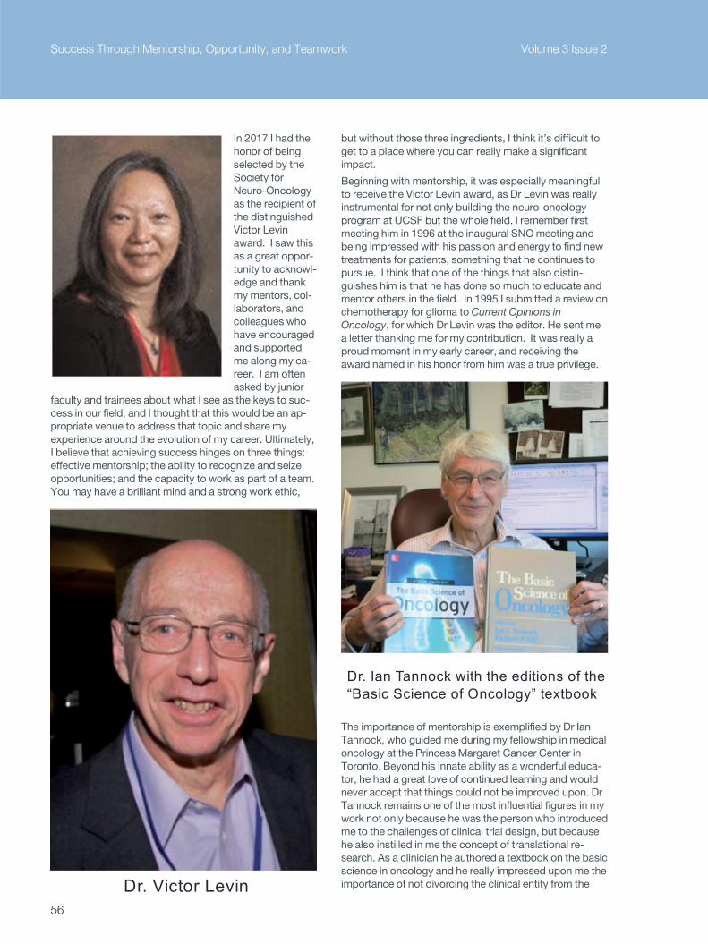
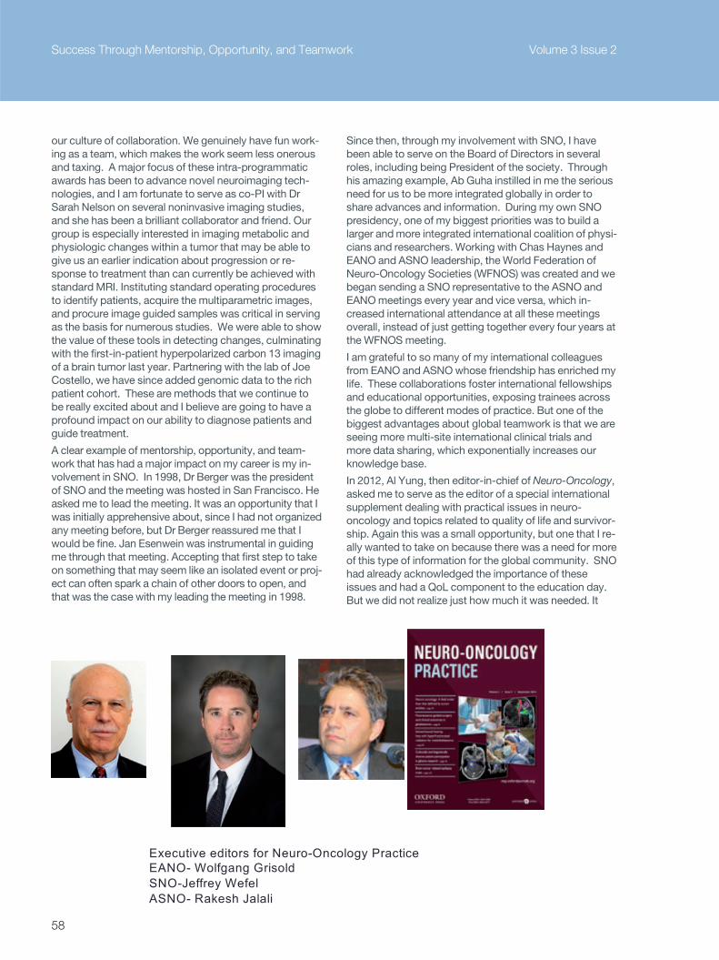
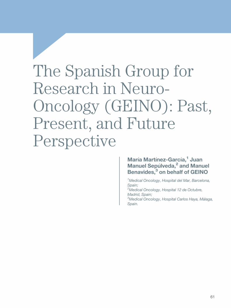
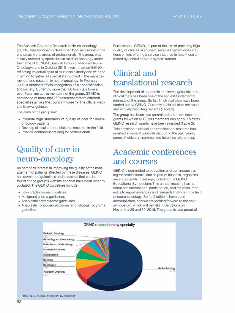

The Egyptian Group of Neuro Oncology (EGNO) the History Meets the Future Volume 3 Issue 2
68
Neuropathology as acornerstoneThe neuropathology group at Ain Shams University was
the first in the country to study MGMT in GBM through
immunohistochemistry years ago Currently they are
conducting a study about the role of PDL1 expression
and its possible effect on the management of GBM in col-
laboration with EGNO
First glioma group inthe countryHaving a glioma group in Egypt was a major step to regis-
ter all the glioma cases diagnosed yearly This was led by
a fellow medical genetics specialist from Ismailia who ini-
tiated a funded project by the government to create the
first genetic mapping of glioma in Egypt The collabora-
tion with EGNO in the second phase of such a project will
spread the call to all the oncology centers all over the
country to collect data and tissue samples opening a
new frontier for research especially with the rising inter-
est in the field of immune and targeted therapy for CNS
tumors
Treating elderlypatients with GBMTwo phase II and III studies were conducted in Egypt in
the past few years at Kasr El Einy and Ain Shams
Universities to explore the advantages of hypofractio-
nated radiotherapy protocols in elderly patients with
GBMwith or without concomitant chemotherapy EGNO
is leading a second interim analysis of the results of both
studies as a foundation for reaching new guidelines in the
management of this group of patients especially with the
presence of some earlier reports that discussed chemo-
therapy as the main line of treatment in frail patients
Radiosurgical re-irradiation of GBMThe Gamma Knife Center Cairo Nasser Institute is initi-
ating a project in collaboration with EGNO aiming at dis-
cussing the efficacy of radiosurgery with Gamma Knife in
re-irradiation of GBM and high-grade glioma as salvage
after failure of chemotherapy and where re-surgery is not
feasible Such a protocol will adopt the fractionated
Gamma Knife ICON radiosurgery (mask based)
Salvage chemotherapyfor GBMEGNO is initiating a protocol for salvage treatment of
GBM using bevacizumab with rechallenge of
temozolomide in good performance patients at time of
recurrence
ldquoIf you donrsquot publish Icanrsquot see yourdquoYears ago Professor Weller an eminent neuro-
oncologist said that in a conference Since then EGNO
Volume 3 Issue 2 The Egyptian Group of Neuro Oncology (EGNO) the History Meets the Future
69
has encouraged its members to get their work presented
in international meetings like those held by EANO ESMO
andWFNOS and to be published in many peer reviewed
journals Such movements had helped us to further col-
laborate with the EORTC brain tumor group and to be
contacted by some international pharmaceutical compa-
nies to participate in some of their funded research about
new therapies for CNS tumors
ChallengesThe lack of funds in a developing country makes it difficult
to finance large projects that could not be done in multi-
ple centers except without the help of pharmaceutical
companies With the establishment of research and ethi-
cal committees in the academic centers many interna-
tionally operated third party companies are operating in
Egypt to monitor and supervise such scientific research
to ensure that the international standards in conducting
research are met
OpportunitiesIn a heavily populated country like Egypt we have more
than 50 oncological academic departments government
hospitals and research centers Many of these have their
own labs and research units which are internationally
accredited and monitored Egypt with the help of many
European countries has established a special funding
authority the Science and Technology Development
Fund (STDF) which helped to fund much basic science
research in the field of neuro-oncology This means that
EGNO is ready to be involved in international trials as the
infrastructure for such trials is already present
What EGNO needsfromWFNOSWFNOS has done a great job in publishing this magazine
and in organizing their first conference in Zurich in 2017
Still we are looking forward to the next step for helping
neuro-oncology societies like EGNO to be active partici-
pants in international multicenter research to use our
data as part of reaching neuro-oncology guidelines and
to help our younger oncologists to find their way to better
training programs and workshops that could improve
their skills and performance in the growing field of neuro-
oncology in Egypt
For further contacts
khalidakmmedasuedueg
wwwegnoegyptorg
The Egyptian Group of Neuro Oncology (EGNO) the History Meets the Future Volume 3 Issue 2
70
EORTC 1709CCTG CE8 a randomizedphase III trial assessing the addition of
marizomib to standard of care in
patients with newly diagnosed
glioblastomaPatrick Roth1 and Michael Weller1
1Department of Neurology and Brain Tumor Center University Hospital
Zurich and University of Zurich Switzerland
Correspondence Dr Patrick Roth Department of Neurology
University Hospital Zurich Frauenklinikstrasse 26 8091 Zurich
Switzerland Tel thorn41 (0)44 255 5511 Fax thorn41 (0)44 255 4507
E-mail patrickrothuszch
Keywords glioblastoma proteasome marizomib radiotherapy
temozolomide EORTC
The standard of care for patients withnewly diagnosed glioblastomaincludes maximum safe resectioninvolved-field radiotherapy (RT) andconcomitant and up to 6 cycles ofmaintenance temozolomide (TMZ)chemotherapy (TMZRTTMZ)Despite this intense treatment theprognosis remains poor with a me-dian overall survival of approximately16 months in contemporary clinicaltrials[1] Within the last 5 years sev-eral novel drugs failed to prolong theoverall survival of patients with newlydiagnosed glioblastoma in random-ized clinical trials including bevaci-zumab targeting vascularendothelial growth factor (VEGF)and the integrin inhibitor cil-engitide[2ndash4] Similarly no immuno-therapeutic approach such asvaccination or immune checkpointinhibition has conferred a survivalbenefit in glioblastoma patients sofar[5]
Consequently novel therapeuticstrategies and targets are urgentlyneeded The proteasome has longbeen considered a promising candi-date for therapeutic targeting in vari-ous types of cancer[6] Severalproteasome inhibitors have shown
pronounced antiglioma activity inpreclinical models[7 8] However de-spite these compelling data no clini-cally meaningful activity was seenwith proteasome inhibitors such asbortezomib in glioblastoma patient-s[9] A major drawback of most of thecurrently available proteasome inhib-itors is their poor ability to cross thebloodndashbrain barrier The emergenceof marizomib (MRZ) a novel brain-penetrant irreversible pan-proteasome inhibitor now offers theopportunity to assess the activity ofthis therapeutic approach in the set-ting of primary brain tumorsMarizomib was derived from a ma-rine actinomycete and inhibits theproteolytic chymotrypsin-like tryp-sin-like and caspase-like activities ofthe 20S unit of the proteasome[10] Ithas been explored in several preclini-cal models demonstrating antigliomaactivity as a single agent[11 12] Inmonkeys drug levels in the CNSreached approximately 30 of thosemeasured in peripheral blood[13]
Most importantly marizomib wassuccessfully investigated in phase Istudies in patients with newly diag-nosed as well as recurrent glioblas-toma In the MRZ-108 study S
tudysynopsis
71
Volume 3 Issue 2 Study synopsis
marizomib was evaluated in patients with recurrent glio-blastoma either as a single agent or in combination withbevacizumab Median overall survival was 94 months[14]The MRZ-112 trial explored the addition of marizomib toradiochemotherapy and maintenance therapy with temo-zolomide in patients with newly diagnosed glioblastomaaiming at defining the recommended dose for furtherstudies Patients were enrolled in separate cohorts forconcomitant (TMZRTthornMRZ) and maintenance(TMZthornMRZ) treatment using a 3thorn 3 dose-escalation de-sign with a subsequent dose-expansion cohort at the rec-ommended dose in concomitant followed bymaintenance treatment Most frequent side effects in-cluded fatigue nausea vomiting and headaches as wellas CNS-related adverse effects including hallucinationsand ataxia The recommended marizomib dose for furtherevaluation was determined to be 08 mgm2 in combina-tion with standard of care[15]
Because of its promising antiglioma activity in various pre-clinical models as well as the data obtained in the MRZ-108 andMRZ-112 studies marizomib will now be investi-gated in the EORTC 1709CCTG CE8 trial a multicenterrandomized controlled open label phase III superioritystudy (ClinicalTrialsgov Identifier NCT03345095)Patients with newly diagnosed glioblastoma who are eli-gible for standard TMZRTTMZ are candidates forstudy participation The trial will enroll a total of 750patients who will be randomized 11 to receive standardtreatment alone or standard treatment plus marizomib(Figure 1) Stratification factors include institution ageKarnofsky performance status and extent of surgeryMarizomib will be administered until tumor progressionuntolerable toxicity withdrawal of consent or up to 18cycles after completion of radiotherapy
The primary objective of this study is to compare overallsurvival in patients receiving marizomib in addition toTMZRTTMZ with patients receiving standard treat-ment only The testing strategy is defined to assess thisobjective in both the intent-to-treat population and thesubgroup of patients with tumors harboring an unme-thylated O6-methylguanine-DNA methyltransferasepromoter Secondary endpoints include progression-free survival safety neurocognitive function and qualityof life
An accompanying translational research program hasbeen set up which involves among others the assess-ment of baseline proteasomal activity in tumor tissue aswell as longitudinal measurements of proteasomal activityin peripheral blood The study will be opened at 50EORTC sites in Europe and done as an intergroup collab-oration with the Canadian Cancer Trials Group (CCTG)with 25 sites in Canada and additional sites in the US Thefirst study sites in Europe and Canada were activated forenrollment in July 2018 and the activation of most sites isexpected at the beginning of 2019
References
1 Weller M van den Bent M Tonn JC et al European Association for
Neuro-Oncology Task Force on Gliomas European Association for
Neuro-Oncology (EANO) guideline on the diagnosis and treatment of
adult astrocytic and oligodendroglial gliomas Lancet Oncol 2017
18e315ndashe329
2 Chinot OL Wick W Mason W et al Bevacizumab plus radiotherapy-
temozolomide for newly diagnosed glioblastoma N Engl J Med
2014 370709ndash722
3 Gilbert MR Dignam JJ Armstrong TS et al A randomized trial of
bevacizumab for newly diagnosed glioblastoma N Engl J Med
2014 370699ndash708
Newly diagnosed
glioblastoma
bull KPS gt 70
bull Eligible for standard
treatment
1 2 3 4 5 6 7-1 8 9 10 11 12 13 14 15 16 17 18 19 20 21 22 23 24 25 26 27 28 29 30 31 32 33 34
weeks
1 2 3 4 5 6 7-1 8 9 10 11 12 13 14 15 16 17 18 19 20 21 22 23 24 25 26 27 28 29 30 31 32 33 34
weeks
Radiotherapy (total dose of 60 Gy in 30 fractions)
Temozolomide concomitant treatment during radiotherapy followed by up to 6 cycles of maintenance therapy
Marizomib at days 1 8 15 29 and 36 during radiotherapy followed by maintenance treatment at days 1 8 and
15 of a 28-day cycle until disease progression unacceptable toxicity withdrawal of consent or up to 18 cycles
Stratification factors
bull le55 vs gt55 years
bull KPS 7080 vs 90100
bull Biopsypartial vs GTR
bull Institution
bull
Figure 1 Design of the EORTC 1709CCTG CE8 trial
Study synopsis Volume 3 Issue 2
72
4 Stupp R Hegi ME Gorlia T et al European Organisation for R
Treatment of C Canadian Brain Tumor C team Cs Cilengitide com-
bined with standard treatment for patients with newly diagnosed
glioblastoma with methylated MGMT promoter (CENTRIC EORTC
26071-22072 study) a multicentre randomised open-label phase 3
trial Lancet Oncol 2014 151100ndash1108
5 Weller M Butowski N Tran DD et al investigators AIt Rindopepimut
with temozolomide for patients with newly diagnosed EGFRvIII-
expressing glioblastoma (ACT IV) a randomised double-blind inter-
national phase 3 trial Lancet Oncol 2017 181373ndash1385
6 Teicher BA Tomaszewski JE Proteasome inhibitors Biochemical
Pharmacology 2015 961ndash9
7 Roth P Kissel M Herrmann C Eisele G Leban JWeller M Schmidt F
SC68896 a novel small molecule proteasome inhibitor exerts antiglioma
activity in vitro and in vivo Clin Cancer Res 2009 156609ndash6618
8 Unterkircher T Cristofanon S Vellanki SH Nonnenmacher L Karpel-
Massler G Wirtz CR Debatin KM Fulda S Bortezomib primes glio-
blastoma including glioblastoma stem cells for TRAIL by increasing
tBid stability and mitochondrial apoptosis Clin Cancer Res 2011
174019ndash4030
9 Kong XT Nguyen NT Choi YJ et al Phase 2 study of bortezomib
combined with temozolomide and regional radiation therapy for
upfront treatment of patients with newly diagnosed glioblastoma
multiforme safety and efficacy assessment Int J Radiat Oncol Biol
Phys 2018 1001195ndash1203
10 Potts BC Albitar MX Anderson KC et al Marizomib a proteasome
inhibitor for all seasons preclinical profile and a framework for clinical
trials Current Cancer Drug Targets 2011 11254ndash284
11 Vlashi E Mattes M Lagadec C Donna LD Phillips TM Nikolay P
McBride WH Pajonk F Differential effects of the proteasome inhibi-
tor NPI-0052 against glioma cells Transl Oncol 2010 350ndash55
12 Manton CA Johnson B Singh M Bailey CP Bouchier-Hayes L
Chandra J Induction of cell death by the novel proteasome inhibitor
marizomib in glioblastoma in vitro and in vivo Sci Rep 2016
618953
13 Di K Lloyd GK Abraham V MacLaren A Burrows FJ Desjardins A
Trikha M Bota DA Marizomib activity as a single agent in malignant
gliomas ability to cross the blood-brain barrier Neuro Oncol 2016
18840ndash848
14 Bota D Desjardins A Mason W Kesari S Magge R Winograd B
Reich S Levin N Trikha M Full enrollment results from the phase 1
2 multicenter open-label study of marizomib (MRZ)thorn- bevacizu-
mab (BEV) in recurrent WHO grade IV malignant glioma (glioblas-
toma rGBM) Neuro Oncol 2017 19vi16
15 Bota D Kesari S Piccioni DE Aregawi D Roth P Stupp R
Desjardins A Elias I Reich S Levin N Winograd B Mason W A
phase 1 multicenter open-label study of marizomib (MRZ) with
temozolomide (TMZ) and radiotherapy (RT) in newly diagnosed WHO
grade IV malignant glioma (glioblastoma ndGBM) Dose-escalation
results J Clin Oncol 2018 36 supple14083
Volume 3 Issue 2 Study synopsis
73
Selection of Brain TumorPatients for ProtonTherapy the DutchApproach
Yvonne LB Klaver1
Hiske L van der Weide2
Ida EM Coremans13
Alejandra Mendez Romero14
Ruud GJ Wiggenraad15 andDanieuroelle BP Eekers67
1Holland Proton Therapy Centre Delft the
Netherlands2Department of Radiation Oncology University
Medical Centre Groningen University of
Groningen the Netherlands3Department of Radiation Oncology Leiden
University Medical Centre Leiden the Netherlands4Department of Radiation Oncology Erasmus
University Medical Centre Rotterdam the
Netherlands5Department of Radiotherapy Haaglanden
Medisch Centrum Leidschendam the
Netherlands6Department of Radiation Oncology (MAASTRO)
GROWmdashSchool for Oncology and Developmental
Biology Maastricht University Medical Centre
Maastricht the Netherlands7Proton Therapy Department South-East
Netherlands (ZON-PTC) Maastricht
74
AbstractProton therapy is a radiation technique which can
reduce toxicity in selected patients compared with
standard photon radiotherapy A careful patient
selection is essential to offer proton therapy to
patients who will benefit the most in terms of
prevention of toxicity and to validate the clinical
benefits compared with photons In the Netherlands
where proton therapy has been introduced in 2018
selection criteria for patients eligible for proton
therapy must be described in a National Indication
Protocol
After extensive deliberation and close collaboration
among experienced radiation oncologists in neuro-
oncology a national consensus on the selection of
patients for proton therapy was reached which is
supported by the neuro-oncologists This paper
describes the development of the Dutch National
Indication Protocol for Neuro-Oncology The pro-
posed protocol is currently under appraisal of the
National Health Care Institute to advise about inclu-
sion in basic health insurance
IntroductionSeveral well-established and highly developed radiother-
apy techniques are available for the treatment of central
nervous system tumors With the most common radiation
modality photon therapy tumors can be irradiated with
very high precision Yet photon therapy has some limita-
tions including an inevitable irradiation of surrounding
(brain) tissue with lower dose Proton therapy is an alter-
native radiation modality which has been applied for many
years worldwide and becomes increasingly available in
Europe The physical principles of proton beams offer
possibilities for superior dose distributions compared with
photon treatment as is shown by many in silico dose
planning comparative studies1ndash3 Based on these in silico
planning studies the main application in which protons
could produce a clinical benefit is reduction of radiation-
induced side effects by reducing the dose to normal tis-
sue The translation of dosimetric advantages to clinical
advantages is challenging For many critical organs or
normal tissues it is observed that the probability of side
effects is directly associated with the radiation dose that
is received by that organ The severity of toxicity following
a given dose of radiotherapy is organ specific4ndash6
Ideally the validation of the benefits of proton therapy
should be performed in a randomized controlled trial with
radiation-induced toxicity as a primary endpoint Yet
many radiation-induced complications can have a long
latency time with incidences increasing over more than
15 years requiring very long monitoring of patients7ndash9 A
randomized controlled trial would take many years to be
completed Given the fact that technological advances
evolve rapidly current techniques will be regarded as out-
dated within several years and it is expected that ran-
domized trials will not generate applicable data To
clinically validate the benefit of protons performance of
randomized controlled trials comparing photon and pro-
ton therapy is therefore often not feasible
In the Netherlands 3 proton therapy centers have been
opened in 2018 Groningen PTC in Groningen Holland
PTC in Delft and ZON PTC in Maastricht A careful selec-
tion of those patients who are expected to benefit most
from proton therapy is needed
In the Dutch health insurance system all primary and cu-
rative care is financed from private mandatory insurance
For this purpose insurance companies must offer a core
universal insurance package for universal primary cura-
tive care for a fixed price The government decides on the
content of the universal package Insurance companies
are not allowed to refuse an applicant for the universal
package The system is financed from payroll taxes paid
by employers the government and the premiums paid
directly by the insured Additional services can be offered
by the insurance companies at extra costs For these
services additional conditions for acceptance may apply
The Dutch Health Council (in Dutch Gezondheidsraad)
and the National Health Care Institute (in Dutch
Volume 3 Issue 2 Selection of Brain Tumor Patients for Proton Therapy
75
Zorginstituut Nederland) have advisory functions for the
Minister of Health for the decisions on the content of the
standard insurance package
Some indications are generally accepted for proton ther-
apy worldwide and are therefore considered standard
indications In the Netherlands proton therapy is part of
the universal insurance package and is regarded as an in-
sured provision for the standard indications of pediatric
tumors chordomas and chondrosarcomas of skull base
or spine and ocular melanoma For selection of patients
with other indications for proton therapy the model-
based approach was chosen by the Dutch Society for
Radiation Oncology (DSRO in Dutch NVRO)10 In the
model-based approach the potential benefit of proton
therapy in reducing side effects is estimated by use of
Normal Tissue Complication Probability (NTCP) models
This concept was approved by the Dutch Health Council
and the National Health Care Institute It is estimated that
with this approach about 3 of all patients in the
Netherlands who have an indication for radiotherapy will
be eligible for proton therapy11 The criteria that must be
fulfilled in order to be eligible for proton therapy (and reim-
bursement in the universal insurance package) are de-
scribed in a National Indication Protocol For every
indication a tumor-specific protocol is written by radia-
tion oncologists with expertise in this area
Before the indication for proton therapy can be regarded as
insured care the indication protocol should be approved by
the National Health Care Institute In this paper the devel-
opment and realization of the proposed National Indication
Protocol for Neuro-Oncological tumors is described
Search for NTCPmodelsAll radiation oncologists working in the Netherlands are
members of the DSRO Within the DSRO radiation oncol-
ogists with expertise of specific tumors are organized in
platforms These platforms are engaged in regulating and
improving radiotherapy treatment for certain tumors and
developing national guidelines The platforms have an ad-
visory function for the board of the DSRO The develop-
ment of national indication protocols for proton therapy is
a combined effort between the tumor-specific platforms
and the National Platform for Proton Therapy (NPPT in
Dutch LPPT) For the National Indication Protocol on
Neuro-Oncology the National Platform for Radiotherapy
in Neuro-Oncology (NPRNO in Dutch LPRNO) estab-
lished a committee consisting of radiation oncologists
with expertise in neuro-oncology and interest in proton
therapy Because the model-based approach is highly
dependent on the use of reliable NTCPmodels the first
step for the committee in the development of the protocol
was to perform a systematic search for NTCPmodels
estimating the risk of toxicity after radiation of organs to a
certain dose Structures that are known to be relevant for
radiation-induced toxicity are often called organs at risk
(OARs) in radiotherapy In neuro-oncology the most rele-
vant OARs are the brain brainstem cochlea cornea
lens retina lacrimal gland optic nerve chiasm pituitary
hypothalamus cerebellum and hippocampus
For the Dutch National Indication Protocol for neuro-onco-
logical tumors a detailed search strategy was composed
in cooperation with a trained librarian of the Leiden
University Medical Center For the National Indication
Protocol several medical databases were searched using
a systematic query which was optimized for every individ-
ual database The results of the search strategies were first
screened by members of the committee in order to select
papers containing NTCPmodels NTCPmodels for neuro-
cognitive function endocrine disorders ototoxicity radio-
necrosis and dry eyes were found The selected papers
were then more thoroughly reviewed and the quality of the
NTCPmodels was assessed according to the TRIPOD
criteria12 The results of this search will be published else-
where During a meeting of the DPRNO the committee
presented the results of the search strategy and the evalu-
ation of the available NTCPmodels to the members of the
DPRNO The members of the DPRNO concluded that the
available evidence and the quality of the available NTCP
models were insufficient for use for patient selection The
use of nonvalidated NTCPmodelsmdashfor instance a model
developed by Gondi et al relating hippocampal dose to
neurocognitive function impairmentmdashwas considered but
rejected after comprehensive deliberation13
Patient selectionBecause of the conclusion from the search a different
approach for patient selection other than the model-
based approach and the use of NTCPmodels was re-
quired After extensive and comprehensive discussion
within the national platform as well as with neuro-oncolo-
gists it was agreed that the therapy should be offered to
patients who are expected to benefit the most in accor-
dance with the principles of the model-based approach
Most of the radiation-induced side effects in neuro-
oncology that are expected to be reduced by proton ther-
apy have a long latency time914 The members of the
DPRNO therefore agreed that the highest potential gain in
quality of life and costs is to be expected for patients with
a good prognosis In order to define a favorable progno-
sis neuro-oncologists were consulted and asked to
share their expert opinion on this topic Good prognosis
was defined as 10-year overall survival of at least 50
which was approved by both radiation oncologists and
neuro-oncologists
The main application of the superior physical properties
of protons in neuro-oncology is the sparing of normal
brain tissue Several studies have shown that irradiation
of normal brain can cause changes in the neurocognitive
Selection of Brain Tumor Patients for Proton Therapy Volume 3 Issue 2
76
function914ndash17 These neurocognitive deficits are among
the most important long-term side effects resulting from
treatment of brain tumors and radiation of the brain in
particular It comes on top of the neurocognitive decline
that may be caused by the brain tumor itself and has a
vast impact on the activities of daily living and thereby on
quality of life of both patients and their families Besides
the brain tissue in general the hippocampus plays an im-
portant role in neurocognitive function especially in
memory It is common practice in the Netherlands to
keep the dose to the aforementioned structures as low as
possible The possibility of sparing of normal brain tissue
and the hippocampi was designated to be the main focus
in selecting patients for proton therapy
Not all patients with brain tumors and a favorable progno-
sis will benefit from proton therapy In silico planning stud-
ies have shown that in tumors with very small volumes the
advantages of protons are limited and sometimes even
unfavorable because of the larger area of dose fall-off
(penumbra) at the edge of a proton beam and the larger
setup uncertainties compared with stereotactic radiother-
apy or radiosurgery with photons Therefore it was stated
in the protocol that proton therapy should not be offered
to patients for whom treatment with stereotactic or radio-
surgical radiotherapy is feasible Furthermore the require-
ment of a quality check was added to the protocol In this
quality check the proton dose distribution plan is com-
pared with a state-of-the-art photon plan The protocol
describes minimal levels of improvement in dose distribu-
tion that the proton plan should offer compared with the
photon plan in order for a patient to be eligible for proton
therapy The ultimate decision whether a patient will be
treated with proton or photon therapy is made by the
treating radiation oncologist in dialogue with the patient
(shared decision making) For an individual patient several
other factors besides dose distributions may influence the
choice for proton or photon therapy such as travel dis-
tance waiting time or personal preferences
For patients with meningioma additional inclusion criteria
were formulated Alternative treatment options to avoid ra-
diation of normal brain tissue andor to postpone radiation
for a significant period of time like additional surgery limit-
ing the target volume for radiation to the progressive post-
operative residual tumor wait-and-scan policy or a
combination of these strategies should be considered be-
fore referring a patient for proton therapy The indication of
craniospinal irradiation in adults was proposed and ac-
cepted bymedical doctors as an additional standard indi-
cation This proposed protocol is currently under appraisal
by the National Health Care Institute to advise about inclu-
sion in the basic health insurance
DiscussionThe model-based approach for selecting patients for pro-
ton therapy was developed and introduced in the
Netherlands to select patients for proton therapy in case a
randomized controlled trial is considered inappropriate10
Yet the feasibility of the approach is highly dependent on
the availability of validated NTCPmodels For neuro-
oncology indications the model-based approach does
not fit due to a lack of NTCPmodels of sufficient quality
In neuro-oncology there are several limiting factors in
creating NTCPmodels First of all radiation-induced tox-
icity is often measurable only after a long follow-up time
of at least 15 years Radiotherapy is a rapidly developing
area of treatment Results from studies that started in-
cluding patients 15 years ago may now be considered
outdated because of the fast development of technical
innovations Outcomes of these studies may not be com-
parable to the outcomes of patients treated with currently
available techniques Furthermore the evaluation of out-
comes in particular neurocognitive function is complex
The neurocognitive function is composed of multiple
domains including executive functions attention learn-
ing and memory perceptual-motor speed and others
These domains can be tested globally by short question-
naires but also by extensive neurocognitive function tests
performed by trained professionals The lack of uniformity
in the testing of neurocognitive function makes results of
studies difficult to compare
Although consensus was reached about the selection cri-
teria for proton therapy in patients with neuro-oncological
tumors not all issues could be solved and the proposed
National Indication Protocol has some limitations and
might need adjustment in time
An age limit as an eligibility criterion for proton therapy
was suggested Rationale for this would be that the brain
of younger people is developing until about the age of 30
years18 and is more susceptible to radiation toxicity
Moreover as the life expectancy of younger people is lon-
ger than that of older patients it was proposed that youn-
ger patients would benefit longer from the advantages of
having less toxicity Yet in literature no definite age limit
was defined for the maturation of brain tissue Many radi-
ation oncologists expressed ethical objections against
selection based on age since solid evidence from litera-
ture to support this is lacking It was therefore decided
not to add an age limit to the list of inclusion criteria
One of the disadvantages of model-based selection and
selection of patients in general from a research perspec-
tive is that the group of patients treated with proton ther-
apy will by definition have different baseline
characteristics than patients treated with photon therapy
This makes comparison of the cohorts very difficult An
alternative approach to compare results would be to
compare patients who have been treated with proton
therapy to a historical cohort of patients who have been
treated with photon therapy in recent years Another op-
tion is to prospectively collect data from a cohort of
patients for whom proton therapy was not available at the
time of introduction of proton in the Netherlands because
of the limited capacity of the proton centers in their
Volume 3 Issue 2 Selection of Brain Tumor Patients for Proton Therapy
77
ramp-up phase For this purpose a national database
and information collecting infrastructure are being cre-
ated in the Netherlands collecting data from patients
treated with either proton or photon therapy This project
is called ProTRAIT (Proton Therapy Research
Infrastructure) and is a collaboration between all univer-
sity hospitals in the Netherlands It is supported by a
grant from the Dutch Cancer Society
In order to collect similar data for all patients who will be
irradiated for brain tumors in the Netherlands the relevant
OARs will be contoured according to an internationally
approved consensus-based atlas created in collabora-
tion with the European Particle Therapy Network1920 This
task force of ESTROwas established to encourage coop-
eration between particle therapy centers in Europe
Registration of toxicity and follow-up of patients was also
one of the conditions of the National Health Care Institute
and the government for proton therapy to become in-
sured care in the Netherlands With this information new
NTCPmodels can be developed and validated in order to
further improve selection of patients in the future This
also implicates that the national protocol will be evaluated
and revised within a few years to incorporate new evi-
dence and information
ConclusionThe members of the DPRNO had extensive deliberation
about the approach toward a model-based National
Indication Protocol for neuro-oncology indications The
close collaboration in the Netherlands between experi-
enced radiation oncologists in neuro-oncology resulted in
a national consensus on the selection of patients for pro-
ton therapy Cooperation and support from other special-
ists in neuro-oncology are essential to introduce this
approach nationwide and to offer each patient custom-
ized care
References
1 Van der Laan HP van de Water TA van Herpt HE et al Rococo co-
operative group The potential of intensity-modulated proton radio-
therapy to reduce swallowing dysfunction in the treatment of head
and neck cancer A planning comparative study Acta Oncol
201352561ndash569
2 Eekers DBP Roelofs E Jelen U et al Benefit of particle therapy in
re-irradiation of head and neck patients Results of a multicentric in
silico ROCOCO trial Radiother Oncol 2016121387ndash394
3 Roelofs E Engelsman M Rasch CR et al ROCOCO Consortium
Results of a multicentric in silico clinical trial (ROCOCO) comparing
radiotherapy with photons and protons for non-small cell lung can-
cer J Thorac Oncol 20127165ndash176
4 Emami B Lyman J Brown A Coia L Goitein M Munzenrider JE
Shank B Solin LJ Wesson M Tolerance of normal tissue to thera-
peutic irradiation Int J Radiat Oncol Biol Phys 1991 21
109ndash122
5 Lambrecht M Eekers DBP Alapetite C et al work package 1 of the
taskforce ldquoEuropean Particle Therapy Networkrdquo of ESTRO
Radiation dose constraints for organs at risk in neuro-oncology the
European Particle Therapy Network consensus Radiother Oncol
2018 128 26ndash36
6 Eekers DBP Lambrecht M Nystrom PDW Swinnen A Wesseling
FWR Roelofs E Troost EGC EPTN consensus-based guideline for
the tolerance dose per fraction of organs at risk in the brain
CancerData 2018 doi1017195candat2018011
7 Aleman BM van den Belt-Dusebout AW KlokmanWJ Vanrsquot Veer
MB Bartelink H van Leeuwen FE Long-term cause-specific mor-
tality of patients treated for Hodgkinrsquos disease J Clin Oncol
2003213431ndash3439
8 Henson KR McGale P Taylor C Darby SC Radiation-related mor-
tality from heart disease and lung cancer more than 20 years after
radiotherapy for breast cancer Br J Cancer 2013108179ndash182
9 Douw L Klein M Fagel SS et al Cognitive and radiological effects
of radiotherapy in patients with low-grade glioma long-term follow-
up Lancet Neurol 20098810ndash818
10 Langendijk JA Lambin P De Ruysscher D Widder J Bos M Verheij
M Selection of patients for radiotherapy with protons aiming at re-
duction of side effects the model-based approach Radiotherapy
and Oncology 2013107267ndash273
11 Gezondheidsraad Signalement Protonenbestraling Den Haag
2009 Rapportnr 200917
12 Collins GS Reitsma JB Altman DG Moons KG Transparent
Reporting of a multivariable prediction model for Individual
Prognosis Or Diagnosis (TRIPOD) Ann Intern Med
201519162735ndash736
13 Gondi V Hermann BP Mehta MP Tome WA Hippocampal dosime-
try predicts neurocognititve function impairment after fractionated
stereotactic radiotherapy for benign or low-grade adult brain
tumors Int J Radiat Oncol Biol Phys 201283 e487ndashe492
14 Habets EJ Taphoorn MJ Nederend S et al Health-related quality
of life and cognitive functioning in long-term anaplastic oligodendro-
glioma and oligoastrocytoma survivors J Neurooncol
2014116161ndash168
15 Klein M Heimans JJ Aaronson NK et al Effect of radiotherapy and
other treatment-related factors on mid-term to long-term cognitive
sequelae in low-grade gliomas a comparative study Lancet
20023601361ndash1368
16 Armstrong CL Hunter JV Ledakis GE et al Late cognitive and ra-
diographic changes related to radiotherapy initial prospective find-
ings Neurology 20025940ndash48
17 Gregor A Cull A Traynor E Stewart M Lander F Love S
Neuropsychometric evaluation of long-term survivors of adult brain
tumours relationship with tumour and treatment parameters
Radiother Oncol 19964155ndash59
18 Lebel C Deoni S The development of brain white matter micro-
structure Neuroimage 201831ndash12
19 Eekers DBP In rsquot Ven L Roelofs E Postma A Alapetite C Burnet
NG Calugaru V Compter I Coremans IEM Hoslashyer M Lambrecht M
Witt Nystrom P Mendez Romero A Paulsen F Perpar A de
Ruysscher D Renard L Timmermann B Vitek P Weber DC van der
Weide HL Whitfield GA Wiggenraad R Troost EGC on behalf of
the taskforce ldquoEuropean Particle Therapy Networkrdquo of ESTRO The
EPTN consensus-based atlas for CT- and MR-based contouring in
Neuro-Oncology Radiother Oncol 201812837ndash43
20 Eekers DBP in rsquot Ven L Roelofs E Postma A Troost EGC EPTN
International Neurological Contouring Atlas CancerData 2017
doi1017195candat2017081
Selection of Brain Tumor Patients for Proton Therapy Volume 3 Issue 2
78
ASCO 2018Highlights
Ugonma N ChukwuekeLakshmi Nayak
Center for Neuro-Oncology Dana-Farber Cancer
Institute Harvard Medical School Boston
Massachusetts USA
79
The American Society of Clinical Oncology hosted its an-
nual meeting on June 1ndash5 2018 in Chicago Illinois This
year there were 190 oral and poster presentations related
to central nervous system tumors including primary and
metastatic brain tumors focusing on areas of basic and
translational science such as immuno-oncology molecu-
lar pathology genomics and the application of precision
medicine Echoing efforts in other solid tumor and hema-
tologic malignancies understanding and addressing so-
cioeconomic determinants and contributors to outcomes
were also presented Here we review the meeting
highlights
Dr Ingo Mellinghoff fromMemorial Sloan Kettering
Cancer Center presented the results from a phase I study
of AG-881 an inhibitor of mutant isocitrate dehydroge-
nase 1 and 2 (IDH1IDH2) In patients with advanced
solid mutant IDH tumors including gliomas In this ses-
sion he presented safety efficacy and pharmacokinetic
pharmacodynamic data As of March 2018 18 patients
were still on study of whom 17 were glioma patients All
patients (glioma and other solid tumors) were treated be-
tween the dose range of 10 mg to 400 mg daily In the gli-
oma cohort there were no treatment-related deaths The
plasma drug exposure increased linearly between doses
10 mg and 200 mg In assessing best response in the gli-
oma cohort 75 (39 of 52 evaluable patients) achieved
stable disease A perioperative study of both AG-881 and
AG-120 an oral IDH1 inhibitor is currently under
evaluation
Dr Michael Platten from Heidelberg University presented
the safety and immunogenicity data from NOA-16 a mul-
ticenter phase I trial of mutation-specific peptide vaccine
targeting IDH1 R132H in 32 patients with newly diag-
nosed malignant astrocytomas Following chemoradia-
tion or 3 cycles of temozolomide patients were treated
with 8 vaccinations over a period of 23 weeks in combina-
tion with adjuvant temozolomide Over 90 of patients
received the entire treatment regimen and there were no
treatment-limiting toxicities Additionally there was evi-
dence of immunogenicity in which patients demonstrated
mutation-specific T-cell or humoral-mediated immune
responses that were not observed prior to treatment In
parallel with signal suggesting immunogenicity were ra-
diographic findings consistent with pseudoprogression
also indicating possible underlying disease response
The clinical molecular and radiographic features of dif-
fuse gliomas with either fibroblast growth factor receptor
1 (FGFR1) mutations or FGFR3ndashtransforming acidic
coiled-coil protein 3 (TACC3) fusions were characterized
by Dr Anna Luisa Di Stefano of Pitie Salpetriere Hospital
Through their work Dr Di Stefano and her group identified
50 gliomas with FGFR-TACC3 fusion across all World
Health Organization (WHO) grades all of which were
IDH1-wildtype and co-occurred with amplification of mu-
rine double minute 2 The fusion was also found to be a
predictor of longer overall survival in glioblastoma
patients FGFR1mutations were found in 13 of 70 midline
gliomas and were associated with K27Mmutation youn-
ger age and longer overall survival (OS) This work high-
lights that recognition of these alterations are critical as
clinical trials targeting FGFR are ongoing
Dr David Reardon from Dana-Farber Cancer Institute dis-
cussed the results of a phase II study of pembrolizumab
with and without bevacizumab in patients with bevacizu-
mab-naıve recurrent glioblastoma A total of 80 patients
were included in this analysis Progression-free survival
at six months (PFS6) was 26 in the combination treat-
ment arm in comparison to 67 in the pembrolizumab
monotherapy arm Patients on dexamethasone had a
poorer survival compared with those who were not The
outcomes of the combination therapy were consistent
with historical controls for bevacizumab monotherapy
Immunocorrelative analyses are ongoing
In the window-of-opportunity clinical trial Dr Amy
Heimberger from the MD Anderson Cancer Center pre-
sented data from 15 patients with recurrent glioblastoma
who were treated with pembrolizumab before and after
surgical resection Immune effector function and PFS6
were the primary objectives in this study Median OS has
not been reached but median PFS was 7 months Mass
cytometry (CyTOF) and multiplex immunohistochemical
analysis following pembrolizumab in resected glioblas-
toma showed minimal T-cell infiltration There was evi-
dence of CD68-positive cells particularly at the bloodndash
brain barrier
Dr Wolfgang Wick from Heidelberg University presented
the results from GAPVAC-101 a first-in-human trial of a
personalized peptide vaccine in patients with newly diag-
nosed glioblastoma In this phase I study feasibility
safety and immunogenicity were evaluated Fifteen
patients started and completed APVAC1 (warehouse se-
lected 7 active peptides against tumor) vaccination 11
patients subsequently were started on APVAC2 (de novo
manufactured after next-generation sequencing 2 pepti-
des) Patients treated with APVAC experienced adverse
events related to the vaccinations (mostly injection site
reactions) and to concurrent treatment Median OS and
PFS were 29 and 142 months respectively
Immunogenicity was demonstrated by CD4thorn T-cell
responses against neo-epitopes in APVAC2 Pre-
vaccination there was no evidence of immunoreactivity
however in tissue resected from a patient with recurrent
disease there was a population of tumor-infiltrating lym-
phocytes with reactivity against one of the APVAC1
peptides
There was an emphasis on the value of obtaining com-
prehensive genomic analysis in gliomas as discussed in 2
oral abstracts Dr Capucine Baldini from Gustave Roussy
investigated outcomes of recurrent glioma patients in
early-phase clinical protocols There were 70 recurrent
glioma patients included in the analysis of whom 41 re-
ceived protocol treatment based upon identification of a
clinically actionable target There was an overall response
rate of 21 in IDH-wildtype patients treated as per
ASCO 2018 Highlights Volume 3 Issue 2
80
molecularly guided protocols compared with 0 in un-
selected patients This work highlighted that molecularly
informed trials may be beneficial to specific patient
populations
Dr Mehdi Touat from Dana-Farber Cancer Institute pre-
sented the efforts of the ALLELE consortium which was
designed to create an infrastructure for prospective geno-
mic and molecular testing in order to inform biomarker-
driven clinical protocols In this feasibility study of 65
patients with glioblastoma 60 underwent prospective ge-
nomic testing across multiple centers with a median turn-
around time of 22 days Although there remain challenges
to generating real-time functional diagnostics including
cost and delays in analysis and data reporting informa-
tion obtained from this testing can be used to guide future
trials
Dr Priscilla Brastianos fromMassachusetts General
Hospital presented data from a phase II study of pembro-
lizumab in leptomeningeal disease from solid tumors The
primary endpoint was OS at 3 months Eighteen patients
were accrued of whom 15 had breast cancer Eleven
patients were alive at 3 months and the study met its pri-
mary endpoint at interim analysis Also highlighted in this
study was the use of cell-free DNA and single-cell RNA
sequencing from CSF to monitor disease activity as well
as immune response
Volume 3 Issue 2 ASCO 2018 Highlights
81
NurseCornerBreaking BadNews Well
Ingela Oberg
Macmillan Lead Neuro-Oncology Nurse Box 166
Division D ndash Neurosurgery Addenbrookersquos Hospital
CUHFT Cambridge Biomedical Campus Hills
Road CB2 0QQ
(Ingelaobergaddenbrookesnhsuk)
In the end the inevitable happens to us allmdashthe only cer-
tain thing in life is that we are born to diemdashand as health
care professionals within the neuro-oncology field we are
more used to seeing the implications of this fact over our
other peers within other health care settings But does this
common repetition of events make us desensitized to the
impending outcome of our patients How can we help
them and their loved ones accept the inevitable and even
achieve a good death How do we break bad news in a
good waymdashis this something that is up to us as nurses
and Allied Health Professionals (AHPs) to do or do we
simply ldquopick up the piecesrdquo once they have been informed
of their diagnosis and prognosis from a senior consultant
colleague
This article aims to explore some of the ways in which
these myths are dispelled and how we as nurses and
AHPs can help streamline this process and aid in the tran-
sition from active treatment to end of life care with greater
ease and acceptance The theme around end of life care is
also the topic of this yearrsquos Nursing and AHP pre-
symposium educational event at EANO Stockholm
(October 2018) which I hope many of you will be able to
attend and join in the discussions
For many of us working in neuro-oncology breaking bad
news is part of everyday practice but how many of us
have undergone formal communication skills training or
undergone a course in palliative care or end of life care or
undertaken a counselling degree Likely not many or at
least not enough of us We learn as we go along and ex-
perience (whether personal or professional) teaches us in-
valuable lessons in both empathy and compassion We
learn how to read nonverbal cues and determine when the
timing may be right to begin to approach the subject
around end of life care with the patient andor his or her
relatives and we are quick at ascertaining how much in-
formation they are likely to be able to retain at any particu-
lar time1 How this is done can greatly influence patientsrsquo
overall experience of their whole health care encounter
and this is where I think as nurses and AHPs we have a
unique opportunity to help lessen the impact of devasta-
tion and prepare them for the next steps in a genteel but
affirmative manner Let me give you a common example
Lisa (fictional name and case) is a 29-year-old mother of
two who presented to the emergency department with her
first tonic-clonic seizure witnessed by her husband A CT
scan picked up a likely glioma and she is discharged
home on anticonvulsants and dexamethasone to await an
outpatient urgent MRI head scan An appointment is also
made in the neurosurgical clinic the following week to dis-
cuss next steps likely surgery with fluorescence-guided
82
resection (with 5-aminolevulinic acid [5-ALA]) She is
given the number for the neuro-oncology specialist nurse
to contact in the interim for advice and support She rings
the following day in a panic
The specialist nurse speaks to her at length about her sei-
zure and medication and can hear how she audibly calms
down over the phone as she understands how the medi-
cation will lessen the risks of further seizures and how
dexamethasone will act to decrease the tumoral edema
Her main worry had been the children having to witness
her having a seizure or for them to find her unconscious
The nurse mentions the likely glioma and explains what a
primary brain tumor is and that the MRI will provide much
more detailed information about its size and location Lisa
is informed about her inability to drive but that she is per-
fectly safe to cuddle her children for example and under-
take normal daily activities with them The specialist
nurse acknowledges that this is a very scary time for
them all and that she is there to offer advice and support
as needed She then talks her through what to expect
from her clinic consultation and likely outcomes of antici-
pated surgery to remove as much of the tumor as is feasi-
ble and that all being well she should be back home
again within 3ndash4 days of her surgery She is strongly en-
couraged to bring family members with her to the
consultation
This is where the first anticipatory clue has already been
ldquodroppedrdquo that surgery is likely to be discussed and is the
foreseen next step Lisa has been informed she has a po-
tential primary brain tumor likely a glioma of some de-
scription It gives Lisa a few days to consider the impact
this will have (and to come up with further questions for
her consultation) and to make preparations for child care
arrangements in the interim as required for her inpatient
stay She is armed with information about her medication
and knows her restrictions in what she cancannot do
Once at the consultation surgery is discussed and she is
consented for the aforementioned fluorescent guided re-
section using 5-ALA She has been shown her MRI scan
so knows where the tumor is located and its size She is
very tearful as the reality dawns on her that she needs
brain surgery and has a serious diagnosis in the form of a
tumor After the consultation the specialist nurse takes
her and her husband aside into another room to go
through the admissions process in a bit more detail Here
is now the perfect opportunity to prepare them for the
next steps and likely outcomes
The nurse explains that 5-ALA is normally administered
as a drink to help the surgeon differentiate between
ldquonormalrdquo brain and fluoresced brain tissue (pink) which is
infiltrated with active tumor cells At this stage the
consultant neurosurgeon explains that the 5-ALA only
glows pink if the tumor is of a high grade meaning some
form of malignancy or cancer2 The grading of malig-
nancy is yet unknown (eg World Health Organization
[WHO] grades IIIndashIV) and that is what the pathology re-
port will confirm but we suspect this may represent
some form of cancerous tumor hence the 5-ALA being
utilized as it will enable the surgeon to remove at least
95 of the tumor
I would strongly recommend using the C-word (cancer) at
this stage even if it is just to say this is what we suspect
she is up against Using terminology like high grade ana-
plastic malignant etc can cause confusion and patients
end up hearing what they want that to mean Everyone
knows what cancer means and it gives her and her hus-
band time to prepare themselves mentally while waiting
for the surgery Hope for the best but plan for the worst
While being realistic in helping them prepare for a likely
cancerous diagnosis you are also instilling hope in that
most (if not all) of the tumor can safely be removed with
the aid of 5-ALA In my opinion prognosis and life expec-
tancy should NOT be entered into at this stage as you
would need both a full pathology report and all relevant
molecular markers to hand to adequately answer this3
What you CAN say though is that surgery is often the
first part of a prolonged treatment pathway and regard-
less of the type and tumor grading it is likely she will re-
quire ongoing treatment with oncology afterward All this
will be confirmed with them once the full pathology report
has been obtained Needless to say these discussions
can be quite emotive so ensure you have time set aside
for them not to feel rushed Offer them plenty of tissues
and above all time
In Lisarsquos case surgery went well and she was discharged
home within 72 hours with no neurological deficits Her
postoperative scan showed complete resection of her en-
hancing tumor and her pathology confirmed a glioblas-
toma (GBM) wildtype (WHO grade IV tumor) requiring
chemotherapy and radiotherapy as discussed in the
neuro-oncology multidisciplinary meeting4 She is seen in
clinic the same week for discussion of her results where
her consultant neurosurgeon informs her of the above in-
formation and treatment plan She is crying as is her hus-
band but they take comfort in knowing the tumor has
been completely removed and that she can have further
treatments
Once again the specialist nurse takes them aside and sits
with them quietly for a while allowing them to grieve
come to terms with their diagnosis and gather their
thoughts From experience the patientrsquos being aware of
the diagnosis and understanding the prognosis are two
different things and from an onward treatment perspec-
tive I feel it is important for the patient to know the differ-
ence to help be prepared for the forthcoming oncology
appointment5 This does not mean you have to spell it out
in regard to life expectancy
Normally I start this conversation by asking them about
their understanding of their surgical procedure and if
they have fully understood what that implies I state that
even though the surgeon was able to completely resect
the tumor (the postoperative scan confirms this) it does
not equate to a cure and does not mean the tumor is
gone for good I go on to state that unfortunately given
Volume 3 Issue 2 Nurse Corner
83
time this tumorwill resurfacemdashnormally in a matter of
months rather than years given her diagnosis5 I then
state that the aim of surgery is to lessen the pressure
effects within the brain and to establish a firm diagnosis
as well as enabling other treatment options such as chemo-
therapy and radiotherapy to bemore effective and for
those treatments to slow down the rate of regrowth Finally
I say that as this is a primary cancer of the brain our treat-
ment aim is to establish control of the disease for as long
as possible (with several arms of treatments available to
us) but ultimately we are not looking at a curative process
In Lisarsquos case while this was difficult for them to hear it
did not come as a shock They had been given potential
clues all along as to what this may represent and they
had gone home and ldquoGoogledrdquo gliomas so were already
well aware of its life limiting implications should this be a
high-grade tumor as initially suspected (and as now con-
firmed) They were obviously devastated but more from a
perspective that their deepest fears had been confirmed
as until now they had naturally been hoping they (and us)
would be wrong They did not ask about prognosis so
this was not discussed at this consultationmdashbut once
again they had been given snippets of what to expect
from oncology in regard to being an incurable disease
that will come back in a matter of months and that the
aim is one of palliation
They had already made plans for school counselor
involvement and had booked a family holiday at the local
seaside resort prior to oncology treatments commencing
to create ldquohappy memoriesrdquo for their children They
thanked the nurse for her unwavering support and profes-
sionalism and even stated that she must have a very diffi-
cult job breaking bad news every day but that she had
done it in such an empathetic and professional way that
they knew what the next steps were at every stage mak-
ing them feel prepared and forewarned They felt that
without her support their journey would not have been as
smooth and as well informed Lisa survived for 10
months
Apart from being good at breaking bad news the special-
ist nurse could offer so much more that would be of great
value and support to both the patient and the family
Providing Lisa and her husband with information leaflets
about their tumor type and treatment options is impera-
tive and most of us do this without a second thought but
as a specialist nurse do you always consider obtaining
other information that may help them holistically6 Do
you provide information about where to access financial
support and advice for example or about local commu-
nity support groups or charities to contact for peer and
online support About how to access grants for things
such as travel reimbursements or how to speak to be-
reaved children and approach the subject of terminal ill-
nesses with themmdashwhere can family members and
children go for counseling and support in your area
would you know Do you regularly offer spiritual advice
and signpost patients to chaplaincies How about
hospices offering daycare services such as music ther-
apy mindfulness courses or dietetic advice in cancer
There is a myriad of information and support out there for
patients families and their carers and loved ones but
knowing how and when to access them is a different en-
tity Being faced with a terminal illness and a very short
life expectancy must be like being a rabbit caught in
headlights it can be very difficult for anyone to know
where to turn or what to expect and patients tend to ei-
ther freeze with fear or bury their heads in the sand not
knowing where to turn It takes time to achieve accep-
tance and we can help them reach that stage in the
knowledge they are not alone on this journeymdashsomeone
to offer them appropriate guidance and support may be
all that is required
Thankfully through dedicated professionals such as you
more and more is being done to support everyone (not
only the patient) through this disease trajectory
Throughout the UK Europe and the USA there are sup-
port programs and even weekend camps for children and
family members of those affected by malignant brain
tumors albeit most of these programs are aimed at pri-
mary malignant tumors and not metastatic7 A network of
carer champions is being established to look after the
needs of the carers and more health care professionals
(as well as governing bodies) are realizing that we need to
look after the carers alongside the patients as they in
turn are the ones looking after the patients Without them
on board we would not be able to provide our current
level of outpatient service
At this yearrsquos Stockholm EANO conference we have the
privilege of once again being able to offer a FREE pre-
symposium educational day for nurses and AHPs attend-
ing the main symposium thanks to funding from the UK-
based Brain Tumor Charity A lot of the aforementioned
topics will be dedicated as speaker slots to further delve
into how we can offer these vital services and continue to
develop our professionalism and dedicated support to
our neuro-oncology cohort We have a lot to learn from
each other and I hope this yearrsquos theme around end of
life care does not demoralize you but rather inspire you
to go out there and make a tangible difference to your
patients and their loved ones arguably in their time of
greatest need
Furthermore I am very excited to announce that as an
EANO first we will also have a dedicated poster viewing
section to encourage the submission of nursing and AHP
abstracts and research Furthermore the previous stand-
alone ldquonursing dayrdquo agenda has been fully incorporated
into the main symposium events as parallel sessions and
breakout sessions over the duration of the conference
minimizing conflicts with other sessions of interest on the
main forum We hope this will encourage more attendees at
the nursing symposiums even if you are not a nurse or
AHP We will also have a ldquomeet the expertsrdquo nursing panel
and sunrise sessions for our benefit We hope it will be a
great success and a format to use for many years to come
Nurse Corner Volume 3 Issue 2
84
I do hope you will access the EANO website to look at the
detailed program in full There is a prize in store for the
best voted nursingAHP abstract submission meaning
free registration for one lucky participant so get
submitting
I hope to see you in October (10thndash14th 2018) in Sweden
and offer you a warm (Brrr) welcome to a beautiful au-
tumnal Stockholm
References
1 Barry M Edgman-Levitan S Shared decision makingmdashthe pinnacle
of patient-centred care N Engl J Med 2012366(9)780ndash781
2 Stummer W Pichlmeier U Meinel T et al (2006) Fluorescence-
guided surgery with 5-aminolevulinic acid for resection of malignant
glioma a randomised controlled multicentre phase III trial Lancet
Oncol 7392ndash401
3 Louis DN Perry A Reifenberger G et al The 2016 World Health
Organization classification of tumors of the central nervous system a
summary Acta Neuropathol 2016131803ndash820
4 National institute for Clinical Health and Excellence (2006) Guidance
on cancer servicesmdashimproving outcomes for people with brain and
other CNS tumours The Manual Chapter 2
5 Lobb EA Halkett GK Nowak AK Patient and caregiver perceptions
of communication of prognosis in high grade glioma J Neurooncol
2011104(1)315ndash322
6 The National Health Service N Holistic needs assessment for people
with cancer London Spring 2011
7 Cavers D Hacking B Erridge S Morris P Kendall M Murray S
Adjustment and support needs of glioma patients and their relatives
serial interviews Psycho-Oncology 201222(6)1299ndash1305
Volume 3 Issue 2 Nurse Corner
85
Atypical SolitaryPresentation ofLeptomeningealMetastases from BreastCancer A Case Report
Amelie Darlix Nelly Firmin
Department of Medical Oncology Institut du
Cancer de Montpellier University of Montpellier
208 Avenue des Apothicaires 34298 Montpellier
France
86
IntroductionBreast cancer is one of the solid tumors most commonly
associated with leptomeningeal metastases1
Leptomeningeal metastases usually occur in a context of
advanced systemic disease and can be associated with
brain intra-parenchymal metastases2 Therapeutic
options are scarce and have limited efficacy3 Current
possibilities include systemic treatments and local thera-
pies such as radiation therapy and intrathecal chemother-
apy Leptomeningeal metastases carry a dismal
prognosis with a median survival of less than 6 months in
most published series but some patients experience lon-
ger survivals up to several years24 Determining the
parameters allowing the identification of appropriate can-
didates for active treatments is challenging Several prog-
nostic factors have been identified including the patientrsquos
age at the time of leptomeningeal metastases diagnosis
the functional and neurological status and the delay be-
tween cancer diagnosis and leptomeningeal
metastases5
Case reportIn June 2013 a 62-year-old woman with no medical his-
tory was diagnosed by biopsy with a negative hormone
receptor amplified human epidermal growth factor re-
ceptor (HER2) ductal carcinoma of the left breast with a
homolateral axillar lymph node involvement (T3N1)
Whole body CT scan bone scan and brain MRI were
normal (M0) She first received neoadjuvant chemother-
apy (3 cycles of a regimen consisting of 5-fluorouracil
epirubicin and cyclophosphamide were followed by 4
cycles of docetaxel and trastuzumab) After completing
the whole protocol she was treated with mastectomy
and axillary node dissection Pathological examination
found no residual invasive carcinoma (breast and lymph
nodes) The patient was treated with adjuvant radiation
therapy delivered to the breast and lymph nodes and
trastuzumab In March 2014 during radiation therapy
she developed acute headache dizziness dysarthria
and nausea The neurological examination revealed cere-
bellar ataxia Physical examination was normal Brain MRI
showed large confluent leptomeningeal enhancing nod-
ules located in the vermis and cerebellar hemispheres
associated with a few smaller leptomeningeal nodules in
the supratentorial compartment There was no hydro-
cephalus Spinal MRI found leptomeningeal enhancing
micronodules at the cervical and lumbar levels Whole
body CT scan showed no visceral progression The pa-
tient was started on corticosteroids and treated with
whole brain radiation in April 2014 (30 Gy in 10 fractions)
An intraventricular catheter (Ommaya reservoir) was then
placed Repeated CSF examinations found a moderate
hyperproteinorachia (045 gL) and the presence of atypi-
cal cells suspected to be breast cancer tumor cells
Intrathecal methotrexate was started in May 2014 asso-
ciated with a systemic regimen consisting of capecitabine
and lapatinib A first follow-up assessment was per-
formed after 5 injections of intrathecal methotrexate and
one cycle of capecitabine-lapatinib The patientrsquos status
was significantly improved Brain MRI revealed a com-
plete resolution of the macroscopic leptomeningeal nod-
ules at both the supratentorial and infratentorial levels It
was decided to continue with the same treatment
Unfortunately the clinical condition of the patient rapidly
deteriorated into an acute prothrombotic state a few days
after the MRI and she was hospitalized The physical ex-
amination showed petechias and multiple hematomas on
the skin A complete biological workup revealed thrombo-
cytopenia (60 gL) and an increased prothrombin time
Repeated CSF analysis revealed a moderate hyperprotei-
norachia (045 gL) and the absence of tumor cells but
brain imaging was not performed Upon hospitalization
she had a respiratory failure chest CT revealed a massive
bilateral pulmonary embolism and the venous Doppler
ultrasound found an extensive thrombus of the left leg
Because of the presence of thrombocytopenia an inferior
vena cava filter placement was programmed but the re-
spiratory status worsened rapidly and she died 5 days
after her admission
DiscussionThis case of a patient diagnosed with breast cancer is
atypical by the type of clinical presentation of the lepto-
meningeal metastases with neurological symptoms
occurring during the adjuvant phase of the initial treat-
ment and in the absence of metastases outside the
central nervous system (CNS) and by the radiological
findings consisting of large macroscopic confluent
lesions affecting predominantly the cerebellum This lo-
cation in the cerebellum made the management of this
patient challenging and a combination of radiation
therapy intrathecal and systemic chemotherapy and
HER2-targeted therapy resulted in a clear clinical im-
provement and a complete disappearance of the ma-
croscopic lesions on MRI
Leptomeningeal metastases commonly occur in the pre-
sence of extra-CNS metastases and rarely reveal the
metastatic disease This case report shows that they can
occur earlier in the oncological history of patients inclu-
ding during postsurgical adjuvant treatment and in the
absence of brain or extra-CNS metastases Indeed
leptomeningeal lesions are rarely isolated in a study of
153 patients with breast cancerndashrelated leptomeningeal
metastases 88 of patients had concomitant extra-CNS
metastases2
Besides this atypical presentation the radiological cha-
racteristics of the leptomeningeal metastases were also
notable Brain MRI showed large macroscopic nodular
enhancing lesions largely predominant in the posterior
Volume 3 Issue 2 Atypical Solitary Presentation of Leptomeningeal Metastases from Breast Cancer
87
fossa Cerebellum is a frequent site of leptomeningeal
metastases in a series of 270 patients with a cytologically
confirmed diagnosis of leptomeningeal metastases from
various solid tumors the cerebellum was involved on im-
aging in 301 of cases6 Cerebellar lesions can be either
nodular as in our case or linear It has been hypothe-
sized that the predominant involvement of the posterior
fossa might be linked with postural deposition and col-
lection of tumor cells6 The correlation of the location of
macroscopic leptomeningeal lesions with outcome has
not been investigated yet It is unknown whether there
is a positive or negative impact of cerebellar leptome-
ningeal involvement in response to treatments or
survival
The management of patients with posterior fossa involve-
ment is indeed challenging for several reasons First
depending on the size of the leptomeningeal nodules it
can be difficult to obtain a cytological confirmation as
the lumbar puncture might not be possible Secondly the
risk of intracranial hypertension andor acute
hydrocephalus must be taken into account when consi-
dering treating patients with radiation therapy Due to the
possible contraindication of lumbar punctures the ad-
ministration of intrathecal chemotherapy can be per-
formed through an intraventricular catheter (Ommaya
reservoir)
Overall this case illustrates the fact that even in the pre-
sence of large nodular leptomeningeal metastases re-
sponsible for a serious deterioration of the neurological
status active treatments (in this case radiation therapy
intrathecal methotrexate and systemic treatment) should
be discussed as it can lead to substantial clinical and
radiological improvement despite a dramatic situation at
diagnosis
References
1 Le Rhun E Weller M Brandsma D et al EANO-ESMO Clinical
Practice Guidelines for diagnosis treatment and follow-up of patients
with leptomeningeal metastasis from solid tumours Ann Oncol
201728(suppl_4)iv84ndashiv99
Figure 1 (A) Brain MRI (gadolinium T1-weighted and FLAIR-weighted) at the diagnosis of leptomeningeal metastases showing large
confluent leptomeningeal enhancing nodules located within the vermis and cerebellar hemispheres and a smaller leptomeningeal
nodule within the left frontal lobe (B) brain MRI after the completion of whole brain radiation therapy 5 administrations of intrathecal
methotrexate and one cycle of Capecitabine-Lapatib showing a complete regression of the leptomeningeal nodules within the
posterior fossa
Atypical Solitary Presentation of Leptomeningeal Metastases from Breast Cancer Volume 3 Issue 2
88
2 Griguolo G Pouderoux S Vittoria Dieci M et al Clinico-pathological
and treatment-associated prognostic factors in patients with breast
cancer leptomeningeal metastases in relation to tumor biology The
Oncologist 2018 Aug 17 pii theoncologist2018-0200 doi 101634
theoncologist2018-0200
3 Chamberlain M Soffietti R Raizer J et al Leptomeningeal
metastasis a Response Assessment in Neuro-Oncology critical
review of endpoints and response criteria of published
randomized clinical trials Neuro Oncol 201416(9)1176ndash1185
4 Gauthier H Guilhaume MN Bidard FC et al Survival of breast cancer
patients with meningeal carcinomatosis Ann Oncol
201021(11)2183ndash2187
5 Lara-Medina F Crismatt A Villarreal-Garza C et al Clinical features
and prognostic factors in patients with carcinomatous meningitis sec-
ondary to breast cancer Breast J 201218(3)233ndash241
6 Debnam JM Mayer RR Chi TL et al Most common sites on MRI of
intracranial neoplastic leptomeningeal disease J Clin Neurosci
201745252ndash256
Volume 3 Issue 2 Atypical Solitary Presentation of Leptomeningeal Metastases from Breast Cancer
89
Hotspots fromNeuro-Oncology
Riccardo Soffietti
Professor of Neurology and Neuro-Oncology
School of Medicine University of Turin Italy
Head Dept Neuro-Oncology University Hospital
Turin
90
EANO guidelines for the diagnosis and treatment of
ependymal tumors
Ruda R et al Neuro Oncol 2018Mar 2720(4)445ndash456
Ependymal tumors are rare CNS tumors and may occur
at any age but their proportion among primary brain
tumors is highest in children and young adults Thus the
level of evidence of diagnostic and therapeutic interven-
tions is higher in the pediatric compared with the adult
patient population
The diagnosis and disease staging is performed by
craniospinal MRI Tumor classification is achieved by his-
tological and molecular diagnostic assessment of tissue
specimens according to the World Health Organization
(WHO) classification 2016 Surgery is the crucial initial
treatment in both children and adults In pediatric patients
with intracranial ependymomas of WHO grades II or III
surgery is followed by local radiotherapy regardless of re-
sidual tumor volume In adults radiotherapy is employed
in patients with anaplastic ependymomaWHO grade III
and in cases of incomplete resection of WHO grade II
ependymoma Chemotherapy alone is reserved for young
childrenlt12 months and for adults with recurrent dis-
ease when further surgery and irradiation are no longer
feasible A gross total resection is the mainstay of treat-
ment in spinal ependymomas and radiotherapy is re-
served for incompletely resected tumors Nine subgroups
of ependymal tumors across different anatomical com-
partments (supratentorial posterior fossa spinal) and pa-
tient ages have been identified with distinct genetic and
epigenetic alterations and with distinct outcomes These
findings may lead to more precise diagnostic and prog-
nostic assessments molecular subgroup-adapted thera-
pies and eventually new recommendations pending
validation in prospective studies
Radiologic progression of glioblastoma on
therapymdashan exploratory analysis of AVAglio
Nowosielski M et al Neuro Oncol 2018 Mar
2720(4)557ndash566
In this exploratory analysis of AVAglio a randomized
phase III clinical study that investigated the addition of
bevacizumab (Bev) to radiotherapytemozolomide in
newly diagnosed glioblastoma (GBM) the authors ana-
lyzed the radiologic characteristics of GBMs on therapy
until progression and investigated whether the type of ra-
diologic progression differs between treatment arms and
is related to survival and molecular data Five progression
types (PTs) were categorized according to MRI behavior
in T1- and T2-weighted images in 621 patients (Bev nfrac14
299 placebo nfrac14 322) Frequencies of PTs (designated
as classic T1 cT1 relapse T2 diffuse T2 circumscribed
and primary nonresponder) time to progressionprogres-
sion-free survival (PFS) and overall survival (OS) were
assessed within each treatment arm and compared with
molecular subtypes and O6-methylguanine DNAmethyl-
transferase (MGMT) promoter methylation status PT fre-
quencies differed between the Bev and placebo arms
except for ldquoT2 diffuserdquo (124 and 71 respectively)
PTs showed differences in PFS and OS with ldquoT2 diffuserdquo
being associated with longest survival Complete disap-
pearance of contrast enhancement during treatment
(ldquocT1 relapserdquo) showed longer survival than only partial
contrast enhancement decrease (ldquoclassic T1rdquo) ldquoT2 dif-
fuserdquo was more commonly MGMT hypermethylated Only
weak correlations to molecular subtypes from primary tis-
sue were detected In conclusion these findings are im-
portant to predict outcome of GBM patients under
treatment but future prospective studies are still needed
Radiation-induced cognitive toxicity pathophysiology
and interventions to reduce toxicity in adults
Wilke C et al Neuro Oncol 2018 Apr 920(5)597ndash607
Radiotherapy is ubiquitous in the treatment of patients
with both primary brain tumors as well as disease which
is metastatic to the brain This therapy is not without cost
however as cognitive decline is frequently associated
with cranial radiation particularly with whole brain radio-
therapy (WBRT) The precise mechanisms responsible for
radiation-induced morbidity remain incompletely under-
stood and continue to be an active area of ongoing re-
search In this article the authors reviewed the
hypothetical means by which cranial radiation induces
cognitive decline as well as potential therapeutic
approaches to prevent minimize or reverse treatment-
induced cognitive deterioration Additionally advances in
imaging modalities that can potentially be used to identify
site-specific radiation-induced anatomic or functional
changes in the brain and their correlation with clinical out-
comes were analyzed
Nivolumab with or without ipilimumab in patients with
recurrent glioblastoma results from exploratory
phase I cohorts of CheckMate 143
Omuro A et al Neuro Oncol 2018 Apr 920(5)674ndash686
Immunotherapies have demonstrated efficacy across a
diverse set of tumors supporting further evaluation in glio-
blastoma The primary objective of this study was to eval-
uate the safetytolerability and describe immune
mediated effects of nivolumab6 ipilimumab in patients
with recurrent glioblastoma Patients were randomized to
receive nivolumab 3 mgkg every 2 weeks (Q2W NIVO3)
or nivolumab 1 mgkgthorn ipilimumab 3 mgkg every 3
weeks (Q3W) for 4 doses then nivolumab 3 mgkg Q2W
(NIVO1thornIPI3) An alternative regimen of nivolumab 3 mg
kgthorn ipilimumab 1 mgkg Q3W for 4 doses then nivolu-
mab 3 mgkg Q2W (NIVO3thornIPI1) was investigated in a
nonrandomized arm
Forty patients were enrolled (NIVO3 nfrac14 10 NIVO1thornIPI3
nfrac14 10 NIVO3thornIPI1 nfrac14 20) The most common
treatment-related adverse events (AEs) were fatigue
(NIVO3 30 NIVO1thornIPI3 80 NIVO3thornIPI1 55) and
diarrhea (10 70 30 respectively) AEs leading to
discontinuation occurred in 10 (NIVO3) 30
(NIVO1thornIPI3) and 20 (NIVO3thornIPI1) of patients Three
patients achieved a partial response (NIVO3 nfrac14 1
Volume 3 Issue 2 Hotspots from Neuro-Oncology
91
NIVO3thornIPI1 nfrac14 2) and 8 had stable disease for12
weeks (NIVO3 nfrac14 2 NIVO1thornIPI3 nfrac14 2 NIVO3thornIPI1
nfrac14 4 [Response Assessment in Neuro-Oncology crite-
ria]) Most patients (68) had tumor-cell programmed
death ligand-1 expression1 Immune mediated
effects mimicking radiographic progression occurred in
2 patients Nivolumab monotherapy was better tolerated
than nivolumabthorn ipilimumab the tolerability of the com-
bination was influenced by ipilimumab dose The authors
concluded that these safety and exploratory findings
were worthy of further investigation of immunotherapies
in glioblastoma however thus far checkpoint inhibitors in
recurrent glioblastomas have failed to show efficacy
Molecular subtyping of tumors from patients with
familial glioma
Ruiz VY et al Neuro Oncol 2018 May 1820(6)810-817
Single-gene mutation syndromes account for some famil-
ial glioma (FG) however they make up only a small frac-
tion of glioma families Gliomas can be classified into 3
major molecular subtypes based on isocitrate dehydro-
genase (IDH) mutation and 1p19q codeletion The
authors hypothesized that the prevalence of molecular
subtypes might differ in familial versus sporadic gliomas
and that tumors in the same family should have the same
molecular subtype Participants in the FG study
(Gliogene) provided samples for germline DNA analysis
Formalin-fixed paraffin-embedded tumors were obtained
from a subset of FG cases and DNA was extracted
Tissue from 75 families was analyzed including 10 fami-
lies containing a second affected family member Copy
number variation data were obtained using a first-
generation Affymetrix molecular inversion probe (MIP) ar-
ray Samples from 62 of 75 (83) FG cases could be
classified into the 3 subtypes The prevalence of the mo-
lecular subtypes was as follows 30 (48) IDH-wildtype
21 (34) IDH-mutant non-codeleted and 11 (19) IDH-
mutant and 1p19q codeleted This distribution of molec-
ular subtypes was not statistically different from that of
sporadic gliomas (Pfrac14054) Of 10 paired FG samples
molecular subtypes were concordant for 7 (jfrac14059) 3
IDH-mutant non-codeleted 2 IDH-wildtype and 2 IDH-
mutant and 1p19q codeleted gliomas These data sug-
gest that within individual families patients develop
gliomas of the same molecular subtype Conversely
differences in the prevalence of the molecular subtypes in
FG compared with sporadic gliomas were not evident
These observations provide further insight into the distri-
bution of molecular subtypes in FG
Radiomic subtyping improves disease stratification
beyond key molecular clinical and standard imaging
characteristics in patients with glioblastoma
Kickingereder P et al Neuro Oncol 2018 May
1820(6)848ndash857
The purpose of this study was to analyze the potential of
radiomics for disease stratification beyond key molecular
clinical and standard imaging features in patients with
glioblastoma Quantitative imaging features (nfrac14 1043)
were extracted from the multiparametric MRI of 181
patients with newly diagnosed glioblastoma prior to
standard-of-care treatment (allocated to a discovery and
a validation set 21 ratio) A subset of 3861043 features
were identified as reproducible (in an independent MRI
test-retest cohort) and selected for analysis A penalized
Cox model with 10-fold cross-validation (Coxnet) was fit-
ted on the discovery set to construct a radiomic signature
for predicting progression-free and overall survival (PFS
and OS) The incremental value of a radiomic signature
beyond molecular (O6-methylguanine-DNAmethyltrans-
ferase [MGMT] promoter methylation DNA methylation
subgroups) clinical (patientrsquos age KPS extent of resec-
tion adjuvant treatment) and standard imaging parame-
ters (tumor volumes) for stratifying PFS and OS was
assessed with multivariate Cox models (performance
quantified with prediction error curves) The radiomic sig-
nature increased the prediction accuracy for PFS and OS
beyond the assessed molecular clinical and standard
imaging parameters (P 001) Prediction errors de-
creased by 36 for PFS and 37 for OS when adding
the radiomic signature (compared with 29 and 27 re-
spectively with molecularthorn clinical features alone) The
radiomic signature wasmdashalong with MGMT statusmdashthe
only parameter with independent significance on multi-
variate analysis (P 001) Overall this study stresses
the role of integrating radiomics into a multilayer deci-
sion framework with key molecular and clinical features
to improve disease stratification and to potentially ad-
vance personalized treatment of patients with
glioblastoma
Hotspots from Neuro-Oncology Volume 3 Issue 2
92
Hotspots inNeuro-OncologyPractice
Susan Chang Wolfgang GrisoldJeffrey Wefel and Rakesh Jalali
93
We highlight several articles from Neuro-Oncology
Practice that represent work on brain cancer survivorship
care plans the importance of patient reported outcomes
in spinal cord decompression treatment the quality of life
results from a randomized trial of donepezil for brain tu-
mor patients and genetic variants that may be associ-
ated with fatigue
(1) Cancer survivorship is a relatively new concept
that refers to the entirety of onersquos life spent living
with through and beyond the active treatment
phase of a cancer diagnosis Patients and their
caregivers often report knowledge gaps associ-
ated with their care during different phases of sur-
vivorship In 2005 the Institute of Medicine
published a report outlining ways to close these
gaps and called for development of a survivorship
care plan (SCP) that each patient would receive
An ad hocmultidisciplinary group from the Society
of Neuro-Oncology Guidelines Committee was or-
ganized and produced the first SCP for adult
neuro-oncology patients which is described by
Leeper et al (1) and is available on the Society for
Neuro-Oncologyrsquos ( SNO) website The authors
hope this document will facilitate the process of
communication between providers patients and
caregivers stimulate research and be adopted
and adapted by health care providers
(2) Researchers from Memorial Sloan Kettering
Cancer Center (MSKCC) reported prospective pa-
tient reported outcome (PRO) data as a primary
endpoint in patients with metastatic spinal cord
compression treated with a hybrid approach of sur-
gery and stereotactic radiosurgery (2) In a cohort
of 111 patients the authors demonstrate signifi-
cant improvement in terms of pain severity inter-
ference of pain with daily life and symptom burden
following this treatment approach Analysis as per
MD Anderson Symptom Inventory (MDASI) scores
also revealed reduction of pain severity and signifi-
cant improvement in patientsrsquo general activities
following this treatment approach With a median
survival of 167 months as expected for this patient
population evaluation of these PROs are critical
assessment tools to evaluate the efficacy of such
technical advances in surgery and radiosurgery
(3) Among various strategies studied to minimize
well-known effects of radiotherapy -induced neu-
rocognitive decline and resultant impact on
health-related quality of life (QoL) in brain tumor
patients several drug-related strategies have
been explored to mitigate these effects Naughton
et al report the QoL component of the phase III
randomized trial of pharmaceutical intervention
strategy using donepezil in brain tumor patients
(3) Although the trial had not shown any significant
improvement in the chosen primary endpoint of
neuro-cognition the present analysis did show
significant improvement in social and emotional
well-being and the overall Functional Assessment
of Cancer TherapyndashBrain (FACT-Br) scores at the
12- week evaluation window Interestingly the ben-
efit with donepezil was particularly and significantly
more apparent in patients with considerable base-
line symptoms Patients with fewer baseline symp-
toms on the other hand randomized to receive
donepezil compared with placebo reported signif-
icantly lower functional well-being at 12 and 24
weeks post evaluation along with greater fatigue at
24 weeks These are likely to be important findings
which if proven in appropriately designed prospec-
tive studies can help us to utilize donepezil judi-
ciously in carefully selected patient populations
(4) Fatigue is a commonly reported and distressing
symptom in patients with cancer Armstrong et al
provide us with unique insights into identification
of patients with gliomas likely to have moderate to
severe fatigue (4) The study was based on a retro-
spective evaluation of occurrence of fatigue in 176
consecutive patients (median age 47 years) with
newly diagnosed malignant glioma Apart from al-
ready known factors such as age and poor per-
formance status unique single nucleotide
polymorphisms (SNPs) were also identified to sig-
nificantly correlate with occurrence and severity of
fatigue Results from multivariate analysis revealed
poor performance status and 2 SNPs were asso-
ciated with fatigue severity Both of these genes
are important in the circadian clock pathway
which has been implicated in diurnal preference
and duration and quality of sleep No genes in the
inflammatory pathway were associated with fa-
tigue in the current study This encouraging prelim-
inary study adds credence to the possible
potential predictor association of unique genetic
susceptibility of patients to the development of fa-
tigue because of disease and treatment A well-
designed study is warranted to confirm these
encouraging findings and may well prove to be a
useful tool in identifying appropriate patient popu-
lations while designing various clinical trials with a
possible impact on routine practice as well
References
1 Leeper HE Acquaye AA Bell S et al Survivorship care planning in
neuro-oncology Neuro-Oncol Prac 20185(1) 3ndash9
2 Barzilai O Amato M-K McLaughlin L et al Hybrid surgerymdash
radiosurgery therapy for metastatic epidural spinal cord compression
a prospective evaluation using patient-reported outcomes Neuro-
Oncol Prac 20185(2)104ndash113
3 Naughton MJ Case LD Peiffer A et al Quality of life of irradiated
brain tumor survivors treated with donepezil or placebo results of the
WFU CCOP research base protocol 91105 Neuro-Oncol Prac
20185(2)114ndash121
4 Armstrong TS Vera E Zhou R et al Association of genetic variants
with fatigue in patients with malignant glioma Neuro-Oncol Prac
20185(2)122ndash128
Hotspots in Neuro-Oncology Practice Volume 3 Issue 2
94
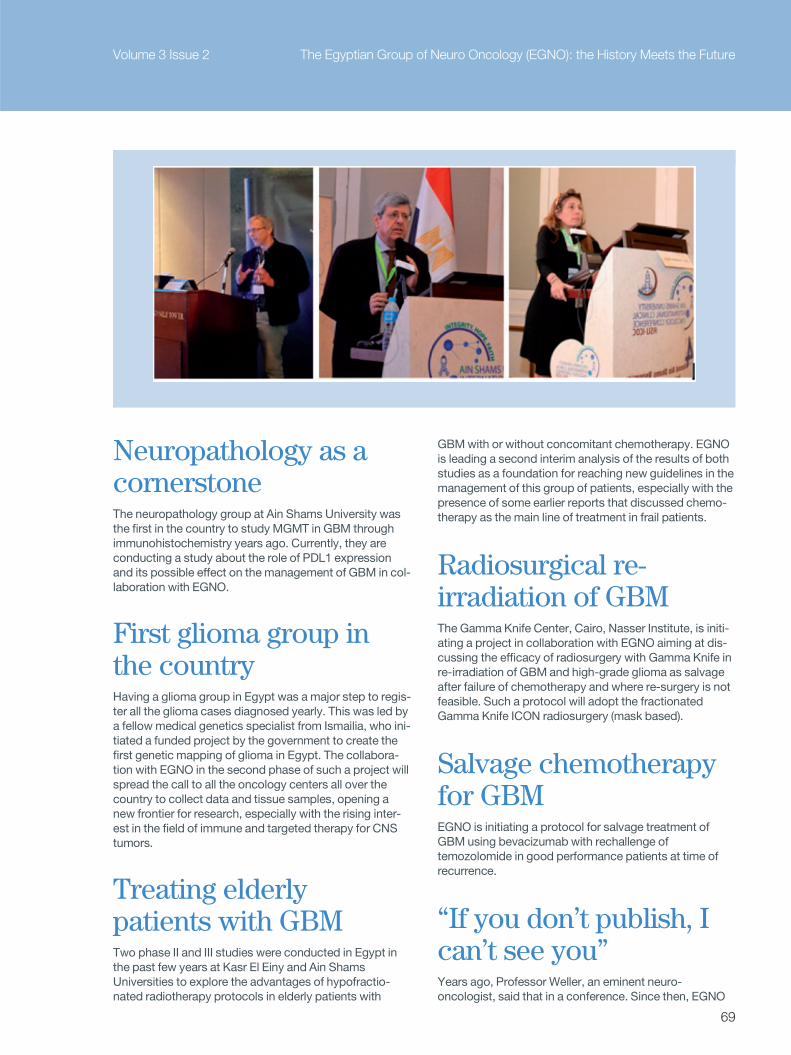
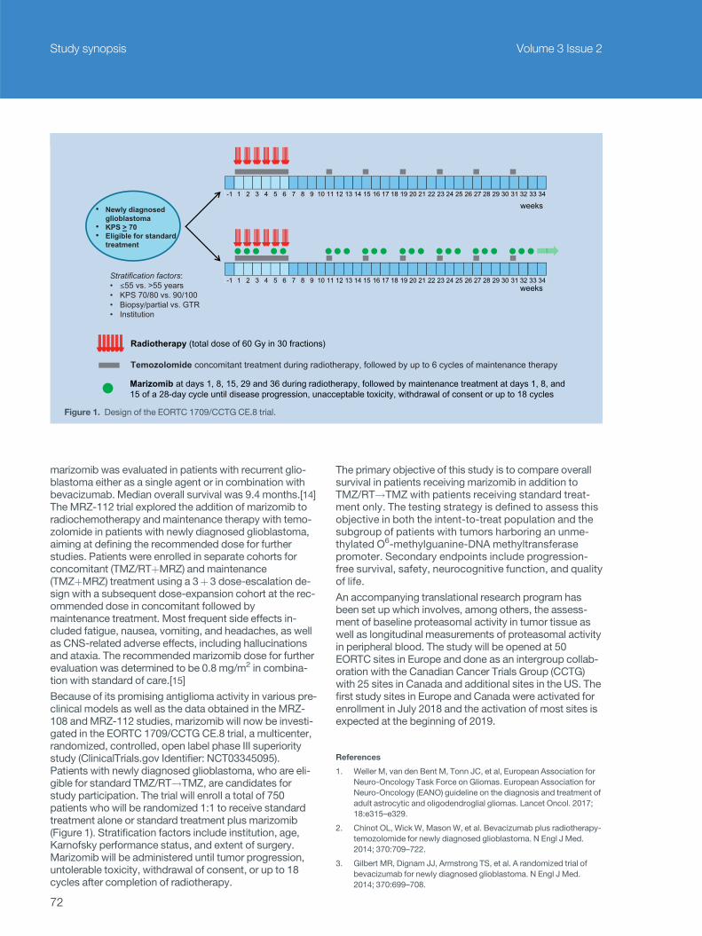
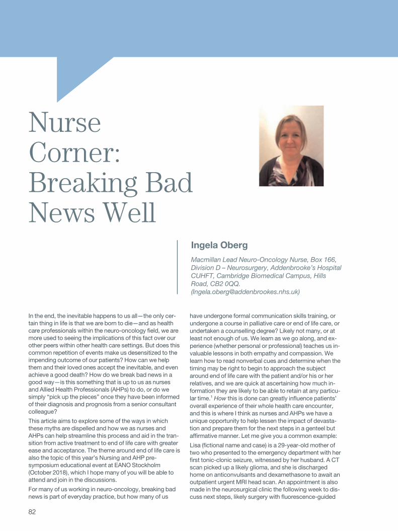
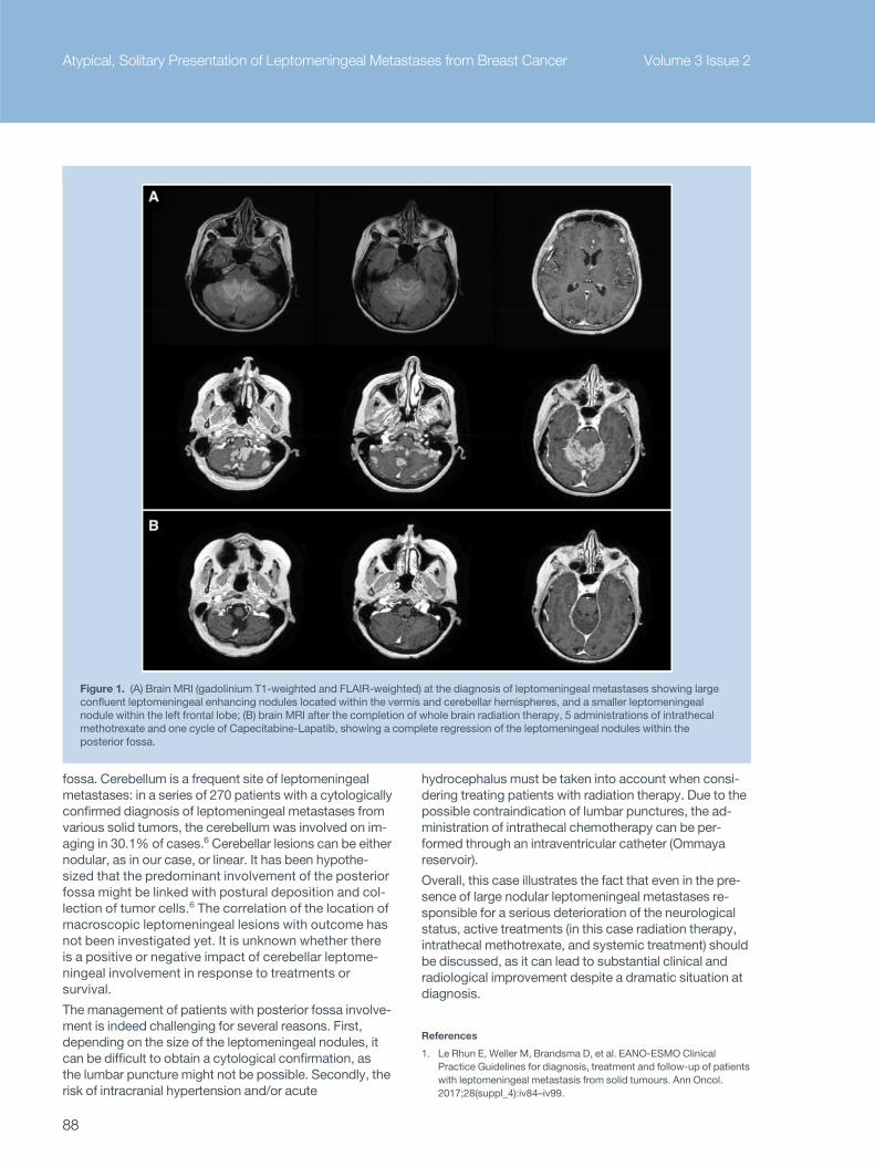