Embed Size (px)
Citation preview

�
Chief Editor:Abdulrazak AbyadMD, MPH, AGSF, AFCHSEEmail: [email protected]
Assistant to the Editor:Ms Rima KhatibEmail: [email protected]
Reporter and Photographer:Dr Manzoor Butt,Email: [email protected]
Ethics Editor and Publisher:Ms Lesley Pocockmedi+WORLD International572 Burwood Road,Hawthorn, Vic Australia 3122Phone: +61 (3) 9819 1224:Fax: +61 (3) 9819 3269Email: [email protected]
Editorial enquiries:[email protected]
Advertising enquiries:[email protected]
While all efforts have been made toensure the accuracy of the infor-mation in this journal, opinions expressed are those of the authors and do not necessarily reflect the views of The Publishers, Editor or the Editorial Board. The publishers, Editor and Editorial Board cannot be held responsible for errors or any consequences arising from the use of information contained in this journal;or the views and opinions expressed.Publication of any advertisements does not constitute any endorse-ment by the Publishers and Editors of the productadvertised.
The contents of this journal arecopyright. Apart from any fair deal-ing for purposes of private study, re-search, criticism or review, as permit-ted under the Australian Copyright Act, no part of this program may be reproduced without the permission of the publisher.
2 Editorial Abdul Abyad
Original Contribution / Clinical Investigation3 The Effects of Some Selected Variables on Child labour at Chapi Nawa-
bganj District in Bangladesh- A Multivariate Analysis Md. Rashed Alam7 The Reference Values of Body Composition for Adult Females Who are
Classified as Normal Weight, Overweight or Obese Accoding to Body Mass Index
Aliye Ozenoglu, Serdal Ugurlu, Gunay Can, Hüsrev Hatemi12 Maternal and Umbilical Cord Blood Lead Levels and pregnancy out-
comes: A Hospital Based Enquiry Asma A. Al- Jawadi, Zina W. A. Al-Mola, Raghad A. Al- Jomard
Medicine and Society19 Changing Face of Measles in Kashmir, India Kadri.S.M, Parray S.H, Rubina Shaheen, Gaash BA, Danish Muzaffar, Aesha.F,
Jan.Yasmeen23 Utilization of Postnatal Care in Al-Hassa, Saudi Arabia Abdel-Hady El-Gilany and Sabry Hammad27 Parental Consanguinity and Idiopathic Dilated Cardiomyopathy in
Children Shahla Roodpeyma, Hootan Salemi31 Stress among medical and law students in Mansoura, Egypt Abdel-Hady El-Gilany, Mostafa Amr, Nabil Awadalla, Ghada El-Khawaga
ISSN 148-4196 November 2008 - Volume 6, Issue 9

MIDDLE EAST JOURNAL OF FAMILY MEDICINE • VOLUME 6 , ISSUE 9�
This is the ninth issue this year and as the year is approaching its end we feel proud of the progress of the journal.
A paper from Turkey studied the reference values for body composition, measured with Bioelectrical Impedance Analyser (BIA), of adult females without any endocrinologic and/or metabolic disturbances, according to their body mass index (BMI) and grouped as normal, overweight, obese and morbidly obese. A total of 327 female subjects were taken into the study. The author concluded that the results could be used as reference values for studies on body composition, especially to predict the degree of body fatness of obese patients and also nutritional status of patients who need nutritional supports.
A paper from India looked at the changing face of measles in Kashmir. The authors looked at the clinical profile of measles in adults and looked at the complications seen in adults suffering from measles as well as the mortality of measles in adults. The author concluded that the study highlights the need for early measles vaccination of infants at 9 months of age and a repeat dose (18-30 years) might be necessary
at a higher age group to prevent adult measles, as is being followed by some developed countries in the west.
A paper from Iran looked at parental Consanguinity and Idiopathic Dilated Cardiomyopathy in Children. The aim of the authors was to determine the incidence of parental consanguinity and its relation with familial type of DCM in a group of children with this disease. The authors found that the parental consanguinity in their patients was significantly higher than that in controls and there was not a significant relationship between parental consanguinity and the familial form of DCM.
A paper from Egypt looked at stress among medical and law students in the university. This study aimed to determine if there is a difference in the perceived stress levels of medical and law students in Mansoura University, Egypt. The authors pointed out that because of stress among medical and law students, counselling and preventive mental health services should be an integral part of the routine clinical facilities caring for university students.
A Hospital Based study from Iraq looked at Maternal and Umbilical Cord Blood Lead Levels and pregnancy. The period of fetal growth is often the stage of development at which an organism is most sensitive to toxic agents. However, fetal.
exposure cannot be directly measured during pregnancy in human research studies. Maternal measurements are the only exposure indices ethically available.
A descriptive study from Saudi Arabia looked at the utilization of postnatal care in Al-Hassa. The authors stressed that Postnatal care coverage is low and is often considered as unnecessary. Therefore, there is an urgent need for an awareness-raising program highlighting the importance and availability of postnatal care. Antenatal care visits are good opportunities to council mothers about postnatal care.
In Bangladesh although child labour has been declined, it is still far from
replacement level. In this study, The Effects of some selected variables on Child labour at Chapi Nawabganj district in Bangladesh- A Multivariate Analysis, an attempt has been made to assess the child labour differentials and determinants in Bangladesh from Chapai Nawabganj district. The purpose of this study is to identify the harmful effects on various aspects of a child’s life of child labour.
A paper from India highlights the need for early measles vaccination of infants at 9 months of age and a repeat dose (18-30 years) might be necessary at a higher age group to prevent adult measles, as is being done by some developed countries in the west.
From the Editor
Abdulrazak Abyad MD, MPH, AGSF, AFCHS(Chief Editor)
Editorial office:Abyad Medical Center & Middle East Longevity InstituteAzmi Street, Abdo CenterPO BOX 618Tripoli, LebanonP + (961) 6 443684F + (961) 6 443685E [email protected] www.amc-lb.com
FROM THE EDITOR

MIDDLE EAST JOURNAL OF FAMILY MEDICINE • VOLUME 6 , ISSUE 9 �
ORIGINAL CONTRIBUTION AND CLINICAL INVESTIGATION
The Effects of Some Selected Variables on Child labour at Chapi Nawabganj District in Bangladesh - A Multivariate Analysis
ABSTRACT Children are the future of the nation and hopes and dreams of the world. But in the least developed countries like Bangladesh they are faced with enormous problems. The most hei-nous problem of children is undoubt-edly “Child Labour”. It is evident that, through the child labourers’ work at various health hazardous situations they have very little access to prima-ry health care and their overall health condition and nutritional status is very low. However, in Bangladesh al-though child labour has declined, it is still far from replacement level. In this study, an attempt has been made to assess the child labour differentials and determinants in Bangladesh from Chapai Nawabganj district. The purpose of this study is to identify the harmful effects on various aspects of a child’s life involved in child labour. Multivariate analysis such as path analysis has been used to find out the direct, indirect, and implied ef-fects of the selected variables. The statistical analysis of socio economic conditions on child labor is a specific means to improve their living condi-tion so that they can contribute to the world in a most effective way. Let’s launch a social campaign against child labor and ensure a happy, healthy, peaceful, hygienic and se-cured environment for children.
IntroductionBangladesh is a developing country,
about 50% of the household lives below the absolute poverty level. Employment of every young child is particularly an alarming problem. In 2002/03, the Bangladesh Bureau of Statistics (BBS) conducted the Second National Child Labour Survey (NCLS). This has been designed and conducted in the context of the commitments made by the government of Bangladesh, following the ratification of the International Labour Organization (ILO) worst forms of child labour convention (No.182) 1999. The child mortality rate under the age of 5 is estimated at 4.1 per 1000 live births and the maternal mortality ratio (MMR) is 3.8 per 1000 live births in 2001.
Considerable progress has been achieved through immunization programs. Percentages of boys and girls aged 12-23 months immunized against DPT (3 or more doses) are 76.8 percent and 71.7 percent respectively. Percentage of boys and girls aged 12-23 months immunized against polio (3 or more doses) are 92.1 and 88.4 respectively. On the other hand, children of the same age immunized against BCG: boys - 93.8 percent, girls - 90.4 percent and against measles (3 or more doses) for boys are 78.5 percent and girls 73.5 percent. National Child Labour Survey 2002-2003, BBS.
Extreme forms of poverty play a crucial role. Child labour is part of a vicious cycle, with poverty as a main cause as well as a main consequence.
This implies that child labour cannot be addressed in isolation. Among factors contributing to child labour are rapid population growth, adult unemployment, bad working conditions, lack of minimum wages, exploitation of workers, low standard of living, low quality of education, lack of legal provisions and enforcement, low capacity of institutions gender discrimination, conceptual thinking about childhood etc. One or more of the above contribute to the large numbers of children working under exploitative or hazardous conditions. Child labour is a persistent problem throughout the world, especially in developing countries (ILO, 1997). It is especially prevalent in rural areas of those countries where poverty is widespread, coupled with the lack of capacity to enforce minimum age requirements for work and schooling. Among the variety of reasons for child labour, the most important is the pressure upon them to escape the plight of poverty (Ahmed, A. & Quasem, M. A. 1991).
Child labour is not a new problem, and there is a long history of international efforts to combat it. The International Labour Organization (ILO), for example, in 1919 developed the first Minimum Age Convention that regulated the age at which children could work. Then, in 1973, a more comprehensive Minimum Age Convention, Number 138, was adopted, and it remains the fundamental standard. Although not new and always a thorny problem, child labour has now become increasingly complex, assuming
Key words: Child labour and its harmful effects, socio-economic conditions, age and Chapai Nawabganj District.
Md. Rashed AlamLecturerDepartment of Population Science and Human Resource DevelopmentUniversity of Rajshahi,Rajshahi-6205, Bangladesh.E-mail: [email protected]

MIDDLE EAST JOURNAL OF FAMILY MEDICINE • VOLUME 6 , ISSUE 9�
ORIGINAL CONTRIBUTION AND CLINICAL INVESTIGATION
new forms as global realities and relations have changed. Among the underlying causes, poverty and economic disparities are of course, critical factors. For much of human history, children have contributed to family welfare in a variety of ways, but intensified urbanization and the breakdown of traditional economic systems have made even basic subsistence more precarious and put children at ever higher risk. The results of a nine-country survey in Latin America, for instance, showed that if teenaged children did not work, poverty rates would increase by 10 to 20 per cent (Ahmed, A. & Quasem, M. A. 1991).
The aims of this study are to investigate the socio-economic conditions and harmful side of child labour and how it can be solved.
Data sources
Data for this study was drawn from a survey, conducted under the authority of the Department of Population Science and Human Resource Development of Rajshahi University. The survey was carried out in Gomastapu Upazila, Chapai-Nawabganj district. To collect the data from the above mentioned areas a survey on child labour was conducted. In addition, fruitful discussion was made with the employers to study several of the functional aspects of child labour. First, a list of child labourers was collected from the studied area and then 200 child labourers were selected for detailed interview using a structured questionnaire.
MethodsPath Analysis
A path analysis is one technique of showing causal linkages among the interrelated variables. The technique of path analysis, which was developed during the 1920s by Sewall Wright as an aid to the quantitative development of genetics, gained popularity in social science studies with the further expositions (Alwin D.F et all., 1975).
Path analysis presumes the existence of a causal framework interlinking different predictor variables with the response variables. Such representation of the causal
variables is called a path model and it is both stochastic and explanatory and is said to be an extension of the multiple regression model . It helps in estimating the magnitude of the linkages between interrelated variables and provides information about the underlying causal processes. This technique explores a chain of relationship among the variables by using standardized regression coefficients of a set of regression equations (Duncan, O.D. 1977). The fundamental to the path analysis is the path diagram which is the outcome of a set of linearly interrelated variables and the assumed causal relationship among them. In the path diagram the principles are as follows:
(i) the variables are arranged from left in such a way that all the endogenous variables are to the right of their exogenous variables
(ii) the unidirectional straight arrows called henceforth as ‘causal paths’ that go from left to right represent the endogenous variables, and
(iii) on the other hand, the two-headed curvilinear arrows represent the non-causal (correlated) relationship among the exogenous variables. This study employs a recursive path model relating to fertility and some of its determinants (Alam et all., 2004).
Methods and Model Specification for Path Analysis
Path analysis is a straightforward extension of multiple regressions. Its aim is to provide estimates of the magnitude and significance of hypothesized causal connections between sets of variables (Chandrasekaran and Hermalin, 1975). This analysis disentangles the specific mechanisms of the socio-economic factors affecting child labour by taking into consideration the intermediate variables involved in the analytical system. Moreover, path analysis provides a theoretical model specified as a system of simultaneous regression equations, which are linear, additive and usually recursive. This is best explained by considering a path diagram.
Table (1) Variables and their measurement used in the path analysis
Variable MeasurementX1 =Age of the respondent.
7 to14 Years
X2 = Respond-ent education
1=Illiterate2=Primary 3=Secondary 3=Other
X3 = Purpose of uses of money
1=Food2=Cloth3=Medical Treatment4=Other
X4 = Respond-ent income
1=<10002= 1001-20008= 2000+
X5= Smoke 1 = Yes2 = No
X6 = Participa-tion in NGO
1 = Yes2 = No
X7= Risky work 1 = Yes2 = No
X8 = Occupation of respondent
Number of workers
The path estimation equations are derived from the structural equations by applying the basic theorem of the path analysis. Thus, it is to be noticed that structural equations are different from the path estimation equations. According to the causal ordering of variables, we may divide the selected set of variables into three groups that are given below:
Exogenous Variable X1, X2, X3, X4, Endogenous variable X6 and X7,Dependent variable X8
This model is a recursive path model in which each variable is assumed to be dependent upon all prior causal variables. From the path analysis the direct effects, indirect effects, joint effects, implied effects and total effects of each selected explanatory variables on Child labour are obtained.
From Table 2 we show that, most children whose age is less than 1o are involved in risky work; on the other hand we observed that most of the child are working for their own needs, not entertainment. Most of them fall prey to diseases and their age range is 13 to 17 years. We also observed that most of the children are illiterate and few have primary and class six and seven. Most of the children are working for their family’s needs and their father’s occupation is agriculture. We also showed that their income is very low and they do not have involvement with any NGO.

MIDDLE EAST JOURNAL OF FAMILY MEDICINE • VOLUME 6 , ISSUE 9 �
ORIGINAL CONTRIBUTION AND CLINICAL INVESTIGATION
Most of the child use their money for family needs. An important matter we observed is that children whose age is less than 10 years do not smoke but most of the children whose age is over13 and above smoke.
Results and DiscussionIf poverty, as Nobel laureate
Amartya Sen argues, is to be defined not merely in terms of low income but as a state of deprivation of basic capabilities, nothing illustrates that more forcefully than child labour. A result and also a cause of poverty, child labour is a prison that withers both capabilities and potential. The prevalence of stunting, under-weight and wasting in children aged 6-71 months has shown a modest decrease over the past decade. The prevalence of stunting amongst girl children has declined from 65.9 in 1989-90 to 49.1 percent in 2000. The prevalence of under-weight girls children declined from 67.8 percent in 1989-90 to 50.9 percent in 2000. The prevalence of wasting has also declined from 15.9 percent to12.0 percent for boys and 17.3 percent to 11.4 percent for girls during 1995-96 to 2000.
From Figure 1 we see that age of the respondents is positively significant correlated with respondent income, purposes/use of money and respondent occupation are positively significant. Respondent education is negatively significant, correlated with respondent smoking, risky work and participation with NGO at 5% level of significance. Again participants in NGOs are negatively significant correlated with age of respondent, education of respondent, purposes/use of money, respondent income and smoking, at 1% level of significance.
According to Figure 1, we observe that there are 18 paths out of 13 hypothesized paths. In our study we have to mention the significant path coefficients only. And out of 7 variables, 3 are found to have a significant direct effect on the index of occupation. Among them are age of respondent (X1), Respondent education (X2), respondent smoking and risky work have a direct significant negative effect.
Total effect of respondent’s education on occupation is found to be more pronounced in all the variables and respondent’s income are positive effects. The total effect of respondent’s education on the occupation (X8) is -0.391 of which about 14 % is transmitted through the age of respondent (X1) about 66% is transmitted through its implied effect in the same direction, then about 18% acts through the risky work (X7). Other indirect effects of respondent’s education are via X6 and X7 and also the joint effects are negligible. Higher total positive influences of purposes of use of money on occupation and belongs to NGO. It is observed that the implied effect (P81) of age at respondent has contributed about 51%, 6% and 37 %of its total effect on occupation while the implied effect (P85) of smoking has contributed to about 55%, 3% and 82%of its total influence on occupation respectively.
ConclusionSome results of path analysis
deserve considerations from the viewpoint of policy implication. It has been found that respondent education and risky work have a direct negative influence on occupation. Thus raising age of respondent by implementing
a minimum-age law may lower occupation and risky work also may indicate lower occupation since at that time children are risk free from reproduction. Again occupation has a direct positive effect by purposes of uses money and belongs to NGO.
Total effect of respondent education on occupation is found to be negative. Education may provide better employment opportunities outside the home and age of respondent can be raised through providing education. Based on the results of this section it may be suggested that attention should be focused on the need of providing educational facilities.
ReferencesAhmed, A. & Quasem, M. A.. 1991. Child Labour in Bangladesh. Department of Economics, Lund University, Sweden.Ahmed, A. & Quasem, M. A.. 1997. Child Labour in Bangladesh, Bangladesh Institute of Develop-ment Studies (BIDS) 1997.Alam M.R., Roy T.K. and Mondal D.K., 2004. Intensity of the effects of the selected socio-economic and demographic factors on fertility in Bangladesh. Man In India, Vol. No. 84(1&2):Pp 51-62.Alwin D.F. and Hauser 1975. The decomposition of the effect in path analysis, American Socio-logical Review Vol. 40 Pp. 37-47.Chandrasekaran, C. and Albert I.Hermalin,1975. “Measuring the Effect of Family Planning pro-grams on Fertility. Paris”: International Union for the Scientific Study of Population Published for the Development Centre of the Organization for Economic Co-operation and Development.Duncan O.D. 1977. Path analysis: Sociological examples (Addenda), Causal models in social science, Chicago, Aldine-Atherton Inc.ILO, 1991. International Labour Organization, Vol. No. 182,1991.
Table 3: Effects of variables used in the path model for explaining respondent’s occupation
Dependent variable
Independent variable
Total as-sociation
Non casual effect
Total effect Indirect effect Other im-plied effect
Direct effect
X6 X7
X8
X1 0.080** .391 -.311 -.010** -.203** -.098**X2 0.021* .412 -.391 -.468** .054** .023X3 0.045 -.186 .231 -.128** 0.237 .122**X4 0.38** .338 .042 -.187** -.117** .346**X5 -0.120 -.094 -.026 -.043 .097 -.080
X6 0.101* -.006 .110** .159** .110**X7 -0.249** .055 -.304 -.304**

MIDDLE EAST JOURNAL OF FAMILY MEDICINE • VOLUME 6 , ISSUE 9�
ORIGINAL CONTRIBUTION AND CLINICAL INVESTIGATION
Non-causal effect = Total association- Total effect.Table 2: Mean number of respondent occupation per children by age and the selected socio-economic characteristics.
socio-economic characteristics
Age of respondentsLess than 10 10-13 13 above
Risky WorkYesNo
4.02.42
3.652.93
3.682.77
Per Month Income150-900901-13001500 above
2.332.521.96
4.334.353.66
03.04.64
Educational StatusNo EducationPrimary EducationSecondary Education
3.102.00
3.283.51.8
3.872.883.06
SmokeYesNo
1.02.86
3.03.3
3.882.89
Participation in NGOYesNo
1.63.3
2.673.69
3.313.11
Purposes & uses of moneyFoodsCloths
3.172.8
3.053.1
3.184.0
Choice of workOwnFamilyEconomic Pressure
.500
1.252.232.6
2.363.454.28
Guardian OccupationAgricultureBusinessService
2.651.240
3.482.46.45
3.872.26.25
EntertainmentYesNo
1.02.86
2.563.3
3.283.89
DiseasesYesNo
3.211.01
3.681.86
4.122.41
Figure 1: Path diagrams of factors affecting respondent occupation through other variables
MIDDLE EAST JOURNAL OF FAMILY MEDICINE • VOLUME 6 , ISSUE 9 �
ORIGINAL CONTRIBUTION AND CLINICAL INVESTIGATION
IntroductionObesity. defined as the increase of
fat tissue in the body, is an important public health problem which leads to increased morbidity and mortality of some diseases, and has negative influences on the duration and quality of life. As well as physiological aspects, it also has social and psychological aspects(1-5). In parallel with technological advances, the ease in obtaining, buying and consuming various kinds of food has increased and physical inactivity, consumption of more cigarettes and alcohol, stress and weakness of the mechanisms for coping with stress are other environmental factors which make it easier for obesity to develop(1,4-7). Presently, obesity,the prevalence of which is increasing in all age groups in many countries, is regarded as a disease which must be treated(4,5). In order to define someone as obese, a person’s body weight, body composition and fat distribution, should be correctly determined. It should not be forgotten that when
evaluated according to body weight there are subjects who are accepted to be obese as they have a lot of muscle mass; but there are also others who have normal body weight but accepted to be obese based on body fat composition and other metabolic parameters(4,8). So, it is quite important to diagnose obesity correctly in order to prevent the organic, metabolic, and psychosocial problems it might cause. On the other hand, the measurement of body composition is highly helpful in understanding whether the individual has need for any nutritional support.
Until now, many methods have been developed to detect body composition(2,3,9). Although direct methods to evaluate body composition in humans do exist, they are not easily applicable in routine clinical practice. So, the applicable methods are indirect. Among these Bioelectrical Impedance Analysis (BIA), the reliability of which was confirmed in many studies, is a practical method which makes use of
The Reference Values of Body Composition for Adult Females Who are Classified as Normal Weight, Overweight or Obese Accoding to Body Mass IndexAliye Ozenoglu, PhDDepartment of Psychiatry, Cerrahpasa Medical Faculty, University of Istanbul, Istanbul, TurkeySerdal Ugurlu, MDDepartment of Medicine, Medical Faculty, University of Cumhuriyet, Sivas, TurkeyGunay Can, MDDepartment of Public Health, Cerrahpasa Medical Faculty, University of Istan-bul, Istanbul, TurkeyHüsrev Hatemi, MDProfessor of Medicine, Division of Endocrinology-Metabolism and Diabetes and Department of Medicine, Cerrahpasa Medical Faculty, University of Istan-bul, Istanbul, Turkey
Corresspondence to:Aliye Özenoglu, PhDCerrahpasa Medical Faculty, Psychiatry DepartmentIstanbul University,34303 Cerrahpasa, Istanbul, TURKEYTel:+90 (212) 414 31 30Fax:+90 (212) 414 31 30
ABSTRACTObjective: The aim of this study was to conduct the reference val-ues for body composition meas-ured with Bioelectrical Impedance Analyser (BIA) of adult females without any endocrinologic and/or metabolic disturbances, ac-cording to their body mass index (BMI) and grouped as normal, overweight, obese and morbidly obese.
Patients and Methods: A total of 327 female subjects were taken into the study. Their body com-positions were measured with BIA, in addition to measurement of their weight, height, and waist and hip circumferences. Results were statistically analysed with ANOVA test.
Results: As BMI got higher, per-centages of body fat and basal metabolism (BM) increased signif-icantly; but percentages of body water and fat free mass, and lean/fat ratio showed a significant de-crease. In addition, waist and hip ratios, percentage of body fat and BM showed a significant positive corelation with BMI.
Conclusion: We concluded that our results could be used as ref-erence values for studies on body composition, especially to pre-dict the degree of body fatness of obese patients and also nutri-tional status of patients who need nutritional supports.
Key Words: Body mass index, body composition, Bioelectrical Imped-ance Analyser.

MIDDLE EAST JOURNAL OF FAMILY MEDICINE • VOLUME 6 , ISSUE 9�
ORIGINAL CONTRIBUTION AND CLINICAL INVESTIGATION
the conductivity of the body, namely the tissues(9-15). The body composition might vary according to age, sex, ethnic background, nutritional status, exercise, climate, the presence of some illnesses and the administration of some drugs; so there need to be standards for different conditions. However, reference values available for this purpose are quite few(10,13,16-
18). In literature, although there are reference values of body composition for different age groups obtained by BIA; there are no reference values of body composition in healthy adults matched for age, sex, and body mass index.
In this study, we aimed to determine reference values of body composition in adult females who were classified as normal, overweight, obese, and morbidly obese according to body mass index (BMI); and who had no endocrine-metabolic disturbances except exogenous obesity and also no history of any drug usage.
Patients and MethodsA total of 327 female subjects (all
>18 years of age) were admitted to our department between 1999 and 2003 with various complaints evaluated retrospectively. Patients who had no endocrine-metabolic disease or denied usage of drugs affecting metabolism, were included in this study. Mean age of subjects was 39.18±12.02 years. As a part of the nutritional status assesments, patients’ height, weights, waist and hip circumferences were determined and the body compositions measurements were performed by BIA. BMI was calculated by adjusting the known formula as weight (kg)/ height2 (m2). Subjects with BMI=18.5-24.9 kg/m2 were accepted to be normal, those with BMI=25.0-29.9 kg/m2 overweight, those with BMI=30.0-39.9 kg/m2 to be obese, and those with BMI³40 kg/m2 to be morbidly obese(1-5,19,20). Waist/hip ratio (WHR) was obtained by dividing the waist circumference into the hip circumference.
BIA depends on the principle that fat is a bad conductor to the applied current; however, lean body mass is a good conductor depending on its content of water and electrolytes. In order to determine the body
impedance, two tetrapolar electrodes are placed on the lateral surfaces of both the right hand and the right foot while the subject is lying down in supine position. There is a low current between the electrodes. Depending on the amplitude of the current, whole body water; or, in low frequencies only extracellular water might be determined(2,3,9,21). As the method mainly measures body water, the hydration of the subject should be normal. Errors in measurement might occur due to the presence of metals on the bed or the subject, daily variations in body weight and composition due to food intake and exercise, intake of drugs, menstrual cycle in females, and placing the electrodes incorrectly(2,9). In this study, optimum care has been taken to minimize errors in measurement and all measurements were performed by the same observer in morning hours.
Temperature of the test room was maintained between 18-20°C. The resistance and reactance values obtained by BIA, together with the age, sex, height and weight of the subject were entered into a computer programme to calculate body water, fat and lean body mass and basal metabolism. Percentage values for body composition, and basal metabolism as kcal. which was obtained automatically via a computer programme, were mainly used in this study. Body fat and lean weight as grams and water as liters were not included in this study. As the body composition of females and males differ, only female subjects were taken into the study. ANOVA test was used in comparing the anthropometric values and body compositions in percentages between groups who were formed according to BMI.
ResultsThe mean BMI, waist and hip
circumferences, and WHR of females according to BMI are given in Table 1.
As the degree of obesity increases, BMI, waist and hip circumferences also increased significantly. WHR showed a parallel increase with BMI significantly (p<0.0001).
The percentages of body water, fat mass (FM) and lean body mass
(LBM), and basal metabolism (BM) obtained by BIA for females are shown in Table 2.
In parallel with the increase in BMI, the percentage of body fat and BM increases; however, the percentages of body water and lean body mass decrease significantly (p<0.0001).
The correlations of the various measurements made in the study are shown in Table 3.
A highly significant positive correlation was found between BMI and waist and hip circumferences and between the percentage of body fat and BM; whereas a negative correlation was present between BMI and lean body mass, and the percentage of body water. In addition, percentage of body fat had significant correlation with waist and hip circumferences and WHR.
DiscussionIn order to know the nutritional
status, it is important to determine the body composition. Body composition might vary according to the stages of growth and development, age, sex, ethnicity, genetic and environmental factors, nutritional and exercise habits, various diseases and different therapies(2,9,10,20-
22). Today, body composition is evaluated at the anatomic, molecular, cellular, tissue-system and the whole body level(1,3,4,20,24-29). As direct measurements in humans cannot be performed under in vivo conditions, body composition might be determined by indirect methods. Direct measurements might be performed only on cadavers. Methods of indirect measurement which can be performed in humans are anthropometric measurements, isotope or chemical dilution method, determination of body density, conductivity measurements, imaging methods, whole body neutron activation analysis and dual-energy X-ray absorbiometry (DEXA)(1-3,9,20-
24). Today, BIA which is a method dependant on conductivity is one of the most preferred methods because it is easily performed, portable, has no danger, is more economic when compared to other methods and the results are reliable(2,3,9,20,21).

MIDDLE EAST JOURNAL OF FAMILY MEDICINE • VOLUME 6 , ISSUE 9 �
ORIGINAL CONTRIBUTION AND CLINICAL INVESTIGATION
To predict the nutritional status of a patient and plan their nutritional treatment, it is helpfull to follow up the changes in body composition. Determination of the body fat composition is an important criterion especially in understanding the risk for obesity and related diseases. In spite of having normal body weight, there might be subjects with insulin resistance, hypertension, dyslipidemia, and above normal body fat mass (normal weight, metabolically obese); on the contrary it must not be forgotten that some others might be obese when body weight is regarded, but their metabolic parameters might be normal in contrast with what is expected (obese, metabolically normal)(4,8).
On the other hand, correct determination of the body compositions of patients who need nutritional support for various reasons is of utmost importance. Follow up of the changes in body composition are useful for determination of the nutritional status and planning therapy and to evaluate the efficiency of administered therapies in disorders of hormones affecting metabolism, or diseases which make it obligatory to use drugs affecting the metabolism; inborn disorders of metabolism in which special diets have vital importance; inflammatory bowel diseases (IBD) which cause deterioration of the patients’ nutritional status; chronic renal failure; neurologic disorders; diseases like cancer the presence of severe disease states or traumas. Body composition is helpful in evaluating the efficacy of diet therapy especially in muscle-type glycogenoses and other muscle diseases in which an increase in the amount of protein in the diet is needed for preservation of the body muscle masses(30,31,32).
Correct determination of the nutritional status in patients with renal failure is important as it is closely related with prognosis. In these patients, total body water (TBW), hypertension and cardiac morbidity are accepted to be independant prognostic markers(12). In one study which was planned to assess TBW and nutritional status in end-stage renal failure patients, it was found out
that TBW varied greatly depending on the method of calculation(12).
IBD patients are frequently faced with malnutrition as a result of malabsorption and decreased food intake due to gastrointestinal symptoms. Many studies conducted in IBD patients in order to assess the nutritional status revealed that body fat mass was significantly decreased when compared to controls, however lipid oxidation rate was increased(33,34,35). It was thought that increased lipid oxidation and insufficient energy intake could explain the decrement in fat mass and it was put forward that enteral diets relatively rich in fat might be useful to sustain the nutritional status of these patients.
Determination of body composition is important for some occupations. It is desirable for athletes, artists, ballet-dancers, and people occupied in military and legal jobs to keep a certain body fat standard. Malina et al.(36) studied the percentage and the distribution of fat of the athletes who took place in the Olympic Games in Montreal in 1976. They reported that percentage of body fat was affected mostly from sports and exercise; however the distribution of fat was dependant on biologic factors. Studies revealed that the distribution of body fat differs between whites and blacks; and that blacks store more fat on the upper part of the trunk when compared to whites(10). In whites, the ratio of extremity skin-fold thickness to trunk skin-fold thickness was found to be higher than in blacks(36). Because of these reasons, when determining body composition age, sex, ethnic background, concurrent diseases and therapies, nutritional habits, activity level, socioeconomic and environmental factors should be carefully investigated.
In order to be able to interpret body composition measurements, reference values formed under various conditions are needed. However, the number of studies about reference values formed under various conditions is limited. In this study, we aimed to determine mean body composition values by grouping adult females according to BMI; and, we found out that body composition
changed significantly in accordance with BMI (Tables 2). This finding shows that while interpretting the body composition of one individual, BMI must be taken into consideration.
The first study in which fat and lean-body masses according to sex and age were determined in healthy subjects was performed by Pichard et al.(13). In this study, they wanted to determine fat and lean-body masses in different decades by BIA, to detect changes in these values with advancing age, and to develop percentile values for these parameters in a population composed of healthy whites (1838 males and 1555 females) between 15-64 years of age. It was demonstrated that mean fat mass and percentage of body fat in males increased progressively; whereas, in females this increase occurred after 45 years of age. It was reported that the data in that study could be used as a reference to evaluate whether body compositions of healthy and sick subjects at certain ages were normal.
In another study(18), 25th-75th percentiles of fat and lean-body masses were formed in 4566 healthy females and males between 20-79 years of age. In this study, subjects were grouped into 20-39, 40-59, 60-79 age ranges and measurements were performed with BIA. The authors concluded that lean-body mass decreased and fat mass increased with age; and that these reference percentile values made it possible to interpret the results of BIA and to determine subjects with abnormal muscle and fat mass.
There are different methods to assess the energy needed for basal metabolism(3,37,38). One of these is indirect calorimetry which depends on the principle of measuring oxygen used in biologic oxidations and it is possible to obtain correct and reliable results with it; however, its use is not widespread because of the difficulties in practice. In daily life, to calculate the energy requirements, methods which can be applied more practically and which depend indirectly on measurement and calculation are used. In our study, it was found that energy for basal metabolism detected by BIA increased significantly in parallel with the increase in BMI

MIDDLE EAST JOURNAL OF FAMILY MEDICINE • VOLUME 6 , ISSUE 9�0
ORIGINAL CONTRIBUTION AND CLINICAL INVESTIGATION
(Tables 2).
Frequently used equations for determination of energy needed for basal metabolism are Harris Benedict, Schofield and WHO equations(3,17,37,38). Some of the studies in which energy for basal metabolism determined by measurement and calculation were compared found differences in values obtained by two different methods; but, some other studies could not find any difference(17,39,40).
Barot et al.(39) compared the measured resting energy expenditure (REE) in 12 IBD patients -9 of whom had Crohn’s disease- with a calculated formula according to Harris Benedict equation. They found that there was no significant difference between these two values in patients having >90% of ideal weight; but, patients <90% of ideal weight were hypermetabolic when compared to controls. Stokes and Hill(40) reported that resting metabolic rate detected by measurement in 13 active Crohn’s patients was 14% higher than that found by calculation.
Recently, we compared BM measured by BIA and calculated by adjusting Harris Benedict equation in female patients with active IBD and women with normal weights according to BMI(41), and saw that the value obtained by measurement was significantly higher than that of calculated (respectively, 1325.75±122.92 kcal vs. 1272.82±102.67 kcal, p=0.02). In this study in which healthy women matched for age and BMI served as controls, BM in the control group was significantly higher than the calculated value (respectively, 1451.88±83.5 kcal vs. 1323.27±74.65 kcal, p<0.0001).
In another study(42), we compared BM in normal weight, healthy adult females and males by two different methods, and found that BM measured by BIA was significantly higher than this calculated by Harris Benedict formula in both sexes (p<0.0001). In a different study(43), we compared BM found by two different methods in females who were of normal weight, overweight or obese according to BMI, had no endocrine-metabolic disorder and no history of any drug usage; the results revealed that BM measured
by BIA was significantly higher than that calculated with Harris Benedict equation (p<0.0001). Depending on the results that we obtained in these studies, we concluded that -not only in healthy subjects; but, also in the presence of diseases affecting metabolism- methods to determine body composition and basal metabolism, which based on measurement might be more reliable in predicting energy needs and planning nutritional therapy.
Obesity which is a major health problem in many developing and developed populations, is an independant risk factor for the development of coronary artery disease (CAD).Also, obesity increases the risk of CAD by its relation with insulin resistance, hypertenion and dyslipidemia(1,4,5,7,8,19,22). Determination of body fat is important in defining obesity which might impair quality of life and cause increased morbidity and mortality. Although techniques like computed tomography (CT), magnetic resonance (MR) make it possible to evaluate regional body fat distribution and abdominal fat depositions, their use for clinical and epidemiological purposes is limited by their high expenses. The determinants of body fat distribution, like WHR, waist diameter, sagittal diameter, are important alternatives in defining individuals with high risk(1,4,5,8,20).
WHR is a simple, useful and sensitive index of body fat distribution. By using this anthropometric index Bray and Gray(44) put forward that in males a WHR>0.95 and in females a WHR>0.85 might be useful in the detection of high-risk subjects. Similarly, Pauliot et al.(45) reported that in females a WHR>0.85 and in males a WHR>1.0 might be related with changes in glucose-insulin homeostasis and lipid-lipoprotein metabolism. It was said that waist circumference determined with techniques like CT and MR was a better indicator of visceral fat than WHR(4,8). It is accepted that independant of BMI and WHR, a waist circumference >88 cm in females and >102 cm in males increase the risk for complications of obesity and mortality(4,9,22). Under the light of this knowledge, females in our study who
were in obese groups (BMI>30.0 kg/m2) according to BMI are accepted to have risk for obesity-related diseases (Table 1). As waist circumference and BMI have a very strong relation with percentage of body fat and WHR has a weak relation, this confirms that waist circumference is a more sensitive indicator (Table 3).
It is known that fat tissue forms 20-25% of body weight in an adult female and 12-15% of that in a male (1,5,19,20). Obesity is accepted to be present when body fat exceeds 30% in females and 20% in males. The results of this study are compatible with the available literature both for body composition and body circumference measurements.
ConclusionCorrect determination of the
nutritional status of an individual is important for planning therapy under various conditions and for evaluating efficiency of administered therapies. Measurements which are dependant on only height and weight are usually misleading and insufficient in evaluation of the nutritional status of one individual. However, as it is unfeasible to determine body composition in the clinical practice, there is need to develop the standards of methods of indirect measurement.Body composition might show variations according to age, sex, ethnic features, nutritional status, genetic and environmental factors, level of exercise, and even the presence of various diseases and administered therapies. For this reason, whenever the body composition of one individual is to be interpretted it will be useful to use reference values formed under similar conditions. In addition; whenever there is no possibility of determining the body composition of the individual, the usage of references in order to predict his nutritional status approximately will lead to more reliable results. To achieve this, it is obvious that standardized indirect measurement must be developed for both healthy individuals and states of disease. We assume that the results of this study which was the first to evaluate body composition by BIA in groups matched for age, sex, and BMI- might be used as reference

MIDDLE EAST JOURNAL OF FAMILY MEDICINE • VOLUME 6 , ISSUE 9 ��
ORIGINAL CONTRIBUTION AND CLINICAL INVESTIGATION
values. We also think that diet and exercise programmes should be used for the treatment of obesity which is defined as excess fat tissue in the body; and the efficacies of diet and medications should be followed up with measurements of body composition.
ReferencesTüzün M. The general features of obesity. In: Yilmaz C (ed). Obesity and its treatment, Izmir, 1999; p. 11-29 (Book in Turkish).Pekcan G. The definition and detection of obesity.In: Proceedings of the Third National Congress on Nutri-tion and Dietetics, Ankara, 2000; 93-104 (Abstract in Turkish).Arslan P. (ed). Obesity, its influence on various dis-eases and scientific applications in dietary treatment. 4 th publication of the Turkish Dietetic Associations, Ankara, 1992 (Book in Turkish).Korugan Ü, Damci T, Özbey N, Özer E. Clinical Obes-ity. Publication of the Obesity Working Group. Istanbul, 2000; 2-14 (Book in Turkish).Yilmaz C (ed). Obesity. Nobel Medical Publications, Izmir, 1995 (Book in Turkish).Bjorntorp P. Visceral obesity: a “civilization syndrome”. Obes Res 1993; 1: 206-22.Siervogel RM, Wisemandle W, Maynard LM, Guo SS, Chumlea WC, Towne B. Lifetime overweight status in relation to serial changes in body composition and risk factors for cardiovascular disease: The feels longitudi-nal study. Obes Res 2000; 8 : 422-30.Brochu M, Poehlman ET, Ades PA. Obesity, body fat distribution and coronary artery disease. J Cardiopulm Rehabil 2000; 20: 96-108.Ellis KJ. Human body composition: In vivo methods. Physiological Reviews 2000; 80: 649-80.Wagner DR, Heyward VH. Measures of body composi-tion in blacks and whites: a comparative review. Am J Clin Nutr 2000; 71: 1392-402.Rosenbaum K, Wang J, Pierson RN, Kotler DP. Time dependent variation in weight and body composition in healthy adults. JPEN 2000; 24 (2): 52-55.Cooper BA, Aslani A, Ryan M, Zhu FY, Ibels LS, Al-len BJ, Pollock CA. Comparing different methods of assessing body composition in end stage renal failure. Kidney Int 2000; 58 : 408-16.Pichard C, Kyle UG, Bracco D, Slosman DO, Morabia A, Schutz Y. Reference values of fat-free and fat mass-es by bioelectrical impedance analysis in 3393 healthy subjects. Nutrition 2000; 16 : 245-54.Lukaski HC. Assessing regional mass with segmental measurements of bioelectrical impedance in obese women during weight loss. Ann NY Acad Sci 2000; 904: 154-8.
1.
2.
3.
4.
5.
6.
7.
8.
9.
10.
11.
12.
13.
14.
Kyle UG, Genton L, Karsegard L, Slosman DO, Pichard C. Validation of a bioelectrical impedance analysis (BIA) equation for the swiss population. Clin Nutr 2000; 19 (Suppl 1): S 6.Atkin LM, Davies PS. Diet composition and body com-position in preschool children. Am J Clin Nutr 2000; 72 : 15-21.Avitzur Y, Singer P, Dagan O, Dinari G, Shamir R. Measured vs calculated resting energy expenditure before and after open heart surgery in children with congenital heart disease. Clin Nutr 2000; 19 (Suppl 1): S 2.Kyle UG, Genton L, Karsegard L, Slosman DO, Pichard C. 25-75 th percentiles for fat-free and fat masses in 4566 healthy adults aged 20-79 years determined by BIA. Clin Nutr 2000; 19 (Suppl 1): S 6.PI-Sunyer FX. Obesity. In: Shils ME, Olson JA, Shike M, Ross AC (eds). Modern Nutrition in Health and Disease. 9th ed. Philadelphia, Lippincott Williams &Wilkins.1999;1395-414.DeHoog S. The assessment of nutritional status. In: Mahan LK, Escott-Stump S (eds). Krause’s Food, Nutrition and Diet Therapy. 9th ed. Philadelphia. W.B. Saunders Company, 1996; 369-74.Jebb SA. Measuring body composition: from the labo-ratory to the clinic. In: Kopelman PG, Stock M. eds. Clinical Obesity. Oxford. Blackwell Science, 1998; 18-50.Weight management and eating disorders. In: Mahan LK, Escott-Stump S (eds). Krause’s Food, Nutrition and Diet Therapy. 9th ed. Philadelphia. W.B. Saunders Company, 1996; 451-63.Brodie D, Moscrip V, Hutcheon R. Body composition measurements . A review of hydrodensitometry, antro-pometry and impedance methods. Nutrition 1998; 14: 296.Wang ZM, Pierson RN, Hemsfield SB. The five-level model: a new approach to organising body composition research. Am J Clin Nutr 1992; 56: 19.Fuller N, Jebb SA, Laskey M, Cowerd M, Elia M. Four compartment model for the assesment of body compo-sition in humans: Comparision with alternative methods and evaluation of the dansity and hydration of fat free mass. Clinical Science 1992; 82: 687.Heymsfield SB, Wang ZM, Baumgartner RN, Ross R. Human body composition. Advances in models and methods. Ann Rev Nutr 1997; 17: 527.Heymsfield SB, Tighe A, Wang ZM. Nutritional asses-ment by anthropometric and biochemical methods. In: Shils ME, Olson JA, Shike M. eds. Modern Nutrition in Health and Disease. 8 th ed. Philadelphia. Lea and Febiger, 1994; 812.WHO. Physical status: The use and interpretation of anthropometry. WHO Tech Rep Ser WHO, Genova. 1995; 854.Nunez C, Kovera AJ, Pietrobelli A, Heshka S, Horlick M, Kehayias JJ, et al. Body composition in children and adults by air displacement plethysmography. Eur J Clin Nutr 1999; 53: 382.Fernandes J, Chen YT. Glycogen storage disease. In: Fernandes J, Saudubray JM, Berghe GV (eds). Inborn Metabolic Disease. 2nd ed. Berlin. Springer-Verlag, 1995; 71-84Dixon M. Disorders of carbohydrate metabolism. In: Shaw V, Lawson M (eds). Clinical Paediatric Dietetics. London. Blackwell Science Ltd. 1995; 210-16.
15.
16.
17.
18.
19.
20.
21.
22.
23.
24.
25.
26.
27.
28.
29.
30.
31.
Forbes GB. Body composition: Influence of nutrition, physical activity, growth and aging. In: Shils ME, Olson JA, Shike M, Ross AC (eds). Modern Nutrition in Health and Disease. 9th ed. Philadelphia. Lippincott Williams & Wilkins, 1999;789-805.Capristo E, Addolorato G, Mingrone G, Greco AV, Gas-barrini G. Effect of disease localization on the anthro-pometric and metabolic features of Crohn’s Disease. Am J Gastroenterol 1998; 93: 2411-19.Mingrone G, Greco AV, Benedetti G, Capristo E, Sem-eraro R, Zoli G, et al. Increased resting lipid oxidation in Crohn’s Disease. Digestive Disease and Science 1996; 41 (1): 72-76.Mingrone G, Capristo E, Greco AV, Benedetti G, De Gaetano A, Tataranni PA, et al. Elevated diet-induced thermogenesis and lipid oxidation rate in Crohn’s Dis-ease. Am J Clin Nutr 1999; 69 (2): 325-30.Malina RM, Mueller WH, Bouchard C, Shoup RF, Lar-iviere G. Fatness and fat patterning among athletes at the Montreal Olympic Games. Med Sci Sports Exerc 1982; 14: 445-52.Arslan P. The calculation of energy and nutritional ele-ment needs in enteral-parenteral nutrition. In: Basoglu S, Karaagaoglu N, Erbas N, Ünlü A (eds). Enteral-Parenteral Nutrition. Turkish Dietetics Associations, publication number: 8, Ankara, 1995;39-49 (Book in Turkish).The calculation of energy needs. In: Özkarabulut A, Özenoglu A, Turanli F, Boneval H. (eds.), Formulas and enteral products in nutrition. Turkish Dietetics As-sociations, publication number: 1. Istanbul, 2000; 10-11 (Book in Turkish).Barot LR, Rombeau JL, Feurer ID, Mullen JL. Caloric requirements in patients with inflammatory bowel dis-ease. Ann Surg 1982; 195: 214-19.Stokes MA, Hill GL. Total energy expenditure in pa-tients with Crohn’s Disease: Measurement by the com-bined body scan technique. JPEN 1993; 17: 3-7.Özenoglu A, Pamuk GE, Pamuk ÖN, Hatemi HH. The comparison of basal metabolism found by two different methods in normal-weight females with inflammatory bowel disease with healthy controls. Goztepe Medical Journal 2004; 19: 15-18. (Journal in Turkish).Özenoglu A, Pamuk ÖN, Pamuk GE, et al. The com-parison of body compositions and basal metabolism found by two different methods between normal-weight healthy females and males. J Endocr Invest. 2001; 10 (2): 21-4. (Journal in Turkish).Özenoglu A, Pamuk GE, Pamuk ÖN, et al. The com-parison of basal metabolism found by two different methods amongst females classified as normal, over-weight or obese according to body mass index. In: Proceedings of the Third Congress of Clinical Enteral-Parenteral Nutrition. Istanbul, 2000; 182 (Abstract in Turkish).Bray GA, Gray DS. Treatment of obesity. An overview. Diabetes Metab Rev 1998; 4: 653-79.Pouliot MC, Despres JP, Lemieux S, et al. Waist circum-ference and abdominal sagittal diameter: Best simple anthropometric indexes of abdominal visceral adipose tissue accumulation and related cardiovascular risk in men and women. Am J Cardiol 1994; 73; 460-68.
32.
33.
34.
35.
36.
37.
38.
39.
40.
41.
42.
43.
44.
45.
Table 1: The mean values of BMI, waist and hip circumferences, and WHR in females.
Group BMI (kg/m2) Waist (cm) Hip (cm) WHRNormal 21.52+2.38 69.41+15.39 95.68+5.44 0.75+0.050Over-weight
27.18+1.35 85.77+6.81 108.58+4.57 0.79+0.056
Obese 34.37+2.60 97.06+14.34 120.13+10.17 0.81+0.057Morbidly obese
46.88+6.74 117.47+11.38 141.37+19.78 0.83+0.059
P 0.0001 0.0001 0.0001 0.0001
Table 2: The percentages of body compositions and basal metabolism in females according to their BMI groups.
Group Water (%) Fat mass (%) Lean-body mass (%)
LBM/ FM BM (kcal)
Normal 59.61+5.01 22.78+4.58 77.22+4.58 3.58+1.16 1397.11+ 111.10Overweight 52.15+3.23 29.69+3.30 70.31+3.30 2.35+0.37 1510.44+ 119.71 Obese 46.10+3.38 35.01+3.25 64.99+3.25 1.87+0.60 1704.86+ 135.24Morbidly obese 40.64+3.44 40.22+3.60 59.78+3.60 1.46+0.23 1978.95+ 198.62P 0.0001 0.0001 0.0001 0.0001 0.0001
Table 3: The correlations between various measurements in females.
BMI Waist circum-ference
Hip cir-cumfer-ence
WHR
Waist circumfer-ence
0.814***
Hip circumfer-ence
0.816*** 0.677***
WHR 0.359*** 0.603*** 0.213**Body water (%) -0.864*** -0.778*** -0.711*** -0.416***Fat mass (%) 0.858*** 0.769*** 0.696*** 0.418***Lean-body mass (%)
-0.858*** -0.769*** -0.696*** -0.418***
LBM/ FM -0.681*** -0.575*** -0.548*** -0.248**BM (kcal) 0.863*** 0.708*** 0.755*** 0.282***

MIDDLE EAST JOURNAL OF FAMILY MEDICINE • VOLUME 6 , ISSUE 9��
Maternal and Umbilical Cord Blood Lead Levels and pregnancy outcomes: A Hospital Based Enquiry
ABSTRACTBackgroundEnvironmental lead exposure is a pub-lic health problem on a global level. The population most sensitive to lead expo-sure from various sources, are pregnant women and children. The aim of the present study is to measure maternal and umbilical cord blood lead levels in asso-ciation with pregnancy outcomes among the study sample in Mosul city.
MethodsTo achieve this aim a cross-sectional study was conducted in the three ma-ternity hospitals in Mosul city, among 350 full term pregnant women. Data was obtained directly from women through a detailed questionnaire before delivery. Physical examination of each woman was done before delivery, as well as neona-tal birthweight, head circumference, and APGAR score being measured. Blood samples were taken from women and from umbilical cords at the time of deliv-ery. Blood lead levels were measured us-ing Lead Care Testing System and Lead Care Blood Lead Test Kits (ESA, Inc.; USA) utilising a (50 µl) whole blood speci-men for each case.
ResultsThe present study demonstrated that the mean maternal blood lead level (MBLL) at delivery was 4.03 ± 2.978 µg / dl, and only 5% of study sample has BLL >10 µg / dl. This study revealed thatincrement in MBLLs were accompanied by a statistically significant decrement in neonatal birth weight and head circum-ference (OR = 43.54, 3.16 respectively). Furthermore, high level of maternal blood lead (>10 µg / dl) was significantly associ-ated with maternal hypertension.
ConclusionsStudy results have provided information needed to be transferred to decision mak-ers to implement measures to effectively eliminate lead from the environment and protect future generations from its delete-rious effects.
List of abbreviationsBLLs Blood lead levelsMBLLs Maternal Blood Lead LevelsPHCC Primary Health Care CenterUBLLs Umbilical Blood Lead Levels
IntroductionAs far as the exposure to
environmental elements is concerned, attention has been directed to study the exposure to lead, and since its health effects may begin during exposure in uterus, the study of maternal exposure is of significance[1].
Potential sources of lead exposure may vary, both within and between countries, however, lead gets into the body through water, food, and air[2].
The blood lead levels (BLLs) of concern for young children, pregnant women, and nursing mothers is 10 microgram per decilitre of blood (µg / dl). For adults, a BLLs of 25 µg / dl is considered to be elevated[3,4].
The Center for Disease Control and Prevention (CDC) has grouped blood lead values into three ranges:
(1) low (1 - 10 µg / dl)
(2) moderate (11 - 20 µg / dl)
(3) high (20 - 38 µg / dl)[5].
Needleman and Landrigan in 2004 stated that, there is no demonstrated safe concentration of lead in blood, adverse health effects can occur at BLLs as low as 2.5 µg / dl[6].
Great attention has been directed to study maternal and children BLLs since pregnant women and young children are the most sensitive populations to the lead exposure from various sources, as
the absorption of lead from the gastro-intestinal tract is higher in children and pregnant women than adults and the developing nervous system in children is thought to be far more vulnerable to the toxic effects of lead than mature brain[7].
During pregnancy stores of lead deposited in bones over a lifetime may be mobilized and transferred to the more bio-available compartment of the maternal circulation with potential toxic effects on the fetus and mother[8]. This possibility of bone resorption during pregnancy is alarming in view of recent studies linking even lower levels of lead exposure with deficits in neurobehavioral function in infants[9]. The early 3rd trimester of pregnancy may constitute a critical period for subsequent intellectual child development during which lead exposure can produce lasting and possibly permanent effects[9,10]. Neuro-developmental effects from prenatal and early childhood exposures have been observed at a relatively low level of lead and it may be the most sensitive end point for its toxicity[11].
The period of fetal growth is often the stage of development at which an organism is most sensitive to toxic agents. However, fetal exposure cannot be directly measured during pregnancy in human research studies. Maternal measurements are the only exposure indices ethically available[12].
Asma A. Al- Jawadi *†Zina W. A. Al-Mola**†Raghad A. Al- Jomard***†*Professor of Public Health & Preventive Medicine. Department of Community Medicine, College of Medicine, University of Mosul, Mosul, Iraq** Community Medicine Specialists, Ninevah Directorate of Health, Mosul, Iraq.E.mail: [email protected]***Assistant researcher, Environmental Health Education & Resources Unit, College of Medicine, Mosul, Iraq.E.mail: [email protected]† Equal contribution.
Corresspondence to:Asma A. Al- JawadiE.mail: [email protected]
ORIGINAL CONTRIBUTION AND CLINICAL INVESTIGATION

MIDDLE EAST JOURNAL OF FAMILY MEDICINE • VOLUME 6 , ISSUE 9 ��
Lead is one of the most significant reproductive toxicant. It is associated with impaired infertility. Additionly, reduction in secondary sex ratio (ratio of live-born males to live-born females) may be associated with lead exposure, because male conceptus may be more susceptible to environmental stressors affecting mothers[13] Maternal BLLs were also associated with increase of systolic and diastolic blood pressure as well as increased risk of 3rd trimester hypertension[14].
The effects of lead on fetal growth, intrauterine development, and postnatal status have long been of concern in occupational and environmental medicine. Lead in large amounts has been shown to be feto-toxic in humans. Prenatal lead exposure likely increases the risk of preterm delivery and is inconsistently associated with reduced APGAR score at delivery, birth weight, head circumference, and recumbent length[15], Moreover Borja-Aburto et al. concluded that low, moderate and high levels produced limited evidence of an association with spontaneous abortion[16].
The present study is the first report of a cross - sectional analysis of heavy and trace metal (lead) in maternal and newborns’ blood at time of delivery in Mosul city. Its main concern is monitoring the association of maternal blood lead levels (MBLLs) with pregnancy outcomes among the study sample.
Method Prior to data collection official
permission was obtained from Ninevah Health Office and Maternity Hospital Administrators who were to be involved in this work. Written consent was taken from
participants prior to the interview and blood sample collection. The present study was conducted in Mosul city, the Center of Ninevah Governorate. For the purpose of data collection three maternity hospitals were selected on the basis of having the largest number of births each month and their accessibility to the whole population living in this region. These hospitals include Al-Batool which is on the right bank of Mosul
city. It was established in 1973 and it contains 179 beds. It has the largest number of births per month.
Al-Khansaa Maternity Hospital was established in 1986 and has 121 beds. In Al-Atheer Maternity and Pediatric Hospital, the maternity section was established in 2000 and it includes 40 beds. These two maternity hospitals are on the left bank of Mosul city.
To achieve the aim of the present study a cross-sectional study design was adopted among women who attended the delivery units in the three hospitals mentioned above. Data were obtained directly from mothers themselves through a detailed questionnaire form before delivery.
In this study a sample of 370 maternal mothers was adopted, which forms 10% of the average monthly attendants in the three maternity hospitals. Out of this number 350 maternal - fetus pairs were involved. Mothers were informed about the nature and the aim of the present work, and they agreed to participate in the study and signed a letter of informed consent .
Participants were to have the following inclusion criteria:
Mother’s age 15 - 49 years old.Mosul city resident for more than 3 years.Have a full term single viable pregnancy.Have no gestational diabetes or seizure.Have no psychiatric illness.Delivered by normal vaginal delivery without any intervention.
Especially designed questionnaires were used to collect the information from participants. It was tested and proved to have a good validity (82.3%) and reliability (86.3%). The form included information related to socio-demographic characteristics, potential lead related variables, life style related behaviour, reproductive history, medical and drug use histories.
Birth weight was measured immediately after birth using an ordinary balance machine which was available in the maternity hospitals
1.2.
3.
4.
5.6.
and recorded to the nearest 0.1 kilograms. This machine was checked each morning and standardized. Neonatse weighing less than 2.5 Kg were considered as a low birth weight baby[17].
Head circumference was measured using non stretchable tape (from the most prominent area of the frontal bone above the eyebrows to the most prominent area of the occipital bone in the midline, returning to the starting point) and recorded to nearest 0.5 cm. The normal head circumference average was 35 cm[18].
APGAR score was determined immediately after birth and 5 minutes later. APGAR score more than 7 was considered a normal neonate[18].
Blood pressure (BP) for each participant was measured before delivery in the sitting position with a cuff that is large enough for the subject’s arm. Pregnant women are considered hypertensive if one measurement of diastolic BP is 110 mmHg or more, or with diastolic BP more than 90 mmHg on two occasions four hours apart[19].
Haemoglobin (Hb) level was obtained from the case sheet of the mother. Pregnant women with Hb level less than 11g / dl were considered as a case of anaemia[12].
Maternal blood samples were taken from pregnant women just before delivery. The venipuncture site was properly wiped clean with alcohol wipes already supplied with the system, for each woman and 5 ml of blood was drawn into a lead free K3 EDTA tube.
Five ml of umbilical cord blood was taken immediately after birth from each newborn, into a lead free K3 EDTA tube. Analysis of blood lead was performed at the Environmental Health Education and
Resources Unit of Mosul Medical College on a batch of 6 - 7 days, using LEAD CARE (Blood Lead Testing System) and LEAD CARE (Blood Lead Testing Kits) from (ESA, Inc.; USA).
Data collection was conducted between May and October 2007.
As all the target hospitals agreed
ORIGINAL CONTRIBUTION AND CLINICAL INVESTIGATION

MIDDLE EAST JOURNAL OF FAMILY MEDICINE • VOLUME 6 , ISSUE 9��
ORIGINAL CONTRIBUTION AND CLINICAL INVESTIGATION
to participate in the present study, a special timetable for visits was arranged for the purpose of data collection. Each maternity hospital was visited 1 - 2 times/week from 8.00am till 1.00pm.
The information regarding each woman was transferred into code sheets and data entry was done using Computer Pentium IV and statistical analysis was done using SPSS package version 13.
The following statistical methods were used for the analysis of data:
Standard statistical methods were used to determine the mean, standard deviation (SD), number, and percentage.Student t test and analysis of variance (ANOVA) were used to determine the presence or absence of any association between lead content of blood and each of the determinant factors. P value of <0.05 was considered to be significant throughout the present study.Linear regression analysis, i.e. Pearson correlation coefficient (r) was performed to find degree of association between MBLLs and UBLLs.chi - square ( _²) test was used to test for the presence or absence of significant association between elevated MBLLs and pregnancy outcomes. Odd ratio (OR) and the corresponding 95% confidence
interval (CI) were also computed.
ResultsOverall, out of 370 women adopted 350 reached the participation rate which equals to 94.59%. All participants accepted the interview and gave blood samples.
Mean MBLLs at delivery was 4.03 ± 2.97 µg/dl and ranged from 0.5 to 22.3 µg/dl. Only 5% of the study sample had BLL > 10 µg/dl. The mean lead concentration in the umbilical cord was 3.05 ± 2.67 µg/dl, the range was 0.30 - 23.10µg/dl.
Figure (1) portrays a significant positive correlation between MBLLs and umbilical blood lead levels (UBLLs) (r = 0.92, P = 0.001). UBLLs is approximately equal to 92% of the
1.
2.
3.
4.
MBLLs. On average,
UBLLs was lower than MBLLs by 0.97µg/dl.
Table (1) exhibits the significant potential lead related to maternal variables. These are maternal age (p=0.000), occupation (p=0.000), year of house building (p=0.000), exposure to chipping paint (p=0.002), parity (p=0.002), physical activity (p=0.000) history of pica (p=0.000), smoking (p=0.001), calcium and iron supplements intake during the current pregnancy (p=0.000). Other variables such as residence, presence of house near traffic jammed areas, presence of electrical generator at house, type of transportation used to place of work, history of abortion and stillbirths, cosmetics use, coffee and tea consumption, history of hyperemesis, chronic diseases and acute diseases during the current pregnancy played no significant role.
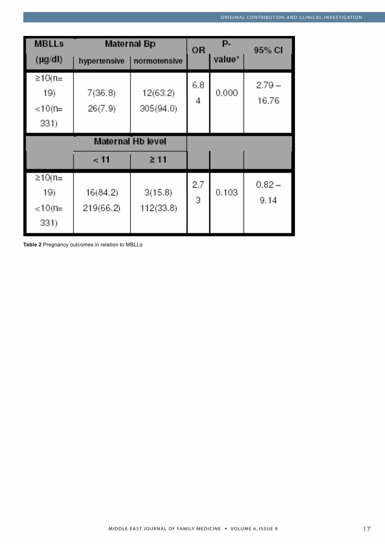
Table (2) clarifies the effect of MBLLs on the development of hypertension and anaemia among the studied women during the current pregnancy. Pregnant women with BLLs > 10 µg/dl are more at risk of developing hypertension than those with BLLs < 10 µg/dl (OR = 6.84, P = 0.000). Although there is an evident risk of anaemia development among women with high BLLs (OR=2.73), this risk does not reach a significant level.
Table (3) presents the distribution of neonatal characteristics among women with different blood lead categories. Overall, the mean birth weight was 3.058 ± 0.515 kg, and the mean head circumference was 35.12 ± 0.838 cm. Babies born to mothers with high BLLs (> 10 µg/dl) are more at risk of developing low birth weight and small head circumference than those born to mothers with low BLLs (OR = 43.54, 3.16 respectively), (P = 0.000, 0.013 respectively). While high BLLs are not considered a risk factor for developing low APGAR score.
DiscussionThis study was done to evaluate
the association between intrapartum risk factors for infection with CRP levels and showed that several such risk factors can cause elevated CRP levels in the absence of infection.
This is in agreement with previously published reports(7,13). Since CRP does not cross the placenta, the elevated levels are due to production of CRP in the neonate. Chorioamnionitis can result in elevation of IL 6 levels even in uninfected neonates(7). Stimuli other than infection, like hypoxia, trauma and metabolic changes can also induce production of proinflammatory mediators(7). Significant association is reported between birth asphyxia and elevated IL 6 levels. In prolonged labour, IL 6 levels rise in the neonate probably related to physical activity of labour. This cytokine stimulates CRP production.
There are few longitudinal studies examining CRP changes in healthy babies with intrapartum risk of infection. Cytokine elevation seen in the early neonatal period in such babies probably reflects physiological stress induced at birth(13). Since CRP levels rise during the initial 24 hours in many babies irrespective of infection or administration of antibiotics, serial determinations in this period may not be of much use in diagnosis but may help in identifying uninfected babies and restricting antibiotic use(14,15). Our data showed lower antibiotic use in babies who were CRP negative.
Various studies utilising varying protocols have suggested different values as upper limit of normal(8) In our study, at 24 h, CRP levels of 6mg/L had a negative predictive value of 99%. This level therefore could be used to guide antibiotic therapy when latex agglutination kits are used. Testing samples in further dilutions to establish the actual amount of CRP may not be necessary since increasing levels were not associated with increasing severity or prognosis.
Cord blood CRP levels estimated using a kit with 6 mg/L as detection limit, could not satisfactorily predict EOS. Recent studies show that cut off values may be different for cord and 24 hour samples(7). More sensitive techniques like nephelometry may help set cut off levels for cord blood. In comparison to leukocyte counts and ratios, CRP levels at 24 hours proved to be the single best indicator for diagnosing EOS. However, the 80% sensitivity obtained is unacceptably low for making critical decisions. If

MIDDLE EAST JOURNAL OF FAMILY MEDICINE • VOLUME 6 , ISSUE 9 ��
ORIGINAL CONTRIBUTION AND CLINICAL INVESTIGATION
utilised with caution, this test can help in reducing antimicrobial use in the new-born.
ConclusionIntrapartum risk factors for early onset sepsis can cause elevation of cord and neonatal CRP levels in the absence of infection. A CRP level of <6mg/L at 24 hour has a good negative predictive value for neonatal sepsis. Serial CRP levels are not useful in diagnosing early onset sepsis.
ReferencesThe WHO young infants study group, Bacterial eti-ology of serious infections in young infants in de-veloping countries: results of a multicentric study, Pediatr Infect Dis J 1999; 18: S17-22.Escobar GJ. Effect of systemic inflammatory re-sponse on biochemical markers of neonatal bacte-
1.
2.
rial infection: A fresh look at old confounders. Clini Chem 2003; 49: 21-22.Ng PC, Cheng SH, Chui KM, Fok TF, Wong MY, Wong W et al. Diagnosis of late onset neonatal sepsis with cytokines, adhesion molecule, and C-reactive protein in preterm very low birthweight in-fants. Arch Dis Child Fetal Neonatal Ed 1997; 77: F221-F227.Chan DK, Ho LY. Usefulness of C-reactive protein in the diagnosis of neonatal sepsis. Singapore Med J 1997; 38: 252-255.Magudumana MO, Ballot DE, Cooper PA, Trusler J, Cory BJ, Viljoen E et al. Serial interleukin 6 meas-urements in the early diagnosis of neonatal sepsis. J Trop Pediatr 2000; 46: 267-271.Dollner H, Vatten L, Linnebo I, Zanussi GF, Laerdal A, Austgulen R. Inflammatory mediators in umbili-cal plasma from neonates who develop early-onset sepsis. Biol Neonate 2001; 80: 41-47.Chiesa C, Pellegrini G, Panero A, Osborn JF, Si-gnore F, Assumma, et al. C-reactive protein, inter-leukin 6 and procalcitonin in immediate post natal period: influence of illness severity, risk status, an-tenatal and perinatal complications and infection. Clin Chem 2003; 49: 60 -68.Vesikari T. Cytokine determinations and rapid di-agnosis of early onset neonatal septicemia. Acta Pediatr 1999; 88: 585-591.Suri M, Thirupuram S, Sharma VK. Diagnostic and
3.
4.
5.
6.
7.
8.
9.
prognostic utility of C-reactive protein, alpha-1-anti-trypsin and alpha-2-macroglobulin in neonatal sep-sis: a comparative account. Indian Pediatr 1991; 28: 1159-1164.Krediet T, Gerards L, Fleer A, van Stekelenburg G. The predictive value of CRP and I/T-ratio in neona-tal infection. J Perinat Med 1992; 20: 479-485.Anwer SK, Mustafa S. Rapid identification of neo-natal sepsis. J Pak Med Assoc 2000; 50: 94-98.Santana C, Guindeo MC, Gonzalez G, Garcia-Mu-noz F, Saavedra P, Domenech E. Cord blood levels of cytokines as predictors of early neonatal sepsis. Acta Pediatr 2001; 90: 1176-1181.Chiesa C, Signore F, Assumma M, Buffone E, Tra-montozzi P, Osborn JF, et al. Serial measurements of C-reactive protein and interleukin-6 in the im-mediate postnatal period: reference intervals and analysis of maternal and perinatal confounders. Clin Chem 2001; 47: 1016-1022.Philip AG, Mills PC. Use of C-reactive protein in minimizing antibiotic exposure: experience with in-fants initially admitted to a well-baby nursery. Pedi-atrics 2000; 106: E4.Bomela HN, Ballot DE, Cory BJ, Cooper PA. Use of C-reactive protein to guide duration of empiric an-tibiotic therapy in suspected early neonatal sepsis. Pediatr Infect Dis J 2000;19: 531-535.
10.
11.
12.
13.
14.
15.
Figure 1 Graphic representation of the correlation between UBLLs and MBLLs

MIDDLE EAST JOURNAL OF FAMILY MEDICINE • VOLUME 6 , ISSUE 9��
ORIGINAL CONTRIBUTION AND CLINICAL INVESTIGATION
Table 1 Significant potential lead related maternal variables Maternal age Mean SD P-value
MIDDLE EAST JOURNAL OF FAMILY MEDICINE • VOLUME 6 , ISSUE 9 ��
ORIGINAL CONTRIBUTION AND CLINICAL INVESTIGATION
Table 2 Pregnancy outcomes in relation to MBLLs

MIDDLE EAST JOURNAL OF FAMILY MEDICINE • VOLUME 6 , ISSUE 9��
ORIGINAL CONTRIBUTION AND CLINICAL INVESTIGATION
Table 3 Neonatal characteristics in relation to MBLLs
MIDDLE EAST JOURNAL OF FAMILY MEDICINE • VOLUME 6 , ISSUE 9 ��
MEDICINE AND SOCIET Y
Changing Face of Measles in Kashmir, India
ABSTRACTMeasles is an acute infection caused by rubella virus, it is a highly contagious disease. It affects virtually everyone in in-fancy or childhood between the ages of 6 months to 3 years.Recovery from illness is the rule but seri-ous complications of respiratory and CNS may occur.1. The study aims to determine the clini-
cal profile of measles in adults.2. To determine the complications seen in
adults suffering from measles, and3. To determine the mortality of measles
in adults.
Results: During a recent outbreak of measles affecting higher age groups, 736 cases were studied: 327 cases were among children aged 1 to 5 years, 128 cases were among 5 to 10 years, 127 cases were among 10 to 15 years, 99 cases were observed in the age group of 15 to 20 years, 25 cases were in the age group of 20 to 25 and 30 cases in the age group of 25 and older. A pregnant woman aged 28 years, in her third trimester (33rd week of gestation) was also included in the study. 20% of the cases were ran-domly selected from different age groups for serological confirmation. Out of these 12 blood samples and throat swabs of different age groups were sent for sero-logical confirmation and genotyping to NIV Pune. 83 samples were reactive for IgG and IgM antibodies to measles virus in serum by ELISA, 64 cases were non reactive and D4 strain of measles virus was isolated.
Conclusions: The study highlights the need for early measles vaccination of infants at 9 months of age and a repeat dose (18-30 years) might be necessary at a higher age group to prevent adult mea-sles, as is being doneby some developed countries in the west.
IntroductionMeasles is caused by an RNA
paramyxovirus of which only one serotype is known. It contains a single strand of RNA, is highly pleomorphic, and ranges from 100 to 300 nm in diameter1.
It affects virtually everyone in infancy or childhood, between 6 months and 3 years in developing countries, while in the developed countries of the West; children between the ages of 3 to 6 years are infected. There is a current trend of measles occurrence in adult age groups, especially in the developed countries, but developing countries are not exempt (e.g. Thailand).
The virus can spread in any season. In temperate climates, measles is a winter disease, because of overcrowding. Epidemics are common in India in the months of winter and early spring.
Cases of measles are the major source of infection, while the subclinical cases are more numerous and are also a source of infection. Infective material is the secretions from the nose, throat and respiratory tract of a case of measles in the prodromal stage and at the time of eruption. Droplet nuclei can remain in the environment for 1 hour.
Communicability declines after appearance of a rash. The period of communicability is 4 days before and 5 days after the appearance of the rash. Isolation of the patient for a week from the onset of rash, more than covers the period of communicability.
Secondary attack rate is 80 percent among household contacts. The incubation period is 10 days. The incubation period with vaccine is shortened to 7 days.
Clinical features include a Prodromal phase (day 10-14), eruptive phase (day 14 -18) + post-measles stage.
Complications are many, especially in malnourished persons, those with poor immunity, and pregnant women. They include Measles associated diarrhoea, pneumonia, other respiratory complications, otitis media, Sub acute Sclerosing Pan Encephalitis (SSPE),
Encephalitis and Severe acute deficiency of vitamin A
Diagnosis is primarily clinical. IgM antibody titer by ELISA can also be carried out. Detection of measles antigen in saliva or urine, where resources permit, or where it is epidemiologically important should be carried out. In vaccinated children, a four-fold increase in measles antibody titre can be demonstrated within 2-6 weeks.
Special Concerns for women
Women who get measles during pregnancy may be at risk for a miscarriage, stillbirth or pre-term (early) delivery. If someone who is pregnant develops measles or has been exposed to a case of measles she should talk with her health care provider. Although in adults, the measles vaccine is usually given in combination with mumps and rubella vaccine (MMR), it cannot however
Kadri.S.M*, Parray S.H**, Rubina Shaheen**, Gaash BA**, Danish Muzaf-far*, Aesha.F*, Jan.Yasmeen***Authors affiliations:*Royal Tropical Institute (KIT), Amsterdam, The Netherlands** Department of epidiomology.Directorate of Health Services, Srinagar.,Kashmir India** Department of Anatomy, Govt.Medical College, Srinagar
Correspondence to:SM KadriRode Kruislaan 1418 C, 1111 XD, Diemen,Amsterdam, The Netherlandsgsm: +31630292620e mail: [email protected], [email protected]
MIDDLE EAST JOURNAL OF FAMILY MEDICINE • VOLUME 6 , ISSUE 9�0
MEDICINE AND SOCIET Y
be given to pregnant women. Thus, depending on the situation, the healthcare provider should administer immunoglobulin, or IG, as soon as possible after known exposure. Immune globulin can either prevent development of measles or result in a milder infection. Immune globulin (IG) is ideally administered within 6 days of exposure to decrease severity of measles. If someone is planning to get pregnant and is not sure if she has had measles or the measles vaccination, the healthcare provider may suggest an MMR vaccine. However, a woman should not get the vaccine if they plan to become pregnant within the next four weeks or may already be pregnant. There are no concerns if a man wishes to receive this vaccine and is planning to have a child. Breast-feeding does not interfere with the response to MMR vaccine and poses no risk to the infant being breastfed.
Materials and MethodsBackground:
Kashmir (the Switzerland of India), its natural beauty, the glorious climate and other attractions of the Kashmir valley have been so much appreciated that the new-comer is apt to picture themselves to be in a paradise on earth.
A team from the Department of Epidemiology, Directorate of Health Services surveyed all the areas and assessed the cases for the clinical manifestation of measles and randomly took blood samples from 147 cases i.e. (20% of cases) for serological confirmation and genotyping.
Findings:
During a recent outbreak of measles epidemic in J&K state, a total of 736 cases of measles occurred within a period of 3 months (March 2007-May 2007) Spring season. Figure 2.
The age breakup of measles cases in Kashmir is tabulated below:
The age breakup of cases shows that measles is shifting from its traditional age group (below 3 years), and affecting higher age groups. In India the majority of cases occur in children below 3 years of age, while we see here that although the maximum number of cases (44.42%) are still occurring
in children (0-5 years of age), a good percentage of cases are occurring in higher age groups (55.58%). Out of these, 20.88% are occurring in adults also (>18 years of age). Even cases above 40 years of age have occurred (30 cases). The severity of cases was mild in adults.
Vaccination status;
Immunization cards were assessed to find the measles vaccination status status. If cards were not available parents/family members were questioned in this regard. Table 3
Serology
A total of 147 blood samples were subjected to serological tests in our Regional laboratory. 83 cases were reactive and 64 cases were non reactive for IgG and IgM antibodies to measles virus in ELISA. 12 blood samples and throat swabs were sent to NIV Pune for genotyping. (D4 strain)
Discussion Measles is one of the most
infectious diseases known to man and remains the leading cause of vaccine preventable deaths in children worldwide. In many countries measles is still a great public health concern.1-
7 despite the easy availability of vaccination for its control, vaccination uptake has been sub -optimal in some countries8.
The diagnosis of measles is often based on the signs and symptoms (fever, malaise, conjunctivitis, coryza and tracheo-bronchitis). Kopliks spots appear on the buccal mucosa 1-2 days before the onset of rash. The rash is typically an erythematous maculopapular rash.4
The most definitive method of diagnosis of measles is either isolating the virus from the throat or by serologic test for antibodies.
Complications include diarrhea, pneumonia, subacute sclerosing pan encephalitis (SSPE), deafness, mental retardation or death.11,13
It has been postulated through various studies carried out around the world that there will be a three fold increase in measles mostly in the developing countries like Africa and
Asia.
Large measles outbreaks continue to occur, and these outbreaks frequently have high case-fatality rates resulting in many measles death.7
The WHO/UNICEF Global Measles strategic plan seeks to reduce measles mortality worldwide by 50%. The strategies recommended for reducing measles deaths include:
Providing a dose of measles vaccine to a very high proportion of infants at 9 months of age through routine immunization service, which will be the foundation of sustainable measles mortality reduction;Ensuring that all children have a second opportunity to receive measles vaccine providing measles immunity to those children who were previously vaccinated yet failed to develop measles immunity;Strengthening measles surveillance with integration of epidemiological and laboratory information andImproving the clinical management of measles cases.12
A study of the Centers for Disease Control and Prevention says that about 3 million Americans between the ages of 20 to 37 are at risk of catching measles. The disease is more dangerous to adults than children. The grown ups are at risk because most of them never got a second dose of measles vaccine.3,6,7
A reported measles outbreak in Victoria, Australia in 1999 has suggested that a substantial proportion of young adults (18-30 years old) may be susceptible to measles infection.5
NICD has pointed out low vaccine coverage and poor surveillance responsible for measles outbreaks in different parts of India.14
Eberhart-Phillips JE, Frederick PD, Baron RC, Mascola L.at Division of Field Epidemiology, Centers for Disease Control and Prevention, Atlanta, Georgia carried out research where 58 pregnant women with measles were identified by county health department records, and their hospital and clinic records were reviewed. It was concluded that the incidence of death and
1.
2.
3.
4.
MIDDLE EAST JOURNAL OF FAMILY MEDICINE • VOLUME 6 , ISSUE 9 ��
MEDICINE AND SOCIET Y
other complications from measles during pregnancy might be higher than expected for age-comparable, non-pregnant women. Measles in pregnancy may lead to high rates of fetal loss and prematurity, especially in the first 2 weeks after the onset of rash.
In this study, a total of 736 cases of measles occurred during the period of three months (March 007 to May 2007). All of these cases were diagnosed on the basis of clinical signs and symptoms like fever, maculopapular rash, cough, conjunctivitis and coryza.
Complications, such as pneumonia and diarrhea were encountered. The pregnant women who was included in the study delivered a healthy male.
However no death was noted in this study.
ConclusionIt is advisable that in any situation
where measles is found to occur in adults, measures are taken to rule out any adverse situations for the women, especially those who are,
or are planning to be pregnant. As there is now a changing pattern in age affliction of measles, i.e. it is shifting towards the adult age groups; it indicates that there is waning immunity in the immunized individuals, leaving aside the un-immunised, and under-immunised children. Also there is a large percentage of those who fail to develop immunity on standard immunisation procedure; i.e. one dose of measles vaccine during infancy at 9 months of age and a repeat dose (18-30 years) might be necessary at a higher age group to prevent adult measles, as is being followed by some developed countries in the west.
ReferencesCenters for disease control and pre-vention: Measles-USA, 2000. M.M.W.R.Morb.Mortal.Wkly.Rep.51 (6):120-123, 2002.Chalmers, I.: Why we need to know whether proph-alytic antibiotics can reduce measles-related mor-bidity, Pediatrics 109:312-315, 2002.Dales, L<Hammer, S.J, Smith, N.J Time trends in autism and in MMR immunization coverage in Cali-fornia. J.A.M.A. 285:1183-1185, 2001.Koplik, H: A new diagnostic sign of Measles.Med.Rec.53:505, 1898.Kelly HA, Riddell MA, Lambert SB Measles immu-nity among young adults in Victoria, Common Dis Intell 1999; 25(3): 129-132
1.
2.
3.
4.
5.
Miller M, Williams WW,Redd SC,Measlesa among adults United States,1985-1995.Am J Prev Med 1999,17(2);114-119.Hopkins RS, Jajosky RA, Hall PA, Adams DA, Connor FJ, Sharp P, et al. Summary of notifiable diseases-United States, 2003. MMWR Morb Mortal Wkly Rep. 2005;52:1-85.King a, Varughese P, De Serres G, Tipples GA, Waters J. Measles elimination in Canada. J In-fect Dis. 2004; 189(Suppl 1):S236-42. [PMID: 15106117].[MedlPapania MJ, Seward JF, Redd SB, Lievano F, Har-paz R, Wharton ME. Epidemiology of measles in the United States, 1997-2001. J Infect Dis. 2004; 189(Suppl 1):S61-8.Vukshich Oster N, Harpaz R, Redd SB, Papania MJ. International importation of measles virus-Unit-ed States, 1993-2001. J Infect Dis. 2004; 189(Sup-pl 1):S48-53.The State of the World’s Children 2006. New York: UNICEF; 2005.World Health Organization. WHO vaccine-prevent-able diseases: monitoring system: 2004 global sum-mary. Geneva: World Health Organization; 2004.Technical instructions to panel physicians for vac-cination requirements. Atlanta: Centers for Disease Control Division of Global Migration and Quaran-tine Health. Accessed at http://www.cdc.gov/nci-dod/dq/pdf/TI.pdf on 5 November 2005.NICD, CD Alert, Measles continuing to remain a major public problem in India, 2000, 4(5); 2-4.
6.
7.
8.
9.
10.
11.
12.
13.
14.
Table 1: Age break up of Measles cases.
DISTRICT 0-5yrs 5-10yrs 10-15yrs 15-20yrs 20-25yrs >25 yrs TotalANANTNAG 4 1 1 3 Nil Nil 09BARAMULLA 15 3 3 Nil Nil Nil 21BUDGAM 04 Nil Nil 01 Nil Nil 05KUPWARA 73 20 14 05 04 Nil 116LADAKH 97 79 94 78 18 30 396PULWAMA 85 13 04 05 Nil Nil 107SRINAGAR 49 12 11 07 03 Nil 82J&K State 327
44.42%12817.39%
127(17.25%)
99(13.45%)
253.39%
304.04%
736
Table (2) Showing gender distribution.
Males 199 78 93 58 06 12 446Females 128 50 34 41 19 18 290
Table 3 Showing vaccination status
District Vaccinated Unvaccinated Vaccine.StatusNot known
TOTAL
Anatnag 13 01 15 29Pulwama 12 03 82 97Budgam 14 01 68 83Srinagar 07 Nil 66 73Baramulla 04 01 54 59Kupwara 23 19 94 136Ladakh 78 36 145 259TOTAL 153 61 522 736

MIDDLE EAST JOURNAL OF FAMILY MEDICINE • VOLUME 6 , ISSUE 9��
MEDICINE AND SOCIET Y
Figure 1 Pie Chart showing percentage distribution of cases in various age groups
Figure 2 showing total no. of cases by Month.

MIDDLE EAST JOURNAL OF FAMILY MEDICINE • VOLUME 6 , ISSUE 9 ��
MEDICINE AND SOCIET Y
Utilization of Postnatal Care in Al-Hassa, Saudi Arabia
ABSTRACT Objectives: To assess utilization of postnatal care and to define the mag-nitude of postnatal maternal morbidi-ties in Al-Hassa, Saudi Arabia.
Methods: A descriptive study in-volved 1,996 mothers, representing 94.7% of mothers giving birth during a four months duration in the catch-ment areas of 15 Primary Health Care Centers (PHCCs) in urban, rural and hegar (deserts collection) localities. Trained Arabic-speaking nurses col-lected data from family files and ma-ternity cards kept at PHCCs as well as by an interview with mothers, two months after delivery, at infant vac-cination sessions.
Results: Less than half of the moth-ers received postnatal care, mostly (88.7%) at PHCCs. Logistic regres-sion analysis revealed that the in-dependent predictors of seeking postnatal care were the presence of maternal morbidities (OR=8.0), long pregnancy spacing (OR=1.9) and antenatal care at private clinic (OR= 1.3). No maternal mortality was re-corded in the chosen PHCCs during the study period. About 5% of moth-ers reported one or more postnatal morbidities. The most frequent post-natal morbidity was breast engorge-ment/abscess.
Conclusions: Postnatal care cover-age is low and is often considered as unnecessary. Therefore, there is an urgent need for an awareness-rais-ing program highlighting the impor-tance and availability of postnatal care. Antenatal care visits are good opportunities to counsel mothers about postnatal care.
Key words: postnatal care - postna-tal morbidities - Saudi Arabia
IntroductionThe postpartum period or
puerperium includes the first six weeks after delivery of the placenta [1-
3]. This period is a very special phase in the life of a woman. She is going through the physiological process of uterine involution and at the same time adapting to her new role in the family. Many postpartum complications occur during this period[1,2,4]. Among the important obstetric morbidities are postpartum hemorrhage, pregnancy-related hypertension, pulmonary embolism and puerperal sepsis. The common surgical complications are wound breakdown, breast abscess and urinary fecal incontinence. Medical conditions such as anemia, headache, backache, constipation and sexual problems may also be present[2,3].
Postpartum care is the most neglected aspect of maternity care and more research is needed on issues related to postpartum maternal health. Early postpartum care is essential to diagnose and treat complications[5]. Although there is little evidence to support the timing and content of postpartum visits, there is evidence that this is a time of increased health needs for both mother and baby[6]. Postpartum care visits provide opportunities to assess the physical and psychological well-being of the mother, counsel her on infant care and family planning and give appropriate referrals for pre-existing or developing chronic conditions[4,5,7].
Utilization of reproductive health services, including maternal health care, is related to their availability, and socioeconomic, demographic and cultural factors such as women’s age, education, employment and autonomy; and perception of women and their families regarding the need for care[8-10]. In contrast to relatively high to moderate levels of antenatal care received by women in the Arab region, the coverage of postnatal care is markedly lower[11]. In other regions of Saudi Arabia postnatal care coverage ranged from a very low rate of 6.5% in the Northern Region[12] (12) and up to 52% in Abha Region[13]. Postnatal care coverage ranged from 48-88% at the national level, according to different surveys[14,15].
This study aims to assess utilization of postnatal care and to define the magnitude of postnatal maternal morbidities in Al-Hassa, Saudi Arabia.
Population and MethodsThis study was carried out in Al-
Hassa, Saudi Arabia, during a period of four months from February 1 to the end of May 2007 G. Al-Hassa is located in the Eastern Zone of the Kingdom, bordering the Arabian Gulf. It is an agricultural area. The population is estimated to be about 800 thousands, distributed into urban, rural and hegar (Bedouin desert collections). Maternity care is provided through a network of 47 PHCCs, maternal and child hospitals, two small district hospitals in addition to the private sector and National Guard hospital and health centers.
Abdel-Hady El-Gilany (M.D., Public Health)(1)and Sabry Hammad (M.D., Public Health)(2)(1) College of Medicine in Al-Hassa, King Faisal University, Saudi Arabia(2) Ministry of Health, Saudi Arabia
Correspondence:Abdel-Hady El-GilanyProf. of Public Health, College of Medicine, Mansoura University, EgyptFamily and Community Medicine Department,College of Medicine in Al-Hassa, King Faisal UniversityP.O. Box: 400Hofuf 31982, Saudi Arabiae-mail: [email protected]: 00966/566482385
MIDDLE EAST JOURNAL OF FAMILY MEDICINE • VOLUME 6 , ISSUE 9��
MEDICINE AND SOCIET Y
After selection of PHCCs and nomination of nurses, the questionnaire was developed and pretested on 150 mothers (10 from each of the 15 PHCCs) to train nurses on data collection, reveal any difficulties and to estimate the percentage of mothers who received postnatal care. The pilot study revealed that it is necessary to review maternity cards and family files in addition to the interview with mothers.
The sample size was calculated using Epi Info 2004 software. From statistics of Al-Hassa Directorate of Health 15032 births were registered during 2006 G. With an expected postnatal care coverage of 51% from the pilot study, confidence interval of 95% and power of 80%, the number of mothers required was 1394. To overcome the attrition due to cluster sampling methodology, 30% were added. Thus, the final sample was 1812 mothers. This sample was distributed proportionally to urban, rural and Hegar localities.
Two-stage stratified sampling technique was used. The first stage is stratification of Al-Hassa into urban, rural and Hegar areas. At the second stage, five primary health care centers (PHCCs) were chosen from each stratum. These centers were chosen based on availability of an Arabic speaking nurse and their geographical distribution to represent different socioeconomic strata of the community.
Mothers were assured that data collected would be dealt with confidentially and the impact of the study would be respected, maintained and used only for research purposes and for improving services. Al-Hassa Directorate of Health approved the study and questionnaire and mothers gave verbal consent before the interview. There is no ethical research committee at the moment.
An Arabic speaking female nurse completed the questionnaires during interview with mothers two months after birth during a vaccination session in each center. Additional data was abstracted from both family file, maternity cards and hospital discharge forms kept in a family file at PHCCs. A maternity card has been
developed to include the appropriate data on prenatal, natal and postnatal care, as well as a list of factors of high-risk pregnancy for referral purposes and maternal morbidities. The Ministry of Health has developed special guidelines for using the card, explaining its contents and how to use it, as well as defining the various measurements and investigation and their normal limits. It is shared by the health centers and hospital. There is continuous stress on completeness of this card.
The outcome variable is receiving one or more postnatal check ups by a health care personnel, whatever its source.
Data were analyzed using SPSS (Statistical Package for Social Sciences) version 11. Descriptive statistics were done. The chi-squared test was used to find significance between categorical variables. Significant predictors of receiving postnatal care in univariate analysis were entered into multivariate stepwise forward Wald logistic regression analysis. Odds ratios and their 95% confidence intervals were presented. P=0.05 was considered statistically significant.
ResultsThe study covered 94.7% (1996 out
of 2108) of mothers who gave birth in the catchment areas of 15 primary health care centers during the study duration. No maternal mortality was recorded.
Table 1 reveals that more than 50% of mothers never received postnatal care and PHC centers were the most common source of care. There was no postnatal home visit. The main reasons for not seeking postnatal care were absence of health problems, busy husbands, the false idea that postnatal care is not necessary (Table 2).
Table 3 shows that mothers with long pregnancy spacing, mothers who received antenatal care at private clinics, and gave birth at private clinics and those with postnatal morbidities are more likely to receive postnatal care. Logistic regression analysis revealed that the independent predictors of seeking postnatal care
are presence of postnatal morbidities, prolonged pregnancy spacing and seeking care at private clinics (Table 4).
The commonest postpartum morbidities were breast engorgement/abscess (4.1) and infected episiotomy/ perineal tear (1.9%) (Table 5).
DiscussionCurrently, the primary health care
program of the Kingdom of Saudi Arabia has become well established, with the attainment of a very wide coverage. Maternal care is the responsibility of physicians assisted by the nurse and midwives[14]. Health care services provided by the governmental sector in Saudi Arabia account for over 80% of total services and are almost provided free of charge. Health care is also provided by other agencies e.g. National Guard, ARAMCO Petroleum Company and the private sector[15].
Like antenatal care, the postpartum care that typically occurs during the six weeks after childbirth is considered important to a new mother’ health. Unlike the tracking of prenatal visits, however, few statistics exists on postpartum health care utilization or postpartum health problems. Thus, postpartum care is a neglected aspect of women’s health care[16,17].
About half of mothers received postnatal care; most of them attended once and only 1.6% attended twice to the health facility; there were no home visits. Reasons cited for not seeking postnatal care were mainly absence of health problems, and no need for care, mother’s ignorance about the importance of postnatal care as well as busy husbands. This relatively high rate of postnatal check-up may be attributed to the policy followed in some PHCC that mothers should attend for check-up as a pre-requisite for registration of newborns. This is evident from the finding that most mothers received a postnatal check-up at the PHCC. Mothers attending postnatal care at hospitals and private clinics are most probably those with morbidities necessitating specialist consultation or hospitalization. Further evidence is that presence of postnatal morbidities is the strongest independent predictor of receiving

MIDDLE EAST JOURNAL OF FAMILY MEDICINE • VOLUME 6 , ISSUE 9 ����
MEDICINE AND SOCIET Y
postnatal care in general.
This coverage rate is much higher than the rate of 6.5% reported from the Northern Region of the Kingdom[12]. However, it is lower than the 52% coverage reported from Abha Region[13]. Review of the data from the Saudis National mothers and child health surveys of 1987 and 1990 showed that the percentage of mothers who received postnatal care increased from 58% to 88%[14]. Rates of postnatal care from other developing countries were reported to be 34% in Nepal[18], 33% in India and 84% in Zambia. In USA, 98% of mothers participated in a Pregnancy Risk Assessment Monitoring System received postnatal care[7].
It was reported that lack of awareness is the main barrier to the utilization of postnatal care. Women’s own occupation, number of pregnancies, children, and husbands’ socioeconomic status, occupation and education were significantly associated with the utilization of postnatal care[7,18]. In Saudi Arabia, there are a lot of problems concerning the understanding of postnatal care home visits:
the mother and her family do not realize the need for postnatal care,traditionally, because of fear of the evil eye, nobody is allowed to see the mother or her baby,many health staff are expatriate who do not understand the language or local customs, so they are not accepted at home level,problems regarding the need for somebody to accompany a female to home in addition to the driver,transport may not be available for home visits,male doctors find it difficult to examine female patients,delivery may take place at home, in other region or in a private clinic without communication with health centers,nursing staff may face isolated, unforeseen bad experiences at home level and (9) home visits are less likely and may not be welcomed for cultural reasons[21].
In Saudi Arabia, women are not
1.
2.
3.
4.
5.
6.
7.
8.
allowed to drive cars so husbands or other adult male family members need to accompany her to the health centre.
Maternal morbidity refers to complications that have arisen during pregnancy, delivery or the past-partum period[19]. Postpartum morbidities continue to be major health issues that need to be looked into critically, not only for curative but also for preventive and promotive care[22]. About 5.4 % of mothers reported one or more severe postnatal morbidities. A higher percentage of mothers with postnatal morbidities, up to 42.9%, was reported from other countries[23,24].
The commonest morbidities encountered were breast abscess/engorgement, infected episiotomy/perineal tear and urinary tract infection. In the Northern region urinary tract infection, puerperal sepsis and postpartum hemorrhage were the commonest postnatal morbidities(12). In the Saudi Arabia Family and Health, about 7% and 6% of respondents reported having severe bleeding and fever, respectively, after delivery[15]. In other countries, the main disorders for which postpartum women sought care were puerperal sepsis, secondary postpartum hemorrhage and postpartum eclampsia. The commonest cause of sepsis was infected episiotomies or tears[19,22-24].
In conclusion, postnatal care is deficient in coverage and number of visits. To improve postnatal care there is a need to establish adequate outreach services in catchment areas. PHC staff should routinely trace defaulting mothers through telephone communication or home visits. Importance of postnatal visits should be communicated to all women at the time of antenatal care and discharge from the hospital after delivery. The policy of obligatory postnatal check-up as a pre-requisite to register of newborns implemented in PHCC can be generalized to all health centers. A clear policy about quality, number, timing and contents of postnatal check-ups will contribute to increase postnatal care coverage. In-depth qualitative study is warranted to explore the socio-cultural and
behavioral factors impeding postnatal care.
ReferencesWHO. Postpartum care of mother and newborn : a practical guide. World Health Organization, Ge-neva. 1998Blenning CE, Pladine H. An Approach to the postpartum office visit. Am Fam Physician 2005;72(12):2443-4Zainur RZ, Loh KY. Postpartum morbidity-what we can do? Med J Malaysia 2006;61(5):651-6Liu N, Mao L, Sun X, Liu X, Chen B, Ding Q. Post-partum practices of puerperal women and their in-fluencing factors in three regions of Hubei, China. BMC Public Health 2006;6:274Ghai OP. Reproductive Health. In: Gupta P, Ghai OP (eds) Textbook of preventive and social medi-cine, 2nd edn. 2007 P.347Gunn J, Lumley J, Young D. Involvement of Victo-rian general practitioners in obstetric and postnatal care. Aust Fam Physician 1998;27(Suppl.2) S73-83CDC (Center for Disease Control). Postpartum care visits-11 States and New York City, 2004. MMWR 2007;56(50):1312-1316Addai I. Determinants of use of maternal and child health services in rural Ghana. J Biosoc Sci 2000;32:1-15Acharya LB, Cleland J. Maternal and child health services in rural Nepal: does access or quality mat-ter more. Health Policy Planning 2000;15(2):223-229International Institute for Population Sciences. Na-tional family survey, 1998-99. International Institute for Population Sciences, Mumbai, India. 2000Nawar L. Reproductive health and reproductive rights in the Arab Region. Background paper pre-sented to the Arab Population Forum. Beirut, No-vember 19-21, 2004El-Gilany AH, Al-Hozem N, Aref Y. Maternity care in primary health care centers, Northern Re-gion, Saudi Arabia. Saudi J Obstet Gynecol 2002; 2(3):99-112Abu-Zeid HA. Health services utilization patterns of two urban communities in Abha, Saudi Arabia. J Community Health 1989;14(2):65-77Baldo MH. Maternal health care. In: Quality as-surance in primary health care manual, Ministry of Health, Saudi Arabia. EMRO-WHO. 1st edn. 1994Khoja TA, Farid SM. (eds). Saudi Family health Sur-vey 1996, Ministry of Health, Saudi Arabia. Prelimi-nary Report. 1997Albers LL. Health Problems after childbirth. J Mid-wifery and Women’s Health 2000;45:55-57Cheng C, Fowles ER, Walker LO. Postpartum ma-ternal health care in the United States: A critical re-view. J Perinatal Educ 2006;15(3):34-42Dhakal S, Champan GN, Simkhada PP, VanTeijlin-gen ER, Stephens J, Raja AE. Utilization of postna-tal care among rural women in Nepal. BMC Preg-nancy and Childbirth 2007;7:19Vallely L, Ahmed Y, Murray S. Postpartum maternal morbidity requiring hospital admission in Lusaka, Zambia - a descriptive study. BMC Pregnancy and Childbirth 2005;5:1-8Yesudian PP. Impact of women’s empowerment, autonomy and attitude on maternal health care uti-lization in India. International Institute for Popula-tion Sciences. Global Forum for Health Research. Forum 8. Mixico City. 2004Ministry of Health, Saudi Arabia. Maternal and child health services manual for health centers physi-cians. 2nd edn. El-Hilal Press, Riyadh. 1997:25-99Shakuntala C, Varma SP, Ritambara B. Qual-ity of postpartum care. J Obstet Gynaecol India 2006;56:142-145Bang RA, Bang AT, Reddy MH, Deshmuskh MD, Baitule SB, Fillippi V. Maternal morbidity during labor and the puerperium in rural homes and the need for medical attention: a prospective observa-tional study in Gadchiroli, India. Br J Obstet Gynae-col 2004;111:231-238
1.
2.
3.
4.
5.
6.
7.
8.
9.
10.
11.
12.
13.
14.
15.
16.
17.
18.
19.
20.
21.
22.
23.

MIDDLE EAST JOURNAL OF FAMILY MEDICINE • VOLUME 6 , ISSUE 9�� ��
MEDICINE AND SOCIET Y
Brace V, Penney G, Hall M. Quantifying severe ma-ternal morbidity: a Scottish population study. Br J Obstet Gynaecol 2004;111(5):481-484
24.
Table 1. Coverage and facility providing postnatal care
Number (%)Postnatal care:NoneOne visitTwo visits
1001(50.2)964(48.3)31(1.6)
Facility providing postnatal care†:Primary health care centersPrivate clinicsGovernmental hospitalsShared care‡
883(88.7)86(8.6)14(1.4)12(1.2)
†Among 995 mothers seeking postnatal care.‡shared care between PHCC, hospital and/or private clinics.
Table 2. Reasons for not seeking postnatal care
Reason Number (%)†
No health problemsFalse idea that postnatal care is not necessaryNo female physicians at PHCCsToo busy mother or husbandsOvercrowded clinics/long waiting timeOthers (not at usual residence, unsuitable working hours, PHCC is far away).
511(51.0)316(31.6)217(21.7)179(17.9)102(10.2)38(3.8)
†Among 1001 mothers not seeking postnatal care and categories are not mutually exclusive.Table 3. Univariate analysis of predictors of seeking postnatal care.
Predictors TotalN (%)
Postnatal careN (%)
OR(95%CI)
Total 1996(100) 995(49.8)Residence:UrbanRuralHegar
971(48.6)799(40.0) 226(11.3)
488(50.3)408(51.1)99(43.8)
1(r)1.0(0.9-1.3)0.8(0.6-1.0)
Family income:SatisfactoryUnsatisfactory
1589(79.6) 407(20.4)
790(49.7)205(50.4)
1(r)1.0(0.8-1.3)
Distance from PHCC:Up to 1 km> 1 Km
1142(57.2) 854(29.3)
568(49.7)427(50.0)
1(r)1.0(0.8-1.2)
Family size:5 or less> 5
1157(58.0)839(42.0)
556(48.1)439(52.3)
1(r)1.2(0.99-1.4)
Maternal education:Illiterate/PrimaryPreparatorySecondaryAbove secondary
482(24.1)267(13.4)756(37.9)491(24.6)
249(51.7)136(50.9)363(48.0)247(50.3)
1(r)1.0(0.7-1.3)0.9(0.7-1.1)1.0(0.7-1.2)
Maternal work:House wifeWork outside homeStudent
1669(83.6)206(10.3)121(6.1)
836(50.1)108(52.4)51(42.1)
1(r)1.1(0.8-1.5)0.7(0.5-1.1)
Husband’s education:Illiterate/PrimaryPreparatorySecondaryAbove secondary
406(20.3)429(21.5)696(34.9)465(23.3)
198(48.8)232(54.1)327(47.0)238(51.2)
1(r)1.2(0.9-1.6)0.9(0.7-1.2)1.1(0.8-1.5)
Husband’s work:Professional/semiprofPolice/militaryTrades/businessOthers
653(32.7)387(19.4)411(20.6)545(27.3)
330(50.5)189(48.8)195(47.4)281(51.6)
1(r)0.9(0.7-1.2)0.9(07-1.1)1.0(0.8-1.3)
Maternal age:<20 years20-35 years35 & more
96(4.8) 1561(78.2)339(17.0)
41(42.7)770(49.3)184(54.3)
1(r)1.3(0.8-2.0)1.6(1.0-2.9)
Gravidity:Primigravida2 & 34 & more
481(24.1)664(33.3)851(42.6)
225(46.8)315(47.4)455(53.5)
1(r)1.0(0.8-1.3)1.3(1.0-1.7)
Spacing†:<1 year1-3 years> 3 years
409(20.5)784(39.3)322(16.1)
179(43.8)383(48.9)208(64.3)
1(r)1.2(1.0-1.6)2.3(1.7-3.2)***
Antenatal care attend-ance:NoYes
46(2.3)1950(97.7)
27(58.7)968(49.6)
1(r)0.7(0.4-1.3)
Antenatal care at private clinic##:NoYes
1328(68.1)622(31.9)
613(46.2)355(57.1)
1(r) 1.6(1.3-1.9)***
Birth at private clinic:NoYes
1685(84.4)311(15.6)
827(49.1)168(54.0)
1(r)1.2(0.95-1.6)
Postnatal morbidity:NoYes
1889(94.6)107(5.4)
900(47.6)95(88.8)
1(r)8.7(4.6-16.8)***
†Primigravidae were excluded, ## Non-attenders for antenatal care were excluded.*** P≤0.001
Table 4. Multivariate logistic regression analysis of significant predic-tors of seeking postnatal care.
Predictor No postnatal careβ P OR(95% CI)
Spacing*:<1 year1-3 years > 3 years
-0.10.6
0.40.000
1(r)0.9(0.7-1.1)1.9(1.4-2.4)
Antenatal care at private clinic:NoYes
-0.3 0.004
1(r)1.3(1.1-1.6)
Postnatal morbid-ity:NoYes
-2.1 0.000
1(r) 8.0(4.4-14.8)
ConstantPercent correctly predicatedModel χ²
-0.357.6118.4,P=0.000
OR= Odds ratio, CI= Confidence Interval, r= reference group
Table 5. Postnatal morbidities reported
Number (%)Any morbidityBreast abscess/engorgementUrinary tract infectionInfected episiotomy/perineal tearPostpartum hemorrhagePostpartum depressionPostpartum fitsOthers†
107(5.4) 68(3.4)41(2.1) 31(1.6)23(1.2) 2(0.1) 1(0.05)36(1.8)
†e.g. piles, varicose veins, pneumonia, umbilical hernia, gastro-en-teritis and severe constipation
MIDDLE EAST JOURNAL OF FAMILY MEDICINE • VOLUME 6 , ISSUE 9 ��
MEDICINE AND SOCIET Y
Parental Consanguinity and Idiopathic Dilated Cardiomyopathy in Children
ABSTRACTBackground: Consanguineous mar-riage is a type of family system in Islamic countries. This type of mar-riage may by a risk factor for heart disease, like dilated cardiomyopathy (DCM).
Objective: The aim of the present study was to determine the incidence of parental consanguinity and its re-lationship with familial type of DCM in a group of children with this disease.
Design: We conducted a retrospec-tive review of medical records on patients with DCM who were hospi-talized in a pediatric ward of a uni-versity hospital in Tehran during a 10 year (1997 - 2007) period. Forty-eight patients with a definite diagnosis of DCM were included in the study. The control group comprised 56 healthy age and sex matched children.
Results: Of the 48 patients 23 were male and 25 were female with an age range from 3 months to 15 years. Sixteen patients and 7 controls had consanguineous parents and the dif-ference was significant (33.3% ver-sus 12.5%, P = 0.01). The familial occurrence of DCM in patients was 16.7%. Thirteen patients (27%) died. There was no significant relationship between mortality rate in familial and non familial cases (P = 0.8) and be-tween parental consanguinity and fa-milial DCM (P = 0.05).
Conclusion: We found that the pa-rental consanguinity in our patients was significantly higher than that in controls and there was no significant relationship between parental con-sanguinity and the familial form of DCM.
Key words: Dilated cardiomyopa-thy, familial cardiomyopathy, parental consanguinity.
IntroductionIdiopathic dilated cardiomyopathy
(DCM) is a disease of the cardiac muscle that is characterized by the presence of ventricular dilatation, systolic and diastolic dysfunction, symptoms of congestive heart failure (CHF), and premature death due to heart failure or arrhythmias(1). As much as 30% of patients have a relative with DCM or LV dilatation which may be an early stage of the disease(1).
There is increasing awareness of the familial nature of DCM(2). Familial DCM (FDCM) was defined as the presence of at least one relative with idiopathic DCM(1). The majority of familial and non-familial cases are of unknown cause(3). Familial analyses are increasingly showing that DCM has a genetic or inherited basis, but, in the pediatric patients, the incidence and proportion of cases with a familial cause have not been defined and many cases are considered idiopathic. Autosomal dominant inheritances have been most frequently described in the familial forms of DCM, but such kindred affected individuals are rarely identified before the age of 10 years. Autosomal recessive inheritance of DCM has been infrequently described, perhaps because a familial association is more difficult to document(4). Familial DCM can be categorized by inheritance pattern and clinical phenotype. Nearly two thirds of familial DCM involves autosomal-dominant inheritance. Penetrability is incomplete and age-related. autosomal recessive DCM
seems to be less frequent and may be characterized by a significantly younger age of onset and a worse prognosis compared with the dominant form(5). The age of onset and the clinical course is highly variable in the same family(3). Patients with hypertrophic cardiomyopathy are identified in the family study of patients with DCM. This is not strange considering that mutations in the same gene can produce both diseases(1).
The existence of consanguinity suggests the possibility of an autosomal dominant or autosomal recessive inheritance(1). In children of consanguineous marriage it is reasonable to assume that the disease gene is contained in a genetic locus that is homozygous by descent(4). First-cousin marriage may be a significant risk factor for specific types of congenital heart disease in a consanguineous population. Thus in a population with a high degree of inbreeding, consanguinity may exacerbate underling genetic risk factors, particularly in the offspring of first cousins. There may be a recessive component in the causation of some cardiac defects(6). An investigation in the United Arab Emirates showed that consanguinity did not result in reproductive wastage, but was found to be an important factor in the causation of specific illnesses in offspring(7). In a study from Qatar, all cardiomyopathy patients below 50 years of age who were citizens or permanent residents in Qatar were included in the study.
Shahla Roodpeyma, MDHootan Salemi, MDDepartment of Pediatrics, Taleghani Medical Center, Shahid Beheshti University (M.C), Tehran, Iran
Correspondence:Dr Shahla Roodpeyma,Pediatric ward, Taleghani hospital,Evin, Tehran 1985717413, Iran.Tel: 0098 - 21 - 22432591Fax: 0098 - 21 - 22432582E-mail: [email protected]

MIDDLE EAST JOURNAL OF FAMILY MEDICINE • VOLUME 6 , ISSUE 9��
DCM was most prevalent (75.8%) in all age groups, and the incidence increased remarkably with age. The consanguinity rate was high among Qatari patients(8).
The purpose of this study was to review all patients with DCM who presented to the pediatric ward of a university hospital in the previous 10 years. The primary aims of this research were to study the frequency of parental consanguinity in our patients, and to study the prevalence of familial DCM and its effect on outcomes of our cases.
Patients and MethodsThe medical records of patients
with discharge diagnosis of cardiomyopathy who were hospitalized in the pediatric ward of Taleghani medical center, Tehran, during 1997 - 2007 were retrospectively reviewed. Cardiomyopathy was defined as a heart muscle disease of unknown cause. Dilated cardiomyopathy was diagnosed on the basis of advanced heart failure, where cardiac dilatation and impaired contractility were recognized in the absence of a known etiology. Secondary cardiomyopathies such as storage disease, Adriamycin toxicity, and transient cardiac dysfunction such as acute myocarditis were excluded. During the study period 52 patients were admitted with a diagnosis of idiopathic cardiomyopathy. There were 4 patients with hypertrophic cardiomyopathy who were excluded. Only 48 patients with DCM were included in the study. The medical histories of the patients were reviewed to determine age, gender, symptoms and signs at presentation. The consanguinity of parents, the history of the same cardiac disease in other family members of patients, or the cause of death in other family members was carefully evaluated. Familial DCM was defined as the history of at least one alive or deceased family member with the same clinical presentation or diagnosis as DCM. Diagnostic tests of patients consisted of chest X-ray examination, 12 leads electrocardiography, and two-dimensional Doppler echocardiography. Cardiac catheterization, endomyocardial
biopsy, and autopsy had not been performed on any patients. Heart transplantation was not available to any patient. The control group consisted of 56 healthy, sex and age matched children who came for a regular visit to the outpatient clinic. All the cases in the control group had normal physical examination without any evidence of cardiac disease. The consanguinity of parents and the possible cause of disease or death in other family members were also carefully questioned in the control group.
The data were presented as a mean ± SD. t test analysis and were used to compare the groups. A p value of < 0.05 was considered statistically significant.
ResultsThere were 23 (47.9%) affected
males and 25 (52.1%) affected females. The mean age of patients was 5.7 ± 4.5 years (range 3 months to 15 years). Age distribution of patients was as follows: less than 1 year old - 8 (16.7%) cases, 1-5 years old - 16 (33.3%) cases, 6-10 years old - 11(22.9%) cases, and 11-15 years old - 13(27.1%) cases. The control group consisted of 27 (48.2%) males and 29 (51.8%) females. The mean age of the control group was 5.2 ± 4.3 years (range 2 months to 14.5 years). All the patients at presentation had congestive heart failure and 9 were in cardiogenic shock. Chest X-ray showed cardiomegaly in all patients. The ECG showed low voltage QRS complex (14 patients), left ventricular hypertrophy (12 patients), normal pattern (12 patients), nonspecific ST, T changes (6 patients), and a variety of different cardiac rhythm disturbances were observed in the remainder 4 patients. Echocardiographic examination revealed low ejection fraction (18 to 30 percent) and low shortening fraction (10 to 20 percent) in the majority of patients. Consanguinity between parents was detected in 16 (33.3%) patients. In 12 patients, parents were first degree cousins, and in 4, parents were second degree cousins. All parents were in a good state of health without any evidence of heart disease. Parental consanguineous marriage
was noted in 7 of 56 children (12.5%) in the control group. Consanguinity between parents in cases, was significantly higher than that in controls (16 of 48 (33.3%) versus 7 of 56 (12.5%), P = 0.01). DCM had not been reported in any live family member of both patients and control groups. Eight patients had siblings who died of DCM, so the familial occurrence of DCM in our patients was 16.7%. Three patients had one dead sibling, 3 patients had 2 dead siblings, 1 patient had 3 dead siblings, and 1 patient had 4 dead siblings. In total 16 affected siblings of 8 patients with familial DCM died. the control group had no sibling death. Parental consanguinity plus sibling death was noted in 5 patients. Thirteen patients died, 2 cases of familial DCM and 11 cases of non familial DCM. We did not find a significant difference between mortality rate in familial and non familial cases (2 of 8 (25%) versus 11 of 40 (27.5%), P = 0.8). The overall mortality rate was 27%. Table 1 summarizes the results of the relationship between parental consanguinity and familial DCM, death of patients, and death of their siblings. As it is shown in this table, we did not find significant correlation between parental consanguinity and familial DCM (P = 0.05), parental consanguinity and patients’ death (P = 0.25), parental consanguinity and siblings’ death (P = 0.05).
DiscussionConsanguineous marriage is a
major feature of family systems in Islamic Asian countries. A previous study from Iran reported that in a total sample of 306,343 couples with different ethnic/religious backgrounds the overall rate of consanguineous marriage was 38.6%. First cousin marriages (27.9%) were the most common form of consanguineous union(9). In the present study parental consanguinity among patients with DCM was significantly higher than that in the control group (33.6% versus 12.5%, P = 0.01). The lower rate of parental consanguinity in the control group may be due to a selection of age and sex matched healthy children. Data showed that the frequency of familial DCM in our patients was 16.7%. In a report
MEDICINE AND SOCIET Y

MIDDLE EAST JOURNAL OF FAMILY MEDICINE • VOLUME 6 , ISSUE 9 ��
MEDICINE AND SOCIET Y
from the Sultanate of Oman 770890 (87%) first - degree relatives of 108 families of hospitalized patients with idiopathic DCM were screened. Thirty percent of the patients were born to consanguineous parents. Familial DCM was found in 7 (6.5%) of families, which is lower than the earlier published figures of 20-25%. Patients with idiopathic DCM were younger at presentation and were more often associated with parental consanguinity but the survival rates of familial patients did not differ significantly. Despite the high prevalence of consanguinity, there was a low proportion of familial DCM in the study population(10). In another study from an eastern province of Saudi Arabia 55 consecutive cases of DCM in patients < 10 yr of age were seen during a 5 year interval. Echocardiography was the primary diagnostic modality. The 55 cases represented 20% of the offspring of 41 families. In 19 families (46%) of parents were first cousins. There was no obvious consanguinity in 22 families (54%)(4). In an article by Bilgic et al from Turkey, the clinical and epidemiological characteristics of 137 children with cardiomyopathy were studied. Consanguinity between their parents was more common than the proportion for Turkey as a whole. Most of the patients had DCM (78.9%) and 10.3% died(11). The onset of DCM in relatives occurred at ages ranging from 9 to 75 years, so there does not appear to be a safe age at which screening can be discontinued. Familial DCM has been considered to have a poorer prognosis than non familial DCM. It was also found that the mean age at diagnosis in the familial DCM group was significantly younger than in the non-familial group and its progression was more rapid than in non-familial cases. The possibility cannot be excluded that subsets of familial DCM patients with younger age at onset or patients with x-linked or autosomal recessive inheritance have more severe disease with a worse prognosis(5). A report by Montserrat et al showed that the prevalence of familial DCM is high in patients who undergo heart transplantation(1). In a report by Michles from USA no significant difference was reported in the 5 year survival,
or time to heart transplantation, in an unselected series of 30 patients with familial DCM compared with 71 patients with non-familial DCM(3). In research from the Mayo clinic 315 relatives of 59 patients with DCM were screened. Twelve of the 59 index patients (20.3%) or at least one in five of the patients had familial disease. There was no difference in age, sex, severity of disease, exposure to selected environmental factors, or electrocardiographic or echocardiographic features between the index patients with familial disease and those with non-familial disease(12).
Patients with DCM commonly have an affected family member and a high proportion of apparently healthy relatives with minor echocardiographic abnormalities(13). Counseling of family members should emphasize the heritable nature of the disease, the age-dependent penetrability and the unpredictable clinical course(14). Careful family history with review of medical records identifies more familial cases than merely asking the patients if there is a family history of DCM. However, even such a careful family history does not identify all familial cases. Some familial cases are identified only by echocardiographic investigation of asymptomatic relatives(15).
Based on the careful family history the frequency of familial DCM in our data was 16.7%. We did not find a higher mortality rate in patients with familial DCM. The incidence of familial DCM increased in patients with consanguineous parents more than that in patients with non-consanguineous parents (62.5% versus 27.5%) but the difference was not significant (P = 0.05). The death of multiple offspring was higher in consanguineous parents than that in non-consanguineous couples but the difference was not significant (P = 0.05). Our report is a retrospective study reflecting the clinical experience of a single pediatrics ward with all the inherent deficiencies of such a study. We were not able to interviewparents or apparently unaffected offspring or other relatives including cousins, aunts, or uncles. The follow-up on individuals who recovered or stabilized
was insufficient and we do not know if these individuals who were critically ill, recovered but will have similar episodes later in life. We were not able to obtain tissue diagnosis on any of our patients (no endomyocardial biopsy or autopsy). However this study provides an opportunity to learn more about DCM and parental consanguinity that may be difficult to recognize in other patient groups in which consanguineous marriage is uncommon and the number of children per family is small.
In conclusion our results indicated that parental consanguinity is significantly higher in patients with DCM. We did not find significant correlation between parental consanguinity and the familial form of DCM, and the mortality from the familial form was not higher than that of non-familial DCM.
ReferencesMonserrat L, Hermida M, Bouzas B, Mosquera l, Mahan N, Peteiro J, et al. Familial dilated cardiomy-opathy in patients transplanted for idiopathic dilated cardiomyopathy. Rev Esp Cardiol 2002; 55(7): 725-732.Bilinska ZT, Michalak E, Piatosa B, Grzybowski J, Skwarek M, Deptuch TW, et al. Familial dilated cardiomyopathy: evidence for clinical and immuno-genetic heterogeneity. Med Sci Monit 2003: 9(5): 67-74.Michels VV, Driscoll DJ, Miller FA, Olson TM, Atkin-son EJ, Olswold CL, Schaid DJ. Progression of fa-milial and non-familial dilated cardiomyopathy: long term follow up. Heart 2003; 89(7): 757-761.Seliem MA, Mansara KB, Palileo M, Ye X, Zhang Z, Benson DW. Evidence for autosomal recessive inheritance of infantile dilated cardiomyopathy: studies from the eastern province of Saudi Arabia. Pediatr Res 2000; 48(6): 770-5.Khositseth A, Michels VV. Familial dilated cardio-myopathy. Cardiovasc Rev Rep 2004; 25(5): 200-202.Becker SM, Al Halees Z, Molina C, Paterson RM. Consanguinity and congenital heart disease in Saudi Arabia. Am J Med Genet 2001; 99(1):8-13.Abdulrazzaq YM, Bener A, al-Gazali LI, al-Khayat AI, Micallef R, Gaber T. A study of possible delete-rious effects of consanguinity. Clin Genet 1997; 51(3): 167-73.EL-Menyar AA, Bener A, Numan MT, Morcos S, Taha RY, AL- Suwaidi J. Epidemiology of idiopathic cardiomyopathy in Qatar during 1996-2003. Med Princ Pract 2006; 15(1): 56-61.Saadat M, Ansari-Lari M, Farhud DD. Consan-guineous marriage in Iran. Ann Hum Biol 2004, 31(2):263-9.Venugopalan P, Agarwal AK, de Bono D. Low proportion of familial dilated cardiomyopathy in an Arab population with a high prevalence of consan-guineous marriage. Acta Paediatr. 2001; 90(11): 1267-70.Bilgic A, Ozbarlas N, Ozkutlu S Ozer S, Ozme S. Cardiomyopathies in children: clinical, epide-miological and prognostic evaluation. Jpn Heart J 1990; 31(6): 789-97.Michels VV, Moll PP, Mellor FA. Tajik AJ, Chu JS, Driscoll DJ, et al. The frequency of familial dilated cardiomyopathy in a series of patients with idio-pathic dilated cardiomyopathy. N Engl J Med 1992; 326(2):77-82.Keeling PJ, Gang Y, Smith G, Seo H, Bent SE, Mur-
1.
2.
3.
4.
5.
6.
7.
8.
9.
10.
11.
12.
13.

MIDDLE EAST JOURNAL OF FAMILY MEDICINE • VOLUME 6 , ISSUE 9�0
MEDICINE AND SOCIET Y
day V, et al. Familial dilated cardiomyopathy in the United Kingdom. Br Heart J 1995; 173(5): 417-21.Crispell KA, Wray A, Ni H, Nauman DJ, Hershberg-er RE. Clinical profiles of four large pedigrees with familial dilated cardiomyopathy: preliminary recom-mendations for clinical practice. J Am Coll Cardiol
14.
1999; 34(3): 837-47.Goerss JB, Michels VV, Burett J, Driscoll DJ, Miller F, Rodeheffer R, et al. Frequency of familial cardio-myopathy. Eur Heart J 1995; 16 Suppl O: 2-4.
15.
Table 1: The relationship between Parental consanguinity and familial DCM, patients’ and siblings’ death
Parental Consanguinity Number (%) P Valve With familial DCM 5 of 8 (62.5)
0.05With non-familial DCM 11 of 40 (27.5)In dead patients 6 of 13 (46.2)
0.25In alive patients 10 of 35 (28.6)In cases with siblings’ death 5 of 8 (62.5)
0.05In cases without siblings’ death 11 of 40 (27.5)

MIDDLE EAST JOURNAL OF FAMILY MEDICINE • VOLUME 6 , ISSUE 9 ��
MEDICINE AND SOCIET Y
Stress Among Medical and Law Students in Mansoura, Egypt
ABSTRACTObjective: This study aims to deter-mine if there is a difference in the perceived stress levels of medical and law students in Mansoura Uni-versity, Egypt.
Method: This is a comparative study between medical and law students, conducted during October and De-cember 2007 G. The sample consist-ed of 283 medical and 277 law stu-dents, selected through the cluster sampling method. The self-reported questionnaire covered four catego-ries including 15 items of sources of stress. Perceived stress scale and Hospital anxiety and depression scale were used to measure stress, anxiety and depression.
Results: Law students were more likely to cite personal, environmental and relationship categories as stres-sors. The top five items of stressors in medical students identified in this study were troubles with the instruc-tors, excessive workload, problems with coursemates, close contact with serious illness and personal injury or illness. The high level of perceived stress was reported by 20.1% and 41.2% of medical and law students, respectively. The other independent predictors of stress among students were anxiety, depression and number of stressors.
Conclusions: Stressors and stress are frequent among medical and law students. Counseling and preventive mental health services should be an integral part of the routine clinical fa-cilities caring for university students.
Key word: Medical students - Law students - perceived stress - Anxiety - Depression
IntroductionThe topic of stress among college
or university students has been the subject of much research for many years. Researchers have found that the perception of high stress levels in students can lead to poor academic performance, depression, attrition and serious health problems[1-3].
Medical school can be a very stressful environment for students. A variety of sources of stress have been identified. These include, among others, the academic pressures, social pressures and financial problems[4]. Other studies suggested that high levels of stress and psychiatric morbidity are not limited to medical students but occur also among law students[5].
Many students experienced stress as they pursue a law degree[6]. Archer and Peter[7] suggested that much of this excessive stress may have its origins in law school. The Socratic method of teaching, high expectations, competitive environment, heavy work load, final examinations, social isolation, and family tension have been identified as stressors, though they are not necessarily ranked in that order. The effect of profession on stress in students varies. While some studies suggested that medical students
have a higher level of stress than their law counterparts[5], other studies do not reveal such differences[8]. There are also suggestions that profession may influence the way medical students perceive stress[9]. To the best of authors’ knowledge no similar comparative studies were done in Egypt, where the situation appears to be different.
The objectives of the study were to
assess the level of perceived stress of medical and law undergraduatesidentify common sources of stress in their livesassess the psychiatric disorders such as anxiety and depressionobserve any association between the high level of stress and sociodemographic profile.
METHODS
Participants
This is a comparative study between medical and law students in Mansoura University, Egypt, conducted during October and December 2007 G.. Filled questionnaires were collected one month before the first term examination period so as to minimise the extra stress symptoms. The undergraduate medical course is six years with the fourth to sixth years consisting of clinical training
1.
2.
3.
4.
Abdel-Hady El-Gilany (MD)Professor of Public Health,Department of Community Medicine, College of Medicine, Mansoura University, EgyptMostafa Amr (MD)Lecturer of Psychiatry, Department of Psychiatry, College of Medicine, Mansoura University, EgyptNabil Awadalla (MD)Lecturer of Public Health, Department of Community Medicine, College of Medicine, Mansoura University, EgyptGhada El-Khawaga (MD)Lecturer of Occupational Medicine, Department of Community Medicine, College of Medicine, Mansoura University, Egypt
Correspondence:Mostafa AmrLecturer of Psychiatry, Departement of Psychiatry,College of Medicine, Mansoura University,Mansoura 35516, EgyptE-mail: [email protected]

MIDDLE EAST JOURNAL OF FAMILY MEDICINE • VOLUME 6 , ISSUE 9��
MEDICINE AND SOCIET Y
along with didactic courses, while the law course is didactic courses of four years. Written approval was obtained from the authorities of both colleges to conduct the survey in the setting. There is no research ethical committee at the moment in our university. After obtaining this approval for data collection the researchers introduced themselves to the students in each grade and informed them about the aim of the study and about guarantees of anonymity and confidentiality and the need for verbal consent. The students were allowed to respond in their own time and privacy. The participation was entirely on a voluntary basis. All students who agreed to participate were given questionnaires; all incomplete questionnaires were excluded. A total of 620 (283 medical, 277 law) students who provided complete questionnaires were involved in the study, representing 4.1% and 2.9% of the enrolled students in both colleges, respectively.
Study tools
Participants were given a packet with three Arabic instruments used and validated in previous studies [10-12]. The first instrument enquires about sociodemographic and sources of stress during the past year. The second is Perceived stress levels, which were measured by Cohen’s Perceived Stress Scale. The third instument is the hospital anxiety and depression scale.
Sources of stress include 15 items divided into 4 categories of potential sources of stress:
3 items representing relationship sources of stress, 5 representing personal sources of stress, 5 representing academic sources of stress, and 2 representing environmental sources of stress.
In respect to the research question what are the greatest stressors in the last year”. The time frame of 12 months was deliberately chosen based on the assumption that a 12-month period is long enough to obtain a reasonable estimate of variation in exposure to recent life events, and short enough to avoid inaccurate recall and perception of the events[13]. Students were asked to list the four categories of stress
viz, relationship, personal, academic, and environmental situations or conditions that they found were most stressful. The four categories and the fifteen items were developed from the responses given by students. Student responses were placed into the categories by a psychatrist and a psychologist working independently. The raters agreed upon 85% of the categorisations.
Relationship sources result from interactions with other people, such as trouble with course mates; personal sources result from internal sources such as personal injury or illness or death of a family member. Academic sources arise from college related activities such as relationship with the instructor. Environmental sources result from problems in the environment outside the academics such as accommodation problems.
Stress was measured by a previously validated 14-item perceived stress scale (PSS). Cronbach coefficient of internal consistency was reported to be 0.85, and test-retest reliability during a short retest interval (several days) was 0.85[14] and the Arabic version was tested among a sample US Arab immigrants[11]. The PSS does not tie appraisal to a particular situation; it is sensitive to the non-occurrence of events as well as to ongoing life circumstances. The stress score was stratified into no, mild, moderate (merged as low level) stress or severe (high level) stress according to first, second and third quartiles. The degree of anxiety and depressive symptoms were measured by Hospital Anxiety and Depression Scale (HAD), where a score of 12 or more for either the anxiety or the depression components denotes possible anxiety or depression[15]. This cut off point had sensitivity of 0.89 and specificity of 0.75[16]. The Arabic version of the HAD scale was validated by El-Rufaie and Absood [12]. The overall Cronbach alpha measures of internal consistency were 0.7836 and 0.8760 for anxiety and depression, respectively.
Sampling
Sample size was calculated using Epi info program version 6.02. According to students’ affairs
administration, the total number of registered medical students in 2007 was 6843 students of both sexes in the six years. A previous study [10] reported that 20.3% of medical students suffer high level of stress. With the worst acceptable level 25%, the sample needed for the study was estimated to be at least 237 students at a study power of 80% and 95% confidence level. To overcome the attrition due to cluster sampling 15% was added to the sample size with a final number of 273 students. An equal number of law students were targeted.
Students were selected through stratified cluster sampling technique. First students were stratified into the different academic years (first to sixth in medicine and first to fourth in law). From each year a section or group (cluster) was randomly chosen. All students in the chosen clusters were included. A total of 366 and 412 students were registered in chosen clusters in medicine and law colleges, respectively. The response rates were 77.9% (285 of 366) in medicine and 67.2% (277 of 412) in law with an overall response rate of 70.2%. No participation was due to lack of interest in the study, absence during the study period and incomplete questionnaires.
Data analysis
We analyzed data using SPSS (Statistical Package for Social Sciences) version 11. In quantitative data, unpaired student’s t-test was used for group comparison. In categorical data, Chi-squared test was used for comparison between groups. Significant factors predicting of high stress level on univariate analysis were entered into multivariate logistic regression analysis to find out the independent predictors of stress. Odds ratio and 95% confidence interval was calculated. P£ 0.05 was considered statistically significant.
ResultsCompared to medical students law
students are more of female sex (66.1% Vs. 47.7%), of younger age (18.9 vs. 20.7 years), of rural residence (53.4% vs. 40.6%), with unsatisfactory family income (26.0%Vs. 11.3%), with less than secondary educated parents (52

MIDDLE EAST JOURNAL OF FAMILY MEDICINE • VOLUME 6 , ISSUE 9 ��
MEDICINE AND SOCIET Y
% Vs. 14.8% for fathers and 58.1% Vs. 23% for mothers, respectively) working as non-professionals. Housewives/ mothers accounted for 71.5% in law Vs. 43.7% in medical students. Thus the two groups of students were not matched regarding their basic sociodemographic characteristics (data unshown in tables).
On comparison three main categories of potential stressors distinguish law from medical students (Table 1). Law students were more likely to cite personal, environmental and relationship categories as stressors. Findings revealed that coping with the course of the study and inconsiderate and insensitive instructors (33.9%, each) and troubles with coursemates (27.6%) were the most common among medical students.The most common items in law students were anxiety and depression (62.8%); time limitation for recreational activities (43.3%); problems with opposite gender (38.6%) and congested classrooms (37.9%).
Table (2) shows that high stress level and anxiety are signficantly more reported by law students than medical students.
Univariate analysis revealed that high levels of stress were reported by law students, of female sex, with unsatisfactory income, low educated parents and non-professional fathers, presence of anxiety and depression (Table 3).
However the logistic regression analysis revealed that the significant independent predictor of high levels of stress are, in order; presence of anxiety (OR=4.5), being a law student (OR=2.3), presence of depression (OR=1.4) and number of stressors (OR=1.3) (Table 4).
DiscussionMarked significant difference in the
response rate to the questionnaire was noted among the medical and law students (77.9% Vs. 67.2%, respectively, P<0.001). Law students take lectures in several departments for varying lengths of time, making them less accesible when compared to the medical students who are fewer
in number and do not have contact with many different departments.
The law compared to medical students had a lower socioeconomic status and belong to the low and middle classes. The study examined the family income, education and occupation of the parents as markers of socioeconomic status. In contrast to our finding, Astin[17] reported that law students do not mirror the socio-economic makeup of the society from which they come; instead, they come from an elite background of higher socioeconomic status than the general population.
PSS scores generally increased as household income decreased. The number of people living in the respondent’s household increased [18]. This social disadvantage is associated with increased stress. One explanation for this pattern is that individuals lower down the socioeconomic status ladder have fewer psychological resources for meeting the stress of the increasingly more challenging environment that may negatively impact physical and psychological well-being[19].
We found that environmental, personal and relationship sources of stress were the more common in law students, with the five most frequently stressors being anxiety and depression, limited time spent for recreational activities, problems with opposite gender, congested classrooms and accomodation problems. Most of the law students come from remote villages and towns outside Mansoura and study for four years in that city. Thus for these students, living in a university campus may cause more stress than for those who live in Mansoura. In addition, this data was collected during the first semester, when students are at the beginning of their courses, being away from home, have difficulty in adjustment to a new environment and take responsibilty for themselves. They try to begin more intimate relationships and seek acceptance from their peers. Also they have diffculty in maintaining relationships with the opposite sex as most of them are from rural conservative communities and facing a world of mixed values. They have
to cope in crammed campuses and congested classrooms, the heavy demands of the law school, and inadequate recreational facilities. In college students, some stress is motivating, whereas too high a level interferes with learning. Excessive stress can be harmful to a student’s academic performance and students who perceive their stress as very high may often lead to depression, anxiety, attrition and serious health problems[20].
The top five items of stressors in medical students identified in this study were troubles with the instructors, excessive workload, problems with coursemates, close contact with serious illness and personal injury or illness. These findings were in keeping with Dyrbye et al[1] in their meta-analysis of stresses in medical students in other parts of the world. Coles[3] commented on enormous overload of information that medical students face and workload was the most common source of stress in the study by Guthrie et al[2].
The problem of poor relationship with teachers appears to be quite widespread in the health professions. Student nurses and medical students regularly mention this stressor[21] and it has been reported that the culture of abuse of medical students is deeply ingrained in medical education, associated with the false belief by teachers that it helps students to learn[22].
As reported in previous studies of major stressors for students [21,23] the importance of perceived financial situation is evident in the study. Egypt, despite its wealth and its natural and human resources, has fared poorly in many aspects of development. Important problems include high inflation, flat wages, lack of job opportunities and slow economic growth because of loss of traditional economy, low productivity and innovation and rapid expansion of population[24].
The results of this study indicate significant higher prevalence of stress and anxiety in our undergraduate law students than medical students. Depression is more frequent in the

MIDDLE EAST JOURNAL OF FAMILY MEDICINE • VOLUME 6 , ISSUE 9��
MEDICINE AND SOCIET Y
law group, although it does not reach a significant statistical difference. This increased level of stress indicates a decrease of psychological health in our students which may impair students’ behavior, diminish learning, and play a role in alcohol and substance abuse.
Ko et al[5] reported that 57% of medical students versus 47.3% of law students scored above cut-off points on the General Health Questionnaire (GHQ). Helmers et al[9] found that medical students are not greatly stressed relative to other groups and they had subjective feelings of stress that are marginally above population norms, but their total-stress scores related to environmental factors, personality mediators, and emotional responses were below those of the general population, the law students, and the graduate students.
On the other hand, a study of American law and medical students revealed no difference in overall stress levels, but law students showed higher perceived stress on the academic and fear-of-failing subscales than medical students [25]. Furthermore, Kellner et al [26] and Benjamin et al[27] reported that law students had significantly more depression, obsessive compulsive disorders, interpersonal sensitivity, anxiety, and hostility as compared to the general population.
The legal profession in Egypt has a long, illustrious history but the current education policy allows an increasing number of admitted law students depending on the total marks alone, and this does not reflect the student’s real desire to choose the field of specialization[28]. This policy increases pressure on the limited resources of universities, reduces performance of faculty members and lowers the efficiency of the graduates. By then, they are dispossessed of the means to make a success of their careers. The legal education program was of the traditional type and and students lack the skills of the labor market such as multi-language abilities, international law specialists and technological skills[29].
Misra and McKean[30] investigated the inter-relationship among independent predictors of stress in
undergraduate university students. It was hypothesized that a student’s academic stress would show a positive correlation with anxiety. Stress as measured by the PSS would be moderately correlated with the number of live events that respondents indicated they had experienced within the last year[18].
Due to the major impact that perceived stress levels may have on a student, it is important that the problem be identified and dealt with effectively. Counseling and preventive mental health services should be an integral part of the routine clinical facilities caring for university students to help students to make smooth transitions between different learning environments with changing learning demands and a growing burden. Studies of the effects of stress on practicing members of the legal profession are needed to further explore the possible effects of law school stress in the subsequent career years and to examine the specific effects of excessive stress on practicing attorneys.
Study limitations
The findings of this study are based on self reported information provided by students and some potential for reporting bias may have occurred because of respondents’ interpretation of the questions or desire to report their emotions in a certain way or simply because of inaccuracies of responses.The study takes place at one university which will affect the generalizability to other institutions. Consequently, the results will only be applicable to similar institutions in similar settings.The study takes place at one point in time which will limit the ability to generalize the findings to other time periods, this is referred to as a threat to temporal validity.The study will be limited to all medical and law students and will not address freshman, sophomores, senior or juniors.The study does not take into account faculty characteristics or teaching styles which could have an effect on student’s perceived stress levels.
1.
2.
3.
4.
5.
ReferencesDyrbye LN, Thomas MR, Shanafelt TD. Systematic review of depression, anxiety and other indicators of psychological distress among U.S. and Canadian Medical students. Acad Med 2006;81(4):354-373.Guthrie EA, Black D, Shaw CM, Hamilton J, Creed FH, Tomenson B. Embarking upon a medical ca-reer: Psychological morbidity in first year medical student. Med Educ1995;35:619-621Coles C. Medicine and stress. Med Educ1994;28:3-4.Morrison J .More on medical student stress. Med Educ 2001;35:617-618.Ko SM, Kua EH, Fones CS. Stress and the under-graduates.Singapore Med J. 1999;40(10):627-30.Dammeyer MM, Nunez N. Anxiety and depression among law students: Current knowledge and future directions. Law Hum Bhav 1999;23(1):55-73.Archer J, Peters MM. Law student stress. NASPAJ 1986;23:48-54.Heins M, Fahey SN, Leiden LI. Perceived stress in medical, law, and graduate students. J Med Educ1984;59(3):169-79.Helmers KF, Danoff D, Steinert Y, Leyton M, Young SN. Stress and depressed mood in medical stu-dents, law students, and graduate students at McGill University. Acad Med1997;72(8):708-14. Amr M, El-Gilany A, El-Sayed M, El-Sheshtawy E. study of stress among medical students at Man-soura University. Banha Medical Journal 2007; 37(5):25-311. Jaber LA, Brown LA, Hammad A, Zhu Q, Her-man WH. Lack of acculturation is a risk factor for diabetes in Arab immigrants in the U.S. Diabetes Care 2003;26:2010-201412. EL-Rufaie OE, Absood GH. Retesting the valid-ity of the Arabic version of the Hospital Anxiety and Dpression (HAD) scale in primary health care. Soc Psychiatr Psychiatr epidemiol 1995;30:26-31Turner RJ, Wheaton B. Checklist measurement of stressful life events. In S. Cohen, RC Kessler, LU Gordon(Ed.) Measuring stress: a guide for health and social scientists. New york:Oxford Univeristy Press. 1995 pp.29-35.Cohen S, Kamarck T, Mermelstein R. A global measure of perceived stress. J Health Soc Behav 1983;24:385-396. Zigmond S, Snaith RP. The hospital anxiety and depression scale (HADS). Acta Psychiat Scand 1983;67:361-370. Olssøn I, Mykletun A, Dahl AA. The hospital anxi-ety and depression rating scale: A cross-sectional study of psychometrics and case finding abilities in general practice. BMC Psychiatry 2005; 5:46. Astin AW. Pre-law Students-A National Profile. J Legal Educ 1984;34(1):73-85. Cohen S, Willamson GM. Perceived stress in a probability sample of the United State. In Spaca-pan S, Oskamp S (Eds.) The social psychology of health. Newbury Park, CA: Sage1988 pp. 31-67. Grant KE, Compas BE, Thurm AE, et al. Stressors and child and adolescent psychopathology: evi-dence of moderating and mediating effects. Clinical Psychology Review 2006;26(3):257-83. Evans W, Kelly B. pre-registration diploma student nurse stress and coping measures. Nurse Educ To-day 2004;24(6):473-482. Trimmins F, Kaliszer M. Aspects of nurse educa-tion programs that frequently cause stress to nurs-ing students -fact finding sample survey. Nurse Educ Today 2002;22:203-211. Kassebaum DG, Culter ER. On the culture of student abuse in medical school. Acad Med 1998;37:49-58. Omigbodun OO, Odukogbe AA, Omigbodun AO, Yusuf OB, Bella TT, Olayemi B. Stressors and psy-chological symptoms in students of medicine and health allied health professions in Nigeria. Soc Psy-chiatry Psychiatr Epidemiol 2006;41:415-421. Okasha A. Focus on Psychiatry in Egypt. British Journal of Psychiatry 2004;185:266-272. Heins M, Fahey SN. Comparison of perceived stress levels among medical and law students. Annu Conf Res Med Educ 1981;20:201-6. Kellner R, Wiggins RJ, Pathak D. Distress in medical and law students. Compr Psychia-
1.
2.
3.
4.
5.
6.
7.
8.
9.
10.
11.
12.
13.
14.
15.
16.
17.
18.
19.
20.
21.
22.
23.
24.
25.
26.

MIDDLE EAST JOURNAL OF FAMILY MEDICINE • VOLUME 6 , ISSUE 9 ��
try1986;27(3):220-223. Benjamin GA, Kazniak A, Sales B, Shanfield SB. The role of legal education in producing psycho-logical distress among law students. Am Bar Found Res J 1986;11(2):225- 252. Moustafa AF. The axes of the future dimension of the development of university education in Egypt to face the unemployment of graduates. J Financial
27.
28.
Commercial Studies. 2004; 2:213-220. Kenawy EM. University Education and its Relation to Development in Egypt. J Applied Sciences Re-search 2006; 2(12): 1270-1284. Misra R, McKean M. College students’ academic stress and its relation to their anxiety, time manage-ment, and leisure satisfaction. Am J Health Studies 2000;16(1):41-51
29.
30.
MEDICINE AND SOCIET Y
Table 1 Stressful factors and circumstances reported by the students
College SignificanceMedicineN (%)
LawN (%)
Number of stressors\Min - MaxX± SD
1-113.3±2.3
1-134.2±2.6 t=4.1,P<0.001
Stressors†Relationships 127(44.9) 155(56.0) χ2=6.9,P=0.009Relationship problems with parents 48(17.0) 64 (23.1) χ2=3.3,P=0.1Problems with the opposite gender 50(17.7) 107(38.6) χ2=30.5,P<0.001Trouble with coursemates 78(27.6) 54(19.5) χ2=5.1,P=0.03Personal problems 195(68.9) 218(78.8) χ2=6.9,P=0.008Personal illness or injury 64(22.6) 34(12.3) χ2=10.4,P=0.001Death of a family member 15(5.3) 44(15.9) χ2=16.7,P<0.001Change of a family member’s health 62(21.9) 59(21.3) χ2=0.03,P=0.9Financial problems 67(23.7) 73(26.4) χ2=0.5,P=0.5Anxiety or depression 82(29.0) 174(62.8) χ2=64.6,P<0.001Academic problems 166(58.7) 154(55.6) χ2=0.5,P=0.5Congested classroms 50(17.7) 105(37.9) χ2=28.6,P<0.001Excessive workload 96(33.9) 67(24.2) χ2=6.4,P=0.01Inconsiderate and insensitive instructors. 96(33.9) 33(11.9) χ2=38.2,P<0.001Fear of future 40(14.1) 95(34.3) χ2=31.1,P<0.001Environmental problems 87(30.7) 162(58.5) χ2=43.6,P<0.001Accomodation problems‡ 62(21.9) 92(33.2) χ2=9.0,P=0.003Close contact with serious diseases and illness 44(15.5) 3(1.1) χ2=38.1,P<0.001Time limitation for recreational activities 43(15.2) 120(43.3) χ2=53.7,P<0.001
†Categories are not mutually exclusive‡e.g. overcrowded accomodation, noisy living environment, transportation problems.
Table 2 Stress, anxiety and depression among studied students
College SignificanceMedicineN (%)
LawN (%)
High stress level 57(20.1) 114(41.2) χ2=29.1,P<0.001Anxiety 95(33.6) 158(57.0) χ2=31.1,P<0.001Depression 46(16.3) 56(20.2) χ2=1.5, P=0.2
Table 3 Univariate analysis of predictors of high levels of stress among total population
Predictor Total High stress Significance OR (95% CI)Overall 620(100) 171(27.6)College:MedicineLaw
283277
57(20.1)114(41.2)
χ2=29.1,P<0.001 1(r)2.8(1.9-4.1)
Sex:MaleFemale
242318
62(25.6)109(34.3) χ2=4.9,P=0.03
1(r)1.5(1.03-2.2)
Family residence:UrbanRural
297263
90(30.3)81(30.8)
χ2=0.02,P=0.9 1(r)1.0(0.7-1.5)
Family income:UsatisfactorySatisfactory
104456
49(47.1)122(26.8) χ2=16.6,P<0.001
1(r)0.4(0.3-0.7)
Family size:Up to 55 and more
298262
90(30.2)81(30.9) χ2=0.03,P=0.9
1(r)1.03(0.7-1.5)

MIDDLE EAST JOURNAL OF FAMILY MEDICINE • VOLUME 6 , ISSUE 9��
MEDICINE AND SOCIET Y
Father’s education:<secondary Secondary > secondary
186135239
76(40.9)44(32.6)51(21.3)
χ2=2.3,P=0.2χ2=19.0,P<0.001
1(r)0.7(0.4-1.1)0.4(0.3-0.6)
Father’s occupation:Farmer/MWProfessional/semiprof.Others
130314102
42(32.3)83(26.4)39(38.2)
χ22=1.6,P=0.2χ2=0.9,P=0.3
1(r)0.8(0.5-1.2)1.3(0.7-2.3)
Mother’s education:<secondary Secondary > secondary
226145189
93(41.2)41(28.3)37(19.6)
χ2=6.4,P=0.012χ2=22.3,P<0.001
1(r)0.6(0.4-0.9)0.4(0.2-0.6)
Mother’s occupation:HousewivesWork ouside home
320236
107(33.4)64(27.1) χ2=2.5,P=0.1
1(r)0.8(0.5-1.1)
Anxiety:NoYes
110450
8(7.3)163(36.2) χ2=34.9,P<0.001
1(r)7.2(3.3-16.5)
Depression:NoYes
249311
59(23.7)112(36.0) χ2=9.9,P=0.002
1(r)1.8(1.2-2.7)
Table 4 Logestic regression analysis of significant independent predictors of high level of stress among total studentsPredictor β P OR (95% CI)College of law 0.8 <0.001 2.3(1.5-3.4)Anxiety 1.5 <0.001 4.5(2.1-9.7)Depression 0.4 =0.04 1.6(1.03-2.4)Number of stressors (continuous)
0.2 <0.001 1.3(1.2-1.4)
ConstantModel χ2
% correctly predicted
-3.8104.0, P<0.001
73.6

MIDDLE EAST JOURNAL OF FAMILY MEDICINE • VOLUME 6 , ISSUE 9 ��


![Lynch CERCA Poster S16 [4196]](https://img.pdfslide.us/doc/110x75/58ed964e1a28ab403a8b45e5/lynch-cerca-poster-s16-4196.jpg)