Embed Size (px)
Citation preview
www.bio-protocol.org/e2447 Vol 7, Iss 15, Aug 05, 2017 DOI:10.21769/BioProtoc.2447
Copyright © 2017 The Authors; exclusive licensee Bio-protocol LLC. 1
Isolation of Guard-cell Enriched Tissue for RNA Extraction
Pirko Jalakas1, Dmitry Yarmolinsky1, Hannes Kollist1 and Mikael Brosché1, 2, *
1Institute of Technology, University of Tartu, Tartu, Estonia; 2Division of Plant Biology, Department of
Biosciences, Viikki Plant Science Centre, University of Helsinki, Helsinki, Finland
*For correspondence: [email protected]
[Abstract] This is a protocol for isolation of guard cell enriched samples from Arabidopsis thaliana plants
for RNA extraction. Leaves are blended in ice-water and filtered through nylon mesh to obtain guard cell
enriched fragments. With guard cell enriched samples, gene expression analysis can be done, e.g.,
comparing different gene expression levels in guard cells versus whole leaf to determine if a gene of
interest is predominantly expressed in guard cells. It can also be used to study the effect of treatments
or different genetic backgrounds in the regulation of the guard cell expressed genes.
Keywords: Arabidopsis thaliana, Guard cell isolation, RNA [Background] Isolation of guard cells for RNA extraction has traditionally relied on guard cell protoplast
extraction (Leonhardt et al., 2004) or epidermal peels (Pandey et al., 2010). Manual dissection of guard
cells from freeze-dried leaves has also been used (Bates et al., 2012). The protoplast procedure may
introduce unwanted changes in gene expression from wounding effects and other methods are time
consuming, require special equipment or expensive reagents (e.g., transcriptional inhibitors). Hence
there is a need for a fast and simple protocol to isolate guard cells for gene expression analysis. Here
we further describe a quick ice blender method for isolation of guard cell enriched tissue (Bauer et al.,
2013), in the form of epidermal fragments without mesophyll and other vascular cells. This method uses
a nylon mesh to collect epidermis from blended leaf tissue. The adequately large pore size of the nylon
mesh allows mesophyll and other vascular cells to pass through while retaining the epidermal fragments.
We propose that the critical factor in this protocol is the type of blender used to process the samples. Materials and Reagents
1. Razor blade
2. 210 μm nylon mesh (A. Hartenstein, laborversand.de, catalog number: PAS1)
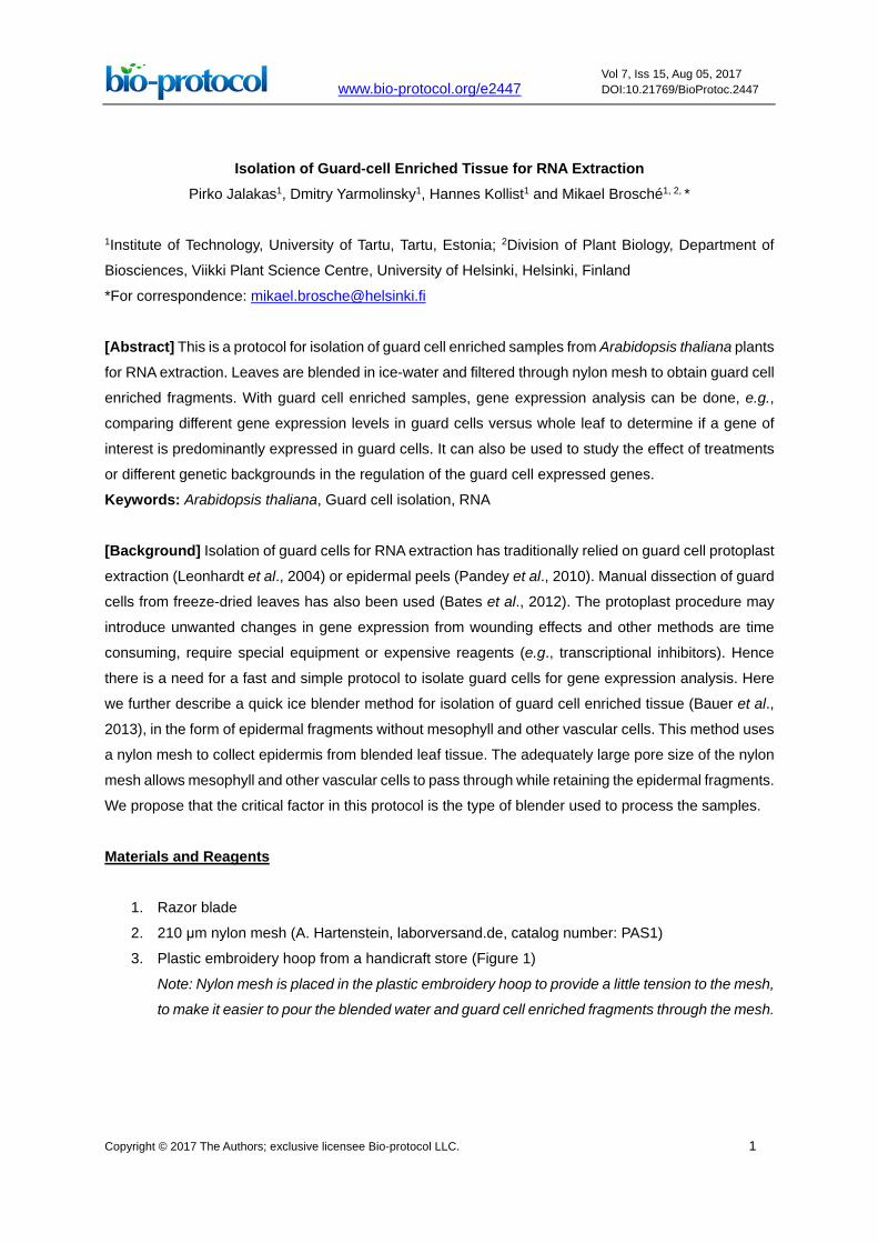
3. Plastic embroidery hoop from a handicraft store (Figure 1)
Note: Nylon mesh is placed in the plastic embroidery hoop to provide a little tension to the mesh,
to make it easier to pour the blended water and guard cell enriched fragments through the mesh.
www.bio-protocol.org/e2447 Vol 7, Iss 15, Aug 05, 2017 DOI:10.21769/BioProtoc.2447
Copyright © 2017 The Authors; exclusive licensee Bio-protocol LLC. 2
Figure 1. Nylon mesh with a plastic embroidery hoop
4. Paper towel
5. Small medical spatula or plastic spoon
6. 1.5 ml tube or aluminum foil
7. Arabidopsis plants
8. Milli-Q or distilled water kept at 4 °C
9. Crushed ice
10. Liquid nitrogen
Equipment
1. Blender (Braun JB 3060 or other 800 W blender) (Braun Household, model: JB 3060)
Note: A blender with lower effect has been tried, but failed to isolate proper fragments, probably
due to insufficient power to fully crush the ice.
2. Microscope (ZEISS, model: SteREO Discovery.v20)
Procedure
Collect leaves from 4-7 week old Arabidopsis plants grown in soil at 12 h/12 h photoperiod, use 17-
18 plants and 4-5 leaves per plant. If plants are smaller in size, use more leaves per plant.
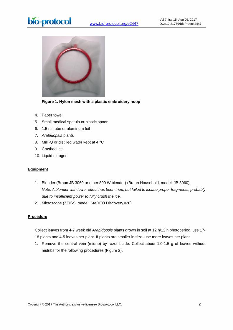
1. Remove the central vein (midrib) by razor blade. Collect about 1.0-1.5 g of leaves without
midribs for the following procedures (Figure 2).
www.bio-protocol.org/e2447 Vol 7, Iss 15, Aug 05, 2017 DOI:10.21769/BioProtoc.2447
Copyright © 2017 The Authors; exclusive licensee Bio-protocol LLC. 3
Figure 2. Midrib is cut and removed with the razor blade
2. Put the cut leaf blades in blender with 250 ml cold Milli-Q water and a handful of crushed ice.
Blend 1 min (program 2 for the Braun blender), pour through mesh placed in a plastic
embroidery hoop.
Note: Pouring through mesh should be done slowly, so that the guard cell enriched fragments
are collected in the middle of the mesh. This is especially important at step 7 after which the
sample is collected.
3. Wash the mesh with cold Milli-Q water above the blender cup so that the guard cell enriched
fragments are transferred inside the blender cup. Add cold Milli-Q water up to 250 ml and
handful of crushed ice.
4. Blend 1 min (program 2), pour through mesh placed in a plastic embroidery hoop.
5. Wash the mesh with cold Milli-Q water above the blender cup so that the guard cell enriched
fragments are transferred inside the blender cup. Add cold Milli-Q water up to 250 ml and
handful of crushed ice.
6. Blend 1 min (program 2), pour through mesh placed in a plastic embroidery hoop.
7. Remove the remaining dark green tissue fragments (small leaf pieces that have not been
blended) from the light green epidermal fraction.
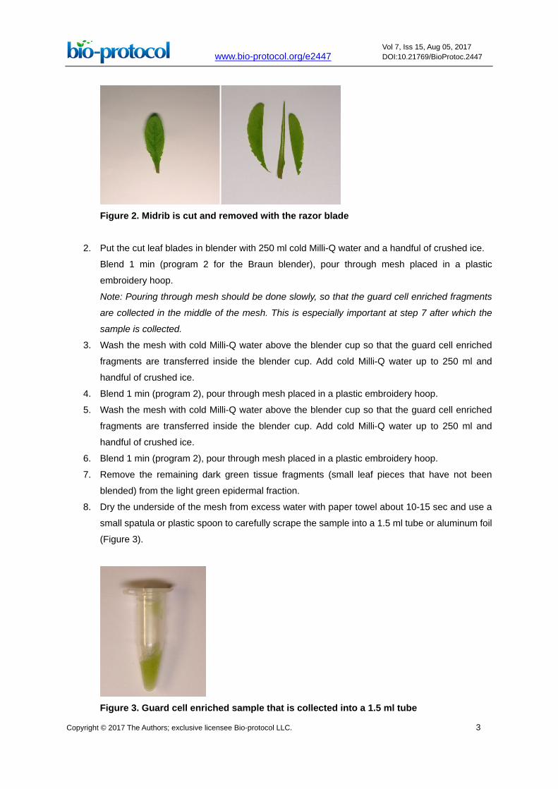
8. Dry the underside of the mesh from excess water with paper towel about 10-15 sec and use a
small spatula or plastic spoon to carefully scrape the sample into a 1.5 ml tube or aluminum foil
(Figure 3).
Figure 3. Guard cell enriched sample that is collected into a 1.5 ml tube
www.bio-protocol.org/e2447 Vol 7, Iss 15, Aug 05, 2017 DOI:10.21769/BioProtoc.2447
Copyright © 2017 The Authors; exclusive licensee Bio-protocol LLC. 4
9. Freeze the sample in liquid nitrogen and keep at -80 °C. Alternatively, the sample can be
immediately ground in mortar with liquid nitrogen and the powder used for RNA isolation.
Note: If an estimation of guard cell enrichment is required, one easy control is to harvest a whole
leaf sample from the same plants used for guard cell enrichment and freeze in liquid nitrogen
and store at -80 °C. RNA can then be extracted in parallel with the guard cell samples. The level
of guard cell enrichment can be estimated by real time quantitative PCR using guard cell
preferentially expressed genes e.g., HT1 (AT1G62400), OST1 (AT4G33950) or GORK
(AT5G37500).
Data analysis
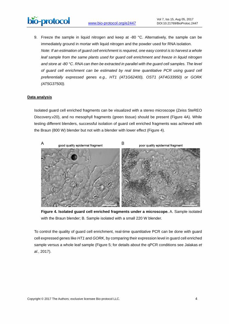
Isolated guard cell enriched fragments can be visualized with a stereo microscope (Zeiss SteREO
Discovery.v20), and no mesophyll fragments (green tissue) should be present (Figure 4A). While
testing different blenders, successful isolation of guard cell enriched fragments was achieved with
the Braun (800 W) blender but not with a blender with lower effect (Figure 4).
Figure 4. Isolated guard cell enriched fragments under a microscope. A. Sample isolated
with the Braun blender; B. Sample isolated with a small 220 W blender.
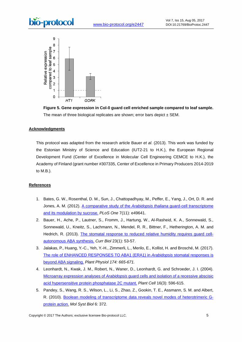
To control the quality of guard cell enrichment, real-time quantitative PCR can be done with guard
cell expressed genes like HT1 and GORK, by comparing their expression level in guard cell enriched
sample versus a whole leaf sample (Figure 5; for details about the qPCR conditions see Jalakas et
al., 2017).
www.bio-protocol.org/e2447 Vol 7, Iss 15, Aug 05, 2017 DOI:10.21769/BioProtoc.2447
Copyright © 2017 The Authors; exclusive licensee Bio-protocol LLC. 5
Figure 5. Gene expression in Col-0 guard cell enriched sample compared to leaf sample. The mean of three biological replicates are shown; error bars depict ± SEM.
Acknowledgments
This protocol was adapted from the research article Bauer et al. (2013). This work was funded by
the Estonian Ministry of Science and Education (IUT2-21 to H.K.), the European Regional
Development Fund (Center of Excellence in Molecular Cell Engineering CEMCE to H.K.), the
Academy of Finland (grant number #307335, Center of Excellence in Primary Producers 2014-2019
to M.B.).
References
1. Bates, G. W., Rosenthal, D. M., Sun, J., Chattopadhyay, M., Peffer, E., Yang, J., Ort, D. R. and
Jones, A. M. (2012). A comparative study of the Arabidopsis thaliana guard-cell transcriptome
and its modulation by sucrose. PLoS One 7(11): e49641.
2. Bauer, H., Ache, P., Lautner, S., Fromm, J., Hartung, W., Al-Rasheid, K. A., Sonnewald, S.,
Sonnewald, U., Kneitz, S., Lachmann, N., Mendel, R. R., Bittner, F., Hetherington, A. M. and
Hedrich, R. (2013). The stomatal response to reduced relative humidity requires guard cell-
autonomous ABA synthesis. Curr Biol 23(1): 53-57.
3. Jalakas, P., Huang, Y.-C., Yeh, Y.-H., Zimmerli, L., Merilo, E., Kollist, H. and Brosché, M. (2017).
The role of ENHANCED RESPONSES TO ABA1 (ERA1) in Arabidopsis stomatal responses is
beyond ABA signaling. Plant Physiol 174: 665-671.
4. Leonhardt, N., Kwak, J. M., Robert, N., Waner, D., Leonhardt, G. and Schroeder, J. I. (2004).
Microarray expression analyses of Arabidopsis guard cells and isolation of a recessive abscisic
acid hypersensitive protein phosphatase 2C mutant. Plant Cell 16(3): 596-615.
5. Pandey, S., Wang, R. S., Wilson, L., Li, S., Zhao, Z., Gookin, T. E., Assmann, S. M. and Albert,
R. (2010). Boolean modeling of transcriptome data reveals novel modes of heterotrimeric G-
protein action. Mol Syst Biol 6: 372.