Embed Size (px)
Citation preview
ISOLATION OF CELL NUCLEUS BY SHORT-TIME CHEMICAL TREATMENT IN CARRIER-MEDIUM EXCHANGE MICROCHANNELS
Kaori Toyama, Masumi Yamada and Minoru Seki* Department of Applied Chemistry and Biotechnology, Chiba University, JAPAN
ABSTRACT
This paper reports a microfluidic system to isolate cell nucleus by short-time chemical treatment. The carrier-medium of cells is exchanged twice, to treat cells by a surfactant solution for a limited span of time. By controlling the treatment (reten-tion) time, the cell membranes could be selectively digested while maintaining the cell nuclei, which were further separated from cytosol components based on size. We successfully isolated the nuclei of cultured mammalian cells by treating with Triton X-100 solution for 0.7-4.0 s, and the isolation efficiency was evaluated by western-blotting. KEYWORDS: Microfluidics, Hydrodynamic filtration, Subcellular separation, Western blotting
INTRODUCTION
Isolation of cell nucleus is an essential procedure for various biological applications, including the extraction of ge-nomic DNA, RNA and nuclear proteins for run-on transcription assays, and apoptosis and signaling studies. Until now, researchers have developed new methods and modified many times to allow rapid isolation of intact nuclei. Physical ex-traction methods using homogenizers are usually employed [1], but they have potential problems including the difficulty in adjusting the stroke conditions to selectively destroy the cell membrane, resulting in low-reproducible isolation effi-ciencies. Additionally, there is a possibility that nucleus are fragmented with the strong shear forces, since the nuclear membrane is so fragile. In contrast, chemical extraction methods using surfactant solution would be free from those problems, but treatment time should be accurately controlled not to digest the nucleus [2]. In this study, we propose a microfluidic system to isolate cell nucleus by the short-time chemical treatment, and evaluated its potential by using cul-tured mammalian cells.
PRINCIPLE
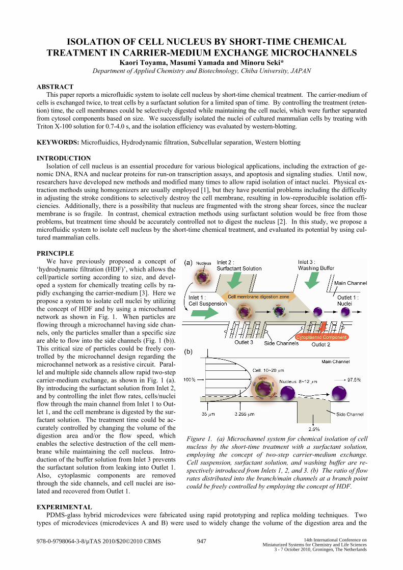
We have previously proposed a concept of ‘hydrodynamic filtration (HDF)’, which allows the cell/particle sorting according to size, and devel-oped a system for chemically treating cells by ra-pidly exchanging the carrier-medium [3]. Here we propose a system to isolate cell nuclei by utilizing the concept of HDF and by using a microchannel network as shown in Fig. 1. When particles are flowing through a microchannel having side chan-nels, only the particles smaller than a specific size are able to flow into the side channels (Fig. 1 (b)). This critical size of particles could be freely con-trolled by the microchannel design regarding the microchannel network as a resistive circuit. Paral-lel and multiple side channels allow rapid two-step carrier-medium exchange, as shown in Fig. 1 (a). By introducing the surfactant solution from Inlet 2, and by controlling the inlet flow rates, cells/nuclei flow through the main channel from Inlet 1 to Out-let 1, and the cell membrane is digested by the sur-factant solution. The treatment time could be ac-curately controlled by changing the volume of the digestion area and/or the flow speed, which enables the selective destruction of the cell mem-brane while maintaining the cell nucleus. Intro-duction of the buffer solution from Inlet 3 prevents the surfactant solution from leaking into Outlet 1. Also, cytoplasmic components are removed through the side channels, and cell nuclei are iso-lated and recovered from Outlet 1.
EXPERIMENTAL
PDMS-glass hybrid microdevices were fabricated using rapid prototyping and replica molding techniques. Two types of microdevices (microdevices A and B) were used to widely change the volume of the digestion area and the
Figure 1. (a) Microchannel system for chemical isolation of cell nucleus by the short-time treatment with a surfactant solution, employing the concept of two-step carrier-medium exchange. Cell suspension, surfactant solution, and washing buffer are re-spectively introduced from Inlets 1, 2, and 3. (b) The ratio of flow rates distributed into the branch/main channels at a branch point could be freely controlled by employing the concept of HDF.
978-0-9798064-3-8/µTAS 2010/$20©2010 CBMS 947 14th International Conference onMiniaturized Systems for Chemistry and Life Sciences
3 - 7 October 2010, Groningen, The Netherlands

treatment time; the typical treatment times are 0.7-1.4 s and 2.0-4.0 s for devices A and B, respectively (Fig. 2 (a, b)). A cell (JM cell, cultured leukemia cell line) suspension, an aqueous solution of a surfactant, and PBS were introduced from Inlets 1, 2, and 3, as shown in Fig. 2 (c). Cell nucleus and membrane were labeled with blue (Hoechst 33342) and red (Vybrant DiI) dyes, respectively, and their changes before and after treatment were observed.
To examine the isolation efficiency, the concentrations of nuclear/cytoplasmic proteins in the recovered samples were evaluated by western blotting, using Oct-1 and -tubulin as nuclear and cytoplasmic markers, respectively [4]. The un-treated cells or the nuclei recovered from Outlet 1 were treated with RIPA buffer containing 1% NP-40 and 0.1% SDS, mixed with SDS-PAGE sample buffer (reducing) containing 5% SDS, 100 mM dithiothreitol (DTT), and then boiled at 98 °C for 5 min. This protein sample was then separated by SDS-PAGE, and electroblotted onto polyvinylidene difluo-ride membranes. After the blocking of the membranes in 0.5% (w/v) casein for 1 h at room temperature, the membranes were incubated with monoclonal anti-human antibodies (anti-Oct-1 and anti--tubulin), for 1 h at room temperature. The protein bands were visualized by using biotinylated anti-mouse antibodies and a commercial kit (Vectastain ABC-AmP; Vector Laboratories, Burlington, CA), and the relative amount of proteins were determined by image processing.
Figure 2. (a) Photographs of the PDMS microdevices, (b) a schematic illustration of the microchannel network for nuc-leus isolation, and (c) the experimental setup. Fluids were introduced by using syringe pumps. The behaviors of cells/nuclei were observed using a CCD camera.
RESULTS AND DISCUSSION
First, to examine if the carrier-medium exchange actually occurs, fluorescent polystyrene particles with diameter of 0.1 m were introduced from Inlet 2 to visualize the flow profile. Particles were suspended in 2.0% Triton X-100 aq., and it was continuously introduced into from Inlet 2, while PBS without particles was introduced from Inlets 1 and 3. As a result, particles were not recovered from Outlets 2 and 3, but not from Outlet 1, showing that two-step exchange of carrier-medium was actually achieved, as shown in Fig. 3 (a). Then we tried to isolate cell nuclei by using two types of surfactant. When cells were treated with SDS, cells and their nuclei were completely destroyed after treated for 0.06 to 1.4 s, and the selective isolation of nuclei was impossible (data not shown). When cells were treated with 2.0% Triton X-100 aq for 1.4 s, cell membranes remained surrounding the nuclei (Fig. 4 (c, g)). By treating cells with the same solu-tion for 3.0 s, cell membranes were almost completely digested and the membrane debris and the cytoplasmic compo-
Figure 3. (a) Visualization of 1st and 2nd carrier-medium exchanges, in zone A and B, respectively. These zones correspond to those shown in Figure 2 (b). (b) Observation of nucleus and cell membrane flowing through the mi-crochannel in zone B, respectively stained with blue (Hoechst 33342) and red dyes (Vybrant DiI), when cells were treated by 2% Triton X-100 aq for 3.0 s.
948

nents flowed through the side channels. On the other hand, most nuclei flowed through the main channel without enter-ing into the side channels (Fig. 3 (b)), which kept the spherical shapes even after the treatment (Fig. 4 (d, h)). On this condition, 90-95% of the introduced cells flowed through the digestion area, and 80-85% of nuclei were recovered from Outlet 1. Finally, by conducting western blotting, we confirmed that the relative amount of the nuclear protein (Oct-1) to the cytoplasmic protein (-tubulin) was dramatically increased after the treatment for 3.0 s (Fig. 5), showing the high ef-ficiency and reproducibility of the presented system for purifying the cell nucleus samples.
Figure 4. Microscopic images of JM cells before (a and e) and after treatment. White arrows indicate the remaining cellular membranes.
CONCLUSIONS The presented microfluidic system provides an alternate way to easily and accurately purify the cellular nucleus sam-
ples. Further studies would be possible to apply this system to other types of cells.
ACKNOWLEDGEMENTS This research was supported in part by Grants-in-aid for Scientific Research A (20241031) from Japan Society for
Promotion of Science (JSPS), and for Improvement of Research Environment for Young Researchers from Japan Sci-ence and Technology Agency (JST).
REFERENCES [1] J. D. Dignam, R. M. Lebovitz and R. G. Roeder, Accurate transcription initiation by RNA polymerase II in a so-
luble extract from isolated mammalian nuclei, Nucleic Acid Res., 11, 1475 (1983). [2] T. M. Antalis and D. Godbolt, Isolation of intact nuclei from hematopoietic cell types, Nucleic Acid Res., 19, 4301
(1991). [3] M. Yamada, J. Kobayashi, M. Yamato, M. Seki and T. Okano, Millisecond treatment of cells using microfluidic
devices via two-step carrier-medium exchange, Lab Chip, 8, 772 (2008). [4] M. Macaluso, M. Montanari, C. M. Marshall, A. J. Gambone, G. M. Tosi, A. Giordano and M. Massaro-Giordano,
Cytoplasmic and nuclear interaction between Rb family proteins and PAI-2: a physiological crosstalk in human corneal and conjunctival epithelial cells, Cell Death Differ., 13, 1515 (2006).
CONTACT *Minoru SEKI, tel: +81-43-290-3436; [email protected]
Figure 5. Quantification result of nuclear and cytoplasmic proteins by western blotting, before and after treatment for 3.0 s by 2% Triton X-100. The graph shows the signal ra-tios of the nuclear protein (Oct-1) to the cytoplasmic protein (-tubulin).
949