Embed Size (px)
Citation preview

International Journal of Science and Research (IJSR) ISSN (Online): 2319-7064
Index Copernicus Value (2013): 6.14 | Impact Factor (2013): 4.438
Volume 4 Issue 2, February 2015
www.ijsr.net Licensed Under Creative Commons Attribution CC BY
Isolation of Antagonistic Actinomycetes SPS from Rhizosphere of BT Cotton
T. Sujatha
1, P. Siva Raagini
2
1 & 2Department of Applied Microbiology, Sri Padmavati Mahila Visvavidyalayam, Tirupati. A.P. India.
1SR&BGNR Government Arts and Science College, Khammam.Telangana state, India .
Abstract: The plant microbe interaction in the rhizosphere is one of the major factors regulating the health and growth of plants.
Actinomycetes are common filamentous soil microorganisms important in maintaining a satisfactory biological balance in the soil,
largely because of the ability to produce antibiotics. In the present study Antagonistic Actinomycetes species was isolated from
rhizosphere of Bt cotton. Seven types of isolated Actinomycetes colonies were isolated from crowded plate method and were screened
primarily by Giant colony technique. Three strains with best antifungal activity were further screened by Well Diffusion method and the
best member which has good antifungal activity was selected and named as BtAS. This strain was studied for it’s morphological,
physiological characteristics according to Bergey’s Manual and further studied by molecular characterization and was identified as
Streptomyces filamentosus. The 16s rRNA gene sequence (1472 bp) of isolate was deposited at NCBI GeneBank with Accession Number
KF939135. The antagonistic nature of the isolated strain was determined for its anti-fungal activity by Well Diffusion method, MIC and
Inhibition of phytopathogenic fungi likeA. alternata,F.moniliformae, M. phaseolina, R. solaniand A. niger in liquid medium. The results indicate that Streptomyces filamentosusisolatedfrom rhizosphere of Bt cotton has Good Antifungal activity and it was more
effective against F.moniliformae when compared with other test fungi.
Keywords: Rhizosphere,Antagonistic,Crowded plate, Giant colony technique,Well Diffusion method, phytopathogenic,MIC 1. Introduction Cotton (Gossypium herbacium arboreu) was the one of the important commercial crop in India. Among the total cultivation of the Cotton crop, Bt cotton occupies 90 % of it. Bt-cotton incorporated with Cry1Ac is highly toxic to the bollworms (Helicoverpa armigera) and other minor pests such as the cotton semilooper (Spodoptera litura) and hairy caterpillar [1].The Rhizosphere contains a large and majority of the soil biota. The plant microbe interaction in the rhizosphere is one of the major factors regulating the health and growth of plants. Soil bacteria living in the rhizosphere can enhance plant growth by several mechanisms like antagonism against plant pathogens, solubilization of phosphates [2], production of phytohormones [3], siderophores production [4], antibiotic production [5], inhibition of plant ethylene synthesis [6] and induction of plant systemic resistance to pathogens [7]. The study of rhizosphere is important as far as control of soil pathogens which pass through the rhizosphere and infect root system. Biological control is a common phenomenon in a soil ecosystem. It is a site for complex diverse microbe mediated processes. Several microorganisms like Actinomycetes secrete low levels of antibiotic compounds as their secondary metabolites. Many of them are effective against bacteria, fungi and actinomycetes which maintain natural soil health. This is a continuous process which can inhibit or kill some of the plant pathogens in that vicinity. Actinomycetes are common filamentous soil microorganisms important in maintaining a satisfactory biological balance in the soil, largely because of the ability to produce antibiotics. They are also known to be actively involved in degradation of complex organic materials in soils and contribute to the biogeochemical transformations.
Most of the actinomycetes are capable of producing wide variety of cell wall degrading enzymes like chitinases, glucanases, cellulases, hemicellulases, amylases etc. These are also known to produce several antifungal compounds that are being exploited commercially for the control of several microbial plant diseases. 2. Materials and Methods
2.1 Soil Sampling
The study area covers Khammam district, Telangana State, India. Three villages namely Ammapalem, V.V.palem and Thallada of Konejerla mandal, were selected for field study. The selected sites are being used to cultivate Bt cotton continuously for more than ten years consecutively without an alternate crop. Rhizospheric soils were collected from 60 days crop of Bt Cotton. Rhizospheric soil samples were taken from five fields from each village. Five transects across each plot were chosen. The soil samples were collected at different points (five points) from each transects to get 125 soil samples from one village. Like this from all the three villages separate 125 Bt cotton rhizospheric soils were collected.
ONE VILLAGE FIVE FIELDSFIVE TRANSECTS FROM EACH FIELD FIVE POINTS FROM EACH TRANSECT (5X5X5=125).
All the 125 samples from each village were mixed to get one representative soil sample. After removal of plant debris, the samples were sieved using 2mm mesh size sieve and air dried. Then they were labeled and transported to the laboratory in polyethylene bags and stored at 40C, and were further used for the isolation of antagonistic Actinomycetes.
Paper ID: SUB151517 1373
International Journal of Science and Research (IJSR) ISSN (Online): 2319-7064
Index Copernicus Value (2013): 6.14 | Impact Factor (2013): 4.438
Volume 4 Issue 2, February 2015
www.ijsr.net Licensed Under Creative Commons Attribution CC BY
2.2 Isolation of actinomycetes by Crowed plate method
The rhizosphere soil (1 gm) was suspended in 10 ml of sterile 0.85% NaCl solution, serially diluted (10-1 to 10-6), centrifuged at 500 rpm for 20 minute to disperse the spore chains. The suspension was allowed to settle for 1hr and plated on to Starch Casein Agar (SCA) [8]. The plates were incubated at 28±20C for 84 hrs. The plates were observed intermittently during incubation for whitish pin point colonies with a zone of inhibition around them. The pin point colonies with inhibitory zone were selected and purified by multiple streaking methods. The isolated seven types of actinomycetes colonies from Bt rhizosphere were maintained on SCA slants at 4 0C [9].
2.3 Screening of Isolates for antagonism against plant
pathogenic fungi
2.3.1 Primary screening by Giant colony technique
The seven isolated Actinomycetes were screened for antimicrobial activity by giant colony technique [10]. Single streak of each Actinomycetes was made on Modified nutrient agar (glucose 5gm, peptone 5gm, beef extract 3gm, NaCl 5gm, agar 15 gm at pH 7) and incubated at 28±2oC for 4 days to test antibacterial activity. After observing a ribbon like growth of the Actinomycetes, the pathogenic bacterial cultures (Escherichia coli, Klebsiella pneumoniae,
Staphylococcus aureus, Proteus vulgaris, Pseudomonas
pyogenes) were streaked at right angles to the original streak of each Actinomycetes and incubated at 37oC. The inhibition zone was measured after 24 h.
Five Fungal cultures of agriculture importance (Alternaria
alternata, Fusarium moniliformae, Macrophomena
phaseolina, Rhizoctonia soloni and Aspergillus niger) were used to determine the antifungal activity of the isolated actinomycetes strain. To test the antifungal activity single streak of actinomycetes were made on Kuster’s agar [8] and the test fungal pathogens were streaked at right angles to the original streak of each Actinomycetes and incubated at 28±2oC. The inhibition zone was measured after 7 days of incubation [11].
2.3.2 Secondary screening of selected strains by Well
Diffusion method
Five isolates which shown most effect on phytopathogenic fungi, were selected for secondary screening. It was carried out by Well Diffusion method.
2.3.3 Preparation of fermentation broth
The strains were cultured on Starch Casein Agar slants at 28±20C for 2 weeks for sporulation. The mature spores were inoculated in Starch Casein Broth. The fermentation set up was incubated on rotary shaker at 200 rpm for 10 days at 28±20C. The fermented broth was centrifuged at 10,000 rpm at 40C for 20 min. The supernatant was filtered using 0.2 µm filters and the filtrate was collected as antibiotic sample [12].
2.4 Testing of antibiotic sample from antagonistic
Actinomycetes
To determine the antagonistic activity the phytopathogenic fungi were cultured in Asthana Hakuer’s broth [11] at 280C for 5 days. The cultures were swapped on Potato Dextrose
Agar (PDA). Four wells (6mm) were prepared in each seeded agar plate and each well was filled with 100 µl of the fermentation broth of the selected strains. These PDA plates were incubated at 28±20C for 5 days. After the incubation the diameter of the inhibition zone was measured. Depending on the zone of inhibition, one strain was selected and named as BtAS.
2.5 Characterization for the taxonomic position of the
selected Actinomycetes strain BtAS
The taxonomic position of the selected strains was determined by studying their morphology, fine structure and spore chain morphology by Gram staining and SEM , Colonial Characteristics were observed for aerial mass color, melanin production, diffusible pigments, reverse side pigmentation of the colony. Nutrition and growth characteristics were determined by growth on different media like Glucose Yeast Extract Malt extract Agar (GYEA) Oat meal Agar (OA) Glycerol Asparagine Agar (GASp) Peptone Yeast extract Iron Agar (PYIA) Tyrosine Agar (TA), Nutrient agar (NA), Malt Yeast Extract Agar (MYEA) and Starch Casein Agar (SCA) [13]. Utilization of Carbon and Nitrogen Sources, Antibiosis and resistance to antibiotics was studied. Physiological characterization was also determined by studying Growth at different temperatures, pH, and Salt concentration. Enzyme activity was studied by testing Chitinolytic activity, Lipolysis activity, lecithinase activity, pectin hydrolysis, urease hydrolysis, starch hydrolysis and gelatin hydrolysis, Denitrification Test, nitrate reduction test and H2S production test [13, 14]. Molecular characterization was done by 16S rRNA analysis. 3. Studies on Antagonistic Activity of the
Isolate
Determinaton of antagonistic activity by Well diffusion
method
The plates were seeded with test fungal inoculum (0.1ml) and wells were punctured (8 mm in diameter) with sterile cork borer. The wells were filled with filtrate of the culture suspension in various concentrations i.e 25 µl/ well, 50 µl/ well, 75 µl/ well and 100 µl/ well. A well with a standard antibiotic (Nystatin 100 µl/well) was also set for reference. The entire set up was incubated at 28±2 0C for 4 days. Clear inhibition zone around wells was measured in millimeters [15].
Calculation of Activity Index
Activity index of BtAS was calculated by comparing the inhibition area of the test sample with that of standard antibiotic [16]. Activity Index = Zone of inhibition in mm of test sample ÷ Zone of inhibition in mm of standard antibiotic.
Inhibition of fungal pathogens in Czapeck’s Broth
The potential antagonistic activity of the BtAS culture was tested against test fungal pathogens in Czapeck’s Broth
Paper ID: SUB151517 1374

International Journal of Science and Research (IJSR) ISSN (Online): 2319-7064
Index Copernicus Value (2013): 6.14 | Impact Factor (2013): 4.438
Volume 4 Issue 2, February 2015
www.ijsr.net Licensed Under Creative Commons Attribution CC BY
(CZB). Fungal inoculum of 0.1 ml capacity was inoculated in to 50 ml of CZB to which 0.5 ml of culture filtrate of BtAS was inoculated separately and incubated for 4 days at 28±20C. All the experimental set up was carried out in triplicates. The difference in dry weight between the mycelia grown with and without BtAS culture were measured [17]. Cultures were passed through pre weighed Whatman No 1 filter paper and dried overnight in an oven at 600C and reweighed. Dry weights of fungal cultures were calculated and compared.
Measurement of Minimum Inhibitory Concentration
(MIC)
The Minimum Inhibitory concentration (MIC) is the least concentration of the antimicrobial agent in µg/ml that will inhibit growth of the phytopathogenic fungi. MIC value of BtAS was determined by serial two fold dilution in Sabouraud Dextrose Broth (SDB) with the dilution ranging from 20-120 µg (20, 40, 60, 80,1 00, 120µg/ml). The100 µl of each dilution was tested against phytopathogenic fungi by well diffusion assay. The definite zone of inhibition of any dimension surrounding the well was measured accurately to the nearest millimeter by means of ruler [18, 19,and 20]. Depending upon the inhibition zone the minimum concentration at which the fungal pathogens were inhibited was noted. 4. Results and Discussion The antagonistic actinomycetes were isolated by crowded plate method. Whitish pin point colonies with the zone of inhibition were observed in a good number on SCA plate with 10-5 dilution. Seven colonies were selected and sub cultured to get pure cultures. All the seven colonies from Bt sample were named as BtAS I, BtAS II, BtAS III, BtAS IV, BtAS V, BtAS VI and BtAS VII and were screened for their antagonistic activity against test phytopathogenic fungi and pathogenic bacteria by giant colony technique.
The inhibition zone in between giant colony and pathogenic organisms was measured for all the isolates. The order of the isolates from Bt rhizosphere for good antagonistic activity against phytopathogenic fungi was BtAS III, BtAS II, BtAS I, BtAS V, BtAS VII, BtAS IV, BtAS VI . The order of the isolates from Bt rhizosphere for good antagonistic activity against pathogenic bacteria was BtAS III, BtAS I, BtAS II, BtAS IV, BtAS V, BtAS VII, and BtAS VI. Three isolates with better antagonistic activity (BtAS III, BtAS II, and BtAS I) were further screened to get one best strain. Three selected isolates were further screened for their antagonistic activity against test fungi by Well Diffusion method. The zone of inhibition for each of the isolates was measured and compared to get one best isolate from rhizosphere of Bt cotton (Table 1). Depending on the results obtained from primary and secondary screening BtAS III was found to have good antagonistic property. This was named as BtAS and further studied for identification, characterized and taxonomic position.
Table1: Antifungal activity of the isolates from Bt soils by Well Diffusion method
Test fungi Zone of inhibition (mm)
BtAS I BtAS II BtAS III
A. alternata ++ +++ ++
F. moniliformae ++ ++ +++
M. phaseolina + + ++
R. solani + + +++
A. niger + + + Weak inhibition 5-9 mm(+), moderate inhibition10-
19mm(++), strong inhibition>20mm(+++)
Photo 1& 2: Zone of inhibition against F.moniliformae &
R. solani by BtAS I (1), BtAS II (2), BtAS III (3) and
control (C).
Characterization For The Taxonomic Position Of The Selected Actinomycetes Strain 1. Growth Characteristics on different media
Growth characteristics of BtAS was observed using different types of media such as ISP2 (Malt- Yeast Extract Agar), ISP5 (Glycerol Yeast extract Malt extract Agar), ISP6 (Peptone Yeast extract Iron Agar), Starch Casein Agar, and Nutrient Agar. All the characteristic features on different media were observed for different growth patterns and recorded (Table. 2).
Table 2: Culture characteristics of BtAS on different types of media
Medium Growth Aerial
mycelium
Substrate
mycelium
Soluble
pigments
SCA Good Light pink Pink to orange No pigment NA Good Orange Yellow orange No pigment
MYEA Good White Whitish pink Grey PYIA Moderate Grayish white Grey Grayish black
(GYMA) Good Grey Light brown Cherry red
Paper ID: SUB151517 1375
International Journal of Science and Research (IJSR) ISSN (Online): 2319-7064
Index Copernicus Value (2013): 6.14 | Impact Factor (2013): 4.438
Volume 4 Issue 2, February 2015
www.ijsr.net Licensed Under Creative Commons Attribution CC BY
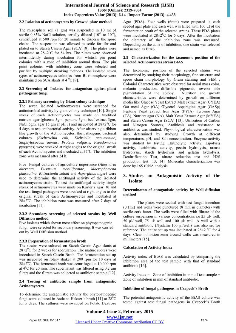
Morphology and fine structure of BtAS Characteristics BtAS
Spore mass Dusty to light pink Spore chain morphology Rectiflexible and filamentous
Spore surface Smooth to warty
Reverse side pigment _ Diffusible pigments +
Melanin — Spore size 845-859nm
Spore shape Oval to rectangular
SEM Image showing Spore chain morphology of BtAS
SEM Image showing Spore size of BtAS
Physiological characteristics of BtAS
Physiological characteristics BtAS
50C -- 150C -- Temperature range280C-450C + Maximum temperature tolerance 500C Optimum temperature 280C pH range 6-8 Optimum pH 7.0 NaCl range 1-9% Optimum NaCl 5% Incubation period range 5-10 days Optimum incubation period 7 days
Degradations and Enzyme activity studies of BtAS
Degradation/hydrolysis: BtAS
Casein + Cellulose + Pectin + Starch + Chitin + Urea + Catalase +
Oxidase +
Lipase activity + Denitrification -- Nitrate reduction + Lecithinase activity + Phosphatase -- Hydrogen sulphate test + Utilization of carbon sources D-Sucrose + L-Raffinose + D-Mannitol -- L-Rhamnose + D-Fructose + D-Glucose + D-Xylose -- L-Aspergine + L-Phenylalanine + L-Histidine + L-Arginine +
Paper ID: SUB151517 1376
International Journal of Science and Research (IJSR) ISSN (Online): 2319-7064
Index Copernicus Value (2013): 6.14 | Impact Factor (2013): 4.438
Volume 4 Issue 2, February 2015
www.ijsr.net Licensed Under Creative Commons Attribution CC BY
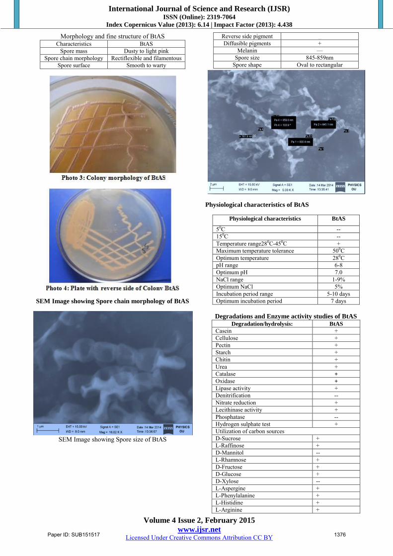
L- Hydroproline +
Antagonistic property and resistance to antibiotics by
BtAS Antagonistic to BtAS E. coli + S. aureus + P. pyogenes + K. pneumoniae + p. vulgaris
+ Resistance to
Rifampicin (50µg/ml) _ Penicillin G (50µg/ml) + Oleandomycin (50µg/ml) + Neomycin (50µg/ml) _
(+) positive, (-) negative From the above colonial, morphological, physiological and nutritional, enzymatic degradation studies, BtAS was identified as member of genus Streptomyces, Category I, cluster Streptomyces exfoliates and the strain has 81% similarity level with Streptomyces filamentosus which was further determined by molecular characterization.
Molecular characterization of BtAS
Molecular characterization was done by isolating the DNA and 16s rDNA analysis for BtAS isolate. From PCR, amplified fragment of 1.5 Kb was obtained and sequenced with the help of Sequencing Technology: Sanger dideoxy sequencing. The 16s rRNA gene sequence (1472 bp) of BtAS isolate was deposited at GeneBank with Accession Number KF939135 (Fig 1). The blast search analysis indicated that the strain BtAS is a close homologue of S.
filamentosus. Multiple sequence alignment of BtAS 16s gene with closely related homologues was done with CLUSTALW.
The BtAS strain has 99% similarity level with Streptomyces
filamentosus which was confirmed by molecular characterization and its taxonomic position was described as
ORGANISM Streptomyces filamentosus
Bacteria; Actinobacteria; Actinobacteridae;
Actinomycetales;
Streptomycineae; Streptomycetaceae; Streptomyces; S.
exfoliatus.
Fig:1Streptomyces filamentosus strain BtAS 16S
ribosomal RNA gene, partial sequence
GenBank: KF939135.1 FASTA Graphics LOCUS KF939135 1472 bp DNA linear BCT 23-FEB-2014 DEFINITION: Streptomyces filamentosus strain BtAS1 16S ribosomal RNA gene, partial sequence. ACCESSION KF939135 VERSION KF939135.1 GI: 585635063 KEYWORDS. SOURCE Streptomyces filamentosus
(Streptomyces roseosporus)
ORGANISM Streptomyces filamentosus
Bacteria ; Actinobacteria; Actinobacteridae;
Actinomycetales;
Streptomycineae; Streptomycetaceae;
Streptomyces.
REFERENCE 1 (bases 1 to 1472) AUTHORS Sujatha.T, Vijayalakshmi.K. TITLE Isolation of actinomycetes from Bt cotton fields
ORIGIN
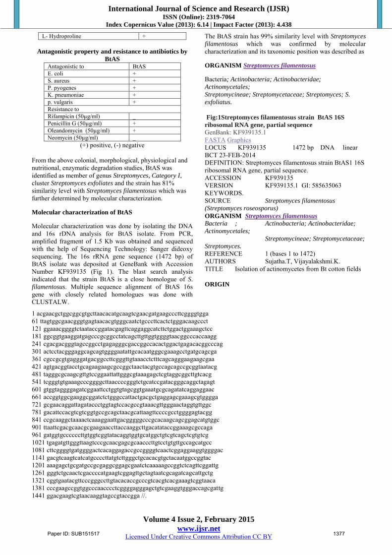
1 acgaacgctggcggcgtgcttaacacatgcaagtcgaacgatgaagcccttcggggtgga 61 ttagtggcgaacgggtgagtaacacgtgggcaatctgcccttcactctgggacaagccct 121 ggaaacggggtctaataccggatacgagttcaggaggcatcttctggactggaaagctcc 181 ggcggtgaaggatgagcccgcggcctatcagcttgttggtggggtaacggcccaccaagg 241 cgacgacgggtagccggcctgagagggcgaccggccacactggactgagacacggcccag 301 actcctacgggaggcagcagtggggaatattgcacaatgggcgaaagcctgatgcagcga 361 cgccgcgtgagggatgacggccttcgggttgtaaacctctttcagcagggaagaagcgaa 421 agtgacggtacctgcagaagaagcgccggctaactacgtgccagcagccgcggtaatacg 481 tagggcgcaagcgttgtccggaattattgggcgtaaagagctcgtaggcggcttgtcacg 541 tcgggtgtgaaagcccggggcttaaccccgggtctgcatccgatacgggcaggctagagt 601 gtggtaggggagatcggaattcctggtgtagcggtgaaatgcgcagatatcaggaggaac 661 accggtggcgaaggcggatctctgggccattactgacgctgaggagcgaaagcgtgggga 721 gcgaacaggattagataccctggtagtccacgccgtaaacgttgggaactaggtgttggc 781 gacattccacgtcgtcggtgccgcagctaacgcattaagttccccgcctggggagtacgg 841 ccgcaaggctaaaactcaaaggaattgacgggggcccgcacaagcagcggagcatgtggc 901 ttaattcgacgcaacgcgaagaaccttaccaaggcttgacatataccggaaagcgccaga 961 gatggtgccccccttgtggtcggtatacaggtggtgcatggctgtcgtcagctcgtgtcg 1021 tgagatgttgggttaagtcccgcaacgagcgcaacccttgtcctgtgttgccagcatgcc 1081 cttcggggtgatggggactcacaggagaccgccggggtcaactcggaggaaggtggggac 1141 gacgtcaagtcatcatgccccttatgtcttgggctgcacacgtgctacaatggccggtac 1201 aaagagctgcgatgccgcgaggcggagcgaatctcaaaaagccggtctcagttcggattg 1261 gggtctgcaactcgaccccatgaagtcggagttgctagtaatcgcagatcagcattgctg 1321 cggtgaatacgttcccgggccttgtacacaccgcccgtcacgtcacgaaagtcggtaaca 1381 cccgaagccggtggcccaacccctcggggagggagctgtcgaaggtgggaccagcgattg 1441 ggacgaagtcgtaacaaggtagccgtaccgga //.
Paper ID: SUB151517 1377
International Journal of Science and Research (IJSR) ISSN (Online): 2319-7064
Index Copernicus Value (2013): 6.14 | Impact Factor (2013): 4.438
Volume 4 Issue 2, February 2015
www.ijsr.net Licensed Under Creative Commons Attribution CC BY
Determination of the antibiotic activity of S. filamentosus
Preparation of fermentation product
The representative filtrate for BtAS was prepared by fermentation setup in which pre inoculated SCB with BtAS was maintained with good aeration and was incubated at 28±20C for 10 days at neutral pH. After incubation the content of the flasks was filtered through Whatman No.1filter paper and filtrate was collected into a vessel.
Determinaton of antagonistic activity by Well Diffusion
method
Antifungal activity of S. filamentosus was tested against selected phytopathogenic fungi by Well Diffusion method. The diameter of Zone of Inhibition was tabulated (Table3). Optimum antifungal activity for S. filamentosus was shown against F. moniliformae, R. solani, A. alternate except A.
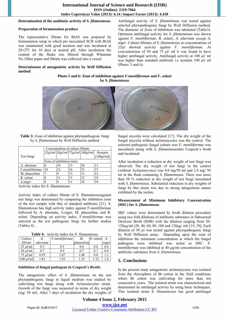
niger. Culture filtrates of S. filamentosus at concentrations of 25µl showed activity against F. moniliformae. At concentrations of 50 and 75 µl/ ml it was found to have higher antifungal activity. Antifungal activity at 100 µl/ ml was higher than standard antibiotic i.e nystatin 100 µl/ ml (Photo: 5 and 6).
Photo 5 and 6: Zone of inhibition against F.moniliformae and F. solani
by S. filamentosus
Table 3: Zone of inhibition against phytopathogenic fungi by S. filamentosus by Well Diffusion method
Test fungi
Concentration of culture filtrate 25µl/ml 50µl/ml 75µl/ml 100µl/ml Nystatin
(100µl/ml)
Zone of inhibition (mm) A. alternata 6 10 13 20 11 F.moniliformae 10 12 16 23 15 M. phaseolina 7 9 13 15 12 R. solani 8 11 15 22 18 A.niger 6 8 11 18 10
Activity index for S. filamentosus Activity index of culture filtrate of S. filamentosusagainst test fungi was determined by comparing the inhibition zone of the test sample with that of standard antibiotic [21]. S. filamentosus has high activity index against F.moniliformae followed by A. alternata, A.niger, M. phaseolina, and R. solani. Depending on activity index, F.moniliformae was selected as the test pathogenic fungi for further studies (Tables 4).
Table 4: Activity index for S. filamentosus Culture
filtrate
A.
alternata
F.moniliformae M.
phaseolina
R. solani A.
niger
25 µl/ml 0.5 0.5 0.6 0.6 0.6 50 µl/ml 0.7 0.9 0.7 0.7 0.8 75 µl/ml 0.93 1.07 1.08 0.8 1.1 100 µl/ml 1.81 1.83 1.25 1.33 1.8
Inhibition of fungal pathogens in Czapeck’s Broth:
The antagonistic effect of S. filamentosus on the test phytopathogenic fungi in liquid medium was studied by cultivating test fungi along with Actinomycetes strain. Growth of the fungi was measured in terms of dry weight (mg/ 50 ml). After 7 days of incubation the dry weights of
fungal mycelia were calculated [17]. The dry weight of the fungal mycelia without actinomycetes was the control. The selected pathogenic fungal culture was F. moniliformae was inoculated along with S. filamentosusinto Czapeck’s broth and incubated.
After incubation a reduction in dry weight of test fungi was observed. The dry weight of test fungi in the control (without Actinomycetes) was 4.0 mg/50 ml and 2.0 mg/ 50 ml in the flask containing S. filamentosus. There was more than 50 % reduction in dry weight of test fungi inoculated with S. filamentosus. Substantial reduction in dry weights of fungi by this strain was due to strong antagonistic nature exhibited by the isolate.
Measurement of Minimum Inhibitory Concentration
(MIC) for S. filamentosus
MIC values were determined by broth dilution procedure using two fold dilutions of antibiotic substance in Sabouraud Dextrose Broth (SDB) with the dilution ranging from 20-120µg/ml (20, 40, 60, 80, 100 and 120µg/ ml) [19, 20]. Each dilution of 50 µl was tested against phytopathogenic fungi by Well Diffusion assay. Depending upon the zone of inhibition the minimum concentration at which the fungal pathogens were inhibited was noted as MIC. F.
moniliformae was inhibited at 40 µg/ml concentration of the antibiotic substance from S. filamentosus.
5. Conclusions In the present study antagonistic actinomycetes was isolated from the rhizosphere of Bt cotton in the field conditions where Bt cotton was cultivating for more than ten consecutive years. The isolated strain was characterized and determined its antifungal activity by using basic techniques. This isolated strain S. filamentosus has good antifungal
Paper ID: SUB151517 1378
International Journal of Science and Research (IJSR) ISSN (Online): 2319-7064
Index Copernicus Value (2013): 6.14 | Impact Factor (2013): 4.438
Volume 4 Issue 2, February 2015
www.ijsr.net Licensed Under Creative Commons Attribution CC BY
activity against all the test fungi and has shown highest activity against F. moniliformae which was a common fungal plant pathogen in the rhizosphere and causative agent of several root rots. Determination of optimum conditions for the fermentation product, it’s other applications, molecular characterization of antibiotic substance was the scope of this study. References
[1] Tulsi Bhardwaj and Sharma J. P. 2013. Impact of Pesticides Application in Agricultural Industry: An
Indian Scenario International Journal of Agriculture
and Food Science Technology (4): 817-822. [2] De Freitas, J. R., Banerjee, M. R. and Germida, J. J.
1997. Phosphate solubilizing rhizobacteria enhance the growth and yeild but not phosphorus uptake of canola (Brassica napus), Biol. Fertil. Soils, 24: 358-364.
[3] Arshad, M. and Frankenberger, Jr. W.T. 1998. Plant growth regulating substances in the rhizosphere: microbial production and functions. Adv. Agron., 62: 46-151.
[4] Kloepper, J. W., Leong, J., Teintze, M. and Schroth, M. N. 1980b. Enhanced plant growth by siderophores produced by plant growth-promoting rhizobacteria. Nature 286: 835–836.
[5] Schneider, M., Schweizer, P., Meuwly, P. and Metraux, J. P. 1996. Systemic acquired resistance in plants. Int. J.
Cytol. 168, 303-340. [6] Glick, B. R., Penrose, D. M. and Jiping, Li. 1998. A
Model For the Lowering of Plant Ethylene Concentrations by Plant Growth-promoting Bacteria. J
of theoretical biology 190:63-68. [7] Kloepper, J. W., Rodrigue-Kabana, R., Zehnder, G. W.,
Marphy, J. F., Sikora, E. and Fermandez, C. 1999. Plant root-bacterial interactions in biological control of soil borne diseases and potential extension to systemic and foliar diseases. Australian Plant Pathology 28: 21-26.
[8] Okami, Y. and Hotta, K. 1988. Search and discovery of new antibiotics, In: Goodfellow M, Williams S. T, Mordarski, M (Eds). Actinomycetes in Biotechnology. Academic Press, Inc., San Diego, 33-67.
[9] Nanjwade, B. K., Chandrashekhara, S., Shamarez, A. M., Goudanavar, P. S. and Manvi, F. V. 2010. Isolation and morphological characterization of antibiotic producing Actinomycetes. Trop. J. Pharmaceut. Res. 9: 231-236.
[10] Lemos, M. L., Toranzo, A. E. and Barja, L. E. 1985. Antibiotic activity of epiphytic bacteria isolated from intertidal seaweeds. Microb. Ecol. 11:149-163.
[11] Baskaran, R., Vijayakumar, R. and Mohan, P. M. 2011. Enrichment method for the isolation of bioactive actinomycetes from mangrove sediments of Andaman Islands, India. Malaysian J. Microbiol., 7: 26-32.
[12] Baecker, A. A. and Ryan, K.C. 1987. Improving the isolation of actinomycetes from soil by high-speed homogenization. S. Afr. J. Plant Soil 4: 165-170.
[13] Williams, S. T., Goodfellow, M. and Alderson, G. 1989. Genus Streptomyces Waksman and Henrici 1943, 339AL.In Williams, S. T., Sharpe M. E. and Holt J. G. (ed) Bergey’s Manual of Determinative Bacteriology, vol. 4, Baltimore: Williams &Willkins. 2453–2492.
[14] Shirling, E. B. and Gottlieb, D. 1969. Cooperative description of type cultures of Streptomyces. IV. Species descriptions from the second, third and fourth studies. Int J SystBacteriol 19:391–512.
[15] Riffat-uz-Zaman, Akhtar, M. S. and Khan, M. S. 2006. In vitro antibacterial screning of Anethumgraveolens L. Fruit, Cichoriumintybus L. leaf, Plantagoovata L. sed husk and Polygonumviviparum L. rot extracts against Helicobacter pylori. Int. J. Pharmacol. 2:674-67.
[16] Sharma, R., Sharma, G. and Meenakshi, S. 2011. Additive and inhibitory effect of antifungal activity of Curcuma longa (Turmeric) and Zingiberofficinale (Ginger) essential oils against Pityriasisversicolor infections. J. of Med Plants Research5;(32), 6987-6990.
[17] Yuan, W. M. and Crawford, D. L. 1995. Characterization of Streptomyces lydicus WYEC108 as a potential biocontrol agent against fungal root and seed rots.Appl. Environ. Microbiol. 61:3119-3128.
[18] Collins, N., McManus, R., Wooster, R., Mangion, J., Seal, S. and Lakhani, S. R. 1995. Consistent loss of the wild type allele in breast cancers from a family linked to the brca2 gene on chromosome 13q12-13. Oncogene
10: 1673–1675. [19] Augustine, S.K., Bhavsar, S.P., Baserisalehi, M. and
Kapadnis, B.P. 2005. A non polyen antifungal antibiotic from Streptomyces albidoflavus PU 23: Indian J Exp.
Biol., 42: 928-932. [20] Cappuccino, J. G. and Shermam, N. 1999.
Microbiology: A Laboratory Manual. Fourth the Benjamin/ Cummins Publishing Company Inc California USA.
[21] Sharma, R. A., Jain, S. C., Jain, R., and Mittal, C. 1988. Antimicrobial activity of cassia species. Ind. J.
pharmaceut. Sci. 60: 29-32.
Paper ID: SUB151517 1379