Embed Size (px)
Citation preview
ANTIMICROBIAL POTENTIAL OF SELECTED
ACTINOMYCETES ISOLATED FROM THE MARINE
ECOSYSTEM
NUR RAHIMATUL HAYATI BINTI ABDUL RAHMAN
FACULTY OF SCIENCE
UNIVERSITY OF MALAYA
KUALA LUMPUR
2010
ANTIMICROBIAL POTENTIAL OF SELECTED
ACTINOMYCETES ISOLATED FROM THE MARINE
ECOSYSTEM
NUR RAHIMATUL HAYATI BINTI ABDUL RAHMAN
DISSERTATION SUBMITTED IN FULFILLMENT
OF THE REQUIREMENTS
FOR THE DEGREE OF MASTER OF BIOTECHNOLGY
FACULTY OF SCIENCE
UNIVERSITY OF MALAYA
KUALA LUMPUR
2010
ii
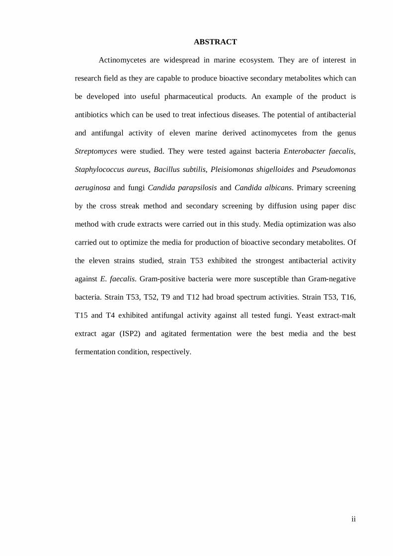
ABSTRACT
Actinomycetes are widespread in marine ecosystem. They are of interest in
research field as they are capable to produce bioactive secondary metabolites which can
be developed into useful pharmaceutical products. An example of the product is
antibiotics which can be used to treat infectious diseases. The potential of antibacterial
and antifungal activity of eleven marine derived actinomycetes from the genus
Streptomyces were studied. They were tested against bacteria Enterobacter faecalis,
Staphylococcus aureus, Bacillus subtilis, Pleisiomonas shigelloides and Pseudomonas
aeruginosa and fungi Candida parapsilosis and Candida albicans. Primary screening
by the cross streak method and secondary screening by diffusion using paper disc
method with crude extracts were carried out in this study. Media optimization was also
carried out to optimize the media for production of bioactive secondary metabolites. Of
the eleven strains studied, strain T53 exhibited the strongest antibacterial activity
against E. faecalis. Gram-positive bacteria were more susceptible than Gram-negative
bacteria. Strain T53, T52, T9 and T12 had broad spectrum activities. Strain T53, T16,
T15 and T4 exhibited antifungal activity against all tested fungi. Yeast extract-malt
extract agar (ISP2) and agitated fermentation were the best media and the best
fermentation condition, respectively.
iii
ABSTRAK
Actinomycete boleh ditemui dengan banyak di dalam ekosistem marin.
Organisma tersebut digunakan dalam bidang penyelidikan berdasarkan keupayaannya
menghasilkan produk bioaktif sekunder. Produk tersebut boleh diproses untuk
menghasilkan produk farmaseutikal. Antibiotik adalah antara produk farmaseutikal
yang boleh dihasilkan untuk merawat jangkitan penyakit. Sebelas actinomycete
daripada ekosistem marin yang telah dikenalpasti sebagai Streptomyces sp. telah dipilih
untuk ujian aktiviti antibakteria dan antikulat. Aktiviti antibakteria dan antikulat telah di
uji terhadap bakteria Enterobacter faecalis, Staphylococcus aureus, Bacillus subtilis,
Pleisiomonas shigelloides dan Pseudomonas aeruginosa serta kulat/yis Candida
parapsilosis dan Candida albicans. Kaedah ‘cross streak’ dan ‘paper disc’ telah
diaplikasikan dalam ujian ini. Kaedah ‘paper disc’ turut diaplikasikan dalam ujian kedua
untuk mengetahui media yang terbaik dalam penghasilan produk bioaktif sekunder.
Daripada sebelas actinomycete yang dikaji, T53 mempamerkan aktiviti penyekatan
pertumbuhan yang paling tinggi terhadap E. faecalis. Bakteria Gram-positif adalah lebih
mudah disekat pertumbuhannya berbanding bakteria Gram-negatif. Aktinomycete T53,
T52, T9 dan T12 menyekat pertumbuhan kedua-dua bakteria Gram-positif dan Gram-
negatif. Actinomycete T53, T16, T15 dan T4 menyekat pertumbuhan semua kulat/yis.
Yis-malta ekstrak agar (ISP2) dan sistem fermentasi ‘agitation’ adalah media dan sistem
fermentasi yang terbaik dalam penghasilan produk bioaktif sekunder.
iv
ACKNOWLEDGEMENTS
I would like to express my gratitude to my supervisor, Prof. Dr. Vikineswary
Sabaratnam and my second supervisor, Dr. G. Y. Annie Tan for their guidance
throughout my project.
I also would like to express my gratitude to Prof. Dr. Thong Kwai Lin for the
pathogens Enterobacter faecalis and Staphylococcus aureus and Prof. Ng Kee Peng for
the pathogens Candida parapsilosis and Candida albicans. Many thanks to my
colleagues, Angelyn Melaya Kloni and Norashimawati binti Ibrahim and members of
Mycology Laboratory.
Last but not least, my special thanks to my beloved family for being very
supportive. My special thanks especially to my beloved parents for your continuous
support, patience and assistance throughout my project. Thank you for everything. May
God bless you.
v
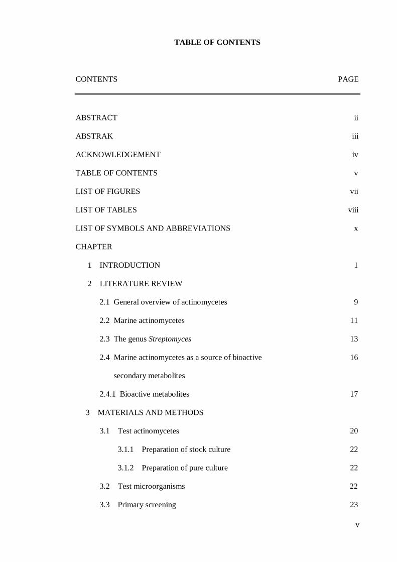
TABLE OF CONTENTS
CONTENTS PAGE
ABSTRACT ii
ABSTRAK iii
ACKNOWLEDGEMENT iv
TABLE OF CONTENTS v
LIST OF FIGURES vii
LIST OF TABLES viii
LIST OF SYMBOLS AND ABBREVIATIONS x
CHAPTER
1 INTRODUCTION 1
2 LITERATURE REVIEW
2.1 General overview of actinomycetes 9
2.2 Marine actinomycetes 11
2.3 The genus Streptomyces 13
2.4 Marine actinomycetes as a source of bioactive 16
secondary metabolites
2.4.1 Bioactive metabolites 17
3 MATERIALS AND METHODS
3.1 Test actinomycetes 20
3.1.1 Preparation of stock culture 22
3.1.2 Preparation of pure culture 22
3.2 Test microorganisms 22
3.3 Primary screening 23
vi
3.4 Secondary screening 24
3.5 Media optimization 27
4 RESULTS, DISCUSSION AND CONCLUSION
4.1 Antagonistic pattern in primary and secondary screening 28
4.1.1 Antibacterial activity 29
4.1.2 Antifungal activity 36
4.2 Antagonistic activity of Streptomyces spp. 39
4.3 Media optimization 52
4.4 Conclusion 62
REFERENCES 64
APPENDIX A : MEDIA 81
APPENDIX B: STATISTICAL TABLES 83
vii
LIST OF FIGURES
FIGURES PAGE
1 Cross streak method for primary screening 23
2 Flow diagram of procedures for antimicrobial screening of
actinomycetes strains 26
3.1 Cultures of selected putative strains of Streptomyces spp. for
bioactivity screening incubated at 28±2oC for 7 – 14 days on ISP4
for T3 and T4 and SA for the remaining strains 21
4.1 Antagonistic activity of different groups of selected
Streptomyces spp. in primary screening against; 1) E.faecalis;
2) S.aureus; 3) P.shigelloides; 4) B.subtilis; 5) P.aeruginosa on
ISP4 for strains T3 and T4 and SA for the remaining strains after
48 hours incubation at 37±2oC 40
4.2 Antagonistic activity of selected Streptomyces sp strains in secondary
screening against test bacteria after 24 hours incubation at 37±2oC 43-44
4.3 Antagonistic activity of crude extracts of T53 against E. faecalis
on ISP2 medium after 24 hours incubation at 37±2oC 53
viii
LIST OF TABLES
TABLES PAGE
3.1 Selected putative strains of Streptomyces spp. for bioactivity
screening on inorganic salts-starch agar (ISP4) for T3 and T4
and sporulation agar (SA) for the remaining strains 20
4.1 Antibacterial and antifungal activities of selected Streptomyces spp.
in primary and secondary screening 28
4.2 Antibacterial activity of selected Streptomyces spp. against test bacteria
in primary and secondary screening 29
4.3 Antibacterial activity of selected Streptomyces spp. against test bacteria
in secondary screening on NA for 24 hours at 37±2oC 31
4.4 Antifungal activity of selected Streptomyces spp. against test
fungi in primary and secondary screening 36
4.5 Antifungal activity of selected Streptomyces spp. against test fungi in
secondary screening on SDA for 24 hours at 37±2oC 37
4.6 Antagonistic activity of different colour groups of selected
Streptomyces spp. against test microorganisms in primary
and secondary screening 39
ix
4.7 Inhibition zones (mm) of different colour groups of selected
Streptomyces spp. against test microorganisms in secondary screening
on NA for test bacteria and SDA for test fungi for 24 hours at 37±2oC 42-43
4.8 Inhibition zones (mm) of crude extracts of submerged cultures
fermentation of strain T53 against E. faecalis in agitation and static
fermentation conditions on SA, ISP2 and ISP4 for 24 hours at 37±2oC 54
4.9 pH value of crude extracts of submerged cultures fermentation of T53
in shaking and static fermentation conditions on SA, I2 and I4 for 24
hours 37±2oC 55
x
LIST OF SYMBOLS AND ABBREVIATIONS
% percentage
oC degree celcius
et al. and others
g gram
µg microgram
mg milligram
ml milliliter
v/v volume/volume
sp. specie
spp. species
1
1. INTRODUCTION
Healthcare-associated infections are problems that affect human life. The problems
have been of concern in our community many years back until today. The infections are
usually caused by pathogens which are bacteria and fungi. Pathogenic bacteria can be
divided into two, Gram-positive bacteria and Gram-negative bacteria. These pathogenic
bacteria can cause diseases in host organisms including human. Examples of Gram-positive
pathogenic bacteria are Enterococcus faecalis, Staphylococcus aureus and Bacillus subtilis.
Examples of Gram-negative pathogenic bacteria are Pleisiomonas shigelloides and
Pseudomonas aeruginosa.
One of the comparative characteristic that differentiates Gram-positive bacteria
from Gram-negative bacteria is their cell walls. Gram-positive bacteria have simpler walls,
with a relatively large amount (multilayered) of peptidoglycan (Campbell et al., 1999)
without lipid outer membrane. On the other hand, Gram-negative bacteria have complex
wall but less peptidoglycan (single-layered). An outer membrane on the Gram-negative
bacteria cell wall contains lipopolysaccharides which are carbohydrates bonded to lipids
(Campbell et al., 1999). Another comparative characteristic is the difference in the Gram-
staining by Gram-staining method. Gram-positive bacteria retain the crystal violet dye and
stain dark violet or purple. While Gram-negative bacteria can be decolorized to accept
counterstain (safranin) and stain red.
Gram-positive pathogenic bacteria as well as Gram-negative pathogenic bacteria
can cause infections in human body. They are usually associated with cancer patients and
nosocomial infections. The bacteria are either pathogens or opportunistic pathogens. The
bacteria have also been reported to cause opportunistic infections of patients after
operations in hospitals. Jones (1996) and Moellering (1998a) reported that the past two
decades have seen a dramatic shift in the incidence of certain bacterial infections in both
2
the community and hospital settings. This shows that infections by pathogenic bacteria are
increasing. With the increase in bacteria infection, human health will be affected.
Gram-positive pathogenic bacteria can be found in the gastrointestinal of human
body as well as mucous membranes such as mouth, vagina and skin. Appelbaum and
Jacobs (2005) reported that Gram-positive bacteria such as staphylococci and streptococci
have historically been and still remain, major causes of human morbidity and mortality
throughout the world. Other than that, there is a report that staphylococci are the most
common pathogens in nosocomial meningitis (Palabiyikoglu, 2003) and these infections
can cause serious clinical outcomes (Palabiyikoglu et al., 2006). Archer (1998) reported
that in the last decade, the staphylococci have again emerged as the predominant organisms
causing infections in the hospital setting. One example is S. aureus which can cause
infections of the skin and other organs. The infections are common in people with frequent
skin injury. It is seen most commonly in pre-pubertal children and certain occupational
groups such as healthcare workers. The bacteria are able to invade via broken skin and
mucous membranes.
Other than the staphylococci, the enterococci are also among the most common
pathogens. Moellering (1998a,b) reported that enterococci have also ascended in
importance. Other researchers also reported that the Enterococcus spp. are now the third or
fourth most common blood stream infection pathogen among hospitalized patients (Pfaller
et al., 1998). Jones et al. (1999) reported that E. faecalis is the dominant species among
hospitalized patients and the most prevalent (59.6%) and the most common cause of
enterococcal infection. E. faecalis can cause endocarditis, as well as bladder, prostate and
epididymal infections. The bacteria can cause life-threatening infections in humans
especially in the nosocomial (hospital) environment.
3
On the other hand, B. subtilis is normally considered to be non-pathogenic.
However, the bacteria has been linked to food borne illness causing diarrhea, nausea,
vomiting, and associated with rice dishes served in oriental restaurants. B.subtilis can
survive the extreme heating that is often used to cook food. The bacteria is also responsible
for causing ropiness, a sticky and stringy consistency caused by bacterial production of
long-chain polysaccharides in spoiled bread dough.
Gram-negative pathogenic bacteria can be found in the gastrointestinal tracts in the
human body. The pathogenic bacteria such as P. aeruginosa have been reported to cause
opportunistic infectious diseases in hospitals. The bacteria can cause nosocomial
pneumonia, hospital-acquired urinary tract infections, surgical wound infections as well as
bloodstream infections. The bacteria can also cause opportunistic infections in
immunocompromised patients such as cancer and bone patients. P. aeruginosa can also
cause chronic infections in cystic fibrosis patients.
Another example of Gram-negative pathogenic bacteria is P. shigelloides which is a
common cause of gastrointestinal complications. The bacteria can also cause septicaemia
and meningitis in patients with underlying disorders and as well as in immunocompromised
patients.
Besides pathogenic bacteria, fungi also cause infections and diseases in human.
Fungi are normally present on the skin and also in mucous membranes such as the vagina,
mouth or rectum. They can also travel through the blood stream and affect the throat,
intestines and heart valves. Fungi including yeasts have also been reported to cause a high
rate of infections in human body. Casadevall et al. (2002) stated that the prevalence of
fungal diseases has increased markedly in hospitalized patients and other individuals with
immune impairment. Kuti et al. (2002) reported that the fungal infections which constituted
a problem exclusively relevant to dermatology can also cause mortality.
4
One example of human pathogenic fungi is yeast. Yeast is identified as a single-cell
organism. Examples of yeasts are Candida albicans and Candida parapsilosis. Yeasts
infections known as candidiasis are very common in human. Pfaller and Wenzel (1992)
reported that Candida spp. became the third most common present isolates found in
hemocultures in the USA. C. albicans is the most common cause of candidiasis. The
pathogen is normally found in the lower bowel, vagina and skin. Another example of
pathogenic fungi is C. parapsilosis which can cause major infection in intensive care units,
premature infants and immunocompromised adults.
The increase in the infections of pathogenic bacteria and fungi happens because of
drug resistance among the pathogens. A number of pathogenic bacteria are multiple drugs
resistant. The bacteria are reported to rapidly develop resistance to multiple classes of
antimicrobial agents (Kwa et al., 2007). Resistance to antibiotics occurs typically as a result
of drug inactivation/modification, target alteration and reduced accumulation owing to
decreased permeability and/or increased efflux (Poole, 2002).
Amyes and Thomson (1995) reported that some hospital-acquired pathogens are
becoming totally resistant to antibiotics. Examples of those pathogens are E. faecalis, S.
aureus and P. aeruginosa. Appelbaum and Jacobs (2005) reported that enterococci are
largely of interest because of their increasing resistance to vancomycin. E. faecalis is
resistant to many commonly used antimicrobial agents such as aminoglycosides,
cephalosporins, oxacillin and vancomycin.
Kitouni et al. (2005) reported that the vancomycin was the antibiotic of choice for
the treatment of the infections caused by methicillin-resistant Staphylococcus aureus, until
the appearance of the first resistance strains to vancomycin. While methicillin resistance in
S. aureus (MRSA) has been reported to become common (Diekema et al., 2001) and has
become a major problem in many countries (Palavecino, 2004). Woo et al. (2006) reported
5
that antibiotic resistant bacteria, such as penicillin resistant Streptococcus pneumoniae and
vancomycin intermediate-resistant Staphylococcus aureus, are often multi-drug resistant,
and thus produced numerous problems and obstacles on the clinical management of these
infections.
Jones (2001) and Landman et al. (2002) reported that the prevalence rates of
multidrug resistance (MDR) among Gram-negative bacteria are on the rise. Other than that,
the increase in the multidrug resistance of Gram-negative bacteria is reported to be high
especially in intensive care units (Flournoy et al., 2000; Obstritch et al., 2004). P.
aeruginosa is reported to be resistant to carbapenem (Amyes and Thomson, 1995).
According to Walsh and Amyes (2004), antibiotic resistance has increased
progressively in the past 20 years and is reaching a crisis because we are running out of
options to treat certain pathogenic bacteria, mainly causing hospital-acquired infection but
with the potential to occur in the community. Kuti et al. (2002) reported that the increasing
emergence of multiresistant bacteria throughout the world and the lack of antibiotics to
combat such pathogenic agents continue to be the major concern of the medical
community. With the increase in drugs resistant among pathogens that happens because of
not enough options to treat certain pathogenic bacteria, we can conclude that infections by
the pathogens in human will also increase.
Increase in fungal infection happens because the antifungal drugs are not very
effective to treat fungal diseases. Casadevall et al. (2002) reported that fungal diseases are
often difficult to diagnose and treat because antifungal drugs are often not very effective in
the setting of impaired immunity. C. albicans can develop resistance to antimycotic drugs
such as fluconazole which is often used to treat candidiasis. Hitchcock et al. (1993)
reported that the frequency of multiazole-resistant strains belonging to Candida species
other than Candida albicans is increasing.
6
This clearly proves that antibiotics or drugs resistance among pathogenic bacteria
and fungi are the causes of infections in human. These are the reasons that influence the
interest in the search for new antibiotics in research field. Palabiyikoglu et al. (2006) stated
that although many of the older antibiotics remain effective, new drug development
remains crucial owing to the increase in drug resistance among these important pathogens.
Search for new antibiotics effective against multidrug resistant pathogenic bacteria is
presently an important area of antibiotic research (Sitachitta et al., 1996) and the scope of
search for various bioactive microbial products had, however, broadened (Berdy, 2005).
New therapeutic agents are urgently needed to treat medical needs that are unmet (Zhang et
al., 2005) and to combat the increasing incidences of resistant pathogenic organisms
including fungi (Vikineswary et al., 1997). Casadevall et al. (2002) stated that there is
considerable interest in the development of vaccines to prevent fungal diseases. This shows
that older antibiotics can still be used. However, development of new antibiotic is important
to improve the treatment of antimicrobial infections.
In researching for new antibiotics, actinomycetes are now of interest because of its
ability to produce bioactive compounds which are useful to be developed as new antibiotic.
Okami and Hotta (1988) stated that actinomycetes are useful biological tools producing
antimicrobials against bacteria. Actinomycetes are now of interest because they are the
most economically and biotechnologically valuable prokaryotes (Lam, 2006). Other than
that, Omura (1992) stated that actinomycetes are the most important source of bioactive
metabolites. Ward and Bora (2006) stated that actinomycetes are the pre-eminent source of
bioactive natural products. The bioactive metabolites or bioactive natural products derived
from actinomycetes are useful compounds to be developed as new antibiotics to treat
pathogenic organism infections.
7
Actinomycetes produce a large amount of useful bioactive compounds. They are
reported to be responsible for the production of about half of the discovered bioactive
secondary metabolites (Berdy, 2005). The metabolites are reported as antibiotics (Berdy,
2005; Strohl, 2004). Okami and Hotta (1988) reported that by the 1980, actinomycetes
accounted for almost 70% of the world’s naturally occurring antibiotics. This clearly shows
that actinomycetes have been proved to be the most important producers of a large amount
of bioactive compounds which can be used as antibiotics.
Actinomycetes isolated from marine ecosystems are now of interest in antibiotic
research field. Marine actinomycetes have been reported to be important producers of a
wide range of bioactive compounds useful in the development of new antibiotics.
Goodfellow and Haynes (1984) have suggested that actinomycetes found in the marine
ecosystem have been relatively neglected and they may be viewed as a selected gene pool
possibly containing organisms capable of producing useful metabolic substances.
The bioactive compounds produced by marine actinomycetes are believed to differ
from those produced by terrestrial actinomycetes because of the difference in the
environmental conditions. Many of these metabolites (bioactive compounds) possess
biological activities and have the potential to be developed as therapeutic agents (Lam,
2006).
Actinomycetes have been frequently isolated from marine waters and sediments but
they are given relatively little attention. They are largely unexplored as a source of
bioactive agents for industrial production. Tan et al. (2004) also reported that actinomyetes
isolated from marine organisms could possibly provide an alternative source of potential
bioactive substances against those fungal pathogens. Jensen and Fenical (1996) reported
that the production of useful compounds by actinomycetes from aquatic environments is
8
just beginning to be studied, but this area looks extremely promising. This obviously
shows that the research for new antibiotics from marine actinomycetes is now of interest.
OBJECTIVES OF STUDY
The objectives of this study were to:
1) study the antibacterial and antifungal potentials of selected marine actinomycetes
2) optimize media for the production of bioactive compounds
9
2 LITERATURE REVIEW
2.1 General overview of actinomycetes
Actinomycetes are Gram-positive bacteria. These bacteria were first regarded as
fungi as early as 1878 (Srinivasan et al., 1991) because of the superficial similarity in the
filaments between actinomycetes and fungi. However, actinomycetes are classified as true
bacteria. Srinivasan et al. (1991) also reported that it is firmly established that these
organisms are prokaryotes with close affinities to the mycobacteria and the coryneforms
and have no phylogenetic relationship with fungi. Das et al. (2006) reported that because of
their well developed morphological and cultural characteristics, actinomycetes have been
considered as a group well separated from common bacteria.
This complex group of bacteria is prokaryotic organisms. They have cell wall but
have no nuclear membrane. They reproduce by spores in two ways, either by fissions or by
conidia. Actinomycetes are called filamentous bacteria because they form branching
filaments or hyphae during their growth which resemble the mycelia of the fungi. However,
their filaments are in the diameter of 1mikron(µ) which is smaller than those of the fungi.
This distinguished the actinomycetes from the fungi.
Actinomycetes belong to the kingdom bacteria ( Phylum: Actinobacteria, Class :
actinobacteria). They can be divided into five subclasses Acidimicrobidae,
Actinobacteridae, Coriobacteridae, Rubrobacteridae and Sphaerobacteridae.
Williams et al. (1984) reported that these bacteria are primarily saprophytic. Most
of the bacteria are aerobic and mesophilic. They are also heterotrophic organisms. The
habitats of actinomycetes are wide spread in our environments.
Zaitlin and Watson (2006) reported that actinomycetes are a complex group of
bacteria present in a wide range of environments, either as dormant spores or actively
growing. The common habitat of actinomycetes is soils. Imada (2005) reported that
10
actinomycetes are representative of terrestrial microorganisms and usually are isolated from
soils. However, these bacteria can also be found in aquatic environments. Bull et al. (2005)
reported that actinomycetes are very widely distributed in the world’s oceans. They are also
reported to be easily isolated from the marine environment (Ward and Bora, 2006).
Other than that, actinomycetes can also be found in marine organisms. Ward and
Bora (2006) reported that actinomycetes are also present in many free-swimming marine
vertebrates and invertebrates, as well as in sessile ones. They can be found living in marine
organisms such as decaying marine algae, seaweed and sponges.
Among the microorganisms, actinomycetes gained special importance due to their
capacity to produce bioactive secondary metabolites and enzymes (Das et al., 2006). They
are the most important producers of bioactive secondary metabolites. They produce
vitamins, enzymes, antitumor agents, immunodefying agents and mainly antibiotic
compounds (Goodfellow et al., 1988; Demain, 1995).
Baltz (2007) reported that most antibiotics in clinical use are direct natural products
or semisynthetic derivatives from actinomycetes or fungi. Many of those products,
including erythromycin and derivatives, vancomycin and teicoplanin, cephalosporins,
rifamicin, tetracyclines and daptomycin were discovered through whole-cell antibacterial
screening procedures.
The bioactive secondary metabolites are very useful. The secondary metabolites
have the properties of interest in medicine and agriculture (Lechevalier, 1992). Other than
that, the bioactive secondary metabolites are said to be of value to humans (Srinivasan et
al., 1991). Lechevalier (1992) also stated that these metabolites will also continue to be
starting points for drug design and chemical modifications. Tens of thousands of such
compounds have been isolated and characterized, many of which have been developed into
11
drugs for treatment of a wide range of human diseases (Bull et al., 1992; Franco et al.,
1991).
Of all the bioactive secondary metabolites produced by actinomycetes, antibiotics
are the most important products. Mincer et al. (2002) stated that actinomycetes are best
known as a source of antibiotics. They produce a very large amount of antibiotics.
Takizawa et al. (1993) reported that approximately two-third of the thousands of naturally
occurring antibiotics have been isolated from actinomycetes. Approximately 7000 of the
compounds (antibiotics) reported in the Dictionary of Natural Products were produced by
actinomycetes (Jensen et al., 2005).
Because of their importance as producers of bioactive secondary metabolites
especially antibiotics, actinomycetes have been the focus of many researches. They play an
important role in pharmaceutical field as well as in biotechnology. Colquhoun et al. (2000)
reported that in many areas of microbiology, there is a growing requirement for rapid and
unambiguous characterization of organisms. Two situations for which this requirement is
especially acute are in clinical diagnosis and biotechnology search and discovery programs
due to failure of drug treatment, reinfection or relapse.
2.2 Marine actinomycetes
Marine ecosystem is one of the habitats of actinomycetes. The world’s oceans
include some of the most biodiverse ecosystems on the planet (Mincer et al., 2002), and
provide the largest inhabitable space for living organisms, particularly microbes (Das et al.,
2006). Lam (2006) reported that recent culture-independent studies have shown that marine
environments contain a high diversity of actinomycetes that are rarely, if at all, recovered
by culture-dependent methods. Zaitlin and Watson (2006) reported that dormant spores of
actinomycetes may be isolated from aquatic environments in high concentrations.
12
Marine actinomycetes are always regarded as wash-in organisms. They were said to
be transported from the terrestrial into the oceans. There have been reports of strains
isolated from aquatic habitats are usually considered as wash-in organisms and, regarding
longer periods, then living there in a dormant state (Bull et al., 2000; Cross, 1981). They
are in many respects regarded as boundary bacteria (Weyland, 1986) and are merely of
terrestrial origin (Mincer et al., 2002).
However, indigenous marine actinomycetes do occur in marine ecosystem. Bull et
al. (2005) reported that truly indigenous marine actinomycetes have now been described.
Recent findings confirm the presence of indigenous marine actinomycetes in the oceans and
indicate that marine actinomycetes are widely distributed in different marine environments
and habitats (Lam, 2006).
The habitats of actinomycetes in the marine ecosystem are wide spread. Ward and
Bora (2006) reported that the actinomycetes, although not all the Actinobacteria, are easy to
isolate from the marine environment. Lam (2006) reported that both culture-dependent and
culture-independent methods demonstrate that novel actinomycetes can be found
everywhere in the oceans – from the deep sea floor to coral reefs, from sediments to
invertebrates and plants.
It has long been recognized that actinomycetes can be recovered from the sea
(Weyland, 1969). Other than the open ocean, the actinomycetes can also be found in the sea
sediments. There have been reports of actinomycetes recovered from the deep sea
sediments (Zhang et al., 2005).
Actinomycetes can also be recovered from the mangroves. Zhang et al. (2005)
reported of the isolation of actinomycetes from the mangroves. Weyland (1986) also
reported that the mangroves exhibited the highest density of actinomycetes amongst the
areas investigated also within their higher salinity regions.
13
Other than that, actinomycetes can also be found living in marine organisms. There
have been reports of actinomycetes isolated from marine organisms such as coral reefs
(Zhang et al., 2005), from sponges (Sponga et al., 1999) and from seaweeds (Genilloud et
al., 1994). Baltz (2007) reported that actinomycetes comprised about 10% of bacteria
colonizing marine aggregates, and can be isolated from marine sediments, including those
obtained at depths of 10898 m from the deepest part of the Marianas Trench. This
obviously shows that actinomycetes are widespread in the marine ecosystem. Hence, the
marine ecosystem must be explored in order to study the actinomycetes in deeper.
2.3 The genus Streptomyces
Streptomyces belong to the order Actinomycetales (Kingdom : Bacteria, Phylum :
Actinobacteria, Subclass : Actinobacteridae, Family : Streptomycetaceae). Many
researchers revealed that this group of actinomycetes species is the majority from many
isolation programs. From 439 actinomycetes isolated, Zheng et al. (2000) reported that
strains belonging to the genera Streptomyces and Micromonospora, especially the former,
represented the majority. Other than that, Kitouni et al. (2005) also reported that from their
studies 93% of active actinomycetes belong to Streptomyces genus. This obviously shows
that Streptomyces genus is the most prolific group of actinomycetes. More than that,
Weyland (1986) also reported that from their studies the bulk of the isolates from sea
sediments could be classified only in three taxonomic groups: streptomycetes,
Micromonospora and nocardioforms.
The species in the genus Streptomyces, as with the other actinobacteria, are Gram-
positive organisms. They are also aerobic and filamentous bacteria. They are saphrophytic
and not highly pathogenic, but are associated with inflammatory disorders of the airways,
and possibly other symptoms of an autoimmune character (Huttunen et al., 2002).
14
The Streptomyces sp. produces substrate mycelium and aerial mycelium. Francisco
and Silvey, (1971) reported that Streptomyces substrate mycelium is described as
facultatively aerobic while the aerial growth was obligately aerobic. Rintala (2003) reported
that the streptomycetes are aerobic, Gram-positive bacteria, which produce extensive
branching vegetative (substrate) mycelium and aerial mycelium bearing chains of
arthrospores. Specifically, the organism produced abundant gray-white aerial mycelium,
which transformed into chains of smooth, cylindrical arthrospores, as shown by scanning-
electron microscopy (Sitachitta et. al., 1996).
Getha et al. (2007) reported that the streptomycete-like group formed substrate
mycelium and abundant aerial mycelium with powdery spore mass. The occurrence of
distinct aerial mycelium with abundant spore mass formation represents an important
macroscopic criterion to identify the genus Streptomyces. They can be differentiated from
other actinomycetes genera that also produce aerial mycelium by other morphological,
biochemical and physiological properties (Goodfellow and Williams, 1983).
The substrate mycelium and spores can be pigmented. However, diffusible pigments
are also produced. On agar plates, they form lichenoid, leathery butyrons colonies
(Williams et al., 1989).
The genus Streptomyces are widespread in the marine environment. They can be
found in coastal and shelf regions (Weyland, 1986). In previous studies enumerating
culturable Streptomyces isolates in marine ecosystems, the majority of isolates were
recovered from coastal environments, such as shallow seas, mangrove swamps, or sea grass
communities (Rehnstam et al., 1993).
Among all Actinobacteria, the genus Streptomyces is regarded as the most potent
producers of bioactive secondary metabolites. After the discovery of streptomycin and later
chloramphenicol, tetracyclins and macrolides the attention turned to the Streptomyces
15
species. Streptomyces in the family Streptomycetacae (Stackebrandt et al., 1997) is one of
the most prolific producers of secondary metabolites. Sanglier et al. (1993) reported
between 1988 and 1992, more than a hundred new molecules from actinomycetes were
discovered. Approximately 75% of these originated from the Streptomyces genus and at
least 5000 documented bioactive compounds are known as being produced by this genus
(Anderson and Wellington, 2001). In fact, the genus Streptomyces also accounts for a
remarkable 80% of the actinomycetes natural products reported to date, a biosynthetic
capacity that remains without rival in the microbial world (Jensen et al., 2005). Wagner et
al. (2002) reported that most secondary metabolites from marine microorganisms found so
far were isolated from Streptomyces and Alteromonas sp.
The bioactive secondary metabolites are very useful. The metabolites have different
functions which is very useful to human life. The bioactive secondary metabolites produced
by the Streptomyces genus have different biological activities such as antibacterial and
antifungal activities (Demain, 1999). Miyadoh (1993) reported that the Streptomyces
species produce about 75% commercially and medically useful antibiotics. Out of the
approximately 10000 known antibiotics, 45-55% are produced by streptomycetes (Demain,
1999; Lazzarini et al., 2000). The secondary metabolites produced by them have a broad
spectrum of biological activities such as antibacterial (streptomycin, tetracycline,
chloramphenicol) and antifungal (nystatin) (Rintala, 2003).
Peela et al. (2005) reported that from 88 actinomycete strains isolated from marine
sediments collected near nine islands of the Andaman coast of the Bay of Bengal, 64
isolates were identified as belonging to the genus Streptomyces, family Streptomycetaceae
(spore chain with coiling and branching). Other than that, Berdy (2005) reported that
obviously various actinomycetales, first of all the Streptomyces species and filamentous
fungi, and to a lesser extent several bacterial species are the most noteworthy producers
16
both in respect of numbers, versatility and diversity of structures of the produced
metabolites.
This obviously shows that the genus Streptomyces are abundant in our ecosystem.
They are the majority species isolated by many researchers. They are also regarded as the
most important producers of bioactive secondary metabolites especially antibacterial and
antifungal. The bioactive secondary metabolites are very useful in human life where they
can be developed to treat infectious diseases.
2.4 Marine actinomycetes as a source of bioactive metabolites
Marine actinomycetes produce many useful bioactive metabolites. They have been
reported to produce highly active substances and are regarded as the most important source
of bioactive metabolites. Mincer et al. (2002) reported that despite their importance in soil
ecology, actinomycetes are best known as a source of antibiotics. More than that, Osada
(1998) and Saadoun and Gharaibeh (2003) also reported that actinomycetes are prolific
producers of antibiotics and other industrially useful secondary metabolites such as
antibiotics, herbicides, pesticides and anti-parasitic. Actinomyctes have long been tapped
by pharmaceutical researchers as a source of novel antibiotics, actinomycin and
streptomycin for instance (Rintala, 2003).
Fiedler et al. (2005) reported that in contrast to marine invertebrates, marine
bacteria seem to be a promising source as producers of drug candidates. They focused their
investigations solely on marine microorganisms whose biotechnological production process
can easily be scaled-up, as in the case of the bacterial order Actinomycetales. Other than
that, Jensen et al. (2005) reported that marine-adapted actinomycetes produce a relatively
high rate of new secondary metabolites and that these bacteria do in fact represent a natural
product resource worthy of thorough exploration.
17
The filamentous actinomycetes account for a significant fraction of microbial
metabolite (Busti et al., 2006). Microbial extracts could be developed into useful drugs to
treat infectious diseases. Microbial extracts have been and continue to be a productive
source of new biologically active molecules for drug discovery (Cragg et al., 1997; Shu,
1998). It is estimated that more than 30% of worldwide human pharmaceutical sales have
compounds from natural sources as their origin (Schmid et al., 1999). More than 70% of
antibiotics (including not only antibacterial agents but also bioactive microbial compounds)
have been reported to be produced by actinomycetes, which were composed of the genus
Streptomyces (68%) and the rare actinomycetes (32%) (Miyadoh, 1993). More than 720
marine metabolites were reported in the literature during 2003, about half of which
demonstrated biological activities (Zhang et al., 2005).
This shows that marine actinomycetes are the most important producers of bioactive
secondary metabolites. The bioactive secondary metabolites have been developed into
useful pharmaceutical products. The products could be used to treat infectious diseases in
human.
2.4.1 Bioactive metabolites
Bioactive metabolites are secondary metabolites. Berdy (2005) reported that the
secondary metabolites are low molecular (MW<3000), chemically and taxonomically
extremely diverse compounds with obscure function, characteristic mainly to some
specific; distinct types of organisms.
The most characteristic features are their incredible array of unique chemical
structures and their very frequent occurrence and versatile bioactivities. The presently
known secondary microbial metabolites, exhibit a great numbers of diverse and versatile
biological effects, first of all antimicrobial activities. Some 60% of the presently known
18
bioactive microbial metabolites, about 14000 compounds exhibit antimicrobial
(antibacterial, antifungal) (Demain and Fang, 2000). Berdy (2005) reported that the
presently known secondary microbial metabolites, exhibit a great numbers of diverse and
versatile biological effects, first of all antimicrobial activities.
In a broad sense, antibiotics include any chemical of natural origin, which has the
effect on the growth of other types of cells. They are antimicrobial agents secreted by
microorganisms that kill or inhibit other microorganisms. They are secondary molecules
(produced only when needed) (Dairi et al., 1999). Srinivasan et al. (1991) reported that
antibiotics are secondary metabolites and, while they have no physiological role in the
growth phase of the actinomycete culture, they are produced as idiophase metabolites after
the active growth phase is over.
Antibiotics are produced as secondary metabolites by certain groups of
microorganisms, especially streptomycetes. Over 6000 of these compounds are produced
by Streptomyces species and many are commercially important medicinal products used
therapeutically as anti-infective (antibiotic, antifungal and antiparasitic), anticancer or
immunosuppressant agents (Takahashi and Mura, 2003). Antibiotics may have a
bactericidal(killing) effect or a bacteriostatic(inhibitory) effect on a range of microbes.
Zheng et al. (2000) reported that as a great promising source for new natural
products which have not been observed from terrestrial microorganisms, marine bacteria
are being developed for the discovery of bioactive substances with new types of structure,
with growing intensive interest. The achievements have been well reviewed (Jensen and
Fenical, 1994; Bernan et al., 1997), where many new antibiotics were obtained from
actinomycetes. Selvin et al. (2004) reported that many bacteria and cyanobacteria
associated with sponges were found to be the sources of antibiotics and other bioactive
compounds in marine environment.
19
From this, we can conclude that actinomycetes are wide spread in our environment.
They are regarded as potent producers of bioactive secondary metabolites. Recently, the
marine actinomycetes have become the focus of studies. Marine actinomycetes are also
widespread in the marine ecosystem. Marine actinomycetes are also regarded as the most
important producers of bioactive secondary metabolites.
The bioactive secondary metabolites are very important especially in the
pharmaceutical fields. The metabolites can be developed into useful pharmaceutical
products which can be used to treat emerging and re-emerging infectious diseases in
human. This helps to improve the human health as well as extend human life.
20
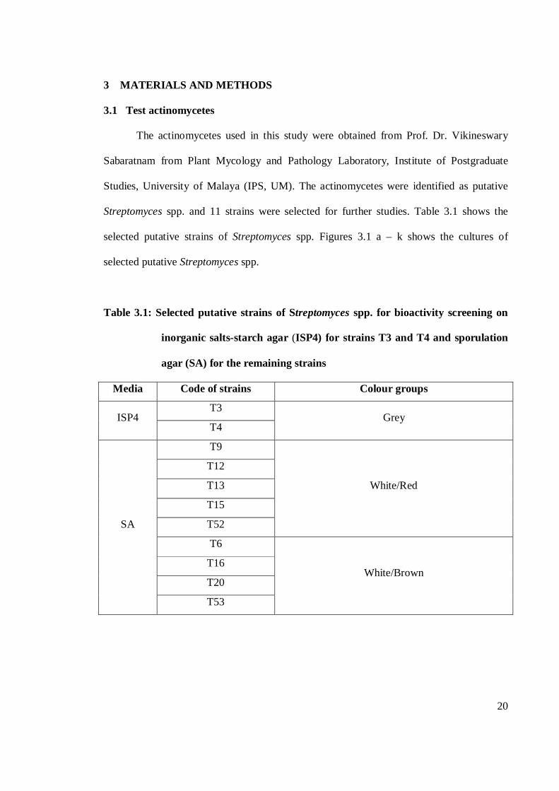
3 MATERIALS AND METHODS
3.1 Test actinomycetes
The actinomycetes used in this study were obtained from Prof. Dr. Vikineswary
Sabaratnam from Plant Mycology and Pathology Laboratory, Institute of Postgraduate
Studies, University of Malaya (IPS, UM). The actinomycetes were identified as putative
Streptomyces spp. and 11 strains were selected for further studies. Table 3.1 shows the
selected putative strains of Streptomyces spp. Figures 3.1 a – k shows the cultures of
selected putative Streptomyces spp.
Table 3.1: Selected putative strains of Streptomyces spp. for bioactivity screening on
inorganic salts-starch agar (ISP4) for strains T3 and T4 and sporulation
agar (SA) for the remaining strains
Media Code of strains Colour groups
ISP4 T3
Grey T4
SA
T9
White/Red
T12
T13
T15
T52
T6
White/Brown T16
T20
T53
21
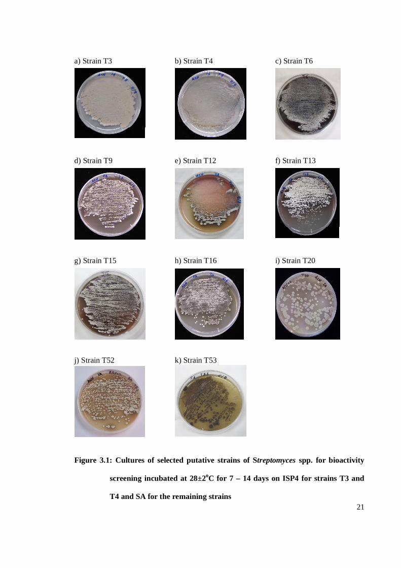
a) Strain T3 b) Strain T4 c) Strain T6 d) Strain T9 e) Strain T12 f) Strain T13 g) Strain T15 h) Strain T16 i) Strain T20 j) Strain T52 k) Strain T53
Figure 3.1: Cultures of selected putative strains of Streptomyces spp. for bioactivity
screening incubated at 28±2oC for 7 – 14 days on ISP4 for strains T3 and
T4 and SA for the remaining strains
22
3.1.1 Preparation of stock cultures
The colonies to be preserved were selected from the agar culture with heavy growth.
The agar culture was aseptically cut into small 0.5x0.5mm pieces using sterilized loop. The
plugs were then transferred into sterilized Bijou bottles containing 4ml of 30% (v/v)
glycerol (Wellington and Williams, 1978) (Appendix A, 1). The stock cultures were then
stored at -20±2oC. For each actinomycetes strain, the stock cultures were prepared in
triplicates.
3.1.2 Preparation of pure cultures
Pure cultures of actinomycetes strains could be prepared using the stock cultures.
The frozen stock cultures were left at 10±2oC overnight and then brought to room
temperature before it was used to prepare pure cultures. The plugs were removed from the
glycerol stock using a sterilized loop and transferred to growth media on agar plates. The
actinomycetes strains were then streaked on the surface of the growth media.
Actinomycetes strains T3 and T4 were streaked on inorganic salts-starch agar (ISP4)
(Appendix A, 2). The remaining strains were streaked on sporulation agar (SA) (Appendix
A, 3). The plates were then incubated at 28±2oC for 7-14 days. The pure cultures were also
prepared in triplicates for each actinomycetes strain.
3.2 Test microorganisms
The test microorganisms used in this study were divided into two groups, the
pathogenic bacteria and fungi. The pathogenic bacteria, Enterobacter faecalis,
Staphylococcus aureus, Bacillus subtilis, Pleisiomonas shigelloides and Pseudomonas
aeruginosa were obtained from Prof. Dr. Thong Kwai Lin from the Institute of
23
Test microorganisms (4-5 streaks) with about 0.5-1cm from the border of the actinomycetes Actinomycetes strains
Postgraduate Studies, University of Malaya (IPS, UM). The fungi, Candida parapsilosis
and Candida albicans were obtained from Prof. Ng Kee Peng.
The pathogenic bacteria were grown on nutrient agar (NA) (Appendix A, 4) and
incubated at 37±2oC for 24 hours. Yeasts were grown on Sabaroud-dextrose agar (SDA)
(Appendix A, 5) at 37±2oC for 24 hours.
3.3 Primary screening
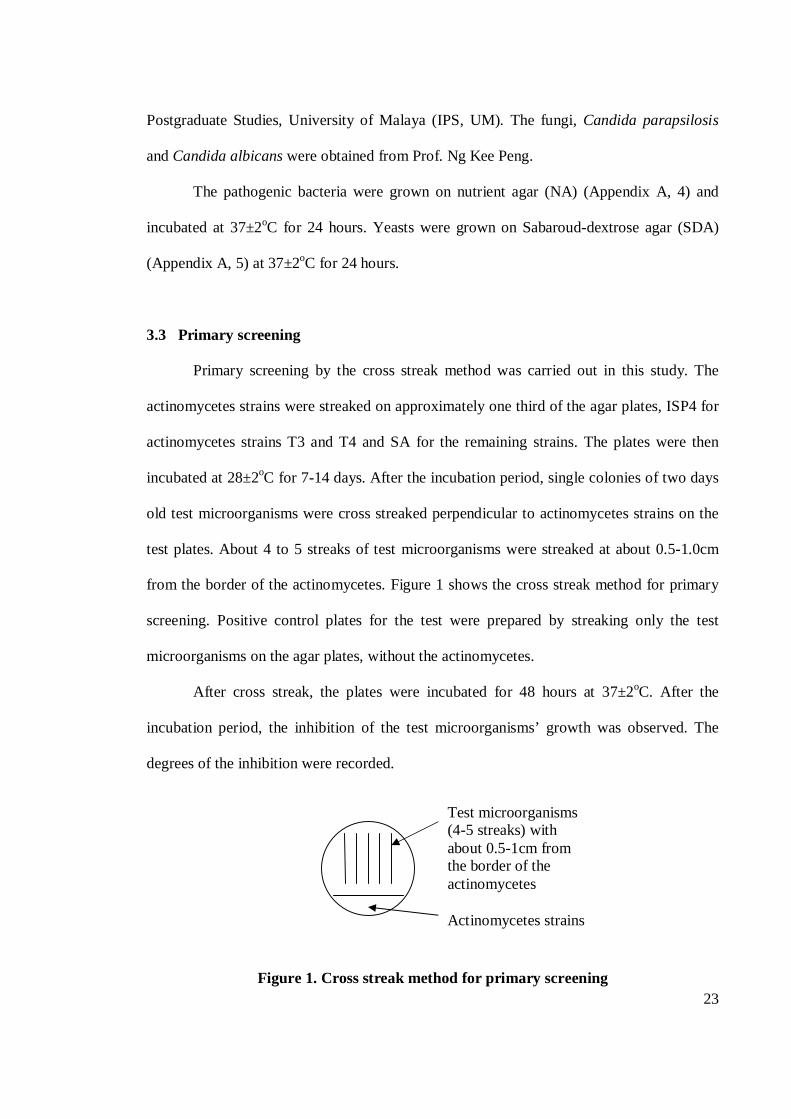
Primary screening by the cross streak method was carried out in this study. The
actinomycetes strains were streaked on approximately one third of the agar plates, ISP4 for
actinomycetes strains T3 and T4 and SA for the remaining strains. The plates were then
incubated at 28±2oC for 7-14 days. After the incubation period, single colonies of two days
old test microorganisms were cross streaked perpendicular to actinomycetes strains on the
test plates. About 4 to 5 streaks of test microorganisms were streaked at about 0.5-1.0cm
from the border of the actinomycetes. Figure 1 shows the cross streak method for primary
screening. Positive control plates for the test were prepared by streaking only the test
microorganisms on the agar plates, without the actinomycetes.
After cross streak, the plates were incubated for 48 hours at 37±2oC. After the
incubation period, the inhibition of the test microorganisms’ growth was observed. The
degrees of the inhibition were recorded.
Figure 1. Cross streak method for primary screening
24
3.4 Secondary screening
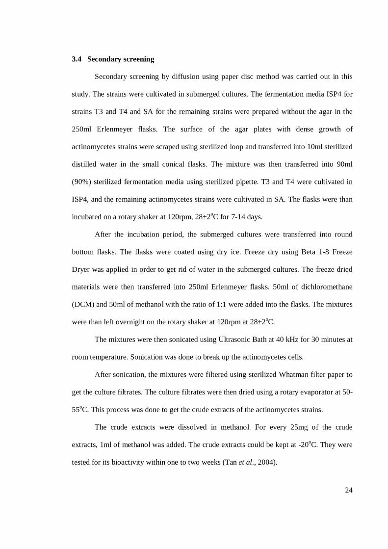
Secondary screening by diffusion using paper disc method was carried out in this
study. The strains were cultivated in submerged cultures. The fermentation media ISP4 for
strains T3 and T4 and SA for the remaining strains were prepared without the agar in the
250ml Erlenmeyer flasks. The surface of the agar plates with dense growth of
actinomycetes strains were scraped using sterilized loop and transferred into 10ml sterilized
distilled water in the small conical flasks. The mixture was then transferred into 90ml
(90%) sterilized fermentation media using sterilized pipette. T3 and T4 were cultivated in
ISP4, and the remaining actinomycetes strains were cultivated in SA. The flasks were than
incubated on a rotary shaker at 120rpm, 28±2oC for 7-14 days.
After the incubation period, the submerged cultures were transferred into round
bottom flasks. The flasks were coated using dry ice. Freeze dry using Beta 1-8 Freeze
Dryer was applied in order to get rid of water in the submerged cultures. The freeze dried
materials were then transferred into 250ml Erlenmeyer flasks. 50ml of dichloromethane
(DCM) and 50ml of methanol with the ratio of 1:1 were added into the flasks. The mixtures
were than left overnight on the rotary shaker at 120rpm at 28±2oC.
The mixtures were then sonicated using Ultrasonic Bath at 40 kHz for 30 minutes at
room temperature. Sonication was done to break up the actinomycetes cells.
After sonication, the mixtures were filtered using sterilized Whatman filter paper to
get the culture filtrates. The culture filtrates were then dried using a rotary evaporator at 50-
55oC. This process was done to get the crude extracts of the actinomycetes strains.
The crude extracts were dissolved in methanol. For every 25mg of the crude
extracts, 1ml of methanol was added. The crude extracts could be kept at -20oC. They were
tested for its bioactivity within one to two weeks (Tan et al., 2004).
25
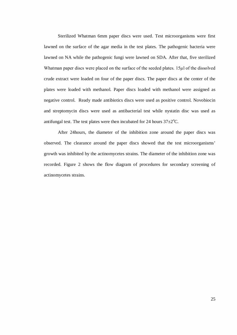
Sterilized Whatman 6mm paper discs were used. Test microorganisms were first
lawned on the surface of the agar media in the test plates. The pathogenic bacteria were
lawned on NA while the pathogenic fungi were lawned on SDA. After that, five sterilized
Whatman paper discs were placed on the surface of the seeded plates. 15µl of the dissolved
crude extract were loaded on four of the paper discs. The paper discs at the center of the
plates were loaded with methanol. Paper discs loaded with methanol were assigned as
negative control. Ready made antibiotics discs were used as positive control. Novobiocin
and streptomycin discs were used as antibacterial test while nystatin disc was used as
antifungal test. The test plates were then incubated for 24 hours 37±2oC.
After 24hours, the diameter of the inhibition zone around the paper discs was
observed. The clearance around the paper discs showed that the test microorganisms’
growth was inhibited by the actinomycetes strains. The diameter of the inhibition zone was
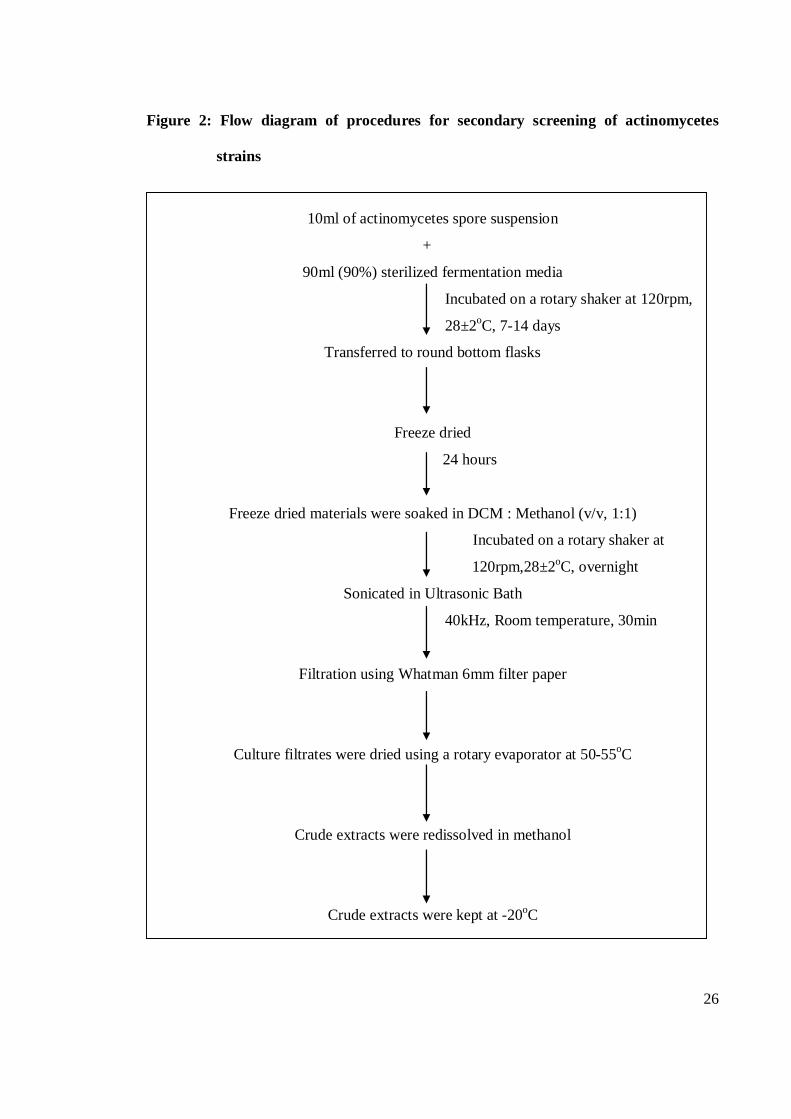
recorded. Figure 2 shows the flow diagram of procedures for secondary screening of
actinomycetes strains.
26
Figure 2: Flow diagram of procedures for secondary screening of actinomycetes
strains
10ml of actinomycetes spore suspension
+
90ml (90%) sterilized fermentation media
Incubated on a rotary shaker at 120rpm,
28±2oC, 7-14 days
Transferred to round bottom flasks
Freeze dried
24 hours
Freeze dried materials were soaked in DCM : Methanol (v/v, 1:1)
Incubated on a rotary shaker at
120rpm,28±2oC, overnight
Sonicated in Ultrasonic Bath
40kHz, Room temperature, 30min
Filtration using Whatman 6mm filter paper
Culture filtrates were dried using a rotary evaporator at 50-55oC
Crude extracts were redissolved in methanol
Crude extracts were kept at -20oC
27
3.5 Media optimization
In optimization of media for production of bioactive compounds, only one
Streptomyces sp. was selected and tested against one selected test microorganism. In this
study, the Streptomyces sp. selected was strain T53. The strain was tested against
pathogenic bacteria, E. faecalis.
In media optimization, only secondary screening method was applied. The
processes applied were as mentioned earlier (Chapter 3, Section 3.4, page 24 – 25).
Howevernthree growth media in liquid form were used. The growth media were yeast
extract-malt extract agar (ISP2) (Appendix A, 6), ISP4 and SA. Dimethylsulfoxide
(DMSO) was used as a solvent.
In optimization of media for production of bioactive compounds, submerged culture
fermentation was carried out in two conditions, static and agitation. For each of the growth
medium, twelve flasks of the submerged cultures were prepared in 250ml Erlenmeyer
flasks. For submerged culture fermentation in static condition, the cultures were incubated
at 28±2oC for twelve days without agitation. On the other hand, submerged cultures
fermentation in agitation condition were incubated at 28±2oC for twelve days and agitated
at 120rpm.
Every three days, three flasks were taken out. The pH of the cultures was recorded.
The submerged cultures were processed as mentioned earlier (Chapter 3, Section 3.4, page
24 – 25) to get crude extracts. The crude extracts were used to test for antibacterial
activities of the actinomycetes strain using the paper disc method. Ready made
streptomycin disc and novobiocin disc were used as positive control for the test. The
diameter of the inhibition zone around the paper discs was observed and recorded.
28
4 RESULTS AND DISCUSSIONS
4.1 Antagonistic pattern in primary and secondary screening
Table 4.1 shows the antibacterial and antifungal activities of selected Streptomyces
spp. in primary and secondary screening. The number of active Streptomyces spp. against
test bacteria was higher in secondary screening compared to primary screening. However,
the number of active strains against test fungi was higher in secondary screening compared
to primary screening.
Table 4.1: Antibacterial and antifungal activities of selected Streptomyces spp. in
primary and secondary screening
Number of active strainsa
Antibacterialb Primary screeningd 9
Secondary screeninge 11
Antifungalc Primary screening 0
Secondary screening 7 a Number of active strains antagonistic against at least one test microorganism in primary and secondary
screening. Total strains tested for bioactivity were eleven; b Antibacterial activity against: E. faecalis, S.
aureus, P. shigelloides, B. subtilis and P. aeruginosa; c Antifungal activity against: C. parapsilosis and C.
albicans; d Primary screening: Cross streak method was applied on ISP4 for T3 and T4 and SA for the
remaining strains. The plates were incubated for 48 hours at 37±2oC; e Secondary screening: Paper disc
method was applied on NA for test bacteria and on SDA for test fungi. The discs were loaded with 15µl of
crude extracts and the plates were incubated for 24 hours at 37±2oC.
In primary screening, nine strains showed antagonistic activity against at least one
test pathogenic bacteria. However, no antifungal activity was recorded against the test
fungi. In secondary screening, all strains showed antagonistic activity against at least one
test pathogenic bacteria. The antifungal activity was also recorded where seven strains
showed antagonistic activity against at least one test fungi.
29
4.1.1 Antibacterial activity
Table 4.2 shows the antibacterial activity of selected Streptomyces spp. against test
bacteria in primary and secondary screening. Total number of Streptomyces spp. tested for
antagonistic activity against test bacteria was eleven. Antibacterial activity against test
bacteria was recorded in both primary and secondary screening. The antibacterial activity
was recorded against both Gram-positive bacteria and Gram-negative bacteria.
Table 4.2: Antibacterial activity of selected Streptomyces spp. against test bacteria in
primary and secondary screening
Antagonistic activity
Test bacteria
Primary screeninga Secondary screeningb
Number of active
strains
Number of active
strains
Gram-positive
bacteria
E. faecalis 8 11
S. aureus 6 11
B. subtilis 6 11
Gram-negative
bacteria
P. shigelloides 6 11
P. aeruginosa 7 4 a Primary screening: Cross streak method was applied on ISP4 for T3 and T4 and SA for the remaining
strains. The plates were incubated for 48 hours at 37±2oC; b Secondary screening: Paper disc method was
applied on NA agar medium. The discs were loaded with 15µl of crude extracts and the plates were incubated
for 24 hours at 37±2oC.
The number of active strains against test bacteria was higher in secondary screening
compared to primary screening (Table 4.1). In primary screening, eight strains showed
antagonistic activity against E. faecalis. Seven strains showed antagonistic activity against
P. aeruginosa and six strains showed antagonistic activity against S. aureus, B. subtilis and
P. shigelloides.
In secondary screening, the number of active strains against all Gram-positive
bacteria increased. All strains showed antagonistic activity against the Gram-positive
30
bacteria. The number of active strains against P. shigelloides also increased. All strains
showed antagonistic activity against the pathogen. However, the number of active strains
against P. aeruginosa decreased to only four strains. In this study, the antagonistic pattern
of P. aeruginosa was not similar with other test bacteria. The antagonistic activity against
the pathogen was higher in primary screening compared to secondary screening.
Table 4.3 shows the antibacterial activity of selected Streptomyces spp. against test
bacteria in secondary screening. The inhibition zones around the paper discs mounted with
crude extracts of selected Streptomyces spp. was recorded. In this study, the degree of
inhibition zones were grouped into weak (<10mm), moderate (10-15mm) and good
(>15mm). Most of the active strains exhibited weak inhibition against all test bacteria. A
few active strains exhibited moderate inhibition against all test bacteria except E. faecalis.
Good inhibition was only recorded against E. faecalis and P. shigelloides.
Ten strains showed weak inhibition against E. faecalis and S. aureus. Nine strains
showed weak inhibition against B. subtilis. Seven and three strains showed weak inhibition
against P. shigelloides and P. aeruginosa, respectively. Moderate inhibition by one active
strain was recorded against S. aureus. One strain also showed moderate inhibition against
P. aeruginosa. Two strains showed moderate inhibition against B. subtilis and P.
shigelloides. Active strains only showed good inhibition against E. faecalis and P.
shigelloides. One strain showed good inhibition against E. faecalis. Two strains showed
good inhibition against P. shigelloides.
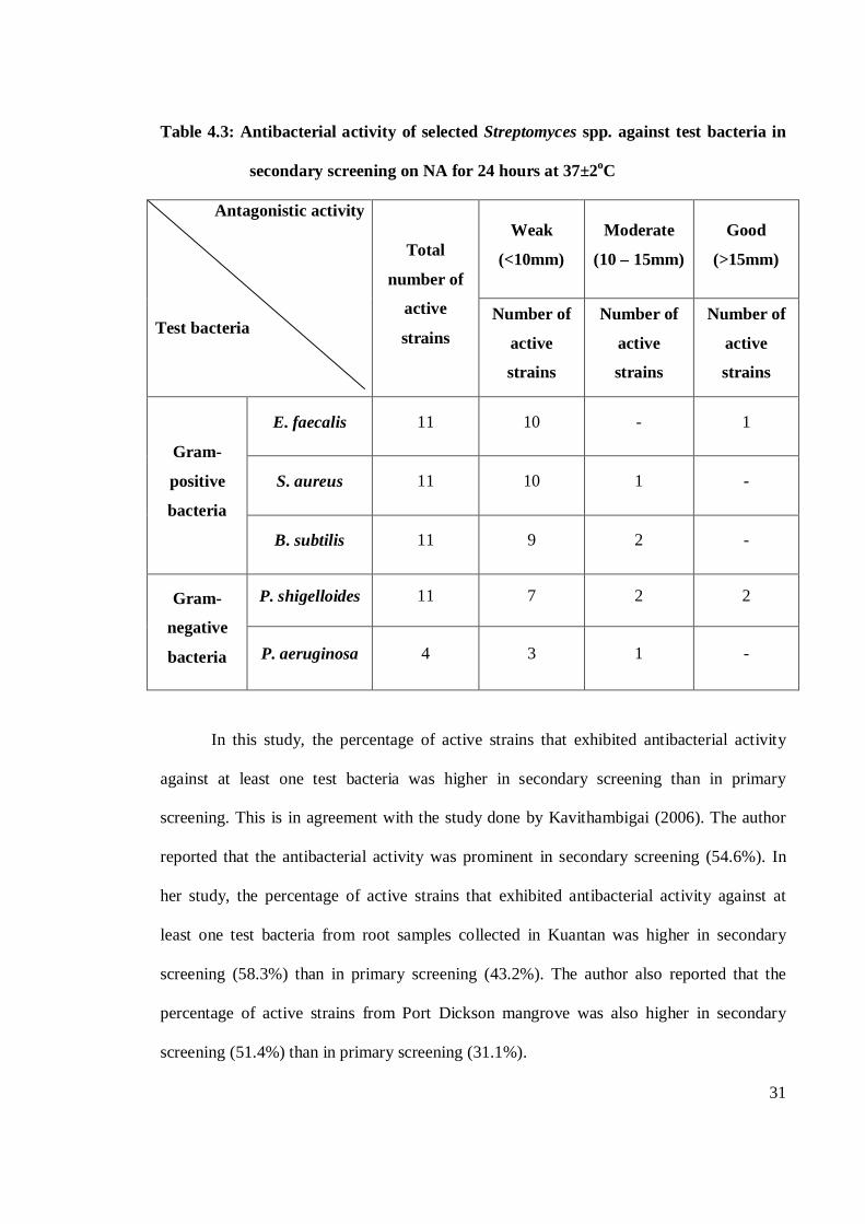
31
Table 4.3: Antibacterial activity of selected Streptomyces spp. against test bacteria in
secondary screening on NA for 24 hours at 37±2oC
Antagonistic activity
Test bacteria
Total
number of
active
strains
Weak
(<10mm)
Moderate
(10 – 15mm)
Good
(>15mm)
Number of
active
strains
Number of
active
strains
Number of
active
strains
Gram-
positive
bacteria
E. faecalis 11 10 - 1
S. aureus 11 10 1 -
B. subtilis 11 9 2 -
Gram-
negative
bacteria
P. shigelloides 11 7 2 2
P. aeruginosa 4 3 1 -
In this study, the percentage of active strains that exhibited antibacterial activity
against at least one test bacteria was higher in secondary screening than in primary
screening. This is in agreement with the study done by Kavithambigai (2006). The author
reported that the antibacterial activity was prominent in secondary screening (54.6%). In
her study, the percentage of active strains that exhibited antibacterial activity against at
least one test bacteria from root samples collected in Kuantan was higher in secondary
screening (58.3%) than in primary screening (43.2%). The author also reported that the
percentage of active strains from Port Dickson mangrove was also higher in secondary
screening (51.4%) than in primary screening (31.1%).
32
Peela et al. (2005) reported that 44% of marine actinomycetes isolated from the Bay
of Bengal showed antibacterial activity. Tan et al. (2004) reported that 25 (51%) out of 49
crude extracts of actinomycetes exhibited inhibition against at least one test bacteria. The
ability of actinomycetes exhibiting antibacterial activity was once again proved in the
present study. Antimicrobial compounds work against bacteria by way of a variety of
mechanisms. Antimicrobial agents act by interfering with cell wall synthesis, cell
membrane function, nucleic acid synthesis, ribosomal function and folate synthesis. Cell
wall synthesis can be affected either by preventing the production of new cell walls,
effectively stopping the cell from reproducing, or by providing analogues for the bacteria to
include as the new cell is produced, leading to cell lysis and death (Appelbaum and Jacobs,
2005).
Tan et al. (2004) reported that seven strains of actinomycetes exhibited broad
spectrum inhibition against both Gram-positive and Gram-negative bacteria. Seventeen
(68%) out of the 25 strains inhibited Gram-positive test bacteria and one (4%) inhibited
Gram-negative test bacteria. Selvin et al. (2004) reported that the extracellular products
(ECPs) of Streptomyces sp. strain BTL7 isolated from marine sponge, Dendrilla nigra
successfully prevented the growth of the Gram-positive bacteria to the extent of 60%
whereas the inhibitory potential was decreased towards the Gram-negative bacteria (40%).
Zheng et al. (2000) reported that eleven (65%) out of seventeen tested strains showed
antibacterial activity against Gram-positive bacteria, eight (47%) against Gram-negative
bacteria and four (23.5%) against both Gram-positive and Gram-negative bacteria. Bernan
et al. (1994) reported that the extracellular proteins (ECPs) of actinomycete strain (LL-
31F508) isolated form an intertidal sediment collected in Key West, Florida, showed potent
antimicrobial activity against Staphylococcus spp. and Enterococcus spp. Pisano et al.
(1992) reported that 68% out of 85 marine actinomycetes capable of degrading chitin
33
exhibited antibacterial activity against Gram-positive bacteria. Only 11% of the
actinomycetes were active against Gram-negative bacteria. This is in agreement with the
results obtained in the present study because the antibacterial activity was prominent
against Gram-positive bacteria compared to Gram-negative bacteria.
Sujatha et al. (2005) reported that streptomycete strain BT-408 showed a broad
spectrum against Gram-positive and Gram-negative bacteria, fungi and yeast including the
pathogenic methicillin resistant S. aureus when tested with crude culture filtrates and also
with purified antibiotic SBR-22. Peela et al. (2005) reported that Streptomyces sp strains
BT606 and BT652 showed antimicrobial activites against S. aureus and P. aeruginosa.
Thorne and Alder (2002) reported that Streptomyces roseosporus produced Daptomycin
which exhibited antibacterial activity against S. aureus. Kavithambigai (2006) reported that
in secondary screening, the percentage of active strains against Gram-positive bacteria
increased to 48.2% and 39.8% for B. subtilis and S. aureus, respectively. This is again in
agreement with the result obtained in the present study because test actinomycetes
exhibited antibacterial activity against Gram-positive bacteria including S. aureus and B.
subtilis.
Pisano et al. (1986) reported that from their studies, 19 strains of actinomycetes
isolated from marine sediments in Sandy Hook Bay, New Jersey displayed antimicrobial
activity. The most prominent inhibitory activity noted was directed against Gram-positive
bacteria (S. aureus and B. subtilis). All of the actinomycetes displayed significant inhibition
of B. subtilis and 12 inhibited the growth of S. aureus, whereas only one inhibited P.
aeruginosa. This is in agreement with the results obtained in the present study because
prominent inhibitory activity was also noted against Gram-positive bacteria (S. aureus and
B. subtilis).
34
Kavithambigai (2006) reported that in secondary screening, the number of active
strains against P. aeruginosa decreased (4.0%). The author also reported that 50% of active
strains showed weak antibiosis (inhibition zone in between 8-10mm) against P. aeruginosa.
This is in agreement with the result obtained in the present study. In this study, the
antagonistic activity against P. aeruginosa decreased from 7 strains in primary screening to
4 strains in secondary screening. Present study also showed that active strains exhibited
weak inhibition zone against P. aeruginosa.
Present study showed that majority of active strains exhibited weak inhibition
against B. subtilis and S. aureus. This result, however, is not in agreement with the results
obtained by Kavithambigai (2006) because the majority of active strains showed moderate
antibiosis (inhibition zone in between 11-20mm) against B. subtilis (54.5%) and S. aureus
(54.1%). The author also reported that 25% of active strains showed good antibiosis
(inhibition zone more than 20mm) against B. subtilis and 16.2% against S. aureus. This
might suggests that the marine actinomycetes might not have the same antibacterial activity
with the actinomycetes isolated from root samples. During the screening of the novel
secondary metabolite, actinomycetes isolates are often encountered which show antibiotic
activity on agar but not in liquid culture. This might explain the higher number of
antibacterial activity against P. aeruginosa in primary screening compared to secondary
screening in this study.
According to Kokare et al. (2004) during the screening of the novel secondary
metabolite, actinomycetes isolates are often encountered which show more active
antimicrobial activity against Gram-positive bacteria than Gram-negative bacteria. Pandey
et al. (2006) reported that the reason for different sensitivity between Gram-positive and
Gram-negative bacteria could be ascribed to the morphological differences between these
microorganisms, Gram-negative bacteria having an outer polysaccharide membrane
35
carrying the structural lipopolysaccharide components. This makes the cell wall
impermeable to lipophilic solutes. On the other hand, the Gram-positive bacteria only have
an outer peptidoglycan layer. Brock et al. (1994) reported that porins structure that situated
in outer membrane of Gram-negative bacteria serve as membrane channels for the entrance
and exit of hydrophilic low-molecular weight substances. When the structure comes in
contact with high molecular antibiotics, such porins can be closed to prevent antibiotic.
This increased the resistance of Gram-negative bacteria to antibiotics. This explains the
higher number of active strains against Gram-positive bacteria compared to Gram-negative
bacteria.
Pisano et al. (1989) reported that 68 (46%) out of 147 strains of actinomycetes
isolated from the sediments of the south shore of Brooklyn, the East River and New Jersey
exhibited antimicrobial activity. Most of the inhibitory activity was directed against Gram-
positive bacteria. B. subtilis was the most susceptible, followed closely by S. aureus.
Activity against Gram-negative bacteria was minimal with only eight or 5% of the isolates
proving effective. Only one strain (strain SG-944) inhibited the growth of P. aeruginosa.
41(28%) of the isolates was inhibitory to one or more of the fungal test species. Pisano et
al. (1992) reported that B. subtilis was inhibited by 58% of the active marine actinomycetes
strains, S. aureus by 50%, P. aeruginosa by 5% and C. albicans by 32%.
According to Kavithambigai (2006), the antibacterial activity might be stimulated
by intracellular metabolites, which were bound inside membrane of actinomycetes and
released during submerged cultivation. This explains the higher number of antibacterial and
antifungal activity in secondary screening compared to primary screening.
36
4.1.2 Antifungal activity
Table 4.4 shows the antifungal activity of selected Streptomyces spp. against test
fungi in primary and secondary screening. Total number of Streptomyces spp. tested for
antagonistic activity against test fungi was eleven. Antifungal activity was only recorded in
secondary screening. In primary screening, selected Streptomyces spp. did not inhibit the
test fungi.
Table 4.4: Antifungal activity of selected Streptomyces spp. against test fungi in
primary and secondary screening
Antagonistic
activity
Test fungi
Primary screeninga Secondary screeningb
Number of active strains Number of active strains
C. parapsilosis - 7
C. albicans - 4 a Primary screening: Cross streak method was applied on SA agar medium and the plates were incubated for
48 hours at 37±2oC; b Secondary screening: Paper disc method was applied on SDA agar medium. The discs
were loaded with 15µl of crude extracts and the plates were incubated for 24 hours at 37±2oC.
In secondary screening, seven strains showed antagonistic activity against C.
parapsilosis. Four strains showed antagonistic activity against C. albicans. Table 4.5 shows
the antifungal activity of selected Streptomyces spp. against test fungi in secondary
screening. All active strains exhibited weak inhibition against the test fungi.
The number of active strains that showed weak inhibition against C. parapsilosis
was seven. Four strains showed weak inhibition against C. albicans. The most susceptible
fungal was C. parapsilosis.
37
Table 4.5: Antifungal activity of selected Streptomyces spp. against test fungi in
secondary screening on SDA for 24 hours at 37±2oC
Antagonistic
activity
Test fungi
Total number
of active strains
Weak
(<10mm)
Moderate
(10 – 15mm)
Good
(>15mm)
Number of
active strains
Number of
active strains
Number of
active strains
C. parapsilosis 7 7 - -
C. albicans 4 4 - -
Present study also showed that active strains exhibited higher percentage of
antifungal activity against at least one test fungi in secondary screening than in primary
screening. This is however not in agreement with the study done by Kavithambigai (2006).
The author reported that active strains exhibited higher antifungal activity in primary
screening for both root samples from Kuantan (69.7%) and Port Dickson (59.7%). This
might suggest that marine actinomycetes might not have the same antifungal substance with
the actinomycetes isolated from root samples.
Kavithambigai (2006) reported that antifungal activity was prominent in secondary
screening. The antifungal activity was more prominent on phytopathogenic fungi (F.
oxysporum cubense race 1; 26.1%, F. oxysporum cubense race 2; 30.4%, F. oxysporum
cubense race 4; 30.4%, Colletotrichum sp.; 21.7%, G. boninense ; 31.5%) compared to C.
albicans (11.6%) and C. parapsilosis (9.8%). Most of the active strains showed moderate
antibiosis (inhibition in between 11-20mm) against C. albicans (83.3%) and C. parapsilosis
(58.8%). Atta and Ahmad (2009) reported that an actinomycete culture was isolated from a
38
soil sample collected from Alam Alroom districted, Marsa Matrouh governorate, Egypt.
This isolate AZ-AR-262 was found to be active against unicellular and filamentous fungi.
Peela et al. (2005) reported that Streptomyces sp. strains BT606 and BT624 were
active against C. albicans. Tan et al. (2004) reported that 15 (31%) of 49 crude extracts
tested exhibited inhibition against the test fungi including C. albicans and C. parapsilosis.
Six out of the fifteen strains exhibited inhibition against C. parapsilosis and all strains
inhibited C. albicans. Zheng et al. (2000) reported that nine (52.9%) out of seventeen tested
strains exhibited antifungal activity against fungi including C. albicans.
Vikineswary et al. (1997) reported that eleven (33%) out of 33 actinomycetes
strains isolated from a tropical mangrove ecosystem showed activity against all the test
fungi including C. albicans. Okazaki and Okami (1976) reported that only two strains out
of 37 antagonistic actinomycetes exhibited antifungal properties. This is also in agreement
with the present study because the percentage of antifungal activity was smaller. Shiomi et
al. (2005) reported that the antimycins (produced by Streptomyces sp. K01-0031) have also
other biological properties such as antifungal activity, inhibition of enzymatic activity as
well as the ability to induce the death of cancer cells.
Vikineswary et al. (1997) reported that 11 strains (33%) out of 33 strains of
actinomycetes isolated from marine environment showed antifungal activity by cross-plug
method. The authors also reported that ten out of eleven actinomycetes strains exhibited
antifungal activity against C. albicans by shake flasks studies. This is again in agreement
with the results obtained in the present study because antifungal activity was only recorded
against fungi including C. albicans only in secondary screening. The authors reported that
only the culture supernatant was tested for bioactivity. The bioactive substance could be
membrane-bound or intracellular. However, in the present study, all the culture mixtures
were tested for bioactivity.
39
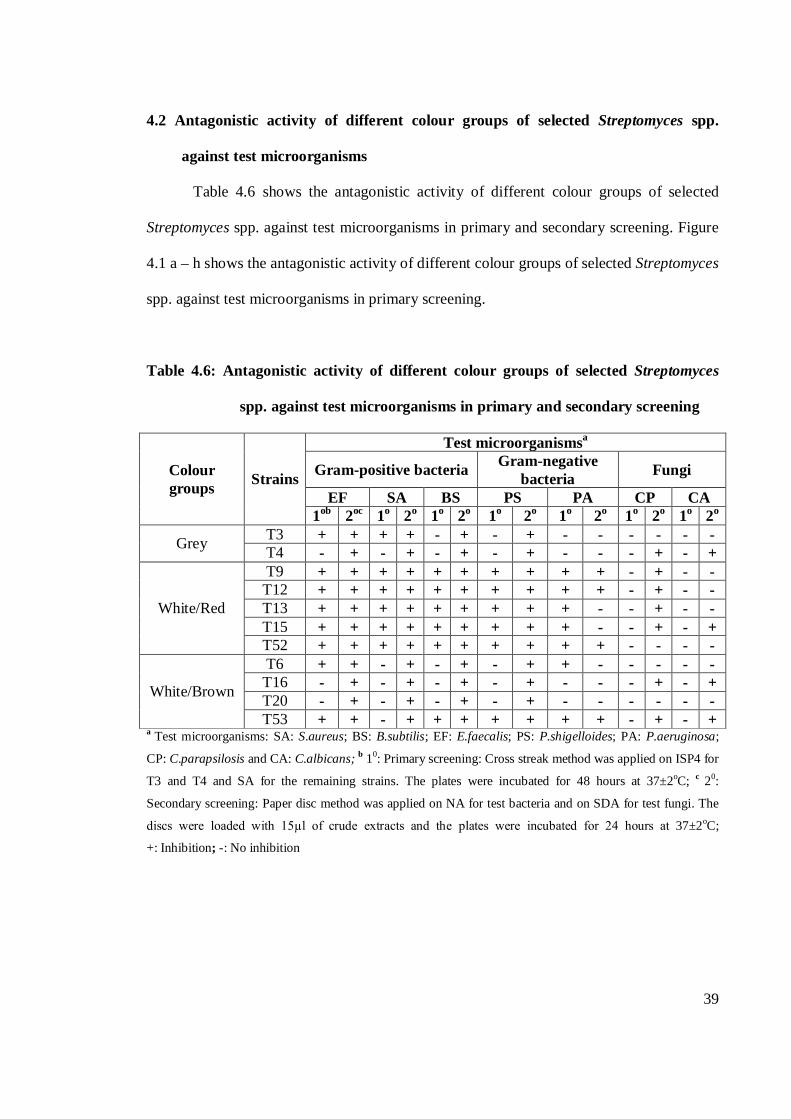
4.2 Antagonistic activity of different colour groups of selected Streptomyces spp.
against test microorganisms
Table 4.6 shows the antagonistic activity of different colour groups of selected
Streptomyces spp. against test microorganisms in primary and secondary screening. Figure
4.1 a – h shows the antagonistic activity of different colour groups of selected Streptomyces
spp. against test microorganisms in primary screening.
Table 4.6: Antagonistic activity of different colour groups of selected Streptomyces
spp. against test microorganisms in primary and secondary screening
Colour groups Strains
Test microorganismsa
Gram-positive bacteria Gram-negative bacteria Fungi
EF SA BS PS PA CP CA 1ob 2oc 1o 2o 1o 2o 1o 2o 1o 2o 1o 2o 1o 2o
Grey T3 + + + + - + - + - - - - - - T4 - + - + - + - + - - - + - +
White/Red
T9 + + + + + + + + + + - + - - T12 + + + + + + + + + + - + - - T13 + + + + + + + + + - - + - - T15 + + + + + + + + + - - + - + T52 + + + + + + + + + + - - - -
White/Brown
T6 + + - + - + - + + - - - - - T16 - + - + - + - + - - - + - + T20 - + - + - + - + - - - - - - T53 + + - + + + + + + + - + - +
a Test microorganisms: SA: S.aureus; BS: B.subtilis; EF: E.faecalis; PS: P.shigelloides; PA: P.aeruginosa;
CP: C.parapsilosis and CA: C.albicans; b 10: Primary screening: Cross streak method was applied on ISP4 for
T3 and T4 and SA for the remaining strains. The plates were incubated for 48 hours at 37±2oC; c 20:
Secondary screening: Paper disc method was applied on NA for test bacteria and on SDA for test fungi. The
discs were loaded with 15µl of crude extracts and the plates were incubated for 24 hours at 37±2oC;
+: Inhibition; -: No inhibition
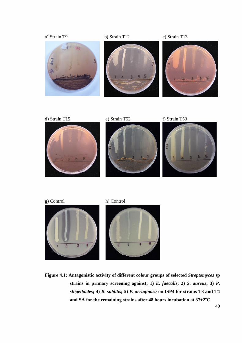
40
a) Strain T9 b) Strain T12 c) Strain T13
d) Strain T15 e) Strain T52 f) Strain T53
g) Control h) Control
Figure 4.1: Antagonistic activity of different colour groups of selected Streptomyces sp
strains in primary screening against; 1) E. faecalis; 2) S. aureus; 3) P.
shigelloides; 4) B. subtilis; 5) P. aeruginosa on ISP4 for strains T3 and T4
and SA for the remaining strains after 48 hours incubation at 37±2oC
41
All strains of grey, white/red and white/brown colour groups inhibited the growth of
all Gram-positive bacteria in secondary screening. Inhibition of the growth of P.
shigelloides by all strains was also recorded in secondary screening. All strains of different
colour groups did not inhibit the growth of test fungi in primary screening.
Table 4.7 shows the inhibition zones (mm) of different colour groups of selected
Streptomyces spp. against test microorganisms in secondary screening. The degree of
inhibition zones was grouped into weak (<10mm), moderate (10-15mm) and good
(>15mm). Figure 4.2 a – l shows the antagonistic activity of different colour groups of
selected Streptomyces spp. against test bacteria in secondary screening. The following
analyses the anatagonistic activity of different colour groups of selected Streptomyces spp.
against test microorganisms in primary and secondary screening.
Grey colour group
The two strains in this colour group, strains T3 and T4 exhibited poor antagonistic
activity against test microorganisms in primary screening (Table 4.6). The antagonistic
activity was only recorded against E. faecalis and S. aureus. The two test microorganisms
were inhibited by strain T3.
In secondary screening, the antagonistic activity was more prominent against Gram-
positive bacteria compared to Gram-negative bacteria. All the two strains inhibited the
growth of all Gram-positive bacteria. Strains T3 and T4 inhibited the growth of only one
test Gram-negative bacteria which was P. shigelloides. Antifungal activity was only
recorded against C. parapsilosis and C. albicans. The test fungi were inhibited by strain T4.
E. faecalis, S. aureus, B. subtilis and P. shigelloides were the most susceptible test
microorganisms in this screening. The microorganisms were inhibited by all strains of grey
colour group.
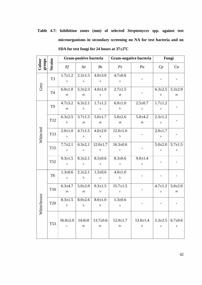
42
Table 4.7: Inhibition zones (mm) of selected Streptomyces spp. against test
microorganisms in secondary screening on NA for test bacteria and on
SDA for test fungi for 24 hours at 37±2oC
Col
our
grou
ps
Stra
ins Gram-positive bacteria Gram-negative bacteria Fungi
Ef Sa Bs Ps Pa Cp Ca
Gre
y T3
1.7±1.2 a
2.3±1.5 a
4.0±3.0 a
4.7±0.6 a
- - -
T4 6.0±1.0
ab
5.3±2.3 ab
4.0±1.0 a
2.7±1.5 ab
- 6.3±2.5
b
5.3±2.9 ab
Whi
te/re
d
T9 4.7±3.2
ab
6.3±2.1 b
1.7±1.2 a
6.0±1.0 b
2.5±0.7 a
1.7±1.2 a
-
T12 6.3±2.5
b
3.7±1.5 ab
5.0±1.7 ab
5.0±2.6 ab
5.0±4.2 ab
2.3±1.2 a
-
T13 2.0±1.0
a
4.7±1.5 a
4.0±2.0 a
12.0±1.0 b
- 2.0±1.7
a -
T15 7.7±2.1
a
6.3±2.1 a
12.0±1.7 b
16.3±0.6 c
- 5.0±2.0
a
5.7±1.5 a
T52 9.3±1.5
a
8.3±2.1 a
8.3±0.6 a
8.3±0.6 a
9.0±1.4 a
- -
Whi
te/b
row
n
T6 1.3±0.6
a
2.3±2.1 b
1.3±0.6 a
4.0±1.0 b
- - -
T16 6.3±4.7
ab
5.0±2.0 ab
9.3±1.5 b
15.7±1.5 c
- 4.7±1.2
a
5.0±2.0 ab
T20 8.3±1.5
b
8.0±2.6 b
8.0±1.0 b
1.3±0.6 a
- - -
T53 16.0±2.0
c
14.0±0 bc
13.7±0.6 bc
12.0±1.7 bc
13.0±1.4 b
5.3±2.5 a
6.7±0.6 a
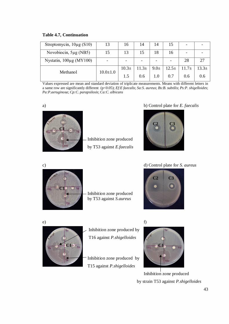
43
Table 4.7, Continuation
Streptomycin, 10µg (S10) 13 16 14 14 15 - -
Novobiocin, 5µg (NB5) 15 13 15 18 16 - -
Nystatin, 100µg (MY100) - - - - - 28 27
Methanol 10.0±1.0 10.3±
1.5
11.3±
0.6
9.0±
1.0
12.5±
0.7
11.7±
0.6
13.3±
0.6 Values expressed are mean and standard deviation of triplicate measurements. Means with different letters in a same row are significantly different (p<0.05); Ef:E faecalis; Sa:S. aureus; Bs:B. subtilis; Ps:P. shigelloides; Pa:P.aeruginosa; Cp:C. parapsilosis; Ca:C. albicans a) b) Control plate for E. faecalis
C2 C3 C1
Inhibition zone produced
by T53 against E.faecalis
c) d) Control plate for S. aureus
C2 C3
C1 Inhibition zone produced
by T53 against S.aureus
e) f)
Inhibition zone produced by
T16 against P.shigelloides
C1 C1
Inhibition zone produced by
T15 against P.shigelloides
Inhibition zone produced
by strain T53 against P.shigelloides
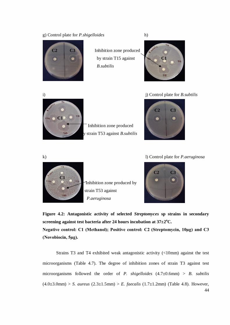
44
g) Control plate for P.shigelloides h)
C2 C3 Inhibition zone produced
by strain T15 against C1
B.subtilis
i) j) Control plate for B.subtilis
C2 C3
C1
Inhibition zone produced
by strain T53 against B.subtilis
k) l) Control plate for P.aeruginosa
C2 C3 C1 Inhibition zone produced by
strain T53 against
P.aeruginosa
Figure 4.2: Antagonistic activity of selected Streptomyces sp strains in secondary
screening against test bacteria after 24 hours incubation at 37±2oC.
Negative control: C1 (Methanol); Positive control: C2 (Streptomycin, 10µg) and C3
(Novobiocin, 5µg).
Strains T3 and T4 exhibited weak antagonistic activity (<10mm) against the test
microorganisms (Table 4.7). The degree of inhibition zones of strain T3 against test
microorganisms followed the order of P. shigelloides (4.7±0.6mm) > B. subtilis
(4.0±3.0mm) > S. aureus (2.3±1.5mm) > E. faecalis (1.7±1.2mm) (Table 4.8). However,
45
there were no significant differences (p>0.05) (Appendix B, Table 1) of the inhibition
zones between the four test microorganisms.
The inhibition zones of strain T4 against test microorganisms followed the order E.
faecalis (6.0±1.0mm) > C. parapsilosis (6.3±2.5mm) > S. aureus (5.3±2.3mm) > B. subtilis
(4.0±1.0mm) > C. albicans (5.3±2.9mm) > P. shigelloides (2.7± 1.5mm) (Table 4.8).
However, there were no significant differences (p>0.05) (Appendix B, Table 2). Strain T4
was the most active strain. The strain exhibited antagonistic activity against four test
bacteria and all test fungi.
White/Red colour group
Strains of white/red colour group were active in both primary and secondary
screening (Table 4.6). All the five strains showed good antagonistic activity against test
bacteria in primary screening. All strains inhibited the growth of all test bacteria. The
antibacterial activity was more prominent than antifungal activity in primary screening. No
antifungal activity was recorded in this screening. E. faecalis, S. aureus, B. subtilis, P.
shigelloides and P. aeruginosa were the most susceptible test bacteria in this screening. The
growth of the bacteria was inhibited by all strains of white/red colour group.
In secondary screening, all the five strains exhibited good antagonistic activity
against all Gram-positive bacteria. All strains also exhibited antagonistic activity against P.
shigelloides. However, only three strains inhibited the growth of P. aeruginosa. The strains
were T9, T12 and T52. Four strains exhibited antagonistic activity against C. parapsilosis.
The strains were T9, T12, T13 and T15. The growth of C. albicans was inhibited by only
one strain which was T15. E. faecalis, S. aureus, B. subtilis and P. shigelloides were the
most susceptible test bacteria in this screening. All strains exhibited antagonistic activity
against the test bacteria.
46
Strain T15 showed good antagonistic activity (>15mm) against P. shigelloides
(16.3±0.6mm) (Table 4.7). This strain also showed moderate antagonistic activity (10-
15mm) against B. subtilis (12.0±1.7mm) and weak antagonistic activity (<10mm) against
other test microorganisms. The inhibition zones of strain T15 against test microorganisms
followed the order P. shigelloides (16.3±0.6mm) > B. subtilis (12.0±1.7mm) > E. faecalis
(7.7±2.1mm) > S. aureus (6.3±2.1mm) > C. albicans (5.7±1.5mm) > C. parapsilosis (5.0±
2.0). The inhibition zones of strain T15 against P. shigelloides was significantly higher than
E. faecalis, S. aureus, B. subtilis, C. parapsilosis and C. albicans, as well as the inhibition
zones of strain T15 against B. subtilis which was significantly higher than E. faecalis, S.
aureus, C. parapsilosis and C. albicans (p=0.05) (Appendix B, Table 10 and 11).
Strain T13 showed moderate antagonistic activity (10-15mm) against P.
shigelloides (12.0±1.0mm) and weak antagonistic activity (<10mm) against other test
microorganisms. The inhibition zones of strain T13 against test microorganisms followed
the order P. shigelloides (12.0±1.0mm) > S. aureus (4.7±1.5mm) > B. subtilis
(4.0±2.0mm) > E. faecalis (2.0±1.0mm) > C. parapsilosis (2.0± 1.7) (Table 4.8). The
inhibition zones of strain T13 against P. shigelloides was significantly higher than E.
faecalis, S. aureus, B. subtilis and C. parapsilosis (p<0.05) (Appendix B, Table 8 and 9).
Strain T9 showed weak antagonistic activity (<10mm) against the test
microorganisms. The inhibition zones of strain T9 against test microorganisms followed the
order P. shigelloides (6.0±1.0mm) > S. aureus (6.3±2.1mm) > P. aeruginosa (2.5±0.7mm)
> E. faecalis (4.7±3.2mm) > B. subtilis (1.7±1.2mm) > C. parapsilosis (1.7±1.2mm). The
inhibition zones of strain T9 against S. aureus and P. shigelloides were significantly higher
than B. subtilis, P. aeruginosa and C. parapsilosis (p<0.05) (Appendix B, Table 5 and 6).
Strain T12 also showed weak antagonistic activity (<10mm) against the test
microorganisms. The inhibition zones of strain T12 against test microorganisms followed
47
the order E. faecalis (6.3±2.5mm) > B. subtilis (5.0±1.7mm) > P. shigelloides
(5.0±2.6mm) > S. aureus (3.7±1.5mm) > C. parapsilosis (2.3± 1.2mm) > P. aeruginosa
(5.0±4.2mm). However, there were no significant differences (p>0.05) (Appendix B, Table
7).
Strain T52 showed weak antagonistic activity (<10mm) against the test
microorganisms. The inhibition zones of strain T52 against test microorganisms followed
the order E. faecalis (9.3±1.5mm) > B. subtilis (8.3±0.6mm) > P. shigelloides
(8.3±0.6mm) > P. aeruginosa (9.0±1.4mm) > S. aureus (8.3±2.1mm). There were no
significant differences between the inhibition zones of strain T52 against all test
microorganisms (p>0.05) (Appendix B, Table 16).
White/Brown colour group
Strains of white/brown colour group exhibited poor antagonistic activity in primary
screening (Table 4.6). Strain T6 only showed antagonistic activity against E. faecalis and P.
aeruginosa in primary screening. Strain T53 inhibited the growth of all test bacteria except
S. aureus. No antifungal activity was recorded in this screening.
In secondary screening, antagonistic activity against Gram-positive bacteria was
more prominent compared to Gram-negative bacteria. All the four strains of white/brown
colour group exhibited antagonistic activity against all Gram-positive bacteria. All strains
also showed antagonistic activity against P. shigelloides. The growth of P. aeruginosa was
only inhibited by strain T53. Only two strains exhibited antagonistic activity against C.
parapsilosis and C. albicans. They were strains T16 and T53. E. faecalis, S. aureus, B.
subtilis and P. shigelloides were the most susceptible test microorganisms in this screening.
All strains of white/brown colour group exhibited antagonistic activity against the test
48
bacteria. Strain T53 was the most active strain. The strain showed antagonistic activity
against all test microorganisms in secondary screening.
Strain T53 exhibited good antagonistic activity (>15mm) against E. faecalis
(16.±2.0mm). The strain also showed moderate antagonistic activity (10-15mm) against S.
aureus (14.0±0.0mm), B. subtilis (13.7±0.6mm), P. shigelloides (12.0±1.7mm) and P.
aeruginosa (13.0±1.4mm) and weak antagonistic activity (<10mm) against the remaining
strains (Table 4.7). The inhibition zones of strain T53 against test microorganisms followed
the order E. faecalis (16.0±2.0mm) > S. aureus (14.0mm)> B. subtilis (13.7±0.6mm) > P.
aeruginosa (13.0±1.4mm) > P. shigelloides (12.0±1.7mm) > C. albicans (6.7±0.6mm) >
C. parapsilosis (5.3±2.5mm). There were significant differences between the inhibition
zones of strain T53 against E. faecalis with P. shigelloides, P. aeruginosa, C. parapsilosis
and C. albicans, as well as the inhibition zones between S. aureus, P. shigelloides, B.
subtilis and P. aeruginosa which were significantly higher than C. parapsilosis and C.
albicans (p=0.05) (Appendix B, Table 17 and 18).
Strain T6 exhibited weak antagonistic (<10mm) against the test microorganisms.
The inhibition zones of strain T6 against test microorganisms followed the order P.
shigelloides (4.0±1.0mm) > E. faecalis (1.3±0.6mm) > B. subtilis (1.3±0.6mm) > S. aureus
(2.3±2.1mm). The inhibition zones of strain T6 against P. shigelloides was significantly
higher than E. faecalis and against B. subtilis as well as the inhibition zone o f B. subtilis
which was significantly higher than S. aureus (p<0.05) (Appendix B, Table 3 and 4).
Strain T16 showed good antagonistic activity (>15mm) against P. shigelloides
(15.7±1.5mm) and weak antagonistic activity (<10mm) against the remaining test
microorganisms. The inhibition zones of strain T16 against test microorganisms followed
the order P. shigelloides (15.7±1.5mm) > B. subtilis (9.3±1.5mm) > C. parapsilosis (4.7±
1.2mm) > S. aureus (5.0±2.0mm) > C. albicans (5.0±2.0mm) > E. faecalis (6.3±4.7mm).
49
The inhibition zones of strain T16 against P. shigelloides was significantly higher than E.
faecalis, S. aureus, B. subtilis, C. parapsilosis and C. albicans, as well as the inhibition
zones of strain T16 against B. subtilis which was significantly higher than C. parapsilosis
(p<0.05) (Appendix B, Table 12 and 13).
Strain T20 exhibited weak antagonistic activity (<10mm) against the test
microorganisms. The inhibition zones of strain T20 against test microorganisms followed
the order B. subtilis (8.0±1.0mm) > E. faecalis (8.3±1.5mm) > S. aurues (8.0±2.6mm) > P.
shigelloides (1.3±0.6mm). The inhibition zones of strain T20 against E. faecalis, S. aureus
and B. subtilis was significantly higher than P. shigelloides (p<0.05) (Appendix B, Table
14 and 15).
The results obtained in present study showed that majority of active strains
exhibited weak antagonistic activity against test microorganisms. Shomura et al. (1979)
reported that the failure of antibiotic productivity during Streptomyces spp. growth in
submerged culture correlated with their mycelial morphology. In agar cultures, the
mycelium of Streptomyces spp. was in filamentous form, but once cultivated in submerged
culture they formed fragments. This resulted in reduced potential of antibiotics production
by Streptomyces spp.
In the present study, majority of strains of grey, white/red and white/brown colour
groups exhibited prominent antagonistic activity against Gram-positive bacteria. Majority
of strains of white/red colour group, strain T3 of grey and two strains of white/brown
colour groups were active against both bacteria and fungi. This might be due to the action
of more than one antibiotic with different targets (Gonzales et al., 1999). The broad
spectrum of the strains might also be due to possible presence of a new antimicrobial
substance which was able to cross both bacterial and fungal cell walls (Tsvetanova and
Price, 2001)
50
Kavithambigai (2006) reported that antimicrobial activity of active strains in grey
colour group was prominent in primary screening. Active strains inhibited the growth of B.
subtilis and S. aureus in both primary and secondary screening. Majority of active strains
showed moderate antibiosis (inhibition between 11-20mm) against B. subtilis and S. aureus
and one strain showed good antibiosis (inhibition more than 20mm) against B. subtilis. This
is however not in agreement with the results obtained in the present study because strains in
grey colour group were more prominent in secondary screening than in primary screening.
The strains also showed weak inhibition zone against the test bacteria. This might explain
the difference in bioactive substances in actinomycetes from marine ecosystem with root
samples.
In this study, strain T53 of white/brown colour group was the most active strain.
The strain inhibited the growth of all test microorganisms. Strain T53 produced inhibition
zone of 13.0±1.4mm against P. aeruginosa, 14.0±0.0mm against S. aureus and 13.7±0.6mm
against B. subtilis. Selvin et al. (2004) reported that the extracellular proteins of
Streptomyces strain BTL7 isolated from marine sponge Dendrilla nigra produced inhibition
zone of 21mm against P. aeruginosa, 16mm against S. aureus and 15mm for B. subtilis.
This is quite similar with the result obtained in the present study. Tan et al. (2004) reported
that comparable inhibition relative to 10µg streptomycin standard was shown by majority
of the strains against S. aureus, B subtilis and P. aeruginosa. Bioactivity of all extracts
tested against P. aeruginosa was the weakest, followed by B. subtilis and strongest against
S. aureus. This is however not comparable with the present study because active strains
produced different degree of inhibition zone against the test bacteria.
Tan et al. (2004) reported that comparable or better inhibition compared to nystatin
standard (15mm) was observed against C. albicans. Strains X13, X46 and X60 produced
inhibition zone bigger by 18% each to nystatin standard. Strain Z250 produced inhibition
51
zone bigger by 29% to nystatin standard. Sacramento et al. (2004) reported that the crude
supernatants of Streptomyces sp. strain 606 showed zones of wide inhibition against yeast
(C. albicans, 23mm). This is however not in agreement with the result obtained in present
study. The test marine actinomycetes produced weak inhibition zone against the test fungi.
This study showed that the inhibiton produced by active strains against the test fungi was
not comparable to nystatin standard. This might suggest that the actinomycetes strains in
this study did not have antifungal substance to inhibit the growth of the test fungi.
Devi et al. (2006) reported that out of three selected and identified actinomycetes,
Streptomyces sp. showed significant antimicrobial activity against human pathogens. The
highest zone of inhibition of 57 mm antimicrobial activity was showed by Streptomyces sp.
against P. aeruginosa. Marine Streptomyces sp. exhibited the highest antibacterial activity
against P. aeruginosa followed by S. aurues. This is not in agreement with the present
result obtained in the present study. The highest inhibition zone was showed against P.
shigelloides, followed by E. faecalis. This might suggest that marine actinomycetes have
different antibacterial substances.
Sacramento et al. (2004) reported that P. aeruginosa was resistant to crude
supernatant, extracts and fractions of Streptomyces sp. strain 606 isolated from a Brazilian
tropical forest soil. This is quite comparable to the result obtained in present study because
only a few strains inhibited the growth the test bacteria.
The search for novel metabolites especially from actinomycetes requires a large
number ofisolates (over thousands) in order to discover a novel compound of
pharmaceutical interest. The search will be more promising if diverse actinomycetes are
sampled and screened (Lo et al., 2002).
52
4.3 Media optimization
Figure 4.3 a – c shows the antagonistic activity of crude extracts of strain T53
against E. faecalis in ISP2 medium. Table 4.8 shows the inhibition zones of crude extracts
of strain T53 against E. faecalis in agitation and static fermentation conditions using three
different media. Inhibition zones of crude extracts of strain T53 against E. faecalis was only
observed in SA and ISP2 media in agitation and static fermentation condition. In SA
medium, in agitation fermentation condition, the inhibition zone was recorded in day 9 and
day 12. In static fermentation condition, the inhibition zone was only observed in day 9. In
ISP2 medium, the inhibition zone was only recorded in day 3, day 6, day 9 and day 12 in
agitation fermentation condition. Plate 4.3 a – c shows the antagonistic activity of crude
extracts of strain T53 against E. faecalis in ISP2 medium. Table 4.9 shows the pH value of
crude extracts of submerged cultures fermentation of T53 in agitation and static
fermentation conditions.
SA medium
In SA medium, antibacterial activity could be observed in both agitation and static
fermentation conditions (Table 4.8). In agitation fermentation, the antibacterial activity was
recorded in day 9 and day 12. Antibacterial activity in static fermentation condition was
only observed in day 9.
The inhibition zones of strain T53 against E. faecalis in SA medium followed the
order of day 12 (Agitation) (10.3±1.5mm) > day 9 (Agitation) (6.0±5.2mm) > day 9 (Static)
(8.7±0.6mm) (Table 4.8). However, there were no significant differences (p>0.05)
(Appendix B, Table 19) of the inhibition zones between the three different days.
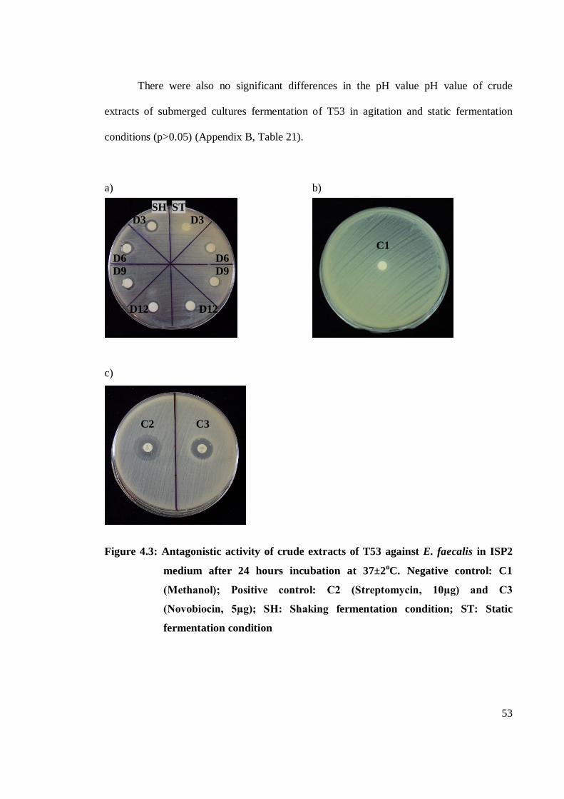
53
There were also no significant differences in the pH value pH value of crude
extracts of submerged cultures fermentation of T53 in agitation and static fermentation
conditions (p>0.05) (Appendix B, Table 21).
a) b)
SH ST D3 D3
C1
D6 D6 D9 D9
D12 D12
c) C2 C3 Figure 4.3: Antagonistic activity of crude extracts of T53 against E. faecalis in ISP2
medium after 24 hours incubation at 37±2oC. Negative control: C1
(Methanol); Positive control: C2 (Streptomycin, 10µg) and C3
(Novobiocin, 5µg); SH: Shaking fermentation condition; ST: Static
fermentation condition
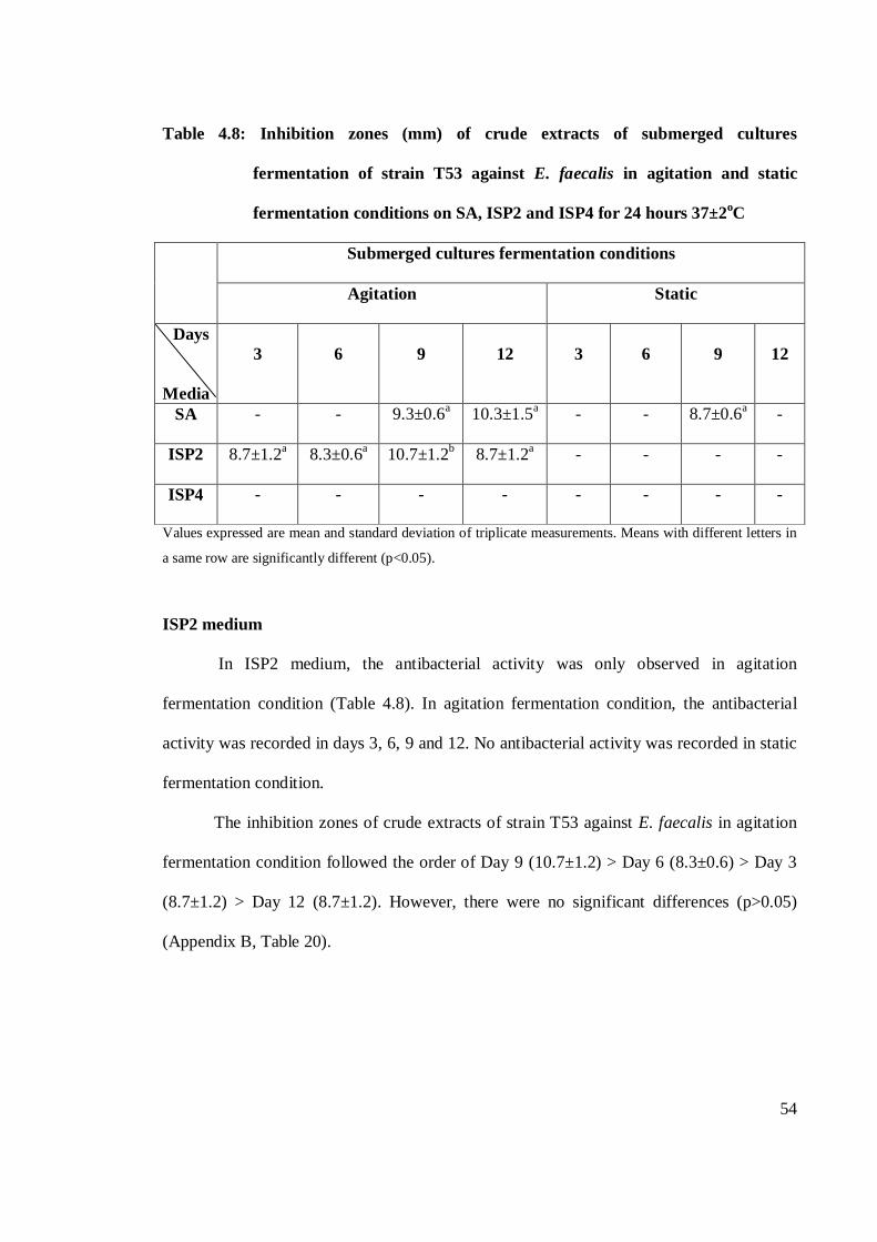
54
Table 4.8: Inhibition zones (mm) of crude extracts of submerged cultures
fermentation of strain T53 against E. faecalis in agitation and static
fermentation conditions on SA, ISP2 and ISP4 for 24 hours 37±2oC
Submerged cultures fermentation conditions
Agitation Static
Days
Media
3 6 9 12 3 6 9 12
SA - - 9.3±0.6a 10.3±1.5a - - 8.7±0.6a -
ISP2 8.7±1.2a 8.3±0.6a 10.7±1.2b 8.7±1.2a - - - -
ISP4 - - - - - - - -
Values expressed are mean and standard deviation of triplicate measurements. Means with different letters in
a same row are significantly different (p<0.05).
ISP2 medium
In ISP2 medium, the antibacterial activity was only observed in agitation
fermentation condition (Table 4.8). In agitation fermentation condition, the antibacterial
activity was recorded in days 3, 6, 9 and 12. No antibacterial activity was recorded in static
fermentation condition.
The inhibition zones of crude extracts of strain T53 against E. faecalis in agitation
fermentation condition followed the order of Day 9 (10.7±1.2) > Day 6 (8.3±0.6) > Day 3
(8.7±1.2) > Day 12 (8.7±1.2). However, there were no significant differences (p>0.05)
(Appendix B, Table 20).
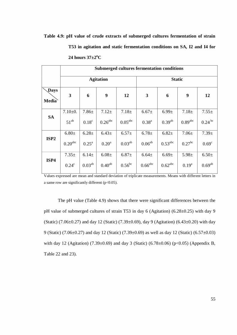
55
Table 4.9: pH value of crude extracts of submerged cultures fermentation of strain
T53 in agitation and static fermentation conditions on SA, I2 and I4 for
24 hours 37±2oC
Submerged cultures fermentation conditions
Agitation Static
Days
Media 3 6 9 12 3 6 9 12
SA 7.10±0.
51ab
7.86±
0.18c
7.12±
0.26abc
7.18±
0.05abc
6.67±
0.38a
6.99±
0.39ab
7.18±
0.89abc
7.55±
0.24 bc
ISP2 6.80±
0.20abc
6.28±
0.25a
6.43±
0.20a
6.57±
0.03ab
6.78±
0.06ab
6.82±
0.53abc
7.06±
0.27bc
7.39±
0.69c
ISP4 7.35±
0.24c
6.14±
0.03ab
6.08±
0.40ab
6.87±
0.56bc
6.64±
0.66abc
6.69±
0.62abc
5.98±
0.19a
6.50±
0.69ab
Values expressed are mean and standard deviation of triplicate measurements. Means with different letters in
a same row are significantly different (p<0.05).
The pH value (Table 4.9) shows that there were significant differences between the
pH value of submerged cultures of strain T53 in day 6 (Agitation) (6.28±0.25) with day 9
(Static) (7.06±0.27) and day 12 (Static) (7.39±0.69), day 9 (Agitation) (6.43±0.20) with day
9 (Static) (7.06±0.27) and day 12 (Static) (7.39±0.69) as well as day 12 (Static) (6.57±0.03)
with day 12 (Agitation) (7.39±0.69) and day 3 (Static) (6.78±0.06) (p<0.05) (Appendix B,
Table 22 and 23).
56
ISP4 medium
In ISP4 medium, no antibacterial activity was recorded in both shaking and static
fermentation condition (Table 4.8). The pH value (Table 4.9) shows that there were
significant differences between day 3 (7.35±0.24) in shaking fermentation condition with
day 6 (6.14±0.03) and day 9 (6.08±0.40) in shaking fermentation condition and day 9
(5.98±0.19) and day 12 (6.50±0.69) in static fermentation condition as well as day 12
(6.87±0.56) in shaking fermentation condition with day 9 (5.98±0.19) in static fermentation
condition (p<0.05) (Appendix B, Table 24 and 25).
From this study, ISP2 was the best medium for bioactive compound production
compared to SA and ISP4 media. Shake flask fermentation was the best fermentation
system compared to static fermentation.
In media optimization, dimethyl sulfoxide (DMSO) was used as a solvent to
dissolve the crude extracts. The usage of methanol in secondary screening (Chapter 3,
Section 3.4, page 24 – 26) was replaced with DMSO because of its low toxicity. Vignes
(2000) reported that DMSO has very low toxicity to humans and the environment,
recyclable after most uses, presents in many foods and plays a significant role in nature’s
Global Sulfur Cycle, highly polar and aprotic solvent. The low toxicity of the solvent
ensured that the inhibition zone of the test pathogens by the actinomycetes strains was not
affected.
The production of bioactive secondary metabolites by actinomycetes is influenced
by a few factors. Srinivasan et al. (1991) reported that antibiotics productivity of strains is
profoundly influenced by factors such as the composition of the nutrient medium,
temperature and duration of incubation. Other than that, the production is also influenced
by fermentation conditions.
57
Antibiotic production generally takes place after the growth reaches the stationary
phase or has passed an optimum level. Bu’lock (1961) reported that the production of
secondary metabolites classically exhibits a two-stage process, with a trophophase (or
growth phase) and an idiophase (or production phase), usually when growth has slowed or
stopped. In the idiophase, productivity depends on maintenance of sub-optimal (cryptic)
growth through control of the concentrations of inorganic phosphate, NH4+, metal ions,
nitrogen or oxygen (Srinivasan et al., 1991).
The nutritional factors strongly influence the production of bioactive secondary
metabolites. The factors include the levels of inorganic phosphorus, metal ions, organic
nitrogen sources and metabolic carbon, and in several cases substrate precursors are also
important for high titres of the desired antibiotics (Higashide, 1984). Pisano et al. (1992)
reported that the nature of the fermentation medium directly influenced the elaboration of
antimicrobial substances.
Carbon sources are one of the most important factors that influence the production
of bioactive secondary metabolites. Glucose is generally used in the fermentation as a
preferred carbon source but catabolite repression is avoided by feeding glucose during the
fermentation. In some cases, carbohydrates, such as lactose, or polysaccharides, such as
starch, which are used more slowly, have been found to be better carbon sources for
antibiotic production (Srinivasan et al., 1991). Tiwaril et al. (2008) reported that
actinomycetes can use peptone as carbon source too. Addition of glucose as carbon source
allows better growth, and up to 0.8% of peptone can be used along with glucose for growth
assessment of actinomcyetes. Farid et al. (2000) reported that natamycin production by
Streptomyces natalensis required glucose in the submerged culture medium with a
concentration of 20g/l. Sujatha et al. (2005) reported that Streptomyces psammmoticus
strain BT-408 isolated from the sediments of Bay of Bengal Ocean produced maximum
58
antibiotic in medium supplemented with glucose as a sole carbon source followed by
fructose and glycerol. The increased in glucose level in the culture from 10 to 12.5 g/l led
to 1.2 fold increase in antibiotic production. Mellouli et al. (2003) reported that the highest
antibacterial activities were obtained when starch at 1% (w/v) was used as sole carbon
source in the presence of traces of mineral oligoelements. In another study reported by
Fguira et al. (2005) highest antimicrobial activities were obtained in Streptomyces spp.
when glucose at 1% (w/v) was used as sole carbon source in the presence of magnesium.
Other than carbon sources, nitrogen sources are also important in the production of
bioactive secondary metabolites. Iwai and Omura (1982) reported that the use of slowly-
consumed antifungal medium carbon sources such as soluble starch and nitrogen sources
such as peptone and yeast-extract have been reported to be effective in the production of
various types of antibiotics by actinomycetes especially the streptomycetes. Marwick et al.
(1999) reported that the nitrogen source is understood to regulate secondary metabolism.
High nitrogen levels have been noted to repress idiophase production of antibiotics
(Marwick et al., 1999). Tiwaril et al. (2008) reported that actinomycetes can use inorganic
nitrogen (KNO3), however organic nitrogen enhanced growth. Farid et al. (2000) reported
that among different nitrogen sources tested, only ammonium sulphate, sodium nitrate and
beef extract were the suitable nitrogen sources in supporting the antibiotic production.
Furthermore, the mixture of beef extract and yeast extract (8 g/l and 2 g/l, respectively)
exhibited a synergistic effect in enhancing the natamycin production reaching about 1.5 g/l.
Sujatha et al. (2005) the highest antibiotic production was obtained in culture of
streptomycete strain BT-408 containing ammonium nitrate as nitrogen source, followed by
cultures containing sodium nitrate, potassium nitrate and alanine.
59
More than that, ammonium is also an important factor that influences the production
of bioactive secondary metabolites. Zhang et al. (1996) found ammonium to stimulate an
antibiotic produced by Streptomyces grisiofuscus. Srinivasan et al. (1991) reported that
ammonium ion often exert a negative effect on production of several antibiotics
(cephamycin, clavulanic acid and streptomycin) and in commercial fermentations use of a
slowly metabolized nitrogen source (releasing ammonia slowly), such as soymeal, partially
overcomes this problem.
Phosphate is also important for the production of bioactive secondary metabolites.
However, Spizek and Tichy (1995) reported that phosphate, although essential for growth,
can at certain concentrations suppress secondary metabolism, inhibiting, for example,
phosphatases and oxygenases. Srinivasan et al. (1991) also reported that fermentative
production of aminoglycoside antibiotics, tetracyclines, polyenes and cephalosporins are
sensitive to the phosphate concentration in the fermentation medium. Farid et al. (2000)
reported that the highest antibiotic production was obtained in a cultivation medium
containing 0.05 g/l of potassium dihydrogen phosphate. Further increase in phosphate
concentration resulted in a significant increase in biomass concomitant with lower
antibiotic production. Sujatha et al. (2005) reported that K2HPO4 at a concentration of 1.2
g/l gave maximum yield of antibiotic. The addition of 0.5 g/l of magnesium sulfate to the
culture medium was optimal for antibiotic production.
Ions might also be important to improve the production of bioactive secondary
metabolites. VanPee (1996) reported that in terms of marine bacteria, the concentration of
bromide ions would seem to be significant in some cases, probably because of the
prevalence of bromide in halogenated marine antibiotics. However, the macrolide antibiotic
productivity tended to decrease when metal ion deficient media were used and also when
the inocula were incubated for long periods and at high temperatures (Higashide, 1984).
60
These might be the reasons that are in agreement with the results obtained in the
present study where ISP2 were the best medium for the production of bioactive secondary
metabolites. Further research should be done to study the compositions of the media.
However, other than nutritional factors, the production of bioactive compounds is
also influenced by fermentation conditions. Submerged fermentation is the most common
submerged culture system that was used to produce bioactive secondary metabolites.
Pelczar and Reid (1972) reported that submerged cultures are widely used to produce many
secondary metabolites because they allow filamentous fungi and bacteria to produce freely
suspended mycelia and pellets, essential for secondary metabolite production.
Agitation fermentation condition is also the most common submerged culture
system that was used to produce bioactive secondary metabolites. Submerged culture
fermentations require a stirred nutrient medium, and in the case aerobic microorganisms, a
supply of oxygen (Duetz and Witholt, 2004). In small-scale fermentation, shake flasks are
usually used. Standard Erlenmeyer flasks is one of the most common types used for small
scale microbial fermentations because they are readily available, their conical shape allows
vigorous shaking without spillages, the small opening limits evaporation so slow-growing
strains can be cultivated without excess moisture loss and the relatively large surface at the
bottom allows a high surface-to-volume ratio (Duetz and Witholt, 2004). This is in
agreement with the results obtained in the present study where antibacterial activity of
selected actinomycetes against test bacteria were recorded in shaking fermentation
conditions. Sacramento et al. (2004) reported that after two weeks incubation under shaking
(agitation) conditions, the supernatants of Streptomyces sp. strain 606 showed the largest
inhibition zones against pathogenic microorganisms.
Other than that, incubation temperature is also an important factor that influences
the production of bioactive secondary metabolites. Sujatha et al. (2005) reported that the
61
increase of the incubation temperature from 20 to 30oC increased the growth of the cells of
streptomycete strain BT-408 by 3-fold. The production of the antibiotic had also increased
by 4.7-fold. Ouhdouch et al. (2001) reported that Streptomyces spp. usually produced
antibiotics at temperatures near 27oC. Iwai and Omura (1992) reported that it would be
interesting to use temperature shifts in antibiotic screening because the cultivation for
antibiotic production was usually performed under constant temperature from the beginning
until the end, but the adequate temperature for growth was not always the same as that for
production.
pH of the submerged cultures was also an important factor that influence the
production of bioactive secondary metabolites. Sujatha et al. (2005) reported that the
maximum antibiotic activity of streptomycete strain BT-408 was obtained at a pH of 7.2 the
results also showed that an incubation time of 96 hours as optimal. Optimization of medium
components and physical parameters (pH, temperature, time) allowed an improvement over
182% in the concentration of antibiotic. Higashide (1984) reported that attainment of high
antibiotic production required optimal concentration of the components in the fermentation
medium, optimum pH and viscosity. Antibiotic production was optimum at 3% NaCl with
slight decrease at 5%. Antibiotic production was comparable in the pH range of 7-9,
whereas at pH10, there was no antibiotic production (Vasavada et al., 2006). Marwick et al.
(1999) reported that the pH level of the growth medium has a marked effect on secondary
metabolism production, with synthesis falling rapidly either side of an optimal level.
Tan et al. (2004) reported that the sensitivity of the antibacterial substances ex-situ
towards light and temperature might influenced the antibacterial activity using crude
extracts. The lower antibacterial activity might also due to naturally unstable antibacterial
substances after prolonged storage or low amount of bioactive compound present in the
crude extracts.
62
4.4 Conclusion
· The antibacterial activity was higher in secondary screening compared to primary
screening. The antifungal activity was only recorded in secondary screening with
significantly higher percentage.
· Gram-positive bacteria were more susceptible than Gram-negative bacteria. E.
faecalis was the most susceptible Gram-positive bacteria. On the other hand, P.
shigelloides was the most susceptible Gram-negative bacteria. The most susceptible
fungi was C. parapsilosis.
· Many of the selected Streptomyces spp. exhibited weak antagonistic activity against
test pathogens. Strains T9, T12, T52 and T53 exhibited broad spectrum activity where
they exhibited antibacterial activity against both Gram-positive and Gram-negative
bacteria. Strains T4, T15, T16 and T53 exhibited antifungal activity against all test
fungi.
· Strains of grey, white/red and white brown colour groups exhibited antagonistic
activity against all Gram-positive bacteria and against one Gram-negative test bacteria
which was P. shigelloides.
· Strain T53 of white/brown colour group exhibited antagonistic activity against all
test bacteria and test fungi. This strain showed good inhibition against E. faecalis.
Hence, it was concluded that strain T53 was the most active strain among the test
microorganisms and E. faecalis was the most susceptible test bacteria.
63
· In media optimization, ISP2 was identified as the best medium for the production of
bioactive secondary metabolites. Agitation fermentation condition was the best
submerged culture fermentation condition for the production of bioactive secondary
metabolites.
· Further investigation should be done to study the composition as well as to identify
the bioactive compounds produced by the selected marine actinomycetes. The
composition of optimized media for the production of bioactive secondary metabolites
should also be studied in further details.
64
REFERENCES
Amyes, S. G. B. and Thomson, C. J. (1995). Antibiotic resistance in the ICU; the eve of
destruction. British Journal of Intensive Care. 5. 263-271.
Anderson, A. L. and Wellington, E. (2001). The taxonomy of Streptomyces and related
genera. International Journal of Systematic and Evolutionary Microbiology. 51. 797-814.
Appelbaum, P. C. and Jacobs, M. R. (2005). Recently approved and investigational
antibiotics for treatment of severe infections caused by Gram-positive bacteria. Current
Opinion in Microbiology. 8. 510-517.
Archer, G. L. (1998). Staphylococcus aureus: A well-armed pathogen. Clinical Infectious
Diseases. 26. 1179-1181.
Atta, H. M. and Ahmad, M. S. (2009). Antimycin-A antibiotic biosynthesis produced by
Streptomyces sp. AZ-AR-262: taxonomy, fermentation, purification and biological
activities. Australian Journal of Basic and Applied Sciences. 3. 126-135.
Baltz, R. H. (2007). Antimicrobials from actinomycetes: back to the future. Microbe. 2.
125-131.
Berdy, J. (2005). Bioactive microbial metabolites. Journal of Antibiotics. 58. 1-26.
65
Bernan, V. S., Montenegro, D. A., Korshalla, J. D., Maiese, W. M., Steinberg, D. A. and
Greenstein, M. (1994). Bioxalomycins new antibiotics produced by the marine
Streptomyces sp. LL-31F508:taxonomy and fermentation. Journal of Antibiotics. 47. 1417-
1424.
Bernan, V. S., Greenstein, M. and Maiese, W. M. (1997). Marine microorganisms as a
source of new natural products. Advance Applied Microbiology. 43. 57-90.
Bull, A. T., Stach, J. E. M., Ward, A. C. and Goodfellow. M. (2005). Marine actinobacteria:
perspectives, challenges and future directions. Antonie van Leeuwenhoek. 87. 65-79.
Bull, A. T., Ward, A. C. and Goodfellow, M. (2000). Search and discovery strategies for
biotechnology: the paradigm shift. Microbiology and Molecular Biology Reviews. 64. 573-
606.
Bull, A. T., Goodfellow, M. and Slater, J. H. (1992). Biodiversity as a source of innovation
in biotechnology. Annual Review of Microbiology. 46. 219-252.
Bu’lock, J. D. (1961). Intermediary metabolism and antibiotic synthesis. Advanced Applied
Microbiology. 3. 293-339.
Busti, E., Monciardini, P., Cavaletti, L., Bamonte, R., Lazzarini, A., Sosio, M. and
Donadio, S. (2006). Antibiotic-producing ability by representatives of a newly discovered
lineage of actinomycetes. Microbiology. 152. 675-683.
66
Campbell, N. A., Reece, J. B. and Mitchell, L. G. (1999). Biology, 5th Edn. Canada:
Benjamin/Cummings. (p.504).
Casadevall, A., Feldmesser, M. and Pirofski, L. (2002). Induced humoral immunity and
vaccination against major human fungal pathogens. Current Opinion in Microbiology. 5.
386-391.
Colquhoun, J. A., Zulu, J., Goodfellow, M., Horikoshi, K., Ward, A. C. and Bull, A. T.
(2000). Rapid characterization of deep-sea actinomycetes for biotechnology screening
programmes. Antonie van Leeuwenhoek. 77. 359-367.
Cragg, G. M., Newman, D. J. and Snader, K. M. (1997). Natural products in drug discovery
and development. Journal of Natural Products. 60. 52-60.
Cross, T. (1981). Aquatic actinomycets: a critical survey of the occurrence, growth and role
of actinomycetes in aquatic habitats. Journal of Applied Bacteriology. 50. 397-423.
Dairi, T., Hamano, Y., Furumai, T. and Oki, T. (1999). Development of a self-cloning
system for Actinomadura verrucosospora and identification of polyketide synthase genes
essential for production of the angycyclic antibiotic pradimicin. Applied and Environmental
Microbiology. 65. 2703-2709.
Das, S., Lyla, P. S. and Khan, S. A. (2006). Marine microbial diversity and ecology:
importance and future prospectives. Current Science. 90. 1325-1334.
67
Demain, A. L. and Fang, A. (2000). The natural functions of secondary metabolites. In
Scheper, T. (Ed), Advances in Biochemical Engineering/Biotechnology, 69. Springer
Verlag: Berlin. (p. 1-39).
Demain, A. L. (1999). Pharmaceutically active secondary metabolites of microorganisms.
Applied Microbiology and Biotechnology. 52. 455-463.
Demain, A. L. (1995). Why do microorganisms produce antimicrobials?. In Hunter, P. A.,
Darby, G. K. and Russel, N. J. (Eds), Fifty years of Antimicrobials: Past, Prospective and
Future Trends – Symposium 53. Society of General Microbiology: Cambridge University
Press. (p. 205-228).
Devi, N. K. A., Jeyarani, M. and Balakrishnan, K. (2006). Isolation and identification of
marine actinomycetes and their potential in antimicrobial activity. Pakistan Journal of
Biological Sciences. 9: 470-472.
Diekema, D. J., Pfaller, M. A., Schmitz, F. J., Smayevsky, J., Bell, J., Jones, R. N. and
Beach, M. (2001). Survey of infections due to Staphylococcus species: frequency of
occurrence and antimicrobial susceptibility of isolates collected in the United States, Latin
America, Europe and the Western Pacific region for the SENTRY Antimicrobial
Surveillance Program, 1997-1999. Clinical Infectious Diseases. 32. S114-S132.
Duetz, W. A. and Witholt, B. (2004). Oxygen transfer by orbital shaking of square vessels
and deepwell microtiter plates of various dimensions. Journal of Biochemical Engineering.
17. 181-185.
68
Fenical, W. and Jensen, P. R. (2006). Developing a new resource for drug discovery:
marine actinomycete bacteria. Nature Chemical Biology. 2. 666-673.
Fiedler, H. P., Bruntner, C., Bull, A. T., Ward, A. C., Goodfelloe, M., Potterat, O., Puder,
C. and Mihm, G. (2005). Marine actinomycetes as a source of novel secondary metabolites.
Antonie van Leeuwenhoek. 87. 37-42.
Flournoy, D. J., Reinert, R. L., Bell-Dixon, C. and Gentry, C. A. (2000). Increasing
antimicrobial resistance in Gram-negative bacilli isolated from patients in intcnsive care
units. American Journal of Infection Control. 28. 244-250.
Francisco, D. E. and Silvey, J. K. G. (1971). The effect of carbon monoxide inhibition on
the growth of an aquatic streptomycete. Canadian Journal of Microbiology. 17. 347-351.
Franco, C. M. M. and Coutinho, L. E. L. (1991). Detection of novel secondary metabolites.
Critical Reviews in Biotechnology. 11. 193-276.
Genilloud, O., Pelaez, F., Gonzalez, I. and Diez, M. T. (1994). Diversity of actinomycetes
and fungi on seaweeds from the Iberian coasts. Microbiology SEM. 10. 413-422.
Getha, K., Ilham, A. M., Lee, S. S., Chang, Y. S., Nimura, S. and Hatsu, M. (2007).
Exploratory studies of actinomyctes biodiversity of FRIM forests in aid of drug discovery.
In website http://info.frim.gov.my/cfdocs/infocenter/Highlight/IRPA_2007/Getha.pdf.
69
Goodfellow, M., Williams, S. T. and Mordarski, M. (1988). Actinomycetes in
Biotechnology. In Goodfellow, M., Williams, S. T. and Mordarski, M. (Eds), London:
Academic Press. ISBN 0-12-289673-4.
Goodfellow, M. and Haynes, J. A. (1984). Actinomycetes in marine sediments. In Ortiz-
ortiz, L., Bojalil, L. F. and Yakoleff, V. (Eds), Biological, Biochemical and Biomedical
Aspects of Actinomycetes. New York, London: Academic Press. (p. 453-472).
Goodfellow, M. and Williams, S. T. (1983). Ecology of actinomycetes. Annual Review of
Microbiology. 37. 189-216.
Higashide, E. (1984). The macrolides: properties, biosynthesis and fermentation. In
Vandamme, E. J. (Ed), Biotechnology of Industrial Antibiotics, New York: Marcel Dekker.
(p. 451-509).
Hitchcock, C. A., Pye, G. W., Tork, D. F., Johnson, E. M. and Warnok, D. W. (1993).
Fluconazole resistance in Candida glabrata. Antimicrobial Agents Chemotherapy. 37.
1962-1965.
Huttunen, K., Hyvarinen, A., Nevalainen, A. Komulainen, H., Hirvonen, M-R. (2002).
More intense production of inflammatory mediator by indoor air bacteria than fungal spores
in mouse and human cells. Environemntal Health Perspectives. In Press.
Imada, C. (2005). Enzyme inhibitors and other bioactive compounds from marine
actinomycetes. Antonie van Leeuwenhoek. 87. 59-63.
70
Iwai, Y. and Omura, S. (1982). Culture conditions for screening of new antibiotics. Journal
of Antibiotics. 35. 124-142.
Iwai, Y. and Omura, S. (1992). Cultural conditions for screening of new antibiotics.
Journal of Antibiotics. 34. 123-141.
Jensen, P. R. and Fenical, W. (1994). Strategies for the discovery of secondary metabolites
from marine bacteria: ecological perspectives. Annual Review of Microbiology. 48. 559-
584.
Jensen, P. R. and Fenical, W. (1996). Marine bacterial diversity as a resource for novel
microbial products. Journal of Industrial Microbiology. 17. 346-351.
Jensen, P. R., Mincer, T. J., Williams, P. G. and Fenical, W. (2005). Marine actinomycete
diversity and natural product discovery. Antonie van Leeuwenhoek. 87. 43-48.
Jones, R. N. (1996). Impact of changing pathogens and antimicrobial susceptibility patterns
in the treatment of serious infections in hospitalized patients. American Journal of
Medicine. 100. 3-12.
Jones, R. N., Low, D. E. and Pfaller, M. A. (1999). Epidemiological trends in nosocomial
and community-acquired infections due to antibiotic-resistant Gram-positive bacteria: The
role of streptogramins and other newer compounds. Diagnostic Microbiology and
Infectious Disease. 33. 101-112.
71
Jones, R. N. (2001). Resistance patterns among nosocomial pathogens : trends over the past
few years. Chest. 119. 397-404.
Kavithambigai, E. (2006) Diversity and biological characteristics of actinomycetes
associated with roots of Rhizospora sp. Master of Science Thesis, University Malaya,
Kuala Lumpur.
Kitouni, M., Boudemagh, A., Oulmi, L., Reghioua, S., Boughachiche, F., Zerizer, H.,
Hamdiken, H., Couble, A., Mouniee, D., Boulahrouf, A. and Boiron, P. (2005). Isolation of
actinomycetes producing bioactive substances from water, soil and tree bark samples of the
north-east of Algeria. Journal de Mycologie Medicale. 15. 45-51.
Kokare, C. R., Mahadik, K. R. and Kadam, S. S. (2004). Isolation of bioactive marine
actinomycetes from sediments isolated from Goa and Maharashtra coast lines (West Coast
of India). Industrial Journal of Marine Science. 33: 248-256.
Kuti, J. L., Capitano, B. and Nicolau, D. P. (2002). Cost-effective approaches to the
treatment of community-acquired pneumonia in the era of resistance. Pharmacoeconomics.
20. 513-28.
Kwa, L. H., Low, G. H., Lee, E., Kurup, A., Chee, HL. And Tam, V., H. (2007). The
impact of multidrug resistance on the outcomes of critically ill patients with Gram-negative
bacterial pneumonia. Diagnostic Microbiology and Infectious Disease. 58. 99-104.
72
Lam, K. S. (2006). Discovery of novel metabolites from marine actinomycetes. Current
Opinion in Microbiology. 9. 245-251.
Landman, D., Quale, J. M., Mayorga, D., Adedeji, A., Vangala, K., Ravishankar, J., Flores,
C. and Brooks, S. (2002). Citywide clonal outbreak of multiresistant Acitenobacter
baumannii and Pseudomonas aeruginosa in Brooklyn, NY: the preantibiotic era has
returned. Archives of Internal Medicine. 162. 1515-1520.
Lazzarini, A., Cavaletti, L., Toppo, G. and Marinelli, F. (2000). Rare genera of
actinomycetes as potential producers of new antibiotics. Antonie van Leeuwenhoek. 78.
399-405.
Lechevalier, H. (1992). Actinomycetes and their products: a look at the future. World
Journal of Microbiology and Biotechnology. 8. 72-73.
Lo, C. W., Lai, N. S., Cheah, H-Y., Wong, N. K. I. and Ho, C. C. (2002). Actinomycetes
isolated from soil samples from the Crocker Range Sabah. ASEAN Review of Biodiversity
and Environmental Conservation.
Marwick, J. D., Wright, P. C. and Burgess, J. G. (1999). Bioprocess intensification for
production of novel marine bacterial antibiotics through bioreactor operation and design.
Marine Biotechnology. 1. 495-507.
73
Mincer, T. J., Jensen, P. R., Christopher, A K. and Fenical, W. (2002). Widespread and
persistant populations of a major new marine actinomycete taxon in ocean sediments.
Applied and Environmental Microbiology. 68. 5005-5011.
Miyadoh, S. (1993). Research on antibiotic screening in Japan over the last decade: a
producing microorganisms approach. Actinomycetologica. 9. 100-106.
Moellering, RC. Jr. (1998a). Introduction : Problems with antimicrobial resistance in Gram-
positive cocci. Clinical Infectious Diseases. 26. 1177-1178.
Moellering, RC. Jr. (1998b). Vancomycin-resistant enterococci. Clinical Infectious
Diseases. 26. 1196-1199.
Okami, Y. and Hotta, K. (1988). Search and discovery of new antibiotics. In Goodfellow,
M., Williams, S. T. and Mordarski, M. (Eds), Actinomycetes in Biotechnology. Orlando:
Academic Press. (p. 33-67).
Okazaki, T. and Okami, Y. (1976). Studies on actinomycetes isolated from shallow sea and
their antibiotic substances. In Arai, T. (Ed), Actinomycetes. The boundary microorganisms.
Tokyo, Singapore: Toppan Co. Ltd. (p. 123-162).
Omura, S. (1992). The Search for Bioactive Compounds from Microorganisms. . New
York: Springer Verlag. (p. 1-44, 213-223, 281-236).
74
Ouhdouch, Y., Barakate, M. and Finance, C. (2001). Actinomycetes of Moroccan habitats:
isolation and screening for antifungal activities. European Journal of Soil Biology. 37. 69-
74.
Osada, H. (1998). Actinomycetes: how fascinating microorganisms. Actinomycetologica.
12: 85–88.
Palabiyikoglu, I. (2003). Pathogenesis of intensive care infections. Turkish Journal of
Intensive Care Medicine. 3. 81-101.
Palabiyikoglu, I., Tekeli, E., Cokca, F., Akan, O., Unal, N., Eberktas, I., Lale, S. and Kiraz,
S. (2006). Nosocomial meningitis in a university hospital between 1993 and 2002. Journal
of Hospital Infection. 62. 94-97.
Palavecino, E. (2004). Community-acquired methicilin-resistant Staphylococcus aureus
infections. Clinical and Laboratory Medicine. 24. 403-418.
Pandey, B., Ghimire, P. and Agrawal, V. P. (2006). Studies on the antibacterial activity of
actinomycetes isolated Khumbu region of Mt. Everest. A paper presented in the
International Conference on the Great Himalayas: climate, health, ecology, management
and conservation, Kathmandu. January 12-15. organized by Kathmandu University and the
Aquatic Ecosystem Health and Management Society, Canada.
Peela, S., Kurada, B. and Terli, R. (2005). Studies on antagonistic marine actinomycetes
from the Bay of Bengal. World Journal of Microbiology & Biotechnology. 21. 583-585.
75
Pelczar, M. and Reid, R. (1972). Microbiology. Mcgraw-Hill, Sydney.
Pfaller, M. A. and Wenzel, R. (1992). Impact of the changing epidemiology of fungal
infections in the 1990s. European Journal of Clinical Microbiology Infectious Diseases. 11.
287-291.
Pfaller, M. A., Jones, R. N., Doern, B. F., Kugler, K. and the SENTRY Participants Group.
(1998). Bacterial pathogens isolated from patients with blood stream infection: Frequencies
of occurrence and antimicrobial susceptibility patterns from the SENTRY Antimicrobial
Surveillance Program (United States and Canada, 1997). Antimicrobial Agents and
Chemotherapy. 42. 1762-1770.
Pisano, M. A., Sommer, M. J. and Lopez, M. M. (1986). Application of pretreatments for
the isolation of bioactive actinomycetes from marine sediments. Applied Microbiology and
Biotechnology. 25. 285-288.
Pisano, M. A., Sommer, M. J. and Brancaccio, L. (1989). Isolation of bioactive
actinomycetes from marine sediments using rifampicin. Applied Microbiology and
Biotechnology. 31. 609-612.
Pisano, M. A., Sommer, M. J. and Taras, L. (1992). Bioactivity of chitinolytic
actinomycetes of marine origin. Applied Microbiology and Biotechnology. 36. 553-555.
76
Poole K. (2002). Mechanisms of bacterial biocide and antibiotic resistance. Journal of
Applied Microbiology. 92. 55–64.
Rhenstam, A. –S., Backman, S., Smith, C., Azam, F. and Hagstrom, A. (1993). Blooms of
sequence-specific culturable bacteria in the sea. FEMS Microbiology and Ecology. 102.
161-166.
Rintala, H. (2003). Streptomycetes in indoor environments – PCR based detection and
diversity. Academic dissertation, University of Kuopio, Finland.
Saadoun, I. and Gharaibeh, R. (2003). The Streptomyces flora of Badia region of Jordan
and its potential as a source of antibiotics active against antibiotic-resistance bacteria.
Journal of Arid. Environment. 53: 365-371.
Sanglier, J. J., Wellington, E. M. H., Behal, V., Fiedler, H. P., Ellouz, G. R., Finance, C.,
Hacene, M., Kamoun, A., Kelly, C., Mercer, D. K., Prinzis, S. and Trigo, C. (1993). Novel
bioactive compounds from actinomycetes. Research on Microbiology. 144. 661-663.
Schmid, I., Sattler, I., Grabley, S. and Thiericke, R. (1999). Natural products in high
throughput screening: automated high-quality sample preparation. Journal of Biomolecular
Screening. 4. 15-25.
Selvin, J., Joseph, S., Asha, K. R. T., Manjusha, W. A., Sangeetha, V. S., Jayaseema, D.
M., Antony, M. C. and Vinitha, A. J. D. (2004). Antibacterial potential of antagonistic
77
Streptomyces sp. isolated from marine sponge Dendrilla nigra. FEMS Microbiology
Ecology. 50. 117-122.
Shu, Y. (1998). Recent natural products based drug development: a pharmaceutical industry
perspective. Journal of Natural Products. 61. 1053-1071.
Sitachitta, N., Gadepalli, M. and Davidson, B. S. (1996). New α-Pyrone-Containing
Metabolites from a Marine-Derived Actinomycete. Tetrahedron. 52. 8073-8080.
Spizek, J and Tichy, P. (1995). Some aspects of overproduction of secondary metabolites.
Floia Microbiology. 40. 43-50.
Sponga, F. L., Cavaletti, A., Lazzarini, A., Borghi, A., Ciciliato, I., Losi, D. and marinelli,
F. (1999). Biodiversity and potentials of marine-derived microorganisms. Journal of
Biotechnology. 70. 65-69.
Srinivasan, M. C., Laxman, R. S. and Deshpande, M. V. (1991). Physiology and nutritional
aspects of actinomycetes: an overview. World Journal of Microbiology and Biotechnology.
7. 171-184.
Stackebrandt, E., Rainey, F. A. and Ward-Rainey, N. L. (1997) Proposal for a new
hierarchic classification system, Actinobacteria clasis nov. International Journal
Systematic Bacteriology. 47. 479-491.
78
Strohl, W. R. (2004). Antimicrobials. In Bull, A.T. (Ed), Microbial Diversity and
Bioprospecting. ASM Press (p. 336-355).
Takahashi, Y.O. and S. Mura, 2003. Isolation of new actinomycete strains for the screening
of new bioactive compounds. Journal of General Applied Microbiology, 49: 141–154.
Takizawa, M., Colwell, R. R. and Hill, R. T. (1993). Isolation and diversity of
actinomycetes in the Chesapeake Bay. Applied and Environmental Microbiology. 59. 997-
1002.
Tan, C. J., Vikineswary, S., Thong, K. L. and Affendi, Y. A. (2004). Antagonistic activities
of selected Actinomycetes isolated from marine organisms against Candida albicans,
C.parasilopsis and selected pathogenic fungus and bacteria. In Phang et al. (Eds), Marine
Science into the New Millenium : New Perspectives & Challenges. (p.489-495).
VanPee, K. H. (1996). Biosynthesis of halogenated metabolites by bacteria. Annual Review
of Microbiology. 50. 375-399.
Vasavada, S. H., Thumar, J. T. and Singh, S. P. (2006). Secretion of a potent antibiotic by
salt-tolerant and alkaliphilic actinomycete Streptomyces sannanensis strain RJT-1. Current
Science. 90. 1393-1397.
Vikineswary, S., Nadaraj, P., Wong, W.H. and Balabaskaran, S. (1997). Actinomycetes
from a tropical mangrove exosystem – Antifungal activity of selected strains. Asia Pacific
Journal of Molecular Biology and Biotechnology. 5. 81-86.
79
Wagner, D. I., Beil, W., Lang, S., Meiners, M. and Latch, H. (2002). Integrated approach to
explore the potential of marine microorganisms for production of bioactive metabolites.
Advance Biochemical Engineering Biotechnology. 74: 207-238.
Walsh, F. M. and Amyes, S. GB. (2004). Microbiology and drug resistance mechanisms of
fully resistant pathogens. Current Opinion in Microbiology. 7. 439-444.
Ward, A. C., and Bora, N. (2006). Diversity and biogeography of marine actinobacteria.
Current Opinion in Microbiology. 9. 279-286.
Wellington, E. M. H. and Williams, S. T. (1978) Preservation of actinomycete inoculum in
frozen glycerol. Microbes Letters. 6.151.
Weyland, H. (1986). Actinomycetes of the bottom sediments of various seas. In GERBAM-
Deuxieme Colloque International de Bacteriologic Marine – CNRS.
Williams, S. T., Lanning, S. and Wellington, M. H. (1984). Ecology of actinomycetes. In
Goodfellow, M., Mordarski, M. and Williams, S. T. (Eds), The Biology of the
Actinomycetes. London: Academic Press. (p. 481-528).
Williams, S. T., Goodfellow, M. and Alderson, G. (1989). Genus Streptomyces waksman
and henrici 1943, 399 AL. In Williams, S. T., Shurpe, M. E. and Holt, J. G. (eds), Bergey’s
manual of systematic Bacteriology. 4. 2452-2492.
80
Woo, P. C. Y., Lau, S. K. P., Huang, Y. and Yuen, K. (2006). Genomic evidence for
antibiotic resistance genes of actinomycetes as origins of antibiotic resistance genes in
pathogenic bacteria simply because actinomycetes are more ancestral than pathogenic
bacteria. Medical Hypotheses. 67. 1297-1304.
Zaitlin, B. and Watson, S. B. (2006). Actinomycetes in relation to taste and odour drinking
water: Myths, tenets and truths. Water Research. 10. 1711-1753.
Zhang, J., Marcin, C., Shifflett, M. A., Brix, T., Salmon, P., Greasham, R., Buckland, B.
and Chartrain, M. (1996). Development of a defined medium fermentation process for
physostigimine production by Streptomyces grisiofuscus. Applied Microbiology and
Biotechnology. 44. 568-575.
Zhang, L., An, R., Wang, J., Sun, N., Zhang, S., Hu, J. and Kuai, J. (2005). Exploring novel
bioactive compounds from marine microbes. Current Opinion in Microbiology. 8. 276-281.
Zheng, Z., Zeng, W., Huang, Y., Yang, Z., Li, J., Cai, H. and Su, W. (2000). Detection of
antitumor and antimicrobial activities in marine organisms associated actinomycetes
isolated from the Taiwan Straits, China. FEMS Microbiology Letters. 188. 87-91.
81
APPENDIX A: MEDIA
1 30% (v/v) GLYCEROL
Glycerol 360ml
Yeast extracts 1.2g
Glucose 4.5g
Casein 1.5g
Distilled water 840ml
2 INORGANIC SALTS-STARCH AGAR (ISP4)
ISP4 18.5g
Sodium chloride (NaCl) 3g
Bacteriological agar 5g
Distilled water 1L
pH 7.2
3 SPORULATION AGAR (SA)
Yeast extract 4g (gly)
CaCO3 1g
Soluble starch 20g
Glucose 15g (gly)
K2HPO4 0.5g
MgSO4.7H2O 0.5g
Casein 5g (gly)
Instant ocean 50% 17g
82
Agar 17g
Distilled water 1L
pH 7.0
4 NUTRIENT AGAR (NA)
Nutrient agar powder 23g
Distilled water 1L
5 SABAROUD DEXTROSE AGAR (SDA)
Sabaroud liquid broth powder 30g
Agar 20g
Distilled water 1L
6 YEAST EXTRACT-MALT EXTRACT AGAR (ISP2)
Yeast extracts 2g
Malt extracts 5g
Glucose 2g
Sodium chloride (NaCl) 3g
Agar 20g
Distilled water 1L
pH 7.3
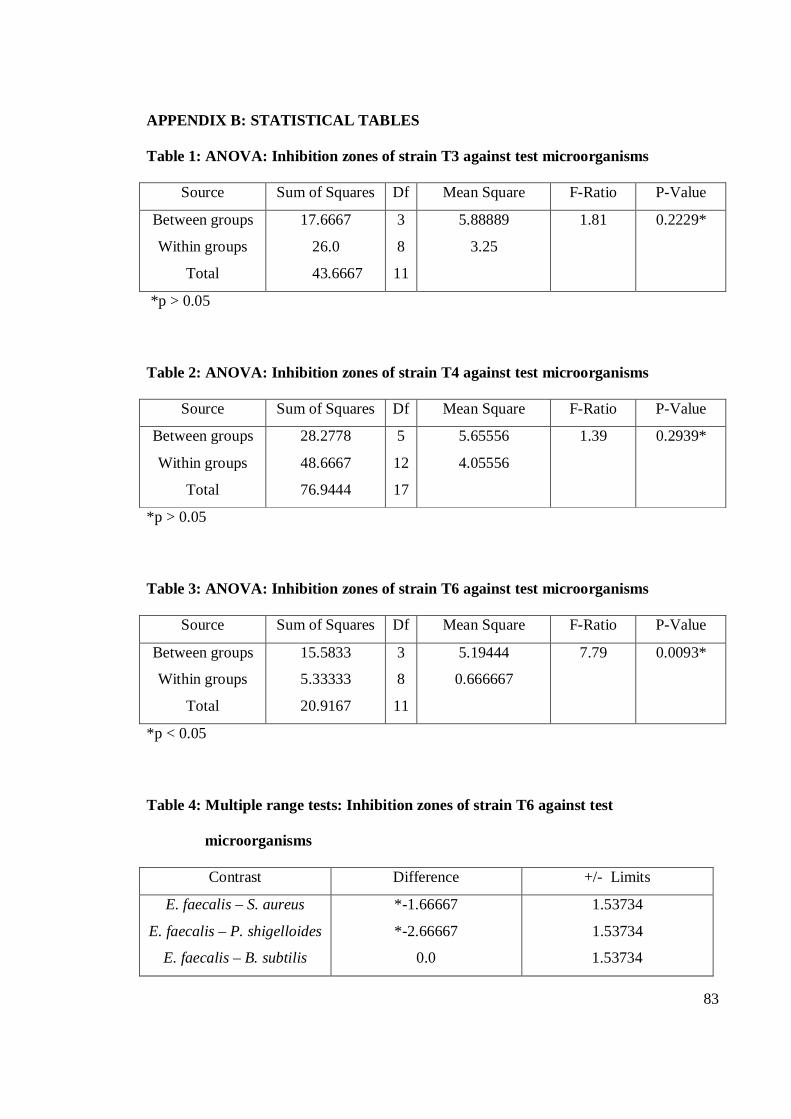
83
APPENDIX B: STATISTICAL TABLES
Table 1: ANOVA: Inhibition zones of strain T3 against test microorganisms
Source Sum of Squares Df Mean Square F-Ratio P-Value
Between groups
Within groups
Total
17.6667
26.0
43.6667
3
8
11
5.88889
3.25
1.81 0.2229*
*p > 0.05
Table 2: ANOVA: Inhibition zones of strain T4 against test microorganisms
Source Sum of Squares Df Mean Square F-Ratio P-Value
Between groups
Within groups
Total
28.2778
48.6667
76.9444
5
12
17
5.65556
4.05556
1.39
0.2939*
*p > 0.05
Table 3: ANOVA: Inhibition zones of strain T6 against test microorganisms
Source Sum of Squares Df Mean Square F-Ratio P-Value
Between groups
Within groups
Total
15.5833
5.33333
20.9167
3
8
11
5.19444
0.666667
7.79 0.0093*
*p < 0.05
Table 4: Multiple range tests: Inhibition zones of strain T6 against test
microorganisms
Contrast Difference +/- Limits
E. faecalis – S. aureus
E. faecalis – P. shigelloides
E. faecalis – B. subtilis
*-1.66667
*-2.66667
0.0
1.53734
1.53734
1.53734
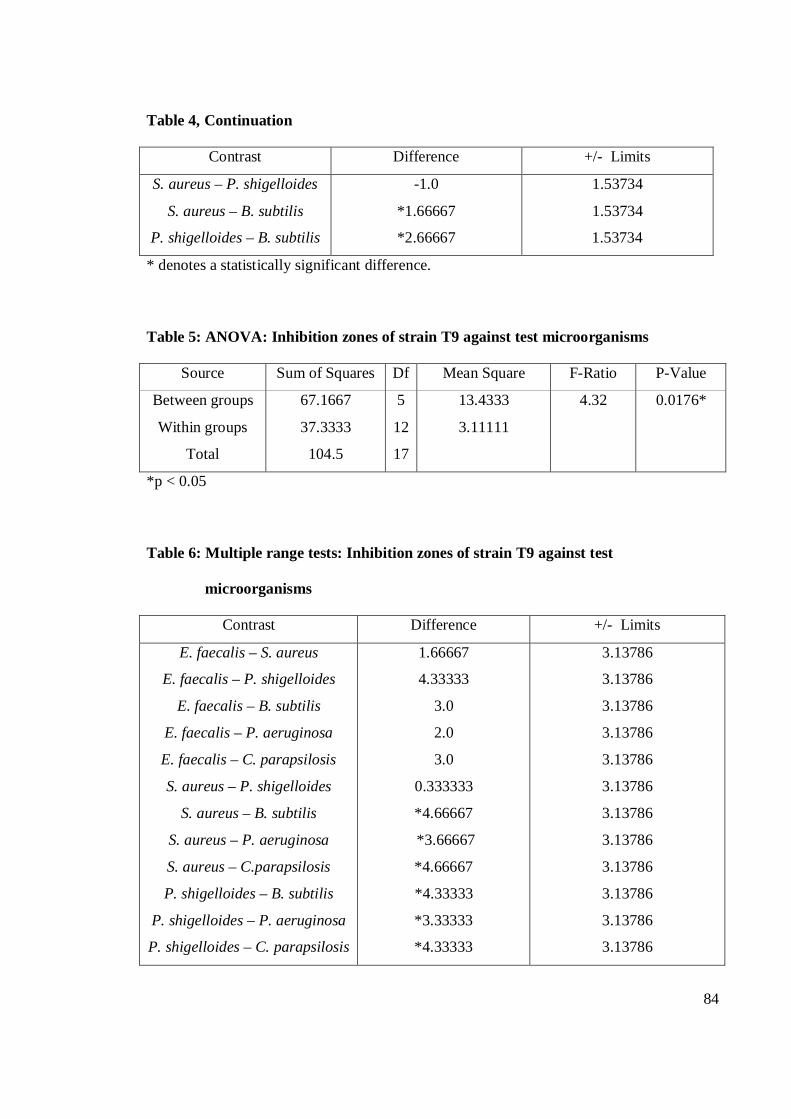
84
Table 4, Continuation
Contrast Difference +/- Limits
S. aureus – P. shigelloides
S. aureus – B. subtilis
P. shigelloides – B. subtilis
-1.0
*1.66667
*2.66667
1.53734
1.53734
1.53734
* denotes a statistically significant difference.
Table 5: ANOVA: Inhibition zones of strain T9 against test microorganisms
Source Sum of Squares Df Mean Square F-Ratio P-Value
Between groups
Within groups
Total
67.1667
37.3333
104.5
5
12
17
13.4333
3.11111
4.32 0.0176*
*p < 0.05
Table 6: Multiple range tests: Inhibition zones of strain T9 against test
microorganisms
Contrast Difference +/- Limits
E. faecalis – S. aureus
E. faecalis – P. shigelloides
E. faecalis – B. subtilis
E. faecalis – P. aeruginosa
E. faecalis – C. parapsilosis
S. aureus – P. shigelloides
S. aureus – B. subtilis
S. aureus – P. aeruginosa
S. aureus – C.parapsilosis
P. shigelloides – B. subtilis
P. shigelloides – P. aeruginosa
P. shigelloides – C. parapsilosis
1.66667
4.33333
3.0
2.0
3.0
0.333333
*4.66667
*3.66667
*4.66667
*4.33333
*3.33333
*4.33333
3.13786
3.13786
3.13786
3.13786
3.13786
3.13786
3.13786
3.13786
3.13786
3.13786
3.13786
3.13786
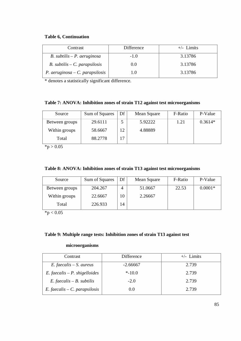
85
Table 6, Continuation
Contrast Difference +/- Limits
B. subtilis – P. aeruginosa
B. subtilis – C. parapsilosis
P. aeruginosa – C. parapsilosis
-1.0
0.0
1.0
3.13786
3.13786
3.13786
* denotes a statistically significant difference.
Table 7: ANOVA: Inhibition zones of strain T12 against test microorganisms
Source Sum of Squares Df Mean Square F-Ratio P-Value
Between groups
Within groups
Total
29.6111
58.6667
88.2778
5
12
17
5.92222
4.88889
1.21 0.3614*
*p > 0.05
Table 8: ANOVA: Inhibition zones of strain T13 against test microorganisms
Source Sum of Squares Df Mean Square F-Ratio P-Value
Between groups
Within groups
Total
204.267
22.6667
226.933
4
10
14
51.0667
2.26667
22.53 0.0001*
*p < 0.05
Table 9: Multiple range tests: Inhibition zones of strain T13 against test
microorganisms
Contrast Difference +/- Limits
E. faecalis – S. aureus
E. faecalis – P. shigelloides
E. faecalis – B. subtilis
E. faecalis – C. parapsilosis
-2.66667
*-10.0
-2.0
0.0
2.739
2.739
2.739
2.739
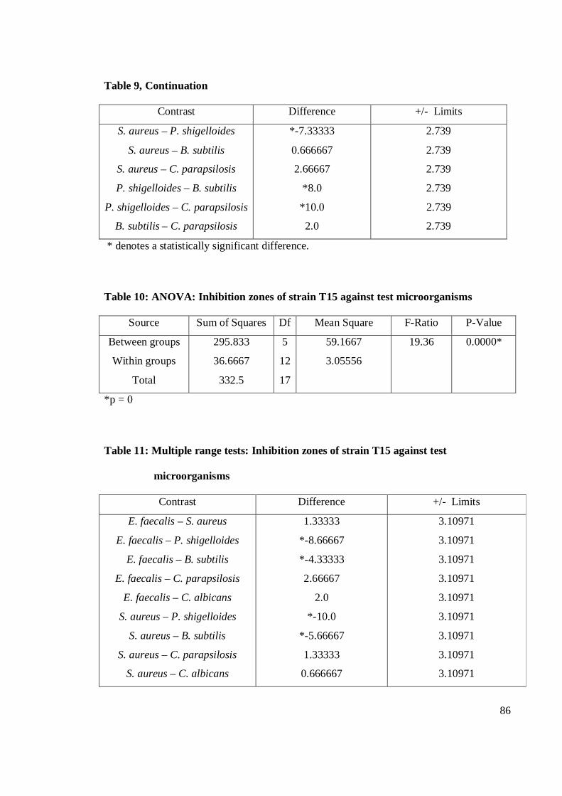
86
Table 9, Continuation
Contrast Difference +/- Limits
S. aureus – P. shigelloides
S. aureus – B. subtilis
S. aureus – C. parapsilosis
P. shigelloides – B. subtilis
P. shigelloides – C. parapsilosis
B. subtilis – C. parapsilosis
*-7.33333
0.666667
2.66667
*8.0
*10.0
2.0
2.739
2.739
2.739
2.739
2.739
2.739
* denotes a statistically significant difference.
Table 10: ANOVA: Inhibition zones of strain T15 against test microorganisms
Source Sum of Squares Df Mean Square F-Ratio P-Value
Between groups
Within groups
Total
295.833
36.6667
332.5
5
12
17
59.1667
3.05556
19.36 0.0000*
*p = 0
Table 11: Multiple range tests: Inhibition zones of strain T15 against test
microorganisms
Contrast Difference +/- Limits
E. faecalis – S. aureus
E. faecalis – P. shigelloides
E. faecalis – B. subtilis
E. faecalis – C. parapsilosis
E. faecalis – C. albicans
S. aureus – P. shigelloides
S. aureus – B. subtilis
S. aureus – C. parapsilosis
S. aureus – C. albicans
1.33333
*-8.66667
*-4.33333
2.66667
2.0
*-10.0
*-5.66667
1.33333
0.666667
3.10971
3.10971
3.10971
3.10971
3.10971
3.10971
3.10971
3.10971
3.10971
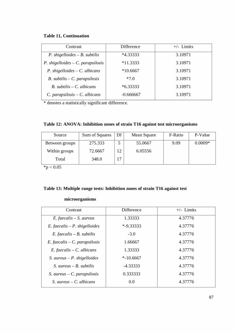
87
Table 11, Continuation
Contrast Difference +/- Limits
P. shigelloides – B. subtilis
P. shigelloides – C. parapsilosis
P. shigelloides – C. albicans
B. subtilis – C. parapsilosis
B. subtilis – C. albicans
C. parapsilosis – C. albicans
*4.33333
*11.3333
*10.6667
*7.0
*6.33333
-0.666667
3.10971
3.10971
3.10971
3.10971
3.10971
3.10971
* denotes a statistically significant difference.
Table 12: ANOVA: Inhibition zones of strain T16 against test microorganisms
Source Sum of Squares Df Mean Square F-Ratio P-Value
Between groups
Within groups
Total
275.333
72.6667
348.0
5
12
17
55.0667
6.05556
9.09 0.0009*
*p < 0.05
Table 13: Multiple range tests: Inhibition zones of strain T16 against test
microorganisms
Contrast Difference +/- Limits
E. faecalis – S. aureus
E. faecalis – P. shigelloides
E. faecalis – B. subtilis
E. faecalis – C. parapsilosis
E. faecalis – C. albicans
S. aureus – P. shigelloides
S. aureus – B. subtilis
S. aureus – C. parapsilosis
S. aureus – C. albicans
1.33333
*-9.33333
-3.0
1.66667
1.33333
*-10.6667
-4.33333
0.333333
0.0
4.37776
4.37776
4.37776
4.37776
4.37776
4.37776
4.37776
4.37776
4.37776
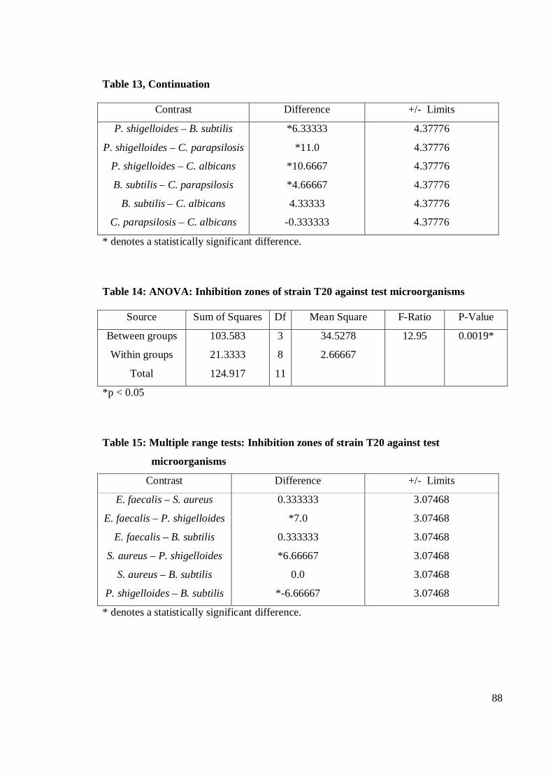
88
Table 13, Continuation
Contrast Difference +/- Limits
P. shigelloides – B. subtilis
P. shigelloides – C. parapsilosis
P. shigelloides – C. albicans
B. subtilis – C. parapsilosis
B. subtilis – C. albicans
C. parapsilosis – C. albicans
*6.33333
*11.0
*10.6667
*4.66667
4.33333
-0.333333
4.37776
4.37776
4.37776
4.37776
4.37776
4.37776
* denotes a statistically significant difference.
Table 14: ANOVA: Inhibition zones of strain T20 against test microorganisms
Source Sum of Squares Df Mean Square F-Ratio P-Value
Between groups
Within groups
Total
103.583
21.3333
124.917
3
8
11
34.5278
2.66667
12.95 0.0019*
*p < 0.05
Table 15: Multiple range tests: Inhibition zones of strain T20 against test
microorganisms
Contrast Difference +/- Limits
E. faecalis – S. aureus
E. faecalis – P. shigelloides
E. faecalis – B. subtilis
S. aureus – P. shigelloides
S. aureus – B. subtilis
P. shigelloides – B. subtilis
0.333333
*7.0
0.333333
*6.66667
0.0
*-6.66667
3.07468
3.07468
3.07468
3.07468
3.07468
3.07468
* denotes a statistically significant difference.
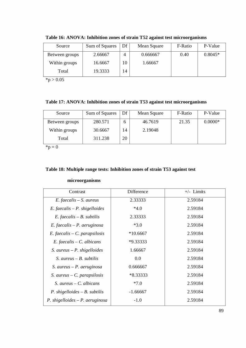
89
Table 16: ANOVA: Inhibition zones of strain T52 against test microorganisms
Source Sum of Squares Df Mean Square F-Ratio P-Value
Between groups
Within groups
Total
2.66667
16.6667
19.3333
4
10
14
0.666667
1.66667
0.40 0.8045*
*p > 0.05
Table 17: ANOVA: Inhibition zones of strain T53 against test microorganisms
Source Sum of Squares Df Mean Square F-Ratio P-Value
Between groups
Within groups
Total
280.571
30.6667
311.238
6
14
20
46.7619
2.19048
21.35 0.0000*
*p = 0
Table 18: Multiple range tests: Inhibition zones of strain T53 against test
microorganisms
Contrast Difference +/- Limits
E. faecalis – S. aureus
E. faecalis – P. shigelloides
E. faecalis – B. subtilis
E. faecalis – P. aeruginosa
E. faecalis – C. parapsilosis
E. faecalis – C. albicans
S. aureus – P. shigelloides
S. aureus – B. subtilis
S. aureus – P. aeruginosa
S. aureus – C. parapsilosis
S. aureus – C. albicans
P. shigelloides – B. subtilis
P. shigelloides – P. aeruginosa
2.33333
*4.0
2.33333
*3.0
*10.6667
*9.33333
1.66667
0.0
0.666667
*8.33333
*7.0
-1.66667
-1.0
2.59184
2.59184
2.59184
2.59184
2.59184
2.59184
2.59184
2.59184
2.59184
2.59184
2.59184
2.59184
2.59184
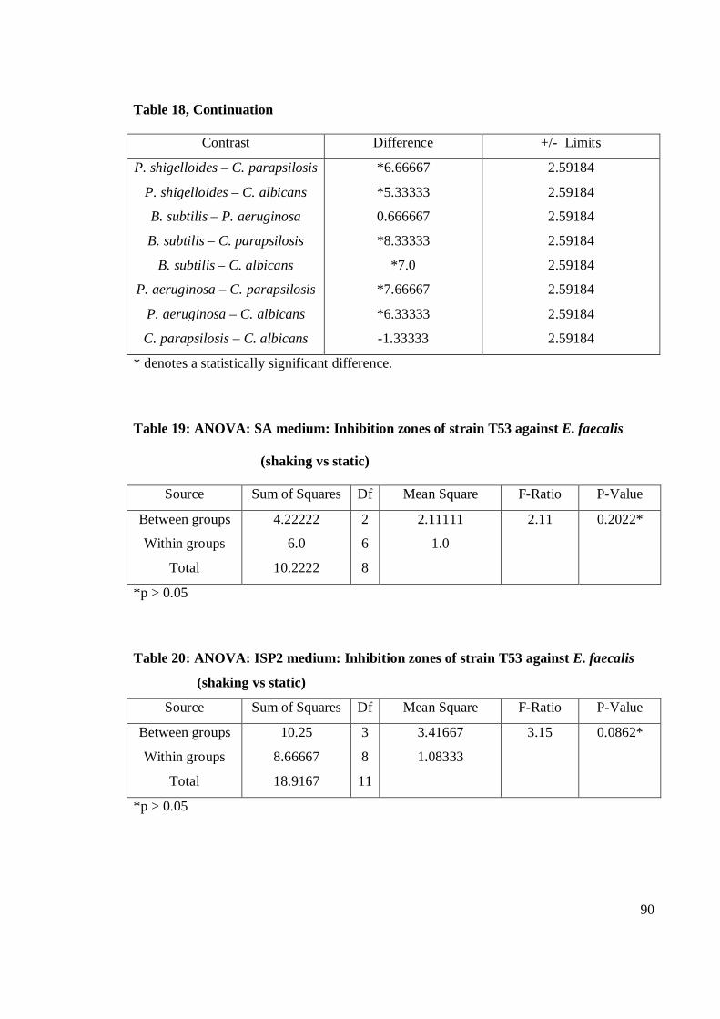
90
Table 18, Continuation
Contrast Difference +/- Limits
P. shigelloides – C. parapsilosis
P. shigelloides – C. albicans
B. subtilis – P. aeruginosa
B. subtilis – C. parapsilosis
B. subtilis – C. albicans
P. aeruginosa – C. parapsilosis
P. aeruginosa – C. albicans
C. parapsilosis – C. albicans
*6.66667
*5.33333
0.666667
*8.33333
*7.0
*7.66667
*6.33333
-1.33333
2.59184
2.59184
2.59184
2.59184
2.59184
2.59184
2.59184
2.59184
* denotes a statistically significant difference.
Table 19: ANOVA: SA medium: Inhibition zones of strain T53 against E. faecalis
(shaking vs static)
Source Sum of Squares Df Mean Square F-Ratio P-Value
Between groups
Within groups
Total
4.22222
6.0
10.2222
2
6
8
2.11111
1.0
2.11
0.2022*
*p > 0.05
Table 20: ANOVA: ISP2 medium: Inhibition zones of strain T53 against E. faecalis
(shaking vs static)
Source Sum of Squares Df Mean Square F-Ratio P-Value
Between groups
Within groups
Total
10.25
8.66667
18.9167
3
8
11
3.41667
1.08333
3.15 0.0862*
*p > 0.05
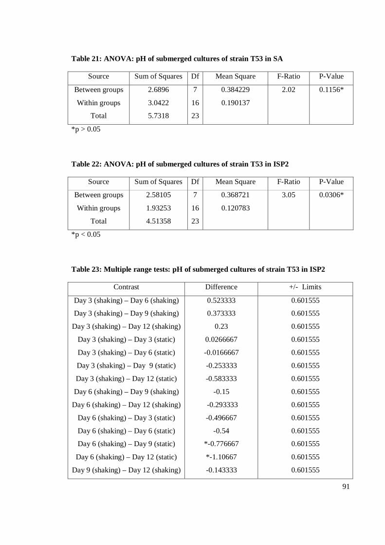
91
Table 21: ANOVA: pH of submerged cultures of strain T53 in SA
Source Sum of Squares Df Mean Square F-Ratio P-Value
Between groups
Within groups
Total
2.6896
3.0422
5.7318
7
16
23
0.384229
0.190137
2.02 0.1156*
*p > 0.05
Table 22: ANOVA: pH of submerged cultures of strain T53 in ISP2
Source Sum of Squares Df Mean Square F-Ratio P-Value
Between groups
Within groups
Total
2.58105
1.93253
4.51358
7
16
23
0.368721
0.120783
3.05 0.0306*
*p < 0.05
Table 23: Multiple range tests: pH of submerged cultures of strain T53 in ISP2
Contrast Difference +/- Limits
Day 3 (shaking) – Day 6 (shaking)
Day 3 (shaking) – Day 9 (shaking)
Day 3 (shaking) – Day 12 (shaking)
Day 3 (shaking) – Day 3 (static)
Day 3 (shaking) – Day 6 (static)
Day 3 (shaking) – Day 9 (static)
Day 3 (shaking) – Day 12 (static)
Day 6 (shaking) – Day 9 (shaking)
Day 6 (shaking) – Day 12 (shaking)
Day 6 (shaking) – Day 3 (static)
Day 6 (shaking) – Day 6 (static)
Day 6 (shaking) – Day 9 (static)
Day 6 (shaking) – Day 12 (static)
Day 9 (shaking) – Day 12 (shaking)
0.523333
0.373333
0.23
0.0266667
-0.0166667
-0.253333
-0.583333
-0.15
-0.293333
-0.496667
-0.54
*-0.776667
*-1.10667
-0.143333
0.601555
0.601555
0.601555
0.601555
0.601555
0.601555
0.601555
0.601555
0.601555
0.601555
0.601555
0.601555
0.601555
0.601555
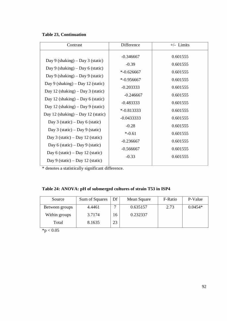
92
Table 23, Continuation
Contrast Difference +/- Limits
Day 9 (shaking) – Day 3 (static)
Day 9 (shaking) – Day 6 (static)
Day 9 (shaking) – Day 9 (static)
Day 9 (shaking) – Day 12 (static)
Day 12 (shaking) – Day 3 (static)
Day 12 (shaking) – Day 6 (static)
Day 12 (shaking) – Day 9 (static)
Day 12 (shaking) – Day 12 (static)
Day 3 (static) – Day 6 (static)
Day 3 (static) – Day 9 (static)
Day 3 (static) – Day 12 (static)
Day 6 (static) – Day 9 (static)
Day 6 (static) – Day 12 (static)
Day 9 (static) – Day 12 (static)
-0.346667
-0.39
*-0.626667
*-0.956667
-0.203333
-0.246667
-0.483333
*-0.813333
-0.0433333
-0.28
*-0.61
-0.236667
-0.566667
-0.33
0.601555
0.601555
0.601555
0.601555
0.601555
0.601555
0.601555
0.601555
0.601555
0.601555
0.601555
0.601555
0.601555
0.601555
* denotes a statistically significant difference.
Table 24: ANOVA: pH of submerged cultures of strain T53 in ISP4
Source Sum of Squares Df Mean Square F-Ratio P-Value
Between groups
Within groups
Total
4.4461
3.7174
8.1635
7
16
23
0.635157
0.232337
2.73 0.0454*
*p < 0.05
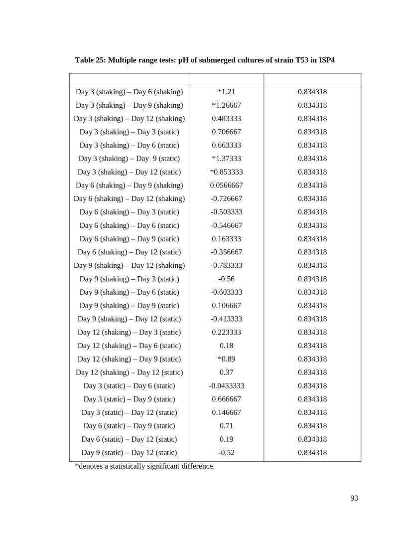
93
Table 25: Multiple range tests: pH of submerged cultures of strain T53 in ISP4
Day 3 (shaking) – Day 6 (shaking)
Day 3 (shaking) – Day 9 (shaking)
Day 3 (shaking) – Day 12 (shaking)
Day 3 (shaking) – Day 3 (static)
Day 3 (shaking) – Day 6 (static)
Day 3 (shaking) – Day 9 (static)
Day 3 (shaking) – Day 12 (static)
Day 6 (shaking) – Day 9 (shaking)
Day 6 (shaking) – Day 12 (shaking)
Day 6 (shaking) – Day 3 (static)
Day 6 (shaking) – Day 6 (static)
Day 6 (shaking) – Day 9 (static)
Day 6 (shaking) – Day 12 (static)
Day 9 (shaking) – Day 12 (shaking)
Day 9 (shaking) – Day 3 (static)
Day 9 (shaking) – Day 6 (static)
Day 9 (shaking) – Day 9 (static)
Day 9 (shaking) – Day 12 (static)
Day 12 (shaking) – Day 3 (static)
Day 12 (shaking) – Day 6 (static)
Day 12 (shaking) – Day 9 (static)
Day 12 (shaking) – Day 12 (static)
Day 3 (static) – Day 6 (static)
Day 3 (static) – Day 9 (static)
Day 3 (static) – Day 12 (static)
Day 6 (static) – Day 9 (static)
Day 6 (static) – Day 12 (static)
Day 9 (static) – Day 12 (static)
*1.21
*1.26667
0.483333
0.706667
0.663333
*1.37333
*0.853333
0.0566667
-0.726667
-0.503333
-0.546667
0.163333
-0.356667
-0.783333
-0.56
-0.603333
0.106667
-0.413333
0.223333
0.18
*0.89
0.37
-0.0433333
0.666667
0.146667
0.71
0.19
-0.52
0.834318
0.834318
0.834318
0.834318
0.834318
0.834318
0.834318
0.834318
0.834318
0.834318
0.834318
0.834318
0.834318
0.834318
0.834318
0.834318
0.834318
0.834318
0.834318
0.834318
0.834318
0.834318
0.834318
0.834318
0.834318
0.834318
0.834318
0.834318
*denotes a statistically significant difference.