Embed Size (px)
Citation preview
Portland State University Portland State University
PDXScholar PDXScholar
Dissertations and Theses Dissertations and Theses
1-1-1976
Isolation and characterization of two sterols from Isolation and characterization of two sterols from
the green alga, Selenastrum capricornutum the green alga, Selenastrum capricornutum
Raymond Mark Owings Portland State University
Follow this and additional works at: https://pdxscholar.library.pdx.edu/open_access_etds
Let us know how access to this document benefits you.
Recommended Citation Recommended Citation Owings, Raymond Mark, "Isolation and characterization of two sterols from the green alga, Selenastrum capricornutum" (1976). Dissertations and Theses. Paper 861. https://doi.org/10.15760/etd.861
This Dissertation is brought to you for free and open access. It has been accepted for inclusion in Dissertations and Theses by an authorized administrator of PDXScholar. Please contact us if we can make this document more accessible: [email protected].

ISOLATIO[\ At\D CHAHACTERIZATIOi\ OF 1\'JO STEROLS FRON
Tr-lE GREEi\ ALGA. SELENASTRL::I CAPRICORl\UTt.:~l
by
RA YNOND HARK O~\ Ii\GS
A thesis submitted in partial fulfillment of the requirements for the degree of
DOCTOR OF PHILOSOPHY in
Ef\VIRONHEl\TAL SCIEI\CE-CHEtv1[STRY
Portland State University 1976

Ai': ABSTRA.CT OF THE THESIS OF Raymond tvlark O\vings for the
Doctor of Philosophy in Environmental Science-Chemistry
presented August 4, 1975.
Title: Isolation and Characterization of Two Sterols From
the Green Alga, Selanastrum capricornutum.
APPROVED BY NEivlBERS OF THE THESIS COHNITTEE:
Karl Dittmer, Chairman
Norman C. Rose
Edward N. Perdue
Dennis \V. Barnum
Richard R. Petersen

2
The green alga, Selenastrum capricornutum, was cul
tured in artificial nutrient medium utilizing five-gallon
carboys, each of which contained 16 1. of culture. The
algal cells were separated from the nutrient medium by con
tinuous-flow centrifugation at 7500 RPM, and were then lyo
philized. The lyophilized cells were extracted by refluxing
with ether, acetone, and chloroform:methanol (2:1). Free
sterols and sterol esters were separated from the crude ex
tract using preparative thin-layer chromatography. Sterol
esters were saponified and both the sterols and the fatty
acids were recovered.
Individual sterols were separated from the free
sterol fraction using argentation thin-layer chromatography.
Gas chromatograms, mass spectra, and ultraviolet spectra
were obtained for these sterols. The free sterol fraction
was found to contain approximately 40% 24-methylcholesta-5,
7-dien-3~-ol and 60% 24-ethylcholesta-5.7-dien-3~-ol.
The sterol ester fraction also contained these two
sterols; however, the composition and amount of esterified
sterols varied as a function of culture age. Sterol ester
content was higher for older cultures, and in older cultures
the composition of the esterified sterols more closely re
sembled that of the free sterols. The fatty acids obtained
from the saponification of the sterol esters were methylated
and were analyzed using gas chromatography. Tentative iden
tifications, based upon comparative retention times, were
made for several of these acids.

3
Sterols were extracted from the nutrient medium after
harvest of the algal cells. Extraction was accomplished by
mixing large quantities of nutrient medium with ether for
several days, or by shaking small aliquots with ether. 24-
methylcholesta-5,7-dien -3p-ol and 24-ethylcholesta-5,7-
dien-3p-ol were isolated from the nutrient medium in approx
imately the same relative amounts as from the algal cells.
The concentration of sterols in the nutrient medium was ap
proximately equal to the water-solubility of cholesterol
(25-29}t g./l.).
Extraction procedures which release sterols from
water soluble complexes were carried out on extracted cells
and on extracted nutrient medium. These procedures failed
to yield measurable quantities of sterols.
Treatment of extracted cells with strong base and
subsequent extraction showed that all sterol.s had been ex
tracted without prior cell lysis or pretreatment. An ex
traction of algal cells was carried out using DMSO:ether as
the extraction solvent. This extraction resulted in com
plete removal of sterols from the cells, and the sterols
were accompanied by only small amounts of other lipid
soluble material.

TO THE OFFICE OF GRADUATE STUDIES AND RESEARCH:
The members of the Co~ittee approve the thesis of
Raymond Mark Owings presented August 4, 1975.
Karl Dittmer, Chairman
Norman C. Rose
Edward No Perdue
Dennis Wo Barnum
Richard R. Petersen
APPROVED:
Chemistry
A CIG\Oh' LEIh ~IEI\TS
I wish to thank Dr. Alfred S. Levinson for his guid
ance and advice during the course of this research.
I also 't'lish to thank Dr. Ed\'lard i'-l. Perdue for his in
terest and assistance, and Nr. Bill Anderson for obtaining
the mass spectra.
Special thanks go to my father and mother for their
encouragement and support, and to my mother for her assist
ance in preparing this thesis.
Ny greatest appreciation is for the 't'lork done by my
wife, Chris, in typing the thesis, and for her constant
enthusiasm far my ,,,ark.

TABLE OF COi.\TE1\TS
ACKl~O~'JLEDGtvlENTS • . . . . · . . · . . . . LIS T OF TABLES. . . . · . . . . . • • • • •
LIST OF FIGURES • . . · . . . . . . . . . . . . CHAPTER
I
II
III
INTRODUCTION. . . . . . . Sterols . • · . · . . . . Algal Sterols . · . . . .
Sterols of Phaeophyta Sterols of Rhodophyta Sterols of Chlorophyta Sterols of Chrysophyta Sterols of Charophyta, Euglenophyta;
and Cyanophy ta
Water Soluble Sterols . · . . . . Systematic Names .••• · . . . .
STATE~1ENT OF THE PROBLEN •• · . . . . MA TERIALS AND ~'1ETHODS • • • . . . . . . .
Culture and Harvest Procedures.
Chromatographic and Spectrometric ~~thods . . • • • . • • • •
Solvents Thin Layer Chromatotjraphy Gas Chromatography Absorption Spectrometry ~~ss Spectrometry
PAGE
iii
vi
vii
1
1
4
19
22
24
29
29
43

CHAPTER
Extraction Methods . . . . • . . • •
Extraction of Lyophilized Cells Extraction of the Supernatant Summary Pretreatment of Lyophilized Cells DMSO as a Solvent for Sterol
Extraction Melting Points
Methods For Obtaining Stearn Volatile
v
PAGE
46
Compounds. . • . •. .•.•• 61
IV
V
VI
RESULTS. . . . . . . . . . . . . . . Free Sterols . • • • • • • • • . . .
Sterols From Lyophilized Cells Sterols From the Supernatant
Bound Sterols .. . . . . . . . . Sterols From Sterol Esters Water Soluble Sterols Sterols Extracted By DMSO:Ether Summary Fatty Acids From Sterol Esters
DISCUSSION . . • . . • . . . . . . . . . Discussion of Results Recommendations
SUMMARY. . . . . . . . . . . . . . . . . BIBLIOGRAPHY •... . . . . . . . . . . . . . . . .
65
65
71
76
81
83
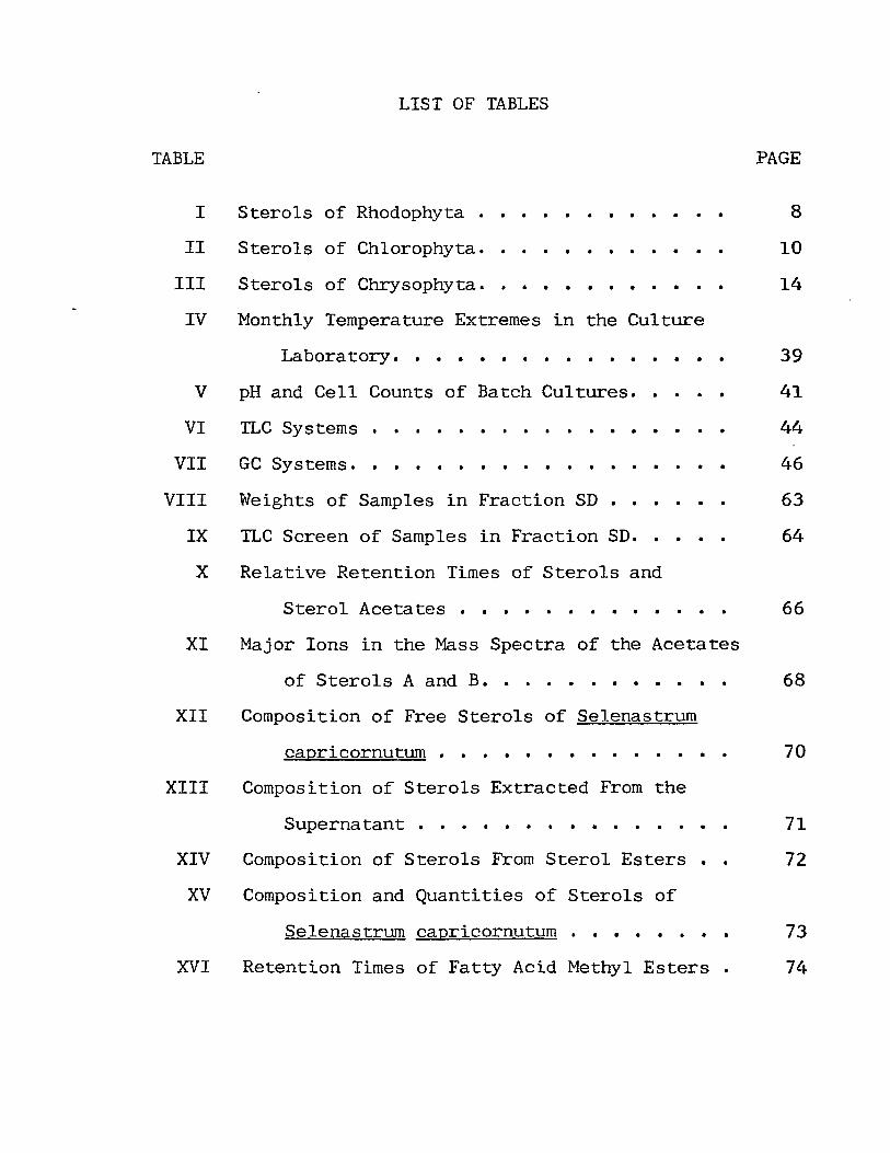
TABLE
I
II
III
LIST OF TABLES
Sterols of Rhodophyta . . . . • . • . . • • •
Sterols of Chlorophyta.
Sterols of Chrysophyta.
. . • • • •
. . . . . · .
. . . . . · . IV Monthly Temperature Extremes in the Culture
V
VI
VII
VIII
IX
X
XI
Labora tory. • • • • • • • . . • •
pH and Cell Counts of Batch Cultures. • •
. . . . . . . . . · . TLC Sy stems . .
GC Systems. . . . . . . . . . . . . . . . Weights of Samples in Fraction SD . . TLC Screen of Samples in Fraction SD. • • • •
Relative Retention Times of Sterols and
Sterol Acetates • . • • . . . • . •
Major Ions in the Mass Spectra of the Acetates
of Sterols A and B. . . . • . • • • . • .
XII Composition of Free Sterols of Selenastrurn
XIII
XIV
XV
XVI
capricornutum . . . . • • . • • . • • • .
Composition of Sterols Extracted From the
Supernatant • . • . • . . • • . .
Composition of Sterols From Sterol Esters
Composition and Quantities of Sterols of
Selenastrum capricornutum . . • . • . • •
Retention Times of Fatty Acid Methyl Esters .
PAGE
8
10
14
39
41
44
46
63
64
66
68
70
71
72
73
74
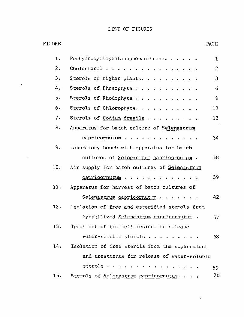
LIST OF FIGURES
FIGURE PAGE
1.
2.
3.
5.
6.
7.
Perhydrocyclopentanophenanthrene.
Cholesterol . . • . . . . . . . · . . . Sterols of higher plants ..
Sterols of Phaeophyta . . . .
Sterols of Rhodophyta • •
Sterols of Chlorophyta. . . . Sterols of Codium fragile
· . . . . . . . . . . . . .
. . . · . . .
• •
· . · .
8. Apparatus for batch culture of Selenastrum
capricornutum . · . . . . . 9. Laboratory bench with apparatus for batch
1
2
3
6
9
12
13
34
cultures of Selenastrum capricornutum 38
10. Air supply for batch cultures of Selenastrum
capricornutum . • . . . . . . . . . . . 39
11. Apparatus for harvest of batch cultures of
Selenastrum cagricornutum . . . • • •. 42
12. Isolation of free and esterified sterols from
lyophilized ;3elenastrum capricornutum. 57
13. Treatment of the cell residue to release
water-soluble sterols o • • • • • •
Ill. Isola tion of free sterols from the superna tant
and treatments for release of water-soluble
sterols . . . • . . . . • . . . . . 15. Sterols of Selenastrum capricornutum .. . .
.58
59
70

CHAPTER I
INTRODUCTION
Mass spectrometry and gas and thin-layer chromato
graphy are techniques which have been utilized for rapid
separation and identification of specific organic compounds,
particularly phytosterols, from mixtures of plant natural
products. These techniques have been extensively applied
in recent investigations of sterol composition of algae.
I. STEROLS
Sterols are one group of steroids which are lipids
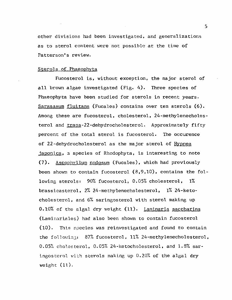
having in common the perhydrocyclopentanophenanthrene skel
eton (Fig. 1). Sterols have been isolated from all major
groups of living organisms. The occurence and biosynthesis
Figure 1. Perhydrocyclopentanophenanthrene.

2
of steroids in animals have been thoroughly investigated and
are well understood. In higher animals cholesterol (Fig. 2)
is the major sterol while cholesterol or mixtures of 27-,
28-, or 29-carbon sterols occur in lower animals (1). The
28- and 29-carbon sterols of lower animals appear to be de
rived from their diet (1,2).
HO
Figure 2. Cholesterol.
The majority of the phytosterols possess an alkyl side
chain at C-24 and one or more double bonds in a ring or the
side chain. In higher plants~-sitosterol is the major
sterol in most species investigated and is often found to
gether with varying amounts of stigmasterol and campesterol
(Fig. 3). Cholesterol and other sterols have been reported
in small amounts in higher plants; however, they are not
common. Plant sterols have been assumed to be secondary
metabolites of little importance to the organisms which syn
thesize them in contrast to the roles of steroids in animal
metabolic processes. Results of recent studies of steroid
metabolism in plants shmv, hmvever, that this assumption may

3
HO
..-J---..... stigmasterol
HO
.;-_---1 campesterol
HO
Figure 3. Sterols of higher plants.

not be correct. ~luch is being done to elucida te the possi
ble roles of phytosterols in plant metabolism (1,3).
4
In contrast to animals and higher plants, the occur
ence of sterols in algae has not been thoroughly investi
gated, and generalizations as to sterol content can be made
for only a few divisions of the algae. Until recently the
analytical techniques available for study of algal sterols
were not sufficiently advanced to allow definite character
ization.of small quantities of sterols isolated from complex
mixtures. This factor, combined with a deficiency in the
number of studies of algal sterols, has led to a confusion
in the field of algal sterols which has yet to be fully re
solved.
II. ALGAL STEROLS
Glenn W. Patterson, affiliated with the University of
Maryland, reviewed the literature dealing with occurence of
sterols in algae i.n 1971 (1), The brm.;n algae (Phaeophyta)
had been previously shown to contain fucosterol almost ex
clusivE:ly as the major sterol. Fucosterol was reported to
occur together with lesser amounts of 24-methylenecholes
terol in some species. The major sterol of most Rhodophyta
varied from sample to sample (4,5). The sterols of Chloro
phyta had been shm.;rn to be much less predictable with C-28
and C-29 sterols predominating and only a few species con
ta ining choles teeol as a major sterol. Fe\·, species of
other divisions had been investigated, and generalizations
as to sterol content \17ere not possible at the time of
Patterson's review.
Sterols of Phaeophyta
5
Fucosterol is, without exception, the major sterol of
all brO\ffi algae investigated (Fig. 4). Three species of
Phaeophyta have been studied for sterols in recent years.
Sargassllm fluitans (Fucales) contains over ten sterols (6).
Among these are fucosterol, cholesterol, 24-rnethylenecholes
terol and trans-22-dehydrocholesterol. Approximately fifty
percent of the total sterol is fucosterol. The occurence
of 22-dehydrocholesterol as the major sterol of Hyonea
japoni.ca, a species of Rhodophyta, is interesting to note
(7). Ascophvllum nodosum (Fucales), which had previously
been sho\ffi to contain fucosterol (8,9,10), contains the fol
Imving sterols: 90% fucos terol, 0.05% cholesterol, 1%
brassicastero1, 2% 2If-methylenecholesterol, 1% 24-keto
cholesterol, and 6% saringosterol with sterol making up
0.10% of the algal dry \vci.ght (11). Laminaria saccharina
(Lamin;lriales) had also been shown to contain fucosterol
(10). This r;pecies was reinvestigated and found to contain
the fol1ouil1':;: 87% fucosterol, 11% 24-methylenecho1esterol,
0.05~~ cholesterol, 0.05% 24-ketocho1esterol, and 1.8% sar
ingosterol ",i::.h sterols making up 0.20% of the algal dry
weight (11).

6
~----~ fucosterol
HO
24-methylenecholesterol
HO
saringosterol
HO
Figure 4. Sterols of Phaeophyta.
7
The presence of 24-methylenecholesterol in brown algae
suggests the possibility that this sterol could serve as a
biosynthetic precursor of fucosterol (1). Saringosterol has
been identified in several species of Phaeophyta and could
arise by air oxidation of fucosterol (12).
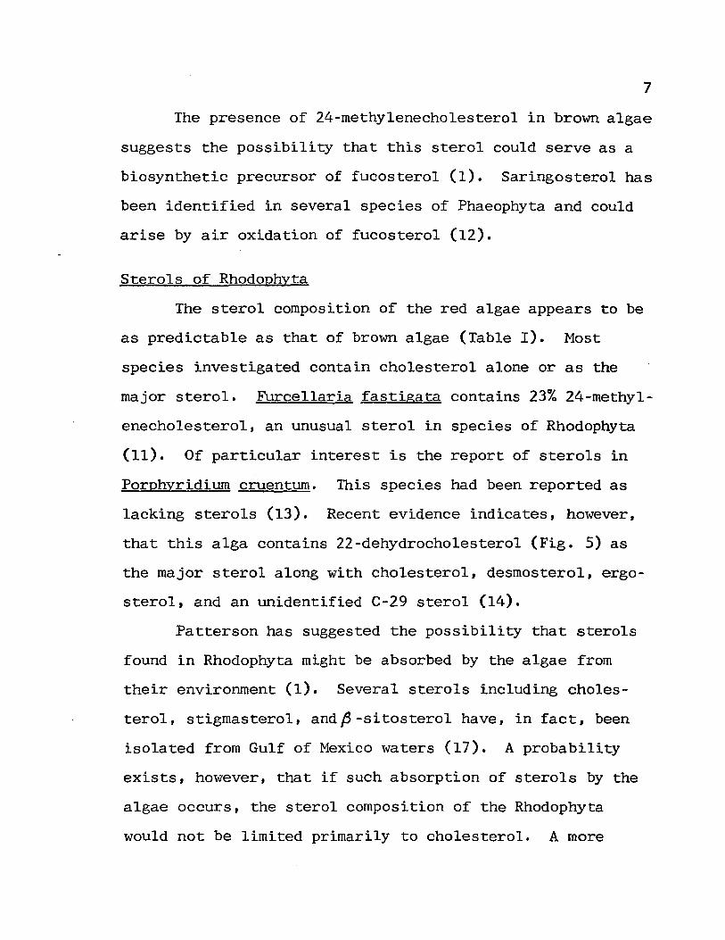
Sterols of Rhodophyta
The sterol composition of the red algae appears to be
as predictable as that of brown algae (Table I). Most
species investigated contain cholesterol alone or as the
major sterol. Furcellaria fastigata contains 23% 24-methyl
enecholesterol, an unusual sterol in species of Rhodophyta
(11). Of particular interest is the report of sterols in
Porphyridiurn cruenturn. This species had been reported as
lacking sterols (13). Recent evidence indicates, however,
that this alga contains 22-dehydrocholesterol (Fig. 5) as
the major sterol along with cholesterol, desmosterol, ergo
sterol, and an unidentified C-29 sterol (14).
Patterson has suggested the possibility that sterols
found in Rhodophyta might be absorbed by the algae from
their environment (1). Several sterols including choles
terol, stigmasterol, and~-sitosterol have, in fact, been
isolated from Gulf of Mexico waters (17). A probability
exists, however, that if such absorption of sterols by the
algae occurs, the sterol composition of the Rhodophyta
would not be limited primarily to cholesterol. A more

Order
Gigartena1es
Porphyridiales Ceramiales
TABLE I
STEROLS OF RHODOPHYTA
Species
Gracilaria textor~i £h verrucosa
Stero1sa
I I I
I,ll I
Meristotheca papu10sa Furce1laria fastigata Phyl1ophora membranifo1ia Porphyridium cruentum Polysiohonia subtilissima Chondria dasyphy1la Ceramium .t::.!Jbrum
I,III,IV,V;VI I
Dasya .Qedicellata Grinne11ia americana Caloglossa 1eprieurie Pti10ta serrata
I I I I I I
% Stero1sb Ref.
14 0.22 15
14 0.004 11 0.022 16
14 0.18 15 0.20 15 0.36 15 0.17 15 0.18 15 0.24 15 0.019 16
a r = cholesterol, II = 24-methylenecholesterol, III = 22-dehydrocholesterol, IV = desmosterol, V = ergosterol, VI = unknown C-29 sterol.
b% of dry weight.
::t)

9
/-----~ desmosterol
HO
~----~ 22-dehydrocholesterol
HO
Figure 5. Sterols of Rhodophyta.
plausible explanation might be that algae and other marine
organisms serve as sources for sterols isolated from marine
waters. Hore experiments in this area are required before
this question can be fully answered.
Sterols of Chlorophyta
The sterols of the Chlorophyta are much more varied
than those of the Phaeophyta or Rhodophyta. Several species
in seven orders of Chlorophyta have been investigated for
sterols in recent years (Table II). In this division the

Classification
Order Chlorococcales: Chlorella ellipsoidea h pringsheimti h fusca Scenedesmus oblio1!.uS Order Cladophor~les: Cladophora flexlto~a PithoplJora sp. Order Zygnemetalcs: Spirogyra .§Q. Order Siphona1&~. Halimeda in~~t~ Codium .!£,agi.le Order Chlorosphaeralcs: Coccomyxa e]oneatn Order Volvocales: Chlamydomona§ .£b.gi.nhard~ Order U1vales: Enteromorp.ha intcstinalis ~ p1umosa U 1 va lac.t.!J£i1
TABLE II
STEROLS OF CHLOROPHYTA
Sterols as % of tota1a
A BCD E F G B I J K L M N 0 % Sterols Ref.
6 4
1 2
6
22 65 7 23 72 1
2 2 6 45 22 21 2 10 14 28 40
12 32 39
4 94
48 19 33
90 90 90
11
2
21 16 18 19 65 16
94 6
44 56
0.32 18 0.22 19 0.18 19 0.27 19
0.06 19 4 0.08 19
0.19 19
0.05 19 20
0.27 19
0.38 19
\ 0.01 15 0.32 15 0.12 15
aA = brassicasterol, B = 65-ergostenol, C = poriferasterol, D =7clionastero1, E = 28-isofucosterol. F = c~olesterol, G = 24-methylenecholestero1, H = A -ergostenol, I = chondrillastero1, J = A -chondrillastenol, K = clerosterol, L = codisterol, M = ergosterol, N = 7-dchydroporiferasterol, 0 = unknown sterol.
t-' o

11
most extensively studied genus is Chlorella. Species of
Chlorella appear to contain ~5-sterols. A7 sterols. or A5 ,7
sterols with none of these classes of sterols occuring to
gether. Patterson (19) reported finding onlyd7-ergostenol,
chondrillasterol, and A7-chondrillastenol (Fig. 6) in
Scenedesmus obliquus in contrast to an earlier report that
this species contained ergosterol and several additional
sterols (21). With few exceptions the other orders of
Chlorophyt~ arc similar to the Chlorococcales. In the
Ulvales 28-isofucosterol is the major sterol; this may be
characteristic for this order. No other orders of Chloro-
phyta have been reported to contain 28-isofucostero1; how
ever, this sterol has been isolated from higher plants (22).
Patterson pointed out the possible importance of the rela
tionship in view of the fact that Ulvales is thought to be
a phylogenetically advanced order (15).
Codium fragile has been shown to contain the unusual
25-methylene sterols. clerosterol and codisterol (Fig. 7)
(20). The Siphonales are thought to have been derived from
the Chlorococcales (23), and the production of 24-ethyl and
24-methyl sterols in one species of Chlorococcales has been
shown to p~oceed via 25-methylene intermediates (24,25).
The lack of or low activity of a 6 25 -reductase has been pro-
posed as the cause for the accumulation of clerosterol and
codisterol in Codium fragile (20).

HO
HO
HO
HO
H
brassicasterol
HO
HO
HO
clionasterol HO
chondrillasterol
HO
I t17 -ergostenol
6 7-chondrillastenol
12
7-dehydroporiferasterol
28-isofucosterol
Figure 6. Sterols of Chlorophyta.

13
clerosterol
HO
codisterol
HO
Figure 7. Sterols of Codium fragile.
Sterols of Chrysophyta
The earliest investigations of the sterol content of
species of Chrysophyta concentrated on a few species of
golden brmvll algae (Chrysophyceae). Since the time of
Patterson's review. several species of Xanthophyceae- and
Bacillariophyceae have been investigated for sterol content
(Table III). The marine dietom. Chaetoceros simplex calci
trans. was cultured in sea \\Tater containing 0.15 pg/m1.

TABLE III
STEROLS OF CHRYSOPHYTA
Class Order
Bacillariophyceae Centrales
Pennales
Xanthophyceae Heterosiphonales
Heterotrichales Heterococcales
Soecles
Chaetoceros simplex calcitrans
Nitzschia !!.1.lli! & closterium Phaeodactylum
tricornutum Botrydium
granulatum Tribonema aeguale Monodus subterraneus
Sterolsa % Sterols Blli
I.IIb 0.47 26 III.IV 27
V 28
V 2
I.IV 29 1, IV 29 I.lV 29
a I = cholesterol. II = 24-methylenecholesterol. III ",. brassicasterol, IV ",. eli.onasterol, V = crinosterol.
bsterols in.culture medium.
J-J .r:-

15
sterols, most of which was cholesterol (26). After harvest
of the algae, the sea water was found to contain O.18~g/ml.
sterols. Approximately 40% of the sterol mixture was
cholesterol while 40% was 24-methylenecholesterol. Small
amounts offi-sitosterol, campesterol, and stigmasterol were
reported both before and after the growth of the algae in
the water. This is one of few investigations of the· oul
ture medium for algal sterols, and it indicates both an up
take and release of sterols by the alga (26).
The diatoms Nitzschia closterium and Phaeodactylum
tricornuturn contain crinosterol which has opposite C-24 con
figuration to brassicasterol (2,28). The identification of
this sterol in these species of Bacillariophyceae is the
first well substantiated identification of a sterol with the
24-~configuration in algae. This sterol also occurs in
esterified form in N. closteriurn. Sterols with the 24-~
configuration have, however, been identified in the diatom,
Nitzschia alba (27). Further investigations into the sterol
content of species of Chrysophyta may provide useful phylo
genetic information (2).
The sterol and sterol ester content have been deter
mined for three species of Xanthophyceae (29). All three
species contain cholesterol and clionasterol with these
sterols present in approximately the same ratio in the free
sterol and sterol ester forms.

16
Sterols of ChCl.rophyta, Euglenophyta, and Cyanophyta
Only a fe\'1 species of these other algal divisions have
recently been investigated for sterol content. I,vo species
of Charophyta, Nitella flexilis and Chara vulgaris. contain
clionasterol and 28-isofucosterol as the primary sterols
(30). ~ iJe~Jli~ contains 58% clionasterol and 36% 28-iso-
fucosterol while in ~ vulgaris the ratio is reversed with
clionasterol and 28-isofucosterol making up 39% and S4% of
the total sterols. Both species contain traces of choles-
terol and 24-~ethylenecholesterol.
Of the Euglenophyta, only Euglena gracilis has been
thoroughly st'!rlj.r.d. Ihis species had been reported to con
tain ergosterol (31,32); however, a reinvestigation has
shown it to contain very little, if any, of this sterol
(33). Sterols, sterol esters, and water soluble sterols
were isolated from both light grown and dark grown E.
gracili§. The free sterols of the light grmm form are
mostly fj7-sterols while those of the dark grown form are
primarily a mixture of llS_ and ~7-sterols. In both types
of E. gracilis only AS-sterols occur in esterified and water
soluble forms. Small quantities of A S, 7 -sterols ,vere also
identified in this species. ~ gracilis appears to be one
of only a few sppcies of algae investigated which contains
,65_,67_, and ,65,7-sterols simultaneously.
Two species of blue-green algae (Cyanophyta) have been
recently investigated for sterols. Anabaena cylindrica con-

17
tains 90% brassicasterol, 8% cholesterol, and 1% 6 5-ergo
stenol (34). Spirulina maxima was reported to contain
cholesterol and ,B-sitosterol (35); hmvever, the identifica
tion of p-sitosterol in this alga was based solely on thin
layer chromatographic evidence. The configuration at C-24
has not been confirmed.
The brown aleae contain fucosterol as the major sterol
while cholesterol or desmosterol predominate in the red
algae. The sterols of the green algae are more varied.
S . fl· . h AS A 7 J\5,7 peC1es 0 green a gae conta1n e1t er Q -, ~ -. or ~ -
sterols, and the 24.-alkyl sterols of these algae all appear
to have the ~ configuration at C-24. With the exception of
two species of diatoms, all 24-alkyl substituted a~gal
sterols for which definitive data have been collected have
been shown to have the 24-~ configuration. This may be a
generalization for most algal sterols as opposed to the 24-~
configuration in higher plant sterols. Euglena gracilis
contains A5 _, ~7_, and 6.5,7-sterols together. It is one of
only a few algal species in which these are found simultan
eously.
Sterol esters have been shown to occur in Euglena
gracilis (Euglenophyta), Nitzschia closterium (Bacillario
phyceae), and in three species of yellow-green algae (Xan
thophyceae). Only in ~ gracilis are the esterified ster
ols quantitatively and qualitatively different to any sig
nificant extent from the free sterols. Very little work

18
has been done toward clarification of the function of
sterol esters in plants. Atallah and Nicholas (36) have
examined the possible relationship between functions of
sterol esters in plants and liquid crystalline properties
of these esters. In spite of the lack of significant con
clusions, the authors were able to show that microsomes and
mitochondria of plant cells are able to esterify free
sterol and to hydrolyze esters of sterols. Further in
vestigations of this type may provide meaningful conclu
sions concerning the role of sterol esters in plant sterol
metabolism.
The biosynthesis of algal sterols has attracted in
creasing attention in recent years (1). Cycloartenol re
places lanosterol as a precursor of sterols in several
species of algae (18,37,38,39). Patterson and co-workers
have utilized substances which inhibit certain steps in
sterol biosynthesis to aid in isolation of sterol pre
cursors from several species of Chlorella. The biosyn
thetic pathways proposed as a result of these experiments
are, with the exception of the last few steps, similar to
biosynthetic schemes proposed for higher plant sterols
(41). The introduction of the side chain at C-24 probably
proceeds by a similar mechanism in higher plants and in
algae. The step of prime importance to the entire mech
anism is alkylation at C-24 by S-adenosyl methionine (18,
39,40,41). This alkylation appears to be under strict

19
stereochemical control in all plants with the 24-~ config
uration predominating in higher plants and the opposite,
24-~ configuration in algae. Few exceptions to this rule
have been observed.
Knights (41) has reviewed several non-random meta
bolic processes involving plant sterols and has concluded
from these processes and from the structural diversity of
plant sterols that the idea that plant sterols are second
ary metabolites of little importance to the plants which
synthesize them is no longer acceptable. Further studies
of the occurence and biosynthesis of algal sterols and of
non-random metabolic processes involving sterols in algae
may lead to a better understanding of the role of sterols
in all plants. Since some algae serve as food for other
organisms and some species have been shmvn to produce ex
tracellular sterols (26,42), such studies may also lead to
a better understanding of the role of algae in the bio
sphere.
III. WATER SOLUBLE STEROLS
\vater soluble complexes of sterols have been studied
in only a few species of plants since the discovery of a
water soluble form of ergosterol in cell free extracts of
yeast (43,44). Adams and Parks demonstrated that some com
plexing agent is present in yeast extracts which can form
water soluble complexes with ergosterol and cholesterol

20
(43). Furthermore,- the sterol can be released from the
complex by several methods including treatment with acid,
alkaline pyrogallol, dimethylsulfoxide, digitonin, or
chromatography on silica gel (43,44,45). These authors
concluded that the binding between the sterol and the com
plexing agen~ was non-covalent, and presented evidence
that the complexing agent \'las a polysaccharide (44).
In Euglena gracilis, water soluble sterols were
found to make up a substantial portion of the total sterols
of both light and dark grmm forms (33). \vater soluble
sterols \rere also found in the culture medium. Also, in
E. gracilis, specific solubilization of cholesterol was ob
served (33,46). The water soluble sterols of ~ gracilis
which are released by acid treatment and by alkaline pyro
gallol treatment differ in composition and the possibility
exists that there are two types of water soluble complexes
of sterols (46).
Water soluble sterols are known to occur in two
species of higher plants, Kalanchoe blossfeldiana (47) and
Zea mays (48). In these plants the water soluble sterols
make up only a small portion of the total plant sterol.
Safe and co-workers (11) extracted water soluble
sterols from three species of marine algae. Laminaria
saccharina and Ascophyllum nodosum contained water soluble
sterols equal in composition and amount to the free sterols -
\V'hile Furcellaria fastiga ta contained water soluble sterols

in about one-third the amount of free sterols.
Patterson and co-workers have investigated some
species of Chlorophyta for water soluble sterol content;
however, none were detected (49).
21
These discoveries of water soluble complexes of
sterols in plants raise the following questions concerning
plant sterol metabolism: What is the function of water
soluble sterols in plants? IVhat is the nature of the com
plex? Do these complexes occur in most green plants?
Why is structural specificity observed in water soluble
sterol complexes in some plants? Are there other chemical
forms of sterols in plants which have not yet been dis
covered? Are water soluble and lipid soluble sterols pre
sent in different parts of the plant or of the cell1 Do
the water soluble sterols serve some purpose for organisms
other than the plants which make them? This last question
seems particularly appropriate for study in the algae
since water soluble sterols are released by Euglena gra
cilis into the culture medium in which it is grown (42).
The answers to these questions and others relating to
water soluble sterols will be known only after further in
vestigations into the occurence and structure of these
complexes.
With the aid of modern analytical tools knowledge
concerning structures and occurence of algal sterols is
increasing at a rapid pace. Some of the most recent re-

search on algal sterols is expanding knowledge in areas
such as mechanisms of biosynthesis of sterols. Bound
22
forms of sterols including sterol esters and water soluble
sterols may be of wide occurence in the algae and should be
included in any study of algal sterols. Continued re-
search into these areas is necessary to elucidate possible
roles the algae may exert in the biosphere.
IV. SYSTEMATIC NAMES
The systematic names of sterols mentioned in this
work are given below.
cholesterol = cholest-S-en-3~-ol
fo-sitosterol = (24R)-24-ethylcholest-S-en-3p-ol
stigmasterol = (24S)-24-ethylcholesta-S,22-dien-3~-ol
campesterol = (24R)-24-methylcholest-S-en-3fo-ol
fucosterol = E-24-ethylidenecholest-S-en-3fo-ol
24-methylenecholesterol = 24-methylenecholest-S-en-~-ol
desmosterol = cholesta-S,24-dien-3~-ol 22-dehydrocholesterol = cholesta-S,22-dien-3fi-ol
brassicasterol = (24R)-24-methylcholesta-S,22-dien-3~-ol
24-ketocholesterol = cholest-S-en-3fi-ol-24-one
ergosterol = (24R)-24-methylcholesta-S,7,22-trien-3#-ol
A7-ergostenol = (24S)-24-methylcholest-7-en-3~-ol
~S-ergostenol = (24S)-24-methylcholest-S-en-3fi-ol
poriferasterol = (24R)-24-ethylcholesta-S,22-dien-3fo-ol
clionasterol = (24S)-24-ethylcholest-S-en-3p-ol

28-isofucosterol = Z-24-ethylidenecholest-S-en-3p-ol
chondrillasterol = (24R)-24-ethylcholesta-7,22-dien-3~-ol L1-chondrillastenol = (24S)-24-ethylcholest-7-en-3~-ol clerosterol = (24S)-24-ethYlcholesta-5,2S-dien-~-ol
codisterol = (24S)-24-methylcholesta-S,2S-dien-3p-ol
7-dehydroporiferasterol = (24R)-24-ethylcholesta-5,7,22-
trien-3~-ol
23
crinosterol = (24S)-24-methylcholesta-S,22-dien-3fi-ol
lanosterol = 4.,4,14~-trimethyl-5~-cholesta-8,24-dien-3#-ol cycloartenol = 4,4,14~-trimethyl-9,19-cyclo-5~-cholest-24-en-3~-ol
In saturated side chains 24~-alkyl substituents are spec
ified as (24R)- and 24fo- substituents are specified as
(24S)-; however, the presence of a ~22_ double bond in the
side chain reverses these specifications (29).
CHAPTER II
STATEMENT OF THE PROBLEM
With few exceptions recent investigations of algal
sterols have been carried out using extraction and isolation
methods which ignore sterols in any chemical form other than
a free state or which fail to distinguish between free
sterols and sterols in other chemical forms. Results from
several species of higher plants and a few species of algae.
however. have shown that esters and water soluble forms of
sterols may make up a considerable percentage of the total
plant sterols (11.29.33.42. l .7.48). In the case of Euglena
gracilis the distribution of s~erols in free and bound forms
is quantitatively and qualitatively different between light
and dark grown forms of the alga (33). The primary objec
tive of the work described herein was to isolate and iden
tify the sterols of the green alga, Selenastrum capri
cornutum, and to determine the distribution of these sterols
among free and bound forms. Bound sterols include sterol
esters and water soluble sterols.
Algae have received considerable attention in recent
years in investigations involving water quality (50.51).
Various algal species have been used extensively in bio
assay studies of water quality whereby an alga is cultured

25
in artificial nutrient medium at varying concentrations of
one or more of the constituents of the medium (51). The
growth of the ·organism in these media is then compared with
growth in samples of natural waters. Selenastrum capri
cornutum has been used for this purpose by several investi
gators and has been chosen by the United States Environ
mental Protection Agency as one of several test species for
bioassay studies. Equally important, some species of algae
have been shown to be contributors to water quality pro
blems (52). Reports on algal extracellular products have
been abundant for several classes of organic compounds;
however, very little is known about release by algae of
sterols into water. The wide range of compound types se
creted into their environment by living algal cells has led
to some speculation that algae may perform functions in the
biosphere other than mere contribution to the carbon and
energy cycle (53,54). The suggestion has been made, for
example, that polypeptides secreted by some algal species
may play a part in species relationships during algal blooms
and may be involved in availing chemically "tied up" nu
trients to algae and other planktonic organisms (53). Evi
dence is available to show that lipids secreted by some
algae may constitute a part of the food supply for zoo
plankton (53).
The use of algae in bioassay studies is one method
for studying the effects of human activity on our environ-

26
mente In order to fully understand the interactions be
tween the algae and their environment, hmoJever, more studies
of the generation and fate of algal extracellular products
are necessary. The sterols deserve attention because of
their importance in animal metabolic processes and because
of the evidence showing that they may function in plants in
roles more important than those of secondary metabolites. A
major objective of this work with S. capricornutum was to
isolate and identify sterols released by the alga into the
culture medium and to compare these results with those ob
tained from the algal cells.
In order to distinguish between free and bound
sterols, extraction of ~ capricornutum cells was carried
out using no chemical pretreatment and no cell lysis. One
problem which might arise from this approach is that ex
tractions may be inefficient. In fact, isolated sterols
may be from cell walls or membranes and may not be qualita
tively or quantitatively identical to sterols contained in
the cells. An objective of this project was to investigate
this problem.
Dimethylsulfoxide has been used recently in studies of
higher plant and fungal sterols (43,44,47). In addition to
its usefulness for isolating sterols from water soluble com
plexes, the solvent properties of this compound may make it
useful for extracting sterols from algal cells particularly
in experiments involving extraction of untreated, intact

cells (55). This possible use of dimethylsulfoxide has
been investigated.
27
Finally, one purpose for this work was to demonstrate
the usefulness of preparative thin layer chromatography,
gas chromatography, and mass spectrometry in isolating and
identifying extremely small quantities of algal sterols,
thus requiring only small amounts of harvested algal cells.
The specific objectives of the project may be outlined
as follows:
1) To culture Selenastrum capricornutum in artificial
nutrient medium and to obtain from these cultures
sufficient algal cells and nutrient medium for
extraction of sterols
2) To isolate and identify sterols of S. capri-
cornutum obtained in the following forms:
a) free sterols from algal cells
b) sterol esters from algal cells
c) water soluble sterols from algal cells by
treatment with acid
d) water soluble sterols from algal cells by
treatment with alkaline pyrogallol
e) water soluble sterols from algal cells by
treatment with dimethylsulfoxide
f) free sterols from the nutrient medium
g) sterol esters from the nutrient medium
h) water soluble sterols from the nutrient

medium by treatment with alkaline pyro
gallol
28
i) water soluble sterols from the nutrient
medium by treatment with dimethylsulfoxide
3) To determine the qualitative and quantitative
differences between sterols extracted from algal
cells before and after pretreatment of the cells
with strong base.
4) To determine the usefulness of dimethylsulfoxide
in extracting sterols from algal cells.
Preliminary chromatographic analysis of the fatty
acids obtained from the sterol esters has been accomplished
and tentative structural assignments have been made for some
of these acids.
CHAPTER III
MATERIALS AND HETHODS
I. CULTURE AND HARVEST PROCEDURES
The alga used in this project was Selenastrum capri
cornutum. The classification of this alga has been a mat-
. ter of some controversy, and it has been given several
names (56). The genus Selenastrum is in the Division
Chlorophyta, Order Chlorococcales, and Family Oocystaceae
(57). The morphology and growth characteristics of Selen
astrum capricornutum have been described in detail (56).
Selenastrum capricornutum was chosen for this project
for several reasons. First, the alga is readily available
and is easy to culture in the nutrient medium used, and its
growth characteristics in this medium are well known. Sec
ond, since the alga is used by many workers in bioassay
studies. knowledge concerning the metabolism and extracel
lular products of the alga would be of value to these
workers. Finally. a green alga was chosen because it is
this division which contains the most diverse array of
sterols among the algae, and it is the division over which
much of the confusion in algal sterol studies "has arisen.
All chemicals used in the preparation of the nutrient
medium were Analytical Reagent grade. Weights were meas-

30
ured using a Hettler model BIO analytical balance. The
water used 'vas distilled and filtered through a charcoal
filter purchased from the Gelman Instrument Company, Ann
Arbor, Nichigan, (model number 12510). Glassware used in
the preparation of nutrient medium and cultures was cleaned
according to the following procedure:
1) two detergent washes
2) sixty-second continuous rinse with tap water
3) two 10% HCl rinses
4.) two-minute continuous rinse with tap water
S) three rinses ''lith distilled water
Johnson, ~ ale found that six rinses were needed to remove
acid wash from glassware under similar conditions (SO).
During glass'vare cleaning and at all stages of the
culturing and harvesting of the alga, procedures were
strictly followed in order to prevent significant variation
in the composition of the nutrient medium between batch cul
tureso
The nutrient medium was that described for use in the
Provisional Algal Assay Procedure Bottle Test (Sl). The
medium consisted of the following salts in aqueous solution.
Macronutrients:
Salt mg!l. Element mg/l.
NaNO 3 2S.500 N 4.200
K2HP04·3H20 1.368 P 0.186
MgCl2 06H2O 12.164 Ng 2.904

31
Salt mg/1. Element mg/l.
CaC12 "2H2O 4.410 Ca 2.143
MgS04 ·7H2O 14.700 S 1.911
NaHC03 15.000 C 1.202
Na 11.001
K 0.469
Micronutrients:
Salt J,\gL1. Element gBL1. , I
H3B03 185.520 B 32.460
MnC12 '4H2O 415.447 Mn 115.374
ZnC12 32.709 Zn 15.691
CoC12 '6H2O 1.428 Co 0.354
CuC12 '2H2O 0.011 Cu 0.004
Na2Mo04 °2H2O 7.260 Mo 2.878
FeC13 '6H2O 159.882 Fe 33.051-
Na2EDTA 02H2O 3000000
Stock solutions of the individual macronutrient salts
were prepared at one thousand times the final concentra
tion. The micronutrients were prepared in a single stock
solution at ten thousand times the final concentration.
All stock solutions were stored in the dark at room temper
ature in one liter glass bottles stoppered with aluminum
foil-\vrapped rubber stoppers. The micronutrient stock solu-
tion was prepared fresh at two month intervals; however,
preparation of fresh macronutrient stocks was unnecessary
during the project.

32
The purpose of culturing Selenastrum capricornutum was
to provide quantities of algal cells and supernatant suffi
cient for the experimental work while maintaining constant
culture conditions between batches. This was accomplished
by culturing the alga in batch cultures in five gallon car
boys. The inoculum for the batch cultures was prepared from
smaller stock cultures.
Stock cultures of S. capricornutum were grown in
100 mI. of sterile nutrient medium in 250 mI. erlenmeyer
flasks stoppered with foam plugs. Stock cultures were in
cubated for seven days. Then aliquots were taken for in
oculation into fresh nutrient medium. No culture older
than seven days was used in preparing inoculum. Two or
three stock cultures were kept at anyone time.
The batch cultures used in the experimental work were
prepared in five gallon Pyrex carboys containing 16 liters
of sterile nutrient medium. The carboys were inoculated
with aliquots from seven-day-old stock cultures after which
they were incubated for 14 to 21 days prior to harvest.
Nutrient medium for stock ~ capricornutum cultures
was prepared by mixing 1.0 mI. each of the macronutrient
stock solutions (measured by volumetric pipet) plus 0.1 mI.
of micronutrient stock solution (measured by serological
pipet) with distilled water in a one-liter volumetric flask.
The prepared medium was dispensed by graduated cylinder
into ten clean 250 mI. erlenmeyer flasks. each of which was

stoppered with a foam plug and autoclaved at 121 °c for 15
minutes. Following sterilization culture flasks were
33
stored in the dark at room temperature until use, never more
than six weeks.
Nutrient medium for each batch culture was prepared by
mixing 16 mI. of each macronutrient stock solution (meas
rued by buret) plus 1.6 mI. of the micronutrient stock solu
tion (measured by serological pipet) with 16 liters of dis
tilled water in a clean five gallon carboy. The carboys
were stoppered with rubber stoppers fitted with aeration
connections (Fig. 8) and autoclaved for 90 minutes at
121 °e. The aeration connections were installed in a #12 rub
ber stopper drilled with two holes. The air inlet consisted
of a piece of 8 mm. glass tubing which extended a few inches
above the stopper and terminated near the carboy bottom with
a 0.5 inch fritted glass disk. The inlet tube was connected
by means of 10 inch length of Tygon tubing to another piece
of glass tubing which was enlarged at the inlet end to ac
comodate a foam plug. This enlarged section of tubing was
fitted with a one-hole rubber stopper which served to con
nect the carboy air inlet to the air source (vide infra).
The air outlet consisted of a piece of 8 mm. glass tubing
terminating approximately two inches below the stopper and
having the outside end enlarged to accomodate a foam plug.
Following sterilization and cooling, the carboys were cov-

34
B
A
D
---------) Figure 8. Apparatus for batch culture of Selenastrum capricornutum. A = five-gallon Pyrex carboy. B = air inlet. C = air outlet. D = fritted-glass disk.

35
ered with aluminum foil bags and stored at room temperature
until use.
On the seventh day of incubation 30 mI. of culture
was removed from a randomly chosen stock culture and placed
in two sterile 15 mI. centrifuge tubes. The culture was
centrifuged; the supernatant was discarded. The cells in
each tube were resuspended in 15 mI. of sterile NaHC03 solution containing 15 mg/l. NaHC03 . They were again cen
trifuged, and the supernatant was discarded. This wash
procedure was repeated twice more, and the cells were fi
nally suspended in 15 mI. of the sterile NaHC03 solution.
By using a sterile disposable pipet a small aliquot was re
moved from one of the tubes, and the concentration of cells
in cells/mI. was determined by direct counting in a Fuchs
Rosenthal hemacytometer. Direct counting is a convenient
method for determination of cell concentrations in cultures
of S. capricornutum. The cells fail to form aggregates or
colonies and lack gelatinous sheaths (56).
An amount of the cell suspension was then transferred
into each of the new stock culture flasks by using a sterile
1.0 mI. serological pipet. The volume of this transfer was
calculated to result in an initial concentration of 1000
cells/mI. in each new stock culture.
Following inoculation, small amounts of each old
stock culture and of the prepared inoculum were streaked
onto nutrient agar plates. These plates were incubated in

the dark at room temperature for seven days after which
they were visually checked for the presence of bacteria.
The nutrient agar plates were prepared by mixing 4.6 grams
of nutrient agar \vith 200 mI. of distilled \va.ter, heating
the mixture to boiling and then autoclaving at 121 °c for
15 minutes. The sterile agar medium \vas then poured into
sterile, disposable petri plates which were refrigerated
until use.
36
\vhen cuI turing of ~ capricornutum was first under
taken, the new stock cultures were prepared by inoculation
of fresh nutrient medium with 1.0 mI. of an old stock cul
ture. The new stock culture was then incubated until used.
The inoculation procedure outlined above was adopted for
several reasons. First, the nutrient medium was totally
inorganic, but the \va.shing of the cells for· the inoculum
and the exclusive use of seven-day-old cultures were neces
sary in order to keep contamination of cultures by bacteria
to a minimum (58). In spite of these precautions and the
use of sterile techniques maintaining bacteria-free stock
cultures proved to be impractical. No bacteria-free batch
cultures were obtained. Second, it was desirable to record
cell concentrations so that cultures differing greatly from
average grmvth could be identified. Finally, a regular
schedule of inoculating fresh stock cultures results in
more viable cultures and more reproducible cell counts (59).

37
Inoculation of batch culture carboys 'vas done by
transferring the entire 100 mI. of a seven-day-old stock
culture into each carboy. The concentration of cells in
the stock culture \.,ras determined prior to inoculation, and
the initial concentration in each carboy was calculated.
Initially S. capricornuturn \\las cultured in a green
house; hmvever, for convenience the cuI turing opera tion was
moved into the laboratory. The culture table consisted of
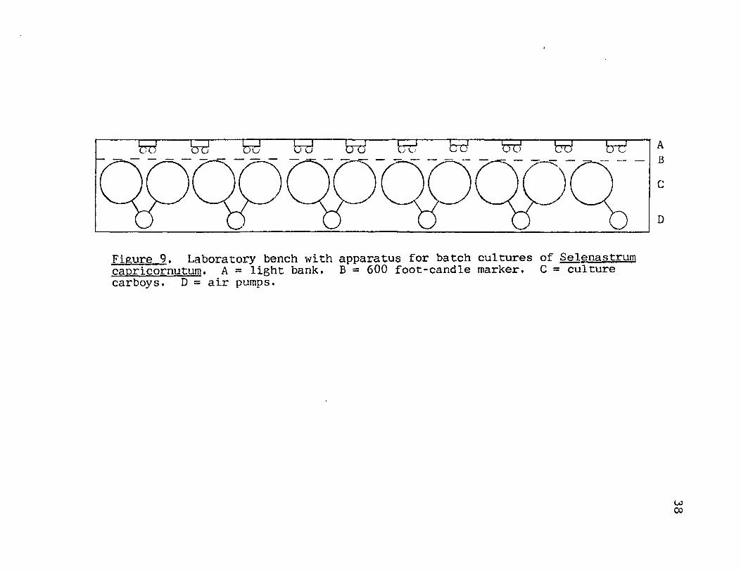
a laboratory bench 3~'' X IS' fitted '<lith a bank of ten
pairs of vertically arranged, 24 inch cool-white fluorescent
lights providing illumination of 600 foot-candles at a dis
tance of six inches from the lights. Culture containers
were placed in a line along the light bank 'vith the edges
of the containers nearest the lights lying on the 600 foot
candle marker (Fig. 9). Illumination was continuous. No
special provision was made to maintain constant temperature
in the room (Table IV). The pH of the nutrient medium was
approximately seven both before and after sterilization.
Two, and sometimes three, stock cultures were kept growing
at anyone time. Throughout the seven day incubation period
the culture flasks '<lere hand s\vi:r:led at least tt<lice daily at
irregular intervals.
After inoculation the ba tch cuI tures \\lere aerated con
tinuously with Silent Giant air pumps fitted with dust traps
(Fig. 10). In most cases the air bubbling provided suffi
cient mixing to maintain the cells in suspension; however,
o -j
Figure 9. Laboratory bench with apparatus for batch cultures of Selenastrum capricornutum. A = light bank. B = 600 foot-candle marker. C = culture carboys, D = air pumps.
A B
c
D
w co

TABLE IV
MONTHLY TEMPERATUP~ EXTREMES IN THE CULTURE LABORATORY
Month
March. 1974 April. 1974· Hay. 1974 June. 1974 July. 1974 August, 1974 September, 1974 October. 1974 November, 1974 December, 1974 January, 1975 February, 1975 Ivlarch, 1975
Temperature °Ca Maximum Minimum
24.0 24.5 24.5 27.0 26.5 25.0 22.0 23.5 21.5 21.0 20.0 21.0 20.5
19.5 20.0 19.0 22.0 25.0 21.5 21.0 18.5 19.0 14.0 18.0 19.0 19.0
aTemperatures were recorded three times daily.
c
B
39
Figure 10. Air supply for batch cultures of Selenastrum capricornutum. A = Silent Giant air pump. B = \vater-fi1led dust trap. C = air supply for two cuI ture carboys.

occasionally a layer of cells collected on the carboy bot
tom during the first few days of incubation. In these in
stances the carboys were hand swirled to resuspend the
cells.
40
Initially batch cultures consisted of ten to twelve
cuI ture carboys per batch. In order to allmv more efficient
harvesting, batches were later reduced to three or four cul
ture carboys each. Most batches included a control carboy
treated in the same manner as the culture carboys except no
inoculum was added.
Stock and batch cultures were labelled by sequential
numbering with the number of each stock culture preceeded
by the letter S and that of each batch culture preceeded by
the letter B. Cell concentrations and other pertinent data
recorded for batch cultures are presented in Table V.
After incubation for 14 to 21 days the aeration of
batch cultures was stopped, and the carboys were immediately
covered with aluminum foil bags. Separation of the algal
cells from the nutrient medium was accomplished by contin
uous-flow centrifugation using a Sorvall RC2-B centrifuge
at 7500 RPM (Fig. 11). The wet cells obtained were lyo
philized, weighed. and stored in the freezer. The super
natant was then ready for extraction. If any delay was ex
perienced between harvest and extraction, the carboys con
taining supernatant were kept covered ,.,ith aluminum foil
bags. Cells from the first four batches were lyophilized

41
TABLE V
pH AND CELL COUNTS OF BATCH CULTURESa
Batch 1ft Carboy 1ft Initial ce1lsLm1. Final cellsLm1. Final ]2H
B-13b 3 1 .• 6 X 10⁴ 2.7 X 10⁶ 7.5c
4 1.1 X 10⁴ 3.0 X 10⁶ 7.5
6 control 7.5
12 control 7.5
B-14d 1 1.3 X 10⁴. 4.4 X 10⁶ 7.2c
5 8.1 X 10³ 3.2 X 10⁶ 7.3
10 5.8 X 10³ 3.5 X 10⁶ 7.3
B-1Sd 2 2.4 X 10⁴ 3.0 X 10⁶ 6.8e
7 1.6 X 10⁴ 2.4 X 10⁶ 6.8
8 1.9 X 10⁴ 2.8 X 10⁶ 6.8
9 control 6.9
B-16b 3 control 6.8e
5 3.1 X 10⁴. 3.7 X 10⁶ 6.9
12 3.3 X 10⁴ 4.2 X 10⁶ 6.9
a These represent only a select group of cultures for which full data were recorded. Throughout the project sixteen batch cultures with a total of eighty-nine carboys were grown, most of which were utilized to standardize culturing, harvesting, and extraction techniques.
bIncubation time 14 days.
CDetermined with a Corning model 6 pH meter.
dlncubation time 21 days.
eDetermined with a Photovolt-digicord pH meter.

A
D
Figure 11. Apparatus for harvest of batch cultures of Selenastrum capricornutum. A = culture carboy. B = Latex-tubing siphon. C = continuous-flow attachment installed in a Sorvall RC2-B centrifuge. D = receiver for supernatant.
42

43
yielding 14.9, 9.1, 11.3, and 10.4 grams respectively of dry
cells.
Prior to harvest, a small sample of culture from each
carboy was streaked onto nutrient agar plates. and 100 mI.
of culture was removed for pH measurements and cell counts.
The nutrient agar plates were visually checked for the pre
sence of bacteria after an incubation period of seven days
in darkness and at room temperature.
I I. CHROMA TOGRAPHIC AND SPECTROMEIRIC METHODS
Solvents
All solvents used for extractions and chromatography
were Reagent grade and were redistilled just prior to use.
Thin Layer Chromatography
Thin layer chromatography was carried out using four
different TLC systems. TLC system I consisted of 20 X 20 cm.
glass plates pre-coated with a 2.0 mm. layer of silica gel
F-254· (EH Laboratories, Inc., Elmsford. New York). The sol
vent for this system 'tvas benzene: ether (4: 1). TLC system II
consisted of 20 X 20 cm. glass plates pre-coated with a
0.5 mm. layer of silica gel F-254 with benzene:ether (4:1)
as the solvent. The plates used in TLC system III were
identical to those used in system II; hmvever, in system III
the solvent used was chloroform. Thin layer chromatography
plates used for these TLC systems were kept in a desiccator
and were activated at 110 °c for 30 minutes just prior to

44
use.
TLC system IV consisted of 20 X 20 cm. Baker-flex
sheets coated with a 0.25 mm. layer of silica gel lB-F
(J.T. Baker Chemical Co., Phillipsburg, New Jersey) which
was impregnated with silver nitrate. Impregnation was ac
complished by dipping the pre-coated sheets in a 10% solu
tion of AgN03 in ethanol:water (4:1) for one minute followed
by drying at room temperature for one hour and activation
at 1100C for 30 minutes. The solvent for TLC system IV was
chloroform. The TLC systems are summarized in Table VI.
TABLE VI
TLC SYSTEMS
System Adsorbent Thickness Solvent
I silica gel F-254 2.0 rnm. benzene: ether (4:1)
II silica gel F-254 0.5 rnm. benzene:ether (4:1)
III silica gel F-254 0.5 rnm. chloroform
IV silica gel lB-F 0.25 rnm. chloroform AgN03
Gas Chromatography
All gas chromatograms were obtained using a Perkin-
Elmer model 990 gas chromatograph fitted with dual flame
ionization detectors which were operated at hydrogen and
air pressures of 21 psi and 23 psi respectively. All in-
jections were made into a glass lined injection port at
275 °c with the detectors also at 275 °c.

45
Gas chromatograms of sterols and sterol acetates were
obtained using two GC systems. GC system I consisted of a
6' X 4 mm. I.D. glass column packed with 3% OV-l on 80/100
mesh Anakrom Q. The column was operated isothermally at
275 °c with a helium flow rate of 30 ml./min. GC system II
was a 6' X 4 mm. I.D. glass column packed with 3% OV-17 on
80/100 mesh Anakrom Q. o It was also operated at 275 Cj
however, the helium flow rate for this system was 60 ml./
min.
GC system III employed the same column and carrier gas
flow as GC system I, but in system III the temperature was
programmed from 100 °c to 275°C at an increase of 12 °C/
min. after an initial hold time of four minutes after in-
jection. GC system IV employed the same column and carrier
gas flow as GC system II and the same temperature program
ming as system III. GC systems III and IV were used to ob
tain chromatograms of fatty acid methyl esters. The GC
systems are summarized in Table VII.
Absorption Spectrometry
Ultraviolet absorption spectra were obtained with a
Cary 14 spectrophotometer. Spectra were obtained for chlor
oform solutions of the samples.
Mass Spectrometry
Mass spectra were obtained using a Dupont 21-49lB
mass spectrometer. Samples were introduced through a di-

rect inlet heated to 150 °C. The ionizing voltage was
70 eVe
System
I
II
III
IV
Liguid J2ha§e
3% OV-l
3% OV-17
3% OV-l
3% OV-17
TABLE VII
GC SYSTEMS
TemQerature Program rate
275 °c isothermal
275 °c isothermal
100-275 °c 12 °C/mina
100-275 °c 12 °C/mina
aAfter four minute initial hold.
III. EXTRACTION METHODS
Extraction of Lyophilized Cells
Carrier gas flow
30 ml/min
60 ml/min
30 ml/min
60 ml/min
46
The general scheme for extraction of lyophilized cells
of s. capricornutum involved extraction with a series of or-
ganic solvents followed by preparative thin layer chromatog-
raphy of the extracts to separate sterols and sterol esters
from the crude extract. The cell residue was then extracted
with water to obtain water soluble sterols.
Extraction With Organic Solven!§. A quantity of
lyophilized ~ capricornutum weighing 7.60 grams was ex
tracted by refluxing for one hour with each of the following
solvents:
300 mI. ether 300 mI. ether 300 mI. acetone 300 mI. acetone

300 mI. chloroform:methanol (2:1) 300 mI. chloroform:methanol (2:1) 300 mI. chloroform:methanol (2:1) 300 mI. ether
47
The organic extracts were combined, filtered, and evaporated
to dryness in a rotary evaporator at reduced pressure.
This procedure yielded 2.99 grams of a dark, green, oily
residue which was then redissolved in 150 mI. of ether. A
small portion of this solution was chromatographed on TLC
system II along with a marker of authentic cholesterol,
and sterols were visualized by spraying with 50% H2S04 fol
lowed by heating for three minutes at 110 °C. The plate
was examined under a long-wave ultraviolet lamp, and two
spots were detected. One spot with Rf 0.17 to 0.23 corre-
sponded to the cholesterol marker and was assumed to con
tain free sterols while the second spot with Rf of 0.70 to
0.84 was assumed to contain sterol esters. These assump
tions were based on the visualization with 50% H2S04 and on
previously reported relative Rf values for sterols and
sterol esters (60). The remainder of the organic extract
was subjected to preparative thin layer chromatography on
TLC system I. Several plates were required to accomplish
this as no more than 300 mg. of sample was applied per
plate. The plates were divided into five zones. The ad-
sorbent in the zones of Rf range equal to those determined
by analytical TLC to contain sterols and sterol esters was
transferred into 250 mI. beakers and washed three times with
100 mI. of ether.
48
The ether extract of the adsorbent from the free
sterol zone was allowed to evaporate to a small volume and
was then subjected to a second preparative TLC separation on
TLC system III. Also, a marker of authentic cholesterol was
spotted at one edge of the plate. Following development.
the marker region was sprayed with 50% H2S04 , the plate was
heated at 110 °c for three minutes, and the marker region
was examined under a long-wave ultraviolet lamp. The ad-
sorbent from the zone having an Rf range equal to that of
the marker spot (0.17-0.31) was transferred into a beaker
and washed with 3 X 25 mI. of ether. The ether washings
were combined and allowed to evaporate to dryness yielding
18.7 mg. of white crystalline residue which was redissolved
in 5 mI. of ether and labelled fraction I. A second extrac-
tion of 10.5 grams of lyophilized S. capricornuturn from a
different batch culture yielded 28.5 mg. of fraction I.
The eluent from the sterol ester zone of the original
separation was allowed to evaporate to dryness, and the res
idue was heated on a steam bath for two hours with 50 mI.
of 10% KOH in methanol:water (9:1). After cooling to room
temperature, this reaction mixture was poured into an equal
volume of water and was shaken with three 25 mI. portions of
ether. The combined ether extracts were dried over anhy-
drous MgS04 and evaporated to dryness under a gentle stream
of nitrogen. The yield was 7.4 mg. of a white. crystalline
residue. This residue, labelled fraction II, was redis-

49
solved in 5 mI. of ether and screened by thin layer chroma
tography on TLC system II. Visualization with iodine vapor
revealed the presence of only one spot with Rf in the range
0.17 to 0.21. This Rf range corresponds to that observed
for cholesterol on TLC system II.
The second extraction of lyophilized S. capricornutum
(10.5 grams) yielded 18.0 mg. of fraction II.
The pH of the aqueous fraction from the saponification
of the sterol esters was adjusted to 2.0 by addition of con
centrated HCI. The acidified solution was shaken with three
25 mI. portions of ether, and the combined extracts were
dried over anhydrous MgS04 and evaporated to dryness under
a gentle stream of nitrogen. The brown, oily residue
(100.0 mg.) obtained was redissolved in 5 mI. of ether and
screened for fatty acid content on TLC system II. Visuali
zation with iodine vapor indicated only one spot with an Rf
range similar to that of authentic steric acid (Rf = 0.23-
0.45). The solution was labelled as fraction III.
Extraction With Water. After the extraction with
organic solvents the cell rasidue was allowed to air dry and
was then extracted three times with water by refluxing for
one hour with 300 mI. of water each time. The combined
aqueous extracts were centrifuged to remove cellular debris,
and the supernatant was shaken with three 50 mI. portions of
ether. The purpose of the ether extraction was to remove
any lipid soluble material from the aqueous extracts before
accomplishing the removal of water soluble sterols. These
ether extracts were combined, dried over anhydrous MgS04
50
and the solvent allowed to evaporate. A residue weighing
3.3 mg. was obtained. An ether solution of this residue was
screened on TLC system II and no sterols were detected.
The aqueous extract was split into two portions. One
part was treated with acid using a modification of the pro
cedure described by R. J. Pryce (47). This involved mixing
with 450 mI. of concentrated HCl:water:methanol (1:2:3) and
stirring this mixture for six days at room temperature. The
resulting solution was shaken with 150 mI. and then three
50 mI. portions of ether. The ether extracts were combined,
dried over anhydrous MgS04' and allowed to evaporate to dry
ness. This procedure yielded 71.6 mg. of a green, oily res
idue which was screened on TLC system II and found to con
tain no sterols. Acid treatment of aqueous extracts was
not performed in subsequent extractions of lyophilized cells
and supernatant.
The aqueous fraction remaining after the acid treat
ment was made basic by addition of solid potassium hydrox
ide and was then treated with pyrogallol using a method sim
ilar to that described by B. G. Adams and L. W. Parks (43).
The alkaline solution was mixed with 150 mI. of 0.5% pyro
gallol in methanol, 100 mI. of 60% aqueous potassium hydrox
ide, and 150 mI. of methanol. This mixture was refluxed for
three hours, cooled, and then shaken with three 100 mI. por-
tions of ether. These ether extracts were combined, dried
over anhydrous NgS04 , and allowed to evaporate to dryness.
A quantity of white, crystalline residue weighing 0.7 mg.
'\vas obtained by this method. The residue '\vas redissolved
51
in 5 mI. of ether and was labelled as fraction IV. The
second extraction of ~ capricornutum cells (10.5 grams)
yielded 26.8 mg. of fraction IV from one half of the aqueous
extract. The aqueous extract from the second extraction was
evaporated to dryness in a rotary evaporator at 50 °c prior
to treatment with alkaline pyrogallol.
The remaining 400 mI. of original aqueous extract '\vas
split into t'\vO 200 mI. portions. The first part was mixed
with 150 mI. of dimethylsulfoxide (DMSO) and heated on a
steam bath for three hours. The second part lvas treated in
an identical manner with the exception that 20 grams of so
dium carbonate lvas added to the mixture prior to heating.
T. Santosusso and D. Swern reported that catalysis arising
from in situ formation of strong acid in DMSO may be impor
tant in several reactions involving D~1S0 (61,62). Sodium
carbonate lvas added to neutralize any strong acids which
might be formed in this reaction and thus to prevent the
isolation of products not directly resulting from the pre
sence of D~lS0 (61). After cooling, each Dt-ISO mixture was
extracted with three 50 ml portions of ether. These ether
extracts were washed with nolO 50 mI. portions of saturated
sodium chloride solution and dried over anhydrous MgS04 .

52
The solvent '\vas removed by evaporation in a gentle stream of
nitrogen; however, no weights '\vere recorded for the residues
obtained. One half of the aqueous extract of the second
batch of S. capricornutum cells (10.5 grams) 'tvas evaporated
to dryness in a rotary evaporator at 50 °c followed by iden
tical treatment with DMSO, but without the sodium carbonate
addition. The extraction of this reaction mixture yielded
10.9 mg. of a green, oily residue.
The three extracts from the D~fSO treatments were
screened using TLC system II. No sterols were detected in
these samples.
Extraction of the Supernatant
Extraction \'1ith Organic Solvent. The extraction of
large quantities of supernatant was accomplished by using
two different methods. The first involved extraction of
several aliquots of supernatant in three-liter separatory
funnels followed by combination of the extracts from each
aliquot. The second method involved simply stirring large
quantities of supernatant 'tvith ether under nitrogen fol
lmved by removal of the ether '\'lith an all-glass siphon.
The firs t extraction method 'vas used to extract 30
liters of supernatant from one batch culture. Aliquots of
2.5 1 i ters of supernatant were firs t shaken 'tvi th 300 mI.
of ether and then with two 100 mI. portions of ether. The
ether extracts 'vere combined, dried over anhydrous MgS04'

53
and then concentrated to a small volume in a rotary evapo
rator. After the concentrate was allowed to evaporate to
dryness, the yield obtained was 7.6 mg. of a light green,
oily residue. This residue was redissolved in 5 mI. of
ether and was screened using TLC system II. Visualization
with iodine vapor revealed the presence of free sterols
(Rf = 0.15-0.21); however, subsequent spraying with 50%
H2S04 and heating at 110 °c indicated that no sterol esters
were present in this sample. The remainder of the extract
was subjected to preparative TLC separation. using TLC sys
tem II. Material in the free sterol zone was recovered
from the plate by transferring the adsorbent in this zone
into a 100 mI. beaker followed by washing with three 25 mI.
portions of ether. The ether extracts were combined and
filtered. Evaporation of the solvent yielded 1.0 mg. of a
white crystalline material which was then dissolved in 5 mI.
of ether and labelled as fraction V.
A total of 105 liters of supernatant from four dif
ferent batch cultures was extracted with ether by using the
second extraction method. Using a magnetic stirrer, fifteen
liter aliquots were mixed under nitrogen with 2000 mI. of
ether in a five-gallon carboy for 5 to 10 days. After mix
ing; the ether phase was removed using an all-glass siphon,
and the aqueous phase was mixed with a second portion of
500 mI. of ether under the same conditions. Following re
moval of the second ether phase, the ether extracts were

combined, dried over anhydrous MgSOq.' and evaporated to a
small volume in a rotary evaporator.
54
The extract from each of the eight aliquots was
screened for sterol content using TLC system II. Visualiza
tion with iodine vapor revealed spots with Rf in the free
sterol range (0.14-0.22) in all eight samples. No sterol
esters were detected in these samples. The eight extracts
were combined, and the solvent was allowed to evaporate
leaving 2l.q· mg. of a light green, oily residue. The en
tire residue was chromatographed on TLC system II, and com
pounds in the zone with Rf corresponding to that of free
sterol were recovered by transferring and extracting the
adsorbent as previously described. The material isolated
by this procedure weighed 2.1 mg. and was also labelled as
fraction V.
Thirty liters of nutrient medium from control carboys
of t\vO different batch cultures was extracted by using the
second extraction method. Chromatographic screening on TLC
system II showed that the extracts contained no compounds
with Rf values in either the free sterol or sterol ester re
gion.
Water Soluble Sterols. Sixteen liters of the super
natant which had been extracted by the first extraction
method was placed in a 22 liter flask and mixed with 1000
mI. of 0.5% pyrogallol in methanol, 1000 mI. of 60% aqueous
potassium hydroxide, and 1000 mI. of methanol. After re-

55
fluxing gently for two hours, this mixture was cooled, and
a total of 17.5 liters, in 2.5 liter a1iquots. was extracted
with ether as described above for untreated supernatant.
This procedure yielded 8.3 mg. of ether soluble material
which was screened using TLC system II. Iodine vapor visu
alization revealed a spot with Rf in the free sterol region
(0.18-0.23). A preparative TLC separation of the components
of the residue using TLC system II and the methods described
above for sample recovery gave 1.5 mg. of a white solid
from the free sterol region of the plate. This sample was
redissolved in 5 mI. of ether and was labelled as fraction
VI.
Thirty liters of supernatant extracted by the second
extraction method was divided into 15 liter aliquots. Each
of these aliquots was treated with alkaline pyrogallol using
the same conditions and quantities of added reagents as de
scribed in the preceding paragraph. The resulting mixtures
were extracted with ether by using the second extraction
method. The individual extracts were screened on TLC system
II. Visualization with iodine vapor showed the same array
of spots in each chromatogram, including a spot in the free
sterol Rf region (0.14.-0.19). These extracts were then com
bined, giving a total weight of 1.07 grams after removal of
the solvent. Preparative TLC of the total extract on TLC
system I resulted in recovery of 5.4· mg. of a green, oily
residue from the free sterol region of the plate. The major

portion of the extract consisted of two unidentified com
pounds of high Rf. The recovered residue was redissolved
in 5 mI. of ether and was also labelled as fraction VI.
56
As in the extraction of lyophilized algal cells,
treatment with DMSO was utilized to check for water soluble
sterols in the supernatant. Thirty liters of supernatant,
extracted by using the second extraction method, was divided
into two 15 liter aliquots. Each aliquot was mixed with
200 mI. of DMSO. The first was stirred under nitrogen for
eight days and then extracted with ether directly in the
carboy. The second aliquot was transferred to a 22 liter
flask and was gently refluxed for one hour after which it
was returned to a five-gallon carboy and extracted with
ether. TLC screening of the two extracts obtained showed
them to be similar in composition. A spot in each chromat
ogram in the range Rf = 0.13-0.23 indicated that free ster
ols were possible components of the samples. The extracts
were combined, giving 7.7 mg. of residue from which the
sterol-like components were removed by preparative TLC as
previously described. A green, oily residue weighing 0.6
mg. was obtained and was labelled as fraction VII.
Summary
The extraction methods used to obtain sterols from
lyophilized s. capricornutum cells and from the nutrient
medium supernatant are summarized in Fig. 12-14.

Lyophilized cells
extract with organic solvents
organic extract ~------'l--------~ residue
sterols
preparative TLC System I
1 Preparative TLC System III
Fraction I
sterol. ,esters
ether fraction Fraction II
acidify,
saponify, extract with ether
aqueous fraction
extract with ether
Fraction III
57
Figure 12. Isolation of free and esterified sterols from lyophilized Selenastrum capricornutum.

58
Lyophilized cells
extract \vi th organic solvents
organic extract~----------~--------------~residue
1 extract \vi th water
aqueous extract
extract with ether
aqueous fraction--------~~~ether
alkaline pyrogallol, extract with ether
aqueous~(----~W~--~)ether fraction fraction
1 fraction
Fraction IV
DMSO, extract \vi th ether
ether~~--~vr------~)aqueous fraction fraction
.., etner fraction
DMSO. Na2C03, extract with ether
... aqueous fraction
Figure 13. Treatment of the cell residue to release water-soluble sterols.

59
Supernatant
extract with ether
ether fraction~----------~~------~aqueous fraction
preparative TLC System II
Fraction V
alkaline pyrogallol, extract with ether
ether~--~----~aqueous fraction fraction
preparative TLC Systems I and II
Fraction VI
DNSO, extract with ether
ether aqueous fraction fraction
preparative TLC System I
Fraction VII
Figure ll,. Isolation of free sterols from the supernatant and treatments for release of \vater-soluble sterols.

60
Pretreatment of Lvophilized Cells
A sample of lyophilized S. capricornuturn 'tvas extracted
by the methods previously described. The remaining cell
residue was mixed with 150 mI. of 'vater and 100 mI. of 20%
potassium hydroxide in ethanol. This mixture was refluxed
for two hours, cooled, and diluted with 100ml. of water.
The final mixture ,vas extracted by shaking 'vi th three 100
mI. portions of ether. A chromatographic screen of this ex
tract on TLC system II shmved that the treatment with
potassium hydroxide failed to release sterols for extrac
tion. Simple extraction with ether, acetone, chloroform:
methanol, and water is, therefore, sufficient for removal
of all sterols from lyophilized cells of S. capricornutum.
D~SO as a Solvent For Sterol Extraction
A sample of lyophilized S. capricornutum (9.38 go)
was extracted 'vi th ether: DHSO (8: 1) by refluxing for two
hours with three separate 500 mI. portions of the solvent.
The extracts were combined and washed with three 500 mI.
portions of saturated sodium chloride solution follmved by
one wash with 500 mI. of water. The resulting ether solu
tion was dried over anhydrous NgS04 • Removal of the solvent
yielded 690 mg. of a dark green residue. This residue ''las
screened on TLC system II and was found to contain a com
ponent with Rf in the free sterol region (Rf ~ 0.21-0.42).
Preparative TLC separation of this component from the res-

61
idue resulted in recovery of 31.0 mg. of a light green res
idue which was labelled as fraction VIII.
Melting Points
Melting points were obtained using a Unitron melting
point apparatus with a Kofler micro hot stage. Melting
point values are uncorrected.
IV. METHODS FOR OB TAINING S TEM-I VOLATILE Cot'WOUNDS
An original objective of this project was to isolate
and characterize any steam-volatile organic compounds pro
duced by the cultured alga and subsequently released into
the culture medium or retained within the algal cells. The
experiments conducted in this regard yielded data which led
to the decision to change from use of rubber tubing to Tygon
tubing for making aeration connections.
Experiments designed to isolate steam-volatile com
pounds from the culture supernatant were conducted according
to the following procedure. Fifteen liters of supernatant
was transferred to a 22 1. round-bottom flask fitted with
a condensor. This was heated and two liters of distillate
was collected. This distillate was shaken in a separatory
funnel with a 200 ml. portion and then three 100 mI. por
tions of ether. The ether fractions were combined. dried,
filtered, and evaporated to dryness.
Two 15 1. samples of distilled water used for prepar
ing the nutrient medium were treated as described for the

62
supernatant. After the original distillation of one of the
distilled water samples, a three-feet-long piece of rubber
tubing was placed in the distilling flask along with the
remaining water. After three days the tubing was removed
and an additional 2 1. of distillate was collected and ex
tracted as described above.
A 5.4633 g. sample of lyophilized cells was mixed with
450 mI. of water in a 1000 mI. round-bottom flask fitted
with a condensor. This mixture was heated and 200 mI.
distillate was collected. The distillate was shaken with
a 75 mI. portion and then three 50 mI. portions of ether.
The ether fractions were combined and treated as described
above.
All ether extractions obtained from steam-distillates
were labelled as Fraction SD. The weights of the extracts
are reported in Table VIII.
With the exception of sample number two, all samples
in Fraction SD were screened by thin-layer chromatography
on a single 20 cm. X 20 cm. plate coated with 0.25 mm.
SilicAR TLC-7GF with benzene:ether (4.:1) as the solvent.
The results of this screening are presented in Table IX.
The results obtained in these experiments indicated
that steam-volatile compounds isolar.ed from culture super
natant and from lyophilyzed cells were also obtained in one
or more of the control experiments. Furthermore, the small,
variable amount of steam-volatile organic material obtained

63
TABLE VIII
WEIGHTS OF SAMPLES IN FRACTION SD
Sample Source Tubing typea Weight(mg)
1 culture
2 culture
3 culture
4 culture
5 control
6 culture
7 culture
8 control
9 control
10 tubing b
11 distilled water
12 distilled water
13 lyophilyzed cells
rubber
rubber
rubber
rubber
rubber
rubber
Tygon
rubber
Tygon
rubber
rubber
2.0
0.8
12.0
14.3
2.5
0.2
0.9
o o 3.7
1.0
o 4.8
aRefers to the type of tubing used in making the aeration connections.
bRefers to the experiment involving distilled water in contact with rubber tubing for three days.
from the cultures was apparently derived from the distilled
water used in preparing nutrient medium and from the rubber
tubing used in aeration connections. The latter was the
more important source. Subsequent to the interpretation of
these results, Tygon tubing was used in the aeration con-
nections.

64
TABLE IX
TLC SCREEN OF SAMPLES IN FRACTION SDa,b
Spot 4ft Sample 4ft
1 3 4 5 6 7 10 11 13
1 0.00 0.00 0.00 0.00 0.00 0.00 0.00 0.00 0.00
2 0.04
3 0.15 0.15 0.16 0.16
4 0.30 0.28 0.31
5 0.41 0.40 0.42
6 0.52 0.50 0.51 0.50 0.53
7 0.56 0.55
8 0.63 0.63 0.61 0.59 0.58 0.60 0.58
9 0.66 0.67
10 0.73 0.71 0.73 0.73 0.75
11 0.78 0.77 0.75 0.79 0.78 0.78 0.81
a See Table VIII for sample source and type of tubing.
bNumbers reported are Rf values of the spots in each chromatogram.

CHAPTER IV
RESULTS
I. FREE STEROLS
Sterols From Lyophilized Cells
Fraction I was analyzed by gas chromatography on GC
systems I and II. Two peaks were observed and comparisons
were made of the relative retention times of the peaks
with those of several authentic sterols (Table X). The
peak of shorter retention time (sterol A) was tentatively
identified as representing a C-28 sterol. The second peak
(sterol B) was identified as representing a C-29 sterol.
Calculation of peak areas showed that fraction I was com
posed of 43% sterol A and 57% sterol B.
Relative retention times of sterols and sterol
acetates were calculated relative to the retention times
of cholesterol and cholesterol acetate, respectively.
Cholestane is often used as a reference for these calcula
tions: however, more reproducible values of relative re
tention times were obtained by using cholesterol and choles
terol acetate as references.
Sterols A and B were acetylated by treatment with
6 mI. of pyridine-acetic anhydride (1:1) for 16 hours at
room temperature. After this time the reaction mixture was

66
TABLE X
REL~TIVE RETENTION TIMES OF STEROLS AND STEROL ACETATESa
Sterol Gas Chromatographic System b
GC system I GC system II SE-30
Sterol A
Sterol B Cholesterol Brassicasterol Ergosterol /l5-Ergostenol ~5,7-Ergostadienol Stigmasterol Poriferastero1 l-Ergostenol Chondril1astero1 f>-Si tostero1 Fucostero1
1.30(1.63) 1.56(1.97) 1. 00(1.00) 1.11
1.25
1.40 1.36 1.24(1.38) 1.33(1.48) 1.50 1.50
(1.84) 28-Isofucostero1 ~5,7-Stigmastadienol h7-Chondri11astenol1.49(1.67)
1.24(1.47) 1.53(1.78) 1.00(1.00)
1.37 1.33(1.38) 1.24(1.48) 1.34(1.59) 1.50(1.55) 1.57(1.64)
(1.70)
1.49(1.77)
1.00(1.00) 1.12(1.12) 1.22(1.22) 1.30(1.30) 1.42(1.42) 1.42(1.42) 1.42(1.42) 1.46(1.46) 1.58(1.58) 1.63(1.63) 1.63(1.63) 1.69(1.69) 1.78(1.78) 1.83(1. 83)
aVa1ues in parentheses are for sterol acetates.
bLiterature values for relative retention times on an SE-30 column (63).
diluted ,,,ith 10 mI. of methanol and 50 mI. of lvater. The
sterol acetates were recovered by extraction of the final
mixture with ether, and they were analyzed by gas chroma
tography (Table X). Comparison of the relative retention
times of these acetates ''lith relative retention times of
authentic sterol acetates confirmed the identification of
sterols A and B as C-28 and C-29 sterols.

67
For purposes of obtaining mass spectra, small quan
tities of the acetates of sterols A and B were separated by
using TLC system IV. The sterol acetate bands were located
on the plate by visualization under a long-wave ultraviolet
lamp, and the acetates were recovered by scraping the bands
and extracting with ether as previously described. The
major ions in the mass spectra of the acetates of sterols
A and B are recorded in Table XI.
The mass spectrum of sterol A acetate had a molecular
ion at mle 4·40 indicating a C-28 sterol with two double
bonds. The base peak at mle 313 resulted from loss of a
C9H19 side chain, indicating that both double bonds were in
the nucleus (29). The molecular ion at mle 454 in the mass
spectrum of sterol B acetate indicated that this was a C-29
sterol acetate with two double bonds. The base peak was at
mle 313 suggesting a C10H2l side chain with both double
bonds in the nucleus. The similarity of the fragmentation
patterns in the mass spectra showed that sterols A and B
differed only in size of the side chain.
Table XI shows comparative mass spectral data for the
acetates of sterols A and B. and the authentic sterols 24-
methylcholesta-5,7-dien-3~-01 and 24-ethylcholesta-S,7-dien-
3(',-01. The configuration at C-24· has no effect on the mass
spectrum. The major differences between the mass spectra
are due to loss of acetic acid from the acetates versus loss
of water from the free sterols. The similarity of the frag-

68
TABLE XI
MAJOR IONS IN THE MASS SPECTRA ~F THE ACETATES OF STEROLS A AND B
Ion A Cb B D F . c ragmentat~on
M+ 440(25) 398(35) 454(40) 412(51) m~lecu1ar ion a 425(22) 383(9) 439(27) 397(12) M -Me b 380(11) 380(20) 394(11) 394(28) M+-HOAc(H 0) c 365(14) 365(58) 379(16) 379(78) M!- Me+Ho~c(H20) d 356(36) 356(11) ~+-SCA e 341(16) 341(16) M -SCB f 339(23) 353(28) M:-(C1~C3+0H+6H) g 313(100) 271(19) 313(100) 271(23) M -SC h 255(32) 255(10) 255(58) 255(6) i 253(20) 253(40) 253(14) 253(45) M+- SC+HOAc(H2O). j 229(18) 229(8) 229(26) 229(8) k 228(14) 228(11) 1 227(23) 227(13) 227(26) 227(13) m 213(35) 213(13) 213(42) 213(14) n 211(15) 211(23) 211(11) 211(28) 0 201(15) 201(14) p 200(10) 200(8) 200(8) 200(7) q 199(15) 199(30) 199(14) 199(32) r 197(13) 197(23) 197(8) 197(22) s 189(15) 189(8) t 187(15) 187(8) 187(13) 187(5) u 185(20) 185(18) 185(14) 185(19) v 183(10) 183(18) 183(10) 183(18) w 175(10) 175(11) x 173(15) 173(8) 173(15) 173(12) y 171(18) 171(25) 171(17) 171(26) z 161(30) 161(8) 161(28) 161(9) aa 160(10) 160(8) 160(15) 160(8) bb 159(20) 159(33) 159(22) 159(31) cc 158(10) 158(28) 158(8) 158(30) dd 157(20) 157(33) 157(17) 157(35)
a lon intensities are in parentheses.
bCo1umns C and D are major peaks in the mass spectra of (24R)-ergosta-5,7-dien-3~-ol and stigmasta-5,7-dien-3~-01, provided by Dr. G. W. Patterson, Department of Botany, University of Maryland.
cSCA = C-23 through side chain end; SCB = C-22 through side chain end; SC = side chain.

69
mentation patterns of the sterol nucleus of the four com
pounds suggests that sterol A is 24·-methylcholesta-S,7-dien-
3~-ol and sterol B is 24-ethylcholesta-S,7-dien-3~-ol.
The melting point of the acetates of sterols A and B
were lSO-lS3 °c and 176-180 °c respectively. Values of lS2-
lS4 °c and lS8 °c have been reported for the melting point
of the acetate of 22-dihydroergosterol.
The ultraviolet spectrum of the sterol acetate mixture
shmved absorption at 284· run., typical of /).S,7 -sterols (33).
On the basis of this evidence sterol A was identified as
24-methylcholesta-S,7-dien-3p-ol, and sterol B was identi
fied as 24-ethylcholesta-S,7-dien-3~-ol. Insufficient
amounts of these sterols were isolated for determination of
NMR spectra or of specific rotation, the only methods al
lowing assignment of configuration at C-24. By analogy
with results obtained for most algal sterols these sterols
were assumed to possess the p configuration at C-24. Sterol
A was tentatively identified as (24S)-24-methylcholesta-S,7-
dien-3p-ol, trivial name 22-dihydroergosterol, and sterol B
was tentatively identified as (24S)-24-ethylcholesta-S,7-
dien-3~-ol, trivial name AS,7-chondrillastenol (Fig. IS).
The composition of the free sterols obtained by two extrac-
tions of lyophilized S. capricornutum are summarized in
Table XII.

TABLE XII
COMPOSITION OF FREE STEROLS OF Selenastrum caoricornutum
Sterol
A
B
% <?f Kree sterol . b first extract10n second extract10n
43
57
32
68
aCu1ture age \11aS 14 days.
bCu1ture age tvas 21 days.
22-dihydroergosterol
HO
~5.7-chondrillastenol
HO
Figure 15. Sterols of Selenastrum capricornutum.
70

Sterols From the Supernatant
The two samples of fraction V were analyzed on GC
system I. Both contained 24-methylcholesta-5,7-dien-3B-ol
and 24-ethylcholesta-5,7-dien-3Aol. The composition of
sterols in these samples is given in Table XIII.
TABLE XIII
COMPOSITION OF STEROLS EXTRACTED FROM THE SUPERNATANT
71
Sterol 1st % o~ Stero~d
extract~on 2 extraction
24-methylcholesta-5,7-dien-3p-ol
24-ethylcholesta-5,7-dien-3~-ol
43
57
48
52
The concentration of sterols in the first supernatant
sample was 33~g/1., and in the second sample it was 20
rs/l. These data were obtained by two different extraction
methods and the differences in sterol concentrations may be
a result of differing efficiencies of extraction.
II. BOUND STEROLS
Sterols From Stero1 Este~s
Gas chromatographic analysis of the two samples of
fraction II revealed that this fraction also contained 24-
methylcholesta-5,7-dien-3p-ol and 2q-ethylcholesta-5,7-dien-
3p-ol. The relative amounts of the two sterols, however,
were more variable in fraction II than in the free sterol

fraction. The composition of fraction II is given in
Table XIV.
TABLE XIV
COMPOSITION OF STEROLS FROM STEROL ESTERS
Sterol 1st % of E~teaifi~a Sterol. b
extract~on 2 extract~on
24-methylcholesta-S,7-dien-3~-ol
24-ethylcholesta-S,7-dien-3p -ol
aCulture age was 14· days.
bCulture age was 21 days.
Water Soluble Sterols
62 39
38 61
72
All samples of fractions IV, VI, and VII were analyzed
on GC systems I and II. These samples were found to contain
only minute quantities of sterols. Sterol peaks were too
small for peak area measurements to be made, and in most
cases the peaks were indistinct, making retention time meas-
urements difficult. It is reasonable to conclude that
sterols present in these fractions were lipid soluble
sterols which were incompletely extracted by the organic
solvents.
Sterols ExtraQ!ed By DMSO:Ether
Analysis of fraction VIII on GC system I showed that
this fraction was a mixture of 24-methylcholesta-5,7-dien-
3p-ol and 24·-ethylcholesta-S,7-dien-3p-ol. The composition
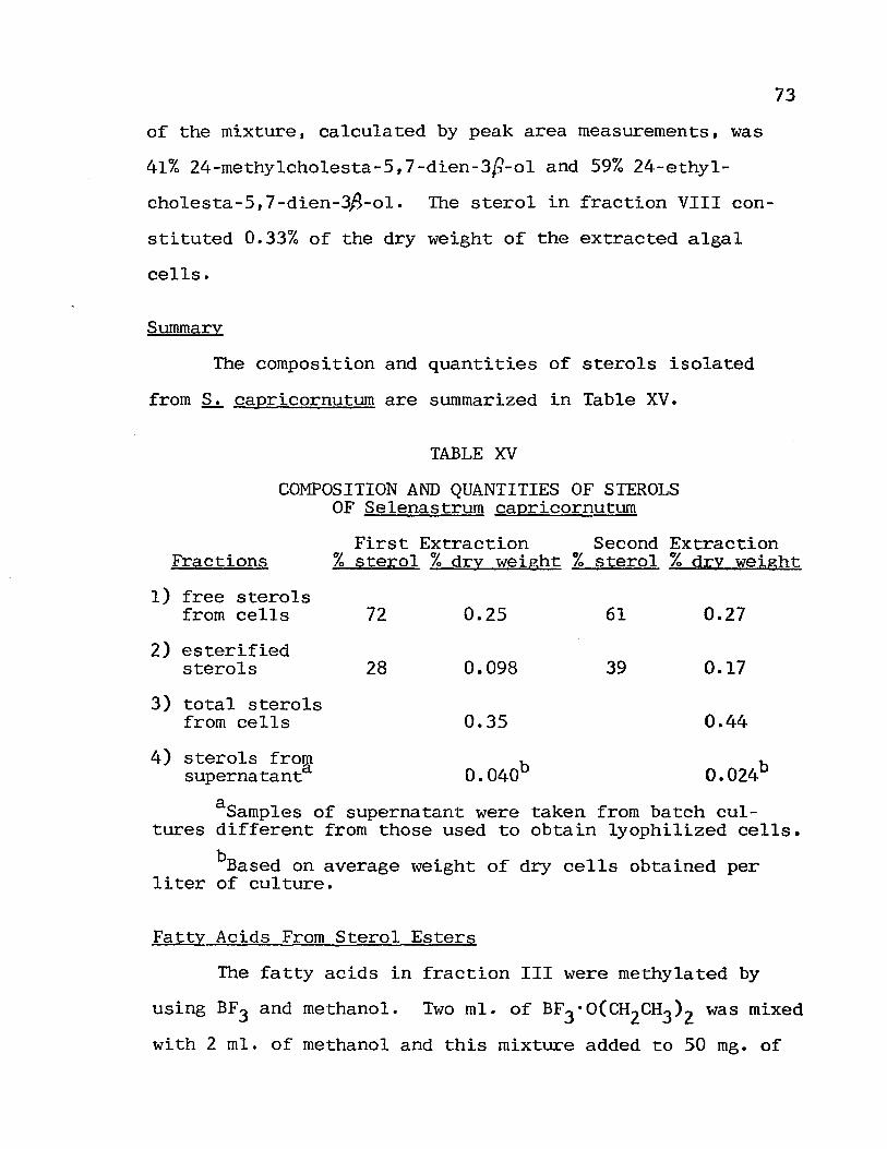
of the mixture, calculated by peak area measurements, was
41% 24-methylcholesta-5,7-dien-3p-ol and 59% 24-ethyl
cholesta-5,7-dien-3fi-ol. The sterol in fraction VIII con
stituted 0.33% of the dry weight of the extracted algal
cells.
Summary
The composition and quantities of sterols isolated
from S. capricornutum are summarized in Table XV.
TABLE XV
COMPOSITION AND QUANTITIES OF STEROLS OF Selenastrum capricorn~
73
First Extraction Second Extraction Fractions % ste~ol % dry weight % sterol % dry weight
1) free sterols from cells 72 0.25 61 0.27
2) esterified sterols 28 0.098 39 0.17
3) total sterols from cells 0.35 0.44
4) sterols from supernatanta O.040b O.024b
aSamples of supernatant were taken from batch cultures different from those used to obtain lyophilized cells.
bBased on average weight of dry cells obtained per liter of culture.
Fatty Acids From Sterol Esters
The fa tty acids in fraction III \17ere methylated by
using BF3 and methanol. Two mI. of BF3 ·O(CH2CH3 )2 was mixed
with 2 mI. of methanol and this mixture added to 50 mg. of

74
the fatty acid fraction. After heating in a boiling water
bath for 3 minutes, the reaction mixture was allowed to cool
to room temperature and was poured into 10 mI. of hot water.
This final mixture was allowed to cool and the fatty acid
methyl esters were extracted with three 15 mI. portions of
ether. The extracts were combined, dried over anhydrous
MgS04' and were analyzed on GC systems III and IV. The re
tention times of the components of this extract, along with
the retention times of some authentic methyl esters of sat-
urated fatty acids, are listed in Table XVI.
TABLE XVI
RETENTION TIMES OF FATTY ACID METHYL ESTERSa
Fatty Acid
unidentified unidentified C-lO C-12 C-14· C-16 unidentified C-1B unidentified
RT of Authentic Ester
8.3 (B.4) 10.B(10.9) 12.9(13.1) 14.7(14.B)
16.3(16.5)
RT of Algal Acid Ester
2.1 3.2 B.4· (B.2)
10.B(10.5) 12.7(13.2) 14.6(14.9) 15.2(15.5) 16.1(16.5) 16.6(17.1)
% of b Mixture
9.0 2.1 2.1 2.1 1.6 9.0
21.0 12.2 40.9
aRetention times are in minutes. Values in parentheses are for GC system IV; others are for GC system III.
b Calculated from peak areas.
The quantity of fatty acids obtained in fraction III
was several times the amount expected from the quantity of
sterols in fraction II. The excess fatty acid most likely
was obtained from sources in the cells other than sterol

esters. Therefore, the data presented in Table XVI do not
necessarily reflect the actual distribution of fatty acids
in the sterol esters.
75

CHAPTER V
DISCUSSION
Discussion of Results
The two sterols, 24-methylcholesta-5,7-dien-3~-ol and
24.-ethylcholesta-5, 7-dien-3~-ol, were the only sterols de-
tected in extracts of Selenastrum capricornutum. By analogy
with results obtained for most algal sterols these sterols
~vere assumed to possess the (!. configuration at C-24. Al
though this is the first report of 24-ethylcholesta-5,7-
dien-lA-ol in a species of Chlorophyta, this sterol was
found in Euglena gracilis (33) and unidentified a5,7-sterols
were reported in several Chlorella species (I). Further
investigation of sterols identified from chromatographic
evidence alone (64) may show thati,7-chondrillastenol is of
common occurence in the green algae. Extractions of S.
capricornutum failed to yield ~7_ or ~5-sterols, supporting
h 1 · ha hI' , 'h 5 t e conc us~on t t t e green a gae conta~n e~t er h - or
A7-, or ~5,7-sterols and that the simultaneous occurence of
these sterol types in algae is unusual.
The composition and quantities of sterols in sterol
esters of S. capricornutum were determined; however, com-
parative data for other species of Chlorophyta are unavail-
able. Furthermore, insufficient data were obtained in all

77
aspects of this experiment to establish the precision of
measured sterol concentrations and compositions. Although
data reported are in line with results of other workers, the
significance of small differences in concentration and com
position cannot be established without further experiments.
The two sterols found in esterified form were identical to
those found in the free form. The composition of the ester
ified sterols varied markedly as a function of culture age,
with the composition in older cultures being similar to that
of the free sterol fraction. The composition and quantity
of the free sterol fraction showed insignificant variation
as a function of culture age. The total sterol content of
S. capricornutum cells was shown to increase with culture
age. This increase was a result of increased sterol ester
content.
Several experiments designed to extract water soluble
sterols from ~ capricornutum cells and from the culture
medium failed to extract measureable quantities of sterols.
Water soluble sterols were found to make up only a small
fraction of the total sterol content of two species of
higher plants; however, other species of Chlorophyta have
been found to lack water soluble sterols. In view of these
reports, a reasonable conclusion is that the sterols iso
lated from aqueous extracts of ~ capricornutum and from the
nutrient medium after organic solvent extraction were lipid
soluble sterols incompletely extracted by the organic

78
solvents.
The composition of the sterol fraction isolated from
the nutrient medium was similar to that of the free sterol
fraction from S. capricornutum cells. The concentration of
sterols in the nutrient medium (20-33ftg./I.) was approxi
mately equal to the solubility of cholesterol (25-29 Mg./I.) I
(17). Cell lysis and active secretion by the algal cells
could serve as sources of the sterols found in the culture
medium. Cell lysis could occur during incubation and during
the harvest process.
Results of treatment of the algal cells with strong
base prior to extraction have indicated that such pretreat
ment is unnecessary for complete extraction of sterols.
Extraction of the algal cells with a DMSO:ether mixture re-
sulted in recovery of a sterol fraction similar in composi-
tion and amount to those extracted by more commonly used
solvents. DMSO did, however, prove to be useful, as lesser
amounts of non-sterol material were extracted by DMSO:ether,
making purification of the sterol fraction less complicated.
Recommendations
Of particular importance in future studies involving
algal sterols, is the investigation of the composition and
quantities of bound sterols. Results from higher plants and
from algae have indicated that in some organisms water solu
ble complexes of sterols may be of major importance in
metabolic processes involving sterols; however, such com-
79
plexes are absent in some organisms. A complete understand
ing of the role of water soluble sterol complexes in plant
sterol metabolism will be possible only after thorough in
vestigations into the occurence and structures of these com
plexes have been made. Sterol esters may also serve some
purpose in sterol metabolism, possibly as a storage or
transport form of sterols.
The mechanisms by which sterol biosynthesis is ac
complished in algae have recently been investigated in a
few algal species. With the diverse array of sterols pro
duced by algae, particularly the green algae, further in
vestigations of biosynthetic pathways will be necessary be
fore algal sterol biosynthesis can be completely understood.
Of particular interest in biosynthetic studies, is the
mechanism and stereochemical control of side chain alkyla
tion at C-24.
The compositions of both free and bound sterols of
some algae appear to have value as guides to taxonomy and
phylogeny. Further investigations of all algal divisions
will make possible generalizations as to sterol content,
and will determine the usefulness of these generalizations
for taxonomic purposes.
Very little is known concerning the release by algae
of sterols into their environment. Results of this work
and of experiments with a few other algal species have sho,m
that sterols are released by these algae into the culture

80
medium; however, the mechanism of the release is unknown.
Elucidation of possible roles of algal sterols in the en
vironment must a\vait further investigations of the occurence
of extracellular sterols and the mechanisms by which they
are released.

CHAPTER VI
SUMMARY
The green alga, Selenastrum capricornutum, has been
cultured in artificial nutrient medium in quantities suf
ficient for extraction of sterols from harvested algal
cells and from the nutrient medium. The two sterols 24-
methylcholesta-5,7-dien-3~-ol and 24-ethylcholesta-5,7-dien-
3~-ol were the only sterols found in all sterol fractions
isolated. The composition of the free sterol fraction from
the algal cells showed insignificant variation as a function
of culture age. The quantity of sterols in the sterol ester
fraction increased as a function of culture age, and the
composition of sterols in this fraction more closely resem
bled that of the free sterol fraction for older cultures.
The sterol fraction isolated from the nutrient medium
was similar in composition to the free sterol fraction from
the algal cells. The concentration of sterols in the nu
trient medium was approximately equal to the solubility of
cholesterol.
Extraction procedures which are known to release
sterols from water soluble complexes were carried out on ex
tracted nutrient medium. These procedures failed to yield
measurable quantities of sterols.
Treatment of extracted algal cells with strong base

82
and subsequent extraction showed that all sterols had been
extracted without prior cell lysis or pretreatment. An
extraction of algal cells was carried out using DMSO:ether
as the extraction solvent, and the usefulness of this sol
vent mixture for sterol extraction was demonstrated.

83
BIBLIOGRAPHY
1. Patterson, G.W. (1971) Lipids 6, 120.
2. Rubinstein, I. and Goad. L.J. (1974) Phytochemistry 13, 485.
3. Heftmann, E. (1972) Lipids 6, 128.
4. Idler, D.R., Saito, A. and \viseman, P. (1968) Steroids 11, 465.
5. Gibbons, G.F., Goad, L.J. and Good\vin, T.W. (1967) Phytochemistry 6, 677.
6. Smith, L.L., Dhar, A.K., Gilchrist, J.L. and Lin, Y.Y. (1973) Phytochemistry 12, 2727.
7. Tsuda, K., Sakai, K., Tanabe, K. and Kishida, Y. (1960) J. Am. Chern. Soc. 82, 1442.
8. Hei1bron, LN. J. Chern. Soc. 1942, 79.
9. Patterson, G.W. (1968) Compo Biochem. Physio1. 24, 501.
10. Black, \\f.A.P. and Cornhil1, tV.J. (1951) J. Sci. Food Agr. 2, 387.
11. Safe, L.M., \vong, C.J. and Chandler, R.F. (1974) J. Pharm. Sci. 63, 464.
12. Knights, B.A. (1970) Phytochemistry 9, 903.
13. Aaronson, S. and Baker, H. (1961) J. Protozoo1. 8, 274.
14. Kanaza\va, A., Yoshioka, N. and Teshirna, S. (1972) Kagoshima Daigaku Sui san Gakubu Kiyo 21, 103.
15. Doyle, P.J. and Patterson, G.\V. (1972) Compo Biochem. Physiol.B 41, 355.
16. Idler, D.R. and \viseman, P. (1970) Compo Biochem. Physiol. 35, 679.
17. Natthews, \\'.S. and Smith, L.L. (1968) Lipids 3, 239.
18. Dickson, L.G. and Patterson, G.W. (1972) Lipids 7, 635.
19. Patterson, G.W. (1974) Compo Biochem. Physiol.B 47, 453.
84
20. Rubinstein, I. and Goad, L.J. (1974) Phytochemistry 13, 481.
21. Belanger, P., Zinte1. J.A., Vanden Heuve1, \v.J.A. and Smith, J.L. (1973) Can. J. Chern. 51, 3294.
22. Baisted, D.J. (1969) Phytochemistry 8, 1697.
23. Klein, R.N. and Cronquist. A. (1967) Quart. Rev. BioI. 42, 105.
2ll. Goad, L.J., Knapp, F.F., Lenton, J.R. and Goodt"in, T.\v. (1972) Biochem. J. 129, 12.
25. Wojchiechowski, Z.A., Goad, L.J. and Goodwin, T.W. (1973) Biochem. J. 136. 405.
26. Bontry, J.L. and Barbier, M. (1974) Mar. Chern. 2, 217.
27. Tornabene, T.G., Kates, H. and Volcani, B.E. (1974) Lipids 9, 279.
28. Orcutt, D.H. and Patterson, G.\-1. (1974) Lipids 9. 1000.
29. t-'lercer, E.1., London, R.A., Kent, I.S.A. and Taylor, A.J. (1974) Phytochemistry 13, 842.
30. Patterson, G.\V. (1972) Phytochemistry 11, 3481.
31. Stern, A.I., Schiff, J.A. and Klein, H.P. (1960) J. Protozool. Z, 52.
32. Avivi, L., Iaron, O. and Ha1evy, S. (1967) Compo Biochem. Physio1. 21, 321.
33. Brandt, R.D .• Pryce, R.J., Anding, C. and Ourisson. G. (1970) Eur. J. Biochem. 17. 344.
34. Teshima, S. and Kanazawa, A. (1972) Nippon Suisan Gakkaishi 38, 1197.
35. Nadal, N.G.N. (1971) Phytochemistry 10, 2537.
36. Atallah, A.M. and Nicholas. H.J. (1974) Lipids 9. 613.
37. Gershengorn, H.C., Smith, A.R.H .• Gou1ston, G., Goad, L.J., Goodt"in, T.\~. and Haines, T.H. (1968) Biochemistry 1, 1698.
38. Goad, L.J. and Goodwin. T.W. (1969) Eur. J. Biochem. Z, 502.

85
39. Doyle, P .. J., Pa tterson, G. \V., Dutky, S. R. and Thompson, N.J. (1972) Phytochemistry 11, 1951.
40. Chan, J.T., Patterson, G.\v., Dutky, S.R. and Cohen, C.F. (1974) Plant Physio1. 53, 244.
ill. Knights, B.A. (1973) Chemistry in Britain 9, 106.
42. Anding, C., Brandt, R.D., Ourisson, G., Pryce, R.J. and Rohmer, ~1. (1972) Proc. R. Soc. Lond. B 180, 115.
43. Adams, B.G. and Parks, L. \'1. (1967) Biochem. Biophys. Res. Comm. 28, 490.
44. Adams, B.G. and Parks, L. vi. (1968) J. Lipid Res. 9, 8.
45. t-1onner, D.A. and Parks, L. ~V. (1968) Anal. Biochem. 25, 61.
46. Brandt, R.D., Ourisson, G. and Pryce, R.J. (1969) Biochem. Biophys. Res. Comm. 37, 399.
47. Pryce, R.J. (1971) Phytochemistry, 10, 1303.
48. Rohmer, M., Ourisson, G. and Brandt, R.D. (1972) Eur. J. Biochem. 31, 172.
49. Patterson, G.\oJ. private communication.
50. Johnson, J.M., Od1aug, T.O., Olson, T.A. and Ruschmeyer, O.R. (1970) Bulletin 20, Water Resources Research Center, University of Ninnesota Graduate School.
51. tveiss, C.N. and Helms, R.\v. (1971) Inter-Laboratory Precision Test, National Eutrophication Research Program, Environreenta1 Protection Agency.
52. Smith, G.M. (1950) The Fresh~vater Algae of the United States. McGra\V'-Hill Book Co. Inc., 1.\. Y., 2nd edt p. 540.
53. Aaronson, S. (1971) Limnol. Oceanog. 16,1.
54. Bednar, T.N'. and Holm-Hansen, O. (1964) Plant Cell Physio!. 5, 297.
55. Adams, B.G. (1972) Anal. Biochem. 45, 137.
56. Schulz, G. (1973) Masters thesis, Portland State University.

57. Smith, G.N. (1950) The Freshwater Algae of the United States. McGraw-Hill Book Co. Inc., N.Y., 2nd ed. p. 266.
58. Reitz, R.C. and Hamilton, J.G. (1968) Compo Biochern. Physiol. 25, 401.
59. Rayburn, W.R. private communication.
60. Rajkowski, K.M. and Broadhead, G.D. (1974) Journal of Chromatography, 89, 374.
61. Santosusso, T.M. and Swern, D. (1974) Tetrahedron Letters, 4255.
62. Santosusso, T .M. a.,j S\Vern, D. (1968) Tetrahedron Letters, 4261.
63. Patterson, G.l';. (1971) Anal. Chern. 4·3, 1165.
86
64. Patterson, G.N. (1969) Compo Biochem. Physio1. 31, 391.