Embed Size (px)
Citation preview
JOURNAL OF CLINICAL MICROBIOLOGY,0095-1137/01/$04.0010 DOI: 10.1128/JCM.39.3.897–905.2001
Mar. 2001, p. 897–905 Vol. 39, No. 3
Copyright © 2001, American Society for Microbiology. All Rights Reserved.
Isolation and Characterization of Polymorphic DNA fromEntamoeba histolytica
MEHREEN ZAKI AND C. GRAHAM CLARK*
Department of Infectious and Tropical Diseases, London School of Hygiene and Tropical Medicine,London WC1E 7HT, United Kingdom
Received 7 July 2000/Returned for modification 13 November 2000/Accepted 20 December 2000
An important gap in our understanding of the epidemiology of amebiasis is what determines the outcome ofEntamoeba histolytica infections. To investigate the possible existence of invasive and noninvasive strains as onefactor, the ability to differentiate individual isolates of E. histolytica is necessary. Two new loci containinginternal repeats, locus 1-2 and locus 5-6, have been isolated. Each contains a single repeat block with two typesof related direct repeats arranged in tandem. Southern blot analysis suggests that both loci are multicopy andmay themselves be arranged in tandem arrays. Three other previously reported, internally repetitive locicontaining at least two repeat blocks each with one or more related repeat units were also investigated. PCRwas used to study polymorphism at each of these loci, which was detected to various degrees in each case.Variation was seen in the total number of bands obtained per isolate and their sizes. Nucleotide sequencecomparison of loci 1-2 and 5-6 in five axenic isolates revealed differences in the number of repeat units, whichcorrelated with the observed PCR product size variation, and in repeat sequence. Use of multiple locicollectively allowed differentiation of a majority of the 13 isolates studied, and we believe that these loci havethe potential to be used as polymorphic molecular markers for investigating the epidemiology of E. histolyticaand the potential existence of genetically distinct invasive and noninvasive strains.
The acceptance of Entamoeba histolytica and Entamoebadispar as distinct species (2, 11) has had a major impact on ourviews of amebiasis, in particular its clinical management andepidemiology. It is likely that at least 90% of the infectionspreviously ascribed to E. histolytica are actually E. dispar, whileonly the remaining 10% are infected with E. histolytica in itsnew sense. However, it also appears that many E. histolyticainfections never progress to become symptomatic and arespontaneously lost. This observation raises some importantquestions. Are the organisms that produce invasive, symptom-atic disease genetically distinct from those that give rise toasymptomatic infections? Or do all E. histolytica isolates havethe potential to become invasive? Do certain invasive isolatesshow tropism for specific organs, with some preferentially end-ing up in the intestinal wall while others reach extraintestinalsites? To address the possibility of a relationship between par-asite variation and infection outcome the ability to differentiateisolates of E. histolytica is necessary.
Our present knowledge of intraspecies variation in E. histo-lytica is limited. Isoenzyme analysis provided the first markers(25), but it now appears that isoenzyme patterns are not fixed(5) and therefore that many ‘zymodeme’ assignments are un-reliable (16). A limited number of DNA markers have beenshown to exhibit intraspecies diversity. Variation has been ob-served in the number of rRNA transcription units present onthe extrachromosomal ribosomal DNA circles; only one rRNAgene copy has been seen in some strains, while the majorityhave two (27). Variation has also been detected in the non-
coding families of short tandem repeats found both upstreamand downstream of the rRNA genes (20, 21, 26). However,variability in the occurrence and instability in the length ofsome of these sequences limits their use for isolate identifica-tion (4). PCR amplification of the Strain-Specific Gene (6) orTr (27), which is present upstream of one rRNA transcriptionunit and contains tandemly repeated internal elements, hasrevealed considerable variation in the number of repeatsamong strains of E. histolytica (8). However, the completeabsence of this locus in certain strains (27) makes it a poorcandidate for intraspecies typing.
At present, the most polymorphic gene of E. histolytica isthat encoding the serine-rich E. histolytica protein (SREHP orK2) a surface antigen with tandem 8- and 12-amino-acid re-peats (17, 28). Repeat number, sequence, and restriction en-zyme site polymorphisms have been reported among differentE. histolytica isolates (8, 14). However, more than one-third ofthe isolates tested gave the same restriction fragment pattern(8). The chitinase gene also encodes tandem repeats of a de-generate 7-amino-acid sequence (10), and a report on the useof chitinase repeat polymorphisms to distinguish isolates of E.histolytica has been published recently (14). However, therestill exists a need for additional reliable polymorphic E. histo-lytica DNA markers.
The use of microsatellite locus analysis has gained consid-erable popularity as a tool for detecting intra- and interspeciesvariations in a number of organisms, including protozoan par-asites such as Trypanosoma (22), Leishmania (24), and Plas-modium (1) spp. Using a method designed to isolate microsat-ellite loci, we have obtained two new polymorphic DNAscontaining tandemly repeated sequences from E. histolytica.We present here the preliminary characterization of the twoloci and the interstrain variations they display. In addition,three other loci showing the presence of tandemly repeated
* Corresponding author. Mailing address: Department of Infectiousand Tropical Diseases, London School of Hygiene and Tropical Med-icine, Keppel Street, London, WC1E 7HT, United Kingdom. Phone:44-207-927-2351. Fax: 44-207-636-8739. E-mail: [email protected].
897
on January 21, 2020 by guesthttp://jcm
.asm.org/
Dow
nloaded from
sequences have been studied for their potential as polymorphicmarkers for use in investigating the molecular epidemiology ofE. histolytica.
MATERIALS AND METHODS
E. histolytica isolates. Except for HM-1:IMSS clone 9, the axenic isolates wereprovided by John Ackers (London School of Hygiene and Tropical Medicine)(Table 1). All axenic isolates were cultured in the casein-free medium YI-S (12)with 15% heat-inactivated adult bovine serum (Sigma-Aldrich).
Xenic isolates were obtained from two sources (Table 1). Four samples werefrom Rashidul Haque of the International Centre for Diarrhoeal Disease Re-search, Bangladesh, via Aura Aguirre (London School of Hygiene and TropicalMedicine), while four others were provided by Terry Jackson of the MedicalResearch Council of South Africa, Durban. The South African isolates werefrom a patient who had recovered from amebic liver abscess (39.0C) or closefamily contacts of such patients who were asymptomatic at the time of isolation.All xenic strains were originally isolated in Robinson’s medium (23); there is noevidence that culture conditions or media have any effect on the markers studied.
Isolation of nucleic acids. DNA was isolated as previously described (7, 9),dissolved in 10 mM Tris-Cl (pH 8.5) and passed over a Microspin S-200 HRcolumn (Amersham Pharmacia Biotech, Inc). RNA was removed by the additionof RNase A (Promega) to 0.05 mg ml21.
Isolation of repeated DNA containing sequences. A nonradioactive methoddesigned for rapid isolation of microsatellite sequences (13, 22) was adapted.Genomic DNA of isolate HM-1:IMSS (ca. 500 ng) was digested for 2 h with arestriction enzyme, either AluI or RsaI (10 U/20-ml reaction) (Gibco-BRL),followed by incubation at 65°C for 15 min to inactivate the enzyme and passagethrough a S-200 column to remove the salts.
59-Phosphorylated 24-mer (59-pAGTCCGGATCCAAGCAAGAGCACA-39)and nonphosphorylated 20-mer (59-CTCTTGCTTGGATCCGGACT-39) oligo-nucleotides with overlapping complementary sequences containing a BamHI sitewere used to generate an adapter. Then, 2.5 pmol of adapter was ligated toapproximately 250 ng of digested DNA with T4 DNA ligase at 14°C.
Ligated fragments (equivalent to ca. 50 ng of DNA) were annealed to 20 pmolof a biotinylated microsatellite oligonucleotide [GATGATCCGACGCAT(CA)12,GATGATCCGACGCAT(CT)12, (CAA)12, (CTT)12, (CAT)12, (CTA)12, or(TAA)12] by denaturing at 95°C for 10 min and annealing at 60°C for 1 min; thehybrids were then bound to 100 mg of streptavidin-coated magnetic beads (Dyna-beads KilobaseBinder kit; Dynal). Following incubation at room temperature for3 h the Dynabead-DNA complexes were washed twice (10 mM Tris-Cl, pH 7.5;1 mM EDTA; 2.0 M NaCl) and resuspended in 50 ml of TE buffer (10 mMTris-Cl, pH 8.0; 1 mM EDTA; pH 8.0). The captured product was used as atemplate for PCR amplification using the 20-mer adapter oligonucleotide understandard conditions. Amplified products were analyzed on a 1.8% agarose gel(Gibco-BRL) using amplified adapter-ligated but unselected digested DNA as acontrol.
After electrophoresis, PCR products appearing to be enriched by the selectionprocess were gel purified and cloned into the vector pGEM-T Easy (Promega).Recombinant plasmids were sequenced using an ABI PRISM 377 (Perkin-Elmer) and Thermo-Sequenase II dye terminator cycle sequencing kit (Amer-sham Pharmacia Biotech).
PCR product size polymorphisms and nucleotide sequence comparison. Prim-ers were designed from repeat flanking region sequences of all the loci. Thegenomic DNA of E. histolytica was amplified using the primers listed in Table 2and 30 cycles of 1 min at 94°C, 1 min at the primer-dependent annealingtemperature, and 2 min at 72°C, with a final extension of 5 min at 72°C. Ampli-fied products were analyzed using 2.4% NuSieve 3:1 agarose gels (FMC) in 13Tris-boric acid-EDTA buffer (TBE).
PCR products from all five axenic isolates of E. histolytica were clonedpGEM-T Easy vector (Promega) and sequenced as described above.
Southern blot analysis. Genomic DNA of isolate HM-1:IMSS clone 9 wasdigested overnight with 10 U each of restriction enzymes AluI, DdeI, DraI,EcoRI, and RsaI (Gibco-BRL or MBI Fermentas), and fragments were sepa-rated by electrophoresis using 0.8% agarose gels in 13 TBE buffer and trans-ferred to BiodyneA membranes (Gibco-BRL) using standard methods.[a-32P]dCTP-labeled double-stranded DNA probes were prepared by using theRediprime II random prime labeling system (Amersham Pharmacia Biotech).Filters were hybridized overnight at 65°C in a solution of 1 M NaCl–1% sodiumdodecyl sulfate–10% dextran sulfate and then washed to a final stringency of0.23 SSC (13 SSC is 0.15 M NaCl plus 0.015 M sodium citrate)–0.1% sodiumdodecyl sulfate at 65°C before autoradiography at 270°C.
The nucleotide sequence data reported here have been submitted to theGenBank database under accession numbers AF276055 to AF276065.
RESULTS
Isolation of repeated DNA containing sequences from the E.histolytica genome. To try and obtain DNA fragments contain-ing microsatellites, we employed a nonradioactive method
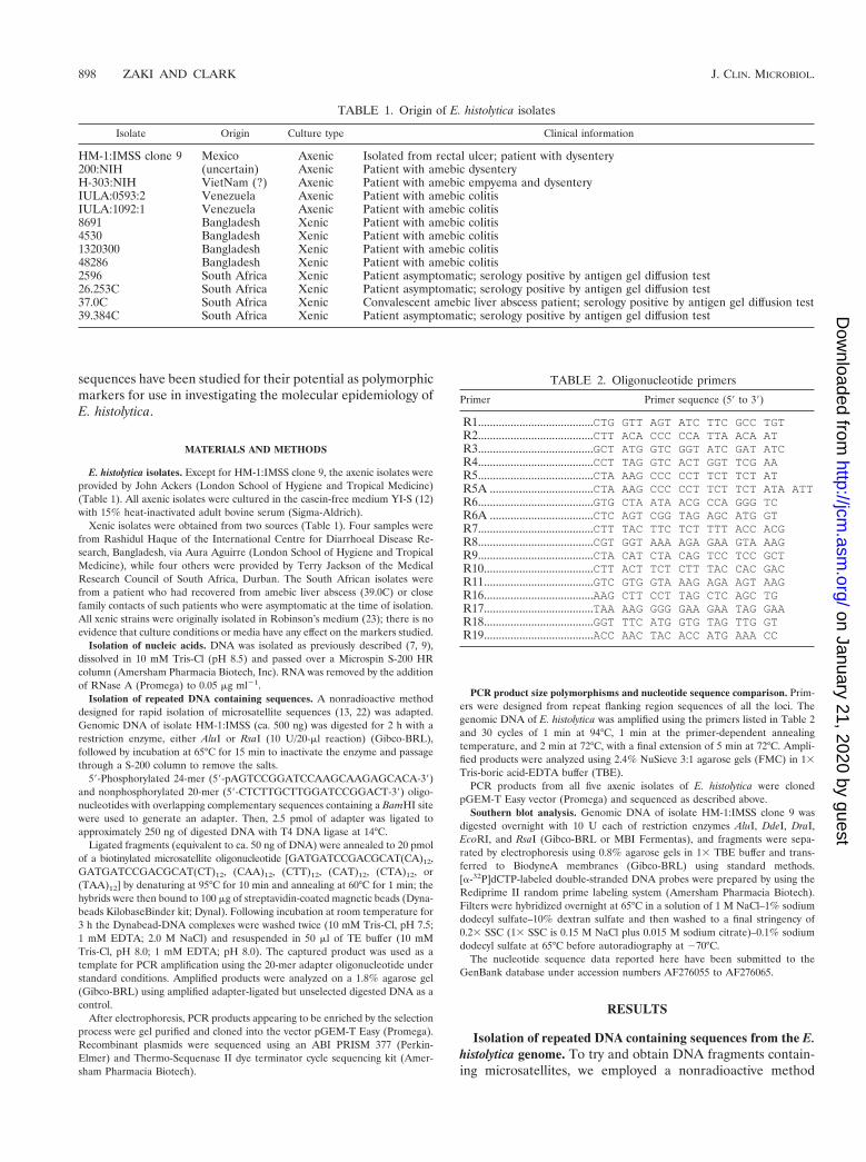
TABLE 1. Origin of E. histolytica isolates
Isolate Origin Culture type Clinical information
HM-1:IMSS clone 9 Mexico Axenic Isolated from rectal ulcer; patient with dysentery200:NIH (uncertain) Axenic Patient with amebic dysenteryH-303:NIH VietNam (?) Axenic Patient with amebic empyema and dysenteryIULA:0593:2 Venezuela Axenic Patient with amebic colitisIULA:1092:1 Venezuela Axenic Patient with amebic colitis8691 Bangladesh Xenic Patient with amebic colitis4530 Bangladesh Xenic Patient with amebic colitis1320300 Bangladesh Xenic Patient with amebic colitis48286 Bangladesh Xenic Patient with amebic colitis2596 South Africa Xenic Patient asymptomatic; serology positive by antigen gel diffusion test26.253C South Africa Xenic Patient asymptomatic; serology positive by antigen gel diffusion test37.0C South Africa Xenic Convalescent amebic liver abscess patient; serology positive by antigen gel diffusion test39.384C South Africa Xenic Patient asymptomatic; serology positive by antigen gel diffusion test
TABLE 2. Oligonucleotide primers
Primer Primer sequence (59 to 39)
R1.......................................CTG GTT AGT ATC TTC GCC TGTR2.......................................CTT ACA CCC CCA TTA ACA ATR3.......................................GCT ATG GTC GGT ATC GAT ATCR4.......................................CCT TAG GTC ACT GGT TCG AAR5.......................................CTA AAG CCC CCT TCT TCT ATR5A ...................................CTA AAG CCC CCT TCT TCT ATA ATTR6.......................................GTG CTA ATA ACG CCA GGG TCR6A ...................................CTC AGT CGG TAG AGC ATG GTR7.......................................CTT TAC TTC TCT TTT ACC ACGR8.......................................CGT GGT AAA AGA GAA GTA AAGR9.......................................CTA CAT CTA CAG TCC TCC GCTR10.....................................CTT ACT TCT CTT TAC CAC GACR11.....................................GTC GTG GTA AAG AGA AGT AAGR16.....................................AAG CTT CCT TAG CTC AGC TGR17.....................................TAA AAG GGG GAA GAA TAG GAAR18.....................................GGT TTC ATG GTG TAG TTG GTR19.....................................ACC AAC TAC ACC ATG AAA CC
898 ZAKI AND CLARK J. CLIN. MICROBIOL.
on January 21, 2020 by guesthttp://jcm
.asm.org/
Dow
nloaded from
based on affinity capture of single-stranded restriction frag-ments annealed to biotinylated microsatellite oligonucleotides,with attachment to streptavidin-coated magnetic beads (Dy-nal), followed by adapter-mediated PCR (13, 22). A total oftwenty two PCR fragments ranging in size from 250 to 700 bpwere gel purified from the total amplification products of AluIor RsaI restriction fragments annealed to one of seven biotin-ylated oligonucleotides. These fragments were chosen on thebasis of their apparent enrichment compared to control am-plification products and were cloned, sequenced, and exam-ined for the presence of microsatellites.
No products contained sequences corresponding to the mi-crosatellite oligonucleotides used in their capture. Further-more, the majority of the sequences did not reveal any tan-demly repeated DNAs. However, two clones (clone 1 andclone 4) derived from an approximately 480-bp fragment, ob-tained from AluI restriction fragments annealed to the(CTT)12 oligonucleotide, showed the presence of internal tan-dem repeats. The repeats seen in clones 1 and 4 were distinct.Two other clones (clone 19 and clone 5) derived from AluIrestriction fragments annealed to (TAA)12 contained the sametype of repeats as clone 4. Clones 19 and 5 contained fragmentsof approximately 480 and 450 bp, respectively. Further analysiswas carried out on clone 1, which represents locus 5-6, andclone 4, which represents locus 1-2.
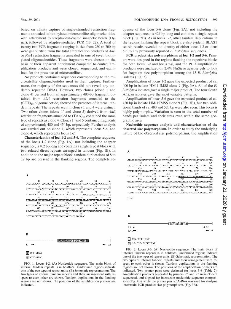
Characterization of loci 1-2 and 5-6. The complete sequenceof the locus 1-2 clone (Fig. 1A), not including the adaptersequence, is 402 bp long and contains a single repeat block withtwo related direct repeats arranged in tandem (Fig. 1B). Inaddition to the major repeat block, tandem duplications of 8 to12 bp are present in the flanking regions. The complete se-
quence of the locus 5-6 clone (Fig. 2A), not including theadapter sequence, is 424 bp long and contains a single repeatblock (Fig. 2B). As in locus 1-2, other tandem duplications inthe regions flanking the repeat block are also evident. BLASTsearch results revealed no identity of either locus 1-2 or locus5-6 to any previously reported E. histolytica sequences.
PCR product size polymorphisms at loci 1-2 and 5-6. Prim-ers were designed in the regions flanking the repetitive blocksfor both locus 1-2 and locus 5-6, and the PCR amplificationproducts were analyzed on 2.4% NuSieve agarose gels to lookfor fragment size polymorphism among the 13 E. histolyticaisolates (Fig. 3).
Amplification of locus 1-2 gave the expected product of ca.400 bp in isolate HM-1:IMSS clone 9 (Fig. 3A). All of the E.histolytica isolates gave a single major product. The four SouthAfrican isolates gave the most variable patterns.
Amplification of locus 5-6 gave the expected product of ca.420 bp in isolate HM-1:IMSS clone 9 (Fig. 3B), but two addi-tional bands of ca. 480 and 520 bp were also seen. This locus ishighly polymorphic. Variation is seen in the total number ofbands per isolate and their sizes even within the same geo-graphic area.
Nucleotide sequence analysis and characterization of theobserved size polymorphism. In order to study the underlyingnature of the observed size polymorphisms, the amplification
FIG. 1. Locus 1-2. (A) Nucleotide sequence. The main block ofinternal tandem repeats is in boldface. Underlined regions indicateone of the two types of repeat units. (B) Schematic representation. Thetwo types of internal tandem repeats and their arrangement with re-spect to each other are shown. Tandem duplications in the flankingregions are not shown. The positions of the amplification primers areindicated.
FIG. 2. Locus 5-6. (A) Nucleotide sequence. The main block ofinternal tandem repeats is in boldface. Underlined regions indicateone of the two types of repeat units. (B) Schematic representation. Thetwo types of internal tandem repeats and their arrangement with re-spect to each other is shown. Tandem duplications in the flankingregions are not shown. The positions of the amplification primers areindicated. Two primer pairs were designed for locus 5-6 (Table 2).Amplification products generated by primers R5 and R6 were cloned,sequenced, and aligned for intrastrain nucleotide sequence compari-sons (Fig. 4B), while the primer pair R5A-R6A was used for studyinginterstrain PCR product size polymorphisms (Fig. 3B).
VOL. 39, 2001 POLYMORPHIC DNA FROM E. HISTOLYTICA 899
on January 21, 2020 by guesthttp://jcm
.asm.org/
Dow
nloaded from
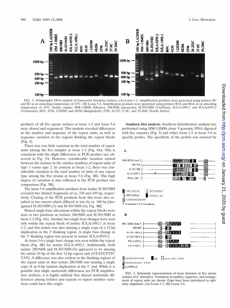
products of all five axenic isolates at locus 1-2 and locus 5-6were cloned and sequenced. This analysis revealed differencesin the number and sequence of the repeat units, as well assequence variation in the regions flanking the repeat blocks(Fig. 4).
There was very little variation in the total number of repeatunits among the five samples at locus 1-2 (Fig. 4A). This isconsistent with the slight differences in PCR product size ob-served in Fig. 3A. However, considerable variation existedbetween the isolates in the relative numbers of repeat units oftype 1 versus type 2. In contrast to locus 1-2, there was con-siderable variation in the total number of units of one repeattype among the five strains at locus 5-6 (Fig. 4B). This highdegree of variation is also reflected in the PCR product sizecomparison (Fig. 3B).
The locus 5-6 amplification products from isolate H-303:NIHrevealed two distinct fragments of ca. 320 and 450 bp, respec-tively. Cloning of the PCR products from this locus also re-sulted in two inserts which differed in size by ca. 100 bp [des-ignated H-303:NIH-(3) and H-303:NIH-(4); Fig. 4B].
Shared single-base alterations within the repeat blocks wereseen at two positions in isolates 200:NIH and H-303:NIH atlocus 1-2 (Fig. 4A). Another two single base changes were seenonly within the repeat block of isolate IULA:1092:1 at locus1-2, and this isolate was also missing a single copy of a 12-bpduplication in the 39-flanking region. A single base change inthe 59-flanking region was present in isolate IULA:0593:2.
At locus 5-6 a single base change was seen within the repeatblock (Fig. 4B) for isolate IULA:1092:1. Additionally, bothisolate 200:NIH and H-303:NIH-(4) appeared to be missingthe initial 10 bp of the first 12-bp repeat unit (GTATGTTTCTAT). A difference was also evident in the flanking regions ofthe repeat units in that isolate 200:NIH was missing a singlecopy of an 8-bp tandem duplication at the 59 end. While it ispossible that single nucleotide differences are PCR amplifica-tion artifacts, it is highly unlikely that shared nucleotide dif-ferences among isolates and repeats or repeat number varia-tions could have this origin.
Southern blot analysis. Southern hybridization analysis wasperformed using HM-1:IMSS clone 9 genomic DNA digestedwith five enzymes (Fig. 5) and either locus 1-2 or locus 5-6 asspecific probes. The specificity of the probes was ensured by
FIG. 3. Polymorphic DNA analysis of Entamoeba histolytica isolates. (A) Locus 1-2. Amplification products were generated using primers R1and R2 at an annealing temperature of 53°C. (B) Locus 5-6. Amplification products were generated using primers R5A and R6A at an annealingtemperature of 56°C. Isolate origins: HM-1:IMSS (Mexico); 200:NIH (uncertain); H-303:NIH (VietNam); IULA:1092:1 and IULA:0593:2(Venezuela); 8691, 4530, 1320300, and 48286 (Bangladesh); 2596, 26.253, 37.0C, and 39.384C (South Africa).
FIG. 4. Schematic representation of locus structure in five axenicisolates of E. histolytica. Variations in number, sequence, and arrange-ment of repeat units are shown. Gaps have been introduced to opti-mize alignment. (A) Locus 1-2. (B) Locus 5-6.
900 ZAKI AND CLARK J. CLIN. MICROBIOL.
on January 21, 2020 by guesthttp://jcm
.asm.org/
Dow
nloaded from
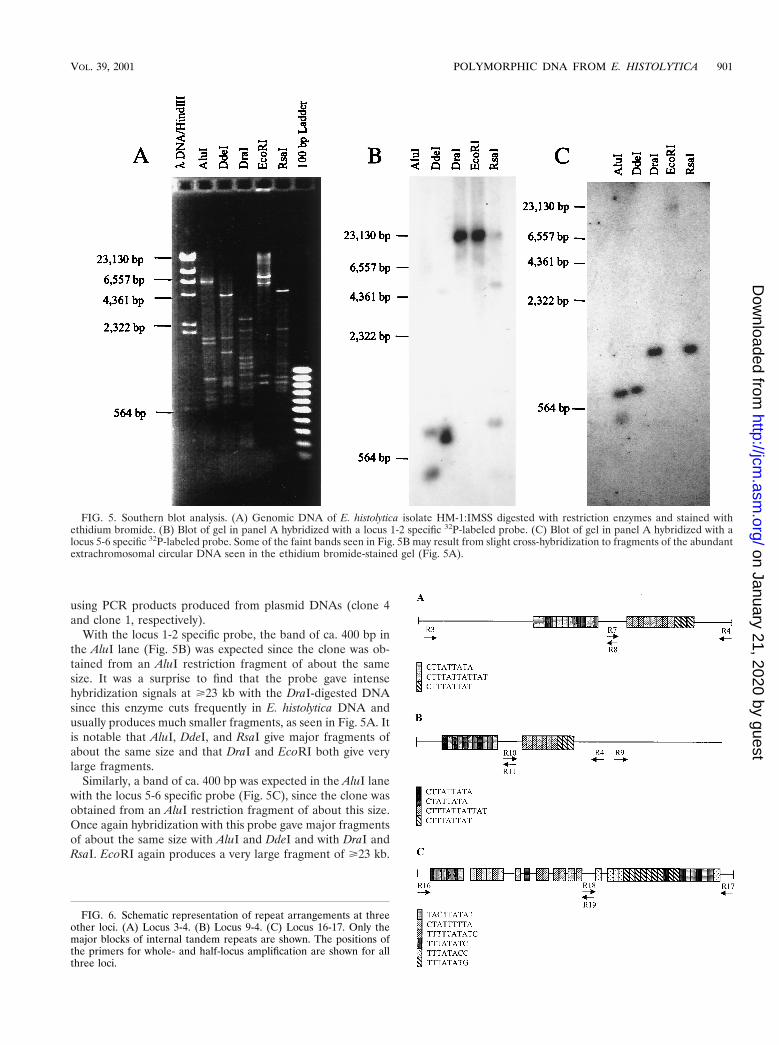
using PCR products produced from plasmid DNAs (clone 4and clone 1, respectively).
With the locus 1-2 specific probe, the band of ca. 400 bp inthe AluI lane (Fig. 5B) was expected since the clone was ob-tained from an AluI restriction fragment of about the samesize. It was a surprise to find that the probe gave intensehybridization signals at Ä23 kb with the DraI-digested DNAsince this enzyme cuts frequently in E. histolytica DNA andusually produces much smaller fragments, as seen in Fig. 5A. Itis notable that AluI, DdeI, and RsaI give major fragments ofabout the same size and that DraI and EcoRI both give verylarge fragments.
Similarly, a band of ca. 400 bp was expected in the AluI lanewith the locus 5-6 specific probe (Fig. 5C), since the clone wasobtained from an AluI restriction fragment of about this size.Once again hybridization with this probe gave major fragmentsof about the same size with AluI and DdeI and with DraI andRsaI. EcoRI again produces a very large fragment of Ä23 kb.
FIG. 5. Southern blot analysis. (A) Genomic DNA of E. histolytica isolate HM-1:IMSS digested with restriction enzymes and stained withethidium bromide. (B) Blot of gel in panel A hybridized with a locus 1-2 specific 32P-labeled probe. (C) Blot of gel in panel A hybridized with alocus 5-6 specific 32P-labeled probe. Some of the faint bands seen in Fig. 5B may result from slight cross-hybridization to fragments of the abundantextrachromosomal circular DNA seen in the ethidium bromide-stained gel (Fig. 5A).
FIG. 6. Schematic representation of repeat arrangements at threeother loci. (A) Locus 3-4. (B) Locus 9-4. (C) Locus 16-17. Only themajor blocks of internal tandem repeats are shown. The positions ofthe primers for whole- and half-locus amplification are shown for allthree loci.
VOL. 39, 2001 POLYMORPHIC DNA FROM E. HISTOLYTICA 901
on January 21, 2020 by guesthttp://jcm
.asm.org/
Dow
nloaded from
Taken together, these data indicate that loci 1-2 and 5-6 existin long tandem arrays.
Characterization of other loci containing internal repeats.A number of other DNA elements containing internal tandemrepeats have been reported in E. histolytica. No attempts havebeen made to study their potential for the detection of in-traspecies polymorphisms. We selected three of these inter-nally repetitive loci for study: a 978-bp element described byMichel et al. (19; GenBank accession number M77091; ourdesignation, locus 3-4), a 931-bp DNA element isolated by J.Rosales-Encina and D. Eichinger (personal communication;GenBank accession number AF265348; our designation, locus9-4), and a 964-bp element reported by Huang et al. (15; ourdesignation, locus 16-17). Schematic representations of therepeat arrangements seen at these loci are given in Fig. 6A, B,and C, respectively.
There is a high degree of identity between the two repeatblocks of locus 3-4 and those of locus 9-4. The ten CTATTATA tandem repeats of locus 9-4 differ from the 11 CTTATTATA tandem repeats of locus 3-4 only in the absence of asingle nucleotide (T) at the second position of each unit. Therepeat unit CTTTATTATTAT in locus 9-4 is identical to the12-bp repeat units of locus 3-4 with the only difference beingthe total number of units seen, i.e., locus 3-4 has eight units,while locus 9-4 has only seven. In fact, this high degree ofidentity between the two loci is apparent in the flanking regionsas well. The sequences from positions 1 to 540 and positions541 to 931 of locus 9-4 are very similar to the nucleotidestretches spanning positions 401 to 977 and positions 1 to 400in locus 3-4, respectively (data not shown).
On comparing the sequences of loci 1-2 and 5-6 with thoseof loci 3-4 and 9-4 we find that the repeat unit CTTTATTAT,which occurs a total of seven times in locus 1-2, is identical tothe three 9-bp units present in the second repeat blocks of bothloci 3-4 and 9-4. The repeat units of locus 5-6, however, werequite unique, as are the repeat flanking regions of both loci.The nucleotide sequence of locus 16-17 is completely differentfrom that of the other loci. There are six major types of inter-nal repeats, with some being arranged in tandem only, whileothers exist as both tandem and solitary copies (Fig. 6C).Besides these, duplications of 5 to 8 bp are also seen inter-spersed among these repeats (not shown).
PCR product size polymorphisms at loci 3-4, 9-4 and 16-17.Primers were designed to amplify all three loci, and the prod-ucts were analyzed on 2.4% NuSieve agarose gels to look forsize polymorphisms among the 13 E. histolytica isolates. Ineach case primers were also designed in the regions betweenthe two main repeat blocks to look additionally for size varia-tions in each half of the locus (Fig. 6).
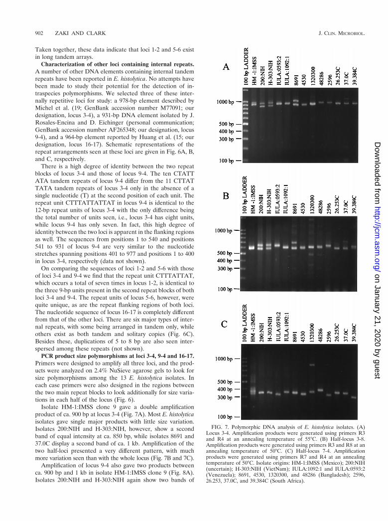
Isolate HM-1:IMSS clone 9 gave a double amplificationproduct of ca. 900 bp at locus 3-4 (Fig. 7A). Most E. histolyticaisolates gave single major products with little size variation.Isolates 200:NIH and H-303:NIH, however, show a secondband of equal intensity at ca. 850 bp, while isolates 8691 and37.0C display a second band of ca. 1 kb. Amplification of thetwo half-loci presented a very different pattern, with muchmore variation seen than with the whole locus (Fig. 7B and 7C).
Amplification of locus 9-4 also gave two products betweenca. 900 bp and 1 kb in isolate HM-1:IMSS clone 9 (Fig. 8A).Isolates 200:NIH and H-303:NIH again show two bands of
FIG. 7. Polymorphic DNA analysis of E. histolytica isolates. (A)Locus 3-4. Amplification products were generated using primers R3and R4 at an annealing temperature of 55°C. (B) Half-locus 3-8.Amplification products were generated using primers R3 and R8 at anannealing temperature of 50°C. (C) Half-locus 7-4. Amplificationproducts were generated using primers R7 and R4 at an annealingtemperature of 50°C. Isolate origins: HM-1:IMSS (Mexico); 200:NIH(uncertain); H-303:NIH (VietNam); IULA:1092:1 and IULA:0593:2(Venezuela); 8691, 4530, 1320300, and 48286 (Bangladesh); 2596,26.253, 37.0C, and 39.384C (South Africa).
902 ZAKI AND CLARK J. CLIN. MICROBIOL.
on January 21, 2020 by guesthttp://jcm
.asm.org/
Dow
nloaded from
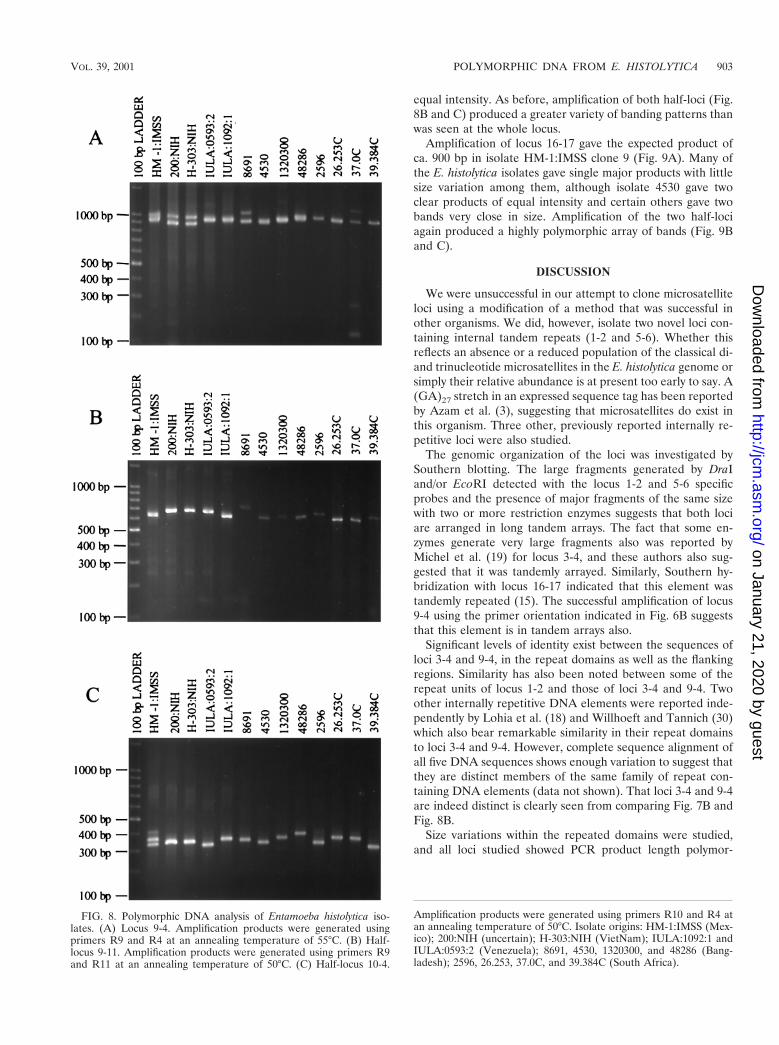
equal intensity. As before, amplification of both half-loci (Fig.8B and C) produced a greater variety of banding patterns thanwas seen at the whole locus.
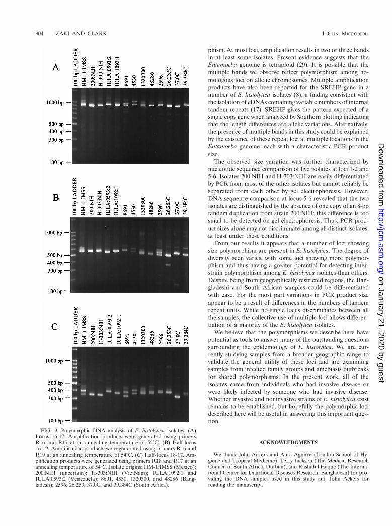
Amplification of locus 16-17 gave the expected product ofca. 900 bp in isolate HM-1:IMSS clone 9 (Fig. 9A). Many ofthe E. histolytica isolates gave single major products with littlesize variation among them, although isolate 4530 gave twoclear products of equal intensity and certain others gave twobands very close in size. Amplification of the two half-lociagain produced a highly polymorphic array of bands (Fig. 9Band C).
DISCUSSION
We were unsuccessful in our attempt to clone microsatelliteloci using a modification of a method that was successful inother organisms. We did, however, isolate two novel loci con-taining internal tandem repeats (1-2 and 5-6). Whether thisreflects an absence or a reduced population of the classical di-and trinucleotide microsatellites in the E. histolytica genome orsimply their relative abundance is at present too early to say. A(GA)27 stretch in an expressed sequence tag has been reportedby Azam et al. (3), suggesting that microsatellites do exist inthis organism. Three other, previously reported internally re-petitive loci were also studied.
The genomic organization of the loci was investigated bySouthern blotting. The large fragments generated by DraIand/or EcoRI detected with the locus 1-2 and 5-6 specificprobes and the presence of major fragments of the same sizewith two or more restriction enzymes suggests that both lociare arranged in long tandem arrays. The fact that some en-zymes generate very large fragments also was reported byMichel et al. (19) for locus 3-4, and these authors also sug-gested that it was tandemly arrayed. Similarly, Southern hy-bridization with locus 16-17 indicated that this element wastandemly repeated (15). The successful amplification of locus9-4 using the primer orientation indicated in Fig. 6B suggeststhat this element is in tandem arrays also.
Significant levels of identity exist between the sequences ofloci 3-4 and 9-4, in the repeat domains as well as the flankingregions. Similarity has also been noted between some of therepeat units of locus 1-2 and those of loci 3-4 and 9-4. Twoother internally repetitive DNA elements were reported inde-pendently by Lohia et al. (18) and Willhoeft and Tannich (30)which also bear remarkable similarity in their repeat domainsto loci 3-4 and 9-4. However, complete sequence alignment ofall five DNA sequences shows enough variation to suggest thatthey are distinct members of the same family of repeat con-taining DNA elements (data not shown). That loci 3-4 and 9-4are indeed distinct is clearly seen from comparing Fig. 7B andFig. 8B.
Size variations within the repeated domains were studied,and all loci studied showed PCR product length polymor-
FIG. 8. Polymorphic DNA analysis of Entamoeba histolytica iso-lates. (A) Locus 9-4. Amplification products were generated usingprimers R9 and R4 at an annealing temperature of 55°C. (B) Half-locus 9-11. Amplification products were generated using primers R9and R11 at an annealing temperature of 50°C. (C) Half-locus 10-4.
Amplification products were generated using primers R10 and R4 atan annealing temperature of 50°C. Isolate origins: HM-1:IMSS (Mex-ico); 200:NIH (uncertain); H-303:NIH (VietNam); IULA:1092:1 andIULA:0593:2 (Venezuela); 8691, 4530, 1320300, and 48286 (Bang-ladesh); 2596, 26.253, 37.0C, and 39.384C (South Africa).
VOL. 39, 2001 POLYMORPHIC DNA FROM E. HISTOLYTICA 903
on January 21, 2020 by guesthttp://jcm
.asm.org/
Dow
nloaded from
phism. At most loci, amplification results in two or three bandsin at least some isolates. Present evidence suggests that theEntamoeba genome is tetraploid (29). It is possible that themultiple bands we observe reflect polymorphism among ho-mologous loci on allelic chromosomes. Multiple amplificationproducts have also been reported for the SREHP gene in anumber of E. histolytica isolates (8), a finding consistent withthe isolation of cDNAs containing variable numbers of internaltandem repeats (17). SREHP gives the pattern expected of asingle copy gene when analyzed by Southern blotting indicatingthat the length differences are allelic variations. Alternatively,the presence of multiple bands in this study could be explainedby the existence of these repeat loci at multiple locations in theEntamoeba genome, each with a characteristic PCR productsize.
The observed size variation was further characterized bynucleotide sequence comparison of five isolates at loci 1-2 and5-6. Isolates 200:NIH and H-303:NIH are easily differentiatedby PCR from most of the other isolates but cannot reliably beseparated from each other by gel electrophoresis. However,DNA sequence comparison at locus 5-6 revealed that the twoisolates are distinguished by the absence of one copy of an 8-bptandem duplication from strain 200:NIH; this difference is toosmall to be detected on gel electrophoresis. Thus, PCR prod-uct sizes alone may not discriminate among all distinct isolates,at least under these conditions.
From our results it appears that a number of loci showingsize polymorphism are present in E. histolytica. The degree ofdiversity seen varies, with some loci showing more polymor-phism and thus having a greater potential for detecting inter-strain polymorphism among E. histolytica isolates than others.Despite being from geographically restricted regions, the Ban-gladeshi and South African samples could be differentiatedwith ease. For the most part variations in PCR product sizeappear to be a result of differences in the numbers of tandemrepeat units. While no single locus discriminates between allthe samples, the collective use of multiple loci allows differen-tiation of a majority of the E. histolytica isolates.
We believe that the polymorphisms we describe here havepotential as tools to answer many of the outstanding questionssurrounding the epidemiology of E. histolytica. We are cur-rently studying samples from a broader geographic range tovalidate the general utility of these loci and are examiningsamples from infected family groups and amebiasis outbreaksfor shared polymorphisms. In the present work, all of theisolates came from individuals who had invasive disease orwere likely infected by someone who had invasive disease.Whether invasive and noninvasive strains of E. histolytica existremains to be established, but hopefully the polymorphic locidescribed here will be useful in answering this important ques-tion.
ACKNOWLEDGMENTS
We thank John Ackers and Aura Aguirre (London School of Hy-giene and Tropical Medicine), Terry Jackson (The Medical ResearchCouncil of South Africa, Durban), and Rashidul Haque (The Interna-tional Center for Diarrhoeal Diseases Research, Bangladesh) for pro-viding the DNA samples used in this study and John Ackers forreading the manuscript.
FIG. 9. Polymorphic DNA analysis of E. histolytica isolates. (A)Locus 16-17. Amplification products were generated using primersR16 and R17 at an annealing temperature of 55°C. (B) Half-locus16-19. Amplification products were generated using primers R16 andR19 at an annealing temperature of 54°C. (C) Half-locus 18-17. Am-plification products were generated using primers R18 and R17 at anannealing temperature of 54°C. Isolate origins: HM-1:IMSS (Mexico);200:NIH (uncertain); H-303:NIH (VietNam); IULA:1092:1 andIULA:0593:2 (Venezuela); 8691, 4530, 1320300, and 48286 (Bang-ladesh); 2596, 26.253, 37.0C, and 39.384C (South Africa).
904 ZAKI AND CLARK J. CLIN. MICROBIOL.
on January 21, 2020 by guesthttp://jcm
.asm.org/
Dow
nloaded from
REFERENCES
1. Anderson, T. J. C., X. Z. Su, M. Bockarie, M. Lagog, and K. P. Day. 1999.Twelve microsatellite markers for characterisation of Plasmodium falciparumfrom finger-prick blood samples. Parasitology 119:113–125.
2. Anonymous. 1997. WHO/PAHO/UNESCO report. A consultation with ex-perts on amoebiasis. Epidemiol. Bull. PAHO 18:13–14.
3. Azam, A., J. Paul, D. Sehgal, J. Prasad, S. Bhattacharya, and A. Bhatta-charya. 1996. Identification of novel genes from Entamoeba histolytica byexpressed sequence tag analysis. Gene 181:113–116.
4. Bhattacharya, S., A. Bhattacharya, and L. S. Diamond. 1992. Entamoebahistolytica extrachromosomal circular ribosomal DNA: analysis of clonalvariation in a hypervariable region. Exp. Parasitol. 74:200–204.
5. Blanc, D. S., and P. G. Sargeaunt. 1991. Entamoeba histolytica zymodemes:exhibition of g and d bands only of glucose phosphate isomerase and phos-phoglucomutase may be influenced by starch content in the medium. Exp.Parasitol. 72:87–90.
6. Burch, D. J., E. Li, S. Reed, T. F. H. G. Jackson, and S. L. Stanley, Jr. 1991.Isolation of a strain-specific Entamoeba histolytica cDNA clone. J. Clin.Microbiol. 29:696–701.
7. Clark, C. G. 1992. DNA purification from polysaccharide-rich cells, p. D-3.1–D-3.2. In J. J. Lee and A. T. Soldo (ed.), Protocols in protozoology, vol. 1.Allen Press, Lawrence, Kans.
8. Clark, C. G., and L. S. Diamond. 1993. Entamoeba histolytica: a method forisolate identification. Exp. Parasitol. 77:450–455.
9. Clark, C. G., and L. S. Diamond. 1991. The Laredo strain and other Ent-amoeba histolytica-like amoebae are Entamoeba moshkovskii. Mol. Biochem.Parasitol. 46:11–18.
10. de la Vega, H., C. A. Specht, C. E. Semino, P. W. Robbins, D. Eichinger, D.Caplivski, S. Ghosh, and J. Samuelson. 1997. Cloning and expression ofchitinases of Entamoebae. Mol. Biochem. Parasitol. 85:139–147.
11. Diamond, L. S., and C. G. Clark. 1993. A redescription of Entamoebahistolytica Schaudinn, 1903 (Emended Walker, 1911) separating it from En-tamoeba dispar Brumpt, 1925. J. Eukaryot. Microbiol. 40:340–344.
12. Diamond, L. S., C. G. Clark, and C. C. Cunnick. 1995. YI-S, a casein-freemedium for axenic cultivation of Entamoeba histolytica, related Entamoeba,Giardia intestinalis and Trichomonas vaginalis. J. Eukaryot. Microbiol. 42:277–278.
13. Fischer, D., and K. Bachmann. 1998. Microsatellite enrichment in organismswith large genomes (Allium cepa L.). BioTechniques 24:796–802.
14. Ghosh, S., M. Frisardi, L. Ramirez-Avila, S. Descoteaux, K. Sturm-Ramirez,O. A. Newton-Sanchez, J. I. Santos-Preciado, C. Ganguly, A. Lohia, S. Reed,and J. Samuelson. 2000. Molecular epidemiology of Entamoeba spp.: evi-dence of a bottleneck (demographic sweep) and transcontinental spread ofdiploid parasites. J. Clin. Microbiol. 38:3815–3821.
15. Huang, M., K. P. Chang, and R. A. Albach. 1997. A 964 bp repetitive DNAin Entamoeba histolytica is associated with linear “chromosomal” DNAs ofvariable sizes. Arch. Med. Res. 28(Suppl.):S1–S4.
16. Jackson, T. F. H. G., and S. Suparsad. 1997. Zymodeme stability of Ent-amoeba histolytica and E. dispar. Arch. Med. Res. 28(Suppl.):S304–S305.
17. Kohler, S., and E. Tannich. 1993. A family of transcripts (K2) of Entamoebahistolytica contains polymorphic repetitive regions with highly conservedelements. Mol. Biochem. Parasitol. 59:49–58.
18. Lohia, A., N. Haider, and B. B. Biswas. 1990. Characterisation of a repetitiveDNA family from Entamoeba histolytica containing Saccharomyces cerevisiaeARS consensus sequences. Gene 96:197–203.
19. Michel, B., A. Alagon, P. Lizardi, and M. Zurita. 1992. Characterization ofa repetitive DNA element from Entamoeba histolytica. Mol. Biochem. Para-sitol. 51:165–168.
20. Mittal, V., A. Bhattacharya, and S. Bhattacharya. 1992. Organization ofrepeated sequences in the region downstream to rRNA genes in the rDNAepisome of Entamoeba histolytica. Arch. Med. Res. 23:17–18.
21. Mittal, V., D. Sehgal, A. Bhattacharya, and S. Bhattacharya. 1992. A secondshort repeat sequence detected downstream of rRNA genes in the Ent-amoeba histolytica rDNA episome. Mol. Biochem. Parasitol. 54:97–100.
22. Oliveira, R. P., N. E. Broude, A. M. Macedo, C. R. Cantor, C. L. Smith, andS. D. J. Pena. 1998. Probing the genetic population structure of Trypanosomacruzi with polymorphic microsatellites. Proc. Natl. Acad. Sci. USA 95:3776–3780.
23. Robinson, G. L. 1968. The laboratory diagnosis of human parasitic amoebae.Trans. R. Soc. Trop. Med. Hyg. 62:285–294.
24. Russell, R., M. P. Iribar, B. Lambson, S. Brewster, J. M. Blackwell, C. Dye,and J. W. Ajioka. 1999. Intra and inter-specific microsatellite variation in theLeishmania subgenus Viannia. Mol. Biochem. Parasitol. 103:71–77.
25. Sargeaunt, P. G. 1987. The reliability of Entamoeba histolytica zymodemes inclinical diagnosis. Parasitol. Today 3:40–43.
26. Sehgal, D., A. Bhattacharya, and S. Bhattacharya. 1993. Analysis of a poly-morphic locus present upstream of rDNA transcription units in the extra-chromosomal circle of Entamoeba histolytica. Mol. Biochem. Parasitol. 62:129–130.
27. Sehgal, D., V. Mittal, S. Ramachandran, S. K. Dhar, A. Bhattacharya, andS. Bhattacharya. 1994. Nucleotide sequence organization and analysis of thenuclear ribosomal DNA circle of the protozoan parasite Entamoeba histo-lytica. Mol. Biochem. Parasitol. 67:205–214.
28. Stanley, S. L., Jr., A. Becker, C. Kunz-Jenkins, L. Foster, and E. Li. 1990.Cloning and expression of a membrane antigen of Entamoeba histolyticapossessing multiple tandem repeats. Proc. Natl. Acad. Sci. USA 87:4976–4980.
29. Willhoeft, U., and E. Tannich. 1999. The electrophoretic karyotype of Ent-amoeba histolytica. Mol. Biochem. Parasitol. 99:41–53.
30. Willhoeft, U., and E. Tannich. 2000. Fluorescence microscopy and fluores-cence in situ hybridization of Entamoeba histolytica nuclei to analyse mitosisand the localization of repetitive DNA. Mol. Biochem. Parasitol. 105:291–296.
VOL. 39, 2001 POLYMORPHIC DNA FROM E. HISTOLYTICA 905
on January 21, 2020 by guesthttp://jcm
.asm.org/
Dow
nloaded from