Embed Size (px)
Citation preview

UNIVERSITY OF LJUBLJANA
BIOTECHNICAL FACULTY
Sandra HOČEVAR
ISOLATION AND CHARACTERIZATION OF PURE
GLIAL CELL POPULATIONS FROM ADULT
MOUSE CENTRAL NERVOUS SYSTEM WHITE
MATTER
M.Sc. Thesis
Ljubljana, 2014

UNIVERSITY OF LJUBLJANA BIOTECHNICAL FACULTY
Sandra HOČEVAR
ISOLATION AND CHARACTERIZATION OF PURE GLIAL CELL
POPULATIONS FROM ADULT MOUSE CENTRAL NERVOUS
SYSTEM WHITE MATTER
M.Sc. Thesis
IZOLACIJA IN KARAKTERIZACIJA ČISTIH GLIJA CELIČNIH
POPULACIJ IZ BELINE CENTRALNEGA ŽIVČNEGA SISTEMA
ODRASLE MIŠI
Magistrsko delo
Ljubljana, 2014

II Hočevar S. Isolation and characterization of pure glial cell populations from adult mouse CNS white matter.
M.Sc. Thesis. Ljubljana, Univ. of Ljubljana, Biotechnical Faculty, 2014
The M. Sc. thesis is a completion of a Master Study Programme in Molecular Biology at
Biotechnical Faculty, University of Ljubljana. The experimental part was carried out in
the laboratory of Cellular Neurophysiology at the Faculty of Science in the School of
Pharmacy and Biomedical Sciences, University of Portsmouth (United Kingdom).
The Council of the 1. and 2. study cycle appointed Professor Robert Zorec, PhD, as a
supervisor, Professor Arthur Butt, PhD, as a co-supervisor and Professor Marko Kreft,
PhD, as a reviewer.
Commission for assessment and defence:
President: prof. dr. Boris BULOG
University of Ljubljana, Biotechnical Faculty, Department of
Biology
Member: prof. dr. Robert ZOREC
University of Ljubljana, Faculty of Medicine
Member: prof. dr. Arthur BUTT
University of Portsmouth, Faculty of Science, School of
Pharmacy and Biomedical Sciences
Member: prof. dr. Marko KREFT
University of Ljubljana, Biotechnical Faculty, Department of
Biology
Date of defence:
The work is the result of my own research work. I agree with publishing of my work in full
text on the internet page Digitalna knjižnica Biotehniške fakultete. I declare that the text in
the electronic version is identical to the printed one.
Sandra HOČEVAR

III Hočevar S. Isolation and characterization of pure glial cell populations from adult mouse CNS white matter.
M.Sc. Thesis. Ljubljana, Univ. of Ljubljana, Biotechnical Faculty, 2014
KEY WORDS DOCUMENTATION (KWD)
DN Du2
DC UDK 602.3:616.8(043.2)=111
CX glia/astrocytes/oligodendrocyte/adult mouse CNS/optic nerve/cell
cultures/immunohistochemistry/MACS system/neurodegenerative diseases
AU HOČEVAR, Sandra, B. Sc. (Biotech)
AA ZOREC, Robert (supervisor)/BUTT, Arthur (co-supervisor)
PP SI-1000 Ljubljana, Večna pot 111
PB University of Ljubljana, Biotechnical Faculty, Molecular Biology
PY 2014
TI ISOLATION AND CHARACTERIZATION OF PURE GLIAL CELL
POPULATIONS FROM ADULT MOUSE CENTRAL NERVOUS SYSTEM
WHITE MATTER
DT M. Sc. Thesis (Master Study Programmes: Molecular Biology)
NO X, 59 p., 10 tab., 14 fig., 109 ref.
LA en
AL en/sl
AB Glial cells are critical participants in the development, function and disease of the
central nervous system (CNS). They play roles in brain homeostasis, synapse
formation, myelin production, and provide the immune system for CNS. There are
several in vitro glia studies which mostly include experiments on embryonic or
young postnatal rodent brains. Therefore, it is important to find a way to isolate
pure glia from adult mouse CNS, since all the major nerurodegenerative diseases,
such as Alzheimer’s disease (AD), Parkinson’s disease (PD) and Multiple sclerosis
(MS), occur in adulthood. With this aim, we tried to develop a reliable technique
for isolating purified astrocytes and oligodendrocytes from adult mouse brain and
optic nerve. Our experiments were based on two different methods of cell
dissociation and isolation, using the Magnetic Activated Cell Sorter (MACS)
system. We managed to dissociate, culture and characterise populations of
oligodendrocytes, astrocytes and oligodendrocyte precursor cells (OPCs) (or
oligodendrocyte-type 2 astrocyte progenitors-O-2A) from adult mouse CNS. Our
optimised in-house dissociation method proved as useful and equally successful as
the Neural Tissue Dissociation Kit (NTDK) method. However, cell isolation from
adult tissue with MACS system turned out to be impracticable and remains difficult
to perform.

IV Hočevar S. Isolation and characterization of pure glial cell populations from adult mouse CNS white matter.
M.Sc. Thesis. Ljubljana, Univ. of Ljubljana, Biotechnical Faculty, 2014
KLJUČNA DOKUMENTACIJSKA INFORMACIJA (KDI)
ŠD Du2
DK UDK 602.3:616.8(043.2)=111
KG glija/astrociti/oligodendrociti/CŽS odrasle miši/optični živec/celične
kulture/imunohistokemija/MACS sistem/nevrodegenerativne bolezni
AV HOČEVAR, Sandra, dipl. bioteh. (UN)
SA ZOREC, Robert (mentor)/BUTT Arthur (somentor)
KZ SI-1000 Ljubljana, Večna pot 111
ZA Univerza v Ljubljani, Biotehniška fakulteta, Molekulska biologija
LI 2014
IN IZOLACIJA IN KARAKTERIZACIJA ČISTIH GLIJA CELIČNIH POPULACIJ
IZ BELINE CENTRALNEGA ŽIVČNEGA SISTEMA ODRASLE MIŠI
TD Magistrsko delo (Magistrski študij- 2. stopnja Molekulska biologija)
OP X, 59 str., 10 pregl., 14 sl., 109 vir.
IJ en
JI en/sl
AI Celice glija so pomembne udeleženke v razvoju, funkciji in boleznih centralnega
živčnega sistema (CŽS). Igrajo vloge pri homeostazi v možganih, produkciji
mielina in zagotavljajo imunski sistem CŽS. Večina študij glije in vitro vključuje
poskuse na embrionalnih ali mladih postnatalnih možganih glodalcev. Ker pa se
večina poglavitnih nevrodegenerativnih bolezni, kot so Alzheimerjeva bolezen,
Parkinsonova bolezen in multipla skleroza, pojavljajo pri odraslem človeku, je
pomembno, da najdemo način za izolacijo čiste glije iz CŽS odrasle miši. S tem
namenom smo poskušali razviti zanesljivo tehniko izolacije čistih astrocitov in
oligodendrocitov iz možganov in optičnega živca odrasle miši. Naši poskusi so
temeljili na dveh različnih disociacijskih metodah in izolaciji z magnetno pogojenim
ločevanjem celic (ang. MACS). Iz CŽS odrasle miši nam je uspelo disociirati,
kultivirati in karakterizirati populacije oligodendrocitov, astrocitov in prekurzorskih
celic oligodendrocitov (ang. OPCs) (ali oligodendrocit-astrocit tipa 2- O-2A). Dokazali
smo, da je naša optimizirana disociacijska metoda enako uspešna kot disociacijska
metoda z uporabo kompleta za disociacijo živčnega tkiva (ang. NTDK). Vendar pa
se je izolacija celic s sistemom MACS iz odraslega tkiva izkazala za težavno in se
je ne uporablja.

V Hočevar S. Isolation and characterization of pure glial cell populations from adult mouse CNS white matter.
M.Sc. Thesis. Ljubljana, Univ. of Ljubljana, Biotechnical Faculty, 2014
TABLE OF CONTENTS
KEY WORDS DOCUMENTATION (KWD) ................................................................ III
KLJUČNA DOKUMENTACIJSKA INFORMACIJA (KDI) ...................................... IV
TABLE OF CONTENTS ................................................................................................... V
LIST OF TABLES ........................................................................................................... VII
LIST OF FIGURES ........................................................................................................ VIII
ABBRIVATIONS AND SYMBOLS ................................................................................ IX
1 INTRUDUCTION ............................................................................................................ 1
1.1 AIM OF STUDY ............................................................................................................. 2
1.2 HYPOTHESIS ................................................................................................................. 2
2 LITERATURE REVIEW ................................................................................................ 3
2.1 BACKGROUND OF GLIA ............................................................................................ 3
2.2 ASTROCYTES ............................................................................................................... 4
2.2.1 Astrocyte morphology and functions ........................................................................ 4
2.2.2 Astrocyte markers ...................................................................................................... 7
2.2.3 Astrocyte pathology .................................................................................................... 7
2.2.3.1 Reactive astrogliosis .................................................................................................. 7
2.2.3.2 Alzheimer’s disease ................................................................................................... 8
2.2.3.3 Parkinson’s disease .................................................................................................... 8
2.3 OLIGODENDROCYTES ............................................................................................... 9
2.3.1 Oligodendrocyte morphology and myelin functions ............................................... 9
2.3.2 Oligodendrocyte progenitor cells (OPCs) .............................................................. 11
2.3.3 NG2-glia ..................................................................................................................... 11
2.3.4 Oligodendrocyte markers ........................................................................................ 12
2.3.5 Oligodendrocyte pathology ...................................................................................... 13
2.3.5.1 Multiple sclerosis ..................................................................................................... 13
2.4 MICROGLIA ................................................................................................................ 13
2.5 ADULT NEURAL STEM CELLS ............................................................................... 14
2.6 Wnt/GSK-3β PATHWAY ............................................................................................. 15
2.7 OPTIC NERVE ............................................................................................................. 17
2.8 In vitro CULTURING OF GLIAL CELLS FROM ADULT MOUSE CNS ................ 18

VI Hočevar S. Isolation and characterization of pure glial cell populations from adult mouse CNS white matter.
M.Sc. Thesis. Ljubljana, Univ. of Ljubljana, Biotechnical Faculty, 2014
3 MATERIALS AND METHODS ................................................................................... 20
3.1 MATERIALS ................................................................................................................ 20
3.1.1 Animals ...................................................................................................................... 20
3.1.2 Agents ........................................................................................................................ 20
3.1.3 Equipment ................................................................................................................. 22
3.2 METHODS .................................................................................................................... 23
3.2.1 Dissection of mouse brain and optic nerve ............................................................. 23
3.2.2 Tissue dissociation of mouse brain and optic nerve .............................................. 23
3.2.2.1 In-house dissociation method .................................................................................. 23
3.2.2.2 Neural Tissue Dissociation Kit (P) and (T) ............................................................. 25
3.2.3 Magnetic Activated Cell Sorting (MACS) of O4+ cells ......................................... 27
3.2.3.1 Magnetic labelling ................................................................................................... 27
3.2.3.2 Magnetic separation ................................................................................................. 28
3.2.4 Cell culturing ............................................................................................................ 28
3.2.5 Immunohistochemistry ............................................................................................ 29
4 RESULTS ........................................................................................................................ 31
4.1 DISSOCIATED GLIAL CELS OF ADULT MOUSE CNS ......................................... 31
4.1.1 Dissociated cells of optic nerve ................................................................................ 31
4.1.2 Dissociated cells of forebrain ................................................................................... 35
4.1.3 Dissociated cells of cerebellum ................................................................................ 37
4.2 ISOLATED GLIAL CELLS ......................................................................................... 38
4.2.1 Isolated cells of young brain .................................................................................... 38
4.2.2 Isolated cells of adult forebrain ............................................................................... 39
5 DISCUSSION .................................................................................................................. 40
6 CONCLUSION ............................................................................................................... 42
7 SUMMARY ..................................................................................................................... 43
7.1 POVZETEK .................................................................................................................. 45
8 REFERENCES ............................................................................................................... 51
AKNOWLEDGEMENTS

VII Hočevar S. Isolation and characterization of pure glial cell populations from adult mouse CNS white matter.
M.Sc. Thesis. Ljubljana, Univ. of Ljubljana, Biotechnical Faculty, 2014
LIST OF TABLES
Tab. 1: Astrocyte markers ..................................................................................................... 7
Tab. 2: Oligodendrocyte markers ........................................................................................ 12
Tab. 3: List of chemicals ..................................................................................................... 20
Tab. 4: Primary antibodies................................................................................................... 21
Tab. 5: Secondary antibodies............................................................................................... 21
Tab. 6: Culture media recipes .............................................................................................. 22
Tab. 7: List of equipment .................................................................................................... 22
Tab. 8: Dissociation solution for brain and optic nerve ...................................................... 23
Tab. 9: Enzyme Mix I for NTDK (P) and (T) tissue dissociation ....................................... 25
Tab. 10: Enzyme Mix II for NTDK (P) and (T) tissue dissociation .................................... 26

VIII Hočevar S. Isolation and characterization of pure glial cell populations from adult mouse CNS white matter.
M.Sc. Thesis. Ljubljana, Univ. of Ljubljana, Biotechnical Faculty, 2014
LIST OF FIGURES
Fig. 1: Neural cell types ......................................................................................................... 1
Fig. 2: Glia-neuron interactions ............................................................................................. 4
Fig. 3: Astrocyte subtypes ..................................................................................................... 5
Fig. 4: The oligodendrocyte and the mature myelin sheath ................................................ 10
Fig. 5: Schematic of progenitor types and lineages in the adult brain SVZ ........................ 15
Fig. 6: Wnt/β-catenin signalling .......................................................................................... 16
Fig. 7: Principle of the MACS Separation ........................................................................... 27
Fig. 8: Dissociated glial cells from the optic nerve of adult mouse, using in-house
dissociation method ............................................................................................................. 32
Fig. 9: Dissociated glial cells from the optic nerve of adult mouse, using NTDK
dissociation method ............................................................................................................. 33
Fig. 10: Comparison of in-house and NTDK dissociation method ..................................... 34
Fig. 11: Dissociated astrocytes from adult mouse forebrain ............................................... 35
Fig. 12: Dissociated glial cells from adult mouse forebrain ................................................ 36
Fig. 13: Dissociated glial cells from cerebellum of transgenic GFAP-eGFP adult mouse . 37
Fig. 14: Isolated cells from young mouse brain .................................................................. 39

IX Hočevar S. Isolation and characterization of pure glial cell populations from adult mouse CNS white matter.
M.Sc. Thesis. Ljubljana, Univ. of Ljubljana, Biotechnical Faculty, 2014
ABBRIVATIONS AND SYMBOLS
AD Alzheimer’s disease
APC Adenomatous polyposis coli
Aβ Aβ-amyloid protein
BBB Blood-brain barrier
C1q Complement component 1 q
CK1 Casein kinase 1
CNS/CŽS Central nervous system/centralni živčni sistem
Dvl Dishevelled
EAAT1 Excitatory amino acid transporter 1
eGFP Enhanced green fluorescent protein
GABA Gamma-aminobutyric acid
GFAP Glial fibrilary acidic protein
GFP Green fluorescent protein
GLAST Glutamate aspartate transporter
Gro Groucho
GSK-3β Glycogen synthase kinase-3β
HDAC Histone deacetylases
HMG High mobility group
IF Intermediate filament
IP3 Inositol trisphosphate
IPCs Intermediate progenitor cells
LEF Lymphoid enhancer factor
LRP5/6 LDL receptor-related proteins 5 and 6
MACS Magnetic activated cell sorter
MAG Myelin associated glycoprotein
MBP Myelin basic protein
MS Multiple sclerosis
NSCs Neural stem cells
NG2 Neuron glia 2
NTDK Neural tissue dissociation kit

X Hočevar S. Isolation and characterization of pure glial cell populations from adult mouse CNS white matter.
M.Sc. Thesis. Ljubljana, Univ. of Ljubljana, Biotechnical Faculty, 2014
Olig1 Oligodendrocyte transcription factor 1
Olig2 Oligodendrocyte transcription factor 2
OPCs Oligodendrocyte progenitor cells
PD Parkinson’s disease
PDGF Platelet-derived growth factor
PDGFαR Platelet-derived growth factor α receptor
PLP Proteolipid protein
PNS/PŽS Peripheral nervous system/periferni živčni sistem
RT Room temperature
SVZ Subventricular zone
TCF T-cell factor
TLE Transducin-like enhancer
TNFα Tumor necrosis factor α
qPCR Quantitative polymerase chain reaction

1 Hočevar S. Isolation and characterization of pure glial cell populations from adult mouse CNS white matter.
M.Sc. Thesis. Ljubljana, Univ. of Ljubljana, Biotechnical Faculty, 2014
1 INTRUDUCTION
Our central nervous system (CNS) consists of neurons and glial cells. The defining
characteristic of a neuron is its ability to transmit rapid electrical signals in the form of
action potentials, whereas glial cells are unable to generate action potentials but instead
surround and ensheath neuronal cell bodies, axons and synapses throughout the nervous
system (Allen and Barres, 2009). Neurons and glia have a common origin in neuronal
precursor cells derived from the embryonic germ layer known as the neuroectoderm.
Astrocytes, oligodendrocytes and ependymal cells are cells of neural origin and often
referred to as macroglial or neuroglial cells. The remaining 5-10 % of glia consist of
microglia, which are a part of immune system and have non-neuronal (mesodermal) origin.
In the peripheral nervous system (PNS), the main class of glia is represented by Schwann
cells which enwrap and myelinate peripheral axons (Verkhratsky and Butt, 2007).
Figure 1: Neural cell types. CNS: Central nervous system, PNS: peripheral nervous system (Verkhratsky and
Butt, 2007: 4)
Glial cells play multiple roles in adult neurogenesis, ranging from functioning as neural
precursor cells to the key determinants of neurogenic permissiveness (Morrens et al.,
2012). Astrocytes are fundamental for brain homeostasis, are critical at the brain-blood

2 Hočevar S. Isolation and characterization of pure glial cell populations from adult mouse CNS white matter.
M.Sc. Thesis. Ljubljana, Univ. of Ljubljana, Biotechnical Faculty, 2014
barrier (Heneka et al., 2009) and provide structural and metabolic support to the nervous
system (Haydon and Carmignoto, 2006).
The human CNS is the most complex system of cells known. Much of our understanding
of normal human brain functions and pathological processes involved in neurodegeneration
have come through the use of in vivo and in vitro non-human models. Studies using
primary cultures and cell lines of neurons, microglia, astrocytes and oligodendrocytes are
numerous and are used to investigate many processes, such as neurotoxicity, inflammation
and neuroprotection, as well as being used to develop novel drugs to treat
neurodegenerative disorders (Allen et al., 2005; Petrova et al., 2004). They are also useful
tools when studying cell differentiation, proliferation, physiology and morphology in a
controlled environment with maintenance of in vivo cell characteristics (Megale de
Almeida-Leite and Esteves Arantes, 2010). In addition, glial cells play a great part in
neurodegenerative diseases such as Alzheimer’s disease (AD), Parkinson’s disease (PD)
and Multiple sclerosis (MS) (Heneka et al., 2010). However, most in vitro studies use cells
derived from embryonic and postnatal brains, whereas the majority of neurodegenerative
diseases occur in adult brains. It is therefore important to develop models that allow us to
study adult glia in vitro so as to develop potential therapeutics on adult humans.
1.1 AIM OF STUDY
Our aim was to develop a reliable technique for isolating purified astrocytes and
oligodendrocytes from adult mouse CNS using model white matter tissue of the optic
nerve and brain.
1.2 HYPOTHESIS
Astrocytes and oligodendrocytes from adult mouse CNS can be isolated by using the
Magnetic Activated Cell Sorter (MACS) system, and maintained in culture for cell
biological experiments.

3 Hočevar S. Isolation and characterization of pure glial cell populations from adult mouse CNS white matter.
M.Sc. Thesis. Ljubljana, Univ. of Ljubljana, Biotechnical Faculty, 2014
2 LITERATURE REVIEW
2.1 BACKGROUND OF GLIA
The concept and term of glia was introduced for the first time in 1858 by Rudolf Virchow.
He derived the term glia (γλια) from the Greek word for something slimy and sticky in
appearance. Camillo Golgi discovered groups of cells located between nerve fibres during
his first observation of oligodendrocytes. The term astrocyte came from the Greek word for
star (astro) and cell (cyte) and was proposed by Michael von Lenhossek in 1893 to
describe stallete shaped glia (Verkhratsky and Butt, 2007). Del Rio-Hortega introduced the
term ‘microglia’ to describe a new central nerve cell type that he considered to be derived
from mesodermal elements (Somjen, 1988) and understood that these cells have the ability
to migrate and act as phagocytes. In 1894, Carl Ludwig Schleich was the first to claim that
neurons and glia have equally important roles and are active cellular elements in the human
brain (Verkhratsky and Butt, 2007). Once glial cells had been properly differentiated from
neurons, the door was opened to speculation over their functions (Somjen, 1988).
To this day the main types of glia remained the same: astrocytes, oligodendrocytes and
microglia. However, in the recent decade, another class of glia was identified; NG2
positive glia, otherwise considered as oligodendrocyte progenitor cells (OPCs) (Butt et al.,
2005). The cellular elements comprising each of these groups, although having common
features, demonstrate a profound functional heterogeneity in different brain regions and at
different developmental stages (Heneka et al., 2010). Moreover, glia became far more
important in health and disease then once thought (Barres, 2008).

4 Hočevar S. Isolation and characterization of pure glial cell populations from adult mouse CNS white matter.
M.Sc. Thesis. Ljubljana, Univ. of Ljubljana, Biotechnical Faculty, 2014
Figure 2: Glia-neuron interactions (Allen and Barres, 2009: 675)
2.2 ASTROCYTES
Astrocytes are the most numerous cells in the mammalian brain. These cells develop from
astrocyte precursors, called radial glia, which participate in neuronal migration during the
embryonic brain development (Anton et al., 1996; Gomes et al., 1999). In the postnatal
brain, astrocytes become a part of heterogeneous family of morphologically distinct cells
and have several important functions in the brain (Gomes et al., 1999; Molofsly et al.,
2012).
2.2.1 Astrocyte morphology and functions
Morphologically we distinguish two types of astrocytes. Fibrous (type 1 in vitro)
astrocytes, which populate the white matter and typically have regular contours and

5 Hočevar S. Isolation and characterization of pure glial cell populations from adult mouse CNS white matter.
M.Sc. Thesis. Ljubljana, Univ. of Ljubljana, Biotechnical Faculty, 2014
cylindrical processes, yielding the more classic ‘star like’ appearance, with dense glial
filaments that stain with the intermediate filament marker, glial fibrilary acidic protein
(GFAP) (Fig. 3A) (Vaughn and Pease, 1967, cit. by Molofsly et al., 2012). This type of
astrocytes contact nodes of Ranvier and vessels in white matter (Barres, 2008).
Protoplasmic (type 2 in vitro) astrocytes populate the grey matter and have more irregular
processes and few glial filaments. This type of astrocytes contact and ensheath synapses by
extending thousands of thin processes, with typically only one or two in contact with blood
vessels or CNS boundaries (Fig. 3B,C) (Bushong et al., 2002).
Figure 3: Astrocyte subtypes: fibrous (type 1 in vitro) astrocyte (A) and protoplasmic (type 2 in vitro)
astrocytes (B, C); cells are marked with GFAP marker (A, B) and A2B5 marker (C) (Compston et al., 1997:
166)
The main function of astrocytes is to allow neurons to function. Firstly, astrocytes are
responsible for the regulation of the microenvironment. They remove excess
neurotransmitters and ions from extracellular space (Heneka et al., 2010). For example, the
accumulation of extracellular potassium is particularly important as it accompanies the
repolarisation phase of action potentials. The control of the extracellular K+ concentrations
is driven by astrocytes trough local K+ uptake involving inward rectifier K
+ channels and
K+ spatial buffering (Kofuji and Nevman, 2004). Furthermore, astrocytes control glutamate
concentrations in the extracellular space. From the bulk of glutamate released during
synaptic transmission, about 20 % is accumulated into postsynaptic neurones and the
remaining 80% is taken up by perisynaptic astrocytes (Verkhratsky and Butt, 2007).
Astroglia enable metabolic support to neurons by collection of neuronal waste and delivery
of nutrients to neurons (Verkhratsky and Butt, 2007). These cells accumulate about 50 %
6 Hočevar S. Isolation and characterization of pure glial cell populations from adult mouse CNS white matter.
M.Sc. Thesis. Ljubljana, Univ. of Ljubljana, Biotechnical Faculty, 2014
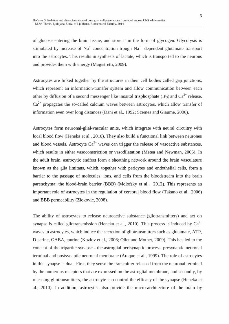
of glucose entering the brain tissue, and store it in the form of glycogen. Glycolysis is
stimulated by increase of Na+ concentration trough Na
+- dependent glutamate transport
into the astrocytes. This results in synthesis of lactate, which is transported to the neurons
and provides them with energy (Magistretti, 2009).
Astrocytes are linked together by the structures in their cell bodies called gap junctions,
which represent an information-transfer system and allow communication between each
other by diffusion of a second messenger like inositol trisphosphate (IP3) and Ca2+
release.
Ca2+
propagates the so-called calcium waves between astrocytes, which allow transfer of
information even over long distances (Dani et al., 1992; Scemes and Giaume, 2006).
Astrocytes form neuronal-glial-vascular units, which integrate with neural circuitry with
local blood flow (Heneka et al., 2010). They also build a functional link between neurones
and blood vessels. Astrocyte Ca2+
waves can trigger the release of vasoactive substances,
which results in either vasoconstriction or vasodilatation (Metea and Newman, 2006). In
the adult brain, astrocytic endfeet form a sheathing network around the brain vasculature
known as the glia limitans, which, together with pericytes and endothelial cells, form a
barrier to the passage of molecules, ions, and cells from the bloodstream into the brain
parenchyma: the blood-brain barrier (BBB) (Molofsky et al., 2012). This represents an
important role of astrocytes in the regulation of cerebral blood flow (Takano et al., 2006)
and BBB permeability (Zlokovic, 2008).
The ability of astrocytes to release neuroactive substance (gliotransmitters) and act on
synapse is called gliotransmission (Heneka et al., 2010). This process is induced by Ca2+
waves in astrocytes, which induce the secretion of gliotransmitters such as glutamate, ATP,
D-serine, GABA, taurine (Kozlov et al., 2006; Oliet and Mothet, 2009). This has led to the
concept of the tripartite synapse - the astroglial perisynaptic process, presynaptic neuronal
terminal and postsynaptic neuronal membrane (Araque et al., 1999). The role of astrocytes
in this synapse is dual. First, they sense the transmitter released from the neuronal terminal
by the numerous receptors that are expressed on the astroglial membrane, and secondly, by
releasing gliotransmitters, the astrocyte can control the efficacy of the synapse (Heneka et
al., 2010). In addition, astrocytes also provide the micro-architecture of the brain by

7 Hočevar S. Isolation and characterization of pure glial cell populations from adult mouse CNS white matter.
M.Sc. Thesis. Ljubljana, Univ. of Ljubljana, Biotechnical Faculty, 2014
forming a scaffold to the nervous system (Verkhratsky and Butt, 2007), have control of
water homeostasis (Simard and Nedergaard, 2004) and may also help oligodendrocytes in
myelination of axons (Meyer-Franke et al., 1999, cit. by Molofsky et al., 2012).
2.2.2 Astrocyte markers
Astroglia express astrocyte specific antigens, which represent important tools for their
detection and characterisation.
Table 1: Astrocyte markers
Marker Function
GLAST/EAAT1 Glutamate Aspartate Transporter (GLAST) or Excitatory Amino Acid Transporter 1
(EAAT1) is a Na+-dependent high-affinity glutamate transporter (Sheldon and Robinson,
2007).This type of glutamate transporters are predicted to have 8 transmembrane domains
and most likely exist as trimmers (Yernool et al., 2003). GLAST was primarily found in
the plasma membranes of astrocytes (Rothstein, 1994) and in radial glia-astrocyte lineage
of developing rodent CNS (Shibata et al., 1997). However, it could also be found in
many other cells, such as oligodendroglia (Domercq, et al., 1999) and macrophages (Gras
et al., 2003). GLAST is enriched in astrocytic processes near synaptic termini (Chaudhry
et al., 1995) and increased in some specialized parts of the brain such as Bergmann glia
and Muller cells of the retina (Sheldon and Robinson, 2007). Its main function is to clear
out extracellular glutamate throughout the synaptic cleft, to keep the level low enough to
prevent neurons from excitotoxicity. Furthermore, GLAST is involved not only in the
regulation of synaptic transmission, but it also plays some roles in the development and
differentiation of the CNS (Shibata et al., 1997).
GFAP GFAP comes from a large multigene family of intermediate filament (IF) proteins. It is
sorted into Class III of IF family, which represents astroglial characteristic. GFAP is a
major IF protein of mature astrocytes and is known as maturation marker (Gomes et al.,
1999). GFAP was found to be important for integrity of CNS white matter architecture
and long-term maintenance of myelination. It also plays an important role in the
formation of BBB (Liedtke, et al., 1996).
Vimentin Vimentin is a Class III IF protein and is present in all mesenchymal tissues. It is
expressed in a wide variety of cells including astrocytes, fibroblasts, endothelial cells,
macrophages, neutrophils and lymphocytes (Evans, 1998). Vimentin is the major IF
expressed in astrocyte precursors and radial glia. During the maturation of astrocyte, its
expression is progressively replaced by expression of GFAP (Gomes et al., 1999).
Vimentin’s major function is to build cytoskeletal structures deep inside the cell and it
plays an important role in supporting the location of the organelles (Katsumoto et al.,
1990).
2.2.3 Astrocyte pathology
2.2.3.1 Reactive astrogliosis
Because astrocytes represent almost one half of the brain cells, there is practically no CNS
disease that does not involve their participation. Astrocytes are important defence

8 Hočevar S. Isolation and characterization of pure glial cell populations from adult mouse CNS white matter.
M.Sc. Thesis. Ljubljana, Univ. of Ljubljana, Biotechnical Faculty, 2014
mechanisms and are genuinely responsible for the protection and survival of the neural
tissue. The reactive astrogliosis is essential for both limiting the areas of damage and for
the post-insult remodelling and recovery of neural function (Henaka et al., 2010).
However, this mechanism can also act as a harmful factor by trying to seal and eliminate
the damage (Barres, 2008). In addition, reactive astrocytes can stimulate unwanted
synapses that can cause epilepsy, neuropathic pain and are involved in several other neuro-
pathological conditions (Boroujerdi et al., 2008).
2.2.3.2 Alzheimer’s disease
Astrocytes are involved in a variety of psychiatric disorders, e.g. depression and
schizophrenia, and neurodegenerative diseases, e.g. Parkinson’s disease (PD), Alzheimer’s
disease (AD) and other types of dementia (Heneka, et al., 2010). AD is the main cause for
senile dementia. The results of this disease are rapid memory lost and severe disruption of
cognitive functions. The main pathological markers of AD are the formation of deposits of
β-amyloid protein (Aβ) in the walls of blood vessels, accumulation of Aβ plaques in the
grey matter and intra-neuronal accumulation of abnormal tau-protein filaments in the form
of neurofibrillary tangles (Verkhratsky and Butt, 2007). Aβ was found to be an activating
signal for astrocytes. The exposure of cultured glial cells to aggregated Aβ or amyloid
plaques triggered harmful reactive astrogliosis (DeWitt, et al., 1997). Thus, it is possible to
observe astrogliosis in adult AD brain, especially in the cells surrounding amyloid plaques
with processes of activated astrocytes joining in formation of neurotic plaques (Nagele et
al., 2004).
2.2.3.3 Parkinson’s disease
PD is a neurodegenerative disorder, theclinical symptoms of which are disturbed
locomotive and motor functions (Parkinson, 2002). These symptoms occur because of the
specific extermination of dopaminergic neurons in substantia nigra, which has severe effect
on nigrostriatal dopaminergic transmission. At the late stages of the disease, reactive
astrocytes are involved in the inflammatory state (McGeer and McGeer, 2008) and may
also fail to support dopaminergic neurons, which contributes to neurodegeneration (Mena,

9 Hočevar S. Isolation and characterization of pure glial cell populations from adult mouse CNS white matter.
M.Sc. Thesis. Ljubljana, Univ. of Ljubljana, Biotechnical Faculty, 2014
et al., 2002). However, it is not fully known what the early changes in astroglia are,
although they may be responsible for the progression of the disease (Heneka et al., 2010).
2.3 OLIGODENDROCYTES
Oligodendrocytes are myelinating cells in the CNS. They develop from migratory and
mitotic OPCs (Baumann and Pham-Dinh, 2001). From their germinal zones, OPCs migrate
to their target regions in white and grey matter, where they maturate into mature myelin-
producing cells and begin to wrap axons by forming myelin sheaths (Fig. 4) (McTigue and
Tripathi, 2008).
2.3.1 Oligodendrocyte morphology and myelin functions
Oligodendrocytes can be distinguished from astrocytes by several features, especially their
smaller size, higher density of cytoplasm and nucleus with dense chromatin, the absence of
intermediate filaments and glycogen in cytoplasm, and the presence of a large number of
microtubules in their processes that might be involved in their stability (Baumann and
Pham-Dinh, 2001; Lunn et al., 1997). On the same axon, myelin sheaths belong to
different oligodendrocytes. Moreover, these cells are able to enwrap up to 50 axonal
segments, depending on the region of the CNS (Baumann and Pham-Dinh, 2001). By their
morphology, size or thickness of the myelin sheath they form, we distinguish four different
types of oligodendrocytes. Oligodendrocyte types I and II arise late in the development and
myelinate many internodes on small diameter axons, while oligodendrocyte types III and
IV arise later and myelinate mainly large diameter axons (Butt, 2013). Though
oligodendrocytes are mostly myelin producing cells, there also exist satellite
oligodendrocytes that may not be directly involved in the formation of myelin sheath. The
satellite oligodendrocytes are perineuronal and most likely serve to regulate the
microenvironment around neurons (Ludwin, 1997, cit. by Baumann and Pham-Dinh,
2001).

10 Hočevar S. Isolation and characterization of pure glial cell populations from adult mouse CNS white matter.
M.Sc. Thesis. Ljubljana, Univ. of Ljubljana, Biotechnical Faculty, 2014
Figure 4: The oligodendrocyte and the mature myelin sheath. Oligodendrocyte- G, node- N (Baumann and
Pham-Dinh, 2001: 873)
The main and most evident function of oligodendrocytes is the production of myelin,
which is significant for the isolation of axons. Moreover, the formation of myelin sheaths
plays an important role in the nervous system functions. Thus, oligodendrocytes provide
structural and electrical properties for axons by controlling their diameter as well as
spacing and clustering of Na+ channels at nodes and paranodes (Barres, 2008). Myelin
represents an essential component of white matter in CNS, which contains approximately
40-50% myelin on a dry weight basis. Myelin dry weight consists of 70 % lipids and 30 %
proteins (Baumann and Pham-Dinh, 2001). There are several specific proteins that we can
find in myelin, including myelin basic protein (MBP), proteolipid protein (PLP) and
myelin associated glycoprotein (MAG) (Ndubaku and de Bellard, 2008). Because of the
high content of lipids and low content of water, myelin sheaths form an isolating layer and
therefore enable fast nerve conduction (Baumann and Pham-Dinh, 2001). Myelinated
axons appear as myelinated membrane segments, called internodes, and are separated by
regions of unsheathed nerve membrane, termed nodes of Ranvier. There is an extremely
high density of Na+ channels in the node of Ranvier, which allows impulse to jump from

11 Hočevar S. Isolation and characterization of pure glial cell populations from adult mouse CNS white matter.
M.Sc. Thesis. Ljubljana, Univ. of Ljubljana, Biotechnical Faculty, 2014
node to node. This fast transmission of impulse is driven by high resistance and low
capacitance of myelin sheath (Shepherd, 1988, cit. by Baumann and Pham-Dinh, 2001).
Oligodendrocytes are important in the development, maintenance and survival of axons
and in control of growth of axon calibre (Baumann and Pham-Dinh, 2001). This is why
injuries and demyelination, due to damage to oligodendrocyte, could lead to variety of
serious diseases (Allen and Barres, 2009).
2.3.2 Oligodendrocyte progenitor cells (OPCs)
OPCs are multipolar cells that represent a big part of mammalian developing CNS and
contact numerous small diameter pre-myelinated axons (Verkhratsky and Butt, 2007).
There are different populations of OPCs that originate from different parts of CNS. These
are specialized domains of spinal cord and forebrain, from where OPCs migrate long
distances to their final destination, where they develop into mature oligodendrocytes (Bradl
and Lassmann, 2010). In adult brain, OPCs persist in significant numbers and are in a
relatively quiescence state, thus providing a potential source for new oligodendrocyres
after injury (Wolswijk et al., 1990). Shi et al. (1998) found that adult OPCs in culture are
not senescent cells and that they retain the capacity to divide rapidly. Studies have shown
that rapidly proliferating OPC-like cells and endogenous precursor cells in the white matter
of the brain can re-myelinate axons (Carroll and Jennings, 1994, cit. by Shi et al., 1998;
Gensert and Goldman, 1997). These findings are important in demyelination injuries and
diseases (Shi et al., 1998).
2.3.3 NG2-glia
NG2-glia are named after the proteoglycan Neuron glia 2, which they express (Stallcup et
al., 2002). NG2 cells have been found in both grey and white matter and are considered as
adult OPCs, as they provide re-myelinating oligodendrocytes following demyelination
(Dawson et al., 2000). However, apart from their assumed functions as OPCs, NG2 cells
have been identified as postmitotic complex cells with stellate morphology and are
considered to be a distinct class of cells called synantocytes or polydendrocytes
(Nishiyama et al., 2009). They have several physiological properties expressing voltage-
gated ion channels and are involved in neural-glial interactions. Nevertheless, NG2 glia

12 Hočevar S. Isolation and characterization of pure glial cell populations from adult mouse CNS white matter.
M.Sc. Thesis. Ljubljana, Univ. of Ljubljana, Biotechnical Faculty, 2014
interacts with nodes of Ranvier and formation of glial-scar, driven by CNS injuries (Butt et
al., 2005).
2.3.4 Oligodendrocyte markers
Table 2: Oligodendrocyte markers
Marker Function
O4
The monoclonal antibody O4 detects sulfatides and an unidentified sulfated glycoconjugate
termed pro-oligodendroblast antigen (Bansal et al., 1999). Sulfatid is one of the major
galactosphingolipid compounds of oligodendrocyte plasma membrane and myelin. It is also
produced by Schwann cells in the peripheral nerve (Takahashi and Suzuki, 2012). In the early
OPCs these sulfatid antigens appear to lack (Deng and Poretz, 2003). Sulfatide has the function
of a negative regulator of oligodendrocyte differentiation (Hirahara et al., 2004), plays role in
oligodendrocyte survival (Shroff et al., 2009) and also appears to function in myelin
maintenance and axon structure (Marcus et al., 2006).
Sox10 Sox10 is one of the transcription factors of a large Sox family. It is characterized by DNA-
binding and bending domain, called the high mobility group box (HMG box) (Kuhlbrodt et al.,
1998). Sox10 plays a crucial role in neuronal crest development (Kim et al., 2003), peripheral
gliogenesis (Ito et al., 2006), terminal oligodendrocyte differentiation and activation of several
key myelin genes (Stolt et al., 2002). It has been reported that Sox10 also functions as an active
nucleocytoplasmic shuttle protein, as it is entering and exiting the nucleus (Rehberg et al.,
2002).
APC Adenomatous polyposis coli (APC) is a 312 kD protein, and acts as a tumor suppressor. Its
mutation reflects as familial adenomatous polyposis, which often develops into colon cancer
(Lang et al., 2013). APC is expressed in oligodendroglial linage during normal oligodendrocyte
development and it is important for oligodendrocyte regeneration and proliferation of OPCs.
APC regulates oligodendrocyte differentiation trough β-catenin intracellular level regulation
(Lang et al., 2013). In CNS it also regulates process formation, proliferation of radial glia,
astrocytes, neurons and neuroblasts (Yokota et al., 2009; Imura et al., 2010).
Olig1 Oligodendrocyte transcription factor 1 (Olig1) is a basic helix-loop-helix transcription factor
expressed exclusively in CNS (Zhou et al., 2000). Olig1 has roles in development and
maturation of oligodendrocytes, evident especially within the brain (Lu, et al., 2002). It also
has an important role in the repairing of demyelinating injuries (Arnett et al., 2004, cit. by
Meijer et al., 2012).
Olig2 Oligodendrocyte transcription factor 2 (Olig2) is expressed very early in CNS development
within the radial glia of the neural tube, which develop into motor neurons and mature
oligodendrocytes (Malatesta et al., 2003; Tsai et al., 2012). Olig2, together with Olig1 also
plays a dominant role in the pattering of the spinal cord (Meijer et al., 2012).
PDGFαR Platelet-derived growth factor α receptor is a tyrosine kinase receptor and binds both A and B
chains of platelet-derived growth factor (PDGF) (Mudhar et al., 1993). PDGFαR is in
gliogenesis uniquely expressed by OPCs at very early stages of oligodendrocyte development
and disappears at O4+ stage of oligodendrocyte maturation (Baumann and Pham-Dinh, 2001).
PDGFαR and its ligands are important players in OPC proliferation and survival (Mudhar et al.,
1993) and also necessary for the neuronal crest development and for normal pattering of the
somites (Soriano, 1997).
NG2 NG2 is an integral membrane chondroitin sulfate proteoglycan with 260 kDa core protein. In
adult CNS, NG2 is expressed together with PDGFαR, which proves the development in
oloigodendrocyre linage (Baumann and Pham-Dinh, 2001). However, NG2 is not expressed in
adult oligodendrocytes, astrocytes, microglia or neurons, proposing that the cells expressing
NG2 antigen are a novel glial cell population (Dawson et al., 2000). Outside the nervous
system, NG2 is expressed in many other tissues by different mesenchymal cell type. NG2
proteoglycan seems to contribute to proliferation and migration of immature progenitor cells
and their responses to injuries such as inflammation or demyelination (Stallcup, 2002).

13 Hočevar S. Isolation and characterization of pure glial cell populations from adult mouse CNS white matter.
M.Sc. Thesis. Ljubljana, Univ. of Ljubljana, Biotechnical Faculty, 2014
2.3.5 Oligodendrocyte pathology
The primary event in oligodentrocyte pathology is the breakdown of BBB which plays a
big role in demyelinating diseases, such as MS (Baumann and Pham-Dinh, 2001).
Secondly, oligodendrocytes have high metabolic activity and consequently use up large
amounts of oxygen and ATP, and have a high iron content (McTigue and Tripathi, 2008).
All this leads to the formation of reactive oxygen species, free radical formation and lipid
peroxidation. Thus, oxidative stress, which can easily effects oligodendrocytes, is one of
the mechanisms responsible for several neurological diseases, especially in the process of
demyelination (Bradl and Lassman, 2010).
2.3.5.1 Multiple sclerosis
MS is one of the most common neurological diseases. It is a chronic, inflammatory disease
of CNS and involves demyelination as a result of autoimmune attack on oligodendrocytes
and myelin (Barres, 2008). Most patients with MS experience repeated attacks on
oligodendrocytes and myelin via the inflammatory cascade that is activated repeatedly over
time. In the early stages of demyelination in MS, re-myelination occurs due to generation
of new oligodendrocytes and myelin formation. However, this repair mechanism fails in
later, progressive stage of the disease and can no longer repair injured oligodendrocytes
and depleted myelin, causing irreversible neurone damage to develop (Dhib-Jabult, 2007).
It is not known whether the repair fails because of the exhaustion of the new OPCs, a
deficiency in the relevant axonal signals or electrical activity that induces OPC
proliferation and myelination, a diversion of OPCs into an astrocyte differentiation
pathway, or the development of inhibitors that prevent migration or myelination by OPCs.
The answer may lead to new drugs that promote myelin repair (Barres, 2008).
2.4 MICROGLIA
Microglia represent the immune system of the CNS and act as macrophages with the
ability of phagocytosis. These cells originate from myeloid lineage, then enter the CNS
and migrate all over the parenchyma where they transform into resting microglia. In case
of insults of the nervous system, such as neurodegenerative diseases, strokes, traumatic
injuries etc., microglial cells are activated (Heneka et al., 2010). With their processes they
14 Hočevar S. Isolation and characterization of pure glial cell populations from adult mouse CNS white matter.
M.Sc. Thesis. Ljubljana, Univ. of Ljubljana, Biotechnical Faculty, 2014
rapidly converge on the site of injury without cell body movement, start to scan the
microenvironment to remove damaged or dead cells and build a potential barrier between
healthy and injured tissue (Davalos et al., 2005).
Microglial cells can secrete numerous cytokines and other pro-inflammatory mediators,
including tumor necrosis factor α (TNFα), which plays a crucial role in promoting
generation of new oligodendrocytes (Arrnet et al., 2001, cit. by Barres, 2008). There is a
dendritic cell type of microglia, which has the capability of presenting myelin antigen to T
cells within the brain (Miller et al., 2007). It has been shown that microglia also have an
important role during the CNS development in eliminating inappropriate synaptic
connections through classical complement cascade called complement component 1 q
(C1q) (Stevens et al., 2007).
However, microglia are also involved in the progression of neurodegenerative diseases like
PD and AD with their neuro-inflammatory mechanisms (Heneka et al., 2010). Cytokines,
reactive oxygen species, complement factors, neurotoxic secretory products, free radical
species and nitric oxide can contribute to neuronal dysfunction and cell death. Thus,
activated microglia can be a potential target for future anti-inflammatory therapeutics
(Sastre et al., 2006).
2.5 ADULT NEURAL STEM CELLS
Neural stem cells (NSCs) are present in the developmental brain and in the specific regions
of postnatal and adult brain. These cells develop not only into neurons but also into glial
cells (Alvarez-Buylla and Lim, 2004). Glial cells were primarily considered to have very
different origin from nerve cells. Recent studies have shown that glial cells in
development, radial glia, and subpopulations of astrocyte in adult mammals actually
function as primary progenitors or NSCs (Kriegstein and Alvarez-Buylla, 2009).
In adult mammals, neurogenesis is most prominent in subventricular zone (SVZ) in the
walls of lateral ventricles. The SVZ contains NSCs, also known as B cells, which are
categorised as subpopulation of astrocytes as they are expressing GFAP, GLAST and other
astroglial markers (Doetsch et al., 1997; Platel et al., 2009). B cells or SVZ astrocytes
15 Hočevar S. Isolation and characterization of pure glial cell populations from adult mouse CNS white matter.
M.Sc. Thesis. Ljubljana, Univ. of Ljubljana, Biotechnical Faculty, 2014
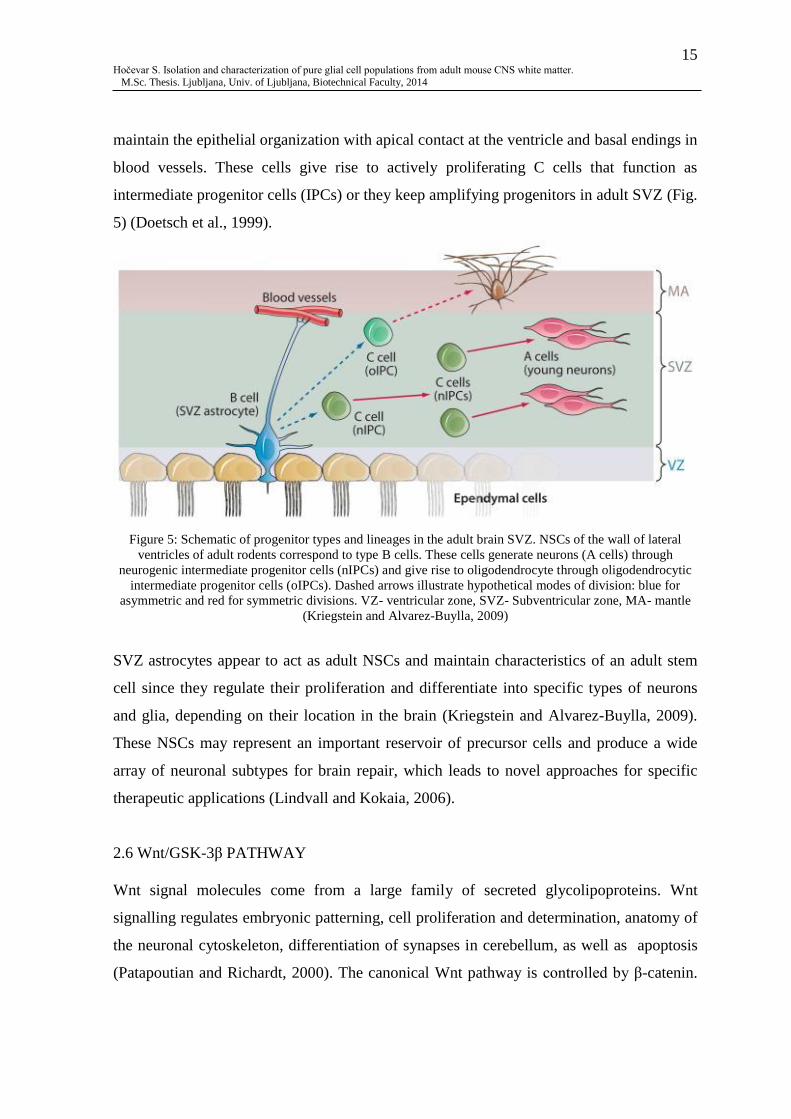
maintain the epithelial organization with apical contact at the ventricle and basal endings in
blood vessels. These cells give rise to actively proliferating C cells that function as
intermediate progenitor cells (IPCs) or they keep amplifying progenitors in adult SVZ (Fig.
5) (Doetsch et al., 1999).
Figure 5: Schematic of progenitor types and lineages in the adult brain SVZ. NSCs of the wall of lateral
ventricles of adult rodents correspond to type B cells. These cells generate neurons (A cells) through
neurogenic intermediate progenitor cells (nIPCs) and give rise to oligodendrocyte through oligodendrocytic
intermediate progenitor cells (oIPCs). Dashed arrows illustrate hypothetical modes of division: blue for
asymmetric and red for symmetric divisions. VZ- ventricular zone, SVZ- Subventricular zone, MA- mantle
(Kriegstein and Alvarez-Buylla, 2009)
SVZ astrocytes appear to act as adult NSCs and maintain characteristics of an adult stem
cell since they regulate their proliferation and differentiate into specific types of neurons
and glia, depending on their location in the brain (Kriegstein and Alvarez-Buylla, 2009).
These NSCs may represent an important reservoir of precursor cells and produce a wide
array of neuronal subtypes for brain repair, which leads to novel approaches for specific
therapeutic applications (Lindvall and Kokaia, 2006).
2.6 Wnt/GSK-3β PATHWAY
Wnt signal molecules come from a large family of secreted glycolipoproteins. Wnt
signalling regulates embryonic patterning, cell proliferation and determination, anatomy of
the neuronal cytoskeleton, differentiation of synapses in cerebellum, as well as apoptosis
(Patapoutian and Richardt, 2000). The canonical Wnt pathway is controlled by β-catenin.

16 Hočevar S. Isolation and characterization of pure glial cell populations from adult mouse CNS white matter.
M.Sc. Thesis. Ljubljana, Univ. of Ljubljana, Biotechnical Faculty, 2014
Wnt binds to Frizzled and LDL receptor-related proteins 5 and 6 (LRP5/6) (He et al.,
2004) and activates the cytoplasmic scaffolding protein Dishevelled (Dvl). The latter
triggers the inhibition of glycogen synthase kinase-3β (GSK-3β), which results in
stabilisation and nuclear translocation of β-catenin (Patapoutian and Richardt, 2000). Free
β-catenin forms a nuclear complex with T-cell factor and lymphoid enhancer factor
(TCF/LEF), DNA-binding transcription factors, activating the transcription of numerous
gens (Fig. 6B) (Arce et al., 2006). In the absence of Wnt signalling, β-catenin is complexed
with GSK-3β, Axin, APC and casein kinase 1 (CK1). GSK-3β and CK1 sequentially
phosphorylate β-catenin, which drives β-catenin ubiquitination and consequently its
degradation by proteasomes (He et al., 2004). Wnt target genes are repressed by inhibitors
Groucho/transducin-like enhancer (Gro/TLE) and histone deacetylases (HDAC) (Fig. 6A)
(Daniels and Weis, 2005).
Figure 6: Wnt/β-catenin signalling. A) The absence of Wnt signal molecule. B) The presence of Wnt signal
molecule. LRP5/6- low-density lipoprotein receptor related proteins 5 and 6, APC- adenomatous polyposis
coli, GSK3- glycogen synthase kinase-3β, CK1- casein kinase 1, ub- ubiquitin, β-Trcp- E3 ubiquitin ligase,
HDAC- histone deacetylase, TLE- transducin-like enhancer, TCF- T-cell factor, Dvl- Dischevelled
(MacDonald et al., 2009)
Hence, GSK-β3 by its actions through regulating Wnt signalling, is an essential molecule
for proliferation and differentiation signals and cell fate determination. Inhibition of GSK-
3β with several inhibitors (ARA-014418, lithium, indirubin and L803-mt) results in

17 Hočevar S. Isolation and characterization of pure glial cell populations from adult mouse CNS white matter.
M.Sc. Thesis. Ljubljana, Univ. of Ljubljana, Biotechnical Faculty, 2014
increased OPCs proliferation and survival, increased oligodendrocyte differentiation and
myelination. Furthermore, GSK-β3 inhibition has positive effects on oligodendrocyte
regeneration and re-myelination, which may give options to develop new therapeutics for
demyelinating disease, e.g. MS (Azim and Butt, 2011). Inhibition of GSK-β3 signalling
with lithium also has an effect on astrogliosis. It results in generation of a novel astrocyte,
different from reactive, scar astrocytes. This may be an important factor in the repairment
of CNS injuries and diseases (Rivera, 2014).
2.7 OPTIC NERVE
The optic nerve is the second pair of cranial nerves that arise from the retina and carry
visual information to the brain. It is the only cranial nerve that is a part of CNS, whereas
the remaining eleven are peripheral nerves. Thus, the optic nerve may be used as a model
of CNS tract, which includes axons of retinal ganglion cells together with
oligodendrocytes, type 1 and type 2 astrocyte (Raff, 1989), NG2-glia and microglia (Butt
et al., 2005). The rodent optic nerve is easy to isolate and represents a suitable model for
studying axonal-glial interactions in their natural state, since there are no neuronal cell
bodies. Furthermore, it is also an ideal preparation for the study of myelination of CNS
axons, relationships between myelination and axonal conduction properties, and
physiological characteristic of glial cells (Bolton and Butt, 2004).
In the early 1980s, oligodendrocyte-type 2 astrocyte bipotential progenitors (O-2A) were
first discovered incultures of the optic nerve by Raff and colleagues. O-2A cells in culture
develop into type 2 astrocytes if cultured in media with fetal calf serum (FCS), and into
oligodendrocytes if cultured in serum free media (Raff et al., 1984). In the absence of
evidence for type-2 astrocytes in vivo, O-2A cells have been referred to as OPCs (Butt et
al., 2005). All this leads to an important aspect of the approach of in vitro culturing, as we
can manipulate the cells and their environment. By exposing them to a variety of signals,
the full differentiation potentiality and characterisation can be explored (Hardy and
Reynolds, 1991).

18 Hočevar S. Isolation and characterization of pure glial cell populations from adult mouse CNS white matter.
M.Sc. Thesis. Ljubljana, Univ. of Ljubljana, Biotechnical Faculty, 2014
2.8 In vitro CULTURING OF GLIAL CELLS FROM ADULT MOUSE CNS
Much of our understanding of glial cell biology comes from the studies of glial cell lines or
cells in cultures that have been isolated from embryonic or early postnatal brain (McCarthy
and de Vellis, 1980; Dincman et al., 2012; Chen et al, 2007; Emery and Dugas, 2013).
McCarthy and de Vellis (1980) contributed an important advance in the development of
astrocyte and oligodendrocyte cultures preparation from rodent neonatal brain. However,
the properties of glial cells isolated from prenatal brain show an unclear relationships to
those of mature glia in the adult brain (Cahoy et al. 2008). Furthermore, it has become
clear that it is very difficult to study mature astrocytes, OPCs and oligodendrocytes in
culture, particularly in pathology/neuro-degeneration and regeneration. There have been
only a few previous studies that isolated pure glial cell populations from adult rodent CNS
(Foo et al., 2011; Cahoy et al., 2008; Shi et al., 1998).
An important aspect of studying glial cells, their functions, involvement in
neurodegenerative diseases and their potential treatments in mouse, is the use of the 3Rs
principle:
REPLACEMENT: the use of non-animal methods such as cell cultures, human
volunteers and computer modelling instead of animal to achieve scientific aim
REDUCTION: the use of methods that enable researchers to obtain comparable
amounts of information from fewer animals or more information from the same
number of animals
REFINEMENT: the use of methods that alleviate or minimise potential pain,
suffering or distress, and that enhance animal welfare for those animals that
cannot be replaced (Robinson, 2005)
To achieve these principles it is important to develop stable glial populations from adult
mouse CNS in vitro. That is how we can replace and reduce the use of in vivo methods. In
addition, as neurons depend on astrocytes and oligodendrocytes for their survival, it has
not been possible to get their functional roles in vivo simply by deleting them, which
makes culture studies a powerful approach (Foo et al., 2011). However, there are still some
limitations, which come along with different isolation and culturing techniques, and can

19 Hočevar S. Isolation and characterization of pure glial cell populations from adult mouse CNS white matter.
M.Sc. Thesis. Ljubljana, Univ. of Ljubljana, Biotechnical Faculty, 2014
result in genomic instability and viability. Thus, it is significant to provide in vitro findings
that will be better recapitulated in vivo (Dincman et al. 2012).

20 Hočevar S. Isolation and characterization of pure glial cell populations from adult mouse CNS white matter.
M.Sc. Thesis. Ljubljana, Univ. of Ljubljana, Biotechnical Faculty, 2014
3 MATERIALS AND METHODS
3.1 MATERIALS
3.1.1 Animals
Mice aged between postnatal day (P)3 and P7, and adults were used throughout. The wild
type mouse strain used was C57BL/6 strain. Transgenic mouse line was used, GFAP-
eGFP, in which enhanced green fluorescent protein (eGFP) is under control of the glial-
specific promotor GFAP. All research involving animals was approved by the University
of Portsmouth Ethics Committee and by the Home Office of the United Kingdom under the
Animals Scientific Procedures Act, 1986. Animals were killed humanely by cervical
dislocation, and brains and optic nerves were removed rapidly and placed in saline.
3.1.2 Agents
Table 3: List of chemicals
Chemical Abbreviation Supplier
Phosphate Buffered Saline tablets PBS Sigma
Triton X-100 Triton Sigma
Poly-L-lysine Poly-L-lysine Sigma
Laminin Laminin Sigma
Ethanol Eth Fisher Scientific
Distilled water dH2O Gibco
Hanks’ Balanced Salt Solution (1x), +CaCl2 +MgCl2 HBSS +CaCl2 +MgCl2 Gibco
Hanks’ Balanced Salt Solution (1x), -CaCl2 -MgCl2 HBSS -CaCl2 -MgCl2 Gibco
Trypsin Trypsin Sigma
Deoxyribonuclease I DNAse I Sigma
Fetal Bovine Serum FBS Gibco
Neuron Goat Serum NGS Gibco
Horse serum HS Gibco
Penicillin/Streptomycin Pen/strep Sigma
Glucose Glucose Sigma
Opti-Minimal Essential Medium Opti-MEM Gibco
Advanced Dulbecco's Modified Eagle Medium/F12 Advanced DMEM/F12 Gibco
Neurobasal- A Medium (1x) NB-A Medium Gibco
L-glutamin L-glutamin Sigma
B27 suplement B27 Millipore
Fibroblast growth factor 2 FGF2 Millipore
Recombinant Human Platelet-derived Growth Factor AA rhPDGF-AA R&D Systems
Bovine Serum Albumin BSA Sigma
N2 suplement N2 Millipore
Sodium hydrogen carbonate NaHCO3 Sigma-Aldrich
continued

21 Hočevar S. Isolation and characterization of pure glial cell populations from adult mouse CNS white matter.
M.Sc. Thesis. Ljubljana, Univ. of Ljubljana, Biotechnical Faculty, 2014
continuation of Table 3. List of chemicals
Chemical Abbreviation Supplier
Paraformaldehyde PFA TAAB
Hoechst Hoechst Invitrogen
Fluoromount Fluoromount Sigma
Neural Tissue Dissociation Kit (P) NTDK (P) Miltenyi Biotec
Neural Tissue Dissociation Kit (T) NTDK (T) Miltenyi Biotec
Anti-O4 Micro Beads: human, mouse, rat Anti-O4 Micro Beads Miltenyi Biotec
Table 4: Primary antibodies
Antigen Host Dilution Supplier
GFAP chicken 1:300 Millipore
GLAST rabbit 1:1000 Abcam
Sox10 rabbit 1:300 Millipore
PDGFαR rabbit 1:300 Santa Cruz Biotechnology
O4 mouse 1:50 R. Reynolds, ICL, UK
Table 5: Secondary antibodies
Conjugate Reactivity Dilution Supplier
Alexa Fluor 488 goat anti-rabbit 1:500 Invitrogen
Alexa Fluor 568 goat anti-rabbit 1:500 Invitrogen
Alexa Fluor 568 goat anti-chicken 1:500 Invitrogen
Alexa Fluor 488 goat anti-mouse 1:500 Invitrogen
Alexa Fluor 568 goat anti-mouse 1:500 Invitrogen
TRITC goat anti-mouse 1:100 Sigma

22 Hočevar S. Isolation and characterization of pure glial cell populations from adult mouse CNS white matter.
M.Sc. Thesis. Ljubljana, Univ. of Ljubljana, Biotechnical Faculty, 2014
Table 6: Culture media recipes
NSC media
50 % Opti-MEM
25 % HS
25 % HBSS +CaCl2 +MgCl2
1 % Pen/strep
1 % Glucose 50 %
NBA-A media
97 % NB-A
2 % B27
1 % L-glutamin
0.5 % Pen/strep
OPC-A media
95 % DMEM/F2
2 % B27
1 % Pen/strep
1 % N2
0.01 % BSA
2.1 g/L NaHCO3
40 ng/mL FGF2
20 ng/mL rhPDGF-AA
Media was filter sterilized through 0.22 micron filter system to prevent contamination.
Media was incubated for 15 min in 37 ˚C, 5 % CO2 incubator before use and was stored at
4 °C if not used.
3.1.3 Equipment
Table 7: List of equipment
Equipment Supplier
Automatic pipetts Gilson
Pipettor Fisher Brand
Laminar flow cabinet (class II) NuAire
CO2 incubator Jouan
Centrifuge, model 1k15 Sigma
Centrifuge, model 5416 Eppendorf
Confocal laser scanning microscope, model 510 Zeiss
Cell counter, Vi-Cell XR Beckman Coulter
24-well plates Cellstar
MACS Pre-Separation Filters, 70 μm Miltenyi Biotec
MACS Separator Miltenyi Biotec
MACS MS Column Miltenyi Biotec
23 Hočevar S. Isolation and characterization of pure glial cell populations from adult mouse CNS white matter.
M.Sc. Thesis. Ljubljana, Univ. of Ljubljana, Biotechnical Faculty, 2014
3.2 METHODS
3.2.1 Dissection of mouse brain and optic nerve
Pieces of mouse forebrain, cerebellum or 6 optic nerves were collected from adult mice.
Whole brains were harvested from pups. The brain was collected by cutting the skin and
skull along the midline and reflected to expose the brain. For the optic nerves, the eyeballs
were cut off and the brain was moved to expose and cut the optic nerves on their opposite
side. Brain samples were placed into 1 ml and optic nerves into 500 µl of HBSS -CaCl2 -
MgCl2.
3.2.2 Tissue dissociation of mouse brain and optic nerve
3.2.2.1 In-house dissociation method
In-house dissociation method is based on enzymatic dissociation with trypsin, which
breaks down the integrity of the tissue by degradation of extracellular adhesion proteins.
This method was used for the dissociation of brain and optic nerve of adult mouse.
The protocol for in-house dissociation method is described below:
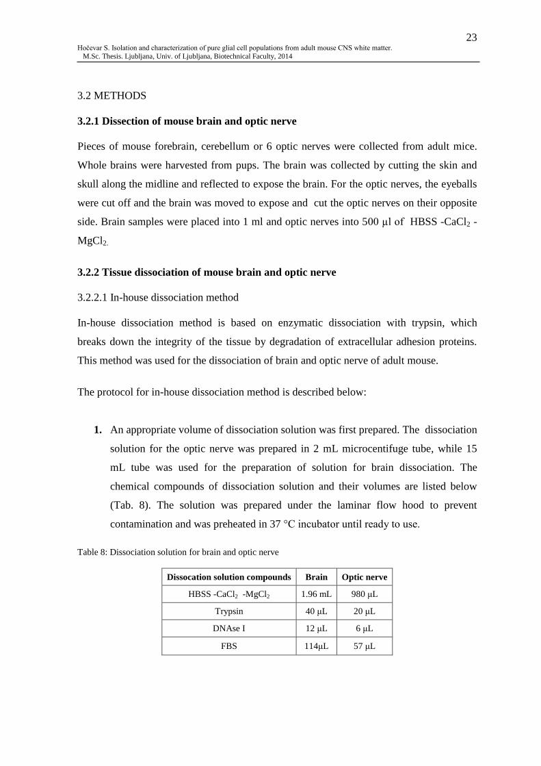
1. An appropriate volume of dissociation solution was first prepared. The dissociation
solution for the optic nerve was prepared in 2 mL microcentifuge tube, while 15
mL tube was used for the preparation of solution for brain dissociation. The
chemical compounds of dissociation solution and their volumes are listed below
(Tab. 8). The solution was prepared under the laminar flow hood to prevent
contamination and was preheated in 37 °C incubator until ready to use.
Table 8: Dissociation solution for brain and optic nerve
Dissocation solution compounds Brain Optic nerve
HBSS -CaCl2 -MgCl2 1.96 mL 980 μL
Trypsin 40 μL 20 μL
DNAse I 12 μL 6 μL
FBS 114μL 57 μL

24 Hočevar S. Isolation and characterization of pure glial cell populations from adult mouse CNS white matter.
M.Sc. Thesis. Ljubljana, Univ. of Ljubljana, Biotechnical Faculty, 2014
2. For tissue collection, appropriate volume of HBSS -CaCl2 -MgCl2 was pre-
prepared and preheated for 10 min in 37 °C incubator.
3. The collected optic nerves were first cut into small pieces for more effective cell
dissociation. For brain dissociation, the brain tissue was placed in a fresh petri dish.
To prevent the tissue to dry out and for the later easier re-collection of the tissue, 1
mL of HBSS -CaCl2 -MgCl2 was added. The tissue sample was then cut with a
scalpel in approximately 1 mm3 pieces and pipetted back into the tube. The petri
dish was rinsed with additional 1-2 mL of HBSS -CaCl2 -MgCl2 to collect any
remaining tissue.
4. The preheated dissociation solution was added to the tissue sample and incubated
for 45 min in 37 °C, 5 % CO2 incubator. The tube was inverted every 10 min to
keep the pieces in the solution.
5. After the incubation, the optic nerve sample was dissociated mechanically by
pipetting slowly up and down 10 times with each of P200, P10 and 1 mL sterile
pastette, respectively, avoiding making air bubbles. For easier brain dissociation,
P1000 was used first in order to break down bigger pieces of the brain tissue.
6. The mechanically dissociated tissue sample was centrifuged at 300 x g for 2 min at
room temperature (RT).
7. The supernatant was carefully removed. 500 μL of HBSS +CaCl2 +MgCl2 was
added to the cells dissociated from the optic nerve and 1 mL to the cells dissociated
from the brain.
8. 70 μm strainer was pre-wetted with 2 mL of HBSS +CaCl2 +MgCl2.
9. Cell suspension was filtered through the strainer to remove cell aggregates or other
large particles. The tube with the tissue was then rinsed with 1-2 mL of HBSS
+CaCl2 +MgCl2 and added to the strainer.
10. The filtered suspension was then centrifuged at 300 x g for 10 min at RT.
11. The supernatant was carefully aspirated and 1 mL of pre-heated media was added.
12. A small aliquot of cells was removed to determine the cell number and viability
with the cell counter. In the meantime the cells were kept on ice.

25 Hočevar S. Isolation and characterization of pure glial cell populations from adult mouse CNS white matter.
M.Sc. Thesis. Ljubljana, Univ. of Ljubljana, Biotechnical Faculty, 2014
3.2.2.2 Neural Tissue Dissociation Kit (P) and (T)
The Neural Tissue Dissociation Kits (NTDKs) have been designed for the dissociation of
the neural tissue to single-cell suspensions by enzymatic degradations of extracellular
adhesion proteins. NTDK (P) contains papain, and NTDK (T) contains trypsin. Some
antigen epitopes are damaged by papain, others by trypsin. The papain based kit was used
as a tissue dissociation method to gain oligodendrocytes, since oligodendrocyte specific O4
antigen is thus preserved . The kit based on trypsin was used in order to obtain cells with
astrocyte specific GLAST antigen. This dissociation kit method was used for both brain
and optic nerve tissue dissociation of adult mouse and pups.
The protocol for NTDKs is described below:
1. An appropriate volume of Enzyme Mix I was first prepared. 1950 μL of Enzyme
Mix I corresponds up to 400 mg of tissue. Solution I was added to Solution II and
was briefly vortexed. This was then preheated in 37 °C, 5 % CO2 incubator for 15
min before use. Volumes of reagents used are listed below (Tab. 9).
Table 9: Enzyme Mix I for NTDK (P) and (T) tissue dissociation
Enzyme Mix I Solution I Solution II
NTDK (P) 50 μL 1900 μL
NTDK (T) 200 μL 1750 μL
2. For tissue collection, appropriate volume of cold HBSS -CaCl2 -MgCl2 was pre-
prepared. The weight of the brain was measured to assure the collected tissue was
not heavier than 400 mg.
3. For more affective cell dissociation, the optic nerves were cut into small pieces.
The brain sample was transfered into a fresh petri dish. To prevent the tissue to dry
out and for later easier re-collection of the tissue, 1 mL of HBSS -CaCl2 -MgCl2
was added. The brain tissue was then cut with scalpel in approximately 1 mm3
pieces and pipetted back into the tube. Petri dish was rinsed with 1-2 mL of HBSS -
CaCl2 -MgCl2 to collect any remaining tissue.

26 Hočevar S. Isolation and characterization of pure glial cell populations from adult mouse CNS white matter.
M.Sc. Thesis. Ljubljana, Univ. of Ljubljana, Biotechnical Faculty, 2014
4. The tissue sample was centrifuged at 300 x g for 2 min at RT.
5. The supernatant was carefully aspirated, which was followed by the addition of a
pre-heated Enzyme Mix I to the pellet, and gently mixed to avoid making air
bubbles.
6. The tissue was then incubated at 37 °C, 5 % CO2 for 15 min in incubator.
7. During the incubation, Enzyme Mix II was prepared by adding Solution 3 to
Solution 4. Volumes of reagents used were appropriate for up to 400 mg of tissue
and are listed below (Tab. 10).
Table 10: Enzyme Mix II for NTDK (P) and (T) tissue dissociation
Enzyme Mix II Solution 3 Solution 4
NTDK (P) 20 μL 10 μL
NTDK (T) 20 μL 10μL
8. After the incubation, Enzyme Mix II was added to the tube with the tissue and
gently inverted. The tissue was then mechanically dissociated by pipetting slowly
up and down 10 times with P1000, avoiding making air bubbles.
9. The mechanically dissociated tissue was incubated in 37 °C, 5 % CO2 incubator for
10 min.
10. After the second incubation, the optic nerve tissue was dissociated mechanically by
pipetting slowly up and down 10 times with each of P200, P10 and 1 mL sterile
pastette, respectively, avoiding making air bubbles.
For easier brain dissociation, P1000 was used first in order to break down the
remaining bigger pieces of the brain tissue.
11. 70 μm strainer was pre-wetted with 2 mL of HBSS +CaCl2 +MgCl2.
12. Cell suspension was filtered through the strainer to remove cell aggregates or other
large particles. The tube with the tissue was then rinsed with 1-2 mL of HBSS
+CaCl2 +MgCl2 and added to the strainer.
13. The cell suspension was then centrifuged at 300 x g for 10 min at RT.
14. After the centrifugation, the supernatant was carefully aspirated and pellet was re-
suspended in 4 mL of HBSS +CaCl2 +MgCl2, which was followed by a second
centrifugation at 300 x g for 10 min at RT.

27 Hočevar S. Isolation and characterization of pure glial cell populations from adult mouse CNS white matter.
M.Sc. Thesis. Ljubljana, Univ. of Ljubljana, Biotechnical Faculty, 2014
15. The supernatant was carefully aspirated. The cells were re-suspended in 1 mL of
pre-heated media.
16. A small aliquot of cells was removed to determine the cell number and viability
with cell counter. In the meantime the cells were kept on ice.
3.2.3 Magnetic Activated Cell Sorting (MACS) of O4+ cells
The principle of MACS Separation is magnetic labelling, e.g. O4+ cells with Anti-O4
MicroBeads. Then the cell suspension is passed through a MACS Column, which is placed
in the magnetic field of the MACS Separator. The magnetically labelled O4+ cells are
retained in the column and the unlabelled cells run through. After removing the column
from the magnetic field, O4+
retained cells can be eluted from the column as a positively
selected cell fraction (Fig. 7).
Figure 7: Principle of the MACS Separation (MACS Miltenyi Biotec, 2014)
3.2.3.1 Magnetic labelling
1. After the determination of the total number of dissociated cells, not more than 107
cells were taken from the cell suspension and centrifuged at 300 x g for 10 min.
2. The supernatant was carefully aspirated. 90 μL of cold BSA 0.5 % and 10 μL of Anti-
O4 MicroBeads were added to the pellet. Cells were re-suspended thoroughly by
pipetting slowly up and down.
3. The labelled cells were incubated for 15 min at 4°C in the dark.

28 Hočevar S. Isolation and characterization of pure glial cell populations from adult mouse CNS white matter.
M.Sc. Thesis. Ljubljana, Univ. of Ljubljana, Biotechnical Faculty, 2014
4. After the incubation, the cells were washed by adding 1 mL of BSA 0.5 % and
pipetted slowly up and down. They were then centrifuged at 300 x g for 10 min.
5. The supernatant was carefully aspirated and the cells were re-suspended in 500 μL of
BSA 0.5 %.
3.2.3.2 Magnetic separation
1. During the centrifugation, the MACS column was placed on the MACS magnetic
stand. The column was rinsed three times with 500 μL of BSA 0.5 %.
2. Cell suspension was applied onto the column. Flow-through with unlabelled cells was
collected.
3. The column was rinsed three times with 500 μL of BSA 0.5 %
4. The column was then removed from the magnetic field and placed onto a fresh 2 mL
tube. 500 μL of BSA 0.5 % was added onto the column. The magnetically labelled
cells were immediately flushed out by the push of plunger into the column.
5. Isolated cells were then centrifuged at 300 x g for 10 min.
6. The supernatant was carefully aspirated. Cells were re-suspended in 1 mL of pre-
heated media.
7. A small aliquot of isolated cells was removed to determine the cell number and
viability with the cell counter. The number of cells in flow-through was also
determined. In the meantime the cells were kept on ice.
3.2.4 Cell culturing
A day before the cell dissociation and isolation, cover slips were prepared:
1. 13 mm cover slips were first dipped into ethanol to avoid contamination. Each of the
cover slips was then placed in a 24 well plate and left resting against the side of the
well to dry out.
2. Poly-L-lysine was diluted in sterile double distilled water in ratio 1:100. 100 μL of
diluted lysine was pipetted onto the centre of each cover slip and left overnight at 37
°C, 5 % CO2 in an incubator.

29 Hočevar S. Isolation and characterization of pure glial cell populations from adult mouse CNS white matter.
M.Sc. Thesis. Ljubljana, Univ. of Ljubljana, Biotechnical Faculty, 2014
3. The following day, laminin (1 mg/mL) was diluted in sterile double distilled water in
ratio 1:200.
4. Lysine was removed and 25 μL of diluted laminin was pipetted onto the centre of
each cover slip and left for 2h at 37 °C, 5 % CO2 in an incubator.
5. After the incubation, the laminin was replaced with 30 μL of distilled water, if clover
slips were not needed straight away.
After the dissociation and isolation of the cells, approximately 106 cells/cm
2 were plated
onto the centre of each pre-prepared cover slip. Approximately 1 mL of preheated media
was added carefully to each well with cells. The cells were kept in culture at 37 °C, 5 %
CO2 for 5-7 days. The media was changed every 3-4 days.
3.2.5 Immunohistochemistry
1. Fixation
After 5-7 days in culture, the media was carefully aspirated from the wells with
cells. The cells were fixed with 4 % PFA and incubated for 10 min at 37 °C in the
incubator. PFA was removed and the cells were washed two times for 10 min in
PBS.
2. Blocking
The blocking solution was prepared with 10 % NGS in PBS for cell surface antigens
and in PBS with 0.01 % triton (PBS-T) for non-surface antigens. Solution was
added to each well with cells and incubated at 4 °C for 1h in the dark.
3. Primary antibodies
The cells were incubated overnight in the dark at 4 °C with primary antibodies in 10
% NGS in PBS or PBS-T solution.
4. Secondary antibodies
The following day, the cells were washed three times for 10 min in PBS to remove
all unlabelled primary antibodies. The cells were then incubated for 1h in the dark at
RT with secondary antibodies in 10 % NGS in PBS or PBS-T solution. Negative
controls for antibody staining did not include primary antibodies to show the
specificity of the secondary antibodies.

30 Hočevar S. Isolation and characterization of pure glial cell populations from adult mouse CNS white matter.
M.Sc. Thesis. Ljubljana, Univ. of Ljubljana, Biotechnical Faculty, 2014
5. Mounting
After the incubation with secondary antibodies, the cells were washed three times
for 10 min in PBS. Approximately 10 μL of mounting media was added onto a glass
slide. The cover slip with cells was then carefully taken from the well and placed
onto the mounting media with the cells facing down. Slides were left to dry and
stored at 4 °C.
6. Confocal microscopy
All cell images were captured with confocal laser scanning microscope and edited in
Zeiss LSM Image Browser software.

31 Hočevar S. Isolation and characterization of pure glial cell populations from adult mouse CNS white matter.
M.Sc. Thesis. Ljubljana, Univ. of Ljubljana, Biotechnical Faculty, 2014
4 RESULTS
4.1 DISSOCIATED GLIAL CELS OF ADULT MOUSE CNS
Cell dissociation experiments were focused on the optic nerve of adult mouse, where in-
house and NTDK dissociation methods were tested. Two different media were used for cell
culture for comparison, one with serum (NSC media) and the other with serum free media
(NB-A media). The dissociation of forebrain and cerebellum, using NTDK method, were
initially performed as positive controls.
4.1.1 Dissociated cells of optic nerve
Using the in-house method, approximately 0.6 x 10⁷ cells/sample from optic nerves of
adult wild type (Wt) mouse were dissociated, with the viability of about 93 %. The in-
house method proved to be a successful technique for glial cell dissociation of adult optic
nerve tissue, as the cells were capable of being maintained in culture for 7 days (Fig. 8).
Both types of glial cells, astrocytes and oligodendrocytes, were detected by their specific
markers. An image of cells characteristic of fibrous astrocytes was captured, expressing the
astrocyte specific GFAP marker (Fig. 8A-D). Cells with the origin of oligodendrocyte
lineage were detected by the Sox10 marker (Fig. 8E-H); the Sox10 transcription factor is
specific for oligodendrocyte lineage cells and is expressed by OPC and oligodendrocytes,
and the cells identified here had the characteristic bipolar morphology of early OPC (or O-
2A) in culture.
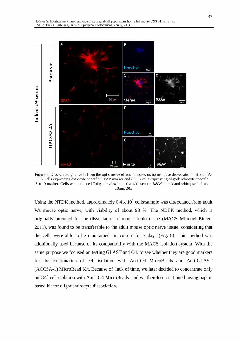
32 Hočevar S. Isolation and characterization of pure glial cell populations from adult mouse CNS white matter.
M.Sc. Thesis. Ljubljana, Univ. of Ljubljana, Biotechnical Faculty, 2014
Figure 8: Dissociated glial cells from the optic nerve of adult mouse, using in-house dissociation method. (A-
D) Cells expressing astrocyte specific GFAP marker and (E-H) cells expressing oligodendrocyte specific
Sox10 marker. Cells were cultured 7 days in vitro in media with serum. B&W- black and white; scale bars =
20μm, 20x
Using the NTDK method, approximately 0.4 x 10⁷ cells/sample was dissociated from adult
Wt mouse optic nerve, with viability of about 93 %. The NDTK method, which is
originally intended for the dissociation of mouse brain tissue (MACS Miltenyi Biotec,
2011), was found to be transferable to the adult mouse optic nerve tissue, considering that
the cells were able to be maintained in culture for 7 days (Fig. 9). This method was
additionally used because of its compatibility with the MACS isolation system. With the
same purpose we focused on testing GLAST and O4, to see whether they are good markers
for the continuation of cell isolation with Anti-O4 MicroBeads and Anti-GLAST
(ACCSA-1) MicroBead Kit. Because of lack of time, we later decided to concentrate only
on O4+ cell isolation with Anti- O4 MicroBeads, and we therefore continued using papain
based kit for oligodendrocyte dissociation.
In-h
ou
se/+
ser
um
Ast
rocy
te
OP
Cs/
O-2
A
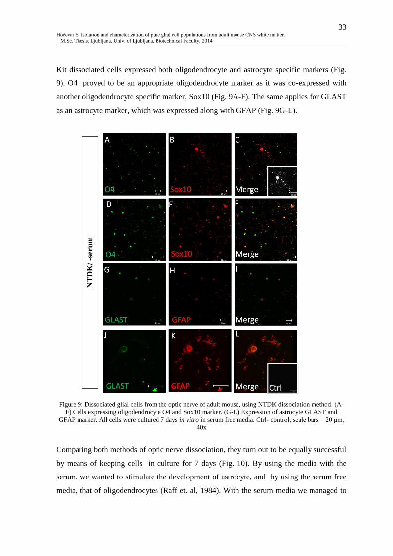
33 Hočevar S. Isolation and characterization of pure glial cell populations from adult mouse CNS white matter.
M.Sc. Thesis. Ljubljana, Univ. of Ljubljana, Biotechnical Faculty, 2014
Kit dissociated cells expressed both oligodendrocyte and astrocyte specific markers (Fig.
9). O4 proved to be an appropriate oligodendrocyte marker as it was co-expressed with
another oligodendrocyte specific marker, Sox10 (Fig. 9A-F). The same applies for GLAST
as an astrocyte marker, which was expressed along with GFAP (Fig. 9G-L).
Figure 9: Dissociated glial cells from the optic nerve of adult mouse, using NTDK dissociation method. (A-
F) Cells expressing oligodendrocyte O4 and Sox10 marker. (G-L) Expression of astrocyte GLAST and
GFAP marker. All cells were cultured 7 days in vitro in serum free media. Ctrl- control; scale bars = 20 μm,
40x
Comparing both methods of optic nerve dissociation, they turn out to be equally successful
by means of keeping cells in culture for 7 days (Fig. 10). By using the media with the
serum, we wanted to stimulate the development of astrocyte, and by using the serum free
media, that of oligodendrocytes (Raff et. al, 1984). With the serum media we managed to
NT
DK
/ -s
eru
m

34 Hočevar S. Isolation and characterization of pure glial cell populations from adult mouse CNS white matter.
M.Sc. Thesis. Ljubljana, Univ. of Ljubljana, Biotechnical Faculty, 2014
culture GFAP positive cells, which refer to astroglia (Fig. 10A-D). On the other hand, we
obtained oligodendroglia by culturing the dissociated cells in the serum free media and
detected expression of oligodentrocyte O4 and Sox10 markers (Fig. 10E-H). However, it is
still likely to get cells positive for oligodendrocytes when cultured in serum media (Fig.
8E-H) and vice versa; cells positive for astrocyte markers in serum free media (Fig. 9G-L).
Figure 10: Comparison of in-house and NTDK dissociation method. (A-D) Astrocytes, expressing GFAP
marker, dissociated with in-house method, 20x. (E-H) Image of oligodendrocyte, expressing Sox10 and O4
markers, dissociated with NTDK method, 40x. All cells were cultured 7 days in vitro. Scale bars = 50 μm
In-h
ou
se/+
ser
um
N
TD
K/-
seru
m

35 Hočevar S. Isolation and characterization of pure glial cell populations from adult mouse CNS white matter.
M.Sc. Thesis. Ljubljana, Univ. of Ljubljana, Biotechnical Faculty, 2014
4.1.2 Dissociated cells of forebrain
The total cell number of cells dissociated from adult forebrain depends on the size of the
tissue sample collected. Approximately 8 x 10⁷ cells/sample was dissociated from adult
forebrain, with viability of about 93 %, using NTDK dissociation method. The dissociation
of the adult mouse forebrain was primarily used as a control to the optic nerve dissociation.
NTDK method was found to be suitable for the adult forebrain tissue dissociation and
provided us with a sufficient number of cells for further MACS cell isolation. Despite the
serum free media we captured GLAST+ and GFAP
+ astrocytes. These markers resulted as
good markers for the detection of astroglia (Fig. 11).
Figure 11: Dissociated astrocytes from adult mouse forebrain. (A-I) Astrocytes, expressing GLAST and
GFAP markers. Cells were cultured for 7 days in vitro in serum free media. Scale bar = 20 μm
GLAST/GFAP Merge
NT
DK
/- s
eru
m

36 Hočevar S. Isolation and characterization of pure glial cell populations from adult mouse CNS white matter.
M.Sc. Thesis. Ljubljana, Univ. of Ljubljana, Biotechnical Faculty, 2014
Interestingly, immunolabelling with PDGFαR primary antibodies was expressed along
with a strong astroglial GFAP marker (Fig. 12). This is characteristic of O-2A cells, which
have a bipolar morphology and co-expressed OPC and GFAP markers (inset, Fig. 12F).
Not all cells co-express GFAP and PDGFαR (Fig. 12A-C), indicating mixed populations of
astrocytes, OPC and O-2A cells; immunolabelling was not observed in the negative control
(inset, Fig. 12C).
Figure 12: Dissociated glial cells from adult mouse forebrain. (A-F) Co-expression of PDGFαR and GFAP
markers, representing O-2A cells. Cells were cultured for 7 days in vitro in serum free media. Ctrl- control;
scale bar = 20 μm, 20x
PDGFαR/GFAP Merge
NT
DK
/ -s
eru
m

37 Hočevar S. Isolation and characterization of pure glial cell populations from adult mouse CNS white matter.
M.Sc. Thesis. Ljubljana, Univ. of Ljubljana, Biotechnical Faculty, 2014
4.1.3 Dissociated cells of cerebellum
Approximately 5 x 10⁷ cells/sample was dissociated from adult GFAP-eGFP transgenic
mouse cerebellum, with viability of about 89 %. Transgenic mice were used in order to
prove the ability of survival of transfected cells after they undergo the cell dissociation
process. Results showed that the dissociation has no effect on transfected cells, which were
able to continue expressing transfected GFP gene (Fig. 13A, E, H). Once again, there were
examples of co-expression of PDGFαR and GFAP (Fig. 13 E-G).
Figure13: Dissociated glial cells from cerebellum of transgenic GFAP-eGFP adult mouse. (A-D) Expression
of GFP under control of GFAP promotor and expression of GLAST marker. (E-G) Expression of GFP under
control of GFAP promotor and PDGFαR. (H-I) Control. Cells were cultured 7 days in vitro in serum media.
Scale bar =50 μm, 20x

38 Hočevar S. Isolation and characterization of pure glial cell populations from adult mouse CNS white matter.
M.Sc. Thesis. Ljubljana, Univ. of Ljubljana, Biotechnical Faculty, 2014
4.2 ISOLATED GLIAL CELLS
4.2.1 Isolated cells of young brain
Using the NTDK dissociation method, approximately 1.4 x 10⁷ cells/brain was dissociated
from pups, aged between P3 and P7, with viability of about 85 %. The dissociated young
mouse brain tissue contained approximately 6 % O4+ cells (cells in the column). For
culturing we used the OPC-A media by Dincman et al. (2012) in order to gain the same
culturing conditions for isolated glial cells from young postnatal brain. The experiments
on young brain served as a positive control and the isolation of glial cells proved to work
very well on the young, developing mouse brain. The cells turned out to be almost entirely
O4+, demonstrating the population of oligodendrocytes (Fig. 14).

39 Hočevar S. Isolation and characterization of pure glial cell populations from adult mouse CNS white matter.
M.Sc. Thesis. Ljubljana, Univ. of Ljubljana, Biotechnical Faculty, 2014
Figure 14: Isolated cells from young mouse brain. (A-F) Isolated O4+ cells from P6 mouse, beforehand
cultured 5 days in vitro in OPC-A media. (G-H) Control; scale bar = 20 μm, 20 x
4.2.2 Isolated cells of adult forebrain
The isolation of O4+ cells from adult forebrain tissue with MACS cells isolation system
turned out as not practicable. Approximately 54 % of the cells that were put through the
column remained in the column. The isolation of adult glia remains difficult to perform
and the right purification step is needed for further optimisation.
Hoechst/O4+
Merge

40 Hočevar S. Isolation and characterization of pure glial cell populations from adult mouse CNS white matter.
M.Sc. Thesis. Ljubljana, Univ. of Ljubljana, Biotechnical Faculty, 2014
5 DISCUSSION
As glia were found to be extremely important for CNS to function, they grew into a
significant part of research in the field of neuroscience. Glial cells became an important
subject in cell culture studies (Megale de Almeida-Leite and Esteves Arantes, 2010), with
the aim to find a way for regeneration of damaged CNS and to study the involvement of
glia in numerous diseases (DeWitt et. al, 1997; Mena et al., 2002; Barres, 2008).
There are very few reports of purified and isolated glial cell populations from adult rodents
as the preparation of adult CNS tissue has proved problematic (Saxena et al., 2012). We
performed dissociation, cell culturing and cell isolation from adult mouse CNS tissue in
order to draw experiments closer to adult human. We successfully managed to dissociate
glial cells from the adult optic nerve, forebrain and cerebellum and maintained them in
vitro. In addition, we were able to confirm that the optic nerve is a reliable and simple
model to study glia (Bolton and Butt, 2004). With the ability to maintain cells in culture we
also achieved and fulfilled the expectations of the 3Rs principles (Robinson, 2005).
However, our experiments of glial isolation and purification with the MACS separation
system preformed on the adult brain tissue remained difficult and unsuccessful. The
problem most likely occurs because of the higher amount of myelin in adult brain tissue
(Young, et al., 2013), which glues all the cells together so the MACS is not able to separate
the cells marked with magnetic beads. Moreover, besides glia, the adult mammalian brain
consists of a variety of projection neurons, local interneurons and has a complex nature of
extracellular matrix (Saxena et al., 2012), which certainly makes the purification and
isolation of glial cells even more problematic.
According to the MACS’s original Anti-O4 MicroBeads protocol, the use of P3-P7
dissociated mouse brain tissue is suggested, and should contain approximately 5-10 % of
O4+ cells (MACS Miltenyi Biotec, 2011a). Our control experiments on the young brain
were consistent with that. As many previous reports have shown (Dincman et al., 2012;
Chen et al., 2007), we were able to confirm that the isolation of glia is practicable and
works well on early postnatal, developing brain.

41 Hočevar S. Isolation and characterization of pure glial cell populations from adult mouse CNS white matter.
M.Sc. Thesis. Ljubljana, Univ. of Ljubljana, Biotechnical Faculty, 2014
Since we have managed to optimise the in-house dissociation method and made it
scientifically useful, it obtained an advantage over NTDK dissociation method in the sense
of financial aspect. As generally known, kits are usually very expensive. Thus, the in-
house method can be used as an alternative dissociation method, when there is no
substantial reason for the use of the kit.
The use of the two different media had no significant effect in our experiments. According
to Raff et al. (1984), populations of astrocytes or oligodendrocyte can be obtained,
depending on presence or absence of serum in the media. This aspect might be of greater
interest if the isolation of adult O4+ cells from adults would be practicable. In that case, we
could determine if the growth and cell proliferation of isolated O4+ cells in culture would
be better in serum free media, which should stimulate the development of
oligodendrocytes. Nevertheless, we managed to characterise oligodendroglia and astroglia
dissociated from adult mouse CNS by their specific markers. We detected O4+, Sox10+
cells which refer to oligodendrocytes (Takahashi and Suzuki, 2012; Stolt et al., 2002) and
on the other hand GLAST+ and GFAP+ cells, which represent astrocytes (Chaudhry et al.,
1995 Gomes et al., 1999). We also characterised populations of OPCs (or O-2A) in culture,
with their typical bipolar morphology (Butt et al., 2005).

42 Hočevar S. Isolation and characterization of pure glial cell populations from adult mouse CNS white matter.
M.Sc. Thesis. Ljubljana, Univ. of Ljubljana, Biotechnical Faculty, 2014
6 CONCLUSION
Although the isolation of pure glia populations from adult mice brain failed in our case,
there is still a possibility that this can be accomplished in the future. The first thing
required, in order to achieve this goal, is to find an appropriate purification process to
resolve the problem of excess myelin before cells go through the magnetic separation
procedure. If the myelin issue turns out to be the main problem in accomplishing glial
isolation, and we overcome it, the doors to research on adult isolated glial cell populations
will be open. By carrying out a variety of methods, such as quantitative polymerase chain
reaction (qPCR), Western blotting and proteomics on these adult glia, we will be able to
learn more about individual diseases such as AD, MS, PD, and their connection to
astrocyte and oligodendrocyte populations in adult humans, in which these diseases most
likely occur. Moreover, testing the important pathways, e.g. GSK3β, related to
neurodegenerative diseases and their potential therapeutics might be much more applicable
on cells isolated from adult mice and could give us more accurate results.
Despite the issues we still need to overcome, we succeeded in dissociating glia from adult
mouse CNS and created a dependable in vitro model of mixed glia populations, which still
represents an advance in the study of glia in the future.

43 Hočevar S. Isolation and characterization of pure glial cell populations from adult mouse CNS white matter.
M.Sc. Thesis. Ljubljana, Univ. of Ljubljana, Biotechnical Faculty, 2014
7 SUMMARY
Glial cells are the most numerous cells in mammalian CNS and have common origin with
neurons from the embryonic germ layer or neuroectoderm. Glia are categorised in
macroglia (astrocytes, oligodendrocytes, ependymal cells) and microglia (the immune
system of the CNS). Schwann cells are the main representative of glia in PNS
(Verkhratsky and Butt, 2007). In vitro cultures of glial cells became a useful approach to
study glial characteristics and physiology in order to develop novel drugs to treat
neurodegenerative disorders (Heneka et al., 2010). However, the majority of in vitro
studies are performed on embryonic and postnatal brains, whereas most of the
neurodegenerative diseases develop in adult brain. Consequently, it is important to develop
models that can allow us to study adult glia in vitro and help us develop drugs to treat adult
neurodegenerative diseases. The aim of our study was to develop a reliable technique for
isolation of pure populations of oligodendrocytes and astrocytes from adult mouse CNS.
The most important functions of astrocytes are the regulation of the microenvironment,
metabolic and structural support to neurons, involvement in the information-transfer
system, as well as building the BBB and secreting numerous gliotransmitters, which
control the efficacy of the synapse. Because of their large number in the brain, astrocytes
take part in several neurodegenerative degenerative diseases such as AD (Heneka et al.,
2010). The main function of oligodendrocytes is the production of myelin, which isolates
axons and provides fast transmission of impulses (Barres, 2008). Oligodendrocytes
develop from OPCs, which in adult brain also represent a source of new oligodendrocytes
after injury (Wolswijk et al., 1990). O-2A cells were discovered in the culture of the optic
nerve. These cells have the ability to develop into type 2 astrocytes or oligodendrocytes,
depending on the presence or absence of serum in the culturing media (Raff et al., 1984).
Our experiments were based on dissection of the forebrain, cerebellum and optic nerve
tissue of adult mouse, cell dissociation, cell isolation with MACS separation system, cell
culturing and immunohistochemistry. The latter was performed with astrocyte (GFAP,
GLAST) and oligodendrocyte (Sox10, O4 and PDGFαR) specific markers. For the
dissociation of the mouse CNS tissue we used in-house and NTDK methods in order to

44 Hočevar S. Isolation and characterization of pure glial cell populations from adult mouse CNS white matter.
M.Sc. Thesis. Ljubljana, Univ. of Ljubljana, Biotechnical Faculty, 2014
compare them. The serum and serum free media were used to see their effect on
differentiation of glial cells. We also performed experiments on young postnatal brains as a
positive control.
We were able to characterise populations of astrocytes, oligodendrocytes and OPCs (or O-
2A) in cultures from the brain and optic nerve of adult mouse. Our results also confirmed
that O4 and GLAST are appropriate markers to preform cell isolation. Both, in-house and
NTDK methods, turned out to be successful dissociation methods. NTDK method was
found to be applicable not only for the dissociation of brain tissue but also for the adult
mouse optic nerve tissue. The dissociation of adult mouse forebrain tissue provided us with
a sufficient number of cells to perform cell isolation. We used the NTDK method in order
to make it compatible with the MACS system. No significant effects were observed despite
using two different media in our experiments. We also proved that the process of
dissociation has no negative effect on cells from transgenic reporter mice, which continued
to express reporters in vitro. The isolation of pure populations of glia from adult mouse
CNS with the MACS isolation system turned out as non-practicable.
We managed to dissociate glia from adult mouse CNS and maintained them in vitro as a
model of mixed glia populations. However, we did not accomplish our aim of isolating
pure cell populations. Because of the high amount of myelin (Young et al., 2013) and
complex extracellular matrix in adult brain (Saxena et al., 2012), the isolation of pure glial
populations from adult mouse CNS was not possible. We successfully optimised an in-
house dissociation method, which is cheaper and thus preferable to the NTDK one, if there
is no special need to use the latter.
Despite our unsuccessful attempt to gain pure isolated adult glia populations in vitro, there
is still hope to accomplish this in the future. To reach this goal, we first need to find a
solution to extracting myelin in the purification process. Once we are able to culture pure
adult astrocyte and oligodendrocyte, we will be able to perform different biological
experiments regarding neurodegenerative disorders. This could potentially lead to new and
more successful ways of treatment of neurodegenerative diseases on adult humans.

45 Hočevar S. Isolation and characterization of pure glial cell populations from adult mouse CNS white matter.
M.Sc. Thesis. Ljubljana, Univ. of Ljubljana, Biotechnical Faculty, 2014
7.1 POVZETEK
Celice glije so najštevilčnejše celice v centralnem živčnem sistemu (CŽS) sesalcev. Imajo
skupen izvor z nevroni iz embrionalne zarodne plasti, nevroektoderma. Glijo delimo na
makroglijo v katero uvrščamo astrocite, oligodendrocite in ependimske celice ter
mikroglijo, ki predstavlja imunski sistem CŽS. Glavni predstavnik glije v perifernem
živčnem sistemu (PŽS) so Schwannove celice (Verkhratsky in Butt, 2007) . In vitro kulture
celic glija so postale uporabno orodje za proučevanje lastnosti in fiziologije glije za razvoj
novih zdravil in zdravljenje nevrodegenerativnih bolezni (Heneka in sod., 2010). Vendar
pa se večina študij in vitro izvaja na embrionalnih in postnatalnih možganih, medtem ko se
največ nevrodegenerativnih bolezni pojavi v možganih odraslih. Zato je pomembno, da se
razvije modele, ki nam bodo omogočili raziskave na odraslih celicah glija in vitro ter nam
tako pomagali pri razvoju zdravil, ki bodo primerna za zdravljenje odraslih ljudi. Namen
naše raziskave je bil razviti zanesljivo tehniko za izolacijo čistih populacij
oligodendrocitov in astrocitov iz CŽS odrasle miši. Postavili smo hipotezo, da lahko
izoliramo astrocite in oligodendrocite iz odraslega CŽS miši z uporabo magnetno
pogojenega ločevanja celic (ang. MACS) ter izolirane celice vzdržujemo v kulturi za
nadaljnje biološke eksperimente.
Astrociti se razvijejo iz prekurzorjev imenovanih radialne glija celice, ki sodelujejo pri
migraciji nevronov v razvijajočih se možganih (Anton in sod., 1996; Gomes in sod., 1999).
Po morfologiji razlikujemo dva tipa astrocitov. Fibrilarni (ali tip 1 in vitro) astrociti
naseljujejo belino CŽS in imajo bolj pravilno, zvezdasto obliko, s celindričnimi izrastki
(Vaughn in Pease, 1967, cit. po Molofsly in sod., 2012). Protoplazemski (ali tip 2) astrociti
naseljujejo sivino in imajo bolj nepravilno obliko (Molofsly in sod., 2012).
Najpomembnejše funkcije astrocitov so regulacija mikrookolja, predstavljajo metabolno in
strukturno podporo nevronom, sodelujejo pri prenosu informacij, gradijo krvno-možgansko
pregrado in izločajo številne gliotransmiterje, kot so glutamat, ATP, GABA, D-serin,
GABA, taurin, ki nadzorujejo učinkovitost sinapse (Heneka in sod., 2010). Astrociti
predstavljajo obrambni mehanizem CŽS s procesom reaktivne astroglioze, ki pa se lahko
razvije v škodljiv proces in povzroči številne nevropatološke motnje, npr. Alzheimerjeva in

46 Hočevar S. Isolation and characterization of pure glial cell populations from adult mouse CNS white matter.
M.Sc. Thesis. Ljubljana, Univ. of Ljubljana, Biotechnical Faculty, 2014
Parkinsonova bolezen (Nagele in sod., 2004). Astrociti izražajo specifične označevalce
(GFAP, GLAST, vimentin), ki predstavljajo pomembno orodje za detekcijo in
karakterizacijo astroglije (Gomes in sod., 1999; Rothstein, 1994).
Oligodendrocit so celice, ki v CŽS proizvajajo mielin. Razvijejo se iz migrirajočih in
mitotičnih prekurzorskih celic oligodendrocitov (ang. OPCs) (Baumann in Pham-Dinh,
2001). Glede na morfologijo, velikost in debelino mielinske ovojnice ločimo štiri različne
tipe oligodendrocitov. Tip I in II se pojavita pozo v razvoju medtem, ko se oligodendrociti
tip III in IV pojavijo kasneje in mielinizirajo predvsem aksone večjih diametrov (Butt,
2013). Glavna funkcija mielina je izolacija aksonov, ki poskrbi za hiter prenos impulzov.
Poleg električnih lastnosti s katerimi oligodendrociti oskrbujejo aksone, za slednje
predstavljajo tudi strukturno podporo (Barres, 2008). Suha masa mielina vsebuje 70 %
lipidov in 30 % proteinov. V mielinu najdemo številne specifične proteine, kot so mielinski
bazični protein (ang. MBP), proteolipid protein (ang. PLP) in z mielinom povezan
glikoprotein (ang. MAG) (Ndubaku in de Bellard, 2008). V zadnjem desetletju so
znanstveniki odkrili nov razred glije imenovan NG2 glija. Te celice, ki so predhodno
veljale za OPC, naseljujejo tako belino kot sivino CŽS. Identificirane so bile kot
kompleksne postmitotične celice, ki se vključujejo v nevro-glija interakcije in formirajo
glialne brazgotine, kot odgovor na poškodbe CŽS (Butt, 2005). Oligodendrogijo je možno
zaznati in karakterizirati preko številnih označevalcev, specifičnih za OPC in zrele
oligodendrocite (O4, Sox10, APC, Olig1, Olig2, PDGFαR, NG2). Oligodendrociti igrajo
veliko vlogo tudi v nevropatologiji, saj so zaradi svoje visoke metabolne aktivnosti
izpostavljeni oksidativnem stresu, ki lahko povzroči številne, predvsem z demielinizacijo
povezane bolezni, npr. multipla skleroza (Bradl in Lassman, 2010).
Mikroglija izvira iz mieloidne linije, njena glavna vloga pa je imunska zaščita CŽS, saj
imajo celice mikroglije značilnosti makrofagov, s sposobnostjo fagocitoze (Heneka in sod.,
2010). V primeru poškodbe CŽS, mikroglija poskrbi za odstranitev poškodovanih ali
odmrlih celic ter zgradi pregrado med zdravim in poškodovanim tkivom (Davalos in sod.,
2005). Poleg tega mikroglija izloča številne citokine in pro-vnetne mediatorje, npr. TNF-α,
ter na ta način spodbuja nastajanje novih oligodendrocitov (Arrnet in sod., 2001, cit. po
Barres, 2008). Kljub temu pa je mikroglija s svojimi nevro-vnetnimi mehanizmi vpletena v

47 Hočevar S. Isolation and characterization of pure glial cell populations from adult mouse CNS white matter.
M.Sc. Thesis. Ljubljana, Univ. of Ljubljana, Biotechnical Faculty, 2014
napredovanje bolezni, kot so Parkinsonova in Alzheimerjeva bolezen (Heneka in sod.,
2010). Aktivirana mikroglija v prihodnosti lahko prispeva k razvoju potencialnih
protivnetnih terapij (Sastre in sod., 2006).
V razvijajočih se možganih in v določenih regijah postnatalnih in odraslih možganov so
prisotne tako imenovane živčne matične celice (ang. NMC). Te celice se razvijejo ne le v
nevrone temveč tudi v celice glije (Alvarez - Buylla in Lim, 2004). Nedavne študije so
pokazale, da glija celice v razvoju, radialna glija in podpopulacije astrocitov pri odraslih
sesalcih, delujejo kot NMC (Kriegstein in Alvarez-Buylla, 2009). Te matične celice so
lahko pomemben rezervoar prekurzorskih celic in proizvedejo širok spekter podtipov
živčnih celic za popravilo možganov, kar lahko vodi do razvoja novih terapevtskih
aplikacij (Lindvall in Kokaia, 2006 ).
Signalne mulekule Wnt so iz velike družine glikoproteinov. Njihova signalna pot regulira
embrionalno vzorčenje, celično proliferacijo in determinacijo, anatomijo nevralnega
citoskeleta, diferenciacijo sinaps v malih možganih in regulira apoptozo (Patapoutian in
Richardt , 2000). Signalna pot Wnt je pod kontrolo β-katenina. Ob vezavi Wnt molekule na
membranske receptorje ta posredno sproži inhibicijo glikogen sintazne kinaze-3β (GSK-
3β), kar se izrazi v stabilizaciji in jedrni translokaciji β-katenina, ki formira jedrni
kompleks z drugimi proteini in tanskripcijskimi dejavniki ter tako aktivira številne gene. V
odsotnosti β-katenina, GSK-3β s pomočjo ostalih dejavnikov povzroči razgradnjo β-
katenina in posledično tudi inhibicijo izražanja genov Wnt. Azim in Butt (2011) sta s
številnimi inhibitorji (ARA-014418, litij, indirubin in L803-mt) inhibirala GSK-3β, kar se
je izrazilo v povečani proliferaciji in preživetju OPC celic, večji diferenciaciji
oligodendrocitov in mielinizaciji. Poleg tega se je izkazalo, da ima inhibicija GSK-3β vpliv
tudi na regeneracijo in remielinizacijo oligodendrocitov, kar predstavlja možnost za razvoj
novih zdravil za bolezni, kot je multipla skleroza. Inhibicija GSK-3β pa se je izkazala za
ključno tudi pri astrogliozi. Povzročila je nastajanje novih astrocitov, ki bi lahko pomenili
ključni faktor pri popravljanju poškodb in zdravljenju bolezni CŽS (Rivera, 2014).
Optični živec se je izkazal kot izvrsten model za raziskovanje številnih lastnostih glije in
interakcij z aksoni, saj ga je enostavno izolirati in ne vsebuje celičnih teles nevronov

48 Hočevar S. Isolation and characterization of pure glial cell populations from adult mouse CNS white matter.
M.Sc. Thesis. Ljubljana, Univ. of Ljubljana, Biotechnical Faculty, 2014
(Bolton in Butt, 2004). V celični kulturi optičnega živca so odkrili O-2A celice. Te celice
imajo sposobnost, da se razvijejo v astrocite tipa 2 ali oligodendrocite, odvisno od
prisotnosti ali odsotnosti seruma v gojitvenem mediju (Raff et al., 1984). Na ta način lahko
manipuliramo celice in njihovo okolje, kar nam omogoča raziskovanje njihovega
diferenciacijskega potenciala in karakterizacijo (Hardy in Reynolds, 1991). Večino znanja
s področja celične biologije glije je bilo pridobljenega s študijami linij glije ali celic v
kulturi, ki so bile izolirane iz embrionalnih ali zgodnjih postnatalnih možganov (McCarthy
in de Vellis, 1980; Dincman in sod., 2012; Chen in sod., 2007; Emery in Dugas, 2013).
Vendar, pa glija celice iz prenatalnih in mladih možganov izražajo nejasen odnos do zrelih
celic v odraslih možganih (Cahoy in sod., 2008). Ker se je izkazalo, da je odraslo glijo zelo
težko izolirati in proučevati je ključno, da razvijemo stabilen in vitro model s čistimi
celičnimi populacijami glije, izolirane iz odraslega CNS tkiva.
Pri naših poskusih smo uporabili divji tip odraslih miši in mlade miši, stare od 3 do 7 dni.
Za kontrolo smo uporabili tudi odrasle transgene GFAP-eGFP miši. Vse so bile usmrčene
na human način, z dislokacijo vratnih vretenc. S seciranjem smo zbrali vzorce velikih
možganov ali malih možganov ali vzorec šestih optičnih živcev. Za celično disociacijo
smo testirali dve metodi-domačo in metodo z uporabo kompleta za disociacijo živčnega
tkiva (ang. NTDK). Domača metoda temelji na encimatski disociaciji s tripsinom, ki
poruši integriteto tkiv z razgradnjo adhezijskih proteinov. NTDK prav tako deluje na
principu encimatske razgradnje, in sicer na bazi papaina ali tripsina, odvisno od tega ali
želimo disociirati pretežno astrocite ali oligodendrocite. Princip MACS izolacijskega
sistema temelji na označevanju celic s protitelesi opremljenimi z magnetnimi delci; v
našem primeru označevanje O4+ celic z Anti-O4 MicroBeads. Nato se celice spusti skozi
kolono, ki se jo predhodno namesti v magnetno polje MACS separatorja. Celice označene
z magnetnimi delci ostanejo v koloni medtem, ko ostale celice stečejo skozi. Kolono se
nato odstrani iz magnetnega polja in magnetno označene celice se eluira kot pozitivno
celično frakcijo. Po disociaciji in izolaciji celic smo celice nanesli na krovna stekelca z
gostoto približno 106 celic/cm
2 in jih kultivirali 5-7 dni na 37 °C in 5 % CO2. Po končani
kultivaciji smo celice uporabili za izvedbo imunohistokemijske metode. Slike imunsko
označenih celic smo na koncu pridobil s konfokalnim laserskim mikroskopom.

49 Hočevar S. Isolation and characterization of pure glial cell populations from adult mouse CNS white matter.
M.Sc. Thesis. Ljubljana, Univ. of Ljubljana, Biotechnical Faculty, 2014
S celično disociacijo smo se osredotočili na odraslo tkivo optičnega živca z uporabo svoje
lastne in NTDK disociacijske metode. Za kultivacijo celic smo uporabili dva različna
gojitvena medija, in sicer s serumom (NCS medij) in brez seruma (NB-A medij).
Disociacija velikih in malih možganov, z uporabo NTDK metode, je bila prvotno izvedena
kot pozitivna kontrola. Domača metoda se je izkazala za uspešno metodo disociacije glija
celic iz odraslega optičnega živca miši, saj smo celice lahko vzdrževali v kulturi 7 dni.
Poleg tega smo uspeli detektirati tako astrocite kot oligodendrocite z njihovima
specifičnima označevalcema, GFAP in Sox10. Celice, identificirane s Sox10, so imele
karakteristike bipolarne morfologije zgodnjih OPC (ali O-2A) v kulturi. Z uporabo NTDK
metode smo dokazali, da je ta primerna tudi za disociacijo tkiva optičnega živca odrasle
miši glede na to, da je ta metoda prvotno namenjena za disociacijo možganskega mišjega
tkiva (MACS Miltenyi Biotec, 2011). Metoda kompleta je bila prvotno uporabljena zaradi
svoje večje kompatibilnosti z izolacijskim sistemom MACS. Z istim namenom smo se
osredotočili tudi na testiranje označevalcev GLAST in O4, da bi preverili ali sta dobra
markerja za nadaljnje poskuse izolacije z Anti-O4 MicroBeads in Anti-GLAST (ACCSA-
1) MicroBead Kit. Zaradi pomanjkanja časa smo se kasneje odločili le za izolacijo O4+
celic z Anti-O4 MicroBeads in nadaljevali z disocijacijskim kompletom baziranem na
papainu za disocaicijo oligodendrocitov. Obe disociacijski metodi sta se izkazali za
uspešni, saj je bilo celice z obema metodama mogoče vzdrževati v kulturi 7 dni.
Disociacija tkiva velikih možganov odrasle miši z metodo NTDK je bila prvotno
namenjena kot kontrola disociaciji tkiva optičnega živca. Vendar, pa nam je odraslo
možgansko tkivo z uporabo metode NDTK doprineslo zadostno število celic za nadaljnjo
izolacijo z sistemom MACS. Imuno označevanje z PDGFαR primarnimi protitelesi je bilo
izraženo skupaj z GFAP, ki je močni označevalec astroglije. To predstavlja karakteristiko
O-2A, ki imajo bipolarno morfologijo in so-izražajo označevalce OPC in GFAP. Dokazali
smo tudi, da proces disociacije nima negativnega učinka na celice pridobljene iz transgene
miši, saj celice obdržijo sposobnost izražanja reporterskega gena in vitro. Izolacija O4+
celice iz možganov mladih miši, starih od 3 do 7 dni se je izkazala za učinkovito in nam je
služila kot pozitivna kontrola. Vendar, pa izolacija O4+ celic iz odraslih velikih možganov
miši z uporabo sistema MACS ni uspela in se je izkazala za neizvedljivo. Od celic, ki so
bile spuščene skozi kolono, jih je v povprečju kar 54 % ostalo v koloni. To pomeni, da je

50 Hočevar S. Isolation and characterization of pure glial cell populations from adult mouse CNS white matter.
M.Sc. Thesis. Ljubljana, Univ. of Ljubljana, Biotechnical Faculty, 2014
za pridobitev pozitivnih rezultatov potrebno najti primeren purifikacijski postopek in na ta
način optimizirati proces izolacije.
Uspelo nam je disociirati celice glija iz CŽS odrasle miši jih in ohranjati in vitro, kot
model mešanih populacij glija. Kljub temu nismo dosegli našega cilja, saj nam ni uspelo
izolirati posameznih populacij glije. Vzrok je predvsem v visoki vsebnosti mielina (Young
in sod., 2013) in kompleksnega ekstracelularnega matriksa v odraslih možganih (Saxena et
al., 2012), kar močno otežuje postopek izolacije in pridobitev čistih celičnih populacij.
Uspešno smo optimizirali lastno disociacijsko metodo. Slednja ima v ekonomskem smislu
prednost pred metodo NTDK, saj je cenejša in tako bolj zaželena za uporabo, kadar ni
posebne potrebe za uporabo kompleta.
Kljub našemu neuspelemu poskusu izolacije odraslih populacij glija in vitro, je še vedno
možnost, da to dosežemo v prihodnje. Za dosega tega cilja moramo najprej najti rešitev za
ekstrakcijo mielina pri procesu purifikacije. Kultivacija čistih odraslih astrocitov in
oligodendrocitov bi nam omogočila izvajanje številnih bioloških ekperimentov v povezavi
z nevrodegenerativnimi boleznimi. To pa bi lahko potencialno vodilo k odkrivanju novih
in bolj uspešnih zdravljenj teh bolezni pri odraslem človeku.

51 Hočevar S. Isolation and characterization of pure glial cell populations from adult mouse CNS white matter.
M.Sc. Thesis. Ljubljana, Univ. of Ljubljana, Biotechnical Faculty, 2014
8 REFERENCES
Allen D.D., Caviedes R., Cárdenas A. M., Shimahara T., Segura-Aguilar J.,Caviedes P. A.
2005. Cell Lines as In Vitro Models for Drug Screening and Toxicity Studies. Drug
Development and Industrial Pharmacy, 31, 8: 757-768
Allen N. J., Barres B. A. 2009. Glia- more than just brain glue. Nature, 457: 675-677
Alvarez-Buylla A., Lim D. A. 2004. For the long run: maintaining germinal niches in the
adult brain. Neuron, 41, 5: 683-686
Anton E. S., Cameron R. S., Rakic P. 1996. Role of Neuron-Glial Junctional Domain
Proteins in the Maintenance and Termination of Neuronal Migration across the
Embryonic Cerebral Wall. The Journal of Neuroscience, 16, 7: 2283-2293
Araque A., Parpura V., Sanzgiri R. P., Haydon P. G. 1999. Tripartite synapses:
glia, the unacknowledged partner. Trends in Neurosciences, 22: 208-215
Arce L., Yokoyama N. N., Waterman M. L. 2006. Diversity of LEF/TCF action in
development and disease. Oncogene, 25, 57: 7492-7504
Azim K., A. M. Butt. 2011. GSK3β negatively regulates oligodendrocyte differentiation
and myelination in vivo. Glia, 59: 540-553
Bansal R., Winkler S., Bheddah S. 1999. Negative Regulation of Oligodendrocyte
Differentiation by Galactosphingolipids. The Journal of Neuroscience, 19, 18: 7913-7924
Barres B. A. 2008. The Mistery and Magic of Glia: Perspective on Their Roles in Health
and Disease. Neuron, 60, 3: 430-440
Baumann N., Pham-Dinh D. 2001. Biology of Oligodendrocyte and Myelin in the
Mammalian Central Nervous System. Physiological Reviews, 81, 2: 871-927
Bolton S., Butt A. M., 2005. The Optic Nerve: A model for axon-glial interactions.
Journal of Pharmacological and Toxicological Methods, 51: 221-233
Boroujerdi A., Kim H. K., Lyu Y. S., Kim D. S., Figueroa K. W., Chung J. M., Luo Z. D.
2008. Injury discharges regulate calcium channel alpha-2-delta-1 subunit upregulation in
the dorsal horn that contributes to initiation of neuropathic pain. Pain, 139, 2: 358-366
Bradl M., Lassmann H. 2010. Oligodendrocytes: biology and pathology. Acta
Neuropathologica, 119: 37-53
Bushong E. A., Martone M. E., Jones Y. Z., Ellisman M. H. 2002.
Protoplasmic Astrocytes in CA1 Stratum Radiatum Occupy Separate Anatomical
Domains. The Journal of Neuroscience, 22, 1: 183-192

52 Hočevar S. Isolation and characterization of pure glial cell populations from adult mouse CNS white matter.
M.Sc. Thesis. Ljubljana, Univ. of Ljubljana, Biotechnical Faculty, 2014
Butt A. M., Pugh M., Hubbard P., James G. 2004. Functions of optic nerve glia: axoglial
signalling in physiology and pathology. Eye, 18: 1110-1121
Butt A. M., Hamilton N., Hubbard P., Pugh M., Ibrahim M. 2005. Synantocytes: the fifth
element. Journal of Anatomy, 207: 695-706
Butt A. M. 2013. Structure and Functions of Oligodendrocytes. In: Neuroglia. Kettenmann
H., Ransom B. R. (eds.). 3rd
ed. Oxford, Oxford University Press: 62-73
Cahoy J. D., Emery B., Kaushal A., Foo L. C., Zamanian J. L., Christopherson K. S., Xing
Y., Lubischer J. L., Krieg P. A., Krupenko S. A., Thompson W. J., Barres B. A. 2008. A
transcriptome database for astrocytes, neurons, and oligodendrocytes: a new resource for
understanding brain development and function. The Journal of Neuroscience, 28, 1: 264-
278
Chaudhry F.A., Lehre K.P., van Lookeren Campagne M., Ottersen O.P., Danbolt
N.C., Storm-Mathisen J. 1995. Glutamate transporters in glial plasma membranes: highly
differentiated localizations revealed by quantitative ultrastructural
immunocytochemistry. Neuron, 15, 3: 711-720
Chen Y., Balasubramaniyan V., Peng J., Hurlock E. C., Tallquist M., Li J., Lu Q. R. 2007.
Isolation and culture of rat and mouse oligodendrocyte precursor cells. Nature Protocols,
2, 5: 1044-1051
Compston A., Zajicek J., Sussman J., Webb A., Hall G., Muir D., Shaw C., Wood A.,
Scolding N. 1997. Glial lineages and myelination in the central nervous system. Journal
of Anatomy, 190: 161-200
Dani J. W., Chernjavsky A., Smith S. J. 1992. Neuronal Activity Triggers Calcium Waves
in Hippocampal Astrocyte Networks. Neuron, 8: 429-440
Daniels D. L., Weis W. I. 2005. β-catenin directly displaces Groucho/TLE repressors from
Tcf/Lef in Wnt-mediated transcription activation. Nature Structural & Molecular
Biology, 12, 4: 364-371
Davalos D., Grutzendler J., Yang G., Kim J. V., Zuo Y., Jung S., Littman D. R., Dustin M.
L., Gan W. B. 2005. ATP mediates rapid microglial response to local brain injury in
vivo. Nature Neuroscience, 8, 6: 752-758
Dawson M. R., Levine J. M., Reynolds R. 2000. NG2-expressing cells in the central
nervous system: are they oligodendroglial progenitors? Journal of Neuroscience
Research, 61, 5: 471-479
Deng W., Poretz R. D. 2003. Oligodendroglia in Developmental Neurotoxicity. Neuro
Toxicology, 24: 161-178

53 Hočevar S. Isolation and characterization of pure glial cell populations from adult mouse CNS white matter.
M.Sc. Thesis. Ljubljana, Univ. of Ljubljana, Biotechnical Faculty, 2014
DeWitt D. A., Perry G., Cohen M., Doller C., Silver J. 1997. Astrocytes regulate
microglial phagocytosis of senile plaque cores of Alzheimer's disease. Experimental
Neurology, 149, 2: 329-340
Dhib-Jalbut S. 2007. Pathogenesis of myelin/oligodendrocyte damage in multiple sclerosis.
Neurology, 68: S13-S21
Dincman T. A., Beare J. E., Saraswat Ohri S., Whittemore S. R. 2012. Isolation of cortical
mouse oligodendrocyte precursor cells. Journal of Neuroscience Methods, 209, 1: 219-
226
Doetsch F., Caillé I., Lim D. A., García-Verdugo J. M., Alvarez-Buylla A. 1999.
Subventricular zone astrocytes are neural stem cells in the adult mammalian brain. Cell,
97, 6: 703-716
Doetsch F., García-Verdugo J. M., Alvarez-Buylla A. 1997. Cellular composition and
three-dimensional organization of the subventricular germinal zone in the adult
mammalian brain. The Journal of Neuroscience, 17, 13: 5046-5061
Domercq M., Sánchez-Gómez M. V., Areso P., Matute C. 1999. Expression of glutamate
transporters in rat optic nerve oligodendrocytes. European Journal of Neuroscience, 11:
2226-2236
Emery B., Dugas J. C. 2013. Purification of oligodendrocyte lineage cells from mouse
cortices by immunopanning. Cold Spring Harbor Protocols, 9: 854-868
Evans R.M. 1998. Vimentin: the conundrum of the intermediate filament gene family.
Bioessays, 20, 1: 79-86
Foo L. C., Allen N. J., Bushong E. A., Ventura P. B., Chung W., Zhou L.,
Cahoy J. D.,
Daneman R.,
Zong H., Ellisman M. H., Barres B. A. 2011. Development of a Novel
Method for the Purification and Culture of Rodent Astrocytes. Neuron, 71, 5: 799-811
Gensert J. M., Goldman J. E. 1997. Endogenous progenitors remyelinate demyelinated
axons in the adult CNS. Neuron, 19, 1: 197-203
Gomes F. C. A., Paulin D., Moura Neto V. 1999. Glial fibrillary acidic protein (GFAP):
modulation by growth factors and its implication in astrocyte differentiation. Brazilian
Journal of Medical and Biological Research, 32: 619-631
Gras G, Chrétien F, Vallat-Decouvelaere A. V., Le Pavec G., Porcheray F., Bossuet C.,
Léone C., Mialocq P., Dereuddre-Bosquet N., Clayette P., Le Grand R., Créminon C.,
Dormont D., Rimaniol A. C., Gray F. 2003. Regulated expression of sodium-dependent
glutamate transporters and synthetase: a neuroprotective role for activated microglia and
macrophages in HIV infection? Brain Pathology, 13, 2: 211-222

54 Hočevar S. Isolation and characterization of pure glial cell populations from adult mouse CNS white matter.
M.Sc. Thesis. Ljubljana, Univ. of Ljubljana, Biotechnical Faculty, 2014
Hardy R., Reynolds R. 1991. Proliferation and differentiation potential of rat forebrain
oligodendroglial progenitors both in vitro and in vivo. Development, 111, 4: 1061-1080
Haydon P. G., Carmignoto G. 2006. Astrocyte Control of Synaptic Transmission and
Neurovascular Coupling. Physiological Reviews, 86: 1009-1031
He X., Semenov M., Tamai K., Zeng X. 2004. LDL receptor-related proteins 5 and 6 in
Wnt/beta-catenin signalling: arrows point the way. Development, 131, 8: 1663-1677
Heneka M. T., Rodríguez J. J., Verkhratsky A. 2010. Neuroglia in Neurodegeneration.
Brain Research Reviews, 63: 189-211
Hirahara Y., Bansal R., Honke K., Ikenaka K., Wada Y. 2004. Sulfatide is a negative
regulator of oligodendrocyte differentiation: development in sulfatide-null mice. Glia,
45, 3: 269-277
Imura T., Wang X., Noda T., Sofroniew M. V., Fushiki S. 2010. Adenomatous Polyposis
Coli Is Essential for Both Neuronal Differentiation and Maintenance of Adult Neural
Stem Cells in Subventricular Zone and Hippocampus. Stem Cells, 28: 2053-2064
Ito Y., Wiese S., Funk N., Chittka A., Rossoll W., Bömmel H., Watabe K., Wegner M.,
Sendtner M. 2006. Sox10 regulates ciliary neurotrophic factor gene expression in
Schwann cells. PNAS, 103, 20: 7871-7876
Katsumoto T., Mitsushima A., Kurimura T. 1990. The role of the vimentin intermediate
filaments in rat 3Y1 cells elucidated by immunoelectron microscopy and computer
graphic reconstruction. Biology of the Cell, 68, 1-3: 139-146
Kim J., Lo L., Dormand E., Anderson D. J. 2003. SOX10 maintains multipotency and
inhibits neuronal differentiation of neural crest stem cells. Neuron, 38, 1: 17-31
Kofuji P., Newman E. A. 2004. Potassium buffering in the central nervous system.
Neuroscience, 129, 4: 1045-1056
Kozlov A. S., Angulo M. C., Audinat E., Charpak S. 2006. Target cell-specific modulation
of neuronal activity by astrocytes. PNAS, 103, 26: 10058-10063
Kuhlbrodt K., Herbarth B., Sock E., Hermans-Borgmeyer I., Wegner M. 1998. Sox10, a
novel transcriptional modulator in glial cells. The Journal of Neuroscience, 18, 1: 237-
250
Lang J., Maeda Y., Bannerman P., Xu J., Horiuchi M., Pleasure D., Guo F. 2013.
Adenomatous polyposis coli regulates oligodendroglial development. The Journal of
Neuroscience, 33, 7: 3113-3130
Lindvall O., Kokaia Z. 2006. Stem cells for the treatment of neurological disorders.
Nature, 441, 7097: 1094-1096

55 Hočevar S. Isolation and characterization of pure glial cell populations from adult mouse CNS white matter.
M.Sc. Thesis. Ljubljana, Univ. of Ljubljana, Biotechnical Faculty, 2014
Lu Q. R., Sun T., Zhu Z., Ma N., Garcia M., Stiles C. D., Rowitch D. H. 2002. Common
developmental requirement for Olig function indicates a motor neuron/oligodendrocyte
connection. Cell, 109, 1: 75-86
Lunn K. F., Baas P. W., Duncan I. D. 1997. Microtubule organization and stability in the
oligodendrocyte. The Journal of Neuroscience, 17, 13: 4921-4932
MacDonald B. T., Tamai K., He X. 2009. Wnt/β-catenin signaling: components,
mechanisms, and diseases. Developmental Cell, 17, 1: 9-26
MACS Miltenyi Biotec. 2011. Neural Tissue Dossociation Kits. Bergisch Gladbach,
Miltenyi Biotec GmbH: 4 p.
https://www.miltenyibiotec.com/~/media/Files/Navigation/Sample%20preparation/Milteny
i%20Protocols/MP2_Dissociation%20of%20neural%20tissue.ashx (December 13th 2013)
MACS Miltenyi Biotec. 2011a. Anti-O4 MicroBeads. Bergisch Gladbach, Miltenyi Biotec
GmbH: 4 p.
https://www.miltenyibiotec.com/~/media/Images/Products/Import/0002000/IM0002007.as
%20%20hx (15th November 2013)
MACS Miltenyi Biotec. 2014. Products & Services: MACS Cell Separation: MACS
Technology: MicroBeads: Basic Principle. Bergisch Gladbach, Miltenyi Biotec GmbH: 1
p.
https://www.miltenyibiotec.com/en/products-and-services/macs-cell-separation/macs-
technology/microbeads_dp.aspx (6th January 2014)
Magistretti P. J. 2009. Role of glutamate in neuron-glia metabolic coupling. The American
Journal of Clinical Nutrition, 90, 3: 875S-880S
Malatesta P., Hack M. A., Hartfuss E., Kettenmann H., Klinkert W., Kirchhoff F., Götz M.
2003. Neuronal or glial progeny: regional differences in radial glia fate. Neuron, 37, 5:
751-764
Marcus J., Honigbaum S., Shroff S., Honke K., Rosenbluth J., Dupree J. L. 2006. Sulfatide
is essential for the maintenance of CNS myelin and axon structure. Glia, 53, 4: 372-381
McCarthy K. D., de Vellis J. 1980. Preparation of separate astroglial and oligodendroglial
cell cultures from rat cerebral tissue. The Journal of Cell Biology, 85, 3: 890-902
McGeer P. L., McGeer E. G. 2008. Glial reactions in Parkinson's disease. Movement
Disorders, 23, 4: 474-483
McTigue D. M., Tripathi R. B. 2008. The life, death, and replacement of oligodendrocytes
in the adult CNS. Journal of Neurochemistry, 107, 1: 1-19

56 Hočevar S. Isolation and characterization of pure glial cell populations from adult mouse CNS white matter.
M.Sc. Thesis. Ljubljana, Univ. of Ljubljana, Biotechnical Faculty, 2014
Megale de Almeida-Leite C., Esteves Arantes R. M. 2010. Primary Culture of Glial Cells
from Mouse Sympathetic Cervial Ganglion: A valuable tool for studying glial cell
biology. Journal of Neuroscience Methods, 194: 81-86
Meijer D. H., Kane M. F., Mehta S., Liu H., Hrrington E., Taylor C. M., Stiles C. D.,
Rowitch D. H. 2012. Seperated at birth? The functional and molecular divergence of
OLIG1 and OLIG2. Nature Reviews Neuroscience, 13, 12: 819-831
Mena M. A., de Bernardo S., Casarejos M. J., Canals S., Rodríguez-Martín E. 2002. The
role of astroglia on the survival of dopamine neurons. Molecular Neurobiology, 25, 3:
245-263
Metea M. R., Newman E. A. 2006. Glial Cells Dilate and Constrict Blood Vessels: A
Mechanism of Neurovascular Coupling. The Journal of Neuroscience, 26, 11: 2862–2870
Miller S. D., McMahon E. J., Schreiner B., Bailey S. L. 2007. Antigen presentation in the
CNS by myeloid dendritic cells drives progression of relapsing experimental
autoimmune encephalomyelitis. Annals of the New York Academy of Sciences, 1103:
179-191
Molofsky A. V., Krenick R., Ullian E. M., Tsai H., Deneen B., Richardson W. D., Barres
B. A., Rowitch D. H. 2012. Astrocytes and disease: a neurodevelopmental perspective.
Genes & Development, 26: 891-907
Morrens J., Van den Broeck W., Kempermann G. 2012. Glial Cells in Adult Neuogenesis.
Glia, 60:159-174
Mudhar H. S., Pollock R. A., Wang C., Stiles C. D., Richardson W. D. 1993. PDGF and its
receptors in the developing rodent retina and optic nerve. Development, 118: 539-552
Nagele R. G., Wegiel J., Venkataraman V., Imaki H., Wang K. C., Wegiel J. 2004.
Contribution of glial cells to the development of amyloid plaques in Alzheimer's disease.
Neurobiology of Aging, 25, 5: 663-674
Ndubaku U., de Bellard M. E. 2008. Glial cells: Old cells with new twists. Acta
Histochemica, 110, 3: 182-195
Nishiyama A. Komitova M., Suzuki R., Zhu X. 2009. Polydendrocytes (NG2 cells):
multifunctional cells with lineage plasticity. Nature Reviews Neuroscience, 10, 1: 9-22
Oliet S. H., Mothet J. P. 2009. Regulation of N-methyl-D-aspartate receptors by astrocytic
D-serine. Neuroscience, 158, 1: 275-283
Parkinson J. 2002. An essay on the shaking palsy. 1817. The Journal of Neuropsychiatry &
Clinical Neurosciences, 158, 1: 275-283

57 Hočevar S. Isolation and characterization of pure glial cell populations from adult mouse CNS white matter.
M.Sc. Thesis. Ljubljana, Univ. of Ljubljana, Biotechnical Faculty, 2014
Petrova P. S., Raibekas A., Pevsner J., Vigo N., Anafi M., Moore M. K., Peaire A.,
Shridhar V., Smith D. I., Kelly J., Duricher Y.,, Commissiong J. W. 2004. Discovering
novel phenotype-selective neurotrophicfactors to treat neurodegenerative diseases.
Progress in Brain Research, 146: 167-183
Patapoutian A., Reichardt L. F. 2000. Roles of Wnt proteins in neuronal development and
maintenance. Current Opinion in Neurobiology, 10: 392-399
Platel J. C., Gordon V., Heintz T., Bordey A. 2009. GFAP-GFP neural progenitors are
antigenically homogeneous and anchored in their enclosed mosaic niche. Glia, 57, 1: 66-
78
Raff M. C. 1989. Glial Cell Diversification in The Rat Optic Nerve. Science, 243:
1450-1455
Raff M. C., Williams B. P., Miller R. H. 1984. The in vitro differentiation of a
bipotential glial progenitor cell. The EMBO Journal, 3, 8: 1857-1864
Rehberg S., Lischka P., Glaser G., Stamminger T., Wegner M., Rosorius O. 2002. Sox10 is
an active nucleocytoplasmic shuttle protein, and shuttling is crucial for Sox10-mediated
transactivation. Molecular and Cellular Biology, 22, 16: 5826-5834
Rivera A. D. 2014. Wnt and Lithium promote gliogenesis in the adult optic nerve: PhD
Thesis, Portsmouth, [Rivera A. D.]: 252 p.
Robinson V. 2005. Finding alternatives: an overview of the 3Rs and the use of animals in
research. School Science Review, 87, 319: 111-114
Rothstein J. D., Martin L., Levey A. I., Dykes-Hoberg M., Jin L., Wu D., Nash N., Kuncl
R. W. 1994. Localization of Neuronal and Clial Glutamate Transporters. Neuron, 13:
713-725
Sastre M., Klockgether T., Heneka M. T. 2006. Contribution of inflammatory processes to
Alzheimer's disease: molecular mechanisms. International Journal of Developmental
Neuroscience, 24: 167-176
Saxena A., Wagatsuma A., Noro Y., Kuji T., Asaka-Oba A., Watahiki A., Gurnot C.,
Fagiolini M., Hensch T. K., Carninci P. 2012. Trehalose-enhanced isolation of neuronal
sub-types from adult mouse brain. Biotechniques, 52, 6: 381-385
Scemes E., Giaume C. 2006. Astrocyte Calcium Waves: What They Are and What They
Do. Glia, 54, 7: 716-725
Sheldon A. L., Robinson M. B. 2007. The Role of Glutamate Transporters in
Neurodegenerative Diseases and Potential Opportunities for Intervention.
Neurochemistry International, 51, 6-7: 333-355

58 Hočevar S. Isolation and characterization of pure glial cell populations from adult mouse CNS white matter.
M.Sc. Thesis. Ljubljana, Univ. of Ljubljana, Biotechnical Faculty, 2014
Shi J., Marinovich A., Barres B. A. 1998. Purification and characterization of adult
oligodendrocyte precursor cells from the rat optic nerve. The Journal of Neuroscience,
18, 12: 4627-4636
Shibata T.,Yamada K., Watanabe M., Ikenaka K., Wada K., Tanaka K., Inoue Y. 1997.
Glutamate Transporter GLAST Is Expressed in the Radial Glia–Astrocyte Lineage of
Developing Mouse Spinal Cord. The Journal of Neuroscience, 17, 23: 9212-9219
Shroff S. M., Pomicter A. D., Chow W. N., Fox M. A., Colello R. J., Henderson S. C.,
Dupree J. L. 2009. Adult CST-null mice maintain an increased number of
oligodendrocytes. Journal of Neurocsience Research, 87, 15: 3403-3414
Simard M., Nedergaard M. 2004. The neurobiology of glia in the context of water and ion
homeostasis. Neuroscience, 129, 4: 877-896
Somjen G. G. 1988. Nervenkitt: Notes on the History of the Concept of Neuroglia. Glia, 1:
2-9
Soriano P. 1997. The PDGFa receptor is required for neural crest cell development and for
normal patterning of the somites. Development, 124: 2691-2700
Stallcup W. B. 2002. The NG2 proteoglycan: Past insights and future prospects. Journal of
Neurocytology, 31: 423-435
Stevens B., Allen N. J., Vazquez L. E., Howell G. R., Christopherson K. S., Nouri N.,
Micheva K. D., Mehalow A. K., Huberman A. D., Stafford B., Sher A., Litke A. M.,
Lambris J. D., Smith S. J., John S. W., Barres B. A. 2007. The classical complement
cascade mediates CNS synapse elimination. Cell, 131, 6: 1164-1178
Stolt C. C., Rehberg S., Ader M., Lommes P., Riethmacher D., Schachner M., Bartsch U.,
Wegner M. 2002. Terminal differentiation of myelin-forming oligodendrocytes depends
on the transcription factor Sox10. Genes & Development, 16, 2: 165-170
Takahashi T., Suzuki T. 2012. Role of sulfatide in normal and pathological cells and
tissues. The Journal of Lipid Research, 53: 1437-1450
Takano T., Tian G. F., Peng W., Lou N., Libionka W., Han X., Nedergaard M. 2006.
Astrocyte-mediated control of cerebral blood flow. Nature Neuroscience, 9, 2: 260-267
Tsai H. H., Li H., Fuentealba L. C., Molofsky A. V., Taveira-Marques R., Zhuang H.,
Tenney A., Murnen A. T., Fancy S. P., Merkle F., Kessaris N., Alvarez-Buylla A.,
Richardson W. D., Rowitch D. H. 2012. Regional astrocyte allocation regulates CNS
synaptogenesis and repair. Science, 337, 6092: 358-362
Verkhratsky A., Butt A. 2007. Glial Neurobiology: A Textbook. Chichester, John Wiley &
Sons Ltd: 215 p.

59 Hočevar S. Isolation and characterization of pure glial cell populations from adult mouse CNS white matter.
M.Sc. Thesis. Ljubljana, Univ. of Ljubljana, Biotechnical Faculty, 2014
Wolswijk G., Riddle P. N., Noble M. 1990. Coexistence of perinatal and adult forms of a
glial progenitor cell during development of the rat optic nerve. Development, 109, 3: 691-
698
Yernool D., Boudker O., Folta-Stogniew E., Gouaux E. 2003. Trimeric Subunit
Stoichiometry of the Glutamate Transporters from Bacillus caldotenax and Bacillus
Stearothermophilus. Biochemistry, 42: 12981-12988
Yokota Y., Kim W.Y., Chen Y., Wang X., Stanco A., Komuro Y., Snider W., Anton E. S.
2009. The adenomatous polyposis coli (APC) protein is an essential regulator of radial
glial polarity and construction of the cerebral cortex. Neuron, 61, 1: 42-56
Young K. M., Psachoulia K., Tripathi R. B., Dunn S. J., Cossell L., Attwell D., Tohyama
K., Richardson W. D. 2013. Oligodendrocyte dynamics in the healthy adult CNS:
evidence for myelin remodeling. Neuron, 77, 5: 873-885
Zhou Q., Wang S., Anderson D. J. 2000. Identification of a novel family of
oligodendrocyte lineage-specific basic helix-loop-helix transcription factors. Neuron, 25,
2: 331-343
Zlokovic B. V. 2008. The Blood-Brain Barrier in Health and Chronic Neurodegenerative
Disorders. Neuron, 57, 2: 178-201

Hočevar S. Isolation and characterization of pure glial cell populations from adult mouse CNS white matter. M.Sc. Thesis. Ljubljana, Univ. of Ljubljana, Biotechnical Faculty, 2014
ACKNOWLEDGMENTS
I am extremely grateful to all who helped me during my placement in Portsmouth. Firstly,
I would like to thank my English supervisor prof. dr. Arthur Butt for giving me this great
opportunity to work in his lab, allowing me to do the research work for my thesis and
giving me guidelines for writing it.
I would like to thank all the members from the lab, past and present, including dr. Andrea
Rivera, Francesca Pieropan, Ilaria Vanzulli, Maria Papanikolaou, Camilla Stagni, dr. James
Hallett and Alessia Giulietti, for helping and advising me and for making the lab
experience so enjoyable.
I am also thankful to prof. dr. Robert Zorec, who without hesitation agreed to mentorship
as my Slovenian supervisor and to reviewer, prof. dr. Marko Kreft.
To conclude, I would like to thank all my friends for their support and encouragement, as
well as my mother and grandmother, knowing they were supporting me in spirit.